Article

First Report on the Emergence of Neopestalotiopsis rosae as a Severe Economic Threat to Strawberry Production in Germany

Department of Phytopathology, Institute of Phytomedicine, Faculty of Agricultural Sciences, University of Hohenheim, Otto-Sander-Str. 5, D-70599 Stuttgart, Germany
*
Authors to whom correspondence should be addressed.
Microorganisms 2025, 13(1), 6; https://doi.org/10.3390/microorganisms13010006
Submission received: 20 November 2024 / Revised: 18 December 2024 / Accepted: 19 December 2024 / Published: 24 December 2024
(This article belongs to the Special Issue Plant Pathogenic Fungi: Genetics and Genomics)

Abstract

:
Strawberries hold significant economic importance in both German and global agriculture. However, their yield is often adversely affected by fungal diseases. This study describes Neopestalotiopsis rosae as a newly emerging pathogen responsible for leaf blight and fruit rot in strawberries in Germany. Infected plants were observed in Hohenheim, Germany. A combination of morphological and molecular analyses, along with pathogenicity tests, confirmed the identity of N. rosae as the causal agent. Morphological examination of conidia and mycelium revealed key characteristics including the presence of versicolorous median cells, conidial appendages, black spherical conidiomata formation as well as changing colony color and fluffy texture. These properties align with the established descriptions for the species. Molecular analysis, particularly the sequencing of the internal transcribed spacer and β-tubulin regions allowed the precise identification of the pathogen. Artificial inoculation of healthy strawberry plants with conidial suspension derived from the isolated strain resulted in the development of characteristic symptoms, including necrotic leaf spots and water-soaked fruit lesions, similar to those observed on the original infected plants. To our knowledge, this study presents the first documented occurrence of N. rosae in Germany, highlighting its emergence as a significant threat to strawberry production in Europe.

1. Introduction

Strawberry (Fragaria × ananassa Duch.) is frequently affected by various pathogens that reduce both yield and quality, resulting in significant economic losses [1]. These pathogens can cause a range of severe diseases, including leaf spot (Mycospaerella fragariae), fruit and rhizome rot (Botrytis sp., Phytophthora spp. and Collototrichum sp.), and wilt (Fusarium spp. and Verticillium sp.), which not only reduce yield quantitively, but also have a negative impact on fruit quality, rendering fruits unsuitable for the market [1,2,3,4]. Fungal pathogens force growers to increase agricultural inputs, i.e., fungicides and labor, thereby raising production costs and complicating agricultural practices [5]. Additionally, emerging fungal pathogens like Pestalotia spp. have been increasingly recognized as severe threats in several strawberry producing countries including India, Israel, Egypt, the United States, and Brazil [6,7,8,9,10]. This highlights the widespread challenges growers face in managing these diseases effectively.
Pestalotia is an outdated genus name, and fungi previously grouped under this genus have been re-classified under various genera such as Pestalotiopsis, Pseudopestalotiopsis, and Neopestalotiopsis, among others [11]. In general, members belonging to the genus Pestalotiopsis, including Neopestalotiopsis spp., have been known to cause diseases in a wide range of host plants, including strawberry [12,13,14]. These diseases often lead to symptoms such as leaf spots, fruit rot, and blight, and can be significant in humid, warm climates that favor fungal growth [15]. Reports from various regions indicate that related pathogens were associated with strawberry production across various countries.
In Mexico, Neopestalotiopsis rosae has been identified as a causal agent of root rot, crown rot, and leaf spot in strawberries, marking the first report of this species infecting strawberries worldwide [15]. At the same time, N. rosae has been reported to cause leaf blight and crown rot in Taiwan [16]. In South America, Neopestalotiopsis clavispora has been implicated in root and crown rot diseases in countries such as Uruguay and Argentina indicating the presence of multiple Neopestalotiopsis species affecting strawberry crops in this region [17,18]. Furthermore, Neopestalotiopsis mesopotamica has been reported in Ecuador, marking its first association with strawberry crown rot [19]. In the United States, particularly in Florida, Neopestalotiopsis spp. have been identified as significant contributors to leaf spot and fruit rot diseases in strawberries [20,21].
The emergence of a new Neopestalotiopsis species in Florida has raised alarms among growers, as this species appears to be more aggressive and destructive compared to previously known pathogens. Genetic analyses of isolates from Florida have shown low genetic diversity among them, yet a clear separation into distinct groups, indicating localized adaptations [20]. This highlights the importance of understanding the genetic diversity and pathogenicity of Neopestalotiopsis species to inform management practices. In Asia, while Neopestalotiopsis chrysea causing leaf spot disease in strawberries was reported in Bangladesh, various Pestalotiopsis species have been associated with crown rot diseases in Vietnam [22]. In China, N. rosae has been identified to cause root rot in strawberries [23]. Recently, the pathogen has been reported to cause leaf blight and crown rot on strawberries in India [24]. In Europe, N. clavispora was reported to cause root and crown rot in strawberries in Spain and Italy [25,26]. However, N. rosae has not yet been reported in Europe. The rapid occurrence and spread of various Neopestalotiopsis species across countries and continents in the past decade is an alarming threat to global strawberry production, especially since there are little to no chemical options for their management [21].
N. rosae and N. clavispora can be considered the most reported Neopestalotiopsis species on strawberries in the world. The symptoms incited by N. rosae are mainly leaf spot and crown rot, but in some cases root rot was also observed [20,23,27]. In contrast, N. clavispora primarily causes root rot in strawberries, making correct identification and differentiation from another species essential for effective management [17,18,25,26]. Furthermore, symptoms on leaves and fruit caused by Neopestalotiopsis spp. can be easily confused with those of other fungal pathogens like Colletotrichum sp., Fusarium sp., Verticillium sp., Phytophthora sp., and Pestalotiopsis sp., all of which can lead to wilting, leaf blight, and fruit and root rot on strawberries [28,29,30,31,32].
Given the serious threats to strawberry cultivation, the emergence of a new fungal pathogen like N. rosae increases the need for reliable pathogen identification and differentiation. This study focuses on the emerging fungal disease, recently observed to cause leaf blight and fruit rot on strawberry plants in Germany. The primary objectives were to isolate the pathogen from infected plant material, carry out comprehensive morphological and molecular analyses, and perform pathogenicity tests to confirm Koch’s postulates, thereby confirming the causal relationship between the isolated pathogen and the observed disease symptoms.

2. Materials and Methods

2.1. Sample Collection and Pathogen Isolation

During the 2023 growing season, strawberry leaves of the cultivar Aprica with wilting symptoms and necrosis were collected from a field trial at the Agricultural Research Station location Heidfeldhof (University of Hohenheim, Stuttgart, Germany). Upon collection, samples were transported to the laboratory in breathable bags to minimize moisture accumulation and were processed within 24 h. To eliminate potential surface contaminants, detached leaves were subjected to surface sterilization using 1% sodium hypochloride for 1 min and subsequently rinsing in sterile water. Leaves were then incubated for 5 days in a humidity chamber. The humidity chambers were sealed airtight with Parafilm and incubated at 21 ± 2 °C and 16/8 h (light/dark) cycle. Regular observations were made to monitor the development of mycelium and conidia.
After incubation, fungal colonies that developed from the tissue were subcultured onto Glucose-Medium-7-Agar (GM7) [33] to obtain pure fungal cultures. After 14 days of incubation, conidia were obtained from the cultures with a sterile pipette and dispersed in 500 µL of sterile water. The conidial suspension was mixed vigorously, spread onto a new Petri dish containing a thin film of GM7. Inoculated cultures were sealed airtight with Parafilm and incubated at a 16/8 h light/dark cycle for 24 h. Subsequently, a sterile needle was used to cut out small pieces of GM7, containing one germinated conidia each. Cut agar pieces with one conidia were transferred to new GM7 petri dishes and incubated for 14 days. The resulting single spore culture was designated as AETS11.

2.2. Morphological Characterization

Colony formation and conidial characteristics were recorded after 14 days. Colony and conidiomata color, shape, and formation were determined using a binocular (Zeiss, Oberkochen, Germany). The mean length, number of septa, length of appendages, as well as the basal, second to fourth cell from the base, median, and apical cells of 30 conidia were determined using the AxioVision release 4.6.3 software and an AxioCam MRm monochromatic camera, attached to an optical microscope Axioskop 2 (Zeiss) (Figure 1).

2.3. Pathogenicity Test

To evaluate the pathogenicity of the isolated fungal strain AETS11 on strawberry plants, a greenhouse experiment was conducted. A conidial suspension was made by adding 4 mL of 0.01% Tween solution to a 14-day-old culture of AETS11. Subsequently, the fungal material was gently scraped off with a spatula. The conidial suspension was filtered through four layers of sterile gauze (Hartmann, Heidenheim, Germany). The concentration of conidia was measured using a Fuchs–Rosenthal counting chamber (Brand, Wertheim, Germany) and adjusted to 105 conidia mL−1. Five healthy strawberry seedlings (cv. Sengana) with three replicates (15 plants in total) were sprayed with the conidial suspension of AETS11 using a chromatography sprayer until runoff (~2 mL each). A total of 15 control plants were sprayed with the same amount of 0.01% Tween solution. To maintain an appropriate relative humidity, plants were placed on fleece saturated with water and covered with translucent plastic boxes. At 24 h intervals, boxes were removed for 8 h. Plants were incubated for 21 days at 21 ± 2 °C under 16/8 h light/dark conditions. Plants were examined for disease symptoms. To monitor the development of fruit rot symptoms on incubated fruit, fruits developed during this experiment were detached when fully ripened and stored in a humidity chamber, made of sterile plastic boxes, filled with one layer of wetted sterile paper tissue.

2.4. Re-Isolation of Fungal Strains from Greenhouse Plants

To fulfill Koch’s postulates, symptomatic leaves from the pathogenicity test were detached after 21 days and the pathogen was isolated as described in Section 2.1. This re-isolated fungal strain was named AETS11R. Additionally, rhizomes from infected plants were thoroughly rinsed with sterile water to remove the soil from their surface. Subsequently, plant rhizomes were cut lengthwise and examined for the presence of discoloration lesions or other symptoms associated with infection. The inner tissue was then cultured on GM7 agar to confirm the presence of the fungal pathogen and to assess its characteristics. This step ensured that the re-isolated strain matched the original isolate, thereby validating the results of the pathogenicity test.

2.5. DNA Extraction, PCR Amplification, and Sequencing

Fungal mycelium was scraped off from 14-day-old cultures of AETS11 and AETS11R and extracted using the rapid mini preparation of fungal DNA for PCR method [34]. For the amplification of the Internal Transcribed Spacers (ITS) and β-Tubulin (TUB) regions, primers ITS5/ITS4 (GGAAGTAAAAGTCGTAACAAGG/TCCTCCGCTTATTGATATGC) [35] and BT2a/BT2b (GGTAACCAAATCGGTGCTGCTTTC/ACCCTCAGTGTAGTGACCCTTGGC) [36,37] were used, respectively.
For PCR, the following components were used: 8 µL 5× HF Phusion buffer (ThermoFisher, Sindelfingen, Germany), 0.8 µL deoxynucleoside triphosphate mix (dNTPs) at 10 µM, 1 µL of each primer at 10 pM, 0.4 µL Phusion Polymerase at 2 U µL−1 (ThermoFisher, Waltham, MA, USA), 1 µL of DNA template at 7.5 ng µL−1, and 27.8 µL of purified water. PCR conditions for the ITS region were as follows: an initial denaturation at 98 °C for 30 s, followed by 35 cycles of denaturation at 98 °C for 10 s, annealing at 55 °C for 15 s, extension at 72 °C for 35 s, and a final extension at 72 °C for 10 min. PCR conditions used for the amplification of TUB gene were the following: an initial denaturation at 98 °C for 30 s, followed by 35 cycles of denaturation at 98 °C for 5 s, annealing at 55 °C for 15 s, extension at 72 °C for 45 s, and a final extension at 72 °C for 5 min. PCR products were visualized on 2% agarose gels, purified using the peqGOLD Cycle-pure-kit (Peqlab, Erlangen, Germany), and sent for sequencing (Mycrosynth Seqlab, Göttingen, Germany). Sequences were deposited to the National Library of Medicine (NCBI) GenBank database (Appendix A, Table A1) [38].

2.6. Sequence Alignment and Phylogenetic Analysis

For phylogenetic analyses, sequences of ITS and TUB regions were chosen according to Maharachchikumbura et al. (2014), Norphanphoun et al. (2019), and Sun et al. (2021) (Appendix A, Table A1) [11,23,39]. Sequences were downloaded from the NCBI GenBank database. ITS and TUB sequences were condensed with BioEdit (version 5.0.9) and aligned using the ClustalW algorithm in the MEGA software (version 11) [40]. Alignments were edited with BioEdit (version 5.0.9) to remove incomplete and corrupted regions. Maximum likelihood (ML), maximum parsimony (MP), and Bayesian inference (BI) analyses were performed. Pestalotiopsis diversiseta (MFLUCC 12–0287) was used as the outgroup for the phylogenetic trees [23].

2.7. Evolutionary Analysis by Maximum Likelihood Method

Evolutionary history was inferred by using the Maximum Likelihood and the Hase-Gawa–Kishino–Yano (HKY) model [41]. The HKY model was determined as the best-fit model with the Jmodeltest (version 2) in the PAUP software (version 4). Initial tree(s) for the heuristic search were obtained automatically by applying Neighbor-Join and BioNJ algorithms to a matrix of pairwise distances estimated using the Maximum Composite Likelihood (MCL) approach, and then selecting the topology with superior log likelihood value. A discrete Gamma distribution was used to model evolutionary rate differences among sites (five categories (+G, parameter = 0.4667)). The rate variation model allowed for some sites to be evolutionarily invariable (+I, 36.51% sites). A total of 1000 bootstrap replicates were developed. Evolutionary analyses were conducted using MEGA software (version 11).

2.8. Maximum Parsimony Analysis of Taxa

Evolutionary history was also inferred using the Maximum Parsimony method. The MP tree was obtained using the Tree-Bisection-Regrafting (TBR) algorithm with search level 1 in which the initial trees were obtained by the random addition of sequences (10 replicates) [42]. A total of 1000 bootstrap replicates were developed. Evolutionary analyses were conducted in MEGA software (version 11).

2.9. Bayesian Inference Analysis

A Bayesian Inference analysis was performed based on the Markov Chain Monte Carlo (MCMC) method using the MrBayes software (version 3.81) [43]. The HKY + G + I model was selected as the best-fit model for the combined gene expression analysis with the MrModeltest software (version 2). For combined sequences, the MCMC method was run for 2,000,000 generations with four chains, starting from a random tree topology. Trees were sampled every 100 generations. Phylogenetic trees were opened with FigTree (version 1.4.4) and edited in MS PowerPoint.

3. Results

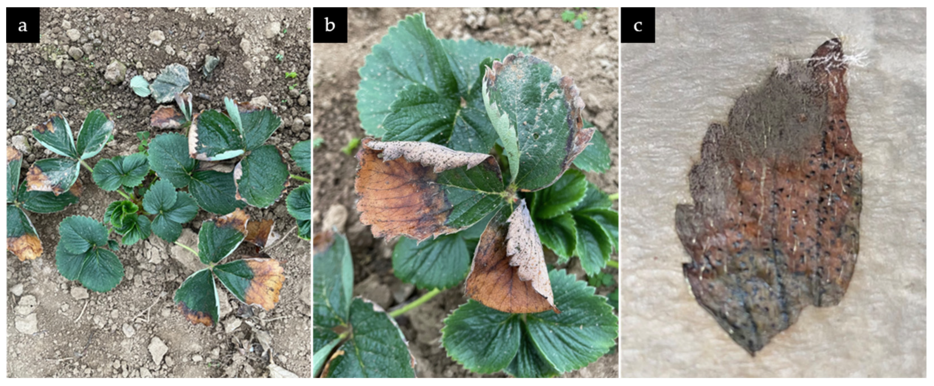
3.1. Symptoms of N. rosae on Naturally Infected Strawberry Plants

Several leaves with necrotic spots on the edges were observed shortly after planting strawberry cv. Aprica (Figure 2a). Starting from the edges, and in later stages commencing to the leaf center, yellow to orange and eventually brown to black necrotic spots were formed. Additionally, leaves started rolling from the edges. Further development showed the formation of black conidiomata on the leaf surface (upper and under side) with a greasy consistency (Figure 2b). Detached leaves in humidity chambers showed further necrosis, formation of white mycelia, and black conidiomata (Figure 2c). Lesions on the fruit started with soft tissue, subsequently forming sunken wet spots and ultimately covered in white mycelia with black structures (conidiomata).

3.2. Morphological Characterization

Isolated colonies on GM7 demonstrated robust mycelial growth, with conidia formation observed after 14 days of incubation. Acervuli were not formed. Fungal colonies exhibited distinctive morphology, displaying circular growth, that initially appeared white and gradually darkened in color. The plate underside displayed a pale orange coloration. The texture of the colonies ranged from velvety to fluffy (Figure 3a).
Microscopic examination of the fungal colonies revealed the presence of typical features consistent with those observed in Neopestalotiopsis rosae [23]. While the sexual morph was not observed on agar, the asexual morph displayed conidiomata that were solitary, spherical to asymmetric, black, and contained glistening conidial masses (Figure 3b,c). Conidiophores were observed as conidiogenous cells (Figure 3d,e).
Conidia were observed to be fusoid or ellipsoidal to cylindrical in shape, straight or slightly curved, typically consisting of five cells. The central three cells were melanized, making them visibly darker (Figure 3f,g). The three middle cells were 15.78 ± 1.01 µm long ( x ¯ ± SD, n = 30). The second cell from the base was hyaline to light grey or brown in color, 5.41 ± 0.63 µm long; the third cell was dark brown to black, 4.77 ± 0.62 µm long; and the fourth cell was grey to dark brown in color, 5.60 ± 0.8 µm long. The Basal cell was 4.60 ± 0.65 µm long, hyaline, conical, and often exhibited one basal appendage, hyaline, unbranched, emerging central from the very tip, and 7.86 ± 1.31 µm long. The Apical cell was 4.39 ± 0.63 µm long, conical, hyaline and exhibited two to four filiform, hyaline, unbranched appendages, 30.33 ± 4.2 µm long.

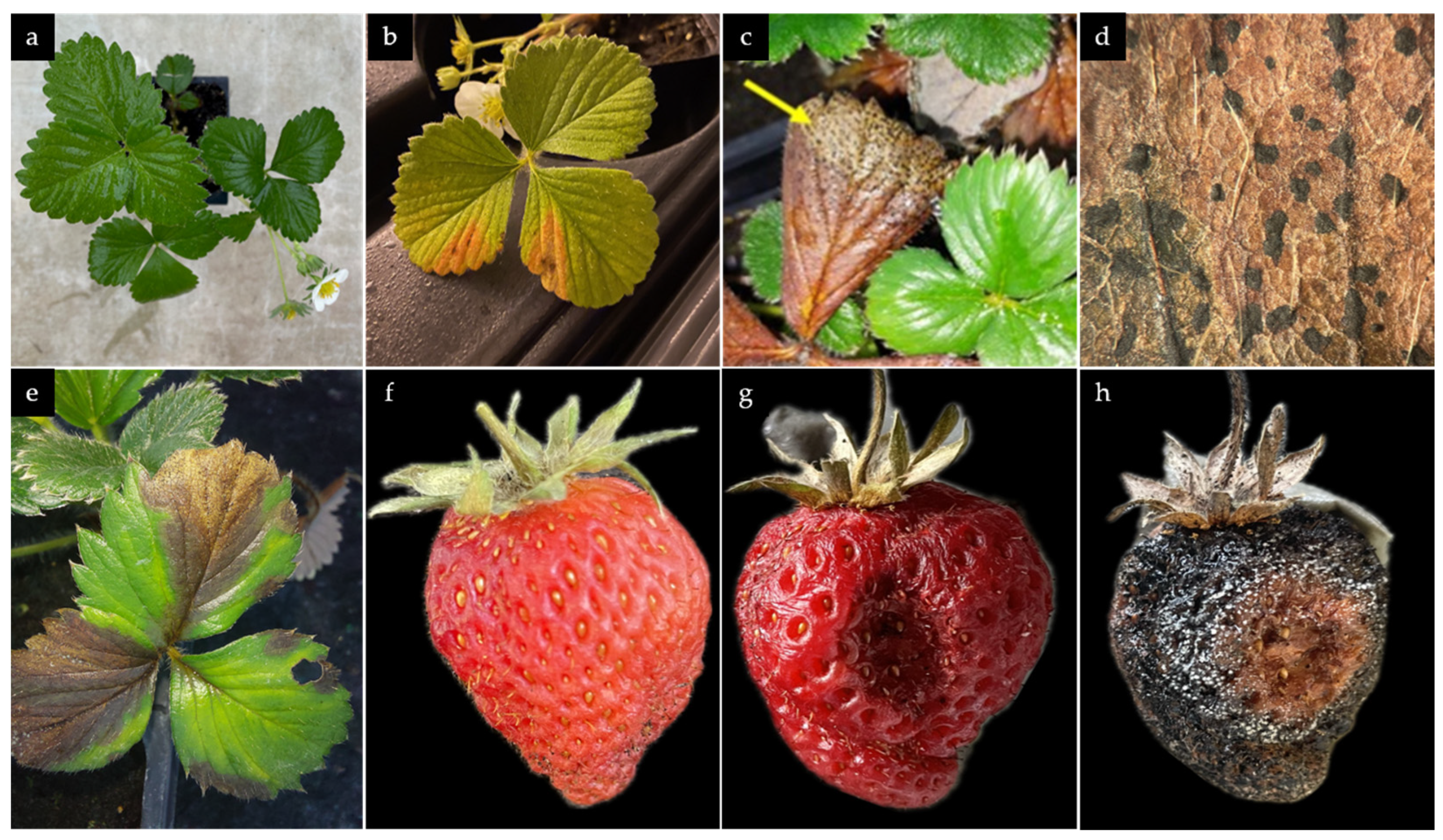
3.3. Pathogenicty Test and Re-isolation of N. rosae from Inoculated Plant Material

At 21 days post inoculation (dpi), uninoculated plants remained healthy (Figure 4a). Plants inoculated with N. rosae showed leaf spots and necrotic lesions, typically V-shaped and starting from the edges, which were later accompanied by conidiomata formation (Figure 4b–e). These symptoms closely resembled those initially observed on the source plants. Additionally, dark lesions on the fruit surface were observed, which typically began as small, water-soaked areas that gradually expanded and darkened, resulting in sunken, necrotic spots (Figure 4f,g). Furthermore, white mycelium covered the whole fruit and ultimately black conidiomata containing spore masses were formed (Figure 4h). A lengthwise cut through the plant rhizome did not reveal any symptoms on inner tissue. Roots were partly darkened. Cultivated root and rhizome tissue on GM7 did not show any fungal growth. Re-isolation and identification of the fungus from conidiomata formed on leaves and fruits confirmed it identity as N. rosae, thus establishing the causal link between the isolated fungus and the observed disease symptoms.

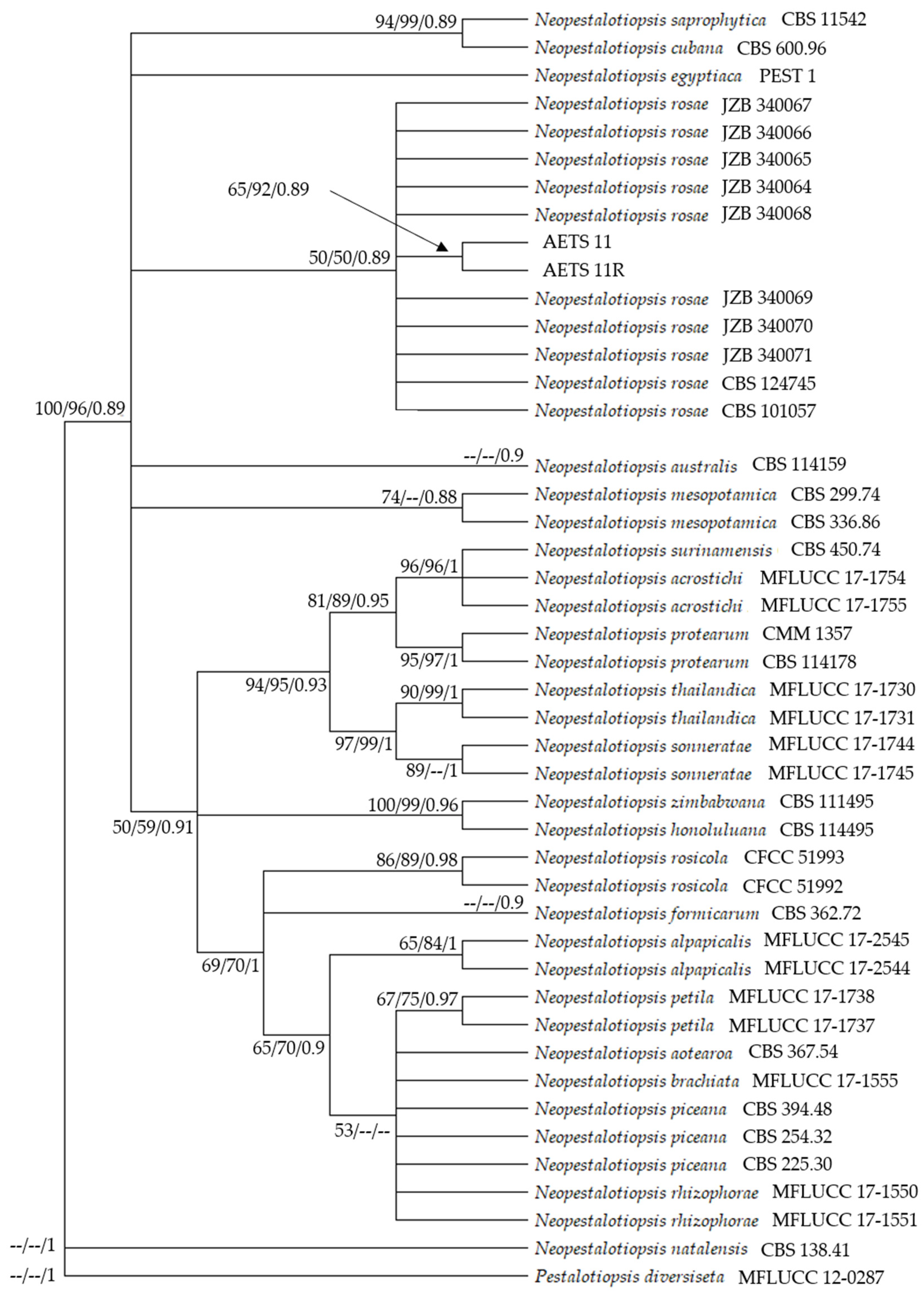
3.4. Phylogenetic Analysis

The phylogenetic tree comprised 45 nucleotide sequences (taxa), with the outgroup P. diversiseta (MFLUCC 12–0287). The combined sequence alignment comprised both the ITS and TUB region. There was a total of 739 positions in the final dataset. The ML tree with the highest log likelihood (−1942.05) is shown (Figure 5).
The percentage of trees in which the associated taxa clustered together is shown next to the branches. ML and MP bootstrap values equal to or greater than 50% are shown in the ML tree. MP Tree one out of three most parsimonious trees (length = 233) was used. The consistency index was 0.884120 (0.640000), the retention index was 0.892000 (0.892000), and the composite index was 0.788635 (0.570880) for all sites and parsimony-informative sites (in parentheses).
The percentage of replicate trees in which the associated taxa clustered together in the bootstrap test (1000 replicates) are shown next to the branches. BI analysis resulted in a tree with the same topology and clades as the ML and MP trees. Phylogenetic analysis proved the strains AETS11 and AETS11R to be N. rosae. The ITS and TUB sequences from AETS11 and AETS11R have been submitted to the NCBI database (Appendix A, Table A1).

4. Discussion

The identity of N. rosae was confirmed through a combination of morphological identification and molecular phylogenetic analysis. Its pathogenicity was demonstrated on strawberry plants. The morphological characteristics of N. rosae, such as the versicolorous median cells and multiple appendages of its conidia, are consistent with those described for other strains within N. rosae [16,23]. When relying solely on ITS sequences, clades in the phylogenetic analysis remained uncertain and yielded inconsistent results across different phylogenetic analyses, which is in line with previous studies [11,23,44]. However, molecular analyses incorporating both ITS and TUB sequences provided additional confirmation of the pathogen’s identity, clustering it with NCBI sequences of N. rosae, and furthermore, positioning it in a distinct clade within Neopestalotiopsis, separated from Pestalotiopsis.
Although Neopestalotiopsis spp. are generally regarded as opportunistic pathogens that typically infect plants under stress conditions, N. rosae has demonstrated notably increased infection severity on strawberry, particularly in Florida, where symptoms are observed across multiple plant tissues [20]. In strawberry plants, Neopestalotiopsis leaf spot disease symptoms resemble those of leaf blotch (Gnomoniopsis spp.), scorch (Diplocarpon spp.), and typical leaf spots caused by other leaf spot pathogens [45,46]. However, leaf lesions caused by N. rosae are characterized by dark brown V-shaped necrotic areas starting at the leaf edge with well-defined borders and light brown centers [47]. Conidiomata are readily produced on the lesions, facilitating the spread of the pathogen.
In fruit tissues, the symptoms caused by N. rosae closely resemble those of anthracnose. However, a key distinguishing feature is the development of black spores on fruit lesions caused by Neopestalotiopsis, in contrast to the pinkish-orange-colored conidiomata produced by Colletotrichum spp. [48].
The symptoms observed in the infected strawberry plants, including necrotic leaf spots and water-soaked lesions on the fruits, align with those caused by N. rosae [16,20]. Although root rot caused by N. rosae has been described by other authors [15,23,27], root rot was not observed in the present study. Given the fact that the examined strain of N. rosae was virulent to various strawberry cultivars such as Aprica or Sengana under both field and greenhouse conditions, this strain is likely to be highly virulent. The symptoms directly impact the photosynthetic capability of the plants and reduce the marketability of the fruit, thereby threatening both yield and quality [49]. The high humidity and temperature conditions in the field likely facilitated the spread and severity of the disease, underscoring the importance of environmental factors in disease development [15].
This study highlights the early emergence and the accurate identification of N. rosae as a significant pathogen of strawberries, which is essential for the implementation of effective disease management strategies. Sequence data, particularly the combined analysis of ITS and TUB sequences, proved to be indispensable for the precise identification of N. rosae. This approach can be applied to other Neopestalotiopsis species to enhance our understanding of their distribution and impact on strawberry crops.

5. Conclusions

This study represents the first documented case of N. rosae causing leaf spot and fruit rot in strawberries across Europe including Germany, contributing valuable information to the understanding of the pathogen’s distribution and impact on strawberry production. Accurate identification of N. rosae through morphological, molecular, and pathogenic analyses is essential for understanding its role in strawberry disease dynamics. The observed virulence of N. rosae across various strawberry cultivars, combined with the impact of environmental conditions on disease severity, underlines the need for proactive and effective disease management strategies to limit its spread and economic impact. Ongoing monitoring and research into the distribution, pathogenicity, and ecological interactions of N. rosae will be essential to inform targeted control measures and preserve strawberry production across Europe and globally.

Author Contributions

Conceptualization, A.E.-H. and T.E.S.; methodology, A.E.-H. and T.E.S.; software, T.E.S.; validation, A.E.-H. and R.T.V.; formal analysis, T.E.S.; investigation, T.E.S.; resources, A.E.-H. and R.T.V.; data curation, T.E.S.; writing—original draft preparation, T.E.S.; writing—review and editing, A.E.-H. and R.T.V.; visualization, T.E.S.; supervision, A.E.-H.; project administration, A.E.-H.; funding acquisition, A.E.-H. and R.T.V. All authors have read and agreed to the published version of the manuscript.

Funding

This research was funded by Deutsche Bundesstiftung Umwelt, grant number DBU AZ38050/01, and the APC was funded by a 100% Feature Paper discount for A.E.-H.

Institutional Review Board Statement

Not applicable.

Informed Consent Statement

Not applicable.

Data Availability Statement

Data are contained within the article; further inquiries can be directed to the corresponding authors.

Conflicts of Interest

The authors declare no conflicts of interest.

Appendix A

The taxa and sequences used for the phylogenetic analysis as well as the NCBI accession numbers for the isolated strains AETS11 and AETS11R are shown.
Table A1. Taxa and sequences of Neopestalotiopsis spp. and Pestalotiopsis diversiseta used in the phylogenetic analyses with NCBI accession numbers.
TaxaStrainHostCountryITSβ-Tubulin
N. alpapicalisMFLUCC 17–2544aRhizophora mucronataThailandMK357772MK463545
MFLUCC 17–2545Symptomatic leaves of Rhizophora apiculataThailandMK357773MK463546
N. aotearoaCBS 367.54aCanvasNew ZealandKM199369KM199454
N. acrostichiMFLUCC 17–1754aAcrostichum aureumThailandMK764272MK764338
MFLUCC 17–1755Acrostichum aureumThailandMK764273MK764339
N. australisCBS 114159aTelopea sp.AustraliaKM199348KM199432
N. brachiataMFLUCC 17–1555aRhizophora apiculataThailandMK764274MK764340
N. cubanaCBS 600.96aLeaf litterCubaKM199347KM199438
N. egyptiacaPEST1Mangifera indicaEgyptKP943747KP943746
N. formicarumCBS 362.72aDead antGhanaKM199358KM199455
N. honoluluanaCBS 114495aTelopea sp.USAKM199364KM199457
N. mesopotamicaCBS 336.86aPinus brutiIraqKM199362KM199441
CBS 299.74Eucalyptus sp.TurkeyKM199361KM199435
N. natalensisCBS 138.41aAcacia mollissimaSouth AfricaNR_156288KM199466
N. piceanaCBS 394.48aPicea sp.UKKM199368KM199453
CBS 254.32Cocos nuciferaIndonesiaKM199372KM199452
CBS 225.30Mangifera indica-KM199371KM199451
N. protearumCBS 114178aLeucospermum cuneiforme cv. “Sunbird”ZimbabweJN712498KM199463
CMM1357--KY549597KY549632
N. rhizophoraeMFLUCC 17–1550aRhizophora mucronataThailandMK764277MK764343
MFLUCC 17–1551Rhizophora mucronataThailandMK764278MK764344
N. rosaeCBS 101057aRosa sp.New ZealandKM199359KM199429
JZB340064Fragaria × ananassaChinaMN495972MN968336
JZB340065Fragaria × ananassaChinaMN495973MN968337
JZB340066Fragaria × ananassaChinaMN495974MN968338
JZB340067Fragaria × ananassaChinaMN495976MN968339
JZB340068Fragaria × ananassaChinaMN495975MN968340
JZB340069Fragaria × ananassaChinaMN495977MN968341
JZB340070Fragaria × ananassaChinaMN495967MN968342
JZB340071Fragaria × ananassaChinaMN495978MN968343
AETS11Fragaria × ananassaGermanyPQ511123PQ584291
AETS11RFragaria × ananassaGermanyPQ511124PQ621801
CBS 124745Paeonia suffruticosaUSAKM199360KM199430
N. rosicolaCFCC 51992aRosa chinensisChinaKY885239KY885245
CFCC 51993Rosa chinensisChinaKY885240KY885246
N. saprophyticaCBS 115452Magnolia sp.ChinaKM199345KM199433
N. sonnerataeMFLUCC 17–1745aSonneronata albaThailandMK764279MK764345
MFLUCC 17–1744Sonneronata albaThailandMK764280MK764346
N. surinamensisCBS 450.74aSoil under Elaeis guineensisSurinameKM199351KM199465
N. thailandicaMFLUCC 17–1730aRhizophora mucronataThailandMK764281MK764347
MFLUCC 17–1731Rhizophora mucronataThailandMK764282MK764348
N. zimbabwanaCBS 111495aLeucospermum cunciforme cv. “Sunbird”ZimbabweJX556231KM199456
Pestalotiopsis diversisetaMFLUCC 12–0287aRhododendron sp.ChinaJX399009JX399040
CBS: Centraalbureau voor Schimmelcultures, Westerdijk Fungal Biodiversity Institute, Utrecht, The Netherlands; JZB: Beijing Academy of Agricultural and Forestry Sciences, Beijing, China; MFLUCC: Mae Fah Luang University Culture Collection, Thailand.

References

  1. Garrido, C.; Carbú, M.; Fernández-Acero, F.J.; González-Rodríguez, V.E.; Cantoral, J. New Insights in the Study of Strawberry Fungal Pathogens. In Genes, Genomes and Genomics; Husaini, A.M., Mercado, J.A., Eds.; Global Science Books: East Sussex, UK, 2011; Volume 5, pp. 24–39. ISBN 978-4-903313-81-8. [Google Scholar]
  2. Bhat, R.; Geppert, J.; Funken, E.; Stamminger, R. Consumers Perceptions and Preference for Strawberries—A Case Study from Germany. Int. J. Fruit Sci. 2015, 15, 405–424. [Google Scholar] [CrossRef]
  3. Bristow, P.R.; McNicol, R.J.; Williamson, B. Infection of Strawberry Flowers by Botrytis cinerea and Its Relevance to Grey Mould Development. Ann. Appl. Biol. 1986, 109, 545–554. [Google Scholar] [CrossRef]
  4. Gunnell, P.S.; Gubler, W.D. Taxonomy and Morphology of Colletotrichum Species Pathogenic to Strawberry. Mycologia 1992, 84, 157–165. [Google Scholar] [CrossRef]
  5. Garrido, C.; González-Rodríguez, V.E.; Carbú, M.; Husaini, A.M.; Cantoral, J. Fungal Diseases of Strawberry and Their Diagnosis. In Strawberry: Growth, Development and Diseases; Husaini, A.M., Neri, D., Eds.; Cabi Publishing: Wallingford, UK, 2016; pp. 157–195. ISBN 978-1-78064-663-3. [Google Scholar] [CrossRef]
  6. Embaby, E.M. Pestalotia Fruit Rot on Strawberry Plants in Egypt. Egypt. J. Phytopathol. 2007, 35, 99–110. [Google Scholar]
  7. Rodrigues, F.A.; Silva, I.T.; Antunes Cruz, M.F.; Carré-Missio, V. The Infection Process of Pestalotiopsis longisetula Leaf Spot on Strawberry Leaves. J. Phytopathol. 2014, 162, 690–692. [Google Scholar] [CrossRef]
  8. Howard, C.M. A Strawberry Fruit Rot Caused by Pestalotia longisetula. Phytopathology 1973, 63, 862. [Google Scholar] [CrossRef]
  9. Kenneth, R.G.; Barkai-golan, R.; Netzer, D. A Pestalotia Fruit Rot of Strawberries in Israel. Plant Dis. Report. 1968, 52, 472–474. [Google Scholar]
  10. Rajnish, K.; Gautam, H.R. Prevalence and Management of Pestalotia Leaf Spot (Pestalotia sp.) of Strawberry. Int. J. Econ. Plants 2022, 9, 250–254. [Google Scholar]
  11. Maharachchikumbura, S.S.N.; Hyde, K.D.; Groenewald, J.Z.; Xu, J.; Crous, P.W. Pestalotiopsis Revisited. Stud. Mycol. 2014, 79, 121–186. [Google Scholar] [CrossRef] [PubMed]
  12. Ayoubi, N.; Soleimani, M.J. Strawberry Fruit Rot Caused by Neopestalotiopsis iranensis sp. nov., and N. mesopotamica. Curr. Microbiol. 2016, 72, 329–336. [Google Scholar] [CrossRef] [PubMed]
  13. Das, R.; Chutia, M.; Das, K.; Jha, D.K. Factors Affecting Sporulation of Pestalotiopsis disseminata Causing Grey Blight Disease of Persea bombycina Kost., the Primary Food Plant of Muga Silkworm. Crop Prot. 2010, 29, 963–968. [Google Scholar] [CrossRef]
  14. Yasuda, F.; Kobayashi, T.; Watanabe, H.; Izawa, H. Addition of Pestalotiopsis spp. to Leaf Spot Pathogens of Japanese Persimmon. J. Gen. Plant Pathol. 2003, 69, 29–32. [Google Scholar] [CrossRef]
  15. Rebollar-Alviter, A.; Silva-Rojas, H.V.; Fuentes-Aragón, D.; Acosta-González, U.; Martínez-Ruiz, M.; Parra-Robles, B.E. An Emerging Strawberry Fungal Disease Associated with Root Rot, Crown Rot and Leaf Spot Caused by Neopestalotiopsis rosae in Mexico. Plant Dis. 2020, 104, 2054–2059. [Google Scholar] [CrossRef] [PubMed]
  16. Wu, H.-Y.; Tsai, C.-Y.; Wu, Y.-M.; Ariyawansa, H.-A.; Chung, C.-L.; Chung, P.-C. First Report of Neopestalotiopsis rosae Causing Leaf Blight and Crown Rot on Strawberry in Taiwan. Plant Dis. 2021, 105, 487. [Google Scholar] [CrossRef]
  17. Obregón, V.G.; Meneguzzi, N.G.; Ibañez, J.M.; Lattar, T.E.; Kirschbaum, D.S. First Report of Neopestalotiopsis clavispora Causing Root and Crown Rot on Strawberry Plants in Argentina. Plant Dis. 2018, 102, 1856. [Google Scholar] [CrossRef]
  18. Machín, A.; González, P.; Vicente, E.; Sánchez, M.; Estelda, C.; Ghelfi, J.; Silvera-Pérez, E. First Report of Root and Crown Rot Caused by Neopestalotiopsis clavispora on Strawberry in Uruguay. Plant Dis. 2019, 103, 2946. [Google Scholar] [CrossRef]
  19. Reyna, H.O.I.; Figueroa, F.J.R.; Casignia, Á.M.R.; Romero, P.I.Á.; Ferreira, A.F.T.A. Outbreaks of Crown Rot in Fragaria x ananassa Caused by Neopestalotiopsis mesopotamica in Ecuador. Emir. J. Food Agric. 2021, 33, 520–527. [Google Scholar] [CrossRef]
  20. Baggio, J.S.; Forcelini, B.B.; Wang, N.-Y.; Ruschel, R.G.; Mertely, J.C.; Peres, N.A. Outbreak of Leaf Spot and Fruit Rot in Florida Strawberry Caused by Neopestalotiopsis spp. Plant Dis. 2021, 105, 305–315. [Google Scholar] [CrossRef]
  21. Kaur, H.; Gelain, J.; Marin, M.V.; Peres, N.A.; Schnabel, G. Development of a Molecular Tool for Identification of a New Neopestalotiopsis sp. Associated with Disease Outbreaks on Strawberry. Plant Dis. 2023, 107, 1544–1549. [Google Scholar] [CrossRef] [PubMed]
  22. Sultana, S.; Maniruzzam, S.; Sabbir, A.; Alam, N. Neopestalotiopsis chrysea Causing Leaf Spot Disease of Strawberry Plants in Bangladesh. J. Plant Sci. 2022, 17, 66–74. [Google Scholar] [CrossRef]
  23. Sun, Q.; Harishchandra, D.; Jia, J.; Zuo, Q.; Zhang, G.; Wang, Q.; Yan, J.; Zhang, W.; Li, X. Role of Neopestalotiopsis Rosae in Causing Root Rot of Strawberry in Beijing, China. Crop Prot. 2021, 147, 105710. [Google Scholar] [CrossRef]
  24. Chandana, R.; Poonacha, T.T.; Chethan, D.; Karan, R.; Kruthika, R.; Khan, F.; Ashwini, K.S.; Bevanur, A.; Vani, Y.; Ramesh, G.V.; et al. Neopestalotiopsis rosae, a Novel Pathogen Causing Leaf Blight and Crown Rot of Strawberries in India. Physiol. Mol. Plant Pathol. 2024, 133, 102377. [Google Scholar] [CrossRef]
  25. Chamorro, M.; Aguado, A.; De los Santos, B. First Report of Root and Crown Rot Caused by Pestalotiopsis clavispora (Neopestalotiopsis clavispora) on Strawberry in Spain. Plant Dis. 2015, 100, 1495. [Google Scholar] [CrossRef]
  26. Gilardi, G.; Bergeretti, F.; Gullino, M.L.; Garibaldi, A. First Report of Neopestalotiopsis clavispora Causing Root and Crown Rot on Strawberry in Italy. Plant Dis. 2019, 103, 2959. [Google Scholar] [CrossRef]
  27. Essa, T.A.; Kamel, S.M.; Ismail, A.M.; El-Ganainy, S. Characterization and Chemical Control of Neopestalotiopsis rosae the Causal Agent of Strawberry Root and Crown Rot in Egypt. Egypt. J. Phytopathol. 2018, 46, 1–19. [Google Scholar] [CrossRef]
  28. Chung, P.-C.; Wu, H.-Y.; Wang, Y.-W.; Ariyawansa, H.A.; Hu, H.-P.; Hung, T.-H.; Tzean, S.-S.; Chung, C.-L. Diversity and Pathogenicity of Colletotrichum species Causing Strawberry Anthracnose in Taiwan and Description of a New Species, Colletotrichum miaoliense sp. nov. Sci. Rep. 2020, 10, 14664. [Google Scholar] [CrossRef]
  29. Basu, P.K. Verticillium Disease of Strawberries. Can. J. Bot. 1961, 39, 165–196. [Google Scholar] [CrossRef]
  30. Koike, S.T.; Gordon, T.R. Management of Fusarium Wilt of Strawberry. Crop Prot. 2015, 73, 67–72. [Google Scholar] [CrossRef]
  31. Alcock, N.L.; Howells, D.V. The Phytophthora Disease of Strawberry. Sci. Hortic. 1936, 4, 52–58. [Google Scholar]
  32. Van Hemelrijck, W.; Ceustermans, A.; Van Campenhout, J.; Lieten, P.; Bylemans, D. Crown Rot in Strawberry Caused by Pestalotiopsis. Acta Hortic. 2017, 1156, 781–786. [Google Scholar] [CrossRef]
  33. Karlovsky, P. Inhibition of Imidazolegly Cerolphosphate Dehydratase of Phytophthora parasitica by Aminotriazole in situ and after Cloning and Expression of the Respective Gene (HIS3) in Escherichia coli. J. Phytopathol. 1994, 141, 121–126. [Google Scholar] [CrossRef]
  34. Liu, D.; Coloe, S.; Baird, R.; Pedersen, J. Rapid Mini-Preparation of Fungal DNA for PCR. J. Clin. Microbiol. 2000, 38, 471. [Google Scholar] [CrossRef] [PubMed]
  35. Innis, M.A.; Gelfand, D.H.; Sninsky, J.J.; White, T.J. PCR Protocols: A Guide to Methods and Applications; Academic Press: Cambridge, MA, USA, 2012; ISBN 978-0-08-088671-8. [Google Scholar]
  36. Glass, N.L.; Donaldson, G.C. Development of Primer Sets Designed for Use with the PCR to Amplify Conserved Genes from Filamentous Ascomycetes. Appl. Environ. Microbiol. 1995, 61, 1323–1330. [Google Scholar] [CrossRef]
  37. O’Donnell, K.; Cigelnik, E. Two Divergent Intragenomic rDNA ITS2 Types within a Monophyletic Lineage of the Fungus Fusarium Are Nonorthologous. Mol. Phylogenetics Evol. 1997, 7, 103–116. [Google Scholar] [CrossRef]
  38. NCBI National Center for Biotechnology; Information. Available online: https://www.ncbi.nlm.nih.gov/ (accessed on 27 September 2024).
  39. Norphanphoun, C. Morphological and Phylogenetic Characterization of Novel Pestalotioid Species Associated with Mangroves in Thailand. Mycosphere 2019, 10, 531–578. [Google Scholar] [CrossRef]
  40. Tamura, K.; Stecher, G.; Kumar, S. MEGA11: Molecular Evolutionary Genetics Analysis Version 11. Mol. Biol. Evol. 2021, 38, 3022–3027. [Google Scholar] [CrossRef] [PubMed]
  41. Hasegawa, M.; Kishino, H.; Yano, T. Dating of the Human-Ape Splitting by a Molecular Clock of Mitochondrial DNA. J. Mol. Evol. 1985, 22, 160–174. [Google Scholar] [CrossRef] [PubMed]
  42. Nei, M.; Kumar, S.; Nei, M.; Kumar, S. Molecular Evolution and Phylogenetics; Oxford University Press: Oxford, UK; New York, NY, USA, 2000; ISBN 978-0-19-513585-5. [Google Scholar]
  43. Ronquist, F.; Teslenko, M.; van der Mark, P.; Ayres, D.L.; Darling, A.; Höhna, S.; Larget, B.; Liu, L.; Suchard, M.A.; Huelsenbeck, J.P. MrBayes 3.2: Efficient Bayesian Phylogenetic Inference and Model Choice across a Large Model Space. Syst. Biol. 2012, 61, 539–542. [Google Scholar] [CrossRef]
  44. Hu, H.; Jeewon, R.; Zhou, D.; Zhou, T.; Hyde, K. Phylogenetic Diversity of Endophytic Pestalotiopsis species in Pinus armandii and Ribes spp.: Evidence from rDNA and Beta-Tubulin Gene Phylogenies. Fungal Divers. 2007, 24, 1–22. [Google Scholar]
  45. Maas, J.L. (Ed.) Compendium of Strawberry Diseases; American Phytopathological Society: St. Paul, MN, USA, 1984; p. 68. ISBN 0-89054-054-3. [Google Scholar]
  46. Pugliese, M.; Gilardi, G.; Guarnaccia, V.; Garibaldi, A.; Gullino, M.L. First Report of Gnomoniopsis fragariae Causing Leaf Spots on Strawberry in Italy. Plant Dis. 2023, 107, 2850. [Google Scholar] [CrossRef] [PubMed]
  47. Madrid, A.M.J.; Munoz, G.; Collins, C.; Brannen, P. First Report of the New Neopestalotiopsis species Causing Strawberry Leaf Spot and Fruit Rot in Georgia. Plant Dis. 2024, 108, 2574. [Google Scholar] [CrossRef] [PubMed]
  48. Aljawasim, B.D.; Samtani, J.B.; Rahman, M. New Insights in the Detection and Management of Anthracnose Diseases in Strawberries. Plants 2023, 12, 3704. [Google Scholar] [CrossRef] [PubMed]
  49. Guthman, J. Wilted: Pathogens, Chemicals, and the Fragile Future of the Strawberry Industry; University of California Press: Berkeley, CA, USA, 2019; p. 322. ISBN 978-0-520-30527-4. [Google Scholar]
Figure 1. Schematic illustration of Neopestalotiopsis rosae conidia with individual cells. Numbers represent first to fourth cell from the base.
Microorganisms 13 00006 g001
Figure 2. (a,b) Necrosis, leaf curling, burning, and conidiomata on naturally infested plants in the field trial; (c) conidiomata and mycelia on detached leaf in humidity chamber.
Microorganisms 13 00006 g002
Figure 3. Various fungal structures used for the microscopic identification of Neopestalotiopsis rosae. (a) Growth on GM7 medium after 14 days. Left: Plate upper side. Right: Plate underside. (b,c) Conidiomata on agar. (d,e) Conidiophores (conidia producing structures, red arrows) (f,g) Conidia with characteristic versicolorous median cells and appendages.
Microorganisms 13 00006 g003
Figure 4. (a) Healthy, uninoculated control plant; (b) wilting and leaf spot formation on inoculated plant 14 dpi; (c,d) Conidiomata formation on inoculated, withered leaf 21 dpi; (e) V-shaped necrotic area on inoculated infected leaf; (fh) fruit rot development on detached fruit test in a humidity chamber (1, 14, 21 dpi, respectively).
Microorganisms 13 00006 g004
Figure 5. Phylogenetic tree resulting from ML analysis of the combined ITS and TUB dataset. ML and MP bootstrap support values (BS) greater than 50% and the Bayesian posterior probabilities (PP) greater than 0.85 are shown at the nodes. Pestalotiopsis diversiseta (MFLUCC 12–0287) was used as an outgroup.
Microorganisms 13 00006 g005
Disclaimer/Publisher’s Note: The statements, opinions and data contained in all publications are solely those of the individual author(s) and contributor(s) and not of MDPI and/or the editor(s). MDPI and/or the editor(s) disclaim responsibility for any injury to people or property resulting from any ideas, methods, instructions or products referred to in the content.

