Review

Inquilinus Species Infections in Humans—A Narrative Review

by
Anastasia Vasilopoulou
1,
Takis Panayiotou
1,
Stella Baliou
1,
Andreas G. Tsantes
2 and
Petros Ioannou
1,*
1
School of Medicine, University of Crete, 71003 Heraklion, Greece
2
Laboratory of Hematology and Blood Bank Unit, “Attikon” University Hospital, School of Medicine, National and Kapodistrian University of Athens, 12462 Athens, Greece
*
Author to whom correspondence should be addressed.
Microorganisms 2025, 13(3), 592; https://doi.org/10.3390/microorganisms13030592
Submission received: 11 February 2025 / Revised: 23 February 2025 / Accepted: 3 March 2025 / Published: 4 March 2025
(This article belongs to the Special Issue Advances in Human Infection)

Abstract

:
Background: Inquilinus species are Gram-negative, non-sporulating, non-pigmented rods that are catalase-positive, indole-negative, and able to grow at various temperatures and in 1% NaCl. Infections due to Inquilinus spp. are increasingly identified, especially in patients with cystic fibrosis. Objective: This review aims to present all reported cases of Inquilinus spp. infections in humans, with an emphasis on data regarding epidemiology, antimicrobial resistance, antimicrobial treatment, and mortality. Methods: A narrative review based on a literature search of the PubMed/MedLine and Scopus databases was performed. Results: In total, 13 articles providing data on 25 patients with Inquilinus infections were included in the analysis. The median age was 19 years, while 60% were male. Cystic fibrosis was the predominant risk factor (92%). Respiratory tract infection was the most common type of infection (96%). Inquilinus limosus was the most commonly identified species. Polymicrobial infection was very common (77.3%). Microbial identification required the use of advanced molecular techniques, such as 16s rRNA sequencing. The pathogen exhibited resistance to beta-lactams, with the exception of carbapenems. The most commonly used antimicrobials included carbapenems (68.4%), followed by quinolones (57.9%) and aminoglycosides (52.6%). Mortality was low (4%). Conclusions: Due to the potential of Inquilinus spp. to cause infection in patients with cystic fibrosis, and given the difficulties in microbial identification, clinicians and laboratory professionals should consider it in the differential diagnosis of patients with cystic fibrosis and respiratory tract infection not responding to beta-lactam treatment or with polymicrobial infections, especially when traditional techniques are used for microbial identification.

1. Introduction

Cystic fibrosis is a genetic, autosomal recessive disease caused by variants of the CFTR gene that manifests most commonly during childhood. CFTR encodes for an ion channel, CFTR, which regulates the balance of water and electrolytes on the surface of several tissues, such as the biliary tract, the pancreas, the cervix, the vas deferens, the intestine, the upper and lower airways, and the sweat glands [1,2]. These variants are associated with fluid imbalance in the lungs and other organs. This fluid imbalance in the lungs is associated with the production of thickened secretions, reduced transport through the mucociliary system, and mucus retention. This retention can lead to reduced bacterial cleaning, leading to the persistence of bacteria that may then increase the likelihood of infections [1,2,3,4]. There is a strong association of cystic fibrosis with specific microorganisms either in the context of colonization or in that of infection. Such microorganisms are Staphylococcus aureus, Pseudomonas aeruginosa, Achromobacter spp., Burkholderia cepacia complex, and Stenotrophomonas maltophilia [1,2]. New species with pathogenic potential are currently being discovered with the use of new molecular identification methods in microbiology. Such previously unknown species include Pandorea apista, Ralstonia pikettii, and Inquinus limosus [5]. Such species, such as Inquilinus spp., have been identified in patients with cystic fibrosis both as colonizers and as pathogens associated with respiratory tract infections [6].
Inquilinus species were first identified in 1999 from Pitulle et al. from a clinical specimen of a patient with lung transplantation and cystic fibrosis [6]. Based on a phylogenetic analysis of the small subunit rRNA and biochemical assays, these species were found to be unrelated to other known bacteria up to that time and represented a new genus within the subdivision of α-proteobacteria [6]. Soon after, Coenye et al. identified eight more isolates from the same genus from patients with cystic fibrosis and proposed the novel genus and species of Inquilinus limosus based on a whole-cell protein analysis, DNA–DNA hybridization, and 16S ribosomal DNA sequencing [7]. Inquilinus spp. are Gram-negative, non-sporulating, non-pigmented rods that are catalase-positive, indole-negative, and able to grow at various temperatures and in 1% NaCl [7]. I. limosus has a size of about 1.5 to 2 μm and a length of 3.5 μm [6,7]. They grow best between 35 °C and 42 °C but can also grow at 25 °C. I. limosus is not motile, and it can utilize many carbohydrate sources, including L-fucose, D-fructose, maltose, D-galactose, D-mannose, D-sorbitol, and D-raffinose, but not D-glucose [6,7]. Thus, it belongs to the non-fermenting bacteria [8]. Additionally, it cannot utilize sorbitol, mannitol, rhamnose, inositol, melibiose, sucrose, amygdalin, citrate, or arabinose [7]. Among the four DNA sequences of I. limosus deposited in GeneBank (DSM 16000, Inq, MP06, and YM_69_17), the total sequence has a length of 5.93–7.69 Mb, a GC content of 69.6–66.9%, and 5877 to 7098 encoding genes [8].
Based on the first studies of Inquilinus spp., a close association with cystic fibrosis was noted, even though whether this identification represents colonization or infection may not always be clear and the potential of this microorganism to cause other infections is not clear [6,7].
The aim of the present study was to comprehensively review all the available information of all types of infections caused by Inquilinus spp. in the literature and to assess clinical data, microbiology, treatment, and outcomes.

2. Materials and Methods

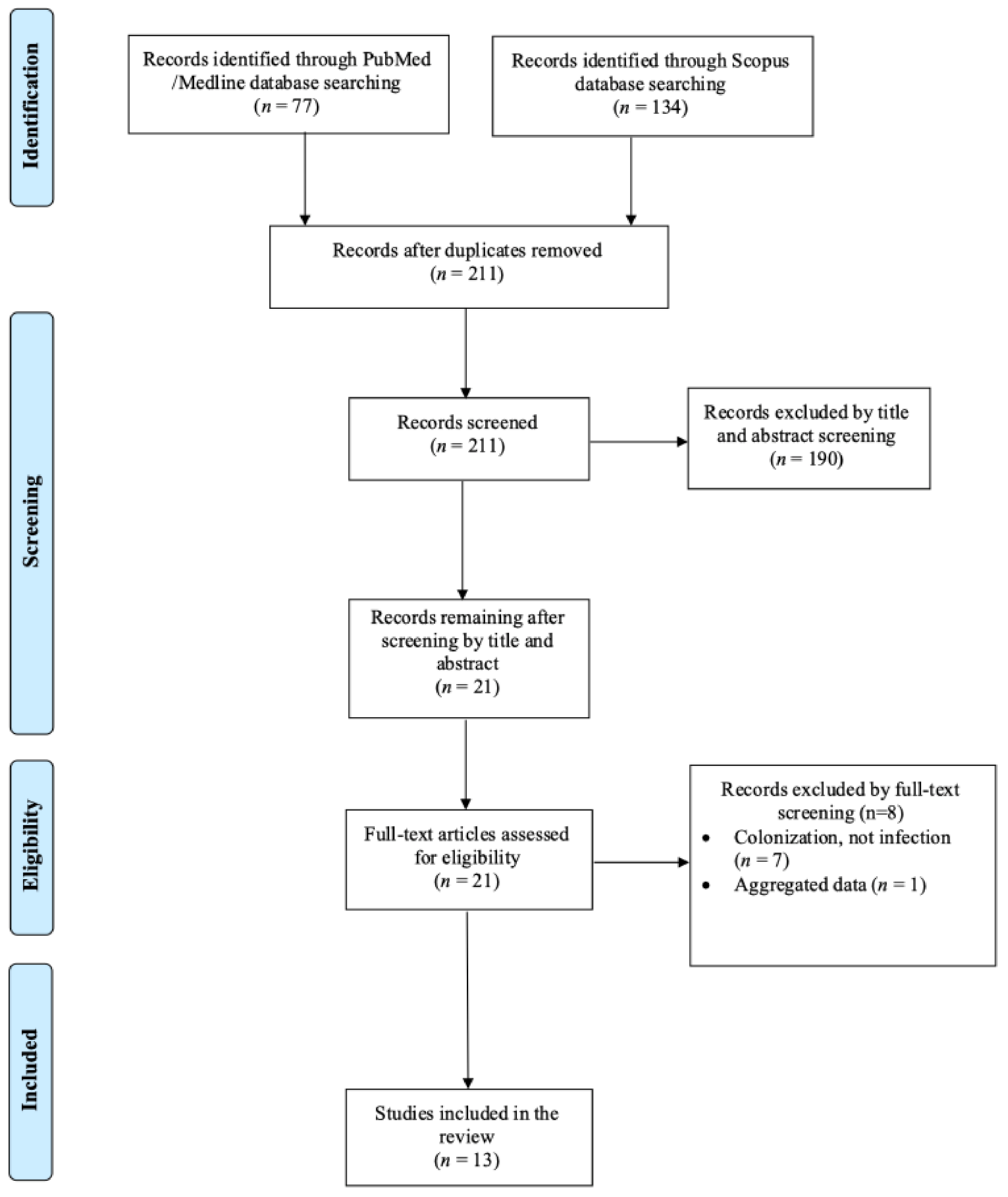
2.1. Search Strategy and Inclusion and Exclusion Criteria

This review aims to collect and present all published data on Inquilinus species infections in humans. The primary aim was to present data on the epidemiology and mortality of the patients. Secondary aims were to gather data about specific infection sites, clinical presentation, microbiological features, and treatment provided. For this review, the PubMed/Medline and Scopus databases were searched up to 13 December 2024. Data were extracted using a predefined template. The following keywords were applied for the search strategy: “Inquilinus” AND “infection”. The inclusion criteria encompassed studies presenting original data, such as case series, case reports, and cohort studies providing information on the epidemiology and clinical outcomes of Inquilinus spp. infections in humans. Only studies in the English language were considered. Reviews and systematic reviews were excluded. Studies involving animals, articles without full-text access, and those lacking information on patients’ mortality and epidemiology were excluded from the analysis. Finally, cases of colonization by Inquilinus species were excluded from the analysis, based on the clinical course depicted and the study authors’ consideration whether the species were pathogens or colonizers. The references of all included articles were examined to identify any studies potentially missed in the initial search.

2.2. Data Extraction and Definitions

The data extracted from each included study were the publication year, article type, country of origin, patient demographics (age and gender), relevant medical history, details of infection, and key clinical characteristics, such as the specific infection site, complications, as well as microbiological characteristics, such as the identified pathogen, antibiotic susceptibilities, and finally, the treatment used and outcome (survival or mortality). The relationship between mortality and the initial infection was documented according to each study’s authors.

2.3. Statistical Analysis

Data are presented as numbers (%) for categorical variables and median (interquartile range, IQR) for continuous variables. Continuous variables were compared using the Mann–Whitney U-test for non-normally distributed variables or the t-test for normally distributed variables. All tests were two-tailed, and a p-value equal to or lower than 0.05 was considered significant. Statistics were calculated with GraphPad Prism 6.0 (GraphPad Software, Inc., San Diego, CA, USA).

3. Results

3.1. Included Studies’ Characteristics

A total of 211 articles were screened from the PubMed and Scopus databases. Eventually, after duplicate removal, record screening, and applying the snowball procedure, only 13 articles met the inclusion criteria and were selected for analysis [6,9,10,11,12,13,14,15,16,17,18,19,20]. These studies presented data on 25 patients. A flow diagram of the selection process is illustrated in Figure 1. Among the included cases, seven occurred in North and South America (53.8%), five in Europe (30.8%), one in Oceania (7.7%), and one in Asia (7.7%). Among the 13 included articles, 7 (53.8%) were case reports. Figure 2 represents the geographical distribution of all Inquilinus spp. infections worldwide. Table 1 shows the characteristics of the included studies in the present review.

3.2. Epidemiology of Inquilinus spp. Infections

The median age of patients with Inquilinus spp. infections was 19 years, varying from 2 to 60 years, while 60% (15 patients) were male. Regarding the patients’ medical history and predisposing risk factors, 24 out of 25 patients (96%) had pulmonary disease, with 23 (92%) having a history of cystic fibrosis and 1 patient (4%) having bronchiectasis and recurrent bronchitis, while 5 (20%) had received a lung transplant—all were patients with cystic fibrosis. The demographic and clinical characteristics of Inquilinus spp. cases are shown in Table 2.

3.3. Antimicrobial Resistance and Microbiology of Inquilinus spp. Infections

Inquilinus spp. were isolated from sputum cultures in 18 patients (72%), from bronchoalveolar fluid in 3 (12%), from blood cultures in 3 (12%), from lung explant culture in 1 (4%), and from pleural fluid, empyema rind, and thoracotomy wound tissue in 1 (4%). In three patients (12%), the method of isolation was not mentioned. Inquilinus limosus was the only identified species, and was identified in 21 patients (84%), while in the rest of the cases, Inquilinus spp. were mentioned as the identified pathogen. In 14 cases (56%), identification was performed with 16s rRNA sequencing. In six cases (24%), identification was performed with matrix-assisted laser desorption/ionization time-of-flight mass spectrometry (MALID-TOF MS). In six cases (24%), the means of pathogen identification was not mentioned. The antimicrobial resistance of Inquilinus spp. is shown in Table 3. In 17 out of 22 patients (77.3%), the infection was polymicrobial. More specifically, in 11 out of 17 patients with polymicrobial infection (64.7%), at least two other pathogens were isolated. The most commonly isolated pathogens were Pseudomonas aeruginosa in 14 patients (82.4%), Aspergillus fumigatus in 8 (47.1%), Staphylococcus aureus in 7 (41.2%), Candida albicans in 3 (17.6%), Stenotrophomonas maltophilia in 2 (11.8%), and non-tuberculous mycobacteria, Paenibacillus amylolyticus, and Proteus mirabilis in 1 (5.9%) each.

3.4. Clinical Presentation of Inquilinus spp. Infections

Inquilinus spp. infections most commonly involved the lower respiratory tract (in 24 patients; 96%) and the bloodstream (in 3 patients; 12%). Infective endocarditis was diagnosed in one patient (4%). The symptoms’ duration ranged from zero (acute onset) to more than three months.

3.5. Treatment and Outcome of Inquilinus Infections

Based on the available data, carbapenems were the most frequently administered antimicrobials (in 13 out of 19 patients with available data; 68.4%), followed by quinolones (in 11 patients; 57.9%) and aminoglycosides (in 10 patients; 52.6%). Of note, cephalosporins or other penicillins were rarely used, if ever. Surgical interventions were applied in combination with antimicrobial treatment in 2 out of 20 patients (10%). The median treatment duration for survivors was 28 days. The overall mortality rate was estimated at 4% (1 out of 25 patients), with the only death being directly associated with the Inquilinus spp. infection.

3.6. Lower Respiratory Tract Infection Due to Inquilinus Infection

Respiratory tract infection was diagnosed in 24 patients. The median age in this patient group was 18.5 years, and 15 patients were male (62.5%). Predisposing risk factors were lung disease in all patients. Lung transplantation had been performed in the past in five patients (20.8%). Among those with available data, 9.1% (2 out of 22 patients) also had bacteremia by Inquilinus spp. Most patients had polymicrobial infection, since 81% (17 out of 21 patients) also had other pathogens isolated from respiratory samples. Fever was present in 19% (4 out of 21), but no patient had sepsis. The median treatment duration was 22 days. Antimicrobial agents most commonly involved carbapenems, quinolones, and aminoglycosides. Overall mortality was 4.2% (1 out of 24 patients) and was specifically attributed to Inquilinus infection.

3.7. Bacteremia Due to Inquilinus

Bacteremia was diagnosed in three patients. Among them, one was male, and the median age was 38 years. Two of those patients had a lower respiratory tract infection due to Inquilinus spp. and were cystic fibrosis patients that had also undergone lung transplantation in the past. Only in one patient with an infection of the lower respiratory tract was the infection polymicrobial (P. aeruginosa and A. fumigatus). All patients had fever, but no patient had sepsis. One patient had infective endocarditis (without lower respiratory tract infection), and the clinical presentation also included heart failure. In this patient group, the median antimicrobial treatment was 42 days; carbapenems and aminoglycosides constituted the most widely used antimicrobials. No patient in this group died.

4. Discussion

This review presents the clinical and microbiological characteristics of infections by Inquilinus spp. in humans by evaluating all cases in the literature where such infections have been recorded. Two types of infections were documented: respiratory tract infection with or without bacteremia and infective endocarditis. All patients with a respiratory tract infection by Inquilinus spp. had a history of chronic respiratory disease, most commonly cystic fibrosis. Antimicrobial resistance to first-line beta-lactams was almost uniform, while quinolones and carbapenems retained significant antimicrobial activity against this pathogen and were very commonly used for treating such infections. Mortality from infections by Inquilinus spp. was very low.
Cystic fibrosis is a hereditary, chronic, progressive disease involving many systems, such as respiratory, gastrointestinal, and reproductive [21]. Due to mutations in the CFTR gene that affect the CFTR transporter that normally transports chloride and bicarbonate across the apical membrane of the secretory epithelia in the above-mentioned tissues, ion exchange is affected, leading to dysregulation of hydration of the tissues. For example, airway surface liquid hydration may be severely affected, leading to thick mucopurulent secretions in the airways that cannot be effectively cleared by the mucociliary system, leading to bacterial colonization and infection and to progressive structural damage of the lungs [2,21,22].
There is an association of cystic fibrosis with some specific microorganisms that tend to colonize patients’ airways and cause infections, while the microbiology of these infections is currently changing. Additionally, the increasing use of new technology that provides precise methods for pathogen identification, such as MALDI-TOF MS, leads to more accurate identification of even rare pathogens that may have been misidentified in the past [23,24,25,26]. For example, in a relatively recent study published in Chest, in which the microbiology of patients with cystic fibrosis reported in the Cystic Fibrosis Foundation Patient Registry from 2006 to 2012 was retrospectively evaluated, the epidemiology of pathogens was changing. The pathogens with the highest incidence were methicillin-sensitive Staphylococcus aureus (MSSA) and Pseudomonas aeruginosa, followed by methicillin-resistant S. aureus (MRSA) [27]. A significant decrease was noted during the evaluated period for P. aeruginosa strains and for the Burkholderia cepacian complex in terms of the annual percent change in overall prevalence and incidence, while a concomitant increase in the respective rates was noted for MRSA. At the same time, the prevalence of non-tuberculous mycobacteria and the Mycobacterium avium complex increased from 2010 to 2012 [27]. Other bacteria that are frequently isolated from patients with cystic fibrosis include Haemophilus influenzae Stenotrophomonas maltophilia, Achromobacter xylosoxidans, Ralstonia species, Burkholderia gladioli, Cupriavidus species, Pandoraea species, and Inquilinus limosus [24]. Other pathogens, such as fungi, have been also implicated in infections in patients with cystic fibrosis. Examples include Aspergillus, Scedosporium, Candida species, and Exophiala dermatidis. Moreover, viruses like the respiratory syncytial virus (RSV) and influenza virus are commonly involved in infections in such patients [24].
Inquilinus species was first isolated in 1999 and was further described in 2002 after taxonomic evaluation of bacterial isolates from the sputum of patients with cystic fibrosis [6,7]. The name of the single species, Inquilinus limosus, is derived from the Latin adjective for “slimy” or “full of slime”, due to the mucoid phenotype of this bacterium. Ever since, several reports of isolation of I. limosus from patients with cystic fibrosis have been published in Europe and North America [10,17,18,19,28,29]. The clinical significance of I. limosus isolation from patients with cystic fibrosis may not be always clear, since its isolation could denote colonization, not infection [30]. Additionally, some patients with cystic fibrosis may remain infected for prolonged periods of time [19,24,29]. The source of acquisition of this microorganism is unclear; however, scarce evidence suggests that the source could be nosocomial [9]. Additionally, there are scarce data regarding the interpatient spread of this microorganism. In a report of six patients with cystic fibrosis who were infected by I. limosus, no shared strains were detected, even among three of those patients who had been treated in the same cystic fibrosis clinic at the same time [17]. Watson et al. presented data of I. limosus infections in three co-habiting brothers suffering from cystic fibrosis, suggesting either inter-sibling transmission or acquisition from the same source [14]. In another study, I. limosus was identified as a commensal microorganism in the oral cavity of a cat, implying the possibility of zoonotic transmission [31].
Inquilinus infections are rare in clinical practice; however, they may be underreported. Their increasing identification in the last years may be associated with the increasing use of modern advanced technology for microbial identification. Indeed, in the vast majority of the studies included in the present review, pathogen identification was performed using 16s rRNA sequencing or MALDI-TOF MS. Indeed, these techniques have revolutionized microbiology, allowing the identification of rare microorganisms that would be very difficult to identify with the use of traditional morphological and biochemical characteristics of the pathogens [32,33,34]. Identifying I. limosus without these molecular methods and instead using routine diagnostics can be challenging [8]. I. limosus grows well on Chocolate and Burkholderia cepacia-selective agar, as well as on Columbia blood. Its growth can be slow, while its growth on MacConkey agar is not always successful [9,12,19,29,35]. The use of metabolic properties and biochemical reactions, as with the use of VITEK2 and API 20NE, is not appropriate for accurately identifying this microorganism since misidentification may occur. The most common misidentification of Inquilinus species involves Roseomonas gilardii, Sphingomonas paucimobilis, Pseudomonas species, or Agrobacterium radiobacter [11,12,19,29,30]. This misidentification could be associated with the fact that corresponding entries for I. limosus were not available in the databases of those identification methods [8,11,18,29]. Notably, even MALDI-TOF MS produced equivocal results in one study, yielding a borderline value, suggesting that identification was not definite and necessitating 16s rRNA sequencing to definitely identify the species causing the infection as Inquilinus [35].
Little is known about the pathogenesis of infections by Inquilinus species. In a relatively recent study investigating the role of emerging bacterial species in patients with cystic fibrosis, Inquilinus was found able to adapt and survive atmospheres with variable oxygen, as is the case in biofilms [36]. Moreover, there were no significant differences in the growth dynamics under anaerobic and microaerophilic conditions, suggesting that Inquilinus could survive under the conditions commonly encountered in biofilms. Given the mucoid morphology of the Inquilinus spp. strains and its resemblance to that of P. aeruginosa that also has a similar mucoid appearance and produces the exopolysaccharide (EPS) alginate, it is plausible to suppose that Inquilinus may also produce alginate. Alginate has been supposed to reduce the likelihood of phagocytosis, acting as a direct barrier and, thus, blocking opsonization [8,37]. In a study evaluating the possibility of EPS production by I. limosus, the bacterium was found to produce two mainly homopolymeric EPSs that had the same charge per sugar residue as alginate of P. aeruginosa [38]. This implies that the EPS produced by I. limosus could also act as a barrier against phagocytosis and opsonization. In another study evaluating the behavior of I. limosus in the presence of bronchial epithelial cells, Inquilinus was shown to invade the epithelial cells. Additionally, they were able to survive in the phagosomes, despite their acidic environment, even though their growth rate was slower [39]. Moreover, the epithelial cells that had been invaded by I. limosus could not induce the production of IL-6 and IL-8, suggesting a blunted pro-inflammatory response [39]. These findings suggest that Inquilinus species may chronically colonize the respiratory tract of people suffering from cystic fibrosis either through growth in biofilms or by invading the epithelial cells. Importantly, the Inquilinus bacterial cells that reside in biofilms have been found to be able to resist the antimicrobial activity of antibiotics, since the minimum concentrations needed to kill bacteria in the biofilm were much higher than the corresponding concentrations for the planktonic cells [40]. The mechanisms through which Inquilinus species have this adaptability are not yet clear; thus, future studies could shed light in these important pathogenetic mechanisms.
It can be difficult to differentiate between airway colonization and infection by Inquilinus spp. given the lack of published data on this particular issue. It is more likely that I. limosus airway colonization can present asymptomatically with patients that are clinically stable, while infections can lead to a range of clinical symptoms. It is not clear how or when a patient moves from a state of colonization to that of infection. Additionally, the pathogenetic mechanisms underlying this transition are also not known. Thus, little can be said regarding whether a particular patient is colonized or infected by Inquilinus spp. in patients with minimal symptoms or in patients with isolation of multiple microorganisms from cultures of respiratory specimens. Moreover, diagnostic tools that could be used to discriminate between colonization and infection are largely unknown, and, to date, clinical judgement is probably the only means to approach patients with Inquilinus spp. isolation in respiratory specimens. It is of note that there are studies supporting that even colonization of the respiratory tract of patients with cystic fibrosis by specific microorganisms can be associated with future respiratory function decline [41]. Whether this could also apply in the case of Inquilinus spp. is unknown. Thus, future studies should address the effect of colonization in patients with cystic fibrosis.
As shown in the studies included in the present review, many patients from whom Inquilinus was isolated suffered from polymicrobial infections. Isolation of Pseudomonas, Aspergillus, and S. aureus was common. This may be associated with underlying pathophysiological mechanisms involving the immune response. For example, a recent study showed that the presence of P. aeruginosa and I. limosus at the same time was associated with a significant increase in the production of neutrophil extracellular traps (NETs) compared to the presence of either of those two microorganisms alone [15]. This may be significant since, even though the presence of NETs is associated with increased microbial killing due to the microbial entrapment on these structures formed by extracellular DNA and proteolytic enzymes, their excessive production could increase sputum viscosity, thus allowing for increased colonization by microorganisms and progressive respiratory decline [42,43]. Thus, it is plausible to assume that I. limosus, along with other microorganisms colonizing or infecting the respiratory tract of a patient with cystic fibrosis, may lead to inflammatory response and the production of NETs, leading to a vicious cycle through bacterial persistence in the thicker secretions [8]. However, since there are no studies specifically evaluating the pathogenic mechanisms of Inquilinus colonization and infection, these speculations remain to be addressed in future studies.
Even though I. limosus has been mainly reported in patients with cystic fibrosis as a cause of respiratory tract infection, often associated with a decline in their respiratory function, other infections could potentially occur. This is underlined by one study where the patient suffered from bacteremia and infective endocarditis [9]. Notably, this patient did not have a history of cystic fibrosis and also developed an episode of infective endocarditis early after surgery, implying that the acquisition may have been nosocomial. This could imply that Inquilinus species could be, theoretically, implicated in other hospital-acquired infections. Yet, this remains to be evaluated in future studies involving a larger number of patients suffering from Iqnuilinus infections.
Previous studies have identified multiple chromosomally encoded multidrug resistance efflux pumps, four putative beta-lactamase genes, and seven penicillin-binding proteins from isolates of I. limosus that can partially explain the antimicrobial resistance patterns of these microorganism [44]. According to data extracted from the studies included in the present review, Inquilinus species had significant antimicrobial resistance to beta-lactams with the exception of carbapenems, as all strains with available data were resistant to aztreonam and the combination of piperacillin with tazobactam. Interestingly, antimicrobial resistance to quinolones was low, and resistance to aminoglycosides was near 50%; thus, these regimens could be used in infections by Inquilinus species if the results of antimicrobial resistance are known. However, empirical treatment with quinolones or carbapenems could be used, with the possibility of carbapenems being reserved for severe infections. Even though this review cannot provide strong recommendations, given the rarity of the evidence, appropriate empirical treatment is of paramount importance, especially in severe infections such as bacteremia, with significant rates of recurrence and mortality of the infection being noted when inadequate empirical treatment is provided [45]. However, given the lack of evidence and the inability for international scientific societies of infectious diseases to provide guidelines on the treatment of infections by these microorganisms, it is reasonable to consider carbapenems, quinolones, and aminoglycosides for treatment. Even though combination treatment is usually discouraged, even in patients with cystic fibrosis, as shown in a recent Cochrane review, in patients suffering severe infections [46], the role of combination treatment of Inquilinus spp. infections has not been evaluated. This is of particular note, since in some of the patients described in the studies included in the present review, more than one antimicrobial had been administered. To that end, it would be important to also have susceptibility testing with combinations of antimicrobials. This synergy testing would allow to better understand whether specific combinations of antimicrobials would be more appropriate for treating patients with severe infections [47].
Given the previously mentioned data on antimicrobial resistance, it is reasonable that carbapenems, aminoglycosides, and quinolones were the most commonly used antimicrobials for the treatment of Inquilinus infections. Notably, the treatment duration was quite long, which could be associated with a patient’s reduced or delayed clinical response that could be linked to the underlying cystic fibrosis. However, there are studies suggesting that a shorter duration of antimicrobials for patients with cystic fibrosis suffering an exacerbation of their pulmonary disease may be non-inferior to a longer duration [48]. This provides room for antimicrobial stewardship even in this population that may be at times difficult to treat.
The mortality of Inquilinus infections was minimal, with only one patient dying from the infection. Infections can be a significant cause of death in patients with cystic fibrosis, especially when lung transplantation has recently been performed [49]. However, the mortality rate of patients with cystic fibrosis and an infection varies depending on the severity and the type of the infection, as well as the rest of the patient’s medical history and overall status of health. For example, in a study involving children with cystic fibrosis admitted to a pediatric intensive care unit with acute respiratory failure due to pulmonary exacerbations, mortality was 17% at discharge, while a study with adult patients with cystic fibrosis who required mechanical ventilation showed a mortality rate of 44.5% during hospitalization [50,51]. However, chronic infections with specific pathogens such as S. maltophilia or a B. cepacia complex may be associated with significantly higher mortality rates. For example, chronic infection by S. maltophilia has been associated with a significantly higher risk of mortality or lung transplantation in patients with cystic fibrosis [52]. To that end, some patients in the studies that were included in the present study developed deterioration in their respiratory function after the diagnosis of the infection as a complication [14,17].
This study has some limitations. First of all, the literature search may not have included all relevant studies providing clinical and microbiologic characteristics, especially given the fact that differentiating between colonization and infection may not have been clear, even for the authors of the studies in some instances. The analysis in the present review was based solely on case reports and case series, which highly depend on accurate record-keeping. Additionally, some studies presented incomplete data, which restricted the scope of the analysis. Thus, this review only presents findings from studies that provided complete data, especially for mortality, basic demographic, and epidemiology. Moreover, this review identified only 13 studies with a small number of cases, which is a very small dataset to draw significant epidemiological or clinical conclusions. Thus, the small sample size and the potential for publication bias should be taken into account when reading this review. Finally, the exclusion of studies in languages other than English could introduce a potential sample bias; however, the number of such articles was minimal.

5. Conclusions

The present review provides important insights into the epidemiology, clinical presentation, antimicrobial resistance, and treatment and outcomes of Inquilinus infections, emphasizing key information regarding the pathogenic potential of this microorganism. I. limosus was the most commonly identified species, while almost all patients suffered a respiratory tract infection and had cystic fibrosis in their medical history. Polymicrobial infections were very common. Inquilinus was resistant to all beta-lactams with the exception of carbapenems, while antimicrobial resistance to quinolones was also low. These antimicrobials, along with aminoglycosides, were the most commonly used for the treatment of infections by this pathogen. Mortality was low. Due to the close association of Inquilinus with cystic fibrosis, Inquilinus should be considered in patients presenting with respiratory exacerbation, especially when treatment with simpler beta-lactams does not lead to clinical improvement. However, since microbial identification requires advanced molecular techniques such as MALDI-TOF MS or 16s rRNA sequencing, clinicians and laboratory professionals should be aware of this rare pathogen to allow adequate and timely diagnosis.

Author Contributions

Conceptualization, P.I.; methodology, P.I., S.B., A.G.T., T.P. and A.V.; software, P.I.; validation, P.I., S.B.; formal analysis, P.I.; investigation, T.P. and A.V.; resources, P.I.; data curation, P.I.; writing—original draft preparation, P.I., T.P. and A.V.; writing—review and editing, S.B. and A.G.T.; visualization, P.I.; supervision, P.I.; project administration, P.I. All authors have read and agreed to the published version of the manuscript.

Funding

This research received no external funding.

Institutional Review Board Statement

Not applicable.

Informed Consent Statement

Not applicable.

Data Availability Statement

No new data were created or analyzed in this study.

Conflicts of Interest

The authors declare no conflicts of interest.

References

  1. Grasemann, H.; Ratjen, F. Cystic Fibrosis. N. Engl. J. Med. 2023, 389, 1693–1707. [Google Scholar] [CrossRef] [PubMed]
  2. Ong, T.; Ramsey, B.W. Cystic Fibrosis: A Review. JAMA 2023, 329, 1859–1871. [Google Scholar] [CrossRef] [PubMed]
  3. Blanchard, A.C.; Waters, V.J. Microbiology of Cystic Fibrosis Airway Disease. Semin. Respir. Crit. Care Med. 2019, 40, 727–736. [Google Scholar] [CrossRef] [PubMed]
  4. Bergeron, C.; Cantin, A.M. Cystic Fibrosis: Pathophysiology of Lung Disease. Semin. Respir. Crit. Care Med. 2019, 40, 715–726. [Google Scholar] [CrossRef]
  5. Elborn, J.S. Identification and Management of Unusual Pathogens in Cystic Fibrosis. J. R. Soc. Med. 2008, 101 (Suppl. S1), S2–S5. [Google Scholar] [CrossRef]
  6. Pitulle, C.; Citron, D.M.; Bochner, B.; Barbers, R.; Appleman, M.D. Novel Bacterium Isolated from a Lung Transplant Patient with Cystic Fibrosis. J. Clin. Microbiol. 1999, 37, 3851–3855. [Google Scholar] [CrossRef]
  7. Coenye, T.; Goris, J.; Spilker, T.; Vandamme, P.; LiPuma, J.J. Characterization of Unusual Bacteria Isolated from Respiratory Secretions of Cystic Fibrosis Patients and Description of Inquilinus Limosus Gen. Nov., Sp. Nov. J. Clin. Microbiol. 2002, 40, 2062–2069. [Google Scholar] [CrossRef]
  8. Akinnurun, O.M.; Riedel, T.; Müller, S.; Bunk, B.; Schröttner, P. Current Knowledge on Inquilinus Limosus, a Scarcely Researched Human Pathogen. BMC Microbiol. 2024, 24, 474. [Google Scholar] [CrossRef]
  9. Kiratisin, P.; Koomanachai, P.; Kowwigkai, P.; Pattanachaiwit, S.; Aswapokee, N.; Leelaporn, A. Early-Onset Prosthetic Valve Endocarditis Caused by Inquilinus sp. Diagn. Microbiol. Infect. Dis. 2006, 56, 317–320. [Google Scholar] [CrossRef]
  10. Hayes, D.; Murphy, B.S.; Kuhn, R.J.; Anstead, M.I.; Feola, D.J. Mucoid Inquilinus Limosus in a Young Adult with Cystic Fibrosis. Pediatr. Pulmonol. 2009, 44, 619–621. [Google Scholar] [CrossRef]
  11. Goeman, E.; Shivam, A.; Downton, T.D.; Glanville, A.R. Bacteremic Inquilinus Limosus Empyema in an Australian Lung Transplant Patient with Cystic Fibrosis. J. Heart Lung Transplant. 2015, 34, 1220–1223. [Google Scholar] [CrossRef] [PubMed]
  12. McHugh, K.E.; Rhoads, D.D.; Wilson, D.A.; Highland, K.B.; Richter, S.S.; Procop, G.W. Inquilinus Limosus in Pulmonary Disease: Case Report and Review of the Literature. Diagn. Microbiol. Infect. Dis. 2016, 86, 446–449. [Google Scholar] [CrossRef] [PubMed]
  13. Poore, T.S.; Virella-Lowell, I.; Guimbellot, J.S. Potential Pathogenicity of Inquilinus Limosus in a Pediatric Patient with Cystic Fibrosis. Pediatr. Pulmonol. 2018, 53, E21–E23. [Google Scholar] [CrossRef] [PubMed]
  14. Watson, L.; Andersson, M.; Ives, A.; Hull, J.; Chapman, S.; Flight, W. Familial Cluster of Inquilinus Limosus Infection among Three Brothers with Cystic Fibrosis. Respir. Med. Case Rep. 2021, 34, 101487. [Google Scholar] [CrossRef]
  15. Ríos-López, A.L.; Bustamante, A.E.; González, G.M.; Llaca-Diaz, J.M.; Sánchez-González, A. Inquilinus Limosus Isolated from a Patient with Chronic Cystic Fibrosis. First Report in Mexico and Evidence That Co-Infection with Pseudomonas aeruginosa Promotes the Accelerated and Increased Formation of Extracellular Neutrophil Traps. Indian J. Med. Microbiol. 2022, 40, 156–159. [Google Scholar] [CrossRef]
  16. Zobell, J.T.; Ferdinand, C.; Young, D.C. Continuous Infusion Meropenem and Ticarcillin-Clavulanate in Pediatric Cystic Fibrosis Patients. Pediatr. Pulmonol. 2014, 49, 302–306. [Google Scholar] [CrossRef]
  17. Schmoldt, S.; Latzin, P.; Heesemann, J.; Griese, M.; Imhof, A.; Hogardt, M. Clonal Analysis of Inquilinus Limosus Isolates from Six Cystic Fibrosis Patients and Specific Serum Antibody Response. J. Med. Microbiol. 2006, 55, 1425–1433. [Google Scholar] [CrossRef]
  18. Bittar, F.; Leydier, A.; Bosdure, E.; Toro, A.; Reynaud-Gaubert, M.; Boniface, S.; Stremler, N.; Dubus, J.-C.; Sarles, J.; Raoult, D.; et al. Inquilinus Limosus and Cystic Fibrosis. Emerg. Infect. Dis. 2008, 14, 993–995. [Google Scholar] [CrossRef]
  19. Chiron, R.; Marchandin, H.; Counil, F.; Jumas-Bilak, E.; Freydière, A.-M.; Bellon, G.; Husson, M.-O.; Turck, D.; Brémont, F.; Chabanon, G.; et al. Clinical and Microbiological Features of Inquilinus Sp. Isolates from Five Patients with Cystic Fibrosis. J. Clin. Microbiol. 2005, 43, 3938–3943. [Google Scholar] [CrossRef]
  20. Farfour, E.; Zrounba, M.; Roux, A.; Revillet, H.; Vallée, A.; Vasse, M. Inquilinus limosus Bacteremia in Lung Transplant Recipient after SARS-CoV-2 Infection. Emerg. Infect. Dis. 2023, 29, 642–644. [Google Scholar] [CrossRef]
  21. Shteinberg, M.; Haq, I.J.; Polineni, D.; Davies, J.C. Cystic Fibrosis. Lancet Lond. Engl. 2021, 397, 2195–2211. [Google Scholar] [CrossRef] [PubMed]
  22. Button, B.; Cai, L.-H.; Ehre, C.; Kesimer, M.; Hill, D.B.; Sheehan, J.K.; Boucher, R.C.; Rubinstein, M. A Periciliary Brush Promotes the Lung Health by Separating the Mucus Layer from Airway Epithelia. Science 2012, 337, 937–941. [Google Scholar] [CrossRef] [PubMed]
  23. AbdulWahab, A.; Taj-Aldeen, S.J.; Ibrahim, E.B.; Talaq, E.; Abu-Madi, M.; Fotedar, R. Discrepancy in MALDI-TOF MS Identification of Uncommon Gram-Negative Bacteria from Lower Respiratory Secretions in Patients with Cystic Fibrosis. Infect. Drug Resist. 2015, 8, 83–88. [Google Scholar] [CrossRef] [PubMed]
  24. Lipuma, J.J. The Changing Microbial Epidemiology in Cystic Fibrosis. Clin. Microbiol. Rev. 2010, 23, 299–323. [Google Scholar] [CrossRef]
  25. Saiman, L.; Waters, V.; LiPuma, J.J.; Hoffman, L.R.; Alby, K.; Zhang, S.X.; Yau, Y.C.; Downey, D.G.; Sermet-Gaudelus, I.; Bouchara, J.-P.; et al. Practical Guidance for Clinical Microbiology Laboratories: Updated Guidance for Processing Respiratory Tract Samples from People with Cystic Fibrosis. Clin. Microbiol. Rev. 2024, 37, e00215-21. [Google Scholar] [CrossRef]
  26. Garrigos, T.; Dollat, M.; Magallon, A.; Folletet, A.; Bador, J.; Abid, M.; Amara, M.; Beauruelle, C.; Belmonte, O.; Boyer, P.; et al. Distribution of Achromobacter Species in 12 French Cystic Fibrosis Centers in 2020 by a Retrospective MALDI-TOF MS Spectrum Analysis. J. Clin. Microbiol. 2022, 60, e02422-21. [Google Scholar] [CrossRef]
  27. Hatziagorou, E.; Orenti, A.; Drevinek, P.; Kashirskaya, N.; Mei-Zahav, M.; De Boeck, K.; on behalf of the ECFSPR. Changing Epidemiology of the Respiratory Bacteriology of Patients with Cystic Fibrosis-Data from the European Cystic Fibrosis Society Patient Registry. J. Cyst. Fibros. 2020, 19, 376–383. [Google Scholar] [CrossRef]
  28. Degand, N.; Carbonnelle, E.; Dauphin, B.; Beretti, J.-L.; Le Bourgeois, M.; Sermet-Gaudelus, I.; Segonds, C.; Berche, P.; Nassif, X.; Ferroni, A. Matrix-Assisted Laser Desorption Ionization-Time of Flight Mass Spectrometry for Identification of Nonfermenting Gram-Negative Bacilli Isolated from Cystic Fibrosis Patients. J. Clin. Microbiol. 2008, 46, 3361–3367. [Google Scholar] [CrossRef]
  29. Wellinghausen, N.; Essig, A.; Sommerburg, O. Inquilinus Limosus in Patients with Cystic Fibrosis, Germany. Emerg. Infect. Dis. 2005, 11, 457–459. [Google Scholar] [CrossRef]
  30. Salvador-García, C.; Yagüe-Guirao, G.; Pastor-Vivero, M.D.; Sáez-Nieto, J.A. Chronic Colonization of Inquilinus limosus in a Patient with Cystic Fibrosis: First Report in Spain. Enferm. Infecc. Microbiol. Clin. 2013, 31, 414–415. [Google Scholar] [CrossRef]
  31. Cheung, K.T.; Chan, H.Y. Oral Flora of Domestic Cats in Hong Kong: Identification of Antibiotic-Resistant Strains. Vet. Med. Sci. 2023, 9, 25–36. [Google Scholar] [CrossRef] [PubMed]
  32. Schröttner, P.; Gunzer, F.; Schüppel, J.; Rudolph, W.W. Identification of Rare Bacterial Pathogens by 16S rRNA Gene Sequencing and MALDI-TOF MS. J. Vis. Exp. JoVE 2016, 113, 53176. [Google Scholar] [CrossRef]
  33. Seng, P.; Abat, C.; Rolain, J.M.; Colson, P.; Lagier, J.-C.; Gouriet, F.; Fournier, P.E.; Drancourt, M.; La Scola, B.; Raoult, D. Identification of Rare Pathogenic Bacteria in a Clinical Microbiology Laboratory: Impact of Matrix-Assisted Laser Desorption Ionization-Time of Flight Mass Spectrometry. J. Clin. Microbiol. 2013, 51, 2182–2194. [Google Scholar] [CrossRef] [PubMed]
  34. Biswas, S.; Rolain, J.-M. Use of MALDI-TOF Mass Spectrometry for Identification of Bacteria That Are Difficult to Culture. J. Microbiol. Methods 2013, 92, 14–24. [Google Scholar] [CrossRef]
  35. Cicatiello, A.G.; Iula, D.V.; Pagliuca, C.; Pastore, G.; Pagliarulo, C.; Catania, M.R.; Catania, M.R.; Colicchio, R.; Picardi, M.; Raia, V.; et al. Identification of Inquilinus limosus in Cystic Fibrosis: A First Report in Italy. New Microbiol. 2014, 37, 567–571. [Google Scholar]
  36. Magalhães, A.P.; Lopes, S.P.; Pereira, M.O. Insights into Cystic Fibrosis Polymicrobial Consortia: The Role of Species Interactions in Biofilm Development, Phenotype, and Response to In-Use Antibiotics. Front. Microbiol. 2016, 7, 2146. [Google Scholar] [CrossRef]
  37. Govan, J.R.; Fyfe, J.A.; Jarman, T.R. Isolation of Alginate-Producing Mutants of Pseudomonas Fluorescens, Pseudomonas putida and Pseudomonas mendocina. J. Gen. Microbiol. 1981, 125, 217–220. [Google Scholar] [CrossRef]
  38. Herasimenka, Y.; Cescutti, P.; Impallomeni, G.; Rizzo, R. Exopolysaccharides Produced by Inquilinus limosus, a New Pathogen of Cystic Fibrosis Patients: Novel Structures with Usual Components. Carbohydr. Res. 2007, 342, 2404–2415. [Google Scholar] [CrossRef]
  39. Lamberti, Y.A.; Debandi, M.; Carrica, M.D.C.; Hayes, J.A.; Rodriguez, M.E. Intracellular Replication of Inquilinus limosus in Bronchial Epithelial Cells. Microb. Pathog. 2022, 171, 105742. [Google Scholar] [CrossRef]
  40. Lopes, S.P.; Azevedo, N.F.; Pereira, M.O. Emergent Bacteria in Cystic Fibrosis: In Vitro Biofilm Formation and Resilience under Variable Oxygen Conditions. BioMed Res. Int. 2014, 2014, 678301. [Google Scholar] [CrossRef]
  41. Hector, A.; Kirn, T.; Ralhan, A.; Graepler-Mainka, U.; Berenbrinker, S.; Riethmueller, J.; Hogardt, M.; Wagner, M.; Pfleger, A.; Autenrieth, I.; et al. Microbial Colonization and Lung Function in Adolescents with Cystic Fibrosis. J. Cyst. Fibros. Off. J. Eur. Cyst. Fibros. Soc. 2016, 15, 340–349. [Google Scholar] [CrossRef] [PubMed]
  42. Brinkmann, V.; Zychlinsky, A. Neutrophil Extracellular Traps: Is Immunity the Second Function of Chromatin? J. Cell Biol. 2012, 198, 773–783. [Google Scholar] [CrossRef] [PubMed]
  43. Martínez-Alemán, S.R.; Campos-García, L.; Palma-Nicolas, J.P.; Hernández-Bello, R.; González, G.M.; Sánchez-González, A. Understanding the Entanglement: Neutrophil Extracellular Traps (NETs) in Cystic Fibrosis. Front. Cell. Infect. Microbiol. 2017, 7, 104. [Google Scholar] [CrossRef] [PubMed]
  44. Pino, M.; Conza, J.D.; Gutkind, G. Draft Genome Sequence of Inquilinus limosus Strain MP06, a Multidrug-Resistant Clinical Isolate. Braz. J. Microbiol. Publ. Braz. Soc. Microbiol. 2015, 46, 943–944. [Google Scholar]
  45. Gradel, K.O.; Jensen, U.S.; Schønheyder, H.C.; Østergaard, C.; Knudsen, J.D.; Wehberg, S.; Søgaard, M.; Danish Collaborative Bacteraemia Network (DACOBAN). Impact of Appropriate Empirical Antibiotic Treatment on Recurrence and Mortality in Patients with Bacteraemia: A Population-Based Cohort Study. BMC Infect. Dis. 2017, 17, 122. [Google Scholar] [CrossRef]
  46. Holland, P.; Jahnke, N. Single versus Combination Intravenous Anti-Pseudomonal Antibiotic Therapy for People with Cystic Fibrosis. Cochrane Database Syst. Rev. 2021, 6, CD002007. [Google Scholar] [CrossRef]
  47. Doern, C.D. When Does 2 plus 2 Equal 5? A Review of Antimicrobial Synergy Testing. J. Clin. Microbiol. 2014, 52, 4124–4128. [Google Scholar] [CrossRef]
  48. Goss, C.H.; Heltshe, S.L.; West, N.E.; Skalland, M.; Sanders, D.B.; Jain, R.; Barto, T.L.; Fogarty, B.; Marshall, B.C.; VanDevanter, D.R.; et al. A Randomized Clinical Trial of Antimicrobial Duration for Cystic Fibrosis Pulmonary Exacerbation Treatment. Am. J. Respir. Crit. Care Med. 2021, 204, 1295–1305. [Google Scholar] [CrossRef]
  49. Martin, C.; Hamard, C.; Kanaan, R.; Boussaud, V.; Grenet, D.; Abély, M.; Hubert, D.; Munck, A.; Lemonnier, L.; Burgel, P.-R. Causes of Death in French Cystic Fibrosis Patients: The Need for Improvement in Transplantation Referral Strategies! J. Cyst. Fibros. 2016, 15, 204–212. [Google Scholar] [CrossRef]
  50. Drummond, D.; Roy, C.; Cornet, M.; Bucher, J.; Boussaud, V.; Pimpec-Barthes, F.L.; Pontailler, M.; Raisky, O.; Lopez, V.; Barbanti, C.; et al. Acute Respiratory Failure Due to Pulmonary Exacerbation in Children with Cystic Fibrosis Admitted in a Pediatric Intensive Care Unit: Outcomes and Factors Associated with Mortality. Respir. Res. 2024, 25, 190. [Google Scholar] [CrossRef]
  51. Siuba, M.; Attaway, A.; Zein, J.; Wang, X.; Han, X.; Strausbaugh, S.; Jacono, F.; Dasenbrook, E.C. Mortality in Adults with Cystic Fibrosis Requiring Mechanical Ventilation. Cross-Sectional Analysis of Nationwide Events. Ann. Am. Thorac. Soc. 2019, 16, 1017–1023. [Google Scholar] [CrossRef]
  52. Waters, V.; Atenafu, E.G.; Lu, A.; Yau, Y.; Tullis, E.; Ratjen, F. Chronic Stenotrophomonas maltophilia Infection and Mortality or Lung Transplantation in Cystic Fibrosis Patients. J. Cyst. Fibros. 2013, 12, 482–486. [Google Scholar] [CrossRef]
Figure 1. Trial flow of this narrative review.
Microorganisms 13 00592 g001
Figure 2. Geographical distribution of Inquilinus spp. studies and infections globally.
Microorganisms 13 00592 g002
Table 1. Characteristics of all included studies.
Author, YearNumber of Patients *GenderAge (Years)Site of Infection (%)Treatment (%)
Kiratisin et al., 2006 [9]1Female38Infective endocarditis, bacteremiaCarbapenem
Hayes et al., 2009 [10]1Male20LRTIQuinolone, aminoglycoside
Goeman et al., 2015 [11]1Male31LRTI, bacteremiaCarbapenem, aminoglycoside
McHugh et al., 2016 [12]1Female60LRTIQuinolone
Poore et al., 2016 [13]1Male6LRTICarbapenem, quinolone, aminoglycoside
Watson et al., 2021 [14]33 Males10, 13, and 22LRTINR 3 (100)
Ríos-López et al., 2022 [15]1Male12LRTIQuinolone
Farfour et al., 2023 [20]31 Male
2 Females
22, 31, and 45LRTI 3 (100),
bacteremia 1 (33.3)
Cephalosporin 1 (33.3)
NA 1 (33.3)
Carbapenem 2 (66.7)
Quinolone 2 (66.7)
Aminoglycoside 2 (66.7)
Zobell et al., 2014 [16]1Female13LRTICarbapenem, aminoglycoside
Pitulle et al., 1999 [6]1Female22LRTINR
Chiron et al., 2005 [19]31 Female
2 Males
10, 13, and 18LRTI 3 (100)Carbapenem 3 (100)
Aminoglycoside 2 (66.7)
Quinolone 2 (66.7)
Schmoldt et al., 2006 [17]52 Males
3 Females
16, 17, 19, 20, and 35LRTI 5 (100)Carbapenem 4 (80)
Quinolone 3 (60)
Aminoglycoside 3 (60)
Bittar et al., 2008 [18]33 Males2, 15, and 21LRTI 3 (100)NR 3 (100)
LRTI: lower respiratory tract infection; NR: not reported; *: 25 patients were included in the present review and only one patient died.
Table 2. Characteristics of patients with Inquilinus species infection.
CharacteristicAll Patients
(n = 25) *
LRTI
(n = 24) *
Bacteremia
(n = 3) *
Age, years, median (IQR)19 (13–26.5)18.5 (13–22)38 (31–45)
Male gender, n (%)15 (60)15 (62.5)1 (33.3)
Predisposing factors
Cystic fibrosis, n (%)23 (92)23 (95.8)2 (66.7)
Lung transplantation, n (%)5 (20)5 (20.8)2 (66.7)
Bronchiectasis and recurrent bronchitis, n (%)1 (4)1 (4.2)0 (0)
Chronic heart disease, n (%)1/19 (5.3)0/18 (0)1/2 (50)
Previous antimicrobial use within three months, n (%)9/10 (90)9/10 (90)2/2 (100)
Lower respiratory tract infection, n (%)24 (96)24 (100)2 (66.7)
Bacteremia, n (%)3/23 (13)2/22 (9.1)3 (100)
Infective endocarditis, n (%)1 (4)0 (0)1 (33.3)
Polymicrobial infection, n (%)17/22 (77.3)17/21 (81)17/22 (77.3)
Clinical characteristics
Fever, n (%)5/22 (22.7)4/21 (19)3 (100)
Sepsis, n (%)0/23 (0)0/22 (0)0 (0)
Treatment
Carbapenem, n (%)13/19 (68.4)12/18 (66.7)3 (100)
Quinolone, n (%)11/19 (57.9)11/18 (61.1)1 (33.3)
Aminoglycoside, n (%)10/19 (52.6)10/18 (55.6)2 (66.7)
Cephalosporin, n (%)1/19 (5.3)1/18 (5.6)1 (33.3)
Surgical management, n (%)2/20 (10)2/19 (10.5)1 (33.3)
Treatment duration, days, median (IQR)28 (14–42)22 (13–42)42 (16–84)
Outcomes
Deaths due to infection, n (%)1 (4)1 (4.2)0 (0)
Deaths overall, n (%)1 (4)1 (4.2)0 (0)
IQR: interquartile range; LRTI: lower respiratory tract infection; *: data are among the number of patients mentioned on top unless otherwise described.
Table 3. Antimicrobial resistance rates.
Antimicrobial AgentNumber of Patients *Resistance (%)
Piperacillin/tazobactam16/16100
Aztreonam14/14100
Trimethoprim/sulfamethoxazole6/785.7
Aminoglycosides5/1145.5
Quinolones1/1010
Carbapenems1/166.3
*: Data show the number of strains being resistant to the respective antimicrobial divided by the number of strains with corresponding data. The rest of the cases did not have available data regarding antimicrobial resistance for the respective antimicrobial agent.
Disclaimer/Publisher’s Note: The statements, opinions and data contained in all publications are solely those of the individual author(s) and contributor(s) and not of MDPI and/or the editor(s). MDPI and/or the editor(s) disclaim responsibility for any injury to people or property resulting from any ideas, methods, instructions or products referred to in the content.

