Article

Inhibitory Effect of Antimicrobial Peptides Bac7(17), PAsmr5-17 and PAβN on Bacterial Growth and Biofilm Formation of Multidrug-Resistant Acinetobacter baumannii

1
Institute of Hygiene and Infectious Diseases of Animals, Department of Veterinary Medicine, Justus Liebig University Giessen, 35392 Giessen, Germany
2
Department of Pharmaceutics and Biopharmaceutics, Philipps-Universität Marburg, 35032 Marburg, Germany
3
Vet Med Labor GmbH, 70806 Kornwestheim, Germany
4
Institute of Medical Microbiology and Infection Control, Hospital of Johann Wolfgang Goethe University, 60596 Frankfurt, Germany
5
NG1, Microbial Genomics, Robert Koch Institute, 13353 Berlin, Germany
*
Author to whom correspondence should be addressed.
Microorganisms 2025, 13(3), 639; https://doi.org/10.3390/microorganisms13030639
Submission received: 10 February 2025 / Revised: 7 March 2025 / Accepted: 10 March 2025 / Published: 11 March 2025
(This article belongs to the Special Issue Therapeutic Potential of Antimicrobial Peptides)

Abstract

:
Acinetobacter (A.) baumannii is a major nosocomial pathogen in human and veterinary medicine. The emergence of certain international clones (ICs), often with multidrug-resistant (MDR) phenotypes and biofilm formation (BF), facilitates its spread in clinical environments. The global rise in antimicrobial resistance demands alternative treatment strategies, such as antimicrobial peptides (AMPs). In this study, 45 human and companion animal MDR-A. baumannii isolates, belonging to the globally spread IC1, IC2 and IC7, were tested for antimicrobial resistance and biofilm-associated genes (BAGs) and their capacity for BF. Of these, 13 were used to test the inhibitory effect of AMPs on bacterial growth (BG) and BF through the application of a crystal violet assay. The two novel AMP variants Bac7(17) (target cell inactivation) and Pasmr5-17 (efflux pump inhibition) and the well-known AMP phenylalanine-arginine-β-naphthylamide (PAβN) were tested at concentrations of 1.95 to 1000 µg/mL. Based on whole-genome sequence data, identical patterns of BAGs were detected within the same IC. AMPs inhibited BG and BF in a dose-dependent manner. Bac7(17) and PAsmr5-17 were highly effective against BG, with growth inhibition (GI) of >99% (62.5 and 125 µg/mL, respectively). PAβN achieved only 95.7% GI at 1000 µg/mL. Similar results were obtained for BF. Differences between the ICs were found for both GI and BF when influenced by AMPs. PAsmr5-17 had hardly any inhibitory effect on the BF of IC1 isolates, but for IC2 and IC7 isolates, 31.25 µg/mL was sufficient. Our data show that the susceptibility of animal MDR-A. baumannii to AMPs most likely resembles that of human isolates, depending on their assignment to a particular IC. Even low concentrations of AMPs had a significant effect on BG. Therefore, AMPs represent a promising alternative in the treatment of MDR-A. baumannii, either as the sole therapy or in combination with antibiotics.

1. Introduction

Acinetobacter (A.) baumannii is a Gram-negative bacterium of the family Moraxellaceae that causes hospital- and community-associated infections such as urinary and respiratory tract infections, bacteremia, sepsis and wound infections [1,2,3]. It possesses several mechanisms of antimicrobial resistance (AMR), often leading to a multidrug-resistant (MDR) phenotype, including drug-modifying enzymes, efflux pump hyperproductivity, membrane permeability defects and target site alteration [4]. Due to its successful dissemination in different environments, A. baumannii belongs to the ESKAPE group, in which treatment failures due to antibiotic resistance are common [5].
In recent years, A. baumannii has also been recognized as a serious pathogen in animals, particularly due to its prevalence in veterinary clinics [6]. Animals often carry the same clones found in humans. To date, eleven international clones (ICs) of A. baumannii have been identified, each representing at least one specific sequence type (ST): IC1 (ST1), IC2 (ST2), IC3 (ST3), IC4 (ST15), IC5 (ST79), IC6 (ST78), IC7 (ST25), IC8 (ST10), IC9 (ST85), IC10 (ST158) and IC11 (ST164) [7,8,9,10]. IC1, IC2 and IC7 are of particular interest as they are responsible for the majority of nosocomial and community-acquired A. baumannii infections worldwide [6,11].
The ability of A. baumannii to form biofilms probably facilitates urinary and respiratory tract infections in humans and animals [6,12,13]. Biofilms enable growth on abiotic surfaces, such as stainless steel and polystyrene, used in medical devices including urinary catheters and endotracheal tubes [14,15,16]. MDR and the ability of biofilm formation (BF) allow A. baumannii to survive in the harsh hospital environment for extended periods of time. Consequently, A. baumannii infections in both human and veterinary medicine are difficult to control [17]. BF increases the antibiotic resistance of A. baumannii, and in veterinary medicine, it is a significant factor in recurrent infections, particularly urinary tract infections [18]. There is limited evidence on the correlation between the ability of BF and the AMR phenotype of A. baumannii isolates. Certain genes that contribute to the virulence of the pathogen are also associated with BF and are referred to as biofilm-associated genes (BAGs) [15,19,20,21,22,23].
The treatment of A. baumannii infections is challenging due to their intrinsic and acquired resistance to many antibiotics. In human medicine, monotherapy with minocycline can be used for mild-to-moderate infections [24]. Severe A. baumannii infections are usually treated with combination therapy, such as carbapenems (imipenem/meropenem) or ampicillin–sulbactam along with an aminoglycoside [25]. Colistin or tigecycline is recommended for MDR infections if no alternative antibiotics are effective [26,27,28]. In veterinary medicine, an antibiotic is selected on the basis of antimicrobial susceptibility [29]. A. baumannii is increasingly recognized as a serious pathogen in animals [6]. Resistance to penicillins, cephalosporins and tetracyclines complicates the treatment of A. baumannii infections [30,31]. More and more isolates are considered MDR or even extensively drug-resistant, which severely limits treatment options [6,24].
With the increase in MDR-A. baumannii, this pathogen was added to the World Health Organization’s (WHO) list of priority pathogens for the research and development of new therapeutic options in 2017 [32,33]. Antimicrobial peptides (AMPs) represent a promising approach for the treatment of A. baumannii infections due to their high activity and efficiency against various bacteria [34]. As part of the natural immune system of prokaryotes and eukaryotes, they fight potential pathogens or regulate the body’s own microbiome [35,36]. AMPs are 11–50-amino-acid-long, amphipathic molecules that bind to bacterial membranes and enter the bacteria by permeabilization or pore formation [37,38]. The mode of action of AMPs is not yet fully understood. However, it is known that the main mechanism of antimicrobial activity is based on a non-receptor-mediated membrane disruption. In addition, AMPs can cause cell death by inhibiting protein/DNA or cell wall synthesis or by inducing apoptosis and necrosis [39]. Many AMPs have already shown inhibitory effects on the bacterial growth (BG) and BF of certain bacterial species [40]. In this study, three AMPs with different modes of action were selected to test their efficacy against BG and BF in MDR-A. baumannii isolates: Bac7(17) (target cell inactivation and protein synthesis inhibition), phenylalanine-arginine-β-naphthylamide (PAβN; efflux pump inhibition) and the synthetic PAsmr analogue PAsmr5-17 (efflux pump inhibition) [41,42,43,44].
While PAβN has been extensively studied, Bac7(17) and PAsmr5-17 represent completely novel AMP variants that have not been examined before. The aim of this study was to analyze the inhibitory effect of these new AMP variants on BG and BF. The AMPs were tested for their effectiveness in human and, for the first time, animal MDR-A. baumannii isolates (dogs and cats). The results might offer a unique opportunity to optimize the future treatment of MDR-A. baumannii infections in both human and veterinary medicine.

2. Materials and Methods

2.1. Bacterial Strains and Growth Conditions

In this study, 13 A. baumannii isolates (Table 1) were examined, which were selected on the basis of the following criteria from a pool of in-house isolates: classified as MDR according to Magiorakos et al. (2012) [45], belonging to a specific ST/IC (ST1/IC1, ST2/IC2 or ST25/IC7), isolated from humans, dogs or cats and have the ability to produce strong biofilm. The isolates were either collected by an in-house microbiological diagnostic laboratory or provided by external veterinary and human medical diagnostic laboratories. The A. baumannii reference strains ATCC 19606T (human, urinary tract) and ATCC 17978 (human, meningitis) were used as controls. The species identification of clinical isolates was performed with matrix-assisted laser desorption time-of-flight mass spectrometry (MALDI-TOF-MS) using the Microflex LT/SH mass spectrometer and the biotyper database (MALDI Biotyper V3.3.1.0) (both Bruker Daltonics, Bremen, Germany). Additionally, species identification was assessed using ribosomal multilocus sequence typing (rMLST) and the online tool “Identify species” offered by PubMLST (https://pubmlst.org/species-id (accessed on 10 February 2025)). All isolates were stored at −70 °C in Brain Heart Infusion Broth (Oxoid, Wesel, Germany) containing 30% glycerol.

2.2. Antimicrobial Susceptibility Testing (AST)

AST was carried out using the VITEK®2 compact automated instrument system (AST-cards GN-97 and AST-N248; bioMérieux, Nürtingen, Germany) during routine laboratory diagnostics [46]. The antibiotics tested were ampicillin (AMP), amoxicillin–clavulanic acid (AMC), piperacillin (PIP), piperacillin–tazobactam (TZP), cephalexin (CEX), cefotaxime (CTX), ceftazidime (CAZ), imipenem (IPM), meropenem (MEM), amikacin (AMK), gentamicin (GEN), tobramycin (TOB), ciprofloxacin (CIP), enrofloxacin (ENR), marbofloxacin (MAR), tetracycline (TET), chloramphenicol (CHL), colistin (COL) and trimethoprim/sulfamethoxazole (SXT). Since no minimal inhibitory concentrations (MICs) were defined by CLSI documents VET01S and VET09 for Acinetobacter spp., MICs were interpreted according to human-derived breakpoints from CLSI document M100 for Acinetobacter spp. [47] with the exception of AMC and CHL (breakpoints for Enterobacterales, CLSI), AMP, CEX and COL (breakpoints for Enterobacterales, EUCAST [48]) and ENR and MAR (breakpoints for ciprofloxacin, Acinetobacter spp., CLSI). Intrinsic resistance was assumed in accordance with the CLSI guideline definitions [47]. Escherichia coli ATCC 25922 and Pseudomonas aeruginosa ATCC 27853 were used as quality-control reference strains for AST performed on the VITEK® 2 compact automated instrument system as recommended by the manufacturer.

2.3. Whole-Genome Sequencing

The DNA for whole-genome sequencing (WGS) was extracted by using the “Master Pure™ DNA Purification Kit” (Biozym Scientific GmbH, Hessisch Oldendorf, Germany). Sequencing libraries were prepared using the Nextera XT Library Preparation Kit (Illumina GmbH, Munich, Germany) for a 250 bp paired-end sequencing run on an Illumina MiSeq sequencer (Illumina Inc., San Diego, CA, USA) with a minimum coverage of 100-fold. FASTQ files were quality-trimmed before they were assembled de novo and annotated using SPAdes v.3.15.5 (https://github.com/ablab/spades/releases/tag/v3.15.5 (accessed on 10 February 2025)) and RAST v.2.0 (http://rast.nmpdr.org/ (accessed on 10 February 2025)).

2.4. Multilocus Sequence Typing, Antibiotic Resistance Genes and Biofilm-Associated Genes

WGS data were used to determine AMR genes and BAGs using the online tools “ResFinder 4.4.2”, provided by the Center for Genomic Epidemiology (CGE) [49,50,51], “single genome analysis”, provided by BacWGSTdb 2.0 [52,53,54], and “MyDBFinder”, provided by CGE (https://cge.food.dtu.dk/services/MyDbFinder/ (accessed on 10 February 2025)), with a custom in-house database containing BAGs. These BAGs include csuA/B/C/D/E (chaperone–usher pilus Csu), ompA (outer membrane protein A), bap (biofilm-associated protein), blp1 and blp2 (Bap-like proteins), AbaIR (quorum-sensing system, consisting of abaI (acyl-homoserine lactone synthase AbaI) and abaR (DNA-binding HTH domain-containing protein AbaR), PNAG (polysaccharide poly-β-(1,6)-N-acetylglucosamine), which is encoded by the gene cluster pgaABCD, and bfmRS (two-component signal transduction, consisting of bfmR (response regulator BfmR) and bfmS (sensor kinase BfmS)) [16,19,20,21,22,23].
A threshold of 90% for both nucleotide identity and length coverage was set for the prediction of AMR genes and BAGs. Ridom v10.0.5 (Ridom GmbH, Münster, Germany) and Geneious 8.1.9 (Biomatters Ltd., Auckland, New Zealand) and subsequent nucleotide BLAST of the National Center of Biotechnology Information (https://blast.ncbi.nlm.nih.gov/Blast.cgi (accessed on 10 February 2025)) were used to identify the exact location (chromosome or plasmid) of the blaOXA genes. Multilocus sequence types were determined using CGE’s “MLST 2.0” (Pasteur scheme) online tool (https://cge.food.dtu.dk/services/MLST/ accessed on 10 February 2025) [55,56].

2.5. Minimum Inhibitory Concentration (MIC) Assay

The AMPs Bac7(17) and PAsmr5-17 were provided by Dr. Daniela Müller. PAβN was purchased from Sigma-Aldrich/Merck (Darmstadt, Gemany). Stock solutions of AMPs were prepared at a concentration of 1 mg/mL in distilled water, and a concentration range of 1.95 μg/mL to 1000 μg/mL was generated by a 1:2 dilution series in distilled water. MIC testing was performed by a microbroth dilution assay [42,57]. For this purpose, 20 μL of each AMP per well was added to a 96-well microtiter plate (MTP). The plates were stored at −20 °C until further use. An overnight culture of each isolate was inoculated into Mueller-Hinton Broth (MHB; Merck KGaA, Darmstadt, Germany) and incubated for 20-24 h at 37 °C on a shaker at 130 rpm. A 1:1000 dilution of each overnight culture was prepared with fresh MHB. The bacterial suspension was added to the prepared 96-well MTPs up to a final volume of 120 µL in each well. The plates were covered with a lid and incubated at 37 °C on a shaker at 130 rpm for an additional 24 h. Growth inhibition was determined photometrically by using the Multiskan FC (Thermo Fisher Scientific, Waltham, MA, USA) at OD595. All assays were performed in triplicate, and the results were averaged. Percentage growth inhibition was determined using the following formula:
Growth inhibition (%) = 1 − ((OD595inhibited − OD595blank)/(OD595uninhibited − OD595blank)).
The optical density is measured at 595 nm (OD595). OD595blank indicates sterile medium without bacteria, OD595uninhibited indicates uninhibited BG without test substance after 24 h, and OD595inhibited indicates BG inhibited by the respective substance after 24 h.
The MIC describes the lowest concentration at which no BG can be detected. In addition to the complete inhibition of BG (100%), called MIC100, other partial inhibitions such as MIC90 or MIC50 can also be specified, at which 90% or 50% growth inhibition can be determined.

2.6. Biofilm Assay

A crystal violet assay, slightly modified from O’Toole et al. (2011) [58], was used to quantify biofilm production. Three to five colonies of A. baumannii isolates were collected from blood agar plates (blood agar base from Merck Chemicals, GmbH, Darmstadt, Germany, supplemented with 5% sheep blood), resuspended in 3 mL MHB and incubated for 18 h at 37 °C on a shaker at 130 rpm. The bacterial suspension was then adjusted to an OD600 of 0.05. To determine the influence of AMPs on BF, 20 μL of each AMP per well with a concentration ranging from 1.95 μg/mL to 1000 μg/mL was added to a 96-well MTP. The bacterial suspension was added to the prepared 96-well MTPs up to a final volume of 120 µL per well. Biofilms were cultured in 96-well MTPs (F-Profile, Rotilabo® from Carl Roth, Karlsruhe, Germany) sealed with Breathe-Easy® sealing membrane (Diversified Biotech) for 24 h at 37 °C. After incubation, supernatants were removed, and each well was washed three times with distilled water. The staining of the bacterial biofilms was performed with a 0.1% (w/v) crystal violet solution (Merck KGaA, Darmstadt, Germany). After three washing steps with distilled water, the bound crystal violet was eluted by adding a solution of 80% ethanol and 20% acetone and shaking the plates for 45 min at 150 rpm. The amount of eluted crystal violet was used as a surrogate for the produced mass of biofilm. The concentration was measured photometrically using the Multiskan FC (Thermo Fisher Scientific) at OD570 and OD595. The A. baumannii reference strains ATCC 19606T and ATCC 17978 were used as positive and negative controls, respectively [59,60]. All assays were performed in triplicate, and the results were averaged. For further statistical analysis, the OD cut-off value (ODc) was determined, defined as three standard deviations above the mean OD of the negative control. The results were classified and categorized according to Stepanovic et al. [61] as shown below.
  • Non-biofilm producer: OD ≤ ODc
  • Weak biofilm producer: ODc < OD ≤ 2× ODc
  • Moderate biofilm producer: 2× ODc < OD ≤ 4× ODc
  • Strong biofilm producer: 4× ODc < OD

2.7. Statistical Analysis

SPSS Statistics version 27 software (IBM, Armonk, NY, USA) was used for statistical analyses. All statistical tests were two-sided, and a p-value of <0.05 was considered statistically significant. A two-factor analysis of variance (ANOVA) was used to conduct multiple comparisons of the effects of AMPs on BG and BF and between the influence of the AMPs and the clonal lineage of the isolates.

3. Results

3.1. Bacterial Strains

The isolates included in this study were selected on the basis of various criteria that were initially determined for a pool of isolates. They should belong to the international clones/sequence types (ICs/STs) IC1/ST1, IC2/ST2 or IC7/ST25, originate from humans, dogs or cats, exhibit an MDR phenotype and have the ability to form strong biofilms. Five isolates from each ST/IC were tested. At least one isolate from each ST was of human origin. Based on the defined selection criteria, only three test isolates could be identified for IC7/ST25 (Table 1).

3.2. Antimicrobial Susceptibility Testing and AMR Genes

All A. baumannii isolates were resistant to all tested penicillins, carbapenems and quinolones. Six isolates showed intermediate susceptibility to CTX and susceptibility to CAZ, AMK and TOB. None of the isolates were resistant to COL (Table 2). All isolates were classified as MDR (i.e., resistant to three or more classes of antibiotics) as defined in Magiorakos et al. (2012) [45].
Among the AMR genes, the intrinsic cephalosporinase gene blaADC-25 was found in all isolates. The ESBL gene blaPER-7 was only present in the human isolate IHIT35349, while the broad-spectrum β-lactamase gene blaTEM-1D was detected in four isolates of IC1. With regard to intrinsic oxacillinases, all A. baumannii isolates carried one blaOXA-51-like gene (blaOXA-69, blaOXA-66 and blaOXA-64), which was consistent with their assignment to IC1, IC2 and IC7. The acquired carbapenemase genes blaOXA-23 (n = 9) and blaOXA-58 (n = 4) were also found in the isolates. In addition, as shown in Table 2, nine different aminoglycoside genes could be identified: aadA1 only in IC1, aac(3)-IIa and aac(6′)-Ian only in IC7, aph(3′)-Ib and aph(6)-Id only in IC2 and IC7, and aac(3)-Ia only in IC1 and IC2. Of the sulfonamides, sul2 was only found in IC2 and IC7, while sul1 was detected in eleven isolates. Among the AMR genes, which are responsible for resistance to phenicols, ABUW_0982 was identified in all isolates and the catA1 and craA genes in five isolates. All isolates revealed mutations in gyrA (S80L) and parC (S84L, A250T), which are associated with fluoroquinolone resistance. Different tetracycline resistance genes were identified in IC1 (tet(A)) and IC2/IC7 isolates (tet(B)). Detailed results of antimicrobial susceptibility testing (AST) data, AMR genes and chromosomal mutations conferring AMR at the isolate level are provided in Supplementary Table S1.

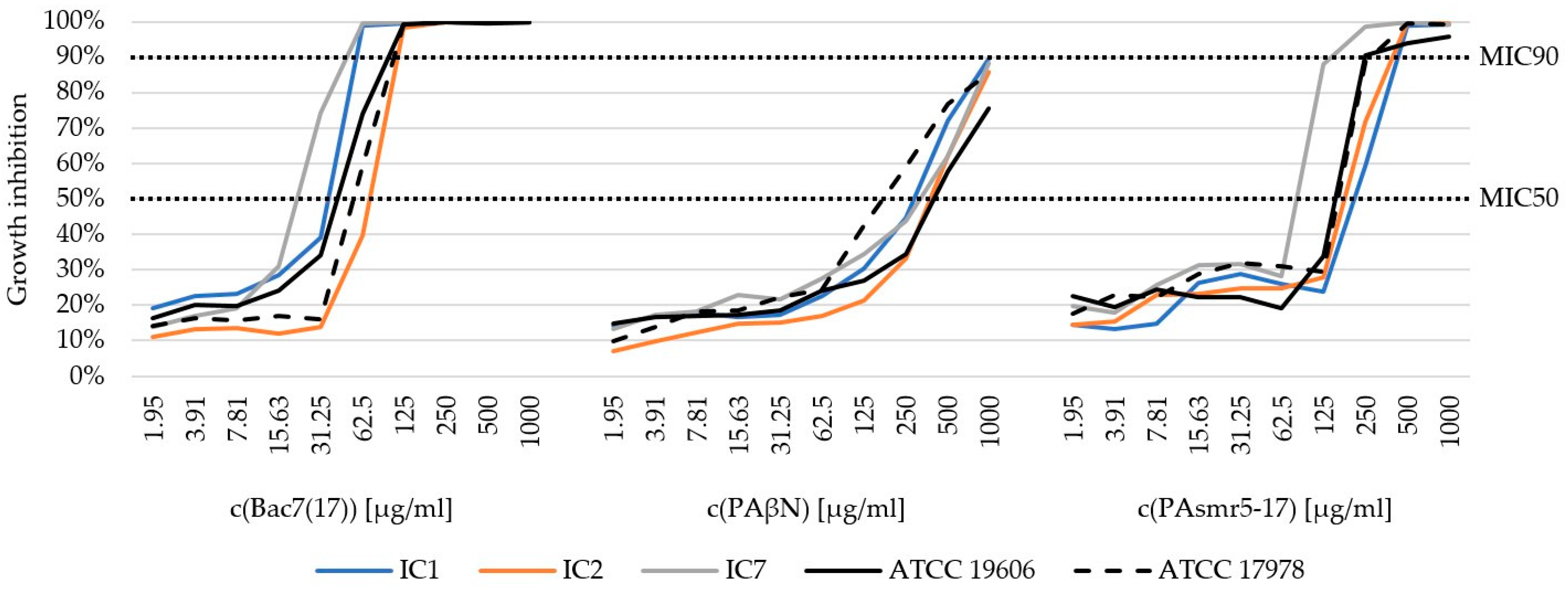
3.3. Minimum Inhibitory Concentration (MIC) of AMPs

Due to the absence of breakpoints, we determined the MIC50 and MIC90 values of the three AMPs (Table 3). In general, BG was increasingly diminished with increasing concentrations of AMPs. Figure 1 shows the average percentage growth inhibition of the respective IC groups after treatment with the different AMPs. The MIC50 and MIC90 values indicate growth inhibition of 50% and 90%, respectively. A detailed graphical representation of individual isolates is provided in Supplementary Figure S1.
Bac7(17) had the strongest inhibitory effect on BG among all AMPs tested. The first MIC90 was observed at 62.5 µg/mL. Significant differences were found between Bac7(17) and PAβN at MIC50 and MIC90. While the MIC50 for Bac7(17) was already reached at 31.35 µg/mL, a concentration of 250/500 µg/mL was required for PAβN (p < 0.001). This was also the case for MIC90, which was only achieved for PAβN at the highest concentration of 1000 µg/mL in five isolates or, in some cases, not at all (p < 0.001). For PAsmr5-17, the first MIC90 was observed at a concentration of 125 µg/mL and MIC50 at an averaged concentration of 250 µg/mL. In addition, differences between the individual ICs could be observed. For Bac7(17), MIC90 was achieved at 62.5 µg/mL in the A. baumannii isolates assigned to IC1 and IC7. For IC2, however, MIC90 was reached at 125 µg/mL (IC1/IC7 vs. IC2: p < 0.001). Similar results were also obtained for PAsmr5-17. A significant difference could be observed between IC7 and IC1/IC2. For IC7, MIC90 was achieved at 125/250 µg/mL. For IC1 and IC2, on the other hand, this concentration was sufficient to achieve MIC50. The MIC90 of these clonal lineages was reached at 250/500 µg/mL (IC1 vs. IC7: p = 0.043; IC2 vs. IC7: p = 0.047).

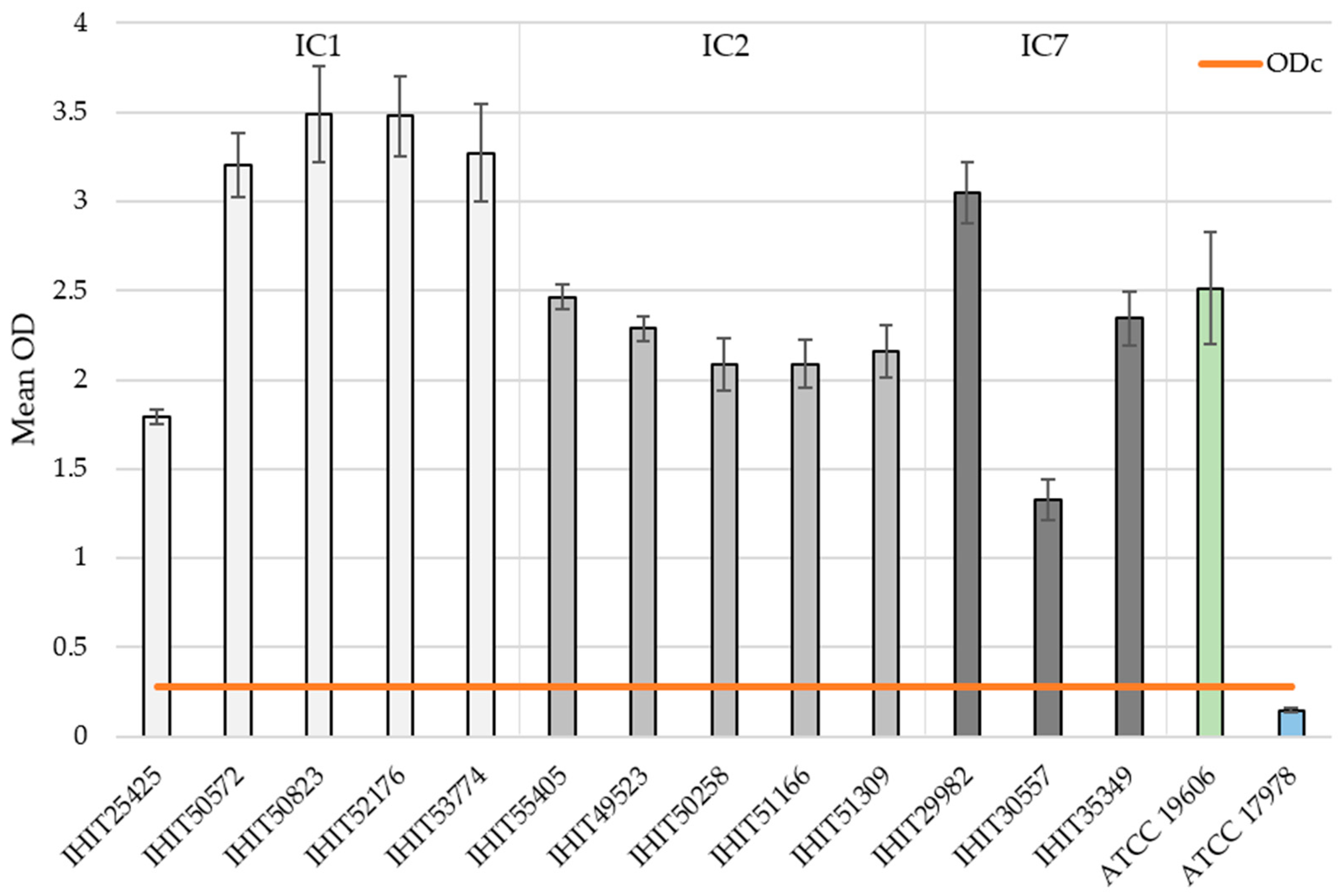
3.4. Biofilm Formation (BF) and Biofilm-Associated Genes (BAGs)

The biofilm-forming capacity of each isolate is summarized in Figure 2. The mean OD values for the reference strains ATCC 19606T (positive control) and ATCC 17978 (negative control) were 2.514 ± 0.316 and 0.242 ± 0.011, respectively. The mean OD values for the clinical isolates ranged from 1.325 ± 0.114 (IHIT30557) to 3.491 ± 0.269 (IHIT29982). All isolates were classified into four groups based on their BF [61]. For this purpose, the cut-off value (ODc) was determined, which is defined as three standard deviations above the mean OD of the negative control. The determined ODc is 0.275. This results in the classification shown below.
  • Non-biofilm producer: OD ≤ 0.275
  • Weak biofilm producer: 0.275 < OD ≤ 0.550
  • Moderate biofilm producer: 0.550 < OD ≤ 1.100
  • Strong biofilm producer: 1.100 < OD
The classification into the corresponding groups of biofilm producers of all A. baumannii isolates is shown in Table 4.
Differences in the degree of biofilm formation were observed within each IC group. For example, the strains belonging to the clonal lineage IC1 show the strongest BF (Ø OD value = 3.339 ± 0.157) with the exception of isolate IHIT25425. Here, only an OD value of 1.793 ± 0.042 could be determined, which represents only 54% of the average OD value of the remaining IC1 isolates. In contrast, IC2 isolates showed a consistent result with an average biofilm OD value of 2.217 ± 0.143 (range from 2.086 to 2.463). In contrast, IC7 isolates did not present consistent BF. As mentioned above, IHIT30557 showed the lowest BF of all isolates with an OD value of 1.325 ± 0.114, followed by IHIT35349 with 2.344 ± 0.150 and IHIT29982 with 3.047 ± 0.173.
Regarding the BAGs, IC2 isolates harbored all 18 genes (Table 5). While IC1 and IC7 showed almost the same gene pattern, 3 of the 18 BAGs are missing, namely, bap, blp1 and ompA. However, BAG profiles differed among IC1 isolates. The A. baumannii isolate IHIT50572 lacked the BAG csuB, and IHIT53774 lacked csuA.

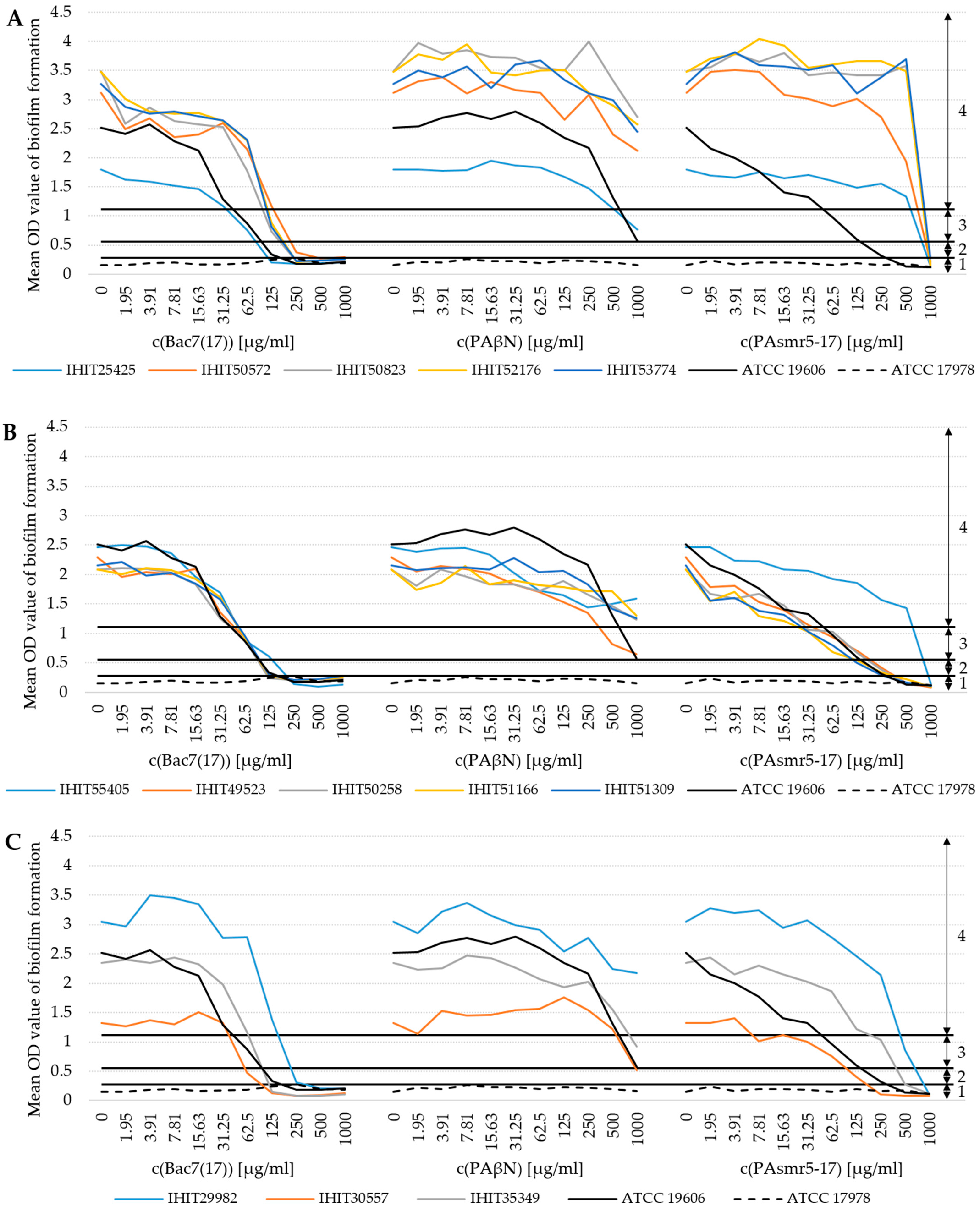
3.5. Influence of AMPs on Biofilm Formation (BF)

Of the three AMPs tested, Bac7(17) showed the strongest and PAβN the lowest inhibitory effect on BF (Figure 3). At a concentration of 62.5 µg/mL of Bac7(17), 6 of the 13 strong A. baumannii biofilm producers achieved moderate BF. At 125 µg/mL, only two isolates were still categorized as strong biofilm producers. At 250 µg/mL, only two isolates showed weak biofilm formation. The remaining isolates were classified as non-biofilm producers. In contrast, PAβN had hardly any inhibitory effect on BF at the same concentrations. At 500 µg/mL, one isolate could be classified as a moderate biofilm producer. At 1000 µg/mL, three isolates showed moderate biofilm formation and only one isolate showed weak biofilm formation. Unlike Bac7(17), the AMP PAsmr5-17 did not show a consistent effect on BF. There was no effect on the BF of IC1 isolates and isolate IHIT55405 (IC2). At 250 µg/mL, the first non-biofilm producer (IHIT30557) and four weak biofilm producers (all IC2, except IHIT55405) were determined. At the highest concentration, all isolates could be classified as non-biofilm producers. Despite the overall low effect of PAβN, we detected a significant difference in BF between IC1 and IC2 with an averaged OD difference of 1.331 (p = 0.047). Regarding PAsmr5-17, differences between IC1 and IC2 already appeared at the lowest concentration of 1.95 µg/mL, with an averaged OD difference of 1.411 (p = 0.050), and 3.91 µg/mL, with an averaged OD difference of 1.679 (p = 0.057). At 500 µg/mL, significant differences were found between IC1 and IC2 (averaged OD difference: 2.382, p = 0.015) as well as IC1 and IC7 (averaged OD difference: 2.401, p = 0.016).

4. Discussion

The increasing global prevalence of MDR-A. baumannii has prompted researchers to investigate the mechanisms of AMR in this pathogen and to develop alternative treatment strategies, both in the human and veterinary domains.
In this study, the antibacterial effect of three AMPs with different modes of action was tested to determine their potential in inhibiting BG and BF. These three AMPs are PAβN and two novel and previously untested variants Bac7(17) and PAsmr5-17. PAβN is a competitive broad-spectrum efflux pump inhibitor (EPI). This effect is particularly beneficial as the efflux pumps of bacterial cells serve as resistance mechanisms against antibiotics. Some antibiotics bind as a substrate to the efflux pump binding site and are thus transported out of the cell. Consequently, these antibiotics have no effect on the pathogen. By inhibiting these pumps, these antibiotics can no longer be transported out of the cell, resulting in a potentiation of antibiotic activity [43,62]. Bac7(17) is a proline-rich peptide derived from Bactenecin, a cathelicidine from bovine neutrophils. It has been shown to be non-toxic to mammalian cells, even at concentrations far above those effective against bacteria. Bac7(17) inactivates bacterial cells by membrane damage via depolarization, permeabilization and the subsequent inhibition of protein synthesis by binding to ribosomes [37,42]. PAsmr5-17 is a synthetic peptide-based EPI, as it contains residues centered on the TM4-TM4 binding interface found in P. aeruginosa small multidrug resistance (SMR) efflux protein [44]. Our study provides a comprehensive analysis of MDR-A. baumannii isolates from humans and animals. The isolates were assigned to either IC1 (ST1), IC2 (ST2) or IC7 (ST25). These ICs/STs are predominantly found in humans but have also often been described in A. baumannii infections in veterinary medicine [6,63,64,65].
The effect of AMPs on BG was determined by growth inhibition tests. Here, Bac7(17) showed the strongest influence on BG with an MIC90 of 62.5 µg/mL, followed by PAsmr5-17, revealing an MIC90 of 125 µg/mL. PAβN did not reach MIC90 and reached a maximum growth inhibition of 87.9%. These results suggest a major effect of the mode of action of the different AMPs. While PAβN as an EPI prevents the removal of metabolic products from the cell, Bac7(17) destroys the bacterial cell and inhibits protein synthesis, which leads to cell death [66]. MIC tests recently performed with pure bactenicin, which is the original source of Bac7(17), revealed an MIC95 in the species Desulfovibrio vulgaris at a concentration of 62.5 µg/mL, which is comparable to what we observed for A. baumannii and Bac7(17) [57].
Furthermore, we observed differences between A. baumannii clonal lineages and the effect of AMPs on BG. While 62.5 µg/mL of Bac7(17) was sufficient to achieve an MIC90 in IC1 and IC7 isolates, 125 µg/mL was required for IC2 isolates, regardless of the host of origin. A stronger effect of PAsmr5-17 was observed on IC7 isolates (MIC90 at 125 µg/mL) than on IC1 and IC2 isolates (MIC90 at 250/500 µg/mL). Overall, IC2 isolates showed the highest resistance to the AMPs, followed by IC1 and IC7 isolates.
As with BG, there was an inhibitory effect of AMPs on the BF of MDR A. baumannii isolates. However, it was expected that a complete inhibition of BG would result in a complete reduction in biofilm production. Therefore, we used the same isolates and applied the same growth conditions (medium, incubation time, temperature) to allow a direct comparison of MICs and data from the biofilm assay. Indeed, there was a clear correlation between growth inhibition and reduction in BF for each isolate. In addition, both Bac7(17) and PAsmr5-17 were able to reduce the biofilm-forming capacity in IC1 and IC7 isolates from the classification “strong” to “non-biofilm producer”. This finding is conclusive in that different patterns of BAGs are found in the respective ICs. IC2 isolates had all 18 of the tested BAGs, while IC1 and IC7 isolates showed an almost identical gene pattern but lacked the three BAGs bap, blp1 and ompA. Loehfelm et al. (2008) could show that bap is responsible for the thickness and volume of the biofilm, among other things [67]. The weaker biofilm in IC1 and IC7 might be due to the absence of bap and two other BAGs, namely, blp1 and ompA. Nevertheless, due to the small number of isolates tested here, further studies are required to validate and extend our results.
A lower concentration was required for Bac7(17) (125/250 µg/mL) than for PAsmr5-17 (500/1000 µg/mL), regardless of whether the isolates originated from humans or animals. A comparison with the MIC testing of AMPs shows that a higher concentration of AMPs is necessary to reduce the biofilm than to inhibit BG. This further supports the assumption that the biofilm acts as a self-protection mechanism by forming a barrier against stress caused by external influences [68]. Although PAβN resulted in biofilm reduction, the maximum reduction achieved with this AMP corresponded to “moderate biofilm producer” at the highest concentration. The minor effect of PAβN on biofilm production is probably related to its mode of action as an EPI. There are several studies on BF and efflux pumps. For example, He et al. (2015) reported that BF by clinical isolates of A. baumannii was associated with an overexpression of the AdeFGH efflux pump [69]. Furthermore, Richmond et al. (2016), examined the ability of different strains of A. baumannii adeB knockout mutants to form biofilms and showed that the knockout of adeB of the adeABC efflux pump resulted in a significant reduction in BF [70]. Our results showed that a complete inhibition of biofilm formation was not possible with PAβN. A similar result was also found by Chen et al. (2020). They achieved a 30% inhibition of the biofilm with 100 µg/mL of PAβN [71].
In summary, we showed that the novel AMPs Bac7(17) and Pasmr5-17 have a major effect on the BG and BF of MDR-A. baumannii isolates regardless of whether they originate from humans or animals. To the authors’ knowledge, the inhibitory effect of AMPs on the BF of MDR-A. baumannii isolates from companion animals was demonstrated here for the first time. Moreover, it seems that the efficacy of an AMP could be dependent on its mode of action. MDR-A. baumannii infections represent an increasing threat to public health. Our data are highly encouraging for future research on AMPs as alternative treatment options for infections in humans and companion animals, either as the sole medical therapy or in combination with antibiotics. Moreover, the development and evaluation of novel AMPs or AMP variants should be intensified in order to overcome the issue of limited treatment options for MDR infections.

5. Conclusions

AMPs are regarded as potential alternatives to antibiotics, which are becoming increasingly ineffective due to the increasing occurrence of MDR bacteria. However, due to their different mechanisms of action, AMPs cannot directly replace antibiotics. Minor changes to a peptide can affect its mode of action. It is therefore important to develop and test new AMP variants such as Bac7(17) and PAsmr7-15 and compare them with a well-researched AMP like PAβN. Our results show that low concentrations of Bac7(17) and PAsmr7-15 can significantly affect the BG and BF of MDR-A. baumannii isolates from humans and animals. While Bac7(17) and PAsmr5-17 were able to significantly reduce BF, PAβN showed limited efficacy. We were also able to show that AMPs with the same mode of action do not necessarily have the same effect. The differences between the individual ICs indicate that consistent results within the same species cannot be expected. This information may be of great interest for future therapies. It shows that the successful treatment of both non-MDR- and MDR-A. baumannii infections may depend on which IC or ST the species belongs to. These results emphasize the need for further research to evaluate the potential of AMPs as new therapeutic options to combat MDR-A. baumannii infections to further explore strain-specific effects. With the growing threat of MDR bacteria, it is critical to develop and explore innovative approaches to treat these infections.

Supplementary Materials

The following supporting information can be downloaded at https://www.mdpi.com/article/10.3390/microorganisms13030639/s1, Figure S1: Detailed graphical representation of MIC assay results of each of the 13 MDR-A. baumannii isolates; Table S1: Detailed list with antimicrobial resistance genes, antimicrobial susceptibility and biofilm-associated genes of the 13 MDR-A. baumannii isolates.

Author Contributions

Conceptualization, C.E.; methodology, C.E., D.M. and J.R.-T.; software, J.R.-T. and T.S.; validation, C.E., D.M. and J.R.-T.; formal analysis, C.E., J.R.-T. and U.L.; investigation, J.R.-T. and U.L.; resources, C.E., S.G., I.S. and T.S.; data curation, J.R.-T. and C.E.; writing—original draft preparation, J.R.-T.; writing—review and editing, J.R.-T., D.M. and C.E.; visualization, J.R.-T. and C.E.; supervision, C.E.; project administration, C.E.; funding acquisition, C.E. All authors have read and agreed to the published version of the manuscript.

Funding

Stephan Göttig is supported by the Rolf. M. Schwiete-Stiftung. The remaining authors received no external funding.

Institutional Review Board Statement

Not applicable.

Informed Consent Statement

Not applicable.

Data Availability Statement

The original contributions presented in this study are included in the article/Supplementary Material. Further inquiries can be directed to the corresponding author.

Acknowledgments

We would like to thank our colleagues from the microbiological diagnostic laboratory of the Institute of Hygiene and Infectious Diseases of Animals, JLU Giessen, for collecting Acinetobacter baumannii isolates from veterinary clinical samples. We would also like to thank Yvonne Pfeifer from RKI Wernigerode for generously providing human isolates.

Conflicts of Interest

Author Ivonne Stamm was employed by the company Vet Med Labor GmbH. The remaining authors declare that the research was conducted in the absence of any commercial or financial relationships that could be construed as a potential conflict of interest.

References

  1. Batt, C.A.; Tortorello, M.L. (Eds.) Encyclopedia of Food Microbiology, 2nd ed.; Elsevier: Amsterdam, The Netherlands, 2014; ISBN 9780123847300. [Google Scholar]
  2. Almasaudi, S.B. Acinetobacter spp. as nosocomial pathogens: Epidemiology and resistance features. Saudi J. Biol. Sci. 2018, 25, 586–596. [Google Scholar] [CrossRef] [PubMed]
  3. Antunes, L.C.S.; Visca, P.; Towner, K.J. Acinetobacter baumannii: Evolution of a global pathogen. Pathog. Dis. 2014, 71, 292–301. [Google Scholar] [CrossRef]
  4. Wareth, G.; Neubauer, H.; Sprague, L.D. Acinetobacter baumannii—A neglected pathogen in veterinary and environmental health in Germany. Vet. Res. Commun. 2019, 43, 1–6. [Google Scholar] [CrossRef]
  5. Rice, L.B. Federal funding for the study of antimicrobial resistance in nosocomial pathogens: No ESKAPE. J. Infect. Dis. 2008, 197, 1079–1081. [Google Scholar] [CrossRef]
  6. Nocera, F.P.; Attili, A.-R.; de Martino, L. Acinetobacter baumannii: Its Clinical Significance in Human and Veterinary Medicine. Pathogens 2021, 10, 127. [Google Scholar] [CrossRef] [PubMed]
  7. Müller, C.; Reuter, S.; Wille, J.; Xanthopoulou, K.; Stefanik, D.; Grundmann, H.; Higgins, P.G.; Seifert, H. A global view on carbapenem-resistant Acinetobacter baumannii. mBio 2023, 14, e0226023. [Google Scholar] [CrossRef]
  8. Karah, N.; Faille, N.; Grenier, F.; Abou-Fayad, A.; Higgins, P.G.; Al-Hassan, L.; Evans, B.A.; Poirel, L.; Bonnin, R.; Hammerum, A.M.; et al. Emergence of Acinetobacter baumannii International Clone 10 predominantly found in the Middle East. bioRxiv 2023. [Google Scholar] [CrossRef]
  9. Hansen, F.; Porsbo, L.J.; Frandsen, T.H.; Kaygisiz, A.N.S.; Roer, L.; Henius, A.E.; Holzknecht, B.J.; Søes, L.; Schønning, K.; Røder, B.L.; et al. Characterisation of carbapenemase-producing Acinetobacter baumannii isolates from danish patients 2014–2021: Detection of a new international clone—IC11. Int. J. Antimicrob. Agents 2023, 62, 106866. [Google Scholar] [CrossRef] [PubMed]
  10. Xu, A.; Li, M.; Hang, Y.; Zeng, L.; Zhang, X.; Hu, Y.; Guo, Q.; Wang, M. Multicenter retrospective genomic characterization of carbapenemase-producing Acinetobacter baumannii isolates from Jiangxi patients 2021–2022: Identification of a novel international clone, IC11. mSphere 2024, 9, e0027624. [Google Scholar] [CrossRef]
  11. Higgins, P.G.; Dammhayn, C.; Hackel, M.; Seifert, H. Global spread of carbapenem-resistant Acinetobacter baumannii. J. Antimicrob. Chemother. 2010, 65, 233–238. [Google Scholar] [CrossRef]
  12. Pour, N.K.; Dusane, D.H.; Dhakephalkar, P.K.; Zamin, F.R.; Zinjarde, S.S.; Chopade, B.A. Biofilm formation by Acinetobacter baumannii strains isolated from urinary tract infection and urinary catheters. FEMS Immunol. Med. Microbiol. 2011, 62, 328–338. [Google Scholar] [CrossRef] [PubMed]
  13. Müller, S.; Janssen, T.; Wieler, L.H. Multidrug resistant Acinetobacter baumannii in veterinary medicine—Emergence of an underestimated pathogen? Berl. Munch. Tierarztl. Wochenschr. 2014, 127, 435–446. [Google Scholar] [PubMed]
  14. Tomaras, A.P.; Dorsey, C.W.; Edelmann, R.E.; Actis, L.A. Attachment to and biofilm formation on abiotic surfaces by Acinetobacter baumannii: Involvement of a novel chaperone-usher pili assembly system. Microbiology 2003, 149, 3473–3484. [Google Scholar] [CrossRef]
  15. Eze, E.C.; Chenia, H.Y.; El Zowalaty, M.E. Acinetobacter baumannii biofilms: Effects of physicochemical factors, virulence, antibiotic resistance determinants, gene regulation, and future antimicrobial treatments. Infect. Drug Resist. 2018, 11, 2277–2299. [Google Scholar] [CrossRef]
  16. Longo, F.; Vuotto, C.; Donelli, G. Biofilm formation in Acinetobacter baumannii. New Microbiol. 2014, 37, 119–127. [Google Scholar] [PubMed]
  17. Bardbari, A.M.; Arabestani, M.R.; Karami, M.; Keramat, F.; Alikhani, M.Y.; Bagheri, K.P. Correlation between ability of biofilm formation with their responsible genes and MDR patterns in clinical and environmental Acinetobacter baumannii isolates. Microb. Pathog. 2017, 108, 122–128. [Google Scholar] [CrossRef]
  18. Abdullahi, U.F.; Igwenagu, E.; Mu’azu, A.; Aliyu, S.; Umar, M.I. Intrigues of biofilm: A perspective in veterinary medicine. Vet World 2016, 9, 12–18. [Google Scholar] [CrossRef]
  19. Sarshar, M.; Behzadi, P.; Scribano, D.; Palamara, A.T.; Ambrosi, C. Acinetobacter baumannii: An Ancient Commensal with Weapons of a Pathogen. Pathogens 2021, 10, 387. [Google Scholar] [CrossRef]
  20. Choi, A.H.K.; Slamti, L.; Avci, F.Y.; Pier, G.B.; Maira-Litrán, T. The pgaABCD locus of Acinetobacter baumannii encodes the production of poly-beta-1-6-N-acetylglucosamine, which is critical for biofilm formation. J. Bacteriol. 2009, 191, 5953–5963. [Google Scholar] [CrossRef]
  21. Pakharukova, N.; Tuittila, M.; Paavilainen, S.; Malmi, H.; Parilova, O.; Teneberg, S.; Knight, S.D.; Zavialov, A.V. Structural basis for Acinetobacter baumannii biofilm formation. Proc. Natl. Acad. Sci. USA 2018, 115, 5558–5563. [Google Scholar] [CrossRef]
  22. Kim, H.-J.; Kim, N.-Y.; Ko, S.-Y.; Park, S.-Y.; Oh, M.-H.; Shin, M.-S.; Lee, Y.-C.; Lee, J.-C. Complementary Regulation of BfmRS Two-Component and AbaIR Quorum Sensing Systems to Express Virulence-Associated Genes in Acinetobacter baumannii. Int. J. Mol. Sci. 2022, 23, 13136. [Google Scholar] [CrossRef]
  23. Roy, S.; Chowdhury, G.; Mukhopadhyay, A.K.; Dutta, S.; Basu, S. Convergence of Biofilm Formation and Antibiotic Resistance in Acinetobacter baumannii Infection. Front. Med. 2022, 9, 793615. [Google Scholar] [CrossRef]
  24. Lee, C.-R.; Lee, J.H.; Park, M.; Park, K.S.; Bae, I.K.; Kim, Y.B.; Cha, C.-J.; Jeong, B.C.; Lee, S.H. Biology of Acinetobacter baumannii: Pathogenesis, Antibiotic Resistance Mechanisms, and Prospective Treatment Options. Front. Cell. Infect. Microbiol. 2017, 7, 55. [Google Scholar] [CrossRef] [PubMed]
  25. Isler, B.; Doi, Y.; Bonomo, R.A.; Paterson, D.L. New Treatment Options against Carbapenem-Resistant Acinetobacter baumannii Infections. Antimicrob. Agents Chemother. 2019, 63, e01110-18. [Google Scholar] [CrossRef] [PubMed]
  26. Giguère, S.; Prescott, J.F.; Dowling, P.M. (Eds.) Antimicrobial Therapy in Veterinary Medicine, 5th ed.; Wiley-Blackwell: Ames, IA, USA, 2013; ISBN 9781118675014. [Google Scholar]
  27. Wong, D.; Nielsen, T.B.; Bonomo, R.A.; Pantapalangkoor, P.; Luna, B.; Spellberg, B. Clinical and Pathophysiological Overview of Acinetobacter Infections: A Century of Challenges. Clin. Microbiol. Rev. 2017, 30, 409–447. [Google Scholar] [CrossRef] [PubMed]
  28. European Medicines Agency. Categorisation of Antibiotics in the European Union. Available online: https://www.ema.europa.eu/en/news/categorisation-antibiotics-used-animals-promotes-responsible-use-protect-public-and-animal-health (accessed on 8 April 2024).
  29. van der Kolk, J.H.; Endimiani, A.; Graubner, C.; Gerber, V.; Perreten, V. Acinetobacter in veterinary medicine, with an emphasis on Acinetobacter baumannii. J. Glob. Antimicrob. Resist. 2019, 16, 59–71. [Google Scholar] [CrossRef]
  30. Schwabe, U.; Paffrath, D. Menge verordneter Antibiotika in Deutschland nach den wichtigsten Substanzgruppen in den Jahren von 2008 bis 2021. Available online: https://de.statista.com/statistik/daten/studie/371902/umfrage/menge-verordneter-antibiotika-in-deutschland-nach-den-wichtigsten-substanzgruppen/ (accessed on 1 February 2024).
  31. Bundesamt für Verbraucherschutz und Lebensmittelsicherheit. Abgegebene Menge Antibiotika in der Veterinärmedizin in Deutschland nach Wirkstoffklassen in den Jahren 2015 bis 2022 (in Tonnen) [Graph]. Available online: https://de.statista.com/statistik/daten/studie/371869/umfrage/abgegebene-menge-antibiotika-in-der-veterinaermedizin-in-deutschland-nach-wirkstoffklassen/ (accessed on 1 February 2024).
  32. Tacconelli, E.; Carrara, E.; Savoldi, A.; Harbarth, S.; Mendelson, M.; Monnet, D.L.; Pulcini, C.; Kahlmeter, G.; Kluytmans, J.; Carmeli, Y.; et al. Discovery, research, and development of new antibiotics: The WHO priority list of antibiotic-resistant bacteria and tuberculosis. Lancet Infect. Dis. 2018, 18, 318–327. [Google Scholar] [CrossRef]
  33. Jesudason, T. WHO publishes updated list of bacterial priority pathogens. Lancet Microbe 2024, 5, 100940. [Google Scholar] [CrossRef]
  34. Mulukutla, A.; Shreshtha, R.; Kumar Deb, V.; Chatterjee, P.; Jain, U.; Chauhan, N. Recent advances in antimicrobial peptide-based therapy. Bioorg. Chem. 2024, 145, 107151. [Google Scholar] [CrossRef]
  35. Bahar, A.A.; Ren, D. Antimicrobial peptides. Pharmaceuticals 2013, 6, 1543–1575. [Google Scholar] [CrossRef]
  36. Huan, Y.; Kong, Q.; Mou, H.; Yi, H. Antimicrobial Peptides: Classification, Design, Application and Research Progress in Multiple Fields. Front. Microbiol. 2020, 11, 582779. [Google Scholar] [CrossRef]
  37. Neshani, A.; Sedighian, H.; Mirhosseini, S.A.; Ghazvini, K.; Zare, H.; Jahangiri, A. Antimicrobial peptides as a promising treatment option against Acinetobacter baumannii infections. Microb. Pathog. 2020, 146, 104238. [Google Scholar] [CrossRef]
  38. Phoenix, D.A.; Dennison, S.R.; Harris, F. (Eds.) Antimicrobial Peptides, 1st ed.; Wiley-VHC: Weinheim, Germany, 2013; ISBN 978-3-527-65288-4. [Google Scholar]
  39. Jaśkiewicz, M.; Neubauer, D.; Kazor, K.; Bartoszewska, S.; Kamysz, W. Antimicrobial Activity of Selected Antimicrobial Peptides Against Planktonic Culture and Biofilm of Acinetobacter baumannii. Probiotics Antimicrob. Proteins 2019, 11, 317–324. [Google Scholar] [CrossRef] [PubMed]
  40. Lewies, A.; Du Plessis, L.H.; Wentzel, J.F. Antimicrobial Peptides: The Achilles’ Heel of Antibiotic Resistance? Probiotics Antimicrob. Proteins 2019, 11, 370–381. [Google Scholar] [CrossRef] [PubMed]
  41. Benincasa, M.; Scocchi, M.; Podda, E.; Skerlavaj, B.; Dolzani, L.; Gennaro, R. Antimicrobial activity of Bac7 fragments against drug-resistant clinical isolates. Peptides 2004, 25, 2055–2061. [Google Scholar] [CrossRef]
  42. Müller, D. Identifizierung, Charakterisierung und Untersuchungen der Struktur-Wirkungs-Beziehung Sowie des Mode of Action Eines Neuen Antimikrobiellen Peptids aus Hirudo Verbana. Ph.D. Thesis, Philipps-Universität Marburg, Marburg, Germany, 2019. [Google Scholar]
  43. Jamshidi, S.; Sutton, J.M.; Rahman, K.M. Computational Study Reveals the Molecular Mechanism of the Interaction between the Efflux Inhibitor PAβN and the AdeB Transporter from Acinetobacter baumannii. ACS Omega 2017, 2, 3002–3016. [Google Scholar] [CrossRef] [PubMed]
  44. Mitchell, C.J.; Stone, T.A.; Deber, C.M. Peptide-Based Efflux Pump Inhibitors of the Small Multidrug Resistance Protein from Pseudomonas aeruginosa. Antimicrob. Agents Chemother. 2019, 63, e00730-19. [Google Scholar] [CrossRef]
  45. Magiorakos, A.-P.; Srinivasan, A.; Carey, R.B.; Carmeli, Y.; Falagas, M.E.; Giske, C.G.; Harbarth, S.; Hindler, J.F.; Kahlmeter, G.; Olsson-Liljequist, B.; et al. Multidrug-resistant, extensively drug-resistant and pandrug-resistant bacteria: An international expert proposal for interim standard definitions for acquired resistance. Clin. Microbiol. Infect. 2012, 18, 268–281. [Google Scholar] [CrossRef]
  46. Klotz, P.; Jacobmeyer, L.; Stamm, I.; Leidner, U.; Pfeifer, Y.; Semmler, T.; Prenger-Berninghoff, E.; Ewers, C. Carbapenem-resistant Acinetobacter baumannii ST294 harbouring the OXA-72 carbapenemase from a captive grey parrot. J. Antimicrob. Chemother. 2018, 73, 1098–1100. [Google Scholar] [CrossRef]
  47. Clinical and Laboratory Standards Institute. Performance Standards for Antimicrobial Susceptibility Testing, 33rd ed.; Clinical and Laboratory Standards Institute: Berwyn, PA, USA, 2023. [Google Scholar]
  48. European Committee on Antimicrobial Susceptibility Testing. Breakpoint Tables for Interpretation of MICs and Zone Diameters, Version 13.0. 2023. Available online: http://www.eucast.org (accessed on 8 April 2024).
  49. Bortolaia, V.; Kaas, R.S.; Ruppe, E.; Roberts, M.C.; Schwarz, S.; Cattoir, V.; Philippon, A.; Allesoe, R.L.; Rebelo, A.R.; Florensa, A.F.; et al. ResFinder 4.0 for predictions of phenotypes from genotypes. J. Antimicrob. Chemother. 2020, 75, 3491–3500. [Google Scholar] [CrossRef]
  50. Clausen, P.T.L.C.; Aarestrup, F.M.; Lund, O. Rapid and precise alignment of raw reads against redundant databases with KMA. BMC Bioinform. 2018, 19, 307. [Google Scholar] [CrossRef]
  51. Camacho, C.; Coulouris, G.; Avagyan, V.; Ma, N.; Papadopoulos, J.; Bealer, K.; Madden, T.L. BLAST+: Architecture and applications. BMC Bioinform. 2009, 10, 421. [Google Scholar] [CrossRef]
  52. Feng, Y.; Zou, S.; Chen, H.; Yu, Y.; Ruan, Z. BacWGSTdb 2.0: A one-stop repository for bacterial whole-genome sequence typing and source tracking. Nucleic Acids Res. 2021, 49, 644–650. [Google Scholar] [CrossRef] [PubMed]
  53. Ruan, Z.; Feng, Y. BacWGSTdb, a database for genotyping and source tracking bacterial pathogens. Nucleic Acids Res. 2016, 44, D682–D687. [Google Scholar] [CrossRef]
  54. Ruan, Z.; Yu, Y.; Feng, Y. The global dissemination of bacterial infections necessitates the study of reverse genomic epidemiology. Brief. Bioinform. 2020, 21, 741–750. [Google Scholar] [CrossRef] [PubMed]
  55. Larsen, M.V.; Cosentino, S.; Rasmussen, S.; Friis, C.; Hasman, H.; Marvig, R.L.; Jelsbak, L.; Sicheritz-Pontén, T.; Ussery, D.W.; Aarestrup, F.M.; et al. Multilocus sequence typing of total-genome-sequenced bacteria. J. Clin. Microbiol. 2012, 50, 1355–1361. [Google Scholar] [CrossRef]
  56. Center for Genomic Epidemiology. MLST. Available online: https://cge.food.dtu.dk/services/MLST/ (accessed on 8 September 2022).
  57. Stillger, L.; Viau, L.; Kamm, L.; Holtmann, D.; Müller, D. Optimization of antimicrobial peptides for the application against biocorrosive bacteria. Appl. Microbiol. Biotechnol. 2023, 107, 4041–4049. [Google Scholar] [CrossRef] [PubMed]
  58. O’Toole, G.A. Microtiter Dish Biofilm Formation Assay. J. Vis. Exp. 2011, 47, 2437–2438. [Google Scholar] [CrossRef]
  59. Qi, L.; Li, H.; Zhang, C.; Liang, B.; Li, J.; Wang, L.; Du, X.; Liu, X.; Qiu, S.; Song, H. Relationship between Antibiotic Resistance, Biofilm Formation, and Biofilm-Specific Resistance in Acinetobacter baumannii. Front. Microbiol. 2016, 7, 483. [Google Scholar] [CrossRef]
  60. Fursova, N.K.; Fursov, M.V.; Astashkin, E.I.; Fursova, A.D.; Novikova, T.S.; Kislichkina, A.A.; Sizova, A.A.; Fedyukina, G.N.; Savin, I.A.; Ershova, O.N. Multidrug-Resistant and Extensively Drug-Resistant Acinetobacter baumannii Causing Nosocomial Meningitis in the Neurological Intensive Care Unit. Microorganisms 2023, 11, 2020. [Google Scholar] [CrossRef]
  61. Stepanović, S.; Vuković, D.; Hola, V.; Di Bonaventura, G.; Djukić, S.; Cirković, I.; Ruzicka, F. Quantification of biofilm in microtiter plates: Overview of testing conditions and practical recommendations for assessment of biofilm production by Staphylococci. APMIS 2007, 115, 891–899. [Google Scholar] [CrossRef] [PubMed]
  62. Soto, S.M. Role of efflux pumps in the antibiotic resistance of bacteria embedded in a biofilm. Virulence 2013, 4, 223–229. [Google Scholar] [CrossRef]
  63. Rühl-Teichner, J.; Jacobmeyer, L.; Leidner, U.; Semmler, T.; Ewers, C. Genomic Diversity, Antimicrobial Susceptibility, and Biofilm Formation of Clinical Acinetobacter baumannii Isolates from Horses. Microorganisms 2023, 11, 556. [Google Scholar] [CrossRef]
  64. Jacobmeyer, L.; Semmler, T.; Stamm, I.; Ewers, C. Genomic Analysis of Acinetobacter baumannii Isolates Carrying OXA-23 and OXA-58 Genes from Animals Reveals ST1 and ST25 as Major Clonal Lineages. Antibiotics 2022, 11, 1045. [Google Scholar] [CrossRef]
  65. Shelenkov, A.; Akimkin, V.; Mikhaylova, Y. International Clones of High Risk of Acinetobacter baumannii-Definitions, History, Properties and Perspectives. Microorganisms 2023, 11, 2115. [Google Scholar] [CrossRef] [PubMed]
  66. Wieczorek, P.; Sacha, P.; Hauschild, T.; Zórawski, M.; Krawczyk, M.; Tryniszewska, E. Multidrug resistant Acinetobacter baumannii--the role of AdeABC (RND family) efflux pump in resistance to antibiotics. Folia Histochem Cytobiol 2008, 46, 257–267. [Google Scholar] [CrossRef] [PubMed]
  67. Loehfelm, T.W.; Luke, N.R.; Campagnari, A.A. Identification and characterization of an Acinetobacter baumannii biofilm-associated protein. J. Bacteriol. 2008, 190, 1036–1044. [Google Scholar] [CrossRef]
  68. Balducci, E.; Papi, F.; Capialbi, D.E.; Del Bino, L. Polysaccharides’ Structures and Functions in Biofilm Architecture of Antimicrobial-Resistant (AMR) Pathogens. Int. J. Mol. Sci. 2023, 24, 4030. [Google Scholar] [CrossRef]
  69. He, X.; Lu, F.; Yuan, F.; Jiang, D.; Zhao, P.; Zhu, J.; Cheng, H.; Cao, J.; Lu, G. Biofilm Formation Caused by Clinical Acinetobacter baumannii Isolates Is Associated with Overexpression of the AdeFGH Efflux Pump. Antimicrob. Agents Chemother. 2015, 59, 4817–4825. [Google Scholar] [CrossRef]
  70. Richmond, G.E.; Evans, L.P.; Anderson, M.J.; Wand, M.E.; Bonney, L.C.; Ivens, A.; Chua, K.L.; Webber, M.A.; Sutton, J.M.; Peterson, M.L.; et al. The Acinetobacter baumannii Two-Component System AdeRS Regulates Genes Required for Multidrug Efflux, Biofilm Formation, and Virulence in a Strain-Specific Manner. mBio 2016, 7, e00430-16. [Google Scholar] [CrossRef]
  71. Chen, L.; Li, H.; Wen, H.; Zhao, B.; Niu, Y.; Mo, Q.; Wu, Y. Biofilm formation in Acinetobacter baumannii was inhibited by PAβN while it had no association with antibiotic resistance. Microbiologyopen 2020, 9, e1063. [Google Scholar] [CrossRef] [PubMed]
Figure 1. Average growth inhibition [%] by the AMPs Bac7(17), PAβN and PAsmr5-17 of the 13 MDR-A. baumannii isolates separated into their clonal linages: IC1 (blue), IC2 (orange) and IC7 (grey). A. baumannii reference strains ATCC 19606T and ATCC 17978 were used as controls. MIC90: 90% growth inhibition. MIC50: 50% growth inhibition. A detailed graphical representation of individual isolates is provided in Supplementary Figure S1.
Microorganisms 13 00639 g001
Figure 2. Mean OD values of BF without the influence of AMPs of the 13 A. baumannii isolates examined in this study sorted by their IC. Reference strains ATCC 19606T (green bar) and ATCC 17978 (blue bar) were used as positive and negative controls, respectively. The orange line indicates the cut-off value (ODc) defined as three standard deviations above the mean OD of the negative control (ATCC 17978).
Microorganisms 13 00639 g002
Figure 3. Effect of the AMPs Bac7(17), PAβN and PAsmr5-17 on the BF of the 13 MDR-A. baumannii isolates separated by their clonal linages: (A) IC1 (n = 5 isolates), (B) IC2 (n = 5 isolates) and (C) IC7 (n = 3 isolates). Reference strains ATCC 19606T and ATCC 17978 were used as positive and negative controls, respectively. Areas of biofilm production: 1 = non-biofilm; 2 = weak; 3 = moderate; 4 = strong.
Microorganisms 13 00639 g003
Table 1. Characteristics of the 13 MDR-A. baumannii isolates examined in this study sorted by IC.
Isolate IDOriginal
Name
HostSourceDate of
Isolation
CountryICSTPaAcquired
OXA 1
Intrinsic OXA
IHIT2542522/09HumanTrachea30.06.2009DE1123PL69
IHIT50572VB939187.1CatUrine22.12.2022DE1158PL69
IHIT50823VB949552.2DogWound16.01.2023DE1158PL69
IHIT52176VB948535DogWound20.04.2023DE1158PL69
IHIT53774VB952338DogUrine03.02.2023DE1158PL69
IHIT5540534/15-1HumanUnknown21.12.2023BG2223PL66
IHIT495232214/22-1CatAbscess eye12.09.2022DE2223PL66
IHIT502582814/22DogCVC23.11.2022DE2223PL66
IHIT51166237/23DogSkin suture08.02.2023DE2223PL66
IHIT51309314/23-1DogWound22.02.2023DE2223PL66
IHIT29982BF135647DogUrine27.07.2015FR72523CH64
IHIT30557BF136700DogUrine09.12.2015FR72523CH64
IHIT35349SG1998HumanGIT06.07.2014DE72523CH64
1 Location of blaOXA gene on plasmid (PL) or chromosome (CH). Abbreviations: CVC—Central venous catheter; GIT—Gastrointestinal tract; DE—Germany; BG—Bulgaria; FR—France; IC—international clone; STPa—multilocus sequence type according to the Pasteur scheme.
Table 2. AMR phenotypes and genes of 13 MDR-A. baumannii isolates examined in this study.
Antibiotic ClassPhenotypic ResistanceGenotypic Resistance
AntibioticAbbr.Resistant
Isolates (n)
AMR GenesPositive
Isolates (n)
β-LactamsAmpicillin *AMP13
Amoxicillin–Clavulanate *AMC13blaADC-like13
Cefalexin *CEX13blaOXA-239
CefotaximeCTX13blaOXA-584
CeftazidimeCAZ7blaOXA-643
CefepimeFEP13blaOXA-665
ImipenemIPM13blaOXA-695
MeropenemMEM13blaPER-71
PiperacillinPIP13blaTEM-1D4
Piperacillin–TazobactamTZP13
Aminoglycosides aadA16
aac(3)-Ia9
aac(3)-IIa3
GentamicinGEN13aac(6′)-Ian3
AmikacinAMK5aac(6′)-Ip3
TobramycinTOB5aph(3′)-Ia4
aph(3′)-VIa2
aph(3″)-Ib7
aph(6)-Id7
Phenicols ABUW_098213
Chloramphenicol *CHL13catA15
craA5
Sulfonamides/
Trimethoprims
Trimethoprim–SulfamethoxazoleSXT13sul111
sul27
TetracyclinesTetracyclineTET13tet(A)5
tet(B)7
FluoroquinolonesCiprofloxacinCIP13none **
EnrofloxacinENR13
MarbofloxacinMAR13
PolymyxineColistinCOL0none
* Intrinsic resistance according to CLSI [47]; ** ENR/MAR-resistant isolates that revealed mutations in GyrA (S80L) (100%) and/or ParC (S84L, A250T) (100%). For Acinetobacter spp. isolates for which veterinary clinical breakpoints were not available, human clinical breakpoints from CLSI document M100 [37] were used where possible. For ENR/MAR, breakpoints for ciprofloxacin (CLSI) [47], for AMC/CPD/CHL, breakpoints for Enterobacterales (CLSI) [47], and for AMP/CEX/COL, breakpoints for Enterobacterales (EUCAST) [48] were used. Accession Nos. of AMR gene sequences are provided in Supplementary Table S1.
Table 3. MIC [µg/mL] at 50% (MIC50) and 90% (MIC90) growth inhibition for the 13 MDR-A. baumannii isolates and 2 reference strains for the three AMPs sorted by their IC.
Bac7(17)PAβNPAsmr5-17
IsolateICMIC50MIC90MIC50MIC90MIC50MIC90
IHIT254251* 31.2562.5250>1000* 125250
IHIT50572131.2562.52501000250500
IHIT50823131.2562.55001000250500
IHIT52176131.2562.52501000250500
IHIT537741* 31.2562.5500>1000250500
IHIT55405262.5125500>1000250500
IHIT495232* 62.5125500>1000250500
IHIT502582* 62.5125250>1000* 125250
IHIT511662* 62.51255001000250500
IHIT513092* 62.5125500>1000250500
IHIT29982731.2562.5500>1000* 62.5125
IHIT30557715.6362.5250100062.5125
IHIT35349731.2562.5500>1000125250
ATCC 19606T-62.5125500>1000* 125250
ATCC 17978-62.5125250>1000* 125500
* These values correspond to the next lowest concentration of MIC90, as MIC50 could not be determined. MIC90: 90% growth inhibition. MIC50: 50% growth inhibition.
Table 4. Mean OD of biofilm formation (BF), standard deviation (SD) and corresponding classification into four different groups of biofilm producers sorted by IC.
IsolateICBFSDClassification
IHIT2542511.792±0.042strong
IHIT5057213.113±0.179strong
IHIT5082313.491±0.269strong
IHIT5217613.480±0.224strong
IHIT5377413.273±0.275strong
IHIT5540522.463±0.069strong
IHIT4952322.289±0.070strong
IHIT5025822.086±0.150strong
IHIT5116622.090±0.134strong
IHIT5130922.158±0.148strong
IHIT2998273.047±0.173strong
IHIT3055771.325±0.114strong
IHIT3534972.344±0.150strong
ATCC 19606T-2.514±0.316strong
ATCC 17978-0.242±0.011none
Table 5. Distribution of biofilm-associated genes among 13 clinical A. baumannii isolates and reference strains ATCC 19606T and ATCC 17978.
IsolateICBiofilm-Associated Genes (BAGs)
abaIabaRbapblp1blp2bfmRbfmScsuAcsuA/BcsuBcsuCcsuDcsuEompApgaApgaBpgaCpgaD
IHIT254251
IHIT505721
IHIT508231
IHIT521761
IHIT537741
IHIT554052
IHIT495232
IHIT502582
IHIT511662
IHIT513092
IHIT299827
IHIT305577
IHIT353497
ATCC 19606Tnt
ATCC 17978nt
Gray-shaded cells indicate presence and white cells indicate absence of genes. nt: unclustered. BAGs were identified by screening WGS data of A. baumannii isolates against BAG sequences of A. baumannii reference strains ATCC 17978 (Accession no. CP018664.1) and AYE (only for blp1 and blp2; Accession no. CU459141.1).
Disclaimer/Publisher’s Note: The statements, opinions and data contained in all publications are solely those of the individual author(s) and contributor(s) and not of MDPI and/or the editor(s). MDPI and/or the editor(s) disclaim responsibility for any injury to people or property resulting from any ideas, methods, instructions or products referred to in the content.

