Article

Interactions Between Endosymbionts Wolbachia and Rickettsia in the Spider Mite Tetranychus turkestani: Cooperation or Antagonism?

College of Agriculture, Shihezi University, Shihezi 832003, China
*
Author to whom correspondence should be addressed.
These authors contributed equally to this work.
Microorganisms 2025, 13(3), 642; https://doi.org/10.3390/microorganisms13030642
Submission received: 18 January 2025 / Revised: 6 March 2025 / Accepted: 7 March 2025 / Published: 12 March 2025
(This article belongs to the Section Plant Microbe Interactions)

Abstract

:
Maternally inherited endosymbionts are widespread in arthropods, with multiple symbionts commonly co-existing within a single host, potentially competing for or sharing limited host resources and space. Wolbachia and Rickettsia, two maternally-inherited symbionts in arthropods, can co-infect hosts, yet research on their combined impacts on host reproduction and interaction remains scarce. Tetranychus turkestani (Acari: Tetranychidae) is an important agricultural pest mite, characterized by rapid reproduction, a short life cycle, and being difficult to control. Wolbachia and Rickettsia are two major endosymbiotic bacteria present in T. turkestani. This study used diverse parthenogenetic backcross and antibiotic screening to explore the reproductive effects of these two symbionts on T. turkestani. The results show that single Rickettsia infection induced male killing in the amphigenesis of T. turkestani, leading to arrhenotokous embryo death and fewer offspring. Single Wolbachia infection induced strong cytoplasmic incompatibility (CI). During dual infection, CI intensity decreased because Rickettsia’s male-killing effect antagonized the Wolbachia-induced CI. Dual-infected mites had increased oviposition, lower mortality, a higher female-to-male ratio, and more offspring, thus enhancing T. turkestani’s fitness. These findings will be helpful for understanding the nature of host–endosymbiont interactions and the potential for evolutionary conflicts, offering insights into their co-evolutionary relationship.

1. Introduction

Endosymbiotic bacteria are common in arthropods, with over 50% of species infected, primarily via maternal inheritance [1,2]. Endosymbiotic bacteria have co-evolved with their hosts, influencing their nutrition, digestion, resistance, and defense against predators, and thereby playing critical role in host colonization and ecological evolution in specific habitats [3,4]. At present, the most studied secondary endosymbionts, including Wolbachia, Cardinium, Rickettsia, and Spiroplasma, are known to manipulate the host reproductive and developmental processes by inducing cytoplasmic incompatibility, male killing, parthenogenesis, heat resistance, and drug resistance in their hosts [5,6,7,8].
Wolbachia is a maternally transmitted Gram-negative bacteria found in arthropods. The host range of Wolbachia is extremely wide, and approximately 65% of insect species naturally carry this endosymbiont. Wolbachia is abundantly present in insect ovaries and testes and is also distributed in non-reproductive tissues, such as the head, muscles, midgut, salivary gland, Malpighian tubules, hemolymph, and fat body of insects [9,10]. The regulatory effects of Wolbachia on its host have always been a hot topic in Wolbachia-related research. Currently, the documented reproductive regulations of the host by Wolbachia include cytoplasmic incompatibility (CI), male killing, feminization, parthenogenesis, etc. CI is the most common reproductive regulation induced by Wolbachia, which refers to the phenomenon where mating between a Wolbachia-infected male and an uninfected female insect results in either no or few offspring. The parthenogenesis induced by Wolbachia in T. turkestani is arrhenotokous parthenogenesis; that is, the oocytes of female individuals develop directly into new individuals without the fertilization process, and the haploid eggs develop into male offspring [11]. In addition, some strains of Wolbachia can also affect the host’s sense of smell, lifespan, immunity, nutrition, fertility, developmental processes, etc. [9,10,12,13,14,15,16,17].
Rickettsia is an intracellular symbiotic bacterium that spreads and causes diseases in humans and animals. It is also a secondary endosymbiont that exists in insects. Rickettsia is a Gram-negative bacterium that belongs to the family Rickettsiaceae in the α subgroup of Proteobacteria. It is widely distributed in nature, and its hosts include vertebrates, arthropods, annelids, amoebas, ciliates, hydrozoans, and plants [7]. Research has found that Rickettsia and its host insects share a mutualistic symbiotic relationship and have co-evolved. Rickettsia can affect the reproductive behavior of their host by inducing male killing and parthenogenesis, and it also has an impact on the fitness of the host insects [8,16,17,18,19].
In nature, co-infection of arthropod hosts by different symbiotic bacteria is quite common. The impacts of multiple infections on the host may be cumulative [20]. The interactions among co-infecting symbiotic bacteria may lead to reproductive phenotypes that are completely different from those seen in singly infected hosts. If co-infection confers a higher fitness than a single infection, it can be stably maintained within the host population [21]. Rickettsia and Wolbachia sometimes co-infect arthropods. However, little research has been conducted on the interactions between these two bacteria [2,22,23,24,25], and studies on co-infections have only focused on the expression of cytoplasmic incompatibility, while the impact of co-infection of these two bacteria on host reproduction has not been reported.
Tetranychus turkestani (Ugarov et Nikolski) is an important agricultural pest mite that is distributed throughout Russia, Kazakhstan, the United States, and the Middle East region. Currently, in China, it is only found in Xinjiang [26,27]. Xinjiang is the largest cotton-growing area in the world. T. turkestani reproduces rapidly and has a short generation cycle, making it the dominant population of pest mites in cotton fields in Xinjiang. T. turkestani has five developmental stages, including egg, larva, protonymph, deutonymph, and adult (Figure 1A). The host plants of T. turkestani include more than 150 species belonging to 25 families. It harms the host plants by sucking their sap. In the early stage of damage, light yellow spots will appear on the front side of the leaves of the damaged plants, and when the damage is severe, purplish red patches will form. In the later stage of damage, the leaves of the plants will wrinkle and deform, finally drying up and falling off [28]. Investigations on cotton fields in different regions of Xinjiang have found that various endosymbiotic bacteria exist in T. turkestani, including Wolbachia, Cardinium, Rickettsia, Spiroplasma, etc., and the densities of the endosymbiotic bacteria are also different in different geographical regions [29]. In this study, we compare the different hybridization types of four different infected strains of T. turkestani (double-infected Rickettsia and Wolbachia strain IWR, single-infected Rickettsia strain IR, single-infected Wolbachia strain IW, and double-uninfected strain IU), and investigate the effects of Wolbachia and Rickettsia on the host and their interaction. These results will further enhance our understanding of the reproductive manipulation induced by the co-infection of symbiotic bacteria in arthropods.

2. Materials and Methods

2.1. Collection and Rearing of Spider Mites

Tetranychus turkestani were collected in 2019 from the experimental field of the College of Agriculture, Shihezi University. Then, they were reared in a light incubator at the Insect Physiology Laboratory of the College of Agriculture, Shihezi University, under controlled conditions (25 °C, a photoperiod of 16 h of light and 8 h of darkness, and relative humidity of 60%) [30]. The mites were fed on Phaseolus vulgaris L. throughout the rearing process, with no exposure to any pesticides.

2.2. Detection of Infections Using Different Symbiotic Bacteria

Extraction of total DNA: First, 25 μL of STE buffer (100 mmol/L NaCl, 10 mmol/L Tris-HCl, 1 mmol/L EDTA, pH = 8.0) was added to a 1.5 mL centrifuge tube. A single spider mite was picked with an insect needle and placed in the tube, and then thoroughly crushed with a plastic pestle. Subsequently, 2 μL of proteinase K (10 mg/mL) was added [30]. The mixture was centrifuged at 3000 r/min for 2 min, incubated at 37 °C for 30 min, heated at 95 °C for 5 min, and centrifuged again at 3000 rpm for 2 min. Finally, 2 μL of the supernatant was used as the template for PCR amplification.
Primers were designed using Beacon Designer 7 software to detect whether T. turkestani was infected with Wolbachia and Rickettsia (see Supplementary Materials, Tables S2 and S5).

2.3. Establishment of Strains Infected with Different Endosymbiotic Bacteria

Establishment of a strain co-infected with Wolbachia and Rickettsia: A complete and fresh kidney bean leaf was put in a Petri dish with a sponge (9 cm diameter) and divided into four approximately equal chambers using moistened cotton strips according to the leaf size [30]. Unmated female mites were selected in the static III state from the laboratory strain and were placed individually into each chamber for parthenogenesis. When the offspring developed into adult male mites, the mother was backcrossed with her male offspring. After two days of backcrossing, the mother was transferred to a new chamber for oviposition. After seven days, PCR was performed to detect the mother. These steps were repeated for five generations with the offspring of the female mites with co-infection of Wolbachia and Rickettsia, and then 30 of them were selected for PCR detection of the infection rates of Wolbachia and Rickettsia. Once all were infected, a strain co-infected with Wolbachia and Rickettsia was obtained.
Experimental strains with a single infection of Rickettsia and a single infection of Wolbachia were obtained using the same method.
Establishment of a completely uninfected strain of T. turkestani: A complete and fresh kidney bean leaf was soaked in a 0.2% tetracycline solution for 24 h and then placed into a 9 cm diameter Petri dish with a sponge. Moist cotton strips were placed around the bean leaf to prevent the spider mites from escaping. Newly hatched T. turkestani larvae (unfed, nearly white) were selected and placed on the leaf, where they were allowed to grow and reproduce naturally. Distilled water was added daily to the Petri dish to maintain the moisture of the sponge, and the leaf was replaced with a fresh one in a timely manner. Once the larvae matured, about 30 individuals were selected for PCR detection of Wolbachia and Rickettsia infections. If no infection was detected, the offspring of this strain were continuously cultured to obtain an experimental strain uninfected by Wolbachia and Rickettsia.
Nomenclature of spider mite strains: IW represents the strain singly infected with Wolbachia, IR represents the strain singly infected with Rickettsia, IWR represents the strain co-infected with Wolbachia and Rickettsia, and IU represents the uninfected strain. F stands for female, and M stands for male.

2.4. Wolbachia and Rickettsia Phylogenetic Tree Construction

The wsp sequence of Wolbachia and the gltA sequence of Rickettsia (see Supplementary Materials, Tables S2–S4) were used in the PCR [31]. PCR amplification products were detected using 1% agarose gel electrophoresis, and positive results were further purified using gel recovery. Then, the purified products were sent to Youkang Biotechnology Co., Ltd. for bidirectional sequencing. Sequences of Wolbachia wsp and Rickettsia gltA from different species were searched and downloaded from the NCBI database. ClustaIW sequence alignment was performed using MEGA11, and an NJ (neighbor-joining) phylogenetic tree was constructed. Bootstrap analysis with 1000 replicates was conducted.

2.5. Detection of the Maternal Inheritance Efficiency of Wolbachia and Rickettsia

The maternal inheritance efficiency of the symbiotic bacteria Wolbachia and Rickettsia was determined by measuring the infection rates of the two bacteria in the male offspring from the parthenogenesis of single female mites or the female offspring from the sexual reproduction of single pairs of T. turkestani [31,32]. The parthenogenetic offspring of IW, IR, and IWR female mites, the bisexual reproductive offspring of IW female mites and IU male mites, the bisexual reproductive offspring of IR female mites and IU male mites, and the bisexual reproductive offspring of IWR female mites and IU male mites were selected. Using the primers for Rickettsia gltA and Wolbachia wsp, the infection statuses of Rickettsia and Wolbachia were detected by PCR. A total of 10/50 female mites were randomly selected (since the number of parthenogenetic offspring of IR female mites was relatively small, 50 IR female mites were selected) to determine whether they underwent arrhenotokous parthenogenesis or bisexual reproduction. Subsequently, 10/2 male or female offspring from each female mite were tested, with a total of 100 offspring individuals in each group. Based on the PCR amplification results, the infection rates of Wolbachia and Rickettsia were calculated.

2.6. Detection of the Titers of Wolbachia and Rickettsia in T. turkestani

Based on the gltA gene sequence of Rickettsia and the wsp gene sequence of Wolbachia, specific quantitative primers were designed to detect the titers of Rickettsia and Wolbachia in T. turkestani. The RPSI8 reference gene was selected as an internal control for data standardization and quantification [33] (see Supplementary Materials, Tables S5–S7). Adult male and female mites from different infected strains were quantified, with 200–300 individuals per group constituting one replicate, and the experiment was repeated three times. Quantitative PCR (qPCR) reactions were performed on an ABI Prism 7500 qPCR instrument. The PCR cycling conditions were as follows: 95 °C for 30 s; 95 °C for 5 s; 60 °C for 30 s; 40 cycles. To verify the specificity of the qPCR products, a melting curve (95 °C for 15 s; 60 °C for 1 min; 95 °C for 15 s) was conducted at the end of the reaction. Three technical replicates were performed for each sample. A negative control was set for each reaction. The titer data of Wolbachia and Rickettsia in T. turkestani were analyzed using SPSS 26.0, and the expression levels were calculated by the 2−ΔΔCt method. Statistical significance analysis was performed using Student’s t-test.

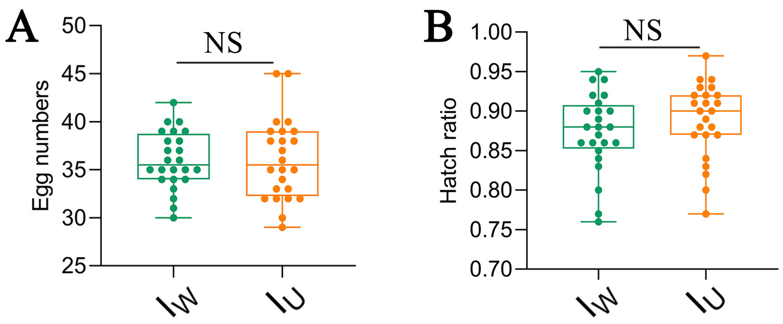
2.7. Effects of Different Symbiotic Bacteria Infections on the Fecundity of T. turkestani

Parthenogenesis: Fresh kidney bean leaves were used, and each leaf was divided into four circular sections with an area of approximately 4 cm2 each. Single female mites in the static III stage with different infection statuses were selected and placed onto each section of the leaf [31]. The number of eggs laid was counted daily, starting on the first day the female mite began to lay eggs. After laying eggs for five consecutive days, the female mite was removed. The daily egg-laying count and the total number of eggs laid were recorded. Once the eggs hatched into larvae, the hatching rate was recorded, and when they developed into adult mites, the sex ratio (female/male) was noted.
Sexual reproduction: Four different strains of T. turkestani were selected using different crossbreeding combinations to conduct hybridization experiments. Fresh leaves were divided into four circular sections, each approximately 4 cm2. A single female and male mite in the static III stage with different infection statuses were placed together in each section of the leaf, with one pair per section. Two days after the female molted into a mature adult, the male was removed. Starting from the first day of egg-laying, the female mites were removed after laying eggs for five days. The daily egg-laying count and the total number of eggs laid were recorded. After the eggs hatched into larvae, the hatching rate was recorded, and the sex ratio (female/male) was noted once the mites reached adulthood. If the parental male adult mite died before the female mite started laying eggs, it was promptly replaced with another male adult mite. If the parental female adult mite died before completing five days of egg laying, the data for that pair were discarded. The cytoplasmic incompatibility (CI) level (CI%) was calculated using the following formula: CI% = (1 − F/FC) × 100, where F represents the number of female offspring from incompatible crosses (♀Iu × ♂IW, ♀Iu × ♂IWR), and FC is the average number of female offspring from the control cross (♀Iu × ♂IU) [34]. The embryonic mortality (EMs) of different T. turkestani strains (parthenogenetic individuals or sexually reproduced individuals) was calculated using the following: formula EM = TE − HE, where TE is the total number of eggs in a single cross, and HE is the number of hatched eggs. The post-embryonic mortality (PEM) of each crossbreeding combination (♀IU × ♂IWR, ♀IU × ♂IW) was calculated using the following formula: PEM% = (1–AO/HE) × EM, where AO is the number of adult offspring in a single cross [35].
The above experiments were repeated 30 times. Under a microscope, the number of eggs laid by a single female mite or each pair of parents was counted, and the number of embryonic deaths and nymph deaths was recorded. Adult mites were collected for gender identification.

2.8. Data Processing

One-way analysis of variance (SPSS 26.0) was used to compare the outputs of arrhenotokous parthenogenesis and bisexual reproduction in the IWR, IW, IR, and IU strains, and to analyze the CI function of Wolbachia in the IWR and IW strains. Pairwise comparisons of all variables were performed using Duncan’s multiple range test. Independent sample t-tests (SPSS 26.0, p < 0.05) were employed to analyze the infection titers of Wolbachia and Rickettsia in T. turkestani of different genders in the IWR, IW, and IR strains, to compare the outputs of arrhenotokous parthenogenesis and bisexual reproduction in the IWR, IR, and IW strains, and to analyze the impact of male killing induced by Rickettsia on the CI function induced by Wolbachia. Graphpad Pism9.5 was used for graphing.

3. Results

3.1. Diagram of the Life Cycle of T. turkestani and Phylogenetic Analysis of Wolbachia and Rickettsia

A phylogenetic tree was constructed based on the Wolbachia wsp sequence and 25 Wolbachia strains from different species in the database, and the Wolbachia-infected T. turkestani were classified into group B. The Wolbachia infecting T. turkestani was found to have a relatively close evolutionary relationship with the Wolbachia infecting Diaphorina citri. A phylogenetic tree was also constructed based on the Rickettsia gltA sequence and 19 Rickettsia strains from different species in the database. The Rickettsia infecting T. turkestani was found to have a relatively close evolutionary relationship with the Rickettsia infecting Leptotrombidium and Ceutorhynchus.

3.2. Analysis of the Maternal Inheritance Efficiency of Wolbachia and Rickettsia

The maternal inheritance efficiency of the two symbiotic bacteria was determined through the infection rates of Wolbachia and Rickettsia in the offspring of T. turkestani. The results show that in all male and female offspring from the IWR, IW, and IR lines (Table 1), regardless of whether they originated from parthenogenesis or sexual reproduction, Rickettsia and Wolbachia were transmitted from the mother with complete infection (100%).

3.3. Detection of the Titers of Wolbachia and Rickettsia in T. turkestani

The target bands were obtained by PCR amplification using Wolbachia-specific primers WSP-236F/44R and Rickettsia-specific primers RICTG—F/R (see Supplementary Materials, Figure S1). The IWR strain of T. turkestani was co-infected with both endosymbiotic bacteria, the Iw strain was infected only with Wolbachia, the IR strain was infected only with Rickettsia, and the Iu strain was not infected with either of these two symbiotic bacteria. Real-time quantitative PCR was used to measure the titers of Wolbachia and Rickettsia in male and female adult mites of different strains. The results show that there were significant differences in the contents of Wolbachia and Rickettsia between the male and female adult mites, with the female adult mites having significantly higher content than the male adult mites. The content of Wolbachia in the IW strain was significantly higher than that in the IWR strain (Figure 2A). In contrast, the content of Rickettsia was the opposite, with the content in female mites of the IWR strain being higher than that in the IR strain (Figure 2B).

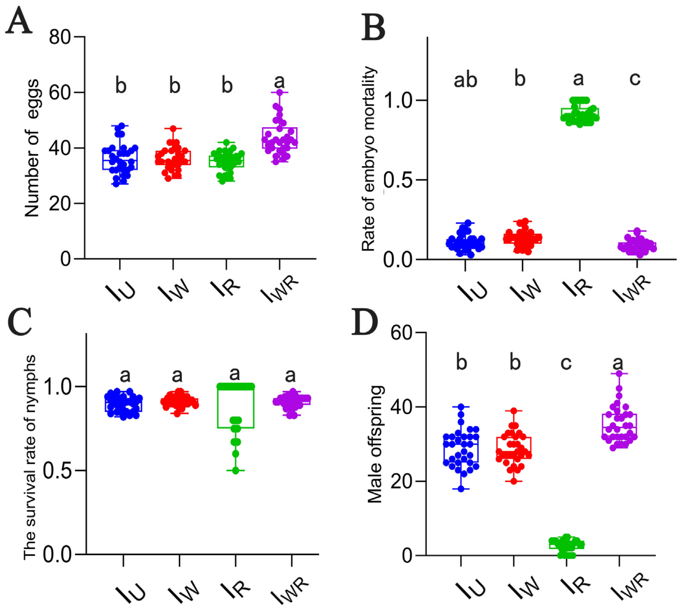
3.4. Effects of Different Endosymbiont Infections on the Parthenogenesis of T. turkestani

Four strains of T. turkestani reproduced parthenogenetically, with all offspring being male. The average number of eggs laid (per female) of the IWR strain was significantly higher than that of the Iu, IW, and IR strains (numbers of eggs laid: 43.80 ± 6.04 vs. 36.20 ± 5.47, 36.20 ± 4.16, and 35.10 ± 3.56, respectively, p < 0.001; Figure 3A). The IWR strain had the lowest embryonic mortality rate, while the IR strain had the highest (0.92 ± 0.05, p < 0.001, Figure 3B). There were no significant differences in the nymph survival rates among the four strains (Figure 3C). The number of male offspring in the IWR strain was the highest, and that in the IR strain was the lowest. The numbers in the IU and IW strains were at an intermediate level and significantly different from those in the IWR and IR strains (35.47 ± 4.89 vs. 2.50 ± 1.57, 28.97 ± 5.04, 28.67 ± 4.20, p < 0.001, Figure 3D). Eggs produced by parthenogenesis from the T. turkestani strain singly infected with Rickettsia failed to hatch normally, and a large number of male embryos died, leading to the lowest number of male offspring in the IR strain.

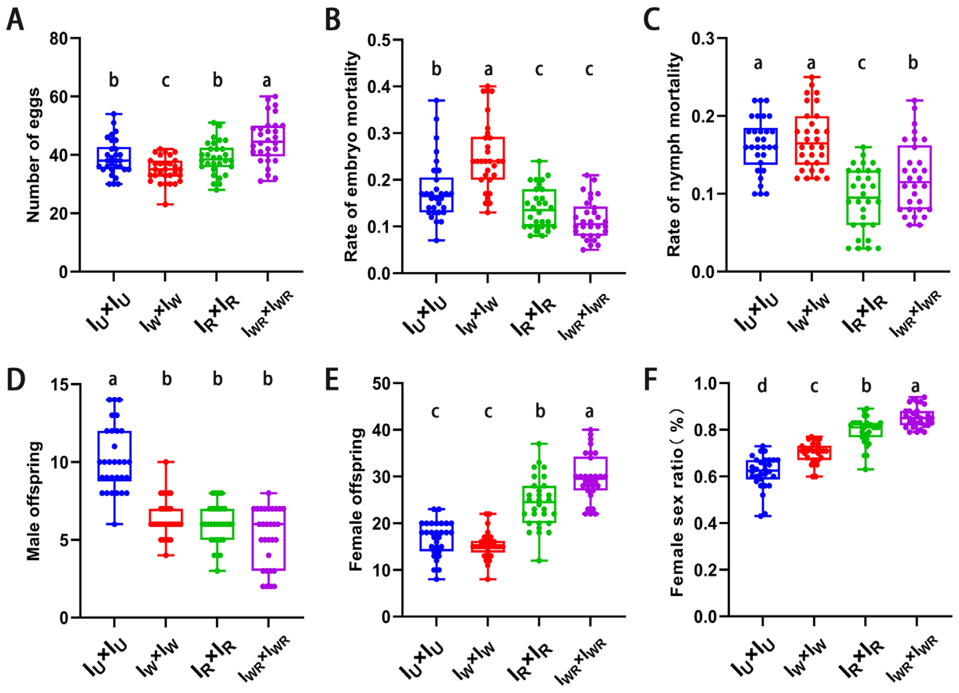
3.5. Effects of Different Endosymbiotic Bacterial Infections on the Sexual Reproduction of T. turkestani

In all four strains, the mated females produced both female and male offspring. The fecundity of the mated female mites of the IWR strain was the highest (45.03 ± 7.78 vs. 38.97 ± 6.13, 34.93 ± 4.32, 38.90 ± 5.63, p < 0.013, Figure 4A). Both the embryonic and nymphal mortality rates of the IWR strain were significantly lower than those of the IW and IU strains (embryonic mortality rate: 0.11 ± 0.04 vs. 0.18 ± 0.07, 0.25 ± 0.08, p < 0.001, Figure 4B; nymphal mortality rate: 0.12 ± 0.05 vs. 0.16 ± 0.04, 0.17 ± 0.04, p < 0.001, Figure 4C). The number of female offspring and the female-to-male sex ratio in the IWR strain were significantly higher than those in the other three strains (number of female offspring: 29.80 ± 5.01 vs. 16.70 ± 3.74, 15.30 ± 2.89, 24.43 ± 5.39, p < 0.001, Figure 4E; female-to-male sex ratio: 0.85 ± 0.04 vs. 0.62 ± 0.07, 0.70 ± 0.04, 0.80 ± 0.06, p < 0.001, Figure 4F). However, at the same time, the number of male offspring in the IWR strain was significantly lower than that in the IU strain (5.20 ± 1.88 vs. 10.17 ± 2.10, p < 0.001, Figure 4D). This indicates that in T. turkestani co-infection with Wolbachia and Rickettsia, male offspring die, resulting in an increase in the female-to-male sex ratio. In other words, co-infection with Wolbachia and Rickettsia can induce male death in T. turkestani.

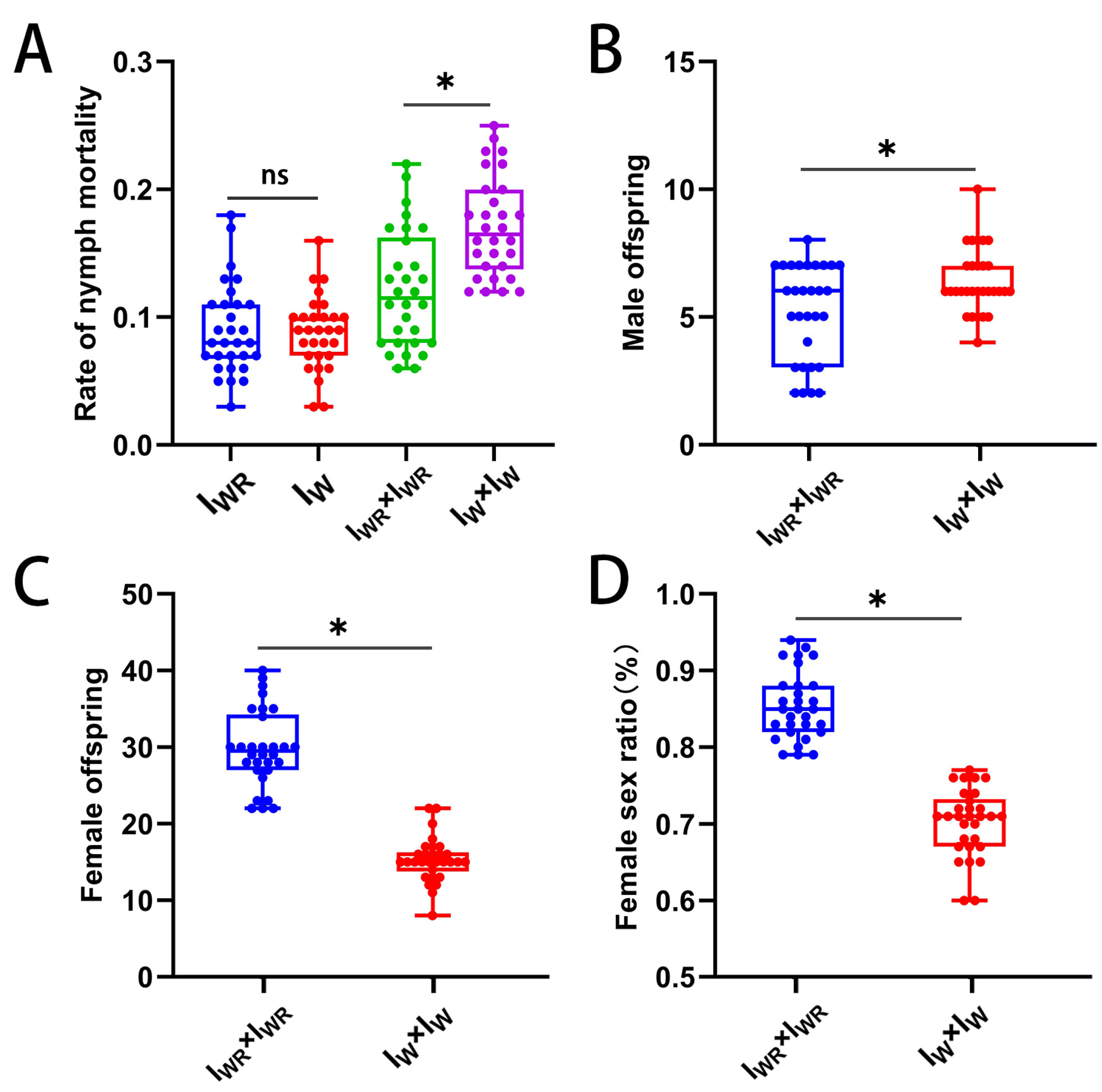
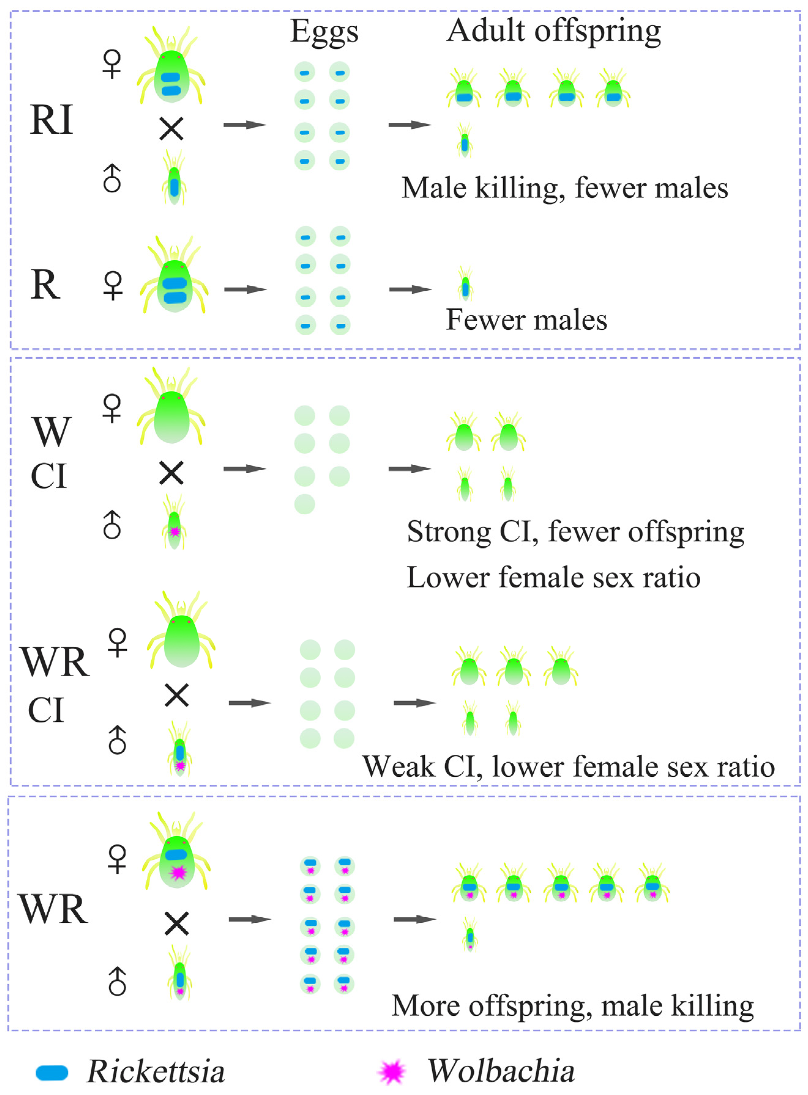
3.6. Verification of the Male-Killing Effect of Rickettsia in T. turkestani

The parthenogenesis and sexual reproduction of female mites in the IWR and IW strains were further investigated to verify the male-killing effect of Rickettsia. Both the IWR and IW strains produced male offspring parthenogenetically, and there was no significant difference in the nymphal mortality rate of the offspring (Figure 5A). Intraspecific sexual reproduction of female mites in the IWR and IW strains simultaneously produced both female and male offspring. The results show that compared with the IW strain without Rickettsia, the IWR strain with Rickettsia had a significantly lower nymphal mortality rate (0.12 ± 0.05 vs. 0.17 ± 0.04, p < 0.001, Figure 5A). Compared with the IW strain, the number of male offspring in the IWR strain was significantly reduced (5.20 ± 1.88 vs. 6.40 ± 1.25, p < 0.01, Figure 5B), while the number of female offspring was significantly increased (29.80 ± 5.01 vs. 15.30 ± 2.89, p < 0.001, Figure 5C), and the female-to-male sex ratio in the IWR strain was significantly higher than that in the IW strain (0.85 ± 0.04 vs. 0.70 ± 0.04, p < 0.001, Figure 5D). It can be inferred that infection with Rickettsia led to the death of more male offspring, resulting in an increase in the female-to-male sex ratio. This verifies that Rickettsia induced male killing in T. turkestani.

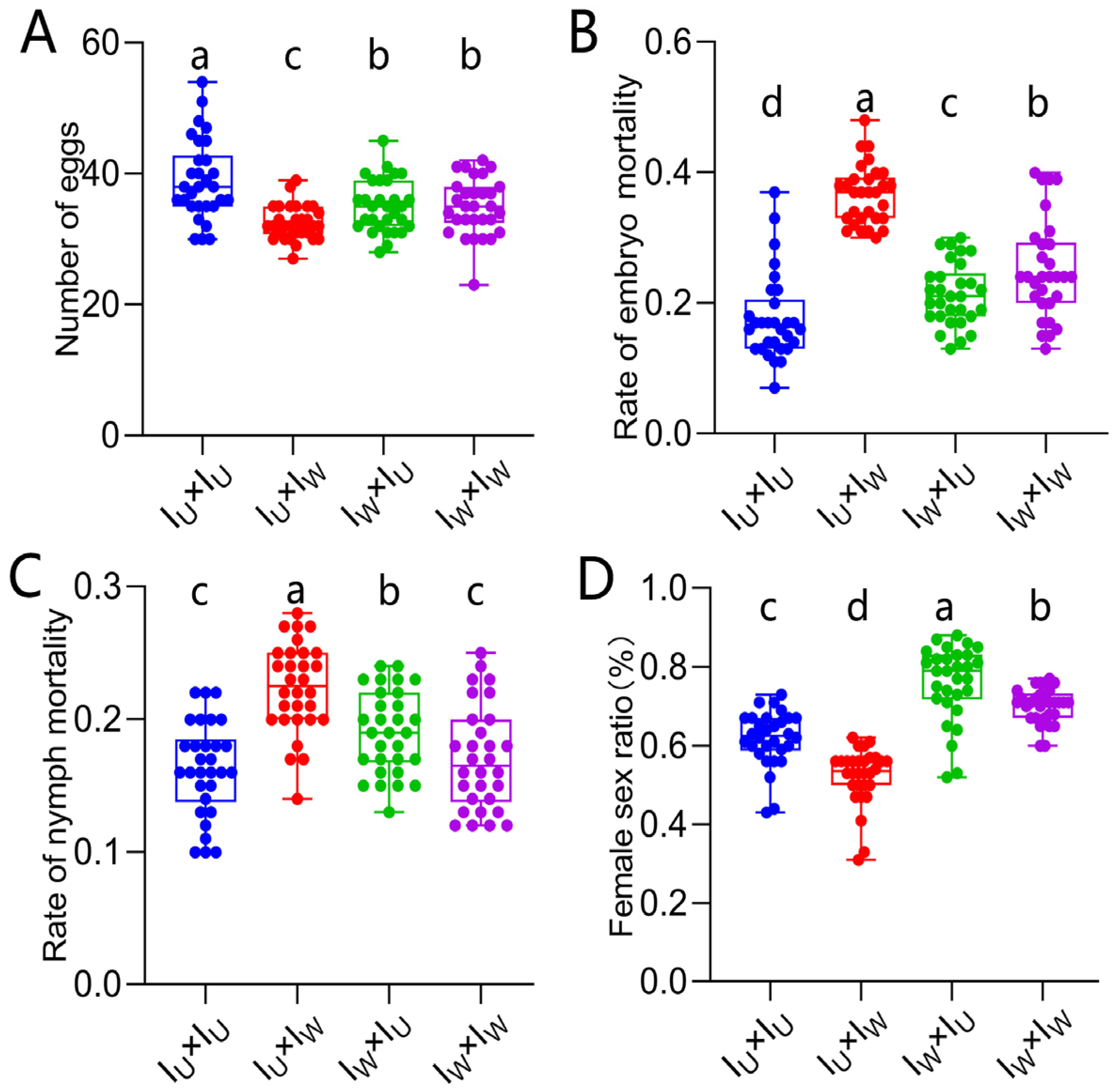
3.7. Verification of the Cytoplasmic Incompatibility (CI) Induced by Wolbachia in Singly-Infected T. turkestani

A hybridization experiment using the IU and Iw strains was carried out to verify that Wolbachia induced CI in singly-infected T. turkestani. Among the four different mating combinations, the fecundity of the ♀IU × ♂IW combination was significantly lower than that of the other three combinations (Figure 6A). The embryonic and nymph mortality rates of the ♀IU × ♂IW combination were significantly higher than those of the other three combinations (embryonic mortality rate: 0.37 ± 0.05, p < 0.001, Figure 6B; nymph mortality rate: 0.22 ± 0.03, p < 0.001, Figure 6C), and the female to male sex ratio was the lowest (0.52 ± 0.07, p < 0.002, Figure 6D). These results all indicate that Wolbachia induced strong CI in T. turkestani.

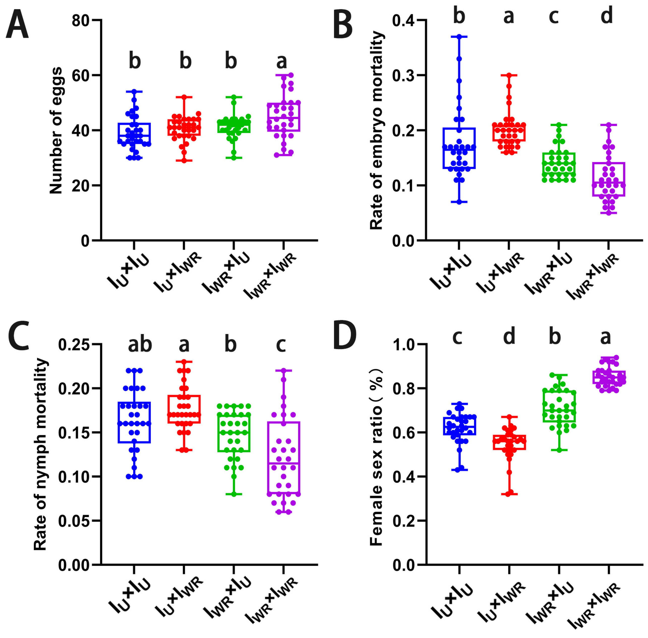
3.8. Verification of the Cytoplasmic Incompatibility (CI) Induced by Wolbachia and Rickettsia in Co-Infected T. turkestani

A hybridization experiment using Iu and IWR strains was conducted to verify that Wolbachia and Rickettsia induced CI in co-infected T. turkestani. The fecundity of the ♀Iu × ♂IWR combination was lower than that of the ♀IWR × ♂IWR combination (40.57 ± 4.53 vs. 45.03 ± 7.78, p < 0.004, Figure 7A), but there was no significant difference compared with the other two hybridization combinations. The embryonic and nymph mortality rates of the ♀Iu × ♂IWR combination were the highest, significantly higher than those of the ♀IWR × ♂IWR combination (embryonic mortality rate: 0.20 ± 0.03 vs. 0.11 ± 0.04, p < 0.001, Figure 7B; nymph mortality rate: 0.18 ± 0.03 vs. 0.12 ± 0.05, p < 0.001, Figure 7C). Meanwhile, the female-to-male sex ratio of the ♀IU × ♂IWR combination was significantly lower than that of the other three groups (0.54 ± 0.08 vs. 0.62 ± 0.07, 0.71 ± 0.08, 0.85 ± 0.04, p < 0.001, Figure 7D). These results indicate that Wolbachia and Rickettsia also induced CI in co-infected T. turkestani.

3.9. Antagonistic Effect of Rickettsia-Induced Male Killing on the Strength of Wolbachia-Induced CI

After verifying that Rickettsia infection can induce male killing in T. turkestani, we further investigated the impact of Rickettsia-induced male killing on Wolbachia-induced CI. Compared with the singly-infected ♀IU × ♂IW combination, the fecundity of the co-infected ♀IU × ♂IWR combination was significantly increased (40.57 ± 4.53 vs. 32.50 ± 2.61, p < 0.001, Figure 8A), and the embryonic and nymphal mortality rates were significantly decreased (embryonic mortality rate: 0.37 ± 0.05 vs. 0.20 ± 0.03, p < 0.001, Figure 8B; nymph mortality rate: 0.22 ± 0.03 vs. 0.18 ± 0.03, p < 0.001, Figure 8C). Compared with the ♀IU × ♂IW combination without Rickettsia infection, the co-infected ♀IU × ♂IWR combination had more female and male offspring (female offspring: 14.47 ± 2.80 vs. 8.33 ± 1.40, p < 0.001, Figure 8D; male offspring: 12.20 ± 2.54 vs. 7.63 ± 1.59, p < 0.001, Figure 8E). Moreover, the CI level of the co-infected ♀IU × ♂IWR combination was significantly lower than that of the singly infected ♀IU × ♂IW combination (9.90 ± 22.77 vs. 48.51 ± 10.13, p < 0.001; Figure 8F). This indicates that the IWR strain induced a weaker CI, which might be due to partial antagonism effect of Rickettsia-induced male killing on the CI induced by Wolbachia.

3.10. Wolbachia Does Not Have a Male-Killing Effect on T. turkestani

Female mites of the IW and IU strains produced male offspring through parthenogenesis, with no significant differences in the fecundity or hatching rate (fecundity: IW vs. IU = 36.00 ± 3.01 vs. 36.08 ± 4.21, p > 0.05, Figure 9A; hatching rate: IW vs. IU = 0.87 ± 0.05 vs. 0.89% ± 0.05, p > 0.05, Figure 9B). This indicates that Wolbachia did not exhibit a male-killing effect on T. turkestani.

4. Discussion

In this study, different strains of T. turkestani infected with different endosymbiotic bacteria (Wolbachia and Rickettsia) were examined, and phylogenetic analysis was carried out. The results show that the Wolbachia-infected T. turkestani belonged to supergroup B and could induce cytoplasmic incompatibility (CI) in the host. The transmission efficiency of Wolbachia and Rickettsia were statistically analyzed. The results reveal that, whether through parthenogenesis or sexual reproduction, both symbiotic bacteria completely followed maternal transmission. Real-time quantitative PCR was used to determine the titers of the endosymbiotic bacteria. The results show significant differences in the abundance of Wolbachia and Rickettsia between male and female adult mites.
Rickettsia is a maternally inherited symbiotic bacterium. In some hosts, it acts as a nutritional symbiont, while in others, it influences the host’s reproduction through reproductive regulations, such as parthenogenesis induction and male killing. It can also enhance the host’s resistance to pesticides and improve the host’s ability to resist predators, high temperatures, or other lethal factors [3,4,17,18,36,37,38,39]. To date, no experimental studies have investigated the reproductive regulation of this bacterium in mites. Our research demonstrates that Rickettsia-infected spider mites resulted in parthenogenesis, producing only male offspring, but the hatching rate of male embryos was extremely low. Sexual reproduction in mites singly infected with Rickettsia produced both female and male offspring, with a higher number of female offspring than male, resulting in a high female-to-male sex ratio. This indicates that Rickettsia infection leads to a male-killing phenotype.
Cytoplasmic incompatibility (CI) is the most common reproductive regulation induced by Wolbachia and typically occurs in two forms. The first form is characterized by a high embryonic mortality rate, resulting in a decrease in the number of female offspring, which is called female lethality. The second form does not decrease the total number of offspring but results in an increase in the number of male offspring, known as male development. Both female lethality and male development induced by Wolbachia may occur simultaneously in a single insect host, such as the Wolbachia wLhetl strain in parasitic wasps [40,41]. In this study, sexual reproduction of T. turkestani (♀IU × ♂IW) significantly increased the mortality rates of embryos and nymphs and significantly decreased the female-to-male sex ratio, indicating the female lethality type. Single infections of Wolbachia in T. turkestani induced strong CI.
Currently, research on the interaction between Rickettsia and Wolbachia is limited, especially regarding the impact of Rickettsia and Wolbachia co-infection on the reproductive regulation of host insects, which has not yet been reported. In this study, we found that the Rickettsia and Wolbachia co-infection induced cytoplasmic incompatibility (CI) in T. turkestani, but the intensity was much weaker than that induced by single infections of Wolbachia. This may be because the male-killing effect of Rickettsia in co-infection reduced the CI level induced by Wolbachia. Subsequent hybridization experiments clearly demonstrated an antagonistic interaction between the male-killing effect of Rickettsia and the CI induced by Wolbachia. Previous studies have shown that there are two types of Wolbachia in nature: one maintains a high prevalence in insect hosts and weakly induces CI, such as Wolbachia in Drosophila melanogaster [42]; the other induces strong CI but maintains a low prevalence and titer, like Wolbachia in D. melanogaster [43]. The Wolbachia in T. turkestani studied here was most similar to the former. Furthermore, compared with the control group (IU × IU) without a symbiotic bacterial infection, RickettsiaWolbachia co-infection had a higher fecundity, lower mortality, a higher female-to-male ratio, and more offspring. In conclusion, the synergistic effect of the two symbiotic bacteria significantly improved the fitness of T. turkestani.
In conclusion, our study reveals five findings (Figure 10): Rickettsia infection induced a male-killing effect in the sexual reproduction of T. turkestani; Rickettsia infection caused the death of parthenogenetically produced male embryos in T. turkestani, leading to a reduction in the number of offspring; Wolbachia single infection induced strong cytoplasmic incompatibility (CI) in T. turkestani, while Rickettsia–Wolbachia co-infection induced weaker CI; the male-killing effect induced by Rickettsia antagonized the strong CI induced by Wolbachia; Rickettsia–Wolbachia co-infection promoted the fitness of T. turkestani, suggesting a synergistic mutualistic relationship between the two symbiotic bacteria within the same host T. turkestani. We gained a better understanding of the complex interactions between symbiotic bacteria and their hosts, as well as between different symbionts, providing strategies and insights for future applications of symbionts in biological control.

Supplementary Materials

The following supporting information can be downloaded at: https://www.mdpi.com/article/10.3390/microorganisms13030642/s1, Table S1: Symbiotic bacteria sequences in the NCBI database. Table S2: Primer sequences for PCR. Table S3: Gene amplification system. Table S4. PCR amplification procedure. Table S5: Primer sequences for genes. Table S6: Quantitative Real-Time PCR Reaction System. Table S7: PCR amplification procedure. Figure S1: S1 PCR detection of different Tetranychus turkestani, Wolbachia and Rickettsia based on wsp and gltA genes.

Author Contributions

Conceptualization, S.W. and Y.Z.; methodology, S.W. and Y.Z.; software, S.W.; validation, S.W., X.W. and A.B.; formal analysis, X.W.; investigation, S.W.; resources, S.W. and X.W.; data curation, S.W.; writing—original draft preparation, S.W.; writing—review and editing, S.W., Y.Z., Q.W., K.Z. and A.B.; visualization, S.W.; supervision, X.W.; project administration, Y.Z.; funding acquisition, Y.Z. All authors have read and agreed to the published version of the manuscript.

Funding

This research was funded by National Nature Science Foundation of China: (32260676, 31860508), and Bingtuan Science and Technology Program (2022CB002-06, 2024DA018).

Institutional Review Board Statement

Not applicable.

Informed Consent Statement

Not applicable.

Data Availability Statement

The original contributions presented in this study are included in the article/Supplementary Material. Further inquiries can be directed to the corresponding author.

Conflicts of Interest

The authors declare no conflicts of interest.

References

  1. Hilgenboecker, K.; Hammerstein, P.; Schlattmann, P.; Telschow, A.; Werren, J.H. How Many Species Are Infected with Wolbachia?—A Statistical Analysis of Current Data: Wolbachia Infection Rates. FEMS Microbiol. Lett. 2008, 281, 215–220. [Google Scholar] [CrossRef] [PubMed]
  2. Weinert, L.A.; Araujo-Jnr, E.V.; Ahmed, M.Z.; Welch, J.J. The Incidence of Bacterial Endosymbionts in Terrestrial Arthropods. Proc. R. Soc. B 2015, 282, 20150249. [Google Scholar] [CrossRef]
  3. Brumin, M.; Levy, M.; Ghanim, M. Transovarial Transmission of Rickettsia Spp. and Organ-Specific Infection of the Whitefly Bemisia tabaci. Appl. Environ. Microbiol. 2012, 78, 5565–5574. [Google Scholar] [CrossRef]
  4. Oliver, K.M.; Russell, J.A.; Moran, N.A.; Hunter, M.S. Facultative Bacterial Symbionts in Aphids Confer Resistance to Parasitic Wasps. Proc. Natl. Acad. Sci. USA 2003, 100, 1803–1807. [Google Scholar] [CrossRef]
  5. Werren, J.H.; Baldo, L.; Clark, M.E. Wolbachia: Master Manipulators of Invertebrate Biology. Nat. Rev. Microbiol. 2008, 6, 741–751. [Google Scholar] [CrossRef] [PubMed]
  6. Hedges, L.M.; Brownlie, J.C.; O’Neill, S.L.; Johnson, K.N. Wolbachia and Virus Protection in Insects. Science 2008, 322, 702. [Google Scholar] [CrossRef] [PubMed]
  7. Perlman, S.J.; Hunter, M.S.; Zchori-Fein, E. The Emerging Diversity of Rickettsia. Proc. Biol. Sci. 2006, 273, 2097–2106. [Google Scholar] [CrossRef]
  8. Liu, B.-Q.; Bao, X.-Y.; Yan, J.-Y.; Zhang, D.; Sun, X.; Li, C.-Q.; Chen, Z.-B.; Luan, J.-B. Rickettsia Symbionts Spread via Mixed Mode Transmission, Increasing Female Fecundity and Sex Ratio Shift by Host Hormone Modulating. Proc. Natl. Acad. Sci. USA 2024, 121, e2406788121. [Google Scholar] [CrossRef]
  9. Dobson, S.L.; Bourtzis, K.; Braig, H.R.; Jones, B.F.; Zhou, W.; Rousset, F.; O’Neill, S.L. Wolbachia Infections Are Distributed throughout Insect Somatic and Germ Line Tissues. Insect Biochem. Mol. Biol. 1999, 29, 153–160. [Google Scholar] [CrossRef]
  10. Ilbeigi Khamseh Nejad, M.; Cappelli, A.; Damiani, C.; Falcinelli, M.; Catapano, P.L.; Nanfack-Minkeu, F.; Mayi, M.P.A.; Currà, C.; Ricci, I.; Favia, G. Wolbachia and Asaia Distribution among Different Mosquito Vectors Is Affected by Tissue Localization and Host Species. Microorganisms 2024, 12, 545. [Google Scholar] [CrossRef]
  11. Fricke, L.C.; Lindsey, A.R.I. Examining Wolbachia-Induced Parthenogenesis in Hymenoptera. Methods Mol. Biol. 2024, 2739, 55–68. [Google Scholar] [CrossRef] [PubMed]
  12. Fast, E.M.; Toomey, M.E.; Panaram, K.; Desjardins, D.; Kolaczyk, E.D.; Frydman, H.M. Wolbachia Enhance Drosophila Stem Cell Proliferation and Target the Germline Stem Cell Niche. Science 2011, 334, 990–992. [Google Scholar] [CrossRef] [PubMed]
  13. Dobson, S.L.; Rattanadechakul, W.; Marsland, E.J. Fitness Advantage and Cytoplasmic Incompatibility in Wolbachia Single- and Superinfected Aedes Albopictus. Heredity 2004, 93, 135–142. [Google Scholar] [CrossRef] [PubMed]
  14. Bian, G.; Xu, Y.; Lu, P.; Xie, Y.; Xi, Z. The Endosymbiotic Bacterium Wolbachia Induces Resistance to Dengue Virus in Aedes Aegypti. PLoS Pathog. 2010, 6, e1000833. [Google Scholar] [CrossRef]
  15. Pan, X.; Zhou, G.; Wu, J.; Bian, G.; Lu, P.; Raikhel, A.S.; Xi, Z. Wolbachia Induces Reactive Oxygen Species (ROS)-Dependent Activation of the Toll Pathway to Control Dengue Virus in the Mosquito Aedes Aegypti. Proc. Natl. Acad. Sci. USA 2012, 109, E23–E31. [Google Scholar] [CrossRef]
  16. Hagimori, T.; Abe, Y.; Date, S.; Miura, K. The First Finding of a Rickettsia Bacterium Associated with Parthenogenesis Induction among Insects. Curr. Microbiol. 2006, 52, 97–101. [Google Scholar] [CrossRef]
  17. Himler, A.G.; Adachi-Hagimori, T.; Bergen, J.E.; Kozuch, A.; Kelly, S.E.; Tabashnik, B.E.; Chiel, E.; Duckworth, V.E.; Dennehy, T.J.; Zchori-Fein, E.; et al. Rapid Spread of a Bacterial Symbiont in an Invasive Whitefly Is Driven by Fitness Benefits and Female Bias. Science 2011, 332, 254–256. [Google Scholar] [CrossRef]
  18. Chen, X.; Li, S.; Aksoy, S. Concordant Evolution of a Symbiont with Its Host Insect Species: Molecular Phylogeny of Genus Glossina and Its Bacteriome-Associated Endosymbiont, Wigglesworthia Glossinidia. J. Mol. Evol. 1999, 48, 49–58. [Google Scholar] [CrossRef]
  19. Fan, Z.-Y.; Liu, Y.; He, Z.-Q.; Wen, Q.; Chen, X.-Y.; Khan, M.M.; Osman, M.; Mandour, N.S.; Qiu, B.-L. Rickettsia Infection Benefits Its Whitefly Hosts by Manipulating Their Nutrition and Defense. Insects 2022, 13, 1161. [Google Scholar] [CrossRef]
  20. Zélé, F.; Weill, M.; Magalhães, S. Identification of Spider-Mite Species and Their Endosymbionts Using Multiplex PCR. Exp. Appl. Acarol. 2018, 74, 123–138. [Google Scholar] [CrossRef]
  21. Frank, S.A. Dynamics of Cytoplasmic Incompatibility with Multiple Wolbachia Infections. J. Theor. Biol. 1998, 192, 213–218. [Google Scholar] [CrossRef] [PubMed]
  22. Zchori-Fein, E.; Perlman, S.J. Distribution of the Bacterial Symbiont Cardinium in Arthropods. Mol. Ecol. 2004, 13, 2009–2016. [Google Scholar] [CrossRef] [PubMed]
  23. Dobson, S.L. Evolution of Wolbachia Cytoplasmic Incompatibility Types. Evolution 2004, 58, 2156–2166. [Google Scholar] [CrossRef] [PubMed]
  24. Ros, V.I.D.; Breeuwer, J.A.J. The Effects of, and Interactions between, Cardinium and Wolbachia in the Doubly Infected Spider Mite Bryobia sarothamni. Heredity 2009, 102, 413–422. [Google Scholar] [CrossRef]
  25. Zhao, D.-X.; Zhang, X.-F.; Hong, X.-Y. Host-Symbiont Interactions in Spider Mite Tetranychus truncates Doubly Infected with Wolbachia and Cardinium. Environ. Entomol. 2013, 42, 445–452. [Google Scholar] [CrossRef]
  26. Ben-David, T.; Melamed, S.; Gerson, U.; Morin, S. ITS2 Sequences as Barcodes for Identifying and Analyzing Spider Mites (Acari: Tetranychidae). Exp. Appl. Acarol. 2007, 41, 169–181. [Google Scholar] [CrossRef]
  27. Navajas, M.; Boursot, P. Nuclear Ribosomal DNA Monophyly versus Mitochondrial DNA Polyphyly in Two Closely Related Mite Species: The Influence of Life History and Molecular Drive. Proc. Biol. Sci. 2003, 270 (Suppl. S1), S124–S127. [Google Scholar] [CrossRef]
  28. Mohammadi, S.; Seraj, A.A.; Rajabpour, A. Effects of Six Greenhouse Cucumber Cultivars on Reproductive Performance and Life Expectancy of Tetranychus Turkestani (Acari: Tetranychidae). Acarologia 2015, 55, 231–242. [Google Scholar] [CrossRef]
  29. Chu, D.; Gao, C.S.; De Barro, P.; Zhang, Y.J.; Wan, F.H.; Khan, I.A. Further Insights into the Strange Role of Bacterial Endosymbionts in Whitefly, Bemisia Tabaci: Comparison of Secondary Symbionts from Biotypes B and Q in China. Bull. Entomol. Res. 2011, 101, 477–486. [Google Scholar] [CrossRef]
  30. Hong, X.-Y.; Gotoh, T.; Nagata, T. Vertical Transmission of Wolbachia in Tetranychus kanzawai Kishida and Panonychus Mori Yokoyama (Acari: Tetranychidae). Heredity 2002, 88, 190–196. [Google Scholar] [CrossRef]
  31. Bing, X.-L.; Lu, Y.-J.; Xia, C.-B.; Xia, X.; Hong, X.-Y. Transcriptome of Tetranychus Urticae Embryos Reveals Insights into Wolbachia-Induced Cytoplasmic Incompatibility. Insect Mol. Biol. 2020, 29, 193–204. [Google Scholar] [CrossRef]
  32. Lv, N.; Peng, J.; Chen, X.-Y.; Guo, C.-F.; Sang, W.; Wang, X.-M.; Ahmed, M.Z.; Xu, Y.-Y.; Qiu, B.-L. Antagonistic Interaction between Male-Killing and Cytoplasmic Incompatibility Induced by Cardinium and Wolbachia in the Whitefly, Bemisia tabaci. Insect Sci. 2021, 28, 330–346. [Google Scholar] [CrossRef] [PubMed]
  33. Sun, W.; Jin, Y.; He, L.; Lu, W.-C.; Li, M. Suitable Reference Gene Selection for Different Strains and Developmental Stages of the Carmine Spider Mite, Tetranychus cinnabarinus, Using Quantitative Real-Time PCR. J. Insect Sci. 2010, 10, 208. [Google Scholar] [CrossRef]
  34. Mouton, L.; Dedeine, F.; Henri, H.; Boulétreau, M.; Profizi, N.; Vavre, F. Virulence, Multiple Infections and Regulation of Symbiotic Population in the Wolbachia-Asobara Tabida Symbiosis. Genetics 2004, 168, 181–189. [Google Scholar] [CrossRef] [PubMed]
  35. Nguyen, D.T.; Morrow, J.L.; Spooner-Hart, R.N.; Riegler, M. Independent Cytoplasmic Incompatibility Induced by Cardinium and Wolbachia Maintains Endosymbiont Coinfections in Haplodiploid Thrips Populations. Evolution 2017, 71, 995–1008. [Google Scholar] [CrossRef]
  36. Sakurai, M.; Koga, R.; Tsuchida, T.; Meng, X.-Y.; Fukatsu, T. Rickettsia Symbiont in the Pea Aphid Acyrthosiphon pisum: Novel Cellular Tropism, Effect on Host Fitness, and Interaction with the Essential Symbiont Buchnera. Appl. Environ. Microbiol. 2005, 71, 4069–4075. [Google Scholar] [CrossRef]
  37. Ferrari, J.; Scarborough, C.L.; Godfray, H.C.J. Genetic Variation in the Effect of a Facultative Symbiont on Host-Plant Use by Pea Aphids. Oecologia 2007, 153, 323–329. [Google Scholar] [CrossRef] [PubMed]
  38. Kontsedalov, S.; Zchori-Fein, E.; Chiel, E.; Gottlieb, Y.; Inbar, M.; Ghanim, M. The Presence of Rickettsia Is Associated with Increased Susceptibility of Bemisia tabaci (Homoptera: Aleyrodidae) to Insecticides. Pest. Manag. Sci. 2008, 64, 789–792. [Google Scholar] [CrossRef]
  39. Shan, H.-W.; Lu, Y.-H.; Bing, X.-L.; Liu, S.-S.; Liu, Y.-Q. Differential Responses of the Whitefly Bemisia tabaci Symbionts to Unfavorable Low and High Temperatures. Microb. Ecol. 2014, 68, 472–482. [Google Scholar] [CrossRef]
  40. Vavre, F.; Dedeine, F.; Quillon, M.; Fouillet, P.; Fleury, F.; Bouletreau, M. Within-Species Diversity of Wolbachia-Induced Cytoplasmic Incompatibility in Haplodiploid Insects. Evolution 2001, 55, 1710–1714. [Google Scholar] [CrossRef]
  41. Vala, F.; Breeuwer, J.a.J.; Sabelis, M.W. Sorting out the Effects of Wolbachia, Genotype and Inbreeding on Life-History Traits of a Spider Mite. Exp. Appl. Acarol. 2003, 29, 253–264. [Google Scholar] [CrossRef] [PubMed]
  42. Kriesner, P.; Conner, W.R.; Weeks, A.R.; Turelli, M.; Hoffmann, A.A. Persistence of a Wolbachia Infection Frequency Cline in Drosophila melanogaster and the Possible Role of Reproductive Dormancy. Evolution 2016, 70, 979–997. [Google Scholar] [CrossRef] [PubMed]
  43. Richardson, K.M.; Schiffer, M.; Griffin, P.C.; Lee, S.F.; Hoffmann, A.A. Tropical Drosophila pandora Carry Wolbachia Infections Causing Cytoplasmic Incompatibility or Male Killing. Evolution 2016, 70, 1791–1802. [Google Scholar] [CrossRef] [PubMed]
Figure 1. Schematic diagram of the life cycle and phylogenetic trees. (A) Schematic diagram of the life cycle of Tetranychus turkestani. (B) Wolbachia. (C) Rickettsia. The numbers at the nodes are bootstrap values, which were used to evaluate the reliability of the branching structure.
Microorganisms 13 00642 g001
Figure 2. Relative abundance of Wolbachia and Rickettsia in female and male Tetranychus turkestani of different infection strains. (A) Relative abundance of Wolbachia. (B) Relative abundance of Rickettsia. IUF: female IU population, IuM: male Iu population, IRF: female IR population, IRM: male IR population, IwF: female Iw population, IwM: male Iw population, IWRF: female IWR population, IWRM: male IWR population. The symbol “*” indicates a statistically significant difference between the two groups (p < 0.05). All error bars represent the standard errors of the means.
Microorganisms 13 00642 g002
Figure 3. The parthenogenetic parameters of IU, IW, IR, and IWR in Tetranychus turkestani. (A) Number of eggs. (B) Rate of embryo mortality. (C) Survival rate of nymphs. (D) Male offspring. The data in the figure are the means ± standard errors. The mean values marked by different letter are statistically significant (p < 0.05).
Microorganisms 13 00642 g003
Figure 4. The sexual reproductive parameters of IW, IR, IWR, and IU mating in Tetranychus turkestani. (A) Number of eggs. (B) Rate of embryo mortality. (C) Rate of nymph mortality. (D) Male offspring. (E) Female offspring. (F) Female sex ratio. The data in the figure are the means ± standard errors. The mean values marked by different letter markers are statistically significant (p < 0.05).
Microorganisms 13 00642 g004
Figure 5. Parthenogenesis and intraspecific sexual reproduction of IWR and IW in Tetranychus turkestani within 5 days of oviposition. (A) Rate of nymph mortality. (B) Male offspring. (C) Female offspring. (D) Female sex ratio. The data in the figure are the means ± standard errors. The star over the bars “*” represents significant differences between each other at p < 0.05, and “ns” represents no significant differences.
Microorganisms 13 00642 g005
Figure 6. CI identification of Wolbachia in Iw of Tetranychus turkestani. (A) Number of eggs. (B) Rate of embryo mortality. (C) Rate of nymph mortality. (D) Female sex ratio. The data in the figure are the means ± standard errors. The mean values marked by different letters are statistically significant (p < 0.05).
Microorganisms 13 00642 g006
Figure 7. CI identification of Wolbachia and Rickettsia in IWR of Tetranychus turkestani. (A) Number of eggs. (B) Rate of embryo mortality. (C) Rate of nymph mortality. (D) Female sex ratio. The data in the figure are the means ± standard errors. The mean values marked by different letters are statistically significant (p < 0.05).
Microorganisms 13 00642 g007
Figure 8. Antagonistic effect of Rickettsia co-infection on CI in Wolbachia. (A) Number of eggs. (B) Rate of embryo mortality. (C) Rate of nymph mortality. (D) Female offspring. (E) Male offspring. (F) CI%. The data in the figure are the means ± standard errors. The mean values marked by different letter are statistically significant (p < 0.05).
Microorganisms 13 00642 g008
Figure 9. The reproductive parameters of different combinations of Tetranychus turkestani. (A) Egg production of IW and IU in parthenogenesis. (B) Hatching rates of IW and IU in parthenogenesis. IU, Wolbachia-uninfected; IW, Wolbachia-infected. Crossing combinations of strains are shown as ‘Female × Male’. The data in the figure are presented as the means ± standard errors. The “NS” represents no significant differences.
Microorganisms 13 00642 g009
Figure 10. The action model of Rickettsia and Wolbachia in the reproductive regulation of Tetranychus turkestani. RI: Tetranychus turkestani with single Rickettsia infection; R: parthenogenesis with single Rickettsia infection; WCI: cytoplasmic incompatibility (CI) induced by single Wolbachia infection; WRCI: cytoplasmic incompatibility (CI) induced by co-infection of WolbachiaRickettsia; WR: Tetranychus turkestani with co-infection of WolbachiaRickettsia.
Microorganisms 13 00642 g010
Table 1. The maternal inheritance efficiency of Rickettsia and Wolbachia.
Tetranychus turkestani
Strains
Number of
Adult
Female Mites
Number
of
Offspring
Total Number
of Specimens
Tested
RickettsiaWolbachia
n+n%n+n%
♀IW1010100010001000100
♀IR502100100010001000
♀IWR101010010001001000100
♀IW × ♂IU1010100010001000100
♀IR × ♂IU1010100100010001000
♀IWR × ♂IU101010010001001000100
Note: n, number; n+, number of positive individuals; n, number of negative individuals.
Disclaimer/Publisher’s Note: The statements, opinions and data contained in all publications are solely those of the individual author(s) and contributor(s) and not of MDPI and/or the editor(s). MDPI and/or the editor(s) disclaim responsibility for any injury to people or property resulting from any ideas, methods, instructions or products referred to in the content.

