Review

The Microbiota–Human Health Axis

1
Department of Intensive Care, Epsom and St. Helier University Hospitals NHS Trust, Wrythe Ln, Sutton SM5 1AA, UK
2
Faculty of Medicine and Pharmacy, University of Oradea, Piaţa 1 Decembrie 10, 410068 Oradea, Romania
3
Lahti Health and Social Services Centre, Harjukatu 48, 15100 Lahti, Finland
4
Department of General Internal Medicine, Northampton General Hospital, NHS Trust, Northampton NN1 5BD, UK
5
Doctoral School of Biomedical Sciences, Faculty of Medicine and Pharmacy, University of Oradea, 410087 Oradea, Romania
6
Department of Psycho-Neuroscience and Recovery, Faculty of Medicine and Pharmacy, University of Oradea, 410073 Oradea, Romania
7
Department of Medical Disciplines, Faculty of Medicine and Pharmacy, University of Oradea, 410073 Oradea, Romania
*
Author to whom correspondence should be addressed.
Microorganisms 2025, 13(4), 948; https://doi.org/10.3390/microorganisms13040948
Submission received: 16 March 2025 / Revised: 15 April 2025 / Accepted: 17 April 2025 / Published: 20 April 2025
(This article belongs to the Section Medical Microbiology)

Abstract

:
Trillions of microorganisms play a pivotal role in maintaining health and preventing disease in humans. Their presence influences daily life, habits, energy levels, and pathologies. The present narrative review synthesized recent studies of microbial diversity across organ systems. The composition of the microbiota regulates the intestinal barrier, modulates the immune response, influences metabolism, and produces essential compounds such as short-chain fatty acids and neurotransmitters. Dysbiosis is associated with numerous pathologies, including metabolic, autoimmune, neurodegenerative, and cardiovascular diseases. The microbiota is key to maintaining physiological balance and reducing disease risk. Therapeutic interventions, such as probiotics, prebiotics, postbiotics, and microbiome transplantation, offer promising perspectives in restoring microbial homeostasis and preventing chronic diseases.

Graphical Abstract

1. Introduction

The human microbiota, a complex and dynamic ecosystem, consists of approximately 100 trillion microorganisms, three times more than human cells, including bacteria, viruses, fungi, archaea, and unicellular eukaryotes. It weighs around 1.1 kg and occupies 1.4 L, with the highest density found in the intestine [1,2]. These microorganisms interact with the host, profoundly influencing physiological homeostasis and immune mechanisms [3].
Recent research highlights the significance of the microbiota across various aspects of human health. The proper functioning of the host organism depends on the microbiota, which is involved in processes such as metabolism, immune responses, digestion, intestinal permeability, bone growth, and development, highlighting its importance in maintaining health and preventing diseases [4,5,6,7,8]. This narrative review explores these complex relationships to provide an updated perspective on the therapeutic potential of microbiota interventions, including probiotics, prebiotics, and fecal microbiota transplantation.

2. Materials and Methods

This narrative review analyzed the medical literature regarding the role of the microbiota in human health. Relevant references were identified from scientific databases, including theNational Center for Biotechnology Information (NCBI, Bethesda, MD, USA), Centers for Disease Control and Prevention (CDC, Atlanta, GA, USA), Scientific Electronic Library Online (SciELO, São Paulo, Brazil), Scopus (Elsevier, Amsterdam, Netherlands), Web of Science (Clarivate Analytics, Philadelphia, PA, USA), and the Cochrane Database (Cochrane, London, UK). A literature search was performed for publications from 2014 to 2024 using the terms “microbiota” or “microbiome”, in combination with “digestive system”, “metabolism”, “immune system”, “integumentary system”, “respiratory system”, “urinary system”, “reproductive system”, “central nervous system”, “cardiovascular system”, and “endocrine system”.
The selection included peer-reviewed articles, reviews, clinical trials, observational studies, and meta-analyses written in English, Italian, or Romanian, focusing on the impact of the microbiota on human health and disease. The studies were selected based on their scientific relevance and contribution to the field. Articles lacking full-text availability, insufficient data to support conclusions, or focused on non-human microbiota were excluded. A total of 46,903 records were initially identified through the keyword search. After screening and eligibility assessment, 129 publications were ultimately included in the final analysis.

3. The Multisystem Impact of Microbiota

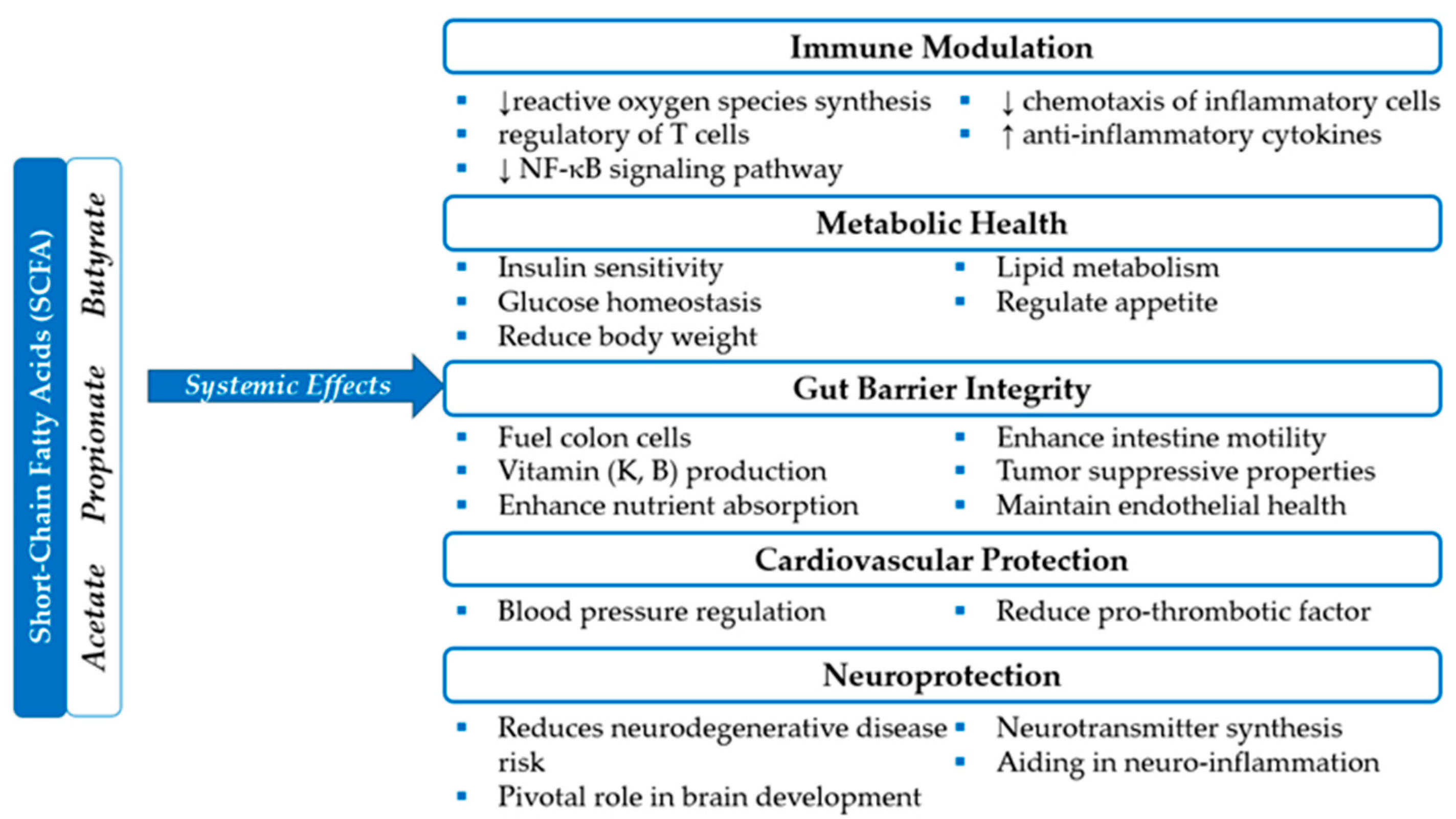
3.1. Digestive System and Metabolism

The human digestive system harbors a vast and complex community of microorganisms known as the microbiota. These microbes are essential for overall health, aiding digestion, supporting immune function, and maintaining metabolic balance. Predominantly composed of bacteria, these microbial communities support digestive health by breaking down indigestible food and synthesizing essential vitamins and short-chain fatty acids (SCFAs). Due to microbial activity, complex polysaccharides are converted into absorbable substrates, with these microorganisms coexisting in the digestive tract without typically causing clinical disorders. The gut flora also plays a crucial role in regulating the immune system by protecting against pathogens and modulating immune responses [9]. Numerous recent studies highlight the role of the gut microbiota in behavioral modulation through the gut–brain axis. Maintaining microbial equilibrium is important to prevent inflammation caused by increased intestinal lining permeability, which can trigger heightened immune reactions.
Fermentation of complex polysaccharides, such as dietary fibers, which human enzymes cannot digest, is one of the primary functions of gut microbes [10]. SCFAs such as butyrate, propionate, and acetate are produced in the large intestine and play a critical role in metabolic homeostasis. These compounds serve as primary energy sources for colonocytes, regulate intestinal motility, protect the intestinal mucosal layer, improve nutrient absorption, and modulate systemic inflammatory responses, Figure 1 [10,11,12]. Butyrate, produced by bacteria such as Faecalibacterium prausnitzii, Eubacterium rectale, and Roseburia spp., has been shown to have anti-inflammatory and antitumor properties, contributing to the maintenance of intestinal mucosal health and reducing the risk of conditions such as leaky gut syndrome [12,13]. Additionally, studies indicate that butyrate stimulates the production of mucin and antimicrobial peptides, which protect the intestinal mucosa from infectious agents [12]. The degradation of glycoproteins in the intestinal mucus, carried out by bacteria such as Akkermansia muciniphila, Bacteroides fragilis, and Ruminococcus gnavus, provides essential nutrients for the gut microbiota and contributes to maintaining the integrity of the intestinal barrier [14,15,16].
The gut microbiota is involved in protein digestion processes (dietary, host-derived, or produced by microorganisms). Species include Clostridium spp., Bacteroides spp., Proteus spp., and Peptostreptococcus spp. which break down undigested proteins into secondary metabolites, such as amino acids and small peptides, which are further transformed, through fermentation and deamination processes, into end products like SCFAs, ammonia, biogenic amines, phenols, and indoles [17]. These metabolites can either be eliminated by the host or used in specific biochemical reactions to synthesize other compounds.
A high-protein diet, as well as the type of protein ingested, influences the composition of the gut microbiota, leading to variations in the concentrations of different bacterial families and major phyla (Actinobacteria, Bacteroidetes, Firmicutes, Proteobacteria, Tenericutes, and Spirochaetes). These microbial shifts may contribute to the onset and progression of pathological conditions such as inflammatory bowel diseases, colorectal cancer, and metabolic disorders (e.g., type 2 diabetes) [17].
The microbiota also plays a key role in xenobiotic metabolism, either by degrading compounds into inactive metabolites (as seen with digoxin in individuals colonized with Eggerthella lenta) or by activating them into prodrugs, such as humimycin antibiotics synthesized by Rhodococcus spp. or the transformation of soy-derived isoflavones into equol, an estrogen-like substance. Additionally, the microbiota can contribute to synthesizing toxic metabolites, especially following their re-entry into the intestine through the enterohepatic circulation of compounds previously detoxified by the host. Certain bacterial species, such as Lactobacillus spp. and Bifidobacterium spp., bind aflatoxins and heavy metals (mercury, cadmium, lead), facilitating their elimination, while Akkermansia muciniphila, by supporting the mucus layer, can limit xenobiotic absorption, acting as a physical barrier [18].
The gut microbiota is essential for the synthesis of B-complex vitamins such as thiamine (B1), riboflavin (B2), niacin (B3), pantothenic acid (B5), pyridoxine (B6), biotin (B7), folate (B9), cobalamin (B12), and K (K2) which collectively support critical metabolic processes including carbohydrate and fat metabolism, DNA synthesis, neurotransmitter production, red blood cell formation, blood clotting, bone health, and maintaining vascular integrity. Vitamin synthesis varies by age. In infants, species such as Escherichia coli, Bacteroides spp., Veillonella parvula, Prevotella copri, Phocaeicola dorei, Bifidobacterium breve, Bifidobacterium bifidum, and Faecalibacterium prausnitzii are involved in the production of B-complex vitamins, while Escherichia coli, Bacteroides spp., Veillonella parvula, and Phocaeicola dorei contribute to the synthesis of vitamin K [19]. In adults, the synthesis of B-complex vitamins is supported by Bacteroides spp., Eubacterium rectale, Phocaeicola dorei, Bifidobacterium adolescentis, Faecalibacterium prausnitzii, Ruminococcus gnavus, and Prevotella copri, whereas vitamin K production involves Bacteroides spp., Eubacterium rectale, and Phocaeicola dorei [19]. In elderly individuals, species such as Escherichia coli, Bacteroides spp., Klebsiella pneumoniae, Prevotella copri, Phocaeicola dorei, Ruminococcus gnavus, and Alistipes putredinis contribute to B-complex vitamin synthesis, while Escherichia coli, Bacteroides spp., Klebsiella pneumoniae, and Phocaeicola dorei are responsible for vitamin K production [19]. These microbial dynamics highlight the age-dependent role of the gut microbiota in supporting metabolic and physiological functions. Any disruption in this balance can negatively impact metabolic health, as the symbiosis between the gut microbiome and humans is vital for optimal digestive health [20]. This also underscores the significance of a healthy and diverse microbiota for the overall well-being of a human being.
The gut microbiome’s impact extends beyond the digestive system, profoundly affecting the human body’s metabolic regulation. One of these roles is lipid metabolism. Processes such as deconjugation and dihydroxylation, carried out by species of the human microbiota, transform primary bile acids (BAs) into secondary BA, which are essential for emulsifying, breaking down, and absorbing lipids [21,22]. Cholesterol metabolism is regulated through the interaction between the gut microbiota and BA [23]. Bacteria such as Eubacterium coprostanoligenes, Bifidobacterium spp., and Lactobacillus spp. convert cholesterol into coprostanol, a less absorbable compound excreted in feces [22]. Trimethylamine N-oxide (TMAO) disrupts cholesterol and lipoprotein metabolism, contributing to vascular inflammation, plaque formation, and an increased risk of cardiovascular disease and diabetes [24,25,26,27,28]. TMAO is produced in the liver from trimethylamine (TMA), which is generated in the gut by bacterial species such as Firmicutes spp., Proteobacteria spp., Clostridium spp., Desulfovibrio spp., and Anaerococcus spp. through dietary choline and carnitine metabolism, primarily sourced from high-fat foods like red meat and eggs [28]. Bacteroides spp. and Bifidobacterium spp. metabolize dietary sphingolipids and simple sphingoid bases from the diet, improving intestinal barrier function, reducing exposure to bacterial toxins, and supporting immunity, with a potential role in regulating metabolic health and neurodevelopment [22]. Additionally, bacteria such as Bacteroides spp. and Lactobacillus spp. modulate the production of eicosanoids (prostaglandins and leukotrienes) by metabolizing essential fatty acids (omega-3 and omega-6), influencing inflammatory processes and overall metabolic balance [22].
The gut–brain axis, a bidirectional communication route between the digestive and central nervous systems, highlights the microbiota’s role in regulating appetite, satiety, and energy production. Gut hormones, such as glucagon-like peptide-1 (GLP-1) and PYY, are released in response to microbial metabolites, including SCFAs. These hormones are critical in maintaining appetite, glucose balance, and insulin sensitivity [24].
Studies have shown that metabolic disorders such as heart disease, hypertension, obesity, insulin resistance, gastrointestinal disorders, and type 2 diabetes, as well as an increased risk of cancers, are associated with dysbiosis [25,26]. Obese individuals show a specific microbial signature, characterized by reduced microbial diversity and an elevated Firmicutes/Bacteroidetes ratio, associated with a higher capability to harvest energy from the diet [27]. A 2022 study by Zhou et al. stated a decline in gut microbial diversity and deterioration of butyrate-producing bacteria in individuals with type 2 diabetes [25].
Certain gut microbes play a direct role in colorectal carcinogenesis. Fusobacterium nucleatum is enriched in colorectal cancer (CRC) tissues and promotes tumor progression via its adhesins Fusobacterium adhesin A (FadA) and Fusobacterial apoptosis protein 2 (Fap2). FadA binds to the epithelial adhesion protein E-cadherin, activating the β-catenin signaling pathway and promoting cellular proliferation. Fap2 impairs antitumor immunity by binding to TIGIT (T cell immunoreceptor with Ig and ITIM domains) on natural killer cells, allowing cancer cells to evade immune detection. Fusobacterium nucleatum has also been detected in primary tumors and liver metastases, suggesting its role in cancer dissemination. Similarly, enterotoxigenic Bacteroides fragilis (ETBF) produces B. fragilis toxin (BFT), a metalloprotease that disrupts epithelial barrier integrity and induces interleukin-17 (IL-17)-mediated inflammation, enhancing tumor development. In mouse models, ETBF colonization leads to rapid colonic tumor formation in genetically susceptible hosts [29].
The gut microbiome can be influenced by various factors, with diet playing a central role. Diets rich in plant-based fiber are generally associated with an increased presence of beneficial bacteria, such as Bifidobacteria and Lactobacillus. In contrast, high-fat and high-sugar diets tend to reduce microbial diversity and promote the growth of pro-inflammatory species, potentially contributing to metabolic imbalance [30].
Prebiotics and probiotics have emerged as promising advancements towards improving digestive health. Prebiotics are non-digestible food compounds, such as inulin, which nourish beneficial microbes and foster their growth, and probiotics are live microorganisms found in fermented foods and supplements that help replenish the microbial balance in the gut. The introduction of both has shown a positive effect on overall human well-being by potentially reducing metabolic diseases by decreasing inflammation, enhancing gut barrier functions, and modulating glucose uptake.
In addition to dietary interventions, fecal microbiota transplantation (FMT) is an approved treatment for recurrent Clostridioides difficile infection, while its use in inflammatory bowel disease (ulcerative colitis and Crohn’s disease), irritable bowel syndrome, severe post-antibiotic gut dysbiosis, metabolic disorders (obesity and type 2 diabetes), microbiota-associated neurological disorders (autism, Parkinson’s disease, and multiple sclerosis), and graft-versus-host disease following hematopoietic stem cell transplantation remains investigational or off-label [31,32,33].

3.2. Integumentary System

Skin, a multidimensional organ, is vital in preserving physical harmony by defending against external threats, regulating fluid balance, controlling temperature, sensing stimuli, and combating pathogens. Humans represent a minority compared to the microbes inhabiting our skin, as shown in Table 1 [34,35].
The first contact begins with vaginal birth, while c-section offers a more sterile environment [36]. After that, different relationships between the human skin and microorganisms develop, encompassing mutualistic, communal, saprophytic, and parasitic interactions. The skin microbiome varies significantly across different body regions and changes with genome, gender, age, and environmental factors such as living conditions, antibiotic use, sun exposure, and diet.
The stratum corneum, consisting of corneocytes surrounded by lipid layers, creates a resilient physical barrier that prevents water loss and protects against external threats [37,38]. Lower temperature, fatty acid production, and sweat gland secretion of lactic acid maintain acidity, supporting beneficial bacteria while inhibiting pathogenic strains like Staphylococcus aureus [39,40]. Most skin areas are dry, which is generally an unfavorable environment for bacterial growth. The continuous shedding of epithelial cells in the stratum corneum renews the skin surface and removes adhering microorganisms [41]. The skin’s defense also includes a chemical barrier of antimicrobial peptides, proteases, cytokines, and chemokines, which can directly inhibit microbial growth or activate immune responses [42].
Age significantly affects the diversity and composition of skin microbiota. Studies suggest that adults have higher microbial diversity than adolescents and the elderly. The increased microbial diversity in adults can be explained by the accumulation of several factors, such as hormonal stabilization, cumulative exposure to a wide range of environments and external factors throughout life, lower use of antibiotics compared to adolescents, and better skin barrier integrity than in older people. Furthermore, the daily activities and diversity of human interactions in which adults are involved, compared to older people, may contribute to the maintenance and diversification of the skin microbiome. Skin moisture retention and sebum production are influenced not only by age but also by gender. Lipophilic bacteria like Propionibacterium increase in abundance during adolescence, peaking in the third decade of life, which correlates with sebum levels [43].
Physiological variances in hormone metabolism, perspiration rate, and skin surface pH between males and females contribute to gender variations in skin environments [44]. While previous research highlighted significant alpha diversity differences between genders at the palm only, current studies identify these distinctions across all body sites, particularly the glabella [44]. The specific reasons for gender variations in the glabella remain theoretical, but differences in facial cosmetic usage within the genders could potentially play a role.
Urban and rural environments differ significantly, as do the lifestyles of their inhabitants. For instance, rural adults and the elderly are more likely to engage in agricultural work, while urbanites typically have indoor occupations. These lifestyle disparities can affect skin conditions and the microbial communities present there. Indoor microbiomes predominantly originate from humans, whereas outdoor microbial sources include soil, water, animals, atmosphere, and plant-associated microbes, which can influence the composition of an outdoor worker’s skin microbiome [45,46].
The primary commensal microbes on the skin are Staphylococcus epidermidis (S. epidermidis), Cutibacterium acnes (C. acnes), formerly Propionibacterium acne, and Corynebacterium. Unfortunately, our knowledge about other bacterial species on the skin is limited due to their low quantity and typically harmless nature [47]. The successful colonization of human skin by S. epidermidis is due to its commensal lifestyle, which prioritizes traits favoring persistence without harming the host. With low cytotoxicity and the ability to evade host defenses, S. epidermidis minimizes immune responses, resembling a mutual non-aggression pact. Certain characteristics suggest a symbiotic relationship, aiding the defense against transient pathogens. However, in compromised host immunity scenarios such as injury or surgery, S. epidermidis may exhibit opportunistic pathogenic behavior [48]. A reduced presence of commensal strains is often linked with an increased risk of pathogenic colonization [49]. Studies show that coagulase-negative S. epidermidis produces antimicrobial peptides (AMPs) that protect against colonization by the pathogenic Staphylococcus aureus [50]. C. acnes exhibits both non-pathogenic and pathogenic characteristics. It shares some properties with the ‘Generally Recognized as Safe’ (GRAS) status microbiome [51]. The relationship between the host and C. acnes consists of both mutualistic and parasitic elements, depending on the host’s tolerance, genetics, hormones, age, and the anatomical site of infection [49]. The sebaceous glands of the scalp, face, chest, and back produce large amounts of sebum, where the lipophilic anaerobe C. acnes predominates [52].
Coryneform spp. bacteria include several species, with Corynebacterium (C) being the most common. Corynebacterium is a Gram-positive pleomorphic rod divided into diphtheria and non-diphtheria species (diphtheroids). They are harmless bacteria that inhabit moist areas such as armpits and groins [53]. Beyond their commensal role, where they compete for nutrients and space, certain Corynebacterium species contribute to colonization resistance by producing bacteriocins (C. jeikeium), inhibiting Staphylococcus aureus virulence factors (C. striatum), or generating antimicrobial fatty acids (C. accolens). Among the more than 130 recognized species, C. jeikeium, C. striatum, C. amycolatum, C. minutissimum, C. kroppenstedtii, and C. xerosis can become opportunistic pathogens, particularly in immunocompromised patients. Corynebacterium has become a clinical concern due to its increasing antibiotic resistance, especially in nosocomial infections [54].
The fungal variety within the skin’s microbiota is limited, with the most predominant species being Malassezia spp. Studies estimate that Malassezia spp. account for 60-90% of the total fungal population on human skin [55]. Due to their inability to synthesize lipids, they are primarily found in sebaceous skin areas such as the scalp, face, back, and chest. Other fungal residents of the skin, like Candida albicans and dermatophytes, are only distantly related to Malassezia. Despite over a century of known associations with various diseases, the role of Malassezia in health and disease remains debated. Culture-based assessments indicate that Candida spp. infrequently inhabit human skin but may cause clinical infections, particularly in individuals with compromised immune systems, diabetes, or following antibiotic therapy. Another additional fungal genus reported to be present in the skin microbiota is Debaryomyces hansenii. [56].
Viruses inhabit healthy skin and are often considered pathogenic agents. This was evidenced by the asymptomatic presence of β- and γ-human papillomaviruses and viruses from the Polyomavirus genus [49,57]. Merkel cell polyomavirus (MCPyV), originally isolated from an aggressive neuroendocrine skin tumor known as Merkel cell carcinoma (MCC), was discovered on the skin surface of most healthy individuals, as well as on normal or pathological skin in patients with benign or non-MCC malignant skin conditions [57]. Viruses can positively impact homeostasis by regulating microbial populations and controlling overgrowth [58].
Skin is the body’s largest organ, extending from head to toe. The roles of the cutaneous microbiota are multiple. The commensal microorganisms at this level form a natural protective barrier against skin colonization by pathogenic microorganisms, competing for space and nutrients and synthesizing antibacterial molecules (natural antibiotics, free fatty acids, AMPc) that inhibit pathogens [35]. The microbiota stimulates the immune system by maintaining the local pro- and anti-inflammatory balance and activating local immune cells. These microorganisms regulate the skin’s pH, maintain the integrity of the stratum corneum, control local inflammatory processes, and preserve the skin’s homeostasis [35]. Some microorganisms can stimulate cellular regeneration processes, promoting the repair of skin lesions. A balanced cutaneous microbiota reduces the risk of skin conditions such as acne, atopic dermatitis, psoriasis, chronic wounds, and bacterial or fungal local infections [35]. Lastly, the microbiota helps the skin adapt to various environmental conditions, such as temperature or humidity variations and exposure to chemical agents.
Cutaneous dysbiosis has been associated with atopic dermatitis, acne, and microbial imbalances responsible for unpleasant body odor. In this context, skin microbiome transplantation (SMT), an emerging strategy, aims to restore the skin microbiota by applying selected beneficial microorganisms or directly transferring microbiota from healthy donors. Preclinical studies support that therapeutic colonization with Staphylococcus epidermidis and Staphylococcus hominis can reduce Staphylococcus aureus presence in patients with atopic dermatitis, with Roseomonas mucosa improves its evolution, while non-inflammatory Cutibacterium acnes strains may help regulate the microbiome in acne vulgaris [59,60]. Additionally, axillary microbiota manipulation has shown a potential to reduce body odor by replacing odor-causing bacteria with non-odorous species [60,61].
Although SMT shows therapeutic promise, research is still in its early stages, and aspects regarding this intervention’s stability, efficacy, and safety require further investigation. Integrating this approach into clinical practice could open new avenues for personalized dermatological therapy.

3.3. Respiratory System

The healthy lung was traditionally considered a sterile organ, particularly the distant alveoli [62]. We now know that it supports a rich and varied microbiota. A large part of these microbial communities, which are made up of bacteria, viruses, and fungi, is responsible for maintaining the normal physiological activities of the respiratory system [63]. Previously, numerous studies have been performed on the impact of the gut microbiota on human health and disease; however, in recent years, the impact of the microbiome on lung health and disease has been increasingly discussed [64,65]. Only recently have the functional mechanisms and consequences of microbiomes on lung health been explained. The respiratory tract microbiota has multiple roles in maintaining respiratory health, the most important being to serve as a gatekeeper to prevent respiratory infections from colonizing the system [66,67].

3.3.1. Upper Respiratory Tract Microbiota

The upper respiratory tract provides a variety of micro-niches, shaped by local anatomy, constant exposure to the external environment, and immune activity. Microbial colonization begins immediately after birth, influenced by the mode of delivery and type of feeding, with breastfeeding promoting dominant species such as Dolosigranulum and Corynebacterium, while formula feeding favors S. aureus [68]. As maturation progresses, the microbiome becomes more diverse in adults, with Actinobacteria, Firmicutes, and Bacteroidetes predominating, while in the elderly, there is a transition toward microbial communities resembling those of the oropharynx, possibly as a result of immunosenescence and reduced local biodiversity [68].
In addition to bacteria, this microbiome includes other microorganisms such as Thaumarchaeota and methanogenic Euryarchaeota archaea, as well as various fungal and viral species, although these remain less well studied [68]. Furthermore, the respiratory mucosa secretes antimicrobial substances such as lysozyme, lactoferrin, defensins, and reactive oxygen species like nitric oxide, which contribute to pathogen control and the maintenance of mucociliary clearance. Processes such as detecting bacterial molecules by specific taste and olfactory receptors can activate rapid immune responses, even before bacteria reach a significant pathogenic load.
Exposure to external factors, such as smoking and inhaled air quality, influences the microbiome at this level, increasing its diversity but reducing its stability, with a tendency to favor potentially pathogenic bacteria. In contrast, interventions such as nasal lavage with saline solutions and the administration of probiotics have proven to be effective and gentle methods for supporting microbiome balance and eliminating inflammatory mediators and pollutants.

3.3.2. Lower Respiratory Tract Microbiota

The lungs are continually exposed to germs suspended in the air and upper airways [69]. Since over 80% of the bacteria that inhabit the human body are undetectable by conventional culture-based methods, it is challenging to demonstrate the presence of the microbiome in the lungs. However, recent advances in sequencing technologies like amplicon have increased our comprehension of the lung microbiome [62,70,71].
A wide variety of microorganisms colonize the lung’s mucosal tissue. The lungs and airways are colonized by microorganisms from the oral cavity and environment [72]. The most common class of microorganisms in the lungs of healthy people are Prevotella, Streptococcus, Veillonella, Neisseria, Haemophilus, and Fusobacterium [73,74,75]. This diverse microbiota forms a complex and heterogeneous ecosystem that supports several key physiological processes [5,62,76]. Most flora is aerobic or facultatively anaerobic, except for Clostridium, Veillonella, and Porphyromonas, specialized anaerobes [63].
The lung microbiota plays essential roles in maintaining immune homeostasis, modulating inflammatory responses, and protecting against respiratory pathogens. The host’s immune system is modulated predominantly by the lung bacteria [77]. The lining of the lungs is covered in a thin layer of mucus with bacteriostatic properties, which offers microorganisms a low-nutrient habitat. The lung microenvironment is characterized by high immunological “tolerance”, which is primarily maintained by subpopulations of alveolar macrophages (AMs) and dendritic cells (DCs) [66]. These cells demonstrate their immunoregulatory capabilities by triggering the production of regulatory T cells (Treg) and releasing prostaglandin E2 (PGE2), tumor growth factor-beta (TGF-β), and interleukin-10 (IL-10). There is growing evidence that the lung microbiota plays a crucial role in fostering lung immunological tolerance by influencing resident immune cells [76,77]. These microorganisms help the lung react to dangerous infections correctly, preventing excess inflammatory reactions to benign antigens [72,78]. Microbial compounds and immune cell pattern recognition receptors (PRRs) interact to accomplish this immunological control [66]. These interactions prevent excessive immune responses and regulate inflammatory pathways. Additionally, immunoglobulin A (IgA), which covers mucosal surfaces and maintains immunological homeostasis by neutralizing pathogens before they can infiltrate deeper into lung tissues, is partly produced by the microbiota [79].
The lung microbiota supports the respiratory system’s physical defenses, such as the mucosal barrier and mucociliary clearance. Inhaled particles and microorganisms are trapped by a mucus layer that protects the epithelial cells lining the airways. The synthesis and composition of this mucus are influenced by microbial communities, which maximize its ability to capture dangerous particles while permitting effective gas exchange. Furthermore, the microbiota facilitates the movement of cilia, which are tiny hair-like projections on the surface of airway epithelial cells that aid in removing mucus and debris from the lungs [63,67,80].
The host’s mucosal barriers and microbial communities must balance to avoid infections and preserve lung health. Because the microbiota impacts mucociliary function, pathogens are effectively eliminated, lowering the risk of respiratory infections and enhancing the respiratory system’s overall resistance [67].
Despite the structural differences between the gut and lungs, possible anatomic communications and intricate networks involving their respective bacteria have supported the possibility of a gut–lung axis. According to the gut–lung axis theory, changes in the gut microbiome’s components brought on by illness, food, or medications are associated with altered immune responses and airway homeostasis [64,77,81,82,83]. Even though our understanding of the gut–lung axis is still developing, new research suggests lung disorders may be treated by modifying the gut microbiota [77]. The gut microbiota produces microbial metabolites, like SCFAs, which can enter the circulation or lymphatic system and reach the lungs, influencing immunological responses and supporting lung homeostasis [84]. Numerous studies show that the gut and lung microbiota are important for maintaining pulmonary health, and as such, they could be targets for nutrition-based preventive strategies against lung disease in older populations. This is because communication along the gut–lung axis becomes more pronounced with age or in elderly patients [82].

3.4. Urinary and Reproductive Systems

The urinary microbiota is the microbial community in the urinary system within the bladder or in bladder-obtained urine [85,86]. Previously, urine was said to be sterile, but bacterial communities have been discovered in the bladder, which has a potential role in urinary health [85,87,88]. Recent studies documented a wide spectrum of microbiota associated with healthy, culture-negative urine [85,86]. Many of these urinary microbial communities are like those seen in the human vagina, colon, and skin [89].
The five predominant bacteria in the urinary system of healthy males were Streptococcus, Lactobacillus, Prevotella, Corynebacterium, and Pseudomonas, and these were also seen in the urethral swab, although at varying degrees [86,89,90]. The microbiota at the penile sulcus is predominantly Pseudomonadaceae spp., which is rare in urine, whereas Sneathia spp. and Lactobacillus spp., which are common in urine, are rare in the sulcus of uncircumcised men [89]. It was observed that some of the penile microbiota, such as Pseudomonadaceae and Oxalobactericeae, were the same irrespective of circumcision status, but before circumcision, the putative anaerobic bacterial microbiota belonging to the families of Clostridiales and Prevotellaceae were more prevalent [90].
The five most predominant urinary microbiota in healthy females were Lactobacillus, Corynebacterium, Gardnerella, Prevotella, and Bacillus [87]. For the female reproductive system, many of the same urinary microbiota were cultured from the vaginal specimen, such as the Lactobacillus, Gardnerella, Bifidobacterium, Enterococcus, Actinomyces, Prevotella, and Atopobium [87]. This shows that both the urinary and vaginal tracts may have a common urogenital microbial community, although another study reported that the urethra and bladder may have distinct microbial communities from the vagina [90]. A healthy vaginal microbial community, however, consists of Lactobacillus, and it seems to play a key role in preventing several urogenital diseases by lowering the environmental pH through lactic acid production [86,87,89,90,91,92,93]. A pH below 4.5 is critical for the ecological balance of the vaginal microbiome, primarily sustained by lactic acid produced by Lactobacilli from epithelial glycogen stores under estrogenic stimulation. It was also observed that most microbial communities are commonly shared by men and women, except Lactobacillus crispatus, found only in healthy women [91]. In summary, the most common microbiota detected in males’ urinary and reproductive systems was Streptococcus, while in females, it was Lactobacillus [86,87,88,89,91,92,93].
There are several ways in which these microbial communities impact the urinary and reproductive systems. The abundance of vaginal lactobacilli prevents urinary tract infections (UTI) as it has been shown that the depletion of Lactobacillus in the microbiota is associated with UTI risk, and replacement with Lactobacillus crispatus intravaginal suppository probiotics reduced recurrent UTI in premenopausal women [93,94].
The female microbial community protects against bacterial vaginosis, which has been shown to result from replacing the microbiota rich in Lactobacillus crispatus with anaerobic species; thus, the urinary microbiota in healthy females differ significantly from those with bacterial vaginosis [91,95].
The urinary microbiota plays a role in preventing urge urinary incontinence (UUI) in females [86]. Both Lactobacillus spp. were cultured in females with and without UUI. In contrast, Lactobacillus gasseri was prevalent in the UUI cohort, and Lactobacillus crispatus was the prevalent species in the non-UUI females [86]. Also, Gardnerella was more frequently cultured in the urine specimen of the females with UUI than in the non-UUI cohort [86].
The urinary microbiota also reduces the risk of UTI in those with neurogenic bladder who showed a decrease in Lactobacillus and an increase in Enterobacteriales, indicating the possibility that this dysbiosis likely predisposed them to UTI [93].
The vaginal microbiome promotes healthy pregnancy outcomes, while Escherichia coli causes adverse events in pregnancy, including preterm birth [95,96]. Secretions from the female genital tract inhibit Escherichia coli. In health, the microbiota rich in Lactobacillus crispatus is associated with healthy pregnancy outcomes as it promotes Escherichia coli inhibitory activity of vaginal secretions, preventing its colonization and subsequently reducing the risk of preterm birth [95]. The Lactobacillus microbiota has also promoted fertility as its abundance in the female urogenital tract decreased in patients with infertility [97].
The microbial community in the urinary tract is important in preventing cellular rejection, as Lactobacillus in females and Streptococcus in males were reduced in those with interstitial fibrosis and tubular atrophy 6-8 months after renal transplantation. In contrast, lactobacillus normalized in females and streptococcus increased in males with excellent transplant function [86].
Vaginal microbiota transplantation (VMT) is an emerging therapeutic strategy aimed at restoring vaginal microbial balance, with the potential to improve the management of bacterial vaginosis and recurrent local infections, as well as to reduce the risk of sexually transmitted diseases and obstetric complications. However, further research is needed to establish donor selection criteria and assess long-term safety [90].

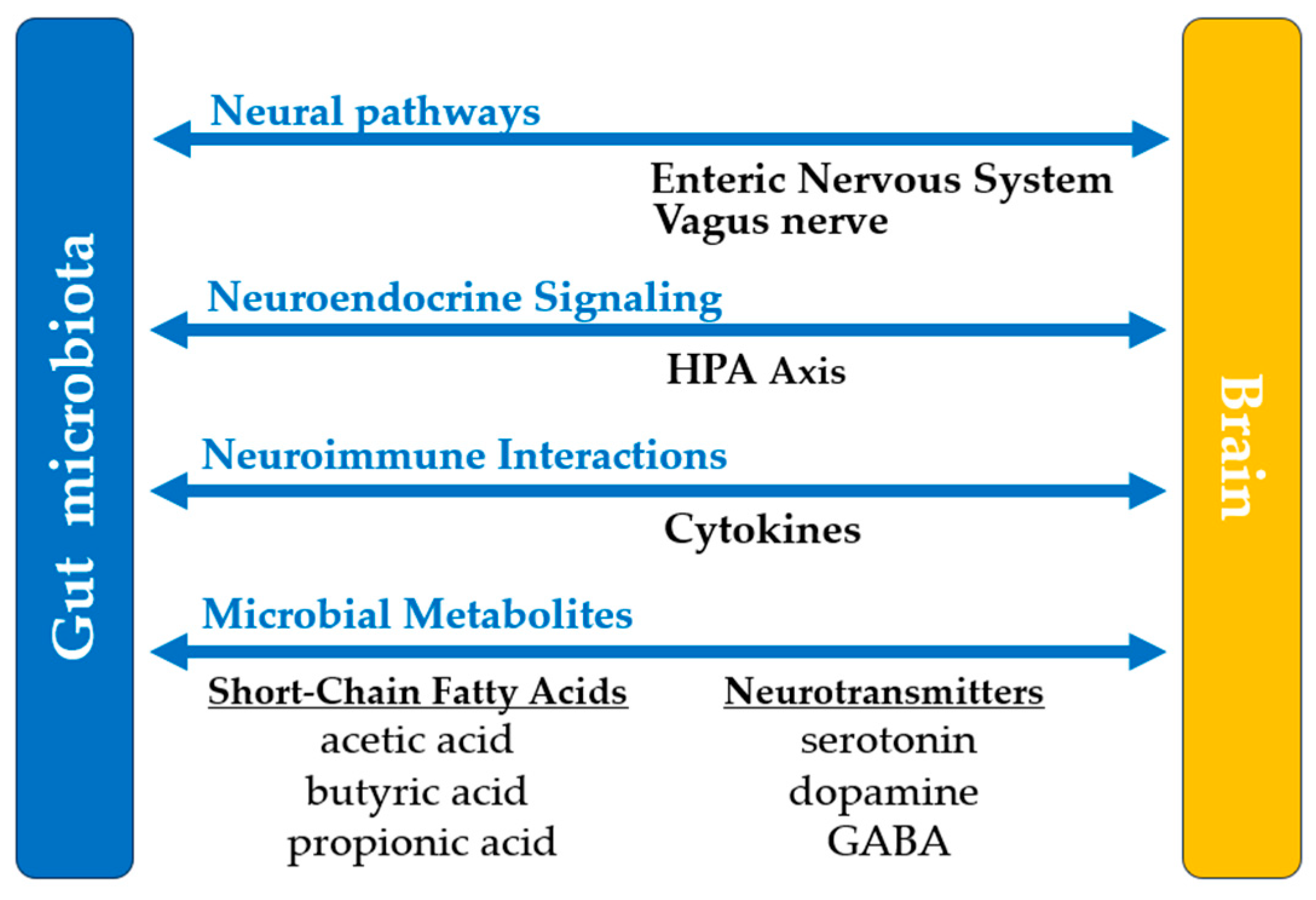
3.5. Central Nervous System

The gastrointestinal microbiome has emerged as a promising avenue for managing and improving neurological well-being. These microorganisms have been shown to influence the effects of neurological disorders through the gut–brain axis. This axis enables bidirectional communication between the central nervous system and gastrointestinal microflora through neural, endocrine, immune, and metabolic circuits, Figure 2. These interactions influence neuroprotection, blood–brain barrier permeability, cognitive function, motor skills, and overall brain health. Dysbiosis has been linked to various mental and neurological disorders. The recovery and symptom management of diseases like Alzheimer’s Disease (AD), Parkinson’s Disease (PD), autism spectrum disorder (ASD), and multiple sclerosis (MS) can be influenced by these microbial factions. In MS, dysbiosis is often characterized by a shift in the Firmicutes/Bacteroidetes ratio and an increased abundance of Akkermansia muciniphila [98]. While some studies have associated Akkermansia with mucin degradation and pro-inflammatory responses, more recent Mendelian randomization analyses indicate a causal protective effect of Akkermansia muciniphila against MS risk (OR 0.43, p = 1.37 × 10⁻⁸) [99]. This suggests that Akkermansia’s role may be context-dependent, influenced by host factors and overall microbial ecosystem composition.
The vagus nerve represents the primary communication pathway between the gut microbiota and the brain. Its intestinal receptors are indirectly stimulated by chemical substances produced by the microbiota, such as SCFAs, neurotransmitters, intestinal hormones [cholecystokinin and peptide YY(YY)], and mechanical stimuli (changes in pressure and intestinal wall distension) [100,101]. Many of these signals are mediated by enteroendocrine cells, which release signaling molecules that activate the afferent fibers of the vagus nerve. Microbial metabolites, such as SCFAs, are vital for optimal CNS functioning. SCFAs, such as acetate and butyrate, are pivotal in brain development, neurotransmitter synthesis, and neuroinflammation [100]. The gastrointestinal microbiota directly produces or regulates the production of neurotransmitters such as serotonin, dopamine, and gamma-aminobutyric acid (GABA).
In addition, gut microbes participate in the metabolism of tryptophan, an essential amino acid that serves as a precursor for serotonin, melatonin, and kynurenine. Several bacterial genera—including Clostridium, Burkholderia, Pseudomonas, Streptomyces, and Bacillus—are involved in transforming tryptophan into kynurenine and quinolinic acid, which deplete serotonin precursors and promote neuroinflammation. Conversely, other taxa such as Holdemania, Ruminococcus, and Tyzzerella favor the production of tryptamine and melatonin, supporting neurotransmitter synthesis and neuroprotection. The dysregulation of these pathways may contribute to neurological and psychiatric conditions, including depression, anxiety, and migraine, via impaired serotonin availability and altered pain perception [102].
The afferent component of the vagus nerve transmits the information received from the intestine to the nucleus of the solitary tract in the brainstem, where it is processed and relayed to brain regions involved in emotion regulation (amygdala and prefrontal cortex) and autonomic functions (hypothalamus). The hypothalamic–pituitary–adrenal (HPA) axis is modulated by signals transmitted from the gut through the vagus nerve, reducing stress and systemic inflammation. Studies have shown that probiotic strains such as Lactobacillus and Bifidobacterium elevate serotonin levels, aiding in the reduction in depressive and anxiety symptoms associated with AD and PD [100,101]. The production of dopamine, which is crucial for motor regulation, is influenced by gut microbes. Following probiotic treatment, an increase in dopamine signaling has been observed in PD patients [103].
The enteric nervous system (ENS), the “second brain”, consists of a complex autonomous neuronal network that plays a vital role in regulating gastrointestinal functions. It maintains close bidirectional communication with the central nervous system through the gut–brain axis. This communication involves the vagus nerve and neurotransmitters (serotonin, dopamine, and acetylcholine), influencing intestinal activity, emotion regulation, behavior, and autonomic responses. Moreover, the ENS plays a fundamental role in coordinating immune responses in the gut by interacting with immune cells, contributing to immunological tolerance and protection against pathogens. ENS dysfunctions can lead to the development of gastrointestinal and neuropsychiatric disorders.
Neurodegenerative diseases are aggravated by systemic inflammation, often caused by dysbiosis. Treatment with prebiotics and probiotics helps reduce inflammatory cytokine levels by restoring gastrointestinal microbial equilibrium. This mechanism has shown potential in conditions like MS, where lowering inflammation leads to a slowdown in disease progression [104]. The bacterial community of the stomach produces butyrate, which helps reduce inflammation by modulating immunity and supporting regulatory T cells [105]. In PD, FMT has demonstrated promise in reducing inflammatory markers [106]. In MS, inflammation markers declined in patients given SCFAs through prebiotics or supplemented with dietary fiber [103].
In AD, the gut microbial community can reduce amyloid-beta deposition by regulating systemic inflammation and oxidative stress. Studies have shown that prebiotic supplementation (which promotes the growth of Bifidobacteria) in individuals with AD is linked to enhanced memory and cognitive scores [107]. Dietary polyphenols broken down by gut bacteria can amplify neuroplasticity and cognitive resilience [108].
Gut dysbiosis is often evident in patients with PD. Supplementing them with Lactobacillus plantarum containing prebiotics has been linked to improved motor function. This effect is achieved by reducing alpha-synuclein buildup and boosting gut motility [103,109].

3.6. Cardiovascular System

Microbial metabolites affect the cardiovascular system, impacting vessels, vascular tone, lipids, and blood pressure. SCFAs produced from the microbial fermentation of dietary fibers in the colon are one of the main interactions between the microbiota and the cardiovascular system [110,111]. SCFAs, absorbed into the bloodstream, induce vasodilatory effects and contribute to regulating blood pressure. They interact with specific receptors on vascular smooth muscle cells, such as free fatty acid receptor (FFAR) 2 and FFAR3 [111,112]. The activation of these receptors promotes blood vessel dilation, thereby improving blood flow. Additionally, butyrate and acetate can stimulate the production of nitric oxide (NO), an essential mediator of vasodilation [113]. NO regulates vascular tone by activating soluble guanylate cyclase, increasing cyclic guanosine monophosphate levels, and activating protein kinase G, which leads to the relaxation of smooth muscle cells. NO also plays a significant role in endothelial protection and preventing thrombosis by inhibiting platelet aggregation and reducing the risk of atherosclerosis [114]. Moreover, SCFAs might modify the renin–angiotensin system, the key regulator of blood pressure. SCFAs can also reduce systemic inflammation, contributing to protection against cardiovascular diseases such as hypertension and atherosclerosis [111]. SCFAs can also exert opposing effects on blood pressure depending on the receptor activated. The activation of olfactory receptor 78 (Olfr78), expressed in renal afferent arterioles, has been shown to increase blood pressure by stimulating renin secretion [115]. The overall effect of SCFAs on blood pressure depends on the relative balance of these receptor pathways.
A second important microbial metabolite is TMA, which gut bacteria (Clostridia, Escherichia, Desulfovibrio, and Anaerococcus) make from dietary choline, phosphatidylcholine, and L-carnitine [28]. The production of TMA and its conversion to TMAO in the liver is associated with cardiovascular disease, but the production of TMAO is also regulated during normal, physiological states of lipid metabolism. The modulated cholesterol and lipid absorption in the intestine by the microbiota supports healthy blood lipid levels (essential to maintaining vascular integrity and function) [116].
Additionally, the gut–brain axis, the linkage of the gut microbiota with central nervous system signaling, also has implications for cardiovascular regulation. For instance, the vagus nerve may convey some effects of microbial metabolites on heart rate and autonomic control of the cardiovascular system. This neural pathway hints that the microbiota can affect the cardiovascular response indirectly through the modulation of heart rate variability as a measure of autonomic balance and cardiovascular fitness [117].
Recent research indicates that gut microbiota can influence salt sensitivity, a crucial factor in blood pressure regulation. The effect is mediated by gut microbial metabolites such as SCFAs and arachidonic acid [118].

3.7. Endocrine Function

The microbiota also heavily impacts the endocrine system, which governs hormone production and regulation. Several of these hormones, involved in metabolic and stress responses, are synthesized or regulated by gut bacteria. SCFAs synthesized by gut bacteria activate FFAR2 and 3 receptors on intestinal L cells, stimulating the secretion of glucagon-like peptide-1 (GLP-1 and PYY), which play a crucial role in glucose metabolism and appetite regulation [119].
The synthesis of ghrelin, a peptide secreted by X/A-like neuroendocrine cells in the gastric mucosa, can be indirectly stimulated by the presence of intestinal bacteria such as Bifidobacterium, Lactobacillus, or Akkermansia [116,120]. Ghrelin subsequently stimulates appetite and promotes the release of growth hormone. Akkermansia modulates the secretion of other key gut hormones, such as leptin, GLP-1, PYY, and cholecystokinin. It also increases satiety and improves metabolism [116,121].
In addition to that, the gut microbiota also interacts with the HPA axis, which comprises the core region for the body’s response to stress. The cortisol release that successively characterizes the HPA axis represents the primary regulator of the adaptive response to stress and of metabolism. Some gut bacteria, such as Lactobacillus, Bifidobacterium, and Akkermansia muciniphila, can activate this axis, affecting the release of the corticotropin-releasing hormone from the hypothalamus [122,123,124].
Microbial communities play an essential role in the metabolism of iodine, an essential component in thyroid hormone synthesis. Small intestine gut bacteria contribute to modulating iodine absorption to maintain normal thyroid gland functioning by producing thyroxine and triiodothyronine, which are responsible for regulating metabolism. Directly supporting endocrine balance and metabolic regulation, the efficiency with which microbes contribute to nutrient absorption, including iodine, indirectly [125].
The gut microbiota is known to influence both metabolic and steroid hormones, particularly estrogens and androgens. A key mechanism involves the production of enzymes, such as β-glucuronidase, by specific bacterial phyla, including Bacteroidetes, Firmicutes, Verrucomicrobia, and Proteobacteria [126]. These enzymes facilitate the deconjugation of estrogens in the gut, enabling their reabsorption into the bloodstream via enterohepatic recycling. This process is crucial in maintaining balanced circulating estrogen levels, essential for reproductive health, bone density, and cardiovascular function [127].
Bile acids’ potential to modulate the gut microbiome is essential for regulating the cardiovascular and endocrine systems. Bile acids derived from the liver are primary, while those altered by the bacteria found in the intestine are secondary forms. These secondary bile acids serve as ligands binding to the FXR and TGR5, which modulate glucose and lipid metabolism and energy expenditure. The right regulation of these pathways by the microbiota benefits metabolic health and the cardiovascular system [120].

3.8. Immune System

The human microbiota interacts with the immune system from the first moments of life, forming a bidirectional axis essential for maintaining a balance between the host and it. The immune system influences the composition of the microbiota, while the microbiota shapes the functions of innate and adaptive immunity.
Immediately after birth, the newborn begins to be colonized by a microbial flora that varies depending on environmental factors, nutrition, and the mode of delivery (vaginal or cesarean section). The microbiota tends to mature within the first three years of life, reaching a stable composition like an adult’s. During this “window of opportunity,” the dynamic relationship between the developing microbiota and the immune system plays a critical role in immune development.
At birth, newborns lack a fully developed immune system, making them vulnerable to microbial agents. The microbiota contributes to the maturation of T lymphocytes [Treg and T helper (Th1, Th2, and Th17)] and B lymphocytes, the synthesis of IgA (which plays a role in local defense) and serum IgG, and the inhibition of IgE production (involved in allergic reactions) [128,129]. Additionally, the microbiota regulates the development and activity of antigen-presenting cells (APCs), neutrophils, natural killer (NK) cells, and mast cells.
In germ-free animals, the absence of a microbiota results in structural lymphoid deficiencies and immunosuppression [128]. Immune homeostasis depends on the balance between pro-inflammatory responses (Th1, Th17) and anti-inflammatory responses (Treg and IgA). A diverse and balanced microbiota maintains this balance, producing metabolites such as SCFAs, essential for Treg regulation and the control of systemic inflammation.
The microbiota also contributes to the integrity of the epithelial barrier by stimulating mucus production, antimicrobial peptides (AMPs produced by Paneth cells), and strengthening intercellular junctions. Dysbiosis compromises this barrier, leading to bacterial translocation, the activation of inflammatory responses, and an increased risk of systemic inflammation.
Commensal microorganisms significantly induce local immune tolerance, reducing the risk of excessive inflammatory reactions. Polysaccharides synthesized by species such as Bacteroides fragilis modulate Th1/Th2 responses, promoting immune balance [128,129].
Dysbiosis caused by unbalanced diets, stress, or antibiotic treatments is associated with chronic inflammation and an increased risk of autoimmune diseases [129].
The human microbiota is pivotal in maintaining overall health and influences multiple physiological systems. Table 2 summarizes the effects of the human microbiota on various body systems.

3.9. Microbiota—A New Frontier in Diagnostic and Treatment

The individual composition of the microbiota presents opportunities for a personalized approach to various pathologies, offering diagnostic, treatment, and prevention solutions tailored to each patient. Genomic sequencing and metabolomic analysis allow for the detailed characterization of the individual microbiome and the identification of specific microbial biomarkers that guide clinicians toward the early detection of predisposition to or presence of pathologies such as metabolic syndrome, autoimmune diseases, or neurodegenerative conditions, Table 3.
In clinical practice, personalizing microbiota-based therapeutic strategies has proven effective in multiple fields, offering innovative solutions for microbial homeostasis regulation and improving patient health. Interventions include diet, probiotics, prebiotics, and postbiotics, as well as advanced strategies such as next-generation probiotics and various microbiota transplantation techniques. Probiotic-containing foods such as yogurt, kefir, and fermented vegetables, and prebiotic-rich sources like inulin-containing vegetables (e.g., garlic, onions, and asparagus) and whole grains contribute significantly to gut microbiota modulation. Targeted probiotics can stimulate beneficial bacterial species such as Lactobacillus, Bifidobacterium, and Akkermansia, reducing inflammation and improving epithelial barrier function. Prebiotics, substances that promote the growth of beneficial bacteria, are already being used to support metabolic and immune health. Postbiotics, active microbial metabolites, play an increasingly important role in maintaining microbial balance and treating inflammatory and metabolic diseases. A recent innovation is represented by next-generation probiotics, such as Faecalibacterium prausnitzii and Akkermansia muciniphila, which directly influence inflammation, intestinal homeostasis, and metabolism [129]. These species are studied extensively for therapeutic applications in obesity, diabetes, inflammatory bowel diseases, and neurodegenerative disorders. However, growing evidence highlights that short-term use of dietary, prebiotic, or probiotic interventions, typically under four weeks, only induces transient changes in the microbiota. A minimum intervention duration of three months is recommended to achieve stable and clinically relevant alterations in microbial composition and function [130].
Microbiota transplants have become promising therapeutic solutions for restoring microbial balance in severe conditions. FMT is an efficient treatment method, demonstrating success rates of over 90% in recurrent Clostridioides difficile infections, more than 60% in inflammatory bowel diseases, and less than 60% in extraintestinal conditions [131]. Other forms of transplantation, such as VMT, and SMT, are currently in experimental stages but show potential in treating dysbiosis related to gynecological, and dermatological conditions.
Moreover, the microbiota plays a key role in the body’s response to various therapies by metabolizing medications, influencing their bioavailability and efficacy. Personalized drug therapy, tailored to each patient’s individual microbiota profile, can prevent adverse reactions and increase therapeutic response rates.
Adapting a diet based on microbiota analysis can reduce the risk of developing and complicating certain metabolic diseases, such as obesity or diabetes mellitus.
Identifying the unique characteristics of the microbiota paves the way for precise, personalized therapies that can optimize health and improve quality of life.
The integration of these microbiota-based therapies opens new directions in personalized medicine, offering innovative solutions for chronic disease prevention and treatment, ultimately improving patients’ quality of life.

4. Conclusions

The microbiota represents a unique ecosystem essential for maintaining human health and is involved in multiple physiological functions, from metabolism to regulating immune responses and neuroprotection. It serves as the individual fingerprint of each human organism, enabling the development of personalized therapies, a promising solution for treating debilitating acute and chronic conditions such as infections, autoimmune diseases, neurodegenerative disorders, and metabolic syndromes. In-depth studies are required to understand the mechanisms through which the microbiota interacts with different bodily systems.
Understanding the role of the microbiota in regulating interactions between various organs and systems could open new perspectives in disease prevention through nutritional and pharmaceutical interventions. An integrated approach to microbiota research could significantly contribute to revolutionizing personalized medicine and improving patients’ quality of life.

Author Contributions

Conceptualization, H.T.J. and N.N.; Methodology, T.C.T., R.A. and Y.Y.T.; Software, E.C.C., Y.Y.T. and R.A. Validation, A.B.A., T.C.T. and N.N.; Formal analysis, E.C.C. and B.B.; Investigation, T.C.T., A.F. and A.B.A.; Resources, P.M. and Y.Y.T.; Data curation, A.F., B.B. and A.B.A.; Writing—original draft preparation, H.T.J., T.C.T., E.C.C., A.B.A., R.A., Y.Y.T., B.B., N.N., A.F. and P.M.; Writing—review and editing, H.T.J., T.C.T., E.C.C., A.B.A., R.A., Y.Y.T., B.B., N.N., A.F. and P.M.; Visualization, E.C.C., P.M., R.A. and B.B.; Supervision, H.T.J., N.N. and P.M.; Project administration, N.N.; Funding acquisition, N.N., H.T.J. and A.F. All authors have read and agreed to the published version of the manuscript.

Funding

This research received no external funding.

Institutional Review Board Statement

Not applicable.

Informed Consent Statement

Not applicable.

Data Availability Statement

No new data were created or analyzed in this study.

Conflicts of Interest

The authors declare no conflicts of interest.

References

  1. FAQ. FAQ: Human Microbiome; American Society for Microbiology: Washington, DC, USA, 2013. Available online: https://www.ncbi.nlm.nih.gov/books/NBK562894/ (accessed on 15 March 2025).
  2. El-Sayed, A.; Aleya, L.; Kamel, M. Microbiota’s Role in Health and Diseases. Environ. Sci. Pollut. Res. Int. 2021, 28, 36967–36983. [Google Scholar] [CrossRef] [PubMed]
  3. Wen, L.; Duffy, A. Factors Influencing the Gut Microbiota, Inflammation, and Type 2 Diabetes. J. Nutr. 2017, 147, 1468S–1475S. [Google Scholar] [CrossRef] [PubMed]
  4. Gomaa, E.Z. Human Gut Microbiota/Microbiome in Health and Diseases: A Review. Antonie Van Leeuwenhoek 2020, 113, 2019–2040. [Google Scholar] [CrossRef]
  5. Man, W.H.; de Steenhuijsen Piters, W.A.; Bogaert, D. The Microbiota of the Respiratory Tract: Gatekeeper to Respiratory Health. Nat. Rev. Microbiol. 2017, 15, 259–270. [Google Scholar] [CrossRef] [PubMed]
  6. Lu, L.; Chen, X.; Liu, Y.; Yu, X. Gut Microbiota and Bone Metabolism. FASEB J. 2021, 35, e21740. [Google Scholar] [CrossRef]
  7. Portincasa, P.; Bonfrate, L.; Vacca, M.; De Angelis, M.; Farella, I.; Lanza, E.; Khalil, M.; Wang, D.Q.; Sperandio, M.; Di Ciaula, A. Gut Microbiota and Short Chain Fatty Acids: Implications in Glucose Homeostasis. Int. J. Mol. Sci. 2022, 23, 1105. [Google Scholar] [CrossRef]
  8. Jandhyala, S.M.; Talukdar, R.; Subramanyam, C.; Vuyyuru, H.; Sasikala, M.; Nageshwar Reddy, D. Role of the Normal Gut Microbiota. World J. Gastroenterol. 2015, 21, 8787–8803. [Google Scholar] [CrossRef]
  9. James, K.R.; Gomes, T.; Elmentaite, R.; Kumar, N.; Gulliver, E.L.; King, H.W.; Stares, M.D.; Bareham, B.R.; Ferdinand, J.R.; Petrova, V.N.; et al. Distinct Microbial and Immune Niches of the Human Colon. Nat. Immunol. 2020, 21, 343–353. [Google Scholar] [CrossRef]
  10. Koh, A.; De Vadder, F.; Kovatcheva-Datchary, P.; Bäckhed, F. From Dietary Fiber to Host Physiology: Short-Chain Fatty Acids as Key Bacterial Metabolites. Cell 2016, 165, 1332–1345. [Google Scholar] [CrossRef]
  11. Fusco, W.; Lorenzo, M.B.; Cintoni, M.; Porcari, S.; Rinninella, E.; Kaitsas, F.; Lener, E.; Mele, M.C.; Gasbarrini, A.; Collado, M.C.; et al. Short-Chain Fatty-Acid-Producing Bacteria: Key Components of the Human Gut Microbiota. Nutrients 2023, 15, 2211. [Google Scholar] [CrossRef]
  12. Hodgkinson, K.; El Abbar, F.; Dobranowski, P.; Manoogian, J.; Butcher, J.; Figeys, D.; Mack, D.; Stintzi, A. Butyrate’s role in human health and the current progress towards its clinical application to treat gastrointestinal disease. Clin. Nutr. 2023, 42, 61–75. [Google Scholar] [CrossRef] [PubMed]
  13. Louis, P.; Hold, G.L.; Flint, H.J. The Gut Microbiota, Bacterial Metabolites, and Colorectal Cancer. Nat. Rev. Microbiol. 2014, 12, 661–672. [Google Scholar] [CrossRef]
  14. Patrick, S. A tale of two habitats: Bacteroides fragilis, a lethal pathogen and resident in the human gastrointestinal microbiome. Microbiology 2022, 168, 001156. [Google Scholar] [CrossRef] [PubMed]
  15. Coletto, E.; Latousakis, D.; Pontifex, M.G.; Crost, E.H.; Vaux, L.; Perez Santamarina, E.; Goldson, A.; Brion, A.; Hajihosseini, M.K.; Vauzour, D.; et al. The role of the mucin-glycan foraging Ruminococcus gnavus in the communication between the gut and the brain. Gut Microbes 2022, 14, 2073784. [Google Scholar] [CrossRef]
  16. Collins, S.L.; Patterson, A.D. The Gut Microbiome: An Orchestrator of Xenobiotic Metabolism. Acta Pharm. Sin. B 2020, 10, 19–32. [Google Scholar] [CrossRef] [PubMed]
  17. Bartlett, A.; Kleiner, M. Dietary protein and the intestinal microbiota: An understudied relationship. iScience 2022, 25, 105313. [Google Scholar] [CrossRef]
  18. Tarracchini, C.; Lugli Gabriele, A.; Mancabelli, L.; van Sinderen, D.; Turroni, F.; Ventura, M.; Milani, C. Exploring the vitamin biosynthesis landscape of the human gut microbiota. mSystems 2024, 9, e00929-24. [Google Scholar] [CrossRef]
  19. Ríos-Covián, D.; Ruas-Madiedo, P.; Margolles, A.; Gueimonde, M.; de Los Reyes-Gavilán, C.G.; Salazar, N. Intestinal Short-Chain Fatty Acids and Their Link with Diet and Human Health. Front. Microbiol. 2016, 7, 185. [Google Scholar] [CrossRef]
  20. Ridlon, J.M.; Kang, D.J.; Hylemon, P.B. Bile salt biotransformations by human intestinal bacteria. J. Lipid Res. 2016, 47, 241–259. [Google Scholar] [CrossRef]
  21. Morales-Sánchez, A.; Fuentes-Pananá, E.M. Human Viruses and Cancer. Viruses 2014, 6, 4047–4049. [Google Scholar] [CrossRef]
  22. Brown, E.M.; Clardy, J.; Xavier, R.J. Gut microbiome lipid metabolism and its impact on host physiology. Cell Host Microbe 2023, 31, 173–186. [Google Scholar] [CrossRef] [PubMed]
  23. Li, T.; Chiang, J.Y.L. Bile acid signaling in metabolic disease and drug therapy. Pharmacol. Rev. 2014, 66, 948–983. [Google Scholar] [CrossRef] [PubMed]
  24. Arora, T.; Vanslette, A.M.; Hjorth, S.A.; Bäckhed, F. Microbial regulation of enteroendocrine cells. Med 2021, 2, 553–570. [Google Scholar] [CrossRef]
  25. Zhou, Z.; Sun, B.; Yu, D.; Zhu, C. Gut Microbiota: An Important Player in Type 2 Diabetes Mellitus. Front. Cell. Infect. Microbiol. 2022, 12, 834485. [Google Scholar] [CrossRef]
  26. DeGruttola, A.K.; Low, D.; Mizoguchi, A.; Mizoguchi, E. Current Understanding of Dysbiosis in Disease in Human and Animal Models. Inflamm. Bowel Dis. 2016, 22, 1137–1150. [Google Scholar] [CrossRef]
  27. Zhang, K.; Zhang, Q.; Qiu, H.; Ma, Y.; Hou, N.; Zhang, J.; Kan, C.; Han, F.; Sun, X.; Shi, J. The complex link between the gut microbiome and obesity-associated metabolic disorders: Mechanisms and therapeutic opportunities. Heliyon 2024, 10, e37609. [Google Scholar] [CrossRef]
  28. Ebrahimzadeh Leylabadlo, H.; Sanaie, S.; Sadeghpour Heravi, F.; Ahmadian, Z.; Ghotaslou, R. From role of gut microbiota to microbial-based therapies in type 2-diabetes. Infect. Genet. Evol. 2020, 81, 104268. [Google Scholar] [CrossRef] [PubMed]
  29. Lopez, L.R.; Bleich, R.M.; Arthur, J.C. Microbiota Effects on Carcinogenesis: Initiation, Promotion, and Progression. Annu. Rev. Med. 2021, 72, 243–261. [Google Scholar] [CrossRef]
  30. Scheithauer, T.P.M.; Rampanelli, E.; Nieuwdorp, M.; Vallance, B.A.; Verchere, C.B.; van Raalte, D.H.; Herrema, H. Gut Microbiota as a Trigger for Metabolic Inflammation in Obesity and Type 2 Diabetes. Front. Immunol. 2020, 11, 571731. [Google Scholar] [CrossRef]
  31. American Society for Microbiology. Fecal Microbiota Transplants (FMT): Past, Present and Future. Available online: https://asm.org/articles/2024/february/fecal-microbiota-transplants-past-present-future (accessed on 15 March 2025).
  32. Reddi, S.; Senyshyn, L.; Ebadi, M.; Podlesny, D.; Minot, S.S.; Gooley, T.; Kabage, A.J.; Hill, G.R.; Lee, S.J.; Khoruts, A.; et al. Fecal microbiota transplantation to prevent acute graft-versus-host disease: Pre-planned interim analysis of donor effect. Nat. Commun. 2025, 16, 1034. [Google Scholar] [CrossRef]
  33. Drwiega, E.N.; Nascimento, D.C. A Comparison of Currently Available and Investigational Fecal Microbiota Transplant Products for Recurrent Clostridioides difficile Infection. Antibiotics 2024, 13, 436. [Google Scholar] [CrossRef] [PubMed]
  34. Carmona-Cruz, S.; Orozco-Covarrubias, L.; Sáez-de-Ocariz, M. The Human Skin Microbiome in Selected Cutaneous Diseases. Front. Cell. Infect. Microbiol. 2022, 12, 834135. [Google Scholar] [CrossRef] [PubMed]
  35. Yang, Y.; Qu, L.; Mijakovic, I.; Wei, Y. Advances in the human skin microbiota and its roles in cutaneous diseases. Microb. Cell Factories 2022, 21, 176. [Google Scholar] [CrossRef]
  36. Oh, J.; Byrd, A.L.; Park, M.; Kong, H.H.; Segre, J.A. Temporal Stability of the Human Skin Microbiome. Cell 2016, 165, 854–866. [Google Scholar] [CrossRef]
  37. Sinha, S.; Lin, G.; Ferenczi, K. The skin microbiome and the gut-skin axis. Clin. Dermatol. 2021, 39, 829–839. [Google Scholar] [CrossRef] [PubMed]
  38. Kengmo Tchoupa, A.; Kretschmer, D.; Schittek, B.; Peschel, A. The epidermal lipid barrier in microbiome-skin interaction. Trends Microbiol. 2023, 31, 723–734. [Google Scholar] [CrossRef]
  39. Lee, H.J.; Kim, M. Skin Barrier Function and the Microbiome. Int. J. Mol. Sci. 2022, 23, 13071. [Google Scholar] [CrossRef] [PubMed]
  40. Swaney, M.H.; Nelsen, A.; Sandstrom, S.; Kalan, L.R. Sweat and Sebum Preferences of the Human Skin Microbiota. Microbiol. Spectr. 2023, 11, e0418022. [Google Scholar] [CrossRef]
  41. Smythe, P.; Wilkinson, H.N. The Skin Microbiome: Current Landscape and Future Opportunities. Int. J. Mol. Sci. 2023, 24, 3950. [Google Scholar] [CrossRef]
  42. Lunjani, N.; Ahearn-Ford, S.; Dube, F.S.; Hlela, C.; O’Mahony, L. Mechanisms of microbe-immune system dialogue within the skin. Genes Immun. 2021, 22, 276–288. [Google Scholar] [CrossRef]
  43. Ying, S.; Zeng, D.N.; Chi, L.; Tan, Y.; Galzote, C.; Cardona, C.; Lax, S.; Gilbert, J.; Quan, Z.X. The Influence of Age and Gender on Skin-Associated Microbial Communities in Urban and Rural Human Populations. PLoS ONE 2015, 10, e0141842. [Google Scholar] [CrossRef] [PubMed]
  44. Boxberger, M.; Cenizo, V.; Cassir, N.; La Scola, B. Challenges in exploring and manipulating the human skin microbiome. Microbiome 2021, 9, 125. [Google Scholar] [CrossRef] [PubMed]
  45. Leung, M.H.Y.; Tong, X.; Lee, P.K.H. Indoor Microbiome and Airborne Pathogens. Compr. Biotechnol. 2019, 31, 96–106. [Google Scholar] [CrossRef]
  46. Hayleeyesus, S.F.; Manaye, A.M. Microbiological quality of indoor air in university libraries. Asian Pac. J. Trop. Biomed. 2014, 4 (Suppl. S1), S312–S317. [Google Scholar] [CrossRef]
  47. Flowers, L.; Grice, E.A. The Skin Microbiota: Balancing Risk and Reward. Cell Host Microbe 2020, 28, 190–200. [Google Scholar] [CrossRef]
  48. Gallo, R.L. S. epidermidis influence on host immunity: More than skin deep. Cell Host Microbe 2015, 17, 143–144. [Google Scholar] [CrossRef]
  49. Christensen, G.J.; Brüggemann, H. Bacterial skin commensals and their role as host guardians. Benef. Microbes 2014, 5, 201–215. [Google Scholar] [CrossRef]
  50. Severn, M.M.; Horswill, A.R. Staphylococcus epidermidis and its dual lifestyle in skin health and infection. Nat. Rev. Microbiol. 2023, 21, 97–111. [Google Scholar] [CrossRef]
  51. Gagliardi, A.; Totino, V.; Cacciotti, F.; Iebba, V.; Neroni, B.; Bonfiglio, G.; Trancassini, M.; Passariello, C.; Pantanella, F.; Schippa, S. Rebuilding the Gut Microbiota Ecosystem. Int. J. Environ. Res. Public Health 2018, 15, 1679. [Google Scholar] [CrossRef]
  52. Rozas, M.; Hart de Ruijter, A.; Fabrega, M.J.; Zorgani, A.; Guell, M.; Paetzold, B.; Brillet, F. From Dysbiosis to Healthy Skin: Major Contributions of Cutibacterium acnes to Skin Homeostasis. Microorganisms 2021, 9, 628. [Google Scholar] [CrossRef]
  53. Chen, Y.E.; Fischbach, M.A.; Belkaid, Y. Skin microbiota-host interactions. Nature 2018, 553, 427–436. [Google Scholar] [CrossRef] [PubMed]
  54. Jensen, M.G.; Svraka, L.; Baez, E.; Lund, M.; Poehlein, A.; Brüggemann, H. Species- and strain-level diversity of Corynebacteria isolated from human facial skin. BMC Microbiol. 2023, 23, 366. [Google Scholar] [CrossRef] [PubMed]
  55. Kanna, B.V.; Latha, R.; Kavitha, K.; Jeyakumari, D. Phenotypic Characterization of Malassezia spp. Isolated from Healthy Individuals. Res. J. Biotechnol. 2023, 18, 74–78. [Google Scholar] [CrossRef]
  56. Schmid, B.; Künstner, A.; Fähnrich, A.; Busch, H.; Glatz, M.; Bosshard, P.P. Longitudinal Characterization of the Fungal Skin Microbiota in Healthy Subjects Over a Period of 1 Year. J. Investig. Dermatol. 2022, 142, 2766–2772.e2768. [Google Scholar] [CrossRef]
  57. Silling, S.; Kreuter, A.; Gambichler, T.; Meyer, T.; Stockfleth, E.; Wieland, U. Epidemiology of Merkel Cell Polyomavirus Infection and Merkel Cell Carcinoma. Cancers 2022, 14, 6176. [Google Scholar] [CrossRef]
  58. Harper, A.; Vijayakumar, V.; Ouwehand, A.C.; ter Haar, J.; Obis, D.; Espadaler, J.; Binda, S.; Desiraju, S.; Day, R. Viral Infections, the Microbiome, and Probiotics. Front. Cell. Infect. Microbiol. 2021, 10, 596166. [Google Scholar] [CrossRef]
  59. Willmott, T.; Campbell, P.M.; Griffiths, C.E.M.; O’Connor, C.; Bell, M.; Watson, R.E.B.; McBain, A.J.; Langton, A.K. Behaviour and sun exposure in holidaymakers alters skin microbiota composition and diversity. Front. Aging 2023, 4, 1217635. [Google Scholar] [CrossRef]
  60. Junca, H.; Pieper, D.H.; Medina, E. The emerging potential ofmicrobiome transplantation on human health interventions. Comput. Struct. Biotechnol. J. 2022, 20, 615–627. [Google Scholar] [CrossRef]
  61. Romano-Bertrand, S.; Bourdier, A.; Aujoulat, F.; Michon, A.L.; Masnou, A.; Parer, S.; Marchandin, H.; Jumas-Bilak, E. Skin microbiota is the main reservoir of Roseomonas mucosa, an emerging opportunistic pathogen so far assumed to be environmental. Clin. Microbiol. Infect. 2016, 22, 737.e1–737.e7. [Google Scholar] [CrossRef]
  62. Li, R.; Li, J.; Zhou, X. Lung microbiome: New insights into the pathogenesis of respiratory diseases. Signal Transduct. Target. Ther. 2024, 9, 19. [Google Scholar] [CrossRef]
  63. Fabbrizzi, A.; Amedei, A.; Lavorini, F.; Renda, T.; Fontana, G. The Lung Microbiome: Clinical and Therapeutic Implications. Intern. Emerg. Med. 2019, 14, 1241–1250. [Google Scholar] [CrossRef] [PubMed]
  64. Natalini, J.G.; Singh, S.; Segal, L.N. The dynamic lung microbiome in health and disease. Nat. Rev. Microbiol. 2023, 21, 222–235. [Google Scholar] [CrossRef] [PubMed]
  65. Sommariva, M.; Le Noci, V.; Bianchi, F.; Camelliti, S.; Balsari, A.; Tagliabue, E.; Sfondrini, L. The lung microbiota: Role in maintaining pulmonary immune homeostasis and its implications in cancer development and therapy. Cell. Mol. Life Sci. 2020, 77, 2739–2749. [Google Scholar] [CrossRef] [PubMed]
  66. Santacroce, L.; Charitos, I.A.; Ballini, A.; Inchingolo, F.; Luperto, P.; De Nitto, E.; Topi, S. The Human Respiratory System and its Microbiome at a Glimpse. Biology 2020, 9, 318. [Google Scholar] [CrossRef]
  67. Prinzi, A. Normal Respiratory Microbiota in Health and Disease. Available online: https://asm.org/Articles/2020/February/Normal-Respiratory-Microbiota-in-Health-and-Diseas (accessed on 24 November 2024).
  68. Kumpitsch, C.; Koskinen, K.; Schöpf, V.; Moissl-Eichinger, C. The microbiome of the upper respiratory tract in health and disease. BMC Biol. 2019, 17, 87. [Google Scholar] [CrossRef]
  69. Whiteside, S.A.; McGinniss, J.E.; Collman, R.G. The Lung Microbiome: Progress and promise. J. Clin. Investig. 2021, 131. [Google Scholar] [CrossRef]
  70. Chen, J.; Li, T.; Ye, C.; Zhong, J.; Huang, J.-D.; Ke, Y.; Sun, H. The Lung Microbiome: A new frontier for lung and brain disease. Int. J. Mol. Sci. 2023, 24, 2170. [Google Scholar] [CrossRef]
  71. Mathieu, E.; Escribano-Vazquez, U.; Descamps, D.; Cherbuy, C.; Langella, P.; Riffault, S.; Remot, A.; Thomas, M. Paradigms of lung microbiota functions in health and disease, particularly in asthma. Front. Physiol. 2018, 9, 1168. [Google Scholar] [CrossRef]
  72. Hérivaux, A.; Willis, J.R.; Mercier, T.; Lagrou, K.; Gonçalves, S.M.; Gonçales, R.A.; Maertens, J.; Carvalho, A.; Gabaldón, T.; Cunha, C. Lung microbiota predict invasive pulmonary aspergillosis and its outcome in immunocompromised patients. Thorax 2021, 77, 283–291. [Google Scholar] [CrossRef]
  73. Yagi, K.; Huffnagle, G.B.; Lukacs, N.W.; Asai, N. The Lung Microbiome during Health and Disease. Int. J. Mol. Sci. 2021, 22, 10872. [Google Scholar] [CrossRef]
  74. Huffnagle, G.B.; Dickson, R.P.; Lukacs, N.W. The Respiratory Tract Microbiome and Lung Inflammation: A Two-Way Street. Mucosal Immunol. 2017, 10, 299–306. [Google Scholar] [CrossRef] [PubMed]
  75. Dickson, R.P.; Erb-Downward, J.R.; Martinez, F.J.; Huffnagle, G.B. The Microbiome and the Respiratory Tract. Annu. Rev. Physiol. 2016, 78, 481–504. [Google Scholar] [CrossRef] [PubMed]
  76. Budden, K.F.; Gellatly, S.L.; Wood, D.L.; Cooper, M.A.; Morrison, M.; Hugenholtz, P.; Hansbro, P.M. Emerging pathogenic links between microbiota and the gut–lung axis. Nat. Rev. Microbiol. 2016, 15, 55–63. [Google Scholar] [CrossRef] [PubMed]
  77. Campbell, C.D.; Barnett, C.; Sulaiman, I.A. A clinicians’ review of the respiratory microbiome. Breathe 2022, 18, 210161. [Google Scholar] [CrossRef] [PubMed]
  78. King, A. Exploring the lung microbiome’s role in disease. Nature 2024. [Google Scholar] [CrossRef]
  79. Ruane, D.; Chorny, A.; Lee, H.; Faith, J.; Pandey, G.; Shan, M.; Simchoni, N.; Rahman, A.; Garg, A.; Weinstein, E.G.; et al. Microbiota Regulate the Ability of Lung Dendritic Cells to Induce IgA Class-Switch Recombination and Generate Protective Gastrointestinal Immune Responses. J. Exp. Med. 2016, 213, 53–73. [Google Scholar] [CrossRef]
  80. Khatiwada, S.; Subedi, A. Lung microbiome and coronavirus disease 2019 (COVID-19): Possible link and implications. Hum. Microbiome J. 2020, 17, 100073. [Google Scholar] [CrossRef]
  81. Mayo Clinic: The Gut-Lung Axis. Available online: https://www.mayoclinic.org/medical-professionals/pulmonary-medicine/news/the-gut-lung-axis-intestinal-microbiota-and-inflammatory-lung-disease/mqc-20483394 (accessed on 24 November 2024).
  82. Enaud, R.; Prevel, R.; Ciarlo, E.; Beaufils, F.; Wieërs, G.; Guery, B.; Delhaes, L. The gut-lung axis in health and respiratory diseases: A place for Inter-organ and inter-kingdom crosstalks. Front. Cell. Infect. Microbiol. 2020, 10, 9. [Google Scholar] [CrossRef]
  83. Dang, A.T.; Marsland, B.J. Microbes, metabolites, and the gut–lung axis. Mucosal Immunol. 2019, 12, 843–850. [Google Scholar] [CrossRef]
  84. Rastogi, S.; Mohanty, S.; Sharma, S.; Tripathi, P. Possible Role of Gut Microbes and Host’s Immune Response in Gut–Lung Homeostasis. Front. Immunol. 2022, 13, 954339. [Google Scholar] [CrossRef]
  85. Perez-Carrasco, V.; Soriano-Lerma, A.; Soriano, M.; Gutiérrez-Fernández, J.; Garcia-Salcedo, J.A. Urinary Microbiome: Yin and Yang of the Urinary Tract. Front. Cell. Infect. Microbiol. 2021, 11, 617002. [Google Scholar] [CrossRef] [PubMed]
  86. Pearce, M.M.; Hilt, E.E.; Rosenfeld, A.B.; Zilliox, M.J.; Thomas-White, K.; Fok, C.; Kliethermes, S.; Schreckenberger, P.C.; Brubaker, L.; Gai, X.; et al. The Female Urinary Microbiome: A Comparison of Women with and without Urgency Urinary Incontinence. mBio 2014, 5, e01283-14. [Google Scholar] [CrossRef] [PubMed]
  87. Robino, L.; Sauto, R.; Morales, C.; Navarro, N.; González, M.J.; Cruz, E.; Neffa, F.; Zeballos, J.; Scavone, P. Presence of intracellular bacterial communities in uroepithelial cells, a potential reservoir in symptomatic and non-symptomatic people. BMC Infect. Dis. 2024, 24, 590. [Google Scholar] [CrossRef] [PubMed]
  88. Modena, B.D.; Milam, R.; Harrison, F.; Cheeseman, J.A.; Abecassis, M.M.; Friedewald, J.J.; Kirk, A.D.; Salomon, D.R. Changes in Urinary Microbiome Populations Correlate in Kidney Transplants With Interstitial Fibrosis and Tubular Atrophy Documented in Early Surveillance Biopsies. Am. J. Transplant. 2017, 17, 712–723. [Google Scholar] [CrossRef]
  89. Onywera, H.; Williamson, A.-L.; Ponomarenko, J.; Meiring, T.L. The Penile Microbiota in Uncircumcised and Circumcised Men: Relationships With HIV and Human Papillomavirus Infections and Cervicovaginal Microbiota. Front. Med. 2020, 7, 383. [Google Scholar] [CrossRef]
  90. Meng, Y.; Sun, J.; Zhang, G. Vaginal microbiota transplantation is a truly opulent and promising edge: Fully grasp its potential. Front. Cell. Infect. Microbiol. 2024, 14, 1280636. [Google Scholar] [CrossRef]
  91. Wright, M.L.; Fettweis, J.M.; Eaves, L.J.; Silberg, J.L.; Neale, M.C.; Serrano, M.G.; Jimenez, N.R.; Prom-Wormley, E.; Girerd, P.H.; Borzelleca, J.F.; et al. Vaginal microbiome Lactobacillus crispatus is heritable among European American women. Commun. Biol. 2021, 4, 872. [Google Scholar] [CrossRef]
  92. Pohl, H.G.; Groah, S.L.; Pérez-Losada, M.; Ljungberg, I.; Sprague, B.M.; Chandal, N.; Caldovic, L.; Hsieh, M. The Urine Microbiome of Healthy Men and Women Differs by Urine Collection Method. Int. Neurourol. J. 2020, 24, 41–51. [Google Scholar] [CrossRef]
  93. Ng, Q.X.; Peters, C.; Venkatanarayanan, N.; Goh, Y.Y.; Ho, C.Y.X.; Yeo, W.-S. Use of Lactobacillus spp. to prevent recurrent urinary tract infections in females. Med. Hypotheses 2018, 114, 49–54. [Google Scholar] [CrossRef]
  94. Ghartey, J.P.; Smith, B.C.; Chen, Z.; Buckley, N.; Lo, Y.; Ratner, A.J.; Herold, B.C.; Burk, R.D. Lactobacillus crispatus dominant vaginal microbiome is associated with inhibitory activity of female genital tract secretions against Escherichia coli. PLoS ONE 2014, 9, e96659. [Google Scholar] [CrossRef]
  95. Liu, X.; Si, S.; Huang, L.; Zhang, M.; Chen, W.; Wang, L.; Yu, Y. Vaginal flora during pregnancy and subsequent risk of preterm birth or prelabor rupture of membranes: A nested case–control study from China. BMC Pregnancy Childbirth 2023, 23, 244. [Google Scholar] [CrossRef]
  96. Dong, Y.-H.; Fu, Z.; Zhang, N.-N.; Shao, J.-Y.; Shen, J.; Yang, E.; Sun, S.-Y.; Zhao, Z.-M.; Xiao, A.; Liu, C.-J.; et al. Urogenital tract and rectal microbiota composition and its influence on reproductive outcomes in infertile patients. Front. Microbiol. 2023, 14, 1051437. [Google Scholar] [CrossRef] [PubMed]
  97. Souza, S.V.; Monteiro, P.B.; Moura, G.A.; Santos, N.O.; Fontanezi, C.T.B.; Gomes, I.A.; Teixeira, C.A. Vaginal microbioma and the presence of Lactobacillus spp. as interferences in female fertility: A review system. JBRA Assist. Reprod. 2023, 27, 496–506. [Google Scholar] [CrossRef]
  98. El-Sayed, M.M.; Mohak, S.; Gala, D.; Fabian, R.; Peterfi, Z.; Fabian, Z. The Role of the Intestinal Microbiome in Multiple Sclerosis-Lessons to Be Learned from Hippocrates. Biology 2023, 12, 1463. [Google Scholar] [CrossRef] [PubMed]
  99. Zancan, V.; Nasello, M.; Bigi, R.; Reniè, R.; Buscarinu, M.C.; Mechelli, R.; Ristori, G.; Salvetti, M.; Bellucci, G. Gut Microbiota Composition Is Causally Linked to Multiple Sclerosis: A Mendelian Randomization Analysis. Microorganisms 2024, 12, 1476. [Google Scholar] [CrossRef] [PubMed]
  100. Morais, L.H.; Schreiber, H.L.; Mazmanian, S.K. The gut microbiota–brain axis in behaviour and brain disorders. Nat. Rev. Microbiol. 2021, 19, 241–255. [Google Scholar] [CrossRef]
  101. Scheperjans, F.; Aho, V.; Pereira, P.A.B.; Koskinen, K.; Paulin, L.; Pekkonen, E.; Haapaniemi, E.; Kaakkola, S.; Eerola-Rautio, J.; Pohja, M.; et al. Gut microbiota are related to Parkinson’s disease and clinical phenotype. Mov. Disord. 2015, 30, 350–358. [Google Scholar] [CrossRef]
  102. Kappéter, Á.; Sipos, D.; Varga, A.; Vigvári, S.; Halda-Kiss, B.; Péterfi, Z. Migraine as a Disease Associated with Dysbiosis and Possible Therapy with Fecal Microbiota Transplantation. Microorganisms 2023, 11, 2083. [Google Scholar] [CrossRef]
  103. Doroszkiewicz, J.; Groblewska, M.; Mroczko, B. The Role of Gut Microbiota and Gut–Brain Interplay in Selected Diseases of the Central Nervous System. Int. J. Mol. Sci. 2021, 22, 10028. [Google Scholar] [CrossRef]
  104. Cekanaviciute, E.; Yoo, B.B.; Runia, T.F.; Debelius, J.W.; Singh, S.; Nelson, C.A.; Kanner, R.; Bencosme, Y.; Lee, Y.K.; Hauser, S.L.; et al. Gut bacteria from multiple sclerosis patients modulate human T cells and exacerbate symptoms in mouse models. Proc. Natl. Acad. Sci. USA 2017, 114, 10713–10718. [Google Scholar] [CrossRef]
  105. Xue, L.J.; Yang, X.-Z.; Tong, Q.; Shen, P.; Ma, S.-J.; Wu, S.-N.; Zheng, J.-L.; Wang, H.-G. Fecal microbiota transplantation therapy for Parkinson’s disease: A preliminary study. Medicine 2020, 99, e22035. [Google Scholar] [CrossRef]
  106. Akbari, E.; Asemi, Z.; Daneshvar Kakhaki, R.; Bahmani, F.; Kouchaki, E.; Tamtaji, O.R.; Taghizadeh, M. Effect of Probiotic Supplementation on Cognitive Function and Metabolic Status in Alzheimer’s Disease. Front. Aging Neurosci. 2016, 8, 256. [Google Scholar] [CrossRef]
  107. V, D.A.; Sarnataro, D. Probiotics, prebiotics and their role in Alzheimer’s disease. Neural Regen Res 2021, 16, 1768–1769. [Google Scholar] [CrossRef]
  108. Westfall, S.; Pasinetti, G.M. The Gut Microbiota Links Dietary Polyphenols With Management of Psychiatric Mood Disorders. Front Neurosci 2019, 13, 1196. [Google Scholar] [CrossRef] [PubMed]
  109. Clemente-Suárez, V.J.; Redondo-Flórez, L.; Rubio-Zarapuz, A.; Martín-Rodríguez, A.; Tornero-Aguilera, J.F. Microbiota Implications in Endocrine-Related Diseases: From Development to Novel Therapeutic Approaches. Biomedicines 2024, 12, 221. [Google Scholar] [CrossRef] [PubMed]
  110. Lymperopoulos, A.; Suster, M.S.; Borges, J.I. Short-Chain Fatty Acid Receptors and Cardiovascular Function. Int. J. Mol. Sci. 2022, 23, 3303. [Google Scholar] [CrossRef]
  111. Yu, F.; Zong, B.; Ji, L.; Sun, P.; Jia, D.; Wang, R. Free Fatty Acids and Free Fatty Acid Receptors: Role in Regulating Arterial Function. Int. J. Mol. Sci. 2024, 25, 7853. [Google Scholar] [CrossRef]
  112. Robles-Vera, I.; Toral, M.; de la Visitación, N.; Aguilera-Sánchez, N.; Redondo, J.M.; Duarte, J. Protective Effects of Short-Chain Fatty Acids on Endothelial Dysfunction Induced by Angiotensin II. Front. Physiol 2020, 11, 277. [Google Scholar] [CrossRef]
  113. Roy, R.; Wilcox, J.; Webb, A.J.; O’Gallagher, K. Dysfunctional and Dysregulated Nitric Oxide Synthases in Cardiovascular Disease: Mechanisms and Therapeutic Potential. Int. J. Mol. Sci. 2023, 24, 15200. [Google Scholar] [CrossRef]
  114. Shanmugham, M.; Bellanger, S.; Leo, C.H. Gut-Derived Metabolite, Trimethylamine-N-oxide (TMAO) in Cardio-Metabolic Diseases: Detection, Mechanism, and Potential Therapeutics. Pharmaceuticals 2023, 16, 504. [Google Scholar] [CrossRef]
  115. Pluznick, J.L. Gut microbiota in renal physiology: Focus on short-chain fatty acids and their receptors. Kidney Int. 2016, 90, 1191–1198. [Google Scholar] [CrossRef] [PubMed]
  116. Canyelles, M.; Borràs, C.; Rotllan, N.; Tondo, M.; Escolà-Gil, J.C.; Blanco-Vaca, F. Gut Microbiota-Derived TMAO: A Causal Factor Promoting Atherosclerotic Cardiovascular Disease? Int. J. Mol. Sci. 2023, 24, 1940. [Google Scholar] [CrossRef]
  117. Wang, L.; Hu, J. Unraveling the gut microbiota’s role in salt-sensitive hypertension: Current evidences and future directions. Front. Cardiovasc. Med. 2024, 11, 1410623. [Google Scholar] [CrossRef]
  118. Christiansen, C.B.; Gabe, M.B.N.; Svendsen, B.; Dragsted, L.O.; Rosenkilde, M.M.; Holst, J.J. The impact of short-chain fatty acids on GLP-1 and PYY secretion from the isolated perfused rat colon. Am. J. Physiol. Gastrointest. Liver Physiol. 2018, 315, G53–G65. [Google Scholar] [CrossRef]
  119. Zhang, Y.; Meng, X.; Ma, Z.; Sun, Z.; Wang, Z. Effects of Probiotic Supplementation on Nutrient Intake, Ghrelin, and Adiponectin Concentrations in Diabetic Hemodialysis Patients. Altern. Ther. Health Med. 2023, 29, 36–42. [Google Scholar]
  120. Hamamah, S.; Hajnal, A.; Covasa, M. Influence of Bariatric Surgery on Gut Microbiota Composition and Its Implication on Brain and Peripheral Targets. Nutrients 2024, 16, 1071. [Google Scholar] [CrossRef] [PubMed]
  121. Rusch, J.A.; Layden, B.T.; Dugas, L.R. Signalling cognition: The gut microbiota and hypothalamic-pituitary-adrenal axis. Front. Endocrinol. 2023, 14, 1130689. [Google Scholar] [CrossRef]
  122. Vagnerová, K.; Vodička, M.; Hermanová, P.; Ergang, P.; Šrůtková, D.; Klusoňová, P.; Balounová, K.; Hudcovic, T.; Pácha, J. Interactions between gut microbiota and acute restraint stress in peripheral structures of the hypothalamic–pituitary–adrenal axis and the intestine of male mice. Front. Immunol. 2019, 10, 2655. [Google Scholar] [CrossRef] [PubMed]
  123. Xu, L.; Chen, D.; Zhao, C.; Jiang, L.; Mao, S.; Song, C.; Gao, F. Decreased abundance of Akkermansia after adrenocorticotropic hormone therapy in patients with West syndrome. BMC Microbiol. 2021, 21, 126. [Google Scholar] [CrossRef]
  124. Shulhai, A.-M.; Rotondo, R.; Petraroli, M.; Patianna, V.; Predieri, B.; Iughetti, L.; Esposito, S.; Street, M.E. The Role of Nutrition on Thyroid Function. Nutrients 2024, 16, 2496. [Google Scholar] [CrossRef]
  125. Hu, S.; Ding, Q.; Zhang, W.; Kang, M.; Ma, J.; Zhao, L. Gut microbial beta-glucuronidase: A vital regulator in female estrogen metabolism. Gut Microbes 2023, 15, 2156157. [Google Scholar] [CrossRef] [PubMed]
  126. Sui, Y.; Wu, J.; Chen, J. The role of gut microbial β-glucuronidase in estrogen reactivation and breast cancer. Front. Cell Dev. Biol. 2021, 9, 631552. [Google Scholar] [CrossRef]
  127. Choden, T.; Cohen, N.A. The gut microbiome and the immune system. Explor. Med. 2022, 3, 219–233. [Google Scholar] [CrossRef]
  128. Zheng, D.; Liwinski, T.; Elinav, E. Interaction between microbiota and immunity in health and disease. Cell Res. 2020, 30, 492–506. [Google Scholar] [CrossRef] [PubMed]
  129. Al-Fakhrany, O.M.; Elekhnawy, E. Next-generation probiotics: The upcoming biotherapeutics. Mol. Biol. Rep. 2024, 51, 505. [Google Scholar] [CrossRef] [PubMed]
  130. Wastyk, H.C.; Fragiadakis, G.K.; Perelman, D.; Dahan, D.; Merrill, B.D.; Yu, F.B.; Topf, M.; Gonzalez, C.G.; Van Treuren, W.; Han, S.; et al. Gut-microbiota-targeted diets modulate human immune status. Cell 2021, 184, 4137–4153.e4114. [Google Scholar] [CrossRef]
  131. Tian, H.; Wang, X.; Fang, Z.; Li, L.; Wu, C.; Bi, D.; Li, N.; Chen, Q.; Qin, H. Fecal microbiota transplantation in clinical practice: Present controversies and future prospects. hLife 2024, 2, 269–283. [Google Scholar] [CrossRef]
Figure 1. The role of SCFA in systemic health. SCFA—short-chain fatty acids; NF-κB—Nuclear Factor kappa-light-chain-enhancer of activated B cells.
Microorganisms 13 00948 g001
Figure 2. The gut–brain axis. HPA—hypothalamic–pituitary–adrenal; gamma-aminobutyric acid—GABA.
Microorganisms 13 00948 g002
Table 1. Skin microbiota diversity.
Skin RegionPredominant Microorganisms
Oily ZonesCutibacterium acnes
Malassezia spp.
Staphylococcus spp.
Corynebacterium spp.
Moist ZonesStaphylococcus spp.
Corynebacterium spp.
Micrococcaceae spp.
Dry ZonesStaphylococcus epidermidis
Micrococcus spp.
Proteobacteria spp.
Bacteroidetes spp.
Actinobacteria spp.
Palms and SolesCorynebacterium spp.
Staphylococcus spp.
Malassezia spp.
Aspergillus spp.
Cryptococcus spp.
Rhodotorula spp.
Epicoccum spp.
ScalpMalassezia spp.
Cutibacterium spp.
Demodex spp.
Perianal AreaEnterococcus spp.
Escherichia coli
Bacteroides spp.
spp.—species.
Table 2. The multisystem impact of microbiota.
SystemMicrobiota–System Relationship
1. Digestive SystemProvides a protective barrier against pathogens.
Supports digestion (polysaccharides and proteins).
Enhances gut barrier integrity and reduces inflammation.
Produces vitamins B-complex and K.
Metabolizes dietary fibers
Regulates glucose and lipid metabolism.
Detoxifies xenobiotics and binds heavy metals.
Influences systemic immune responses through modulation of gut-associated lymphoid tissue.
2. Integumentary SystemProvides a protective barrier against pathogens.
Regulates skin pH and supports lipid production.
Stimulates skin regeneration and wound healing.
Adapts to environmental factors.
Modulates immune responses to maintain local homeostasis.
3. Respiratory SystemPrevents colonization by respiratory pathogens.
Modulates immune responses via alveolar macrophages and regulatory T cells.
Supports mucosal barrier integrity and facilitates mucociliary clearance.
Influences respiratory health through the gut–lung axis, producing SCFAs that modulate lung immunity.
Contributes to IgA production, enhancing mucosal defenses.
4. Urinary SystemMaintains urinary tract health by balancing microbial populations and preventing infections.
Modulates local pH.
Shares microbial communities with the reproductive system, supporting urogenital health.
5. Reproductive SystemRegulates vaginal pH.
Supports healthy pregnancy by modulating local immune responses.
Protects against sexually transmitted infections through microbial competition and lactic acid production.
Enhances fertility.
Interacts with the urinary microbiota to maintain overall urogenital health.
6. Central Nervous SystemFacilitates gut–brain communication via the vagus nerve.
Produces neurotransmitters, influencing mood and cognition.
Modulates stress responses.
Reduces neuroinflammation.
Promotes neuroprotection.
Influences the progression and management of neurodegenerative diseases.
7. Cardiovascular SystemRegulates blood pressure.
Modulates lipid metabolism.
Reduces systemic inflammation.
Protects vascular integrity.
Prevents thrombosis.
Produces TMA, which is linked to cardiovascular health.
Supports endothelial function and vascular tone.
8. Endocrine SystemRegulating appetite and glucose metabolism.
Modulates bile acid metabolism, influencing energy expenditure and lipid homeostasis.
Affects cortisol production and stress adaptation.
Supports thyroid hormone synthesis.
Regulates estrogen levels.
9. Immune systemInfluences the maturation and differentiation of T and B cells.
Regulates the production of immunoglobulins.
Balancs pro-inflammatory and anti-inflammatory responses.
Modulates Treg activity and systemic inflammation.
Promoting local immune tolerance.
Reinforces the intestinal barrier.
Prevents chronic inflammation and autoimmune diseases.
SCFAs—short-chain fatty acids; IgA—immunoglobulin A; TMA—trimethylamine; Treg—regulatory T cells.
Table 3. Microbial biomarkers and relevant bacteria across systems for diagnostic applications.
SystemRelevant MicroorganismsProducts/Biomarkers
1. Digestive SystemFaecalibacterium prausnitzii
Akkermansia muciniphila
Roseburia spp.
Bacteroides spp.
Lactobacillus spp.
Eggerthella lenta
Firmicutes spp.
Firmicutes/Bacteroidetes ratio
SCFAs
mucin
lipopolysaccharides
GLP-1
PYY
2. Integumentary SystemCutibacterium acnes
Staphylococcus epidermidis
Staphylococcus aureus
Corynebacterium jeikeium
Malassezia spp.
Candida spp.
Merkel cell polyomavirus
free fatty acids
AMPs
lactic acid
3. Respiratory SystemPrevotella spp.
Streptococcus spp.
Veillonella spp.
Haemophilus influenzae
Moraxella catarrhalis
IL-10
IL-6
IL-8
4. Urinary and Reproductive SystemLactobacillus crispatus
Lactobacillus gasseri
Gardnerella vaginalis
Corynebacterium spp.
Escherichia coli
5. Central Nervous SystemLactobacillus plantarum
Bifidobacterium spp.
SCFAs
PYY
serotonin
dopamine
GABA
6. Cardiovascular SystemEubacterium coprostanoligenes
Lactobacillus spp.
Bifidobacterium spp.
Ruminococcus spp.
SCFAs
TMA
arachidonic acid
7. Endocrine SystemAkkermansia muciniphila
Bacteroides spp.
Bifidobacterium spp.
SCFAs
GLP-1
PYY
β-glucuronidase
leptin
cholecystokinin
8. Immune SystemBacteroides fragilis
Lactobacillus spp.
Bifidobacterium spp.
Faecalibacterium prausnitzii
SCFAs
IL-10
AMPs
SCFAs—short-chain fatty acids; TMA—trimethylamine; GLP-1—glucagon-like peptide-1; PYY—peptide YY; AMPs—antimicrobial peptides; GABA—gamma-aminobutyric acid; IL — interleukin.
Disclaimer/Publisher’s Note: The statements, opinions and data contained in all publications are solely those of the individual author(s) and contributor(s) and not of MDPI and/or the editor(s). MDPI and/or the editor(s) disclaim responsibility for any injury to people or property resulting from any ideas, methods, instructions or products referred to in the content.

