Article

Analysis of clasp2 Transcription Pattern in Male Germ Cells during Spermatogenesis: A Comparative Study in Zebrafish (Danio rerio) and Guppy (Poecilia reticulata)

Department of Biology, Geology and Environmental Sciences, University of Bologna, 40126 Bologna, Italy
*
Author to whom correspondence should be addressed.
Animals 2023, 13(23), 3617; https://doi.org/10.3390/ani13233617
Submission received: 13 October 2023 / Revised: 18 November 2023 / Accepted: 20 November 2023 / Published: 22 November 2023
(This article belongs to the Special Issue Morphological and Physiological Research on Fish)

Abstract

:

Simple Summary

Fertility, in males and females, is linked to the correct formation of sexual line cells (spermatozoa and oocytes) through a physiological process called meiosis. In all vertebrates, meiosis is orchestrated by different molecular effectors, and among these, we focused our attention on CLASP2, a protein involved in cytoskeleton function. In several animal models, such as mice, flies, or frogs, this protein is known to participate in spermatozoa maturation. However, in fish, nothing was known about the presence of CLASP2 in sexual organs or whether it could have a role in spermatozoa formation. To investigate this point, we used two excellent vertebrate aquatic models: guppy and zebrafish. Our results open a window to knowledge about male sex cell formation effectors in fish. Considering the great affinity between zebrafish and humans, the study of CLASP2 in fish spermatogenesis could uncover a new target of male fertility in humans.

Abstract

Cytoplasmic linker-associated protein-2 (CLASP2) is a member of the CLIP-associating proteins (CLASPs) family involved in the structure and function of microtubules and Golgi apparatus. Several studies performed using different mammalian and non-mammalian model organisms reported that CLASP2 controls microtubule dynamics and the organization of microtubule networks. In Drosophila and mice, an important role of CLASP2 during the development of germ cell lines has been uncovered. However, no study has clearly defined its role during fish germ cell differentiation. In the present study, we used two excellent aquatic animal models among teleost fish: zebrafish (Danio rerio) and guppy (Poecilia reticulata). Using qPCR, we found that the clasp2 transcript level is significantly high in the testis of both fish. Then, by in situ hybridization, we localized the clasp2 transcript in the spermatozoa of zebrafish and the spermatozeugmata of guppy. Our data suggest a potential role for this gene in the last stage of spermiogenesis in fish.

1. Introduction

Cytoplasmic linker-associated protein-2 (CLASP2) is one of the two mammalian conserved CLIP-associating proteins (CLASPs) involved in microtubule stabilization and acentrosomal nucleation [1,2,3]. In detail, CLASP2 strictly cooperates with its paralogue, CLASP1, to achieve functional mitotic spindle assembly [4]. In addition, several studies in epithelial [5,6] and mainly in neuronal [7,8] cell lines have defined a key role of CLASP2 in Golgi apparatus morphology, dynamics, and function. CLASP2 has received a lot of attention as a target in cell division aberration on the premise of carcinogenesis because of these important roles during cell division. Clasp2 expression, indeed, was found to be upregulated in bladder cancer tissues, where it seems to promote endothelial-to-mesenchymal transition (EMT) and to be associated with tumor progression, gaining attention as a prognostic factor. Similarly, enrichment analysis on histological human specimens of lung cancer defined CLASP2 and its paralogue, CLASP1, as predictive markers of tumor recurrence [9,10,11].
The main studies that attempted to establish the role of CLASP2 in pathophysiological processes were reported in mice (Mus musculus). In particular, CLASP2 appears as a key factor during mouse neocortical development as it regulates neuron formation, migration, polarity, and synaptic function [7,12]. Interestingly, recent studies defined CLASP2 as a target “sensitive” to metabolic condition changes. Phospho-proteomic wide analysis of the brain from C57BL6 mice undergoing a high-fat diet showed a higher phosphorylation pattern of CLASP2 than tissues from mice on a chow diet. Further studies in vitro then clarified that CLASP2 undergoes insulin-stimulated phosphorylation, which has a crucial role in cytoskeleton modification and is necessary for GLUT4 transporter trafficking [13,14,15]. Also, CLASP2 was observed in mice to participate in microtubule destabilization, which is at the base of endothelial permeability and inflammation [16,17].
Although a wide body of literature is present about CLASP2 roles in mammalian cell pathways, the first evidence about CLIP protein functions was observed in Caenorhabditis elegans. In C. elegans, the synergism between the CLASP2 orthologues CLS-2, BUB-1, and KLN-1, recruited by the kinetochore, seems to be crucial during the mitotic central spindle microtubule assembly [18,19]. Similarly, during Xenopus laevi embryogenesis, CLASPs, mainly transcribed in cranial nerves, cooperate with microtubule plus-end tracking proteins (+TIPs) in the regulation of microtubule dynamics [20,21]. In Drosophila melanogaster, the gene Orbit/Mast was identified as the orthologue of the mammalian CLASP2, and it was observed to be involved in the bundling of mitotic microtubules [22,23]. However, multiple studies showed that Orbit/Mast protein plays a key role in the meiotic division of germline cells [24]. In detail, Orbit/Mast is required at several stages of oogenesis, specifically during the formation of polarized 16-cell cysts before oocyte differentiation [25]. On the other hand, studies of immunofluorescence performed in male germline cells showed a very dynamic expression of Orbit/Mast, suggesting this protein plays a role in spermatogenesis. In spermatogonia, expression of Orbit was observed to be higher in the fusomes (germline-specific cytoskeleton), where it is involved in the proper orientation of spindles during synchronous mitosis of spermatogonium cysts [26]. During cytokinesis, the accumulation of myosin was seen to be dependent on Orbit, as the myosin heavy chain localizes in proximity to Orbit in the cleavage furrow [27,28]. However, Orbit/Mast seems to also participate in the premeiotic stage of male germ cells, as it was observed to be expressed during centriole elongation [29].
As it concerns the role of CLASP2 in fish models, at present, very scant literature is present. One comparative study in mammalian cardiomyocytes and zebrafish (Danio rerio) embryos showed that the formation of complex CLASP2-EB1 (microtubule end-binding protein 1), as it happens in human cells, is essential for cardiac sodium channel NaV1.5 trafficking, suggesting a similar scaffold role of this protein in microtubule stabilization [30]. Moreover, interesting insights have risen on CLASP2’s involvement in the regeneration processes concerning fish nervous and hemopoietic systems. Specifically, gene expression and pathway enrichment studies in the zebrafish spinal cord injury model underlined an upregulation of the CLASP2 gene associated with axon regeneration, as it acts on microtubules to favor axon extension [30,31]. Similarly, a comparative study in mice and zebrafish showed that CLASP2 deficiency affects Hematopoietic Stem Cells (HSCs) expansion and maturation during animal development as it regulates c-Kit protein levels and Golgi apparatus integrity [32]. These previous observations seem to confirm a key role of CLASP2 in fish mitotic cell division as well as in mammals and other vertebrate models. However, there are no studies that have clearly defined its role during fish germ cell formation.
In the present studies, we aim to analyze clasp2 gene transcription levels during fish germ line cell formation. We decided to use two excellent fish models, zebrafish and guppy (Poecilia reticulata) teleost fishes, in which oogenesis and spermatogenesis processes are best described [33,34,35,36]. In detail, by quantitative PCR, we measured total clasp2 transcription levels in different zebrafish and guppy tissues, observing a higher transcription in the testis, similarly in the two models. Then, we specifically localized clasp2 mRNA by in situ hybridization, showing clasp2 high transcription in spermatozoa in zebrafish and spermatozeugmata in guppy.

2. Materials and Methods

2.1. Phylogenetic Analysis

Sequences of the homology cluster of the clasp gene family were retrieved from the results of Piccinini and Milani (2023) [37] for a subset of metazoan species (namely, zebrafish, P. reticulata, other Gnathostomata species, Drosophila melanogaster, and Caenorhabditis elegans). Sequences were aligned with MAFFT [38], trimmed with BMGE [39], and a Maximum Likelihood tree was inferred with IQTREE2 [40]. Amphimedon queenslandica (Porifera) was used as an outgroup to root the tree.

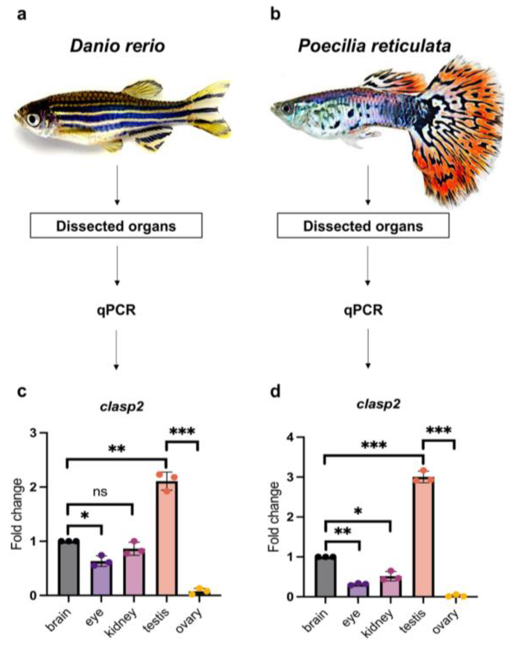
2.2. Dissection of Animals and Organs

The study was performed by using *AB zebrafish (Danio rerio) and guppy (Poecilia reticulata) adult males and females, respectively, obtained from a local supplier and recently analyzed for other purposes, as described in our previous published reports [34,41]. Fish were housed in an aquarium under standard photoperiod conditions (14 h light and 10 h dark) and a temperature of 28 °C. The animals have not received any medical treatment before or during the experiments.
All procedures have been performed by Italian Government Decree 26/2014 and approved by the Animal Ethical Committee of the University of Bologna (protocol no. 17/79/2014). Euthanasia of animals has been performed by using specific anesthetic drugs (ethyl 3-aminobenzoate and methanesulfonate 0.1%, Sigma Chemicals Co., St. Louis, MO, USA), and organs have been dissected for the experimental procedures.

2.3. RNA Extraction

Total RNA was extracted from different organs (3 adult males and 3 adult females for zebrafish), and 3 adult males and 3 adult females for guppy were sacrificed as described above. The following organs, from different specimens: brains, eyes, kidneys, testis, and ovaries, were dissociated using RLT buffer with the RNAeasy mini kit (Qiagen, Frankfurt, Germany). To obtain purified RNA, we followed the manufacturer’s protocol. This procedure was repeated in three independent experiments.

2.4. Reverse Transcriptase PCR

Reverse transcription of mRNA into cDNA was obtained by using the Superscript III First-Strand Synthesis System kit (Invitrogen, Boston, MA, USA), according to the manufacturing procedure. In detail, 0.5 μg of total RNA was incubated with buffer and enzyme mix for 10 min at 25 °C, 30 min at 50 °C, and 5 min at 85 °C. After, samples were treated with RNase-H for 30 min at 37 °C and stored at −20 °C.

2.5. Quantitative Real-Time PCR

Quantitative real-time polymerase chain reaction (PCR) was performed by using the thermocycler with the MyiQ detector (Bio-Rad, Hercules, Dallas, TX, USA). Specifically, cDNA has been mixed with specific forward and reverse primers, SYBR-Green (Bio-Rad), and RNase-free water according to the manufacturer’s protocol. The above mix was incubated for 15 min at 95 °C, then 15 s at 95 °C, 30 s at 60 °C, and 30 s at 72 °C for 40 cycles. Primer sequences used for amplification of clasp2 and gapdh (housekeeping) in zebrafish and guppy are listed in Table 1 and Table 2:
qPCR data indicate the fold change of clasp2 transcript levels in the brain, eye, kidney, testis, and ovary of zebrafish and guppy, using gapdh to normalize the absolute quantification, calculated using 2−∆∆Ct. Correct amplification and PCR efficiency were confirmed by melting curve analysis. Each qPCR experiment has been performed in biological triplicates. In the analysis of qPCR data, each n represents the average of triplicates from a single experiment. Each experiment has been repeated three times.

2.6. Synthesis of Riboprobes for clasp2 in Zebrafish and Guppy

All digoxigenin (DIG)-labeled antisense and sense riboprobes were generated using the protocol described in our previous studies [42]. In detail, clasp2 riboprobes for zebrafish and guppy were produced by using primers listed in Table 3 and Table 4.
After amplification by PCR, each insert was cloned into the TOPO-TA vector (Invitrogen). Subsequently, transformation into thermos-competent cells was performed. After applying heat shock, bacteria were plated onto Luria–Bertani (LB) agar plates containing the appropriate antibiotic to select only the transformed cells. The transformants were screened, and the white colonies containing the insert were then grown. Then, white bacterial colonies were picked and inoculated in an LB medium containing the appropriate antibiotic. Bacteria were grown for 16 h in an orbital rotator at 37 °C. Subsequently, purification of plasmid DNA was performed using the Quick Plasmid Miniprep Kit (Invitrogen, Boston, MA, USA). Next, we confirmed the antisense and sense orientation by sequencing, and the plasmids were linearized by the enzymatic restriction. Next, using T7 polymerase (Roche-Diagnostic) and SP6 polymerase (Roche-Diagnostic, Barrington, IL, USA) with DIG-RNA Labeling Mix (Roche Diagnostic, Indianapolis, IN, USA), we performed in vitro transcription. All riboprobes were purified using NucleoSpin RNA Clean-up columns (Qiagen, Frankfurt, Germany). Verification of reaction specificity was performed by hybridizing the sense and antisense riboprobes on adjacent sections.

2.7. In Situ Hybridization

Testis from zebrafish and guppy (n = 3 males per species) were quickly dissected and fixed in paraformaldehyde (PFA) at 4% overnight at 4 °C. After 24 h, tissues were processed for paraffin embedding. Sections (7 µm thick) were cut by using a rotary microtome and mounted on slides. All paraffin sections were immersed in xylene two times for 3 min and rehydrated in ethanol at 100%, 95%, 80%, 70%, 50%, and 30% (1 min each). Next, sections of zebrafish and guppy testis were immersed in PBS, adding proteinase K diluted at 2 mg/mL at 37 °C for 5 min. All slides were fixed in paraformaldehyde 4% for 20 min, washed in PBS, and then washed in standard saline sodium citrate (SSC) twice (10 min each). Then, slides were incubated at 63 °C for 24 h in a moist chamber with the probes (2 µg/mL) diluted in a specific medium (Denhart 5×; SSC 2×; 50% formamide; ethylenediamine-tetra acetic acid 4 mM; 5% dextran sulfate; yeast tRNA 50 µg/mL). After 24 h, slides were washed with SCC 2×; 50% formamide/SCC 2×; SSC 0.2×; and SSC 0.1×. After, sections were immersed in Tris-HCl/NaCl buffer (mixing 100 mM Tris-HCl pH 7.5 and 150 mM NaCl) and washed in the same buffer containing 0.5% milk powder and 0.1% Triton. For chromogenic revelation, all sections have been incubated with anti-digoxigenin alkaline phosphatase Fab fragments, dilution 1:5000 (Roche Diagnostic Company, Chicago, IL, USA), overnight at room temperature. After 24 h, all sections were washed in Tris-HCl/NaCl buffer and with 110 mM HCl-Tris (pH 8) containing 10 mM MgCl2 and 100 mM NaCl. Staining was performed using NBT/BCIP buffer (Roche, Mannheim, Germany) (pH 9.5). For fluorescence revelation, all slides were immersed in anti-DIG POD antibody (Roche, Germany) at a 1:200 dilution in the above blocking solution at room temperature for 24 h. Slides were washed for 5 min in PBS 4 times. Next, the sections were visualized by the HNPP/Fast Red Fluorescent Detection set (Roche Diagnostics, Chicago, IL, USA, #11758888001) according to the kit instructions. Mounted with DAPI (Thermo Fisher, Waltham, MA, USA) and cover slide and then observed with a fluorescence microscope (Olympus Life Science, Segrate, Milan, Italy), equipped with a DP71 digital camera, or a confocal microscope (Leica SP2). The images were processed with either the Olympus (Cell), Zeiss (AxioVision4) (Zeiss, Jana, Germany), or Leica (LCS Lite) (Leica, Wetzlar, Germania) software V-10. For all reagents and kits see Table 5.

2.8. Histology (Haematoxylin and Eosin Staining)

Adult zebrafish testes were fixed in 4% paraformaldehyde in PBS for 24h at 4 °C. After washing, the tissues were stored in 70% ethanol at 4 °C. Subsequently, tissues were embedded in paraffin, sectioned using a rotary microtome, and sections were mounted on slides. To deparaffinize, sections were put in xylene and rehydrated in an ethanol series. After, paraffin sections were washed 3× in distilled water for 1 min each time. Then, to examine cell and tissue morphology, hematoxylin and eosin (HE) staining was performed.

2.9. Statistical Analysis

Data were processed for statistical analysis using GraphPad Prism version 9.4.1 by applying one-way ANOVA with Tukey–Kramer post hoc tests, adjusted for multiple comparisons. Values of p equal to or less than 0.5 were considered statistically significant.
Catalog number for all kits and reagents used:
Table 5. Catalog number of kits and reagents.
RNAeasy minikit (Qiagen)cat # 74104
Superscript III First-Strand Synthesis kitcat # 12574026
TOPO™ TA Cloning™cat # K4575J10
Dig-RNA label mix (Roche)cat # 11277073910
HNPP/Fast Red Fluorescent Detection setcat # 11758888001
Anti-Dig antibody (Roche)cat # 11093274910
Proteinase Kcat # 25530049
Ethanolcat # 100983

3. Results

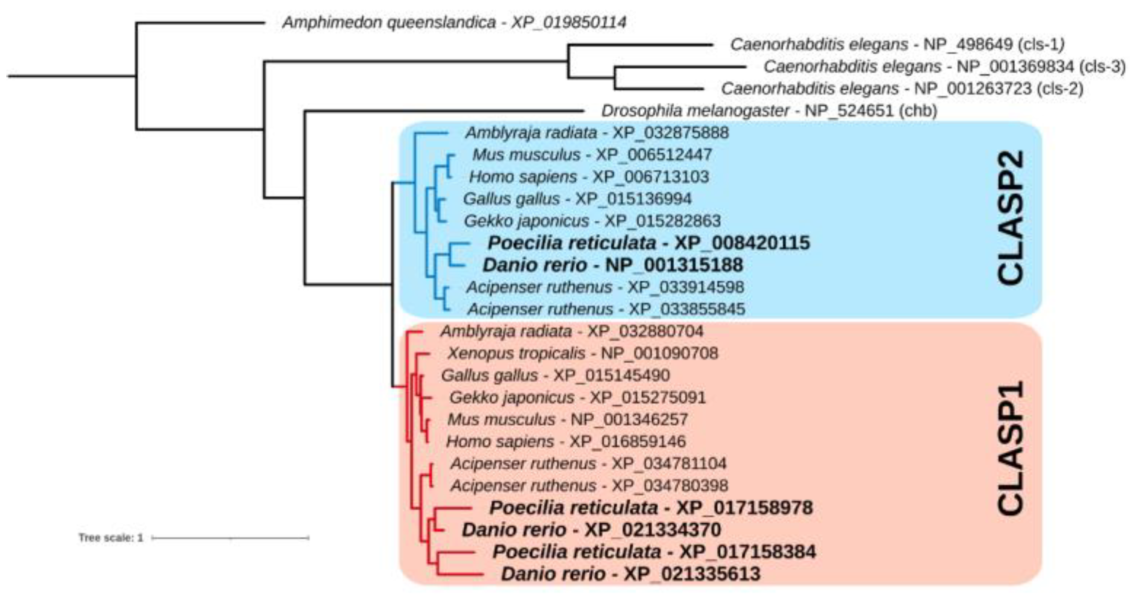
3.1. Phylogenetic Tree of the Clasp Gene Family

The Maximum Likelihood tree showed that the emergence of the CLASP1 and CLASP2 paralogues preceded the evolution of gnathostomes but followed at least the emergence of deuterostomes. A subsequent duplication of CLASP1 occurred in the teleost lineage (Figure 1).

3.2. Quantitative Analysis of clasp2 Transcription Level in Different Organs of Zebrafish and Guppy

For qPCR experiments, firstly, we dissected adult males and females of zebrafish (Danio rerio) and guppy (Poecilia reticulata) to remove different organs from each teleost fish (Figure 2a,b). Next, we measured the transcription level of clasp2 in the brain, eye, kidney, testis, and ovary.
In the ovary of zebrafish and guppy, clasp2 was undetected. In the kidney and eye of both teleost fish, clasp2 presented lower transcription compared with the brain. The most relevant data obtained from this analysis is that clasp2 is highly transcribed in the testis of adult zebrafish and guppy compared with other organs (Figure 2c,d).

3.3. Clasp2 Transcript Is Transcribed in Spermatozoa of Adult Zebrafish Testis

We first performed hematoxylin-eosin staining of adult zebrafish testis sections to better show germ line cell morphology. In zebrafish, it is well described that spermatogenesis occurs in cysts [43]. As shown in Supplementary Figure S1, we observed numerous cysts, characterized by spermatogonia displaying heterochromatin in elongated and/or round nuclei. In the figure, it is possible to identify spermatocytes (at different meiotic division stages). Instead, spermatids, and finally, spermatozoa, are identifiable by the significant decrease in cellular and nuclear volumes.
Subsequently, to identify clasp2 expressing cells in adult zebrafish testis, we performed fluorescence in situ hybridization. Clasp2 mRNA was specifically transcribed in spermatozoa (Figure 3a–f).
We confirmed previous data by using confocal microscopy with higher magnification (Figure 4a–f).

3.4. Clasp2 Transcript Is Transcribed in Spermatozeugmata of Adult Guppy Testis

As we mentioned before, the present study aimed to compare clasp2 mRNA transcription patterns between zebrafish and guppy testis. Therefore, to identify the distribution of clasp2 transcripts in guppy testis, we performed chromogenic and fluorescence in situ hybridization. By chromogenic revelation, we found that the clasp2 transcript was specifically transcribed in spermatozeugmata. In detail, the staining results are dashed and present only at the periphery of spermatozeugmata where the spermatozoa heads make contact with the Sertoli cells (Figure 5a,b).
We confirmed these previous results by using fluorescence in situ hybridization. We found that clasp2 transcripts are specifically distributed in spermatozoa contained in spermatozeugmata cysts (Figure 6a–e).

4. Discussion

Microtubule dynamics and function during spindle assembly in meiotic cell divisions are crucial for the correct development of both somatic and germ cell lines in most animal species. The CLASPs protein family is a highly important regulator of the growing microtubule plus-ends, as indicated by their enrichment at these sites via tip-binding proteins such as end-binding proteins [44] (EB). Microtubule ends are also regulated by localized multimolecular ensembles of CLASPs [16,45]. For example, membrane-associated clusters of CLASP molecules capture microtubule ends at the postsynaptic membrane, whereas cortical clusters of CLASPs tether and stabilize microtubule ends near focal adhesions in motile cells or at the cell cortex [46]. Despite several animal models, such as Drosophila, Xenopus, and C. elegans, CLASP2 activity in meiotic divisions has been well described, and very scant knowledge is present for fish models.
For the first time in the present study, we showed the transcription pattern of clasp2 during spermatogenesis in two small teleost fish, zebrafish and guppy. We found that clasp2 is transcribed in several tissues, such as the brain, eye, and kidney, in a comparable fashion between zebrafish and guppy; however, higher transcription was observed in the testis. These data agree with what has been previously observed in other animal models, such as Mus musculus or Drosophila [47,48]. Thus, this might suggest a similar potential role in male germ cell differentiation. In detail, by in situ hybridization in zebrafish, we found that clasp2 is specifically transcribed in the spermatozoa of adult testes. Interestingly, in the guppy, clasp2 is highly transcribed in spermatozeugmata. Therefore, we could hypothesize that clasp2 plays an important role in male germ cells during the late stage of spermatogenesis (i.e., spermiogenesis). Indeed, our results confirm previous studies performed using mouse models. In detail, breedings between homozygous CLASP2 knockout mice did not yield any pregnancies; hence, separate breedings between homozygous males and heterozygous or wild-type females did not produce any litter. This evidence led the authors to hypothesize that both male and female CLASP2−/− mice might be infertile. Further examination of the ovaries showed no pronounced difference in size but a difference in coloration due to hemorrhages. By contrast, testes from CLASP2 knockout mice were severely reduced in size in comparison to wild-type testes. Further histological analysis of the testes of CLASP2 knockout mice showed absent mature sperm, and the interstitium of the tubule was increased in size and filled with blood vessels [48]. In humans as well as in other animals, it is well established that spermatogenesis relies on dynamic changes in the cytoskeleton, which implies a strict regulation of microtubule dynamics [49]. Thus, microtubule dysfunction can alter the correct formation of male germ cells, and it is associated with infertility [50]. Interestingly, one recent study observed that ablation of the CLIP-170 protein (belonging to the same family of proteins as CLASP2) is associated with abnormal nucleus shapes of spermatozoa and consequent infertility [51]. However, to the best of our knowledge, no evidence has been reported about the specific role of CLASP2 in human male spermatogenesis. A possible speculation about the role of CLASP2 in male fertility could derive from preliminary data reported on The Human Protein Atlas on CLASP2 expression in Testis Cancer (https://www.proteinatlas.org/ENSG00000163539-CLASP2/pathology/testis+cancer (accessed on 12 October 2023). Indeed, it is well recognized that testicular cancer exerts profound detrimental effects on the reproductive health of men [52,53]. However, the data reported on the Protein Atlas are still preliminary, and, at present, they have not reached any prognostic value.
Thus, despite more extensive studies being needed to decipher whether and how CLASP2 is involved in fish spermatogenesis, our study represents the very first evidence of clasp2 expression in developing male germ line cells, giving insight to further investigate this protein as a target in teleost fish meiotic cell division.

Supplementary Materials

The following supporting information can be downloaded at: https://www.mdpi.com/article/10.3390/ani13233617/s1, Supplementary Figure S1: Hematoxylin-eosin staining of adult zebrafish testis. Spg: spermatogony. Spc: spermatocyte. Spt: spermatid. Spz: spermatozoa.

Author Contributions

Conceptualization, P.C.; methodology, S.R., L.M. and P.C.; software, P.C.; validation S.R., L.M., M.L., V.F. and P.C.; formal analysis, S.R.; resources, P.C.; data curation, S.R., L.M. and M.L.; writing—original draft preparation, S.R.; writing—review and editing, P.C., L.M., S.R., M.L., M.G.M. and V.F.; visualization, S.R.; project administration, P.C.; funding acquisition, P.C. All authors have read and agreed to the published version of the manuscript.

Funding

This work was supported by national public funds grant number RFO2023-Cacialli from the Italian Ministry of University and Research (MIUR).

Institutional Review Board Statement

All procedures were performed according to the Italian Decree 26/2014 and approved by the Animal Committee of the University of Bologna (protocol no. 17/79/2014).

Informed Consent Statement

Not applicable.

Data Availability Statement

All data generated in this study are available from the corresponding author on reasonable request.

Acknowledgments

The authors thank Giovanni Piccinini for his assistance in the phylogenetic analysis.

Conflicts of Interest

The authors declare no conflict of interest.

References

  1. Drabek, K.; van Ham, M.; Stepanova, T.; Draegestein, K.; van Horssen, R.; Sayas, C.L.; Akhmanova, A.; Ten Hagen, T.; Smits, R.; Fodde, R.; et al. Role of CLASP2 in microtubule stabilization and the regulation of persistent motility. Curr. Biol. 2006, 16, 2259–2264. [Google Scholar] [CrossRef]
  2. Al-Bassam, J.; Chang, F. Regulation of microtubule dynamics by TOG-domain proteins XMAP215/Dis1 and CLASP. Trends Cell Biol. 2011, 21, 604–614. [Google Scholar] [CrossRef]
  3. Lawrence, E.J.; Arpag, G.; Norris, S.R.; Zanic, M. Human CLASP2 specifically regulates microtubule catastrophe and rescue. Mol. Biol. Cell 2018, 29, 1168–1177. [Google Scholar] [CrossRef]
  4. Pereira, A.L.; Pereira, A.J.; Maia, A.R.; Drabek, K.; Sayas, C.L.; Hergert, P.J.; Lince-Faria, M.; Matos, I.; Duque, C.; Stepanova, T.; et al. Mammalian CLASP1 and CLASP2 cooperate to ensure mitotic fidelity by regulating spindle and kinetochore function. Mol. Biol. Cell 2006, 17, 4526–4542. [Google Scholar] [CrossRef]
  5. Matsui, T.; Watanabe, T.; Matsuzawa, K.; Kakeno, M.; Okumura, N.; Sugiyama, I.; Itoh, N.; Kaibuchi, K. PAR3 and aPKC regulate Golgi organization through CLASP2 phosphorylation to generate cell polarity. Mol. Biol. Cell 2015, 26, 751–761. [Google Scholar] [CrossRef]
  6. Adachi, A.; Kano, F.; Tsuboi, T.; Fujita, M.; Maeda, Y.; Murata, M. Golgi-associated GSK3beta regulates the sorting process of post-Golgi membrane trafficking. J. Cell Sci. 2010, 123, 3215–3225. [Google Scholar] [CrossRef]
  7. Dillon, G.M.; Tyler, W.A.; Omuro, K.C.; Kambouris, J.; Tyminski, C.; Henry, S.; Haydar, T.F.; Beffert, U.; Ho, A. CLASP2 Links Reelin to the Cytoskeleton during Neocortical Development. Neuron 2017, 93, 1344–1358.e1345. [Google Scholar] [CrossRef]
  8. Sayas, C.L.; Basu, S.; van der Reijden, M.; Bustos-Moran, E.; Liz, M.; Sousa, M.; Van Ijcken, W.F.; Avila, J.; Galjart, N. Distinct Functions for Mammalian CLASP1 and -2 During Neurite and Axon Elongation. Front. Cell Neurosci. 2019, 13, 5. [Google Scholar] [CrossRef]
  9. Zhu, B.; Qi, L.; Liu, S.; Liu, W.; Ou, Z.; Chen, M.; Liu, L.; Zu, X.; Wang, J.; Li, Y. CLASP2 is involved in the EMT and early progression after transurethral resection of the bladder tumor. BMC Cancer 2017, 17, 105. [Google Scholar] [CrossRef]
  10. Chen, L.; Xiong, W.; Guo, W.; Su, S.; Qi, L.; Zhu, B.; Mo, M.; Jiang, H.; Li, Y. Significance of CLASP2 expression in prognosis for muscle-invasive bladder cancer patients: A propensity score-based analysis. Urol. Oncol. 2019, 37, 800–807. [Google Scholar] [CrossRef] [PubMed]
  11. Valter, A.; Luhari, L.; Pisarev, H.; Truumees, B.; Planken, A.; Smolander, O.P.; Oselin, K. Genomic alterations as independent prognostic factors to predict the type of lung cancer recurrence. Gene 2023, 885, 147690. [Google Scholar] [CrossRef]
  12. Beffert, U.; Dillon, G.M.; Sullivan, J.M.; Stuart, C.E.; Gilbert, J.P.; Kambouris, J.A.; Ho, A. Microtubule plus-end tracking protein CLASP2 regulates neuronal polarity and synaptic function. J. Neurosci. 2012, 32, 13906–13916. [Google Scholar] [CrossRef]
  13. Siino, V.; Amato, A.; Di Salvo, F.; Caldara, G.F.; Filogamo, M.; James, P.; Vasto, S. Impact of diet-induced obesity on the mouse brain phosphoproteome. J. Nutr. Biochem. 2018, 58, 102–109. [Google Scholar] [CrossRef]
  14. Langlais, P.; Dillon, J.L.; Mengos, A.; Baluch, D.P.; Ardebili, R.; Miranda, D.N.; Xie, X.; Heckmann, B.L.; Liu, J.; Mandarino, L.J. Identification of a role for CLASP2 in insulin action. J. Biol. Chem. 2012, 287, 39245–39253. [Google Scholar] [CrossRef]
  15. Kruse, R.; Krantz, J.; Barker, N.; Coletta, R.L.; Rafikov, R.; Luo, M.; Hojlund, K.; Mandarino, L.J.; Langlais, P.R. Characterization of the CLASP2 Protein Interaction Network Identifies SOGA1 as a Microtubule-Associated Protein. Mol. Cell Proteomics 2017, 16, 1718–1735. [Google Scholar] [CrossRef] [PubMed]
  16. Karki, P.; Ke, Y.; Zhang, C.O.; Li, Y.; Tian, Y.; Son, S.; Yoshimura, A.; Kaibuchi, K.; Birukov, K.G.; Birukova, A.A. SOCS3-microtubule interaction via CLIP-170 and CLASP2 is critical for modulation of endothelial inflammation and lung injury. J. Biol. Chem. 2021, 296, 100239. [Google Scholar] [CrossRef]
  17. Karki, P.; Ke, Y.; Tian, Y.; Ohmura, T.; Sitikov, A.; Sarich, N.; Montgomery, C.P.; Birukova, A.A. Staphylococcus aureus-induced endothelial permeability and inflammation are mediated by microtubule destabilization. J. Biol. Chem. 2019, 294, 3369–3384. [Google Scholar] [CrossRef]
  18. Maton, G.; Edwards, F.; Lacroix, B.; Stefanutti, M.; Laband, K.; Lieury, T.; Kim, T.; Espeut, J.; Canman, J.C.; Dumont, J. Kinetochore components are required for central spindle assembly. Nat. Cell Biol. 2015, 17, 953. [Google Scholar] [CrossRef] [PubMed]
  19. Nahaboo, W.; Zouak, M.; Askjaer, P.; Delattre, M. Chromatids segregate without centrosomes during Caenorhabditis elegans mitosis in a Ran- and CLASP-dependent manner. Mol. Biol. Cell 2015, 26, 2020–2029. [Google Scholar] [CrossRef]
  20. Park, E.C.; Lee, H.; Hong, Y.; Kim, M.J.; Lee, Z.W.; Kim, S.I.; Kim, S.; Kim, G.H.; Han, J.K. Analysis of the expression of microtubule plus-end tracking proteins (+TIPs) during Xenopus laevis embryogenesis. Gene Expr. Patterns 2012, 12, 204–212. [Google Scholar] [CrossRef]
  21. Grimaldi, A.D.; Zanic, M.; Kaverina, I. Encoding the microtubule structure: Allosteric interactions between the microtubule +TIP complex master regulators and TOG-domain proteins. Cell Cycle 2015, 14, 1375–1378. [Google Scholar] [CrossRef]
  22. Akhmanova, A.; Hoogenraad, C.C.; Drabek, K.; Stepanova, T.; Dortland, B.; Verkerk, T.; Vermeulen, W.; Burgering, B.M.; De Zeeuw, C.I.; Grosveld, F.; et al. Clasps are CLIP-115 and -170 associating proteins involved in the regional regulation of microtubule dynamics in motile fibroblasts. Cell 2001, 104, 923–935. [Google Scholar] [CrossRef]
  23. Aonuma, M.; Miyamoto, M.; Inoue, Y.H.; Tamai, K.; Sakai, H.; Kamasawa, N.; Matsukage, A. Microtubule bundle formation and cell death induced by the human CLASP/Orbit N-terminal fragment. Cell Struct. Funct. 2005, 30, 7–13. [Google Scholar] [CrossRef]
  24. Moriwaki, T.; Goshima, G. Five factors can reconstitute all three phases of microtubule polymerization dynamics. J. Cell Biol. 2016, 215, 357–368. [Google Scholar] [CrossRef]
  25. Mathe, E.; Inoue, Y.H.; Palframan, W.; Brown, G.; Glover, D.M. Orbit/Mast, the CLASP orthologue of Drosophila, is required for asymmetric stem cell and cystocyte divisions and development of the polarised microtubule network that interconnects oocyte and nurse cells during oogenesis. Development 2003, 130, 901–915. [Google Scholar] [CrossRef]
  26. Miyauchi, C.; Kitazawa, D.; Ando, I.; Hayashi, D.; Inoue, Y.H. Orbit/CLASP is required for germline cyst formation through its developmental control of fusomes and ring canals in Drosophila males. PLoS ONE 2013, 8, e58220. [Google Scholar] [CrossRef]
  27. Inoue, Y.H.; Savoian, M.S.; Suzuki, T.; Mathe, E.; Yamamoto, M.T.; Glover, D.M. Mutations in orbit/mast reveal that the central spindle is comprised of two microtubule populations, those that initiate cleavage and those that propagate furrow ingression. J. Cell Biol. 2004, 166, 49–60. [Google Scholar] [CrossRef]
  28. Kitazawa, D.; Matsuo, T.; Kaizuka, K.; Miyauchi, C.; Hayashi, D.; Inoue, Y.H. Orbit/CLASP is required for myosin accumulation at the cleavage furrow in Drosophila male meiosis. PLoS ONE 2014, 9, e93669. [Google Scholar] [CrossRef]
  29. Shoda, T.; Yamazoe, K.; Tanaka, Y.; Asano, Y.; Inoue, Y.H. Orbit/CLASP determines centriole length by antagonising Klp10A in Drosophila spermatocytes. J. Cell Sci. 2021, 134, jcs251231. [Google Scholar] [CrossRef]
  30. Marchal, G.A.; Jouni, M.; Chiang, D.Y.; Perez-Hernandez, M.; Podliesna, S.; Yu, N.; Casini, S.; Potet, F.; Veerman, C.C.; Klerk, M.; et al. Targeting the Microtubule EB1-CLASP2 Complex Modulates Na(V)1.5 at Intercalated Discs. Circ. Res. 2021, 129, 349–365. [Google Scholar] [CrossRef] [PubMed]
  31. Shen, W.Y.; Fu, X.H.; Cai, J.; Li, W.C.; Fan, B.Y.; Pang, Y.L.; Zhao, C.X.; Abula, M.; Kong, X.H.; Yao, X.; et al. Identification of key genes involved in recovery from spinal cord injury in adult zebrafish. Neural Regen. Res. 2022, 17, 1334–1342. [Google Scholar] [CrossRef]
  32. Klaus, A.; Clapes, T.; Yvernogeau, L.; Basu, S.; Weijts, B.; Maas, J.; Smal, I.; Galjart, N.; Robin, C. CLASP2 safeguards hematopoietic stem cell properties during mouse and fish development. Cell Rep. 2022, 39, 110957. [Google Scholar] [CrossRef]
  33. Leal, M.C.; Cardoso, E.R.; Nobrega, R.H.; Batlouni, S.R.; Bogerd, J.; Franca, L.R.; Schulz, R.W. Histological and stereological evaluation of zebrafish (Danio rerio) spermatogenesis with an emphasis on spermatogonial generations. Biol. Reprod. 2009, 81, 177–187. [Google Scholar] [CrossRef]
  34. Cacialli, P. Expression of Nerve Growth Factor and Its Receptor TrkA in the Reproductive System of Adult Zebrafish. Vet. Sci. 2022, 9, 225. [Google Scholar] [CrossRef]
  35. Cacialli, P.; D’Angelo, L.; de Girolamo, P.; Avallone, L.; Lucini, C.; Pellegrini, E.; Castaldo, L. Morpho-Functional Features of the Gonads of Danio rerio: The Role of Brain-Derived Neurotrophic Factor. Anat. Rec. 2018, 301, 140–147. [Google Scholar] [CrossRef]
  36. Torres-Martinez, A.; Ruiz de Dios, L.; Hernandez-Franyutti, A.; Uribe, M.C.; Sanchez, W.C. Structure of the testis and spermatogenesis of the viviparous teleost Poecilia mexicana (Poeciliidae) from an active sulfur spring cave in Southern Mexico. J. Morphol. 2019, 280, 1537–1547. [Google Scholar] [CrossRef] [PubMed]
  37. Piccinini, G.; Milani, L. Germline-related molecular phenotype in Metazoa: Conservation and innovation highlighted by comparative transcriptomics. Evodevo 2023, 14, 2. [Google Scholar] [CrossRef]
  38. Katoh, K.; Standley, D.M. MAFFT multiple sequence alignment software version 7: Improvements in performance and usability. Mol. Biol. Evol. 2013, 30, 772–780. [Google Scholar] [CrossRef]
  39. Criscuolo, A.; Gribaldo, S. BMGE (Block Mapping and Gathering with Entropy): A new software for selection of phylogenetic informative regions from multiple sequence alignments. BMC Evol. Biol. 2010, 10, 210. [Google Scholar] [CrossRef]
  40. Minh, B.Q.; Schmidt, H.A.; Chernomor, O.; Schrempf, D.; Woodhams, M.D.; von Haeseler, A.; Lanfear, R. IQ-TREE 2: New Models and Efficient Methods for Phylogenetic Inference in the Genomic Era. Mol. Biol. Evol. 2020, 37, 1530–1534. [Google Scholar] [CrossRef]
  41. Milani, L.; Cinelli, F.; Iannello, M.; Lazzari, M.; Franceschini, V.; Maurizii, M.G. Immunolocalization of Vasa, PIWI, and TDRKH proteins in male germ cells during spermatogenesis of the teleost fish Poecilia reticulata. Acta Histochem. 2022, 124, 151870. [Google Scholar] [CrossRef]
  42. Mahony, C.B.; Cacialli, P.; Pasche, C.; Monteiro, R.; Savvides, S.N.; Bertrand, J.Y. Hapln1b, a central organizer of the ECM, modulates kit signaling to control developmental hematopoiesis in zebrafish. Blood Adv. 2021, 5, 4935–4948. [Google Scholar] [CrossRef]
  43. Menke, A.L.; Spitsbergen, J.M.; Wolterbeek, A.P.; Woutersen, R.A. Normal anatomy and histology of the adult zebrafish. Toxicol. Pathol. 2011, 39, 759–775. [Google Scholar] [CrossRef]
  44. Girao, H.; Okada, N.; Rodrigues, T.A.; Silva, A.O.; Figueiredo, A.C.; Garcia, Z.; Moutinho-Santos, T.; Hayashi, I.; Azevedo, J.E.; Macedo-Ribeiro, S.; et al. CLASP2 binding to curved microtubule tips promotes flux and stabilizes kinetochore attachments. J. Cell Biol. 2020, 219, e201905080. [Google Scholar] [CrossRef]
  45. Rodgers, N.C.; Lawrence, E.J.; Sawant, A.V.; Efimova, N.; Gonzalez-Vasquez, G.; Hickman, T.T.; Kaverina, I.; Zanic, M. CLASP2 facilitates dynamic actin filament organization along the microtubule lattice. Mol. Biol. Cell 2023, 34, br3. [Google Scholar] [CrossRef]
  46. Mimori-Kiyosue, Y.; Grigoriev, I.; Lansbergen, G.; Sasaki, H.; Matsui, C.; Severin, F.; Galjart, N.; Grosveld, F.; Vorobjev, I.; Tsukita, S.; et al. CLASP1 and CLASP2 bind to EB1 and regulate microtubule plus-end dynamics at the cell cortex. J. Cell Biol. 2005, 168, 141–153. [Google Scholar] [CrossRef]
  47. Sousa, A.; Reis, R.; Sampaio, P.; Sunkel, C.E. The Drosophila CLASP homologue, Mast/Orbit regulates the dynamic behaviour of interphase microtubules by promoting the pause state. Cell Motil. Cytoskeleton 2007, 64, 605–620. [Google Scholar] [CrossRef]
  48. Drabek, K.; Gutierrez, L.; Vermeij, M.; Clapes, T.; Patel, S.R.; Boisset, J.C.; van Haren, J.; Pereira, A.L.; Liu, Z.; Akinci, U.; et al. The microtubule plus-end tracking protein CLASP2 is required for hematopoiesis and hematopoietic stem cell maintenance. Cell Rep. 2012, 2, 781–788. [Google Scholar] [CrossRef]
  49. O’Donnell, L.; O’Bryan, M.K. Microtubules and spermatogenesis. Semin. Cell Dev. Biol. 2014, 30, 45–54. [Google Scholar] [CrossRef]
  50. Gunes, S.; Sengupta, P.; Henkel, R.; Alguraigari, A.; Sinigaglia, M.M.; Kayal, M.; Joumah, A.; Agarwal, A. Microtubular Dysfunction and Male Infertility. World J. Mens. Health 2020, 38, 9–23. [Google Scholar] [CrossRef]
  51. Lehti, M.S.; Sironen, A. Formation and function of the manchette and flagellum during spermatogenesis. Reproduction 2016, 151, R43–R54. [Google Scholar] [CrossRef]
  52. Paduch, D.A. Testicular cancer and male infertility. Curr. Opin. Urol. 2006, 16, 419–427. [Google Scholar] [CrossRef]
  53. Ostrowski, K.A.; Walsh, T.J. Infertility with Testicular Cancer. Urol. Clin. N. Am. 2015, 42, 409–420. [Google Scholar] [CrossRef]
Figure 1. Phylogenetic tree of the clasp gene family.
Animals 13 03617 g001
Figure 2. (a) Schematic experimental design outline for organs dissected from adult zebrafish (Danio rerio). (b) Schematic experimental design outline for organs dissected from an adult guppy (Poecilia reticulata). (c) qPCR analysis of clasp2 in the brain, eye, kidney, testis, and ovary of adult zebrafish. Clasp2 is most transcribed in the testis. Statistical analysis was completed using a one-way ANOVA with Tukey–Kramer post hoc tests, adjusted for multiple comparisons (n = 3 animals used). * p < 0.01; ** p < 0.001; *** p < 0.0001. ns—not significant. (d) qPCR analysis of clasp2 in the brain, eye, kidney, testis, and ovary of an adult guppy. Clasp2 is most transcribed in the testis. Statistical analysis was completed using a one-way ANOVA with Tukey–Kramer post hoc tests, adjusted for multiple comparisons (n = 3 animals used). * p < 0.01; ** p < 0.001; *** p < 0.0001.
Animals 13 03617 g002
Figure 3. Clasp2 mRNA transcription pattern in adult zebrafish testis at widefield fluorescence microscopy. (a) DAPI staining marks cell nuclei. (b) Fluorescence in situ hybridization of clasp2 in adult zebrafish testis. (c) Fluorescence in situ hybridization of clasp2 and cell nuclei (DAPI). (d) High magnification of spermatozoa cell nuclei (DAPI). (e) High magnification of clasp2 expressing cells. (f) High magnification of fluorescence in situ hybridization of clasp2 and cell nuclei (DAPI). Abbreviations: Spz: spermatozoa; Spc: spermatocytes; Spg: spermatogonia (inside the dashed line) in adult zebrafish testis. Scale bars: 100 µ (ac) and 25 µ (df).
Animals 13 03617 g003
Figure 4. Clasp2 transcription pattern in adult zebrafish testis at confocal microscopy. (a) DAPI staining marks cell nuclei. (b) Fluorescence in situ hybridization of clasp2 in adult zebrafish testis. (c) Fluorescence in situ hybridization of clasp2 and cell nuclei (DAPI) in adult zebrafish testis. (d) DAPI staining marks cell nuclei at high magnification. (e) Fluorescence in situ hybridization of clasp2 in adult zebrafish testis at high magnification. (f) Fluorescence in situ hybridization of clasp2 and cell nuclei (DAPI) in adult zebrafish testis at high magnification. Abbreviations: Spz; spermatozoa. Spt; spermatids. Scale bars: 100 µ (ac) and 25 µ (df).
Animals 13 03617 g004
Figure 5. Clasp2 transcript distribution in sections of adult guppy testis. (a) In this section, we identified the organization of the testis. Cysts containing germ cells in the early stages of spermatogenesis (spermatogonia and spermatocytes) are located near the periphery of the testis, while cysts with numerous spermatids at different stages of spermiogenesis are located deeper, near the efferent ducts. In the same region, there are present spermatozeugmata cysts, in which spermatozoa are tightly packed with the sperm heads oriented towards the Sertoli cells and flagella oriented towards the center of the cyst. Spermatozeugmata is also found inside the efferent ducts (located in the central region of the testis), where mature spermatozoa are released. Chromogenic in situ hybridization of clasp2 in this section shows a clear marking at the periphery of the spermatozeugmata. The peripheral region of spermatozeugmata cysts located inside the efferent ducts shows weak staining (Sg: spermatogonia; Sc: spermatocytes; St: spermatids; Sz: spermatozeugmata; Ed: efferent ducts). (b) The strong marking of spermatozeugmata. (Sz: spermatozeugmata). Scale bars: 100 µ (a); 50 µ (b).
Animals 13 03617 g005
Figure 6. Clasp2 mRNA distribution in adult guppy testis, confocal microscopy. (a) Fluorescence in situ hybridization of clasp2 shows the labeling concentrated at the periphery of numerous spermatozeugmata. (b) High magnification of a portion of spermatozeugmata in a white rectangle is shown in panel a. (c) Fluorescence in situ hybridization of clasp2 is seen at the periphery of spermatozeugmata cysts. (d) DAPI staining marks all the cell nuclei of the testis. (e) The merge of fluorescence in situ hybridization of clasp2 and DAPI in adult guppy testis. Abbreviation: Sz; spermatozeugmata. Scale bars are 50 µ (a); 10 µ (b); and 30 µ (ce).
Animals 13 03617 g006
Table 1. qPCR forward and reverse primer sequences of clasp2 and gapdh in zebrafish.
Zebrafish(Amplicon Size)
clasp2F: TGGAGGCACATAAAGACC(122 bp)
R: TGACTGGATGATGGGACA
GapdhF: GTGTAGGCGTGGACTGTGG(151 bp)
R: TGGGAGTCAACCAGGACAAA
Table 2. qPCR forward and reverse primer sequences of clasp2 and gapdh in guppy.
Guppy(Amplicon Size)
clasp2F: GAAGGACGTTACACGTAGAC(181 bp)
R: CCCACAGATGTCTATCCCT
GapdhF: CTCCACTCATGGTGTCTG(140 bp)
R: CAACATAGTCTACGGCAGC
Table 3. Forward and reverse primer sequences for clasp2 riboprobes in zebrafish.
ZebrafishLength of Riboprobe
clasp2F: GCATTGCTGGGGATCGATA (532 bp)
R: CGTCGAAACTACGGTCTTG
Table 4. Forward and reverse primer sequences for clasp2 riboprobes in guppy.
GuppyLength of Riboprobe
clasp2F: CTCAGCTCAGGCTGCTTT(494 bp)
R: CGCAGTTGGGAATGAGGT
Disclaimer/Publisher’s Note: The statements, opinions and data contained in all publications are solely those of the individual author(s) and contributor(s) and not of MDPI and/or the editor(s). MDPI and/or the editor(s) disclaim responsibility for any injury to people or property resulting from any ideas, methods, instructions or products referred to in the content.

