Article

Effect of Sempervivum tectorum Extract on Some Biomarkers of Reproductive Function and Levels of Some Trace Elements in Male Rats Exposed to Aluminum

1
Faculty of Veterinary Medicine, University of Life Sciences “King Michael I”, 300645 Timisoara, Romania
2
Working Group for Xenobiochemistry, Romanian Academy-Branch, 300645 Timisoara, Romania
3
Faculty of Medicine Bucharest, Titu Maiorescu University, 300645 Timisoara, Romania
4
Faculty of Medicine Timisoara, University of Medicine and Pharmacy ”Victor Babeș”, 300645 Timisoara, Romania
*
Authors to whom correspondence should be addressed.
Animals 2024, 14(8), 1196; https://doi.org/10.3390/ani14081196
Submission received: 1 April 2024 / Revised: 9 April 2024 / Accepted: 16 April 2024 / Published: 16 April 2024
(This article belongs to the Special Issue Role of Trace Element in Animal Health and Metabolic: Second Edition)

Abstract

:

Simple Summary

Although aluminum was extensively studied in the last decades, questions still arise and present increasing interest regarding the reproductive toxicity, with the research still having lacunas, as was observed in a up-to-date and very ample review. In the present study, we show that aluminum is accumulating in the genital organs and also in sexual accessory glands, impairing the histoarchitecture of the reproductive system, except for bulbo-urethral glands and seminal vesicles, and it is negatively correlated with the hormone levels and the trace minerals from reproductive organs and accessory glands. At the same time, we observed that the use of Sempervivum tectorum aqueous extract can protect against deleterious effects of aluminum upon the main studied reproductive biomarkers. Some of the results are, for the first time, presented since we did not found research related to this, such as the aluminum content in the bulbo-urethral glands and seminal vesicles, the correlation between the trace minerals from the genital organs and sexual accessory glands and the hormone levels, and the correlation between aluminum level and the trace element levels in studied organs.

Abstract

Aluminum, a contentious trace element found in the environment, has been demonstrated to have harmful effects on both humans and animals. In contrast, Sempervivum tectorum, an evergreen plant, has been found to offer numerous beneficial effects for both humans and animals. Therefore, this study aims to assess the protective effect of S. tectorum on certain reproductive biomarkers in male rats exposed to aluminum. Thirty-five Wistar rats were randomly divided into five groups: NTC (no-treatment control) received distilled water; NC (negative control) received drinking water containing 1 mg/L aluminum sulfate (AS); E1 received 1 mg/L AS along with an 8% S. tectorum extract; PC (positive control) received only 8% S. tectorum extract; E2 received 1 mg/L AS for three months followed by 8% extract for one month. The study analyzed testosterone, LH, FSH, body weight, and the histological structure of the testis, epididymis, and prostate, as well as the levels of zinc, manganese, copper, and iron in these organs. Significant decreases in body weight, testis, and epididymis size were observed in the aluminum-exposed groups compared to the control, whereas these decreases were not significant in the S. tectorum-treated groups compared to the control. Aluminum exposure led to significant decreases in testosterone and LH levels, with FSH levels showing a nonsignificant decrease in males, which were mitigated significantly by the administration of the plant extract. Histological analysis revealed alterations in the testis, epididymis, and prostate of the AS-exposed groups, including necrosis of seminiferous tubule epithelium and Leydig cells in the testis, and basal epithelial necrosis in the epididymis and prostate. Aluminum levels increased in all organs studied, while levels of zinc, copper, iron, and manganese decreased, showing a negative and significant correlation with aluminum levels. The aqueous extract of S. tectorum demonstrated a protective effect on certain studied biomarkers in male rats affected by aluminum exposure.

1. Introduction

Aluminum (Al) is a ubiquitous and abundant metal in the environment, placed as the third most common constituent of the Earth’s crust [1,2,3]. While initial studies on aluminum suggested potential biological significance, over the last two decades, numerous studies have contradicted this notion. Recent research has consistently demonstrated the lack of bio-utility of this metal, emphasizing its association with adverse effects on various target organs, including the kidneys [4], brain [5,6], and the male and female reproductive systems [3,7,8,9,10,11,12,13].
Excessive amounts of aluminum can result in its accumulation in specific organs, potentially causing damage to testicular tissues in both humans and animals. Elevated levels of aluminum in testes, Leydig cells, spermatozoa, seminal plasma, and blood have been linked to compromised sperm quality and viability [14].
The detrimental effects of aluminum are varied and can lead to a complex systemic toxicity. Its toxic actions target various molecular pathways within cells, disrupting cellular balance and resulting in damage that contributes to systemic toxicity, manifesting as structural and functional abnormalities in organs [15]. Primarily, aluminum’s toxic effects stem from its pro-oxidant properties, inducing oxidative stress, free radical damage, and the oxidation of cellular proteins and lipids [16].
Despite extensive investigations into aluminum, questions persist, particularly in the realm of reproductive toxicity. Existing research still exhibits gaps, as highlighted in a comprehensive and up-to-date review by Yokel [2].
Sempervivum tectorum L., an evergreen plant, known as the common houseleek, is a perennial herb with a dense basal rosette of fleshy sharp-tipped, obovate to oblong leaves having red-colored flowers arranged in terminal cymes [17,18,19]. The houseleek is a drought-resistant plant, native to central and southern Europe, growing on roofs, scree, and weathered rocks, traditionally used fresh rather than dried [18,20]. S. tectorum contains phytochemical compounds such as flavonoids, phenolic acids, and tannins, which possess antioxidant and metal-chelating properties [20,21,22]. These compounds have the potential to scavenge reactive oxygen species (ROS) generated by aluminum toxicity and inhibit the harmful effects of aluminum on the reproductive system. Aluminum-induced oxidative stress is considered a major cause of male reproductive disorders [7,8,9,10,11]. S. tectorum has been reported to exhibit significant antioxidant activity due to its phytochemical constituents [21]; the antioxidative capacity of S. tectorum can help mitigate the oxidative damage caused by aluminum exposure and protect against reproductive disorders [17].
Nowadays, the plant is linked to numerous effects, such as antinociceptive, liver protective, and membrane-stabilizing effects, which are believed to be due to the antioxidant activity of its highly phenolic content [21,23].
The aim of the present study was to underline the effect of the aqueous extract of S. tectorum on some morphological and biochemical markers of male reproductive function, as well as the correlation between aluminum level and the levels of sexual hormones and some trace minerals with importance for reproductive function.

2. Materials and Methods

2.1. Plant Extract

The S. tectorum freshly gathered leaves were separated from the rosette and roots, washed, and then dried in an oven at 50 °C until reaching a consistent weight. Extraction was carried out, in Erlenmeyer flasks, from the leaves with a particle size of 0.4 mm, using tap water (at 0.8/10 w/v ratio). The mixture was heated to 90 °C for 10 min in a water bath, filtered twice, and subsequently stored at 4 °C for a maximum duration of three days [17]. The chemical and mineral composition is presented as Appendix A.

2.2. Animals and the Experimental Protocol

Thirty-five six-month-old Wistar rats weighing 350 ± 15 g were utilized for the study, sourced from the authorized animal facility at the University of Medicine and Pharmacy “Victor Babeş” Timisoara. The rats were housed in five groups (n = 7) in standard polycarbonate cages (dimensions: length × width × height = 750 × 720 × 360 mm), maintaining a 12 h light/dark cycle and a controlled temperature of 23 ± 2 °C. They were fed a Biovetimix standard diet (Bioveti code 140-501, Romania, details in Appendix B) and acclimatized to the experimental conditions for one week before the start of the experiment. The handling of animals adhered to directive 2010/63/EU [24] and the guidelines of the National Research Council (NRC) [25].
The experimental period, lasting three months for all groups except E2, which spanned four months, received approval from the Ethical Committee of the University of Life Sciences “King Michael I” in Timisoara (No. 120/2018). Following the acclimatization period, the rats were randomly assigned to five groups:
  • Group I (NTC—no-treatment control, received distilled water).
  • Group II (NC—negative control) received 1 mg/L aluminum as aluminum sulfate (AS) in drinking water (1 mg/L being the level that demonstrated marked effects on reproductive function as were observed in a previous study) [3].
  • Group III (E1 received 1 mg/L AS + 8% S. tectorum extract as drinking water).
  • Group IV (PC—positive control received 8% S. tectorum as drinking water).
  • Group V (E2 received 1 mg/L AS for three months, and thereafter, 8% S. tectorum for one month).

2.3. Samples Collection and Analysis

Daily food and water/S. tectorum intake in controls and experimental groups were monitored and are presented in Appendix C.
At the end of the exposure period, the animals were euthanized by overdosing a combination of anesthetics, specifically 300 mg/kg.BW ketamine (ketamine 10%, CP Pharma, Burgdorf, Germany) and 30 mg/kg.BW xylazine (Narcoxyl, Intervet International, The Netherlands). Blood and organ samples were then collected for subsequent analyses.
The blood was collected in a BD vacutainer (Serum Plain BD ref no. 367837), without anticoagulant, in order to obtain serum samples. The serum testosterone (T), luteinizing hormone (LH), and follicle stimulating hormone (FSH) levels were determined by chemiluminescence using the Randox Evidence Evolution Biochip Array (Randox Laboratories, UK); the analyses were performed by Tody Laboratories, Bucharest, Romania (Certified laboratory).
For histological examination, the genital organs and sexual accessory glands were washed in saline buffer, immersed and fixed in Bouin’s solution, and embedded in paraffin, then were sliced at 5 µm and stained by hematoxylin and eosin method. The histoarchitecture changes were studied using an Olympus CX 41 microscope (Olympus, Leicestershire, UK) with image capture software and data interpretation, at 200× magnification.
In order to analyze trace elements, the organs samples were prepared by microwave digestion (Multiwave GO, Anton Paar, GmbH, Graz, Austria) using nitric acid 10 mL and hydrogen peroxide 2 mL, which were added to a 0.5 g sample and submitted to the digester with the following parameters: 120 °C, 800 W for 20 min. The samples were analyzed by atomic absorption spectroscopy (AAS) using a Perkin Elmer Analyst 800 (Perkin Elmer Inc., Waltham, MA, USA), and the operating conditions were in accordance with the manufacturers’ recommendations for each element. The atomization was carried out in an air-acetylene flame (ratio 13.5:2) for Cu, Zn, Fe, and Mn, and graphite furnace for Al, using hollow cathode lamps as radiation source. The calibration standards were prepared from a Merck CertiPur ICP 1000 mg/L stock standard solution (Merk, Darmstadt, Germany).

2.4. Statistical Analysis

The results are presented as mean ± SEM (standard error of the mean) and were subjected to statistical analysis using one-way ANOVA with Bonferroni correction and Tukey’s post hoc test for group comparisons. Additionally, the Pearson correlation test was employed to assess the correlation between aluminum, hormone levels, and trace element concentrations. The statistical analyses were performed using Graph Pad Prism 9.1 for Windows (Graph Pad Software, version 9.1.2, San Diego, CA, USA), with significance set at p < 0.05 for all values.

3. Results

3.1. Body Weight and the Weight of Genital Organs and Sexual Accessory Glands

The total body weight (Table 1) was significantly (p < 0.01) decreased in rats exposed to aluminum compared to the control group (NC/NTC: +12.58%). The administration of S. tectorum extract led to a significant (p < 0.01) increase in body weight compared to the group that received only aluminum (E1/NC: +11.93%), reaching almost the weight of the control group (E2/C: −2.15%, p > 0.05). When the extract was administered for one month after aluminum exposure, the body weight was significantly higher (p < 0.05) comparative to rats exposed to aluminum (E2/E1: +9.05%), but still remained lower than in control (E2/C: −4.67%).
Testes (TS), epididymis (EP), bulbo-urethral glands (BU), and seminal vesicles (SV) weights were significantly lower in groups that received aluminum compared to control (NC/NTC: TS: −18.62%, EP: −53.16%, BU: −29.03%, SV: −16.03%), except for prostate (PR) weight, which was found to be significantly (p < 0.05) higher in the aluminum-exposed group compared to control (NC/NTC: +16.67%). In both sexual organs and sexual accessory glands (except prostate), in the group that received S. tectorum extract, the weight was higher than in the group exposed to aluminum, but not statistically significant (p > 0.05), even if an increase was observed (E1/NC: TS +9.95%, EP +21.62%; PR −23.46%; BU +27.27%; SV +10.01%), and although the weight was increased, it remained lower than in the control group (E1/NTC: TS −10.52%, EP −43.03%, p < 0.05; PR −10.71%; BU −9.67%; SV −7.63%).
There were not significant (p > 0.05) differences in the group that received S. tectorum extract compared to control, but there were some significant differences when the extract was administered for one month after the aluminum administration compared to the group that received the extract and aluminum at the same time (E2/NC: TS +16.91%, EP +72,97%, PR −26.53%, BU +31.81%, SV +17.27%).

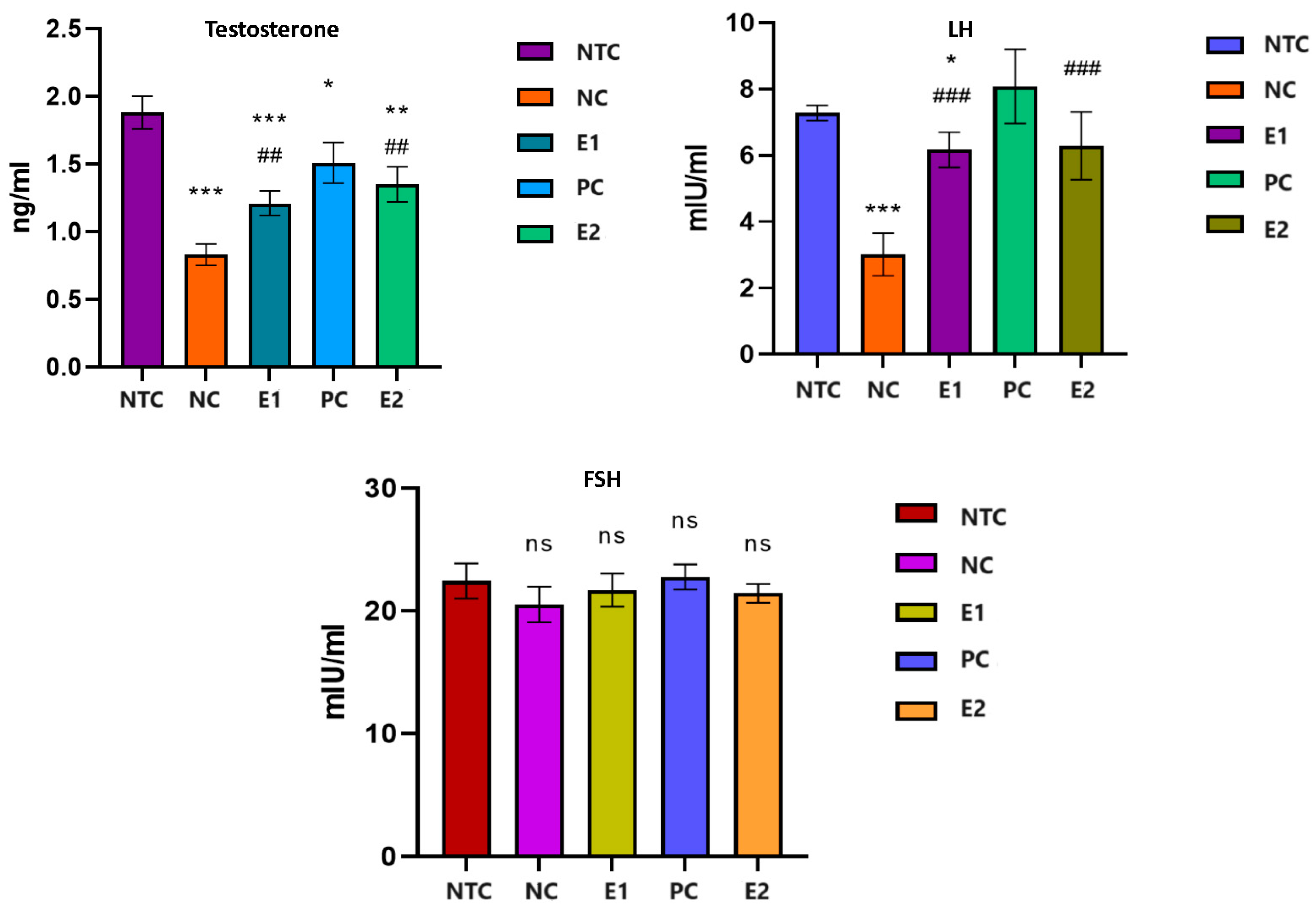
3.2. Biochemical Reproductive Biomarkers

Following the statistical analysis of the results (Figure 1), we found that the serum testosterone level decreased strongly significantly (p < 0.001) in the rats from the groups that received aluminum compared to that of the individuals in the control group (NC/NTC: −56.11%). The administration of aqueous extract of S. tectorum led to a significant increase (p < 0.05) in the serum level of testosterone compared to individuals in the NC group (E1/NC: +45.78%). In individuals who received aqueous extract of S. tectorum one month after aluminum exposure, testosterone levels increased significantly (p < 0.01) compared to individuals in the aluminum-exposed group (E2/NC: +62.65%), but did not reach the level of the control group; they remained at a significantly (p < 0.05) lower level in comparison (E2/NTC: −28.19%).
Serum LH decreased significantly (p < 0.001) in individuals exposed to aluminum compared to those in the control group (NC/NTC: −58.76%). The application of the aqueous extract of S. tectorum had a notable impact (p < 0.05) on the LH level compared to the group exposed solely to aluminum, resulting in a significant increase (E1/NC: +104.98%). This increase brought the level close to that of the control group, albeit it was still significantly lower than the level recorded in the control group (E1/NTC: −15.24%). Administration for one month of S. tectorum extract after exposure to aluminum led to an increase in LH, which was significantly higher than in individuals exposed to aluminum alone (E2/NC: +108.97%), but did not reach the level of the control group (E2/NTC: −13.59%).
Regarding the level of serum FSH, we found not-significant fluctuations (p > 0.05) in all groups.

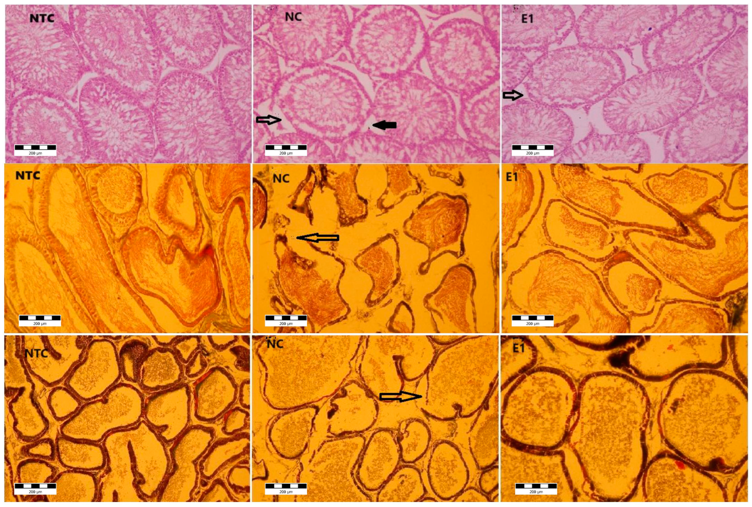
3.3. Histological Structure

Microscopic examination of testis (Figure 2) in the control group showed the apparently normal architecture, with the compact seminiferous tubules having a regular circumference.
In the aluminum (NC) and aluminum plus S. tectorum extract (E1) groups, several microscopic changes in different degrees were noticed. The seminiferous tubules exhibited an altered appearance characterized by a thickened basement membrane, which, in certain instances, was disrupted. Additionally, there was a moderate to complete degeneration (necrosis) of the spermatogenic cells within some seminiferous tubules. If the seminiferous tubules are the main producers of sperm cells, the epididymis (Figure 2) is the androgen-dependent segment of male genital tract for sperm cells maturation and their storage until ejaculation. Under microscopic examination, the epididymis in the control group displayed a pseudo-stratified epithelium comprising diverse types of columnar cells. These cells were characterized by stereocilia that played a crucial role in maintaining the luminal microclimate essential for the maturation of sperm cells.
Microscopic analysis revealed structural alterations in the epididymis of the NC and E1 groups, with varying degrees of severity. Disruptions in the epididymis duct wall were observed in specific areas, accompanied by the presence of atrophic cells within the epithelium. Furthermore, certain regions exhibited a hyperplastic epithelium.
The prostate presents a tubulo-alveolar structure, with the alveoli being separated and supported by lamellae, made up of thin connective tissue. The epithelium is columnar with secretory cells and basal cells. For the control group, the microscopic examination revealed the normal structural appearance. For the NC and E1 groups, at the prostate level, morphological changes were noticed. Thus, the enlargement of spaces between alveoli suggests the interstitial edema. The alveoli were distorted with disruptions of the basement membrane. The epithelium was atrophied and hyperplasic in certain zones.
There were no structural changes in the groups PC and E2, and there were also no changes observed in the bulbo-urethral glands and seminal vesicles in all groups.

3.4. Trace Elements Content

The levels of aluminum (Al) showed a significant increase (Table 2) in all examined tissues of rats exposed to this element compared to the control (NC/NTC: TS +193.58%; EP +135.61%; PR +111.83%; BU +90.27%; SV +181.91%). In the group that received aluminum and plant extract, the levels decreased compared to those observed in the group exposed solely to aluminum. The decrease was statistically significant for the testes, epididymis, and prostate (E1/NC: TS −42.35%; EP −20.57%; PR −36.80%), and not statistically significant (p > 0.05) for the coagulating glands and seminal vesicles (BU −28.46%; SV −46.79%). Even though it decreased, the Al level remained higher than in the control group (E1/NTC: TS +69.23%; EP +87.12%; PR +33.87%; BU +36.11%; SV +50.00%).
Administration of plant extract for one month after the aluminum exposure proved to have a better effect regarding Al accumulation comparative to the group that received it together with the aluminum (E1/NC: TS −14.01%; EP −53.44%; PR −34.75%; BU +4.08%; SV −25.53%).
The examined trace elements demonstrated a similar pattern (Table 2), showing a decrease in both genital organs and sexual accessory glands of rats exposed to aluminum compared to the control.
The administration of S. tectorum extract exhibited a beneficial effect when administered concurrently with aluminum or for one month following aluminum exposure. This resulted in the restoration of trace element levels to a level nearly equivalent to those observed in the control group.
Upon correlating the levels of aluminum with the primary trace elements studied (Table 3), we observed a negative correlation between aluminum levels in genital organs and sexual accessory glands and those of zinc (Zn) and copper (Cu). This negative correlation was significant (p < 0.05) only in the testes, bulbo-urethral glands, and seminal vesicles for Zn, and in the testes for Cu. While iron (Fe) and manganese (Mn) levels were predominantly negatively correlated, there were some distinctions, with positive and nonsignificant (p > 0.05) correlations observed between Al levels in the epididymis for Fe and in the seminal vesicles for both Fe and Mn.
Also, we performed a statistical correlation between trace element levels and the reproductive biochemical biomarkers (Table 4), showing a significant (p < 0.05) negative correlation between aluminum and the levels of testosterone and LH, and a positive correlation between sexual hormones and Zn, Cu, Fe, and Mn, significantly (p < 0.05) mainly for the LH level and the levels of Cu and Fe.

4. Discussion

The results of our study revealed that exposure to Al caused alterations in body weight, sexual organs, and sexual accessory glands biometry and histoarchitecture. Our study pointed out the significant decrease in testes and epididymis weight in rats exposed to 1 mg/L, compared to control. We think that the decrease in the body and sexual organs weights might be due to the mitochondrial dysfunction and a disruption in glucose metabolism [26] or it could be due to the decrease in testosterone level, which may result from the oxidative damage induced by Al, as suggested by Murshidi et al. [27] in male mice exposed to 1200 ppm aluminum chloride for twelve weeks that presented the same decrease in body weight, testes, and epididymis weight as this experiment. On the other hand, the decrease in the testosterone levels that were found in our study may explain the low epididymis weight that was observed in the Al-exposed animals; since the epididymis is an androgen-dependent organ, the weight could be linked to the action of testosterone on the epididymis epithelial cells [28].
Testes are specialized organs whose basic function is to produce germ cells and steroid hormones. LH stimulates Leydig cells in males to synthesize and secrete testosterone; these levels are regulated and controlled by the negative testosterone feedback, and accordingly, decreased testosterone will cause LH levels to increase through pituitary stimulation [29,30]. In our study we observed significant decrease in the testosterone level and LH level, but no influence on the FSH level, denoting a lack of pituitary response. In accordance with our findings, Sun et al. [30] noted the decrease in testosterone and LH without affecting FSH level in middle-dose and high-dose aluminum-exposed rats, concluding that Al exposure suppressed T and LH secretion and decreased androgen receptor protein and mRNA expression, which weaken the binding of androgen with the androgen receptor. The stability of FSH levels may be attributed to the fact that any alterations observed were within a compensatory range [30].
In a prior investigation, we observed a strong antioxidant effect in the S. tectorum aqueous extract when addressing Al-induced oxidative stress [17].
Guo et al. [31] documented that aluminum exposure in mice, specifically through aluminum chloride at 34 mg/kg.BW/intraperitoneal, heightened the activity of nitric oxide synthase, leading to an increased nitric oxide presence in the testis. Nitric oxide is recognized for its potential to regulate androgen synthesis, as highlighted in the study [32].
This association suggests a plausible explanation for the decline in testosterone levels, given that oxidative stress is acknowledged to disrupt endocrine processes and impede testosterone production [33]. Several studies have consistently indicated that aluminum (Al) can lead to damage in the testes and epididymis, resulting in hormonal imbalances and fertility issues [3,34,35], thereby supporting our findings.
For example, Yousef et al. [36] conducted a study on rats exposed to 70 mg/kg.BW aluminum oxide nanoparticles, revealing detrimental effects on reproductive function. They observed decreases in body, testes, and epididymis weight, an increase in prostate weight, reduced testosterone levels, and alterations in testicular tissue structure, aligning with our findings observed at levels of 1 mg/L. However, their study reported increased levels of luteinizing hormone (LH) and follicle-stimulating hormone (FSH), which contrasted with our results.
Conversely, Moselhy et al. [37], in rats administered 34 mg/kg.BW aluminum chloride daily for sixty days, and Shahraki et al. [38], in rats injected with aluminum chloride into the lateral ventricle for twenty days, noted significant decreases in LH and FSH levels, partially corroborating our findings, where the FSH level remained within the compensatory range in our experiment. In contrast to our findings and those of the aforementioned authors, Mayyas et al. [39] reported an increase in testosterone and LH in male mice exposed to 1000 to 1400 ppm/day aluminum chloride in drinking water for 12 weeks.
The present study’s histological assessments consistently revealed adverse impacts on the reproductive system in the testis, epididymis, and prostate. For instance, da Silva Lima et al. [12] documented disruptions in part of the seminiferous tubules within the testes, epithelial hyperplasia, and the accumulation of lipofuscin granules in secretory luminal cells within the prostate of gerbils treated with 10 mg/kg.BW AlCl3 for 30 days. Interestingly, these specific effects were not observed in our study with rats exposed to 1 mg/L Al. Furthermore, rats administered S. tectorum extract exhibited an improvement in the histological impairments observed in Al-exposed rats.
However, Martinez et al. [40] indicated that while Al impaired testis histology, it did not affect epididymis structure. Their study also noted alterations in body mass, genital organ weight, and sexual accessory gland weight in rats receiving AlCl3 at 100 mg/kg.BW for 60 days.
In the present study, we observed a restoration in the histological structure of organs, hormonal levels, and other parameters studied when utilizing S. tectorum aqueous extract. While we could not find direct research on the use of houseleek S. tectorum to counteract aluminum’s reproductive effects for comparison, there are studies that obtained results akin to ours by employing other extracts or substances possessing antioxidant properties. For instance, Mohammad et al. [41] observed that AlCl3 significantly decreased serum testosterone, LH, FSH, testicular weight, zinc levels, and Leydig cell count, all of which were restored through the use of coenzyme Q10 and fish oil, aligning with our findings.
Olarewaju et al. [42] reported impaired reproductive function, noting a decrease in luteinizing hormone and an increase in FSH, leading to a substantial reduction in testosterone levels. They also observed degenerative changes in the testicular structure and Leydig cells in rats exposed to 300 mg/kg.BW aluminum chloride. These changes were mitigated by administering 200 mg/kg.BW quercetin over a 21-day exposure period. Similarly, Odo et al. [43] achieved positive outcomes in testosterone levels and the restoration of testicular structure using an ethanolic extract of Citrullus lanatus in rats treated with 100 to 200 mg/kg.BW aluminum chloride. This illustrates how various plants can protect against aluminum-induced reproductive harm, akin to what we observed in our study using S. tectorum extract.
A substantial body of research [3,7,8,9,11,17,27,32,36,38,39,40,41,42,43] has highlighted a significant increase in aluminum (Al) levels in the testes, epididymis, prostate, seminal vesicles, and bulbo-urethral glands compared to control groups, supporting our findings. However, it is important to note that we did not find studies assessing Al levels in sexual accessory glands for comparison.
Upon analyzing the correlation matrix, we noted a negative correlation between the Al level and sexual hormones, as well as important trace elements crucial for reproductive function, such as Zn, Cu, Fe, and Mn, in nearly all organs studied, except for Fe in the epididymis and Fe/Mn in seminal vesicles. Additionally, we identified a positive correlation between all examined trace elements and sexual hormone levels. These trace elements play essential roles as cofactors in enzymes involved in responding to oxidative stress, participating in various enzymatic reactions to counteract harmful reactive oxygen species [31,36].
In our current study, we observed a decrease in Zn levels among the Al-exposed groups, with a significant increase noted upon the administration of S. tectorum. Numerous studies have highlighted the vital role of Zn in regulating oxidative stress within the reproductive system, closely tied to various dehydrogenase activities [44]. Zn serves as a defender against free radicals and lipid peroxidation, thereby sustaining the proper functioning of antioxidant enzymes [45]. As previously mentioned, oxidative stress has been implicated in many reproductive system structural and functional impairments, a factor we believe was at play in our study.
Iron also holds significance in oxidative stress dynamics. Elevated concentrations in testicular tissue have been linked to antioxidant depletion, potentially escalating oxidative damage in rat testes [46], potentially elucidating part of our findings.
On another note, Zhu et al. [33], in a sub-chronic study on Al-exposed rats, proposed that reduced spermatogenesis and male reproductive disorders were primarily due to decreased testicular enzyme activity and imbalanced concentrations of other trace elements (Zn, Fe, Cu). They observed increased Al and Cu levels alongside decreased Fe and Zn content, suggesting that aluminum disrupted testicular energy and upset the trace element equilibrium within the testes, aligning with our study’s observations.

5. Conclusions

Drawing from our observations, which align with those of other researchers in this field, we can conclude that aluminum accumulation occurs in the genital organs and sexual accessory glands. This accumulation detrimentally affects the histological structure of the reproductive system, with exceptions noted in the bulbo-urethral glands and seminal vesicles. Furthermore, aluminum shows a negative correlation with hormone levels and the trace minerals present in reproductive organs and accessory glands. Concurrently, our findings suggest that the administration of S. tectorum aqueous extract can offer protection against the harmful effects of aluminum on the key reproductive biomarkers studied. Complementary investigations are required, incorporating additional measurements conducted throughout the experimental phase, to track the progression and identify the point at which the efficacy of the S. tectorum extract initiates.

Author Contributions

Conceptualization: F.M.; methodology: F.M., A.O.D., E.D. and R.T.C.; software: F.M. and E.D.; formal analysis: D.M.D., I.G., J.S. and J.D.; investigation: D.M.D., I.G., J.D. and D.B.; resources: C.C.G.; data curation: F.M. and A.O.D.; writing original draft preparation: F.M.; writing review and editing: F.M. and R.T.C.; visualization: R.T.C. and C.C.G.; supervision: F.M.; project administration: F.M.; funding acquisition: C.C.G. and F.M. All authors have read and agreed to the published version of the manuscript.

Funding

The work was supported by University of Life Sciences ”Regele Mihai I”—Project for financing excellence in CDI, code PFE/2024.

Institutional Review Board Statement

This study received approval from the Ethical Committee of the University of Life Sciences “King Michael I” from Timisoara, Romania, No. 120/2018. The handling of animals adhered to directive 2010/63/EU and the guidelines of the National Research Council (NRC).

Informed Consent Statement

Not applicable.

Data Availability Statement

Acknowledgments

Thank to Mihai Folescu, a Ph.D. student at ULS Timisoara, for offering English editing and proofreading assistance.

Conflicts of Interest

The authors declare no conflicts of interest. The funders had no role in the design of the study; in the collection, analyses, or interpretation of data; in the writing of the manuscript; or in the decision to publish the results.

Appendix A

Table A1. Chemical and mineral content of S. tectorum extract.
CompositionUnitContent Value
Polyphenols%0.621
Tannins%0.169
Proanthocyanidins%0.122
Flavonoids (O-glycosides)%0.137
Flavonoids (C-glycosides)%0.195
Anthocyanidins%0.019
Almg/kg92.14
Camg/kg82,351.01
Pmg/kg2431.66
Mnmg/kg316.15
Znmg/kg195.28
Femg/kg36.45
Mgmg/kg7258.14
Cumg/kg1.85
Nimg/kg0.59
Kmg/kg32,574.11

Appendix B

Table A2. Composition of standard rat diet according to producer.
CompositionUnitContent Value
Protein%15
Fat %10
Cellulose %8
Ash %3
Ca%0.5
P%0.4
Vit. AIU/kg8500
Vit. D3IU/kg1500
Vit. Emg/kg20
Mnmg/kg52
Znmg/kg30
Femg/kg40
Mgmg/kg55
Cumg/kg8
Img/kg1.5
Semg/kg0.1

Appendix C

Table A3. Daily food and water/S. tectorum intake in controls and experimental groups.
SpecificationGroups
NTC
(n = 7)
NC
(n = 7)
E1
(n = 7)
PC
(n = 7)
E2
(n = 7)
Food intake (g)25.8 ± 1.1525.3 ± 1.54 25.1 ± 1.18 24.9 ± 1.2224.5 ± 1.65
Water/S. tectorum extract intake (mL)29.2 ± 0.1230.1 ± 1.03 29.5 ± 0.5229.1 ± 0.628.8 ± 0.55
NTC—no-treatment control, NC—negative control (1 mg/L Al), E1 (1 mg/L Al + 8% S. tectorum extract), PC—positive control (8% S. tectorum extract), E2 (1 mg/L Al for three months followed by 8% S. tectorum extract one month).

References

  1. Wang, B.; Zhu, Y.; Zhang, H.; Liu, L.; Li, G.; Song, Y.; Li, Y. Effects of aluminium chloride on the serum protein, bilirubin and hepatic trace elements in chickens. Toxicol. Ind. Health 2016, 32, 1693–1699. [Google Scholar] [CrossRef] [PubMed]
  2. Yokel, R.A. Aluminum reproductive toxicity: A summary and interpretation of scientific reports. Crit. Rev. Toxicol. 2020, 50, 551–593. [Google Scholar] [CrossRef] [PubMed]
  3. Muselin, F.; Cristina, R.T.; İgna, V.; Dumitrescu, E.; Brezovan, D.; Trif, A. The consequences of aluminium intake on reproductive function in male rats: A three-generation study. Turk. J. Med. Sci. 2016, 46, 1240–1248. [Google Scholar] [CrossRef] [PubMed]
  4. D’Haese, P.C.; Couttenye, M.M.; Lamberts, L.V.; Elseviers, M.M.; Goodman, W.G.; Schrooten, I.; Cabrera, W.E.; de Broe, M.E. Aluminum, iron, lead, cadmium, copper, zinc, chromium, magnesium, strontium, and calcium content in bone of end-stage renal failure patients. Clin. Chem. 1999, 45, 1548–1556. [Google Scholar] [CrossRef] [PubMed]
  5. Domingo, J.L. Aluminum and other metals in Alzheimer’s disease: A review of potential therapy with chelating agents. J. Alzheimers Dis. 2006, 10, 331–341. [Google Scholar] [CrossRef] [PubMed]
  6. Zatta, P.; Drago, D.; Bolognin, S.; Sensi, S.L. Alzheimer’s disease, metal ions and metal homeostatic therapy. Trends Pharmacol. Sci. 2009, 30, 346–355. [Google Scholar] [CrossRef] [PubMed]
  7. Llobet, J.M.; Colomina, M.T.; Sirvent, J.J.; Domingo, J.L.; Corbella, J. Reproductive toxicology of aluminum in male mice. Fundam. Appl. Toxicol. 1995, 25, 45–51. [Google Scholar] [CrossRef] [PubMed]
  8. Guo, C.H.; Lu, Y.F.; Hsu, G.S.W. The influence of aluminum exposure on male reproduction and offspring in mice. Environ. Toxicol. Pharmacol. 2005, 20, 135–141. [Google Scholar] [CrossRef] [PubMed]
  9. Guo, C.H.; Hsu, G.S.W.; Chuang, C.J.; Chen, P.C. Aluminum accumulation induced testicular oxidative stress and altered selenium in mice. Environ. Toxicol. Pharmacol. 2009, 27, 176–181. [Google Scholar] [CrossRef] [PubMed]
  10. Yousef, M.I.; Kamel, I.K.; El-Guendi, M.I.; El-Demerdash, F.M. An in vitro study on reproductive toxicity of aluminum chloride on rabbit sperm: The protective role of some antioxidants. Toxicology 2007, 239, 213–223. [Google Scholar] [CrossRef] [PubMed]
  11. Akinola, B.K.; Olawuyi, T.S.; Ukwenya, V.O.; Daniel, L.D.; Faleye, B.C. Protective effects of aloe vera gel (aloe baberdensis Miller) on aluminum chloride-induced reproductive toxicity in male Wistar rats. JBRA Assist. Reprod. 2021, 25, 193–201. [Google Scholar] [CrossRef] [PubMed]
  12. da Silva Lima, D.; da Silva Gomes, L.; de Sousa Figueredo, E.; Silva, Y.I.F.; Silva, E.M.; de Souza Bovi, T.; Taboga, S.R.; Marques, M.R.; Biancardi, M.F.; Dos Santos, F.C.A. Subacute exposure to aluminum chloride causes prolonged morphological insults in the ventral male prostate and in the female prostate of adult gerbils. Environ. Toxicol. 2022, 37, 299–309. [Google Scholar] [CrossRef]
  13. Yalçın, T.; Kaya, S.; Kuloğlu, T. N-Acetylcysteine May Regulate Altered Meteorin-Like Levels in Testicular Tissue due to Aluminum Exposure. Biol. Trace Elem. Res. 2023, 201, 5335–5345. [Google Scholar] [CrossRef] [PubMed]
  14. Berihu, B.A. Histological and Functional Effect of Aluminium on Male Reproductive System. Int. J. Pharma Sci. Res. 2015, 6, 1122–1132. [Google Scholar]
  15. Igbokwe, I.O.; Igwenagu, E.; Igbokwe, N.A. Aluminium toxicosis: A review of toxic actions and effects. Interdiscip. Toxicol. 2019, 12, 45–70. [Google Scholar] [CrossRef] [PubMed]
  16. Exley, C. Human exposure to aluminium. Environ. Sci. Process. Impacts 2013, 15, 1807–1816. [Google Scholar] [CrossRef] [PubMed]
  17. Muselin, F.; Trif, A.; Stana, G.L.; Cristina, R.T.; Gavrila, C.; Macinic, I.; Dumitrescu, E. Protective Effects of Aqueous Extract of Sempervivum tectorum L. (Crassulaceae) on Aluminium-Induced Oxidative Stress in Rat Blood. Trop. J. Pharm. Res. 2014, 13, 179–184. [Google Scholar] [CrossRef]
  18. Gentscheva, G.; Karadjova, I.; Minkova, S.; Nikolova, K.; Andonova, V.; Petkova, N.; Milkova-Tomova, I. Optical Properties and Antioxidant Activity of Water-Ethanolic Extracts from Sempervivum tectorum L. from Bulgaria. Horticulturae 2021, 7, 520. [Google Scholar] [CrossRef]
  19. Kovac-Besovic, E.E.; Duric, K.; Suljevic, L. Investigation of Flavonoids Presence in Houseleek, Sempervivum tectorum L. Sci. Pharm. 2009, 77, 256. [Google Scholar] [CrossRef]
  20. Cattaneo, F.; De Marino, S.; Parisi, M.; Festa, C.; Castaldo, M.; Finamore, C.; Duraturo, F.; Zollo, C.; Ammendola, R.; Zollo, F.; et al. Wound healing activity and phytochemical screening of purified fractions of Sempervivum tectorum L. leaves on HCT 116. Phytochem. Anal. 2019, 30, 524–534. [Google Scholar] [CrossRef] [PubMed]
  21. Alberti, Á.; Béni, S.; Lackó, E.; Riba, P.; Al-Khrasani, M.; Kéry, Á. Characterization of phenolic compounds and antinociceptive activity of Sempervivum tectorum L. leaf juice. J. Pharm. Biomed. Anal. 2012, 70, 143–150. [Google Scholar] [CrossRef] [PubMed]
  22. Jankov, M.; Ristivojević, P.; Cvijetić, I.; Milojković-Opsenica, D. Assessing radical scavenging capacity of Sempervivum tectorum L. leaf extracts: An integrated high-performance thin-layer chromatography/in silico/chemometrics approach. J. Chromatogr. A 2023, 1703, 464082. [Google Scholar] [CrossRef] [PubMed]
  23. Alberti, Á.; Riethmüller, E.; Béni, S.; Kéry, Á. Evaluation of Radical Scavenging Activity of Sempervivum tectorum and Corylus avellana Extracts with Different Phenolic Composition. Nat. Prod. Commun. 2016, 11, 469–474. [Google Scholar] [PubMed]
  24. Directive 2010/63/EU of the European Parliament and of the Council of 22 September 2010 on the Protection of Animals Used for Scientific Purposes. Available online: https://eur-lex.europa.eu/LexUriServ/LexUriServ.do?uri=OJ:L:2010:276:0033:0079:en:PDF (accessed on 20 February 2022).
  25. National Research Council, Institute of Laboratory Animal Research (NRC). Guide for the Care and Use of Laboratory Animals, 8th ed.; The National Academies Press: Washington, DC, USA, 2011. Available online: https://grants.nih.gov/grants/olaw/guide-for-the-care-and-use-oflaboratory-animals.pdf (accessed on 20 May 2022).
  26. Xu, F.; Liu, Y.; Zhao, H.; Yu, K.; Song, M.; Zhu, Y.; Li, Y. Aluminum chloride caused liver dysfunction and mitochondrial energy metabolism disorder in rat. J. Inorg. Biochem. 2017, 174, 55–62. [Google Scholar] [CrossRef] [PubMed]
  27. Al Murshidi, M.M.H.; Raheem, S.A.; Razaq, R.A. Some Histological and Physiological Effects of Aluminum Chloride on Some Reproductive Organs of Male Albino Mice (Musmusculus). Ann. Rom. Soc. Cell Biol. 2021, 25, 5906–5918. Available online: https://annalsofrscb.ro/index.php/journal/article/view/6609 (accessed on 15 January 2024).
  28. Mouro, V.G.S.; Menezes, T.P.; Lima, G.D.A.; Domingues, R.R.; Souza, A.C.F.; Oliveira, J.A.; Matta, S.L.P.; Machado-Neves, M. How Bad Is Aluminum Exposure to Reproductive Parameters in Rats? Biol. Trace Elem. Res. 2018, 183, 314–324. [Google Scholar] [CrossRef] [PubMed]
  29. Yeap, B.B. Testosterone and ill-health in aging men. Nat. Clin. Pract. Endocrinol. Metab. 2009, 5, 113–121. [Google Scholar] [CrossRef] [PubMed]
  30. Sun, H.; Hu, C.; Jia, L.; Zhu, Y.; Zhao, H.; Shao, B.; Wang, N.; Zhang, Z.; Li, Y. Effect of aluminium exposure on serum sex hormones and androgen receptors expression in male rats. Biol. Trace Elem. Res. 2011, 144, 1050–1058. [Google Scholar] [CrossRef] [PubMed]
  31. Guo, C.H.; Lin, C.Y.; Yeh, M.S.; Hsu, G.S.W. Aluminum-induced suppression of testosterone through nitric oxide production in male mice. Environ. Toxicol. Pharmacol. 2005, 19, 33–40. [Google Scholar] [CrossRef]
  32. Yousef, M.I.; Salama, A.F. Propolis protection from reproductive toxicity caused by aluminium chloride in male rats. Food Chem. Toxicol. 2009, 47, 1168–1175. [Google Scholar] [CrossRef] [PubMed]
  33. Wang, Y.; Chen, F.; Ye, L.; Zirkin, B.; Chen, H. Steroidogenesis in Leydig cells: Effects of aging and environmental factors. Reproduction 2017, 154, R111–R122. [Google Scholar] [CrossRef] [PubMed]
  34. Zhu, Y.Z.; Sun, H.; Fu, Y.; Wang, J.; Song, M.; Li, M.; Li, Y.F.; Miao, L.G. Effects of sub-chronic aluminum chloride on spermatogenesis and testicular enzymatic activity in male rats. Life Sci. 2014, 102, 36–40. [Google Scholar] [CrossRef] [PubMed]
  35. Lappano, R.; Malaguarnera, R.; Belfiore, A.; Maggiolini, M. Recent advances on the stimulatory effects of metals in breast cancer. Mol. Cell. Endocrinol. 2017, 457, 49–56. [Google Scholar] [CrossRef] [PubMed]
  36. Yousef, M.I.; Al-Hamadani, M.Y.I.; Kamel, M.A. Reproductive toxicity of aluminum oxide nanoparticles and zinc oxide nanoparticles in male rats. Nanoparticle 2019, 1, 3–13. [Google Scholar] [CrossRef]
  37. Moselhy, W.A.; Helmy, N.A.; Abdel-Halim, B.R.; Nabil, T.M.; Abdel Hamid, M.I. Role of ginger against the reproductive toxicity of aluminium chloride in albino male rats. Reprod. Domest. Anim. 2012, 47, 335–343. [Google Scholar] [CrossRef] [PubMed]
  38. Shahraki, M.R.; Palan Mony, E.Y.; Zahed Asl, S.; Sarkaki, A.R.; Shahraki, A.R. Effect of aluminium chloride injection in lateral ventricle on serum gonadotropines, testosterone and spermatogenesis in rats. J. Med. Sci. 2008, 8, 410–414. [Google Scholar] [CrossRef]
  39. Mayyas, I.; Elbetieha, A.; Khamas, W.; Khamas, W.A. Evaluation of reproductive and fertility toxic potential of aluminum chloride on adult male mice. J. Anim. Vet. Adv. 2005, 4, 224–233. [Google Scholar]
  40. Martinez, C.S.; Uranga-Ocio, J.A.; Peçanha, F.M.; Vassallo, D.V.; Exley, C.; Miguel-Castro, M.; Wiggers, G.A. Dietary Egg White Hydrolysate Prevents Male Reproductive Dysfunction after Long-Term Exposure to Aluminum in Rats. Metabolites 2022, 12, 1188. [Google Scholar] [CrossRef] [PubMed]
  41. Mohammad, N.S.; Arafa, M.H.; Atteia, H.H. Coenzyme Q10 and fish oil synergistically alleviate aluminum chloride-induced suppression of testicular steroidogenesis and antioxidant defense. Free Radic. Res. 2015, 49, 1319–1334. [Google Scholar] [CrossRef] [PubMed]
  42. Olanrewaju, J.A.; Akinpade, T.G.; Olatunji, S.Y.; Owolabi, J.O.; Enya, J.I.; Adelodun, S.T.; Fabiyi, S.O.; Desalu, A.B. Observable Protective Activities of Quercetin on Aluminum Chloride-Induced Testicular Toxicity in Adult Male Wistar Rat. J. Hum. Reprod. Sci. 2021, 14, 113–120. [Google Scholar] [CrossRef] [PubMed]
  43. Odo, R.I.; Uchendu, C.N.; Okeke, S.E. Protective effects of Citrullus lanatus seed ethanol extract on aluminum chloride-induced testosterone, testicular and hematological changes in an experimental male rat model. Vet. Res. Forum 2021, 12, 7–13. [Google Scholar] [CrossRef] [PubMed]
  44. Herbein, G.; Varin, A.; Fulop, T. NF-kappaB, AP-1, Zinc-deficiency and aging. Biogerontology 2006, 7, 409–419. [Google Scholar] [CrossRef] [PubMed]
  45. Taysi, S.; Okumus, S.; Akyuz, M. Zinc administration modulates radiation-induced oxidative injury in lens of rat. Pharmacogn. Mag. 2012, 8, 245–249. [Google Scholar] [CrossRef] [PubMed]
  46. Wellejus, A.; Poulsen, H.E.; Loft, S. Iron-induced oxidative DNA damage in rat sperm cells in vivo and in vitro. Free Radic. Res. 2000, 32, 75–83. [Google Scholar] [CrossRef] [PubMed]
Figure 1. The levels of testosterone, LH, and FSH in rats exposed to aluminum and S. tectorum extract. Comparative to NTC: * p < 0.05, ** p < 0.01, *** p < 0.001; comparative to NC: ## p < 0.01, ### p < 0.001. NTC—no-treatment control, NC—negative control (1 mg/L Al), E1 (1 mg/L Al + 8% S. tectorum extract), PC—positive control (8% S. tectorum extract), E2 (1 mg/L Al for three months followed by 8% S. tectorum extract one month).
Animals 14 01196 g001
Figure 2. Histoarchitecture of testis, epididymis, and prostate in rats exposed to aluminum and S. tectorum extract (×200). Testis (first row NTC, NC, E1) Animals 14 01196 i001—seminiferous tubules epithelial necrosis; Animals 14 01196 i002—Leydig cells necrosis; epididymis (second row NTC, NC, E1) Animals 14 01196 i003—epithelial basal necrosis; prostate (third row NTC, NC, E1); Animals 14 01196 i004—epithelial basal necrosis. NTC—no-treatment control, NC—negative control (1 mg/L Al), E1 (1 mg/L Al + 8% S. tectorum extract).
Animals 14 01196 g002
Table 1. The total body weight and the weight of genital organs and sexual accessory glands in rats exposed to aluminum and S. tectorum extract.
SpecificationGroups
NTC
(n = 7)
NC
(n = 7)
E1
(n = 7)
PC
(n = 7)
E2
(n = 7)
Body weight (g)278 ± 2.15243 ± 2.22 **272 ± 3.18 ##281 ± 2.39265 ± 3.15 *,#
Testis (g)2.47 ± 0.082.01 ± 0.09 *2.21 ± 0.072.59 ± 0.162.35 ± 0.18 #
Epididymis (g)0.79 ± 0.020.37 ± 0.03 ***0.45 ± 0.04 *0.81 ± 0.040.64 ± 0.03 ##
Prostate (g)0.84 ± 0.030.98 ± 0.02 *0.75 ± 0.03 #0.86 ± 0.030.72 ± 0.02 **,##
Bulbo-urethral glands (g)0.31 ± 0.010.22 ± 0.04 *0.28 ± 0.080.36 ± 0.040.29 ± 0.03 #
Seminal vesicles (g)1.31 ± 0.021.10 ± 0.03 *1.21 ± 0.111.42 ± 0.131.29 ± 0.14
Comparative to NTC: * p < 0.05, ** p < 0.01, *** p < 0.001; comparative to NC: # p < 0.05, ## p < 0.01. NTC—no-treatment control, NC—negative control (1 mg/L Al), E1 (1 mg/L Al + 8% S. tectorum extract), PC—positive control (8% S. tectorum extract), E2 (1 mg/L Al for three months followed by 8% S. tectorum extract one month).
Table 2. Trace element levels in genital organs and sexual accessory glands of rats exposed to aluminum and S. tectorum extract (mean ± SEM).
OrgansGroupsStudied Trace Elements (µg/g)
AlZnCuFeMn
TestisNTC (n = 7)3.12 ± 0.07143.21 ± 9.417.56 ± 0.93164.29 ± 16.391.12 ± 0.15
NC (n = 7)9.16 ± 0.94 ***98.18 ± 7.32 **4.25 ± 0.74 **111.42 ± 12.12 ***0.82 ± 0.04
E1 (n = 7)5.28 ± 0.69 **,##112.24 ± 5.28 *6.35 ± 1.12 #134.89 ± 16.87 *,#1.08 ± 0.07
PC (n = 7)2.17 ± 0.18 *139.47 ± 6.38 8.22 ± 0.98183.28 ± 21.221.31 ± 0.09
E2 (n = 7)4.54 ± 0.57 *,###,$141.58 ± 11.25 #6.89 ± 0.65 #145.87 ± 12.28 *,#1.29 ± 0.12 #
EpididymisNTC (n = 7)1.32 ± 0.0442.56 ± 3.872.84 ± 0.15107.65 ± 12.150.58 ± 0.05
NC (n = 7)3.11 ± 0.45 **28.42 ± 3.63 **1.65 ± 0.08 *109.58 ± 16.180.49 ± 0.11
E1 (n = 7)2.47 ± 0.61 *,#39.51 ± 2.76 #1.91 ± 0.11 *108.24 ± 18.270.51 ± 0.15
PC (n = 7)0.98 ± 0.0640.22 ± 5.132.25 ± 0.09106.29 ± 15.550.55 ± 0.09
E2 (n = 7)1.15 ± 0.09 ##,$38.65 ± 5.29 #1.98 ± 0.08 *108.97 ± 14.860.52 ± 0.08
ProstateNTC (n = 7)2.45 ± 0.14136.22 ± 8.920.94 ± 0.0875.88 ± 11.350.91 ± 0.16
NC (n = 7)5.19 ± 0.68 **128.48 ± 7.55 *0.88 ± 0.0473.27 ± 16.280.88 ± 0.09
E1 (n = 7)3.28 ± 0.35 *,##131.54 ± 6.880.91 ± 0.0373.98 ± 12.540.87 ± 0.06
PC (n = 7)1.99 ± 0.64 *132.81 ± 9.110.91 ± 0.0474.29 ± 14.960.85 ± 0.19
E2 (n = 7)2.14 ± 0.58 ##,$$130.11 ± 8.260.89 ± 0.0572.51 ± 12.570.93 ± 0.16
Bulbo-urethral glandsNTC (n = 7)0.72 ± 0.0824.3 ± 6.251.12 ± 0.1168.59 ± 8.450.37 ± 0.08
NC (n = 7)1.37 ± 0.19 *18.5 ± 3.38 *0.84 ± 0.02 *66.74 ± 5.890.33 ± 0.07
E1 (n = 7)0.98 ± 0.0721.2 ± 4.480.96 ± 0.0865.38 ± 6.220.34 ± 0.06
PC (n = 7)0.52 ± 0.0725.31 ± 6.550.93 ± 0.0967.29 ± 7.880.34 ± 0.09
E2 (n = 7)1.02 ± 0.09 *20.35 ± 8.11 *,#0.93 ± 0.0566.81 ± 6.340.36 ± 0.04
Seminal vesiclesNTC (n = 7)0.94 ± 0.0820.18 ± 3.454.81 ± 0.18115.38 ± 12.860.64 ± 0.03
NC (n = 7)2.65 ± 0.64 ***16.47 ± 1.12 **4.22 ± 0.11121.86 ± 19.970.63 ± 0.02
E1 (n = 7)1.41 ± 0.14 *18.39 ± 2.15 *4.39 ± 0.15119.41 ± 12.550.65 ± 0.08
PC (n = 7)0.59 ± 0.0919.58 ± 3.284.28 ± 0.18118.28 ± 13.650.62 ± 0.04
E2 (n = 7)1.05 ± 0.18 #18.66 ± 4.45 #4.35 ± 0.15120.55 ± 16.210.60 ± 0.08
Comparative to NTC: * p < 0.05, ** p < 0.01, *** p < 0.001; comparative to NC: # p < 0.05, ## p < 0.01, ### p < 0.001; comparative to E1: $ p < 0.05, $$ p < 0.01. NTC—no-treatment control, NC—negative control (1 mg/L Al), E1 (1 mg/L Al + 8% S. tectorum extract). PC—positive control (8% S. tectorum extract), E2 (1 mg/L Al for three months followed by 8% S. tectorum extract one month).
Table 3. Pearson r and p values of aluminum levels in genital organs and sexual accessory glands in correlation matrix with trace element levels.
Al Level in Specification Studied Trace Elements
ZnCuFeMn
Testisr−0890 −0.999 −0.963 −0.878
p0.0430.00030.0080.050
Epididymisr−0.780−0.6610.668−0.755
p0.1200.2240.2180.140
Prostater−0.604−0.534−0.248−0.193
p0.2810.3530.6870.756
Bulbo-urethral gl.r−0.981−0.584−0.448−0.405
p0.0030.3010.4500.499
Seminal vesiclesr−0.930−0.3990.6780.220
p0.0220.5050.2080.722
Table 4. Correlation matrix between hormones and testis trace element levels of rats exposed to aluminum and S. tectorum extract.
HormonesSpecificationStudied Trace Elements
AlZnCuFeMn
Testosteroner−0.8770.8710.8630.7990.648
p0.0500.0540.0590.1040.236
LHr−0.9970.8590.9950.9580.859
p0.0010.0610.0040.0100.062
FSHr−0.8640.6840.8450.8190.530
p0.0580.2020.0710.0890.358
Disclaimer/Publisher’s Note: The statements, opinions and data contained in all publications are solely those of the individual author(s) and contributor(s) and not of MDPI and/or the editor(s). MDPI and/or the editor(s) disclaim responsibility for any injury to people or property resulting from any ideas, methods, instructions or products referred to in the content.

