Review

Microgravity, Bone Homeostasis, and Insulin-Like Growth Factor-1

Departments of Academic Affairs and Biomedical Sciences, James H. Quillen College of Medicine, East Tennessee State University, P.O. Box 70300, Johnson City, TN 37614, USA
Appl. Sci. 2020, 10(13), 4433; https://doi.org/10.3390/app10134433
Submission received: 17 May 2020 / Revised: 23 June 2020 / Accepted: 24 June 2020 / Published: 27 June 2020
(This article belongs to the Special Issue Advances in Space Biology: Cell Behavior in Microgravity)

Abstract

:
Astronauts at are risk of losing 1.0–1.5% of their bone mass for every month they spend in space despite their adherence to high impact exercise training programs and diets high in nutrients, potassium, calcium, and vitamin D, all designed to preserve the skeletal system. This article reviews the basics of bone formation and resorption and details how exposure to microgravity or simulated microgravity affects the structure and function of osteoblasts, osteocytes, osteoclasts, and their mesenchymal and hematologic stem cell precursors. It details the critical roles that insulin-like growth factor-1 and its receptor insulin-like growth factor-1 receptor (GFR1) play in maintaining bone homeostasis and how exposure of bone cells to microgravity affects the function of these growth factors. Lastly, it discusses the potential of tumor necrosis factor-related apoptosis-inducing ligand, syncytin-A, sclerostin inhibitors and recombinant IGF-1 as a bone-saving treatment for astronauts in space and during their colonization of the Moon.

1. Introduction

All terrestrial forms of life have had millions of years to adapt to Earth’s gravity (9.8 m/sec2). Cytoskeletons constructed with actin, microfilaments and microtubules resist compressive forces and allow internal load bearing [1]. Adhesive molecules such as integrins deal with external compressive forces and allow cells to maintain their positions in space [2]. Mechanosensors shuttling between cytoplasmic membranes and nuclei and matrix clusters of cell extension kinases keep the cell informed of needed changes in gene expression [3,4]. Gravisensing organelles inform roots and stems which direction in space they should occupy [5]. And cells use thermal convection in which heated fluids rise to the top of the gravity vector and are then exchanged by cooler fluids, establishing a convection current that dissipates heat, renews nutrient supplies, and removes waste materials [6].
Nature is the ultimate architect and has done well by its creatures on Earth. But man has thrown nature a curve ball and ventured into space where all these adaptations are obsolete.
The adverse effects of microgravity are particularly severe on the musculoskeletal system, particularly bone. Astronauts at are risk of losing 1.0–1.5% of their bone mass for every month they spend in space despite their adherence to high impact exercise training programs, and diets high in nutrients, potassium, calcium, and vitamin D, all designed to preserve the skeletal system. And the adverse effects of space travel on the skeletal system may persist for years after returning to Earth.
In this review, I detail the effects of microgravity on bone homeostasis and review the prominent roles that insulin-like growth factor-1 and its receptor, IGF-1R, play in the growth and maintenance this vital tissue.

2. Bone Formation

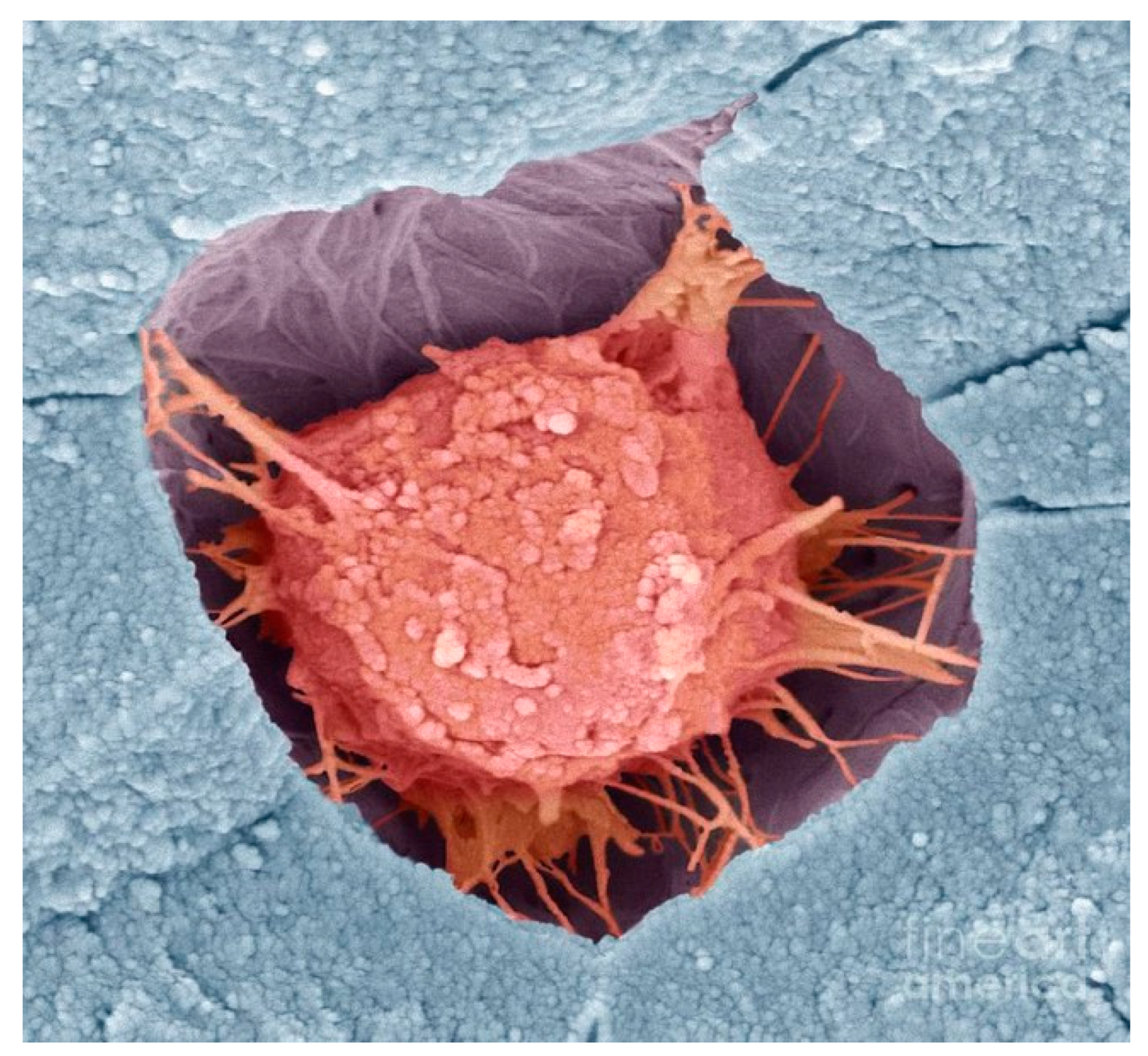
In its initial phase mesenchymal stem cells and osteoblasts occupying the surface of cortical bone, secrete receptor activator of nuclear factor kappa B ligand (RANKL) which upregulates osteoclast differentiation by binding to RANK receptors on the surface of osteoclast precursors. This process is counterbalanced by osteoprotegerin (OPG), a soluble decoy receptor that inhibits osteoclastogenesis by binding RANKL. In the second phase, newly evolved osteoclasts secrete hydrochloric acid and acid phosphatases that degrade surface bone and the bone matrix, releasing imbedded growth factors, including bone morphogenic proteins, transforming growth factor-β and insulin-like growth factor-1. Osteoclasts form Howship’s lacunae in trabecular bone and a cutting zone in cortical bone; once these cavities reach a certain size, osteoclasts undergo apoptosis, terminating bone resorption. In the third phase of bone remodeling, the newly liberated growth factors stimulate osteoblasts to evolve from their mesenchymal stem cell precursors to control bone mineralization and secrete collagen to form the bone matrix. In the fourth phase of bone remodeling, osteoblasts trapped in the bone matrix evolve into osteocytes, cells that connect with one another, osteoblasts, and osteoclasts through a myriad of dendritic processes that constitute the lacunar-canalicular network (see Figure 1 and Figure 2). Osteocytes are the most abundant cell type in bone (90–95% of all bone cells); they respond to hormonal and mechanical signals and are the primarily cell responsible for the control of bone homeostasis [7,8].

3. Mechanotransduction

3.1. Osteocytes

Exposure of osteocytes to shear stress generated in blood vessels within the Volkmann and Haversian canals and interstitial fluid in the lacunar-canalicular network and in spaces between the crystallites of the mineral hydroxyapatite and collagen fibers distorts their shape and activates the Wnt signaling pathway via translocation of β-catenin to the nucleus [8]. Wnt signaling is critical for bone mass accrual, bone remodeling, and fracture repair, and loss of function in Wnt or in mutations of its co-receptor LRP5 result in low bone mass and an increase in fragility fractures [8,9,10]. Cell shape distortion involves cytoskeletal rearrangement of actin fibers and microtubules, both of which are known to be integral players in mechanotransduction. Osteocyte cilia, dendritic extensions, and integrins in the extracellular matrix also act as mechanosensors [10,11]. Hillsley and Frangos propose that the bone loss observed in disuse and microgravity is due to a lack or decreased interstitial fluid flow [12].
Liu and associates have shown that osteocytes also respond to changes in hydraulic pressure. Cultures of osteocyte-like MLO-Y4 cells sensed cyclic changes in hydraulic pressure and responded by increasing intracellular calcium concentrations, COX-2 levels, and RANKL/osteoprotegerin mRNA ratios, and by altering cytoskeleton organization of actin and microtubules [13]. Mobilization of intracellular calcium activates downstream signaling of protein kinase A (PKA), MAPK, c-Fos, and prompts nuclear translocation of NF-κB. Blocking calcium release attenuates the mechanically induced upregulation of osteogenic gene expression in vitro and abrogates load-induced bone formation in vivo. COX-2 is the gene that encodes cyclooxygenase-2, a mediator of prostaglandins that play a role in bone homeostasis [13,14].

3.2. Osteoblasts

Young and associates found that reduced levels of osteoblast mechanostressors activates mechanotransduction via focal adhesion kinase (FAK) contained within extracellular matrix signaling complexes. Osteoblasts respond by increasing the expression of proteins involved in bone formation, such as COX-2, c-Fos and osteopontin E2 [15]. Ban and associates found that osteoblasts subjected to low levels of fluid shear stress proliferated and increased their production of bone markers including alkaline phosphatase and osteocalcin [16].

3.3. Osteoclasts

Osteoclast responses to mechanical loading are mediated by osteoblasts and osteocytes. Mechanically stimulated osteoblasts inhibit osteoclastogenesis by producing protegerin; this decoy receptor binds RANKL and prevents it from binding to receptors on osteoclast hematologic stem cells. Osteocytes control osteoclast activity by secreting sclerostin, a mechanosensing protein that increases bone resorption while decreasing bone formation [17].
Other structures within bone may also play a role in the response of bone to mechanical forces, including ion channels, connexins, integrins, and the glycocalyx [8,11].

4. Mechanotransduction in Space (1 × 10⁻⁶ g)

4.1. Osteocytes

Osteocytes are particularly susceptible to the adverse effects of zero gravity. In mice, one month of spaceflight decreased femoral trabecular bone volume by 64% and increased bone resorption by 140% when compared to ground controls. These changes were associated with osteocyte apoptosis, reductions in osteocyte lacunar volumes, and increases in lacunar vacancies (+344% vs. ground controls); fatty metamorphosis of mesenchymal stem cell precursors was also observed [18]. Similar findings were noted in iliac crest biopsies of monkeys flown for 14 days at zero gravity [19].

4.2. Osteoblasts

A site-specific bone loss of 1–2% occurs in astronauts and in-flight animals after 1 month of spaceflight. The loss is associated with changes in osteoblast cellular and nuclear morphology and altered mRNA expression of type I collagen, osteocalcin, and insulin-like growth binding proteins [20]. Nabavi and associates found that osteoblasts subjected to 5 days of microgravity had extended shapes, fragmented or condensed nuclei, shorter microtubules, and smaller and fewer focal adhesions [21].
Hughs-Fulford found that serum-activated osteoblasts had a 60% reduction in growth as compared to ground controls, presumably due to alterations in growth factor receptors and/or cell-connected matrix kinase pathways. After 4 days in space there was a loss of focal adhesions and a complete collapse of the osteoblast cytoskeleton. He postulated that the collapse was due to F-actin microfilament failure and/or inhibition of microtubular polymerization [22]. The same investigator had previously reported that cultured osteoblasts had increased levels of C-fos mRNA and diminished levels of Cox-2 mRNA [23]. C-fos is a protooncogene that encodes a 62 kDa protein that forms a heterodimer with c-jun (part of the jun family of transcription factors) resulting in the formation of activator protein 1 (AP-1) complex which binds DNA at AP-1 specific sites at the promoter and enhancer regions of target genes and converts extracellular signals into signals of gene expression. Cox-2 catalyzes the rate-limiting step in prostaglandin synthesis. Using cultures of ROS 17/2.8 osteoblasts, Guignandon and associates reported that osteoblastic integrin-mediated cell adhesion was disrupted in cells flown for six days on the space shuttle. The changes were characterized by disorganization of the cytoskeleton associated with disassembling of vinculin spots and phosphorylated proteins within the focal contacts [24]

4.3. Osteoclasts

Differentiated osteoclasts cultured on bone substrates for 10 days of zero gravity increased their expression of genes involved in osteoclast maturation and bone resorption when compared to ground controls [25].

5. Mechanotransduction in Simulated Microgravity (g ≥ 10−3)

5.1. Osteocytes

Robling and associates investigated the mechanoregulation of sclerostin and its regulatory gene Sost in rodent osteocytes under enhanced (ulnar loading) and reduced (hindlimb unloading) conditions. Sost transcripts and sclerostin protein levels were reduced in loaded bone and increased in unloaded bone [17]. Lin and associates found that mechanical unloading of wildtype mice caused a decrease in Wnt/beta-catenin signaling accompanied by increases in Sost and sclerostin production and reductions in bone formation and in the viability of osteocytes and osteoblasts [26].

5.2. Osteoblasts

Using NASA’s rotary cell culture system, Saxena and associates found that modeled microgravity inhibited osteoblastogenesis and increased adipose differentiation in human stem cells. The transformation involved reductions in RhoA activity and cofilin phosphorylation, disruption of F-actin stress fibers, and decreased integrin signaling through focal adhesion kinase [27]. Employing the same culture system, Ontiveros and McCabe reported a 50–80% reduction in alkaline phosphatase, Runx2, osteocalcin expression, and AP-1 transactivation in cultures of rat osteoblasts [28], and Patel and associates noted that 3 days of rotation altered the expression of 61 mechanosensitive genes, including osteomodulin, Runx2, and osteoglycin in cultures of mouse tibial pre-osteoblasts [29].
Using two-axis random positioning machines, Shi and associates found that simulated microgravity inhibited rat calvarial osteoblast differentiation, maturation, and ciliogenesis and attributed the change to atrophy of osteoblast cilia [30]. Sun and associates found that simulated microgravity reduced intracellular free calcium levels by inhibiting calcium channels in mouse osteoblasts [31]. As previously noted, low levels of intracellular calcium impairs osteogenic gene expression in vitro and abrogates load-induced bone formation in vivo. Dai and associates found that simulated microgravity inhibited the proliferation and osteogenesis of rat bone marrow mesenchymal stem cells [32]. Using cultures of human bone marrow mesenchymal stem cells, Li and associates found that simulated microgravity inhibited genes regulating proliferation and differentiation while upregulating genes involved in adipogenesis [33]. Chen and associates found that simulated microgravity inhibited osteogenic differentiation of mesenchymal stem cells by inhibiting the expression of transcriptional coactivator with PDZ-binding motif, an important regulator of osteogenesis [34]
Experiments measuring the metabolomics and proteomics of cultured human osteoblasts found that simulated microgravity caused a decrease in mitochondrial proteins, a reduction in mitochondrial energy potential, and an oxidative stress response (decreases in oxidized glutathione and antioxidant enzymes) [35].

5.3. Osteoclasts

Using NASA’s rotary cell culture system and mouse bone marrow cultures, Sambandam and associates found that simulated microgravity enhanced the maturation of osteoclast precursors [36,37]. Ethiraj and associates reported similar findings noting that osteoclastogenesis was associated with increased stem cell expression of syncytin-A [38]. Rucci and associates found that modeled microgravity stimulated osteoclastogenesis and bone resorption by increasing osteoblast RANKL/osteoprotegerin ratios [39], and Saxena and associates found that modeled microgravity and hindlimb unloading sensitized osteoclast precursors to RANKL-mediated osteoclastogenesis [40] (see Table 1).

6. The Somatotrophic Axis

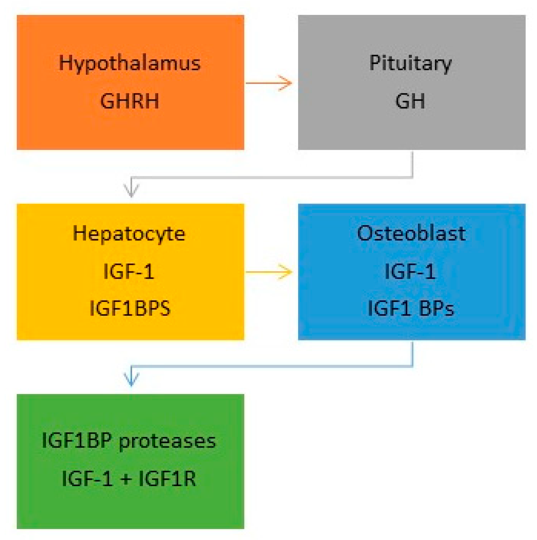
The somatotropic axis consists of hormonal circuits that regulate somatic growth, including body and bone mass, body adiposity, and lipid and carbohydrate metabolism. The axis includes growth hormone-releasing hormone (GHRH), growth hormone (GH), insulin-like growth factors (IGFs) 1 & 2 and their receptor, IGF1R, six IGF binding proteins (IGFBPs), the acid-labile subunit (ALS), somatostatin, and ghrelin (see Figure 3). Also included are growth hormone receptor’s downstream mediators, Janus kinase 2 (JAK2), signal transducer and activator of transcription 5 (STAT5) and suppressors of cytokine signaling 1–3 (SOCS1-3) [7]. IGF-2 is primarily involved in the growth and development of preadolescents and will not be discussed in this article.

6.1. Insulin-Like Growth Factor-1 (IGF-1)

IGF-1 is structurally related to insulin. It consists of 70 amino acids in a single chain with three intramolecular bridges and has a molecular weight of 7649 Daltons [41,42]. This growth factor is produced by GH-stimulated hepatocytes (the main source of circulating IGF-1) and by all non-hepatic tissues where it acts in a paracrine/autocrine fashion [7].
In serum, ~75% of IGF-1 exists in a 150-kDa complex consisting of IGF, IGFBP-3 or -5 and the ALS [7,43]. About 20% of IGF-1 is bound in binary complexes with IGFBPs, and ~5% is found in the free, biologically active state, which has a half-life of ~10 min. IGFBP binding prolongs the half-life of IGF-1 and assists in its delivery to specific tissues [7].
When in close proximity to its receptor, IGF-1 is liberated from its BP by the actions of one of the serine proteases, pregnancy-associated plasma protein-A or prostate-specific antigen. In tissue, kallikreins, cathepsins, and matrix metalloproteinases have been shown to assist in tissue remodeling and repair by cleaving the IGFBPs and releasing IGF-1 [7]

6.2. Insulin-Like Growth Factor-1 Receptor (IGF-1R)

Although IGF-1 can bind to insulin receptors, its primary action is mediated when it binds to its main receptor, IGF-1R. IGF-1R is a class II receptor tyrosine kinase (RTK) which plays a critical role in cell growth and differentiation. Binding of one IGF-1 molecule is sufficient to break the autoinhibited state of IGF-1R and initiate receptor activation. This results in the activation of a variety of intracellular transduction cascades including phosphatidlyinositol-3 kinase (PI3K) and its downstream target, mammalian target of rapamycin (mTOR) [41,42]. Recently, IGF-IR has been shown to have extensive cross-talk with many other receptors and can function as an RTK/GPCR hybrid. In addition, after activation, IGF-IR may translocate to the nucleus and function as a transcriptional cofactor; alternatively, it is recycled to the surface or degraded [43].

7. IGF-1 and Bone Homeostasis

IGF-1 is the most abundant growth factor in bone matrix [42] where it is synthesized by osteoblast mesenchymal precursors, mature osteoblasts, osteoclasts and osteocytes [44,45]. Osteoblasts also produce all the IGFBPs [45]. Hepatocyte synthesized IGF-1 is distributed to the matrix via the bone’s nutrient and periosteal arteries located within the Volkmann and Haversian canals.
In bone, IGF-1 regulates periosteal bone expansion (radial bone growth) by its effects on osteoprogenitors at the periosteal or endocortical bone surfaces. Linear bone growth is controlled by growth hormone which stimulates chondrocyte precursors to produce IGF-1 which then acts in an autocrine fashion to induce the production of type II collagen1a via activation of the phosphatidylinositol 3-kinase/phosphoinositide-dependent kinase-1 pathway. With progression of chondrocytes to terminal differentiation, IGF1R signaling is terminated [46]. IGF-1 regulates bone mass by activation of mTOR in mesenchymal stem cell progenitors of osteoblasts [47].
IGF-1 has pro-differentiating and pro-survival effects on osteoblasts and their mesenchymal precursors and is essential for the anabolic actions of parathyroid hormone (PTH) on bone [48]. Activation osteoblast IGF1R initiates the expression of the runt-related transcription factor 2 (Runx2) which stimulates the transition of mesenchymal stem cells into osteoprogenitors. Downstream of Runx2 is osterix (osx) which controls the maturation of osteoprogenitors into mature osteoblasts. Osteoblasts are responsible for the production and secretion of the extracellular matrix and expresses genes involved in matrix calcification [45].
Osteoclasts differentiate from mononuclear progenitors in the presence of macrophage colony stimulating factor (M-CSF) and RANKL. Wang and associates found that mRNA expression of RANKL, RANK, and M-CSF was diminished by 35–60% in IGF-1 knockout mice and led to a reduction in osteoclastogenesis [49]; the results confirm the importance of IGF-1 in promoting osteoclast differentiation from hematologic stem cell precursors.
Both animal and human studies have shown that circulating levels and locally produced autocrine/paracrine sources of IGF-1 each play an important role in achieving peak bone mass and strength [7,44].

8. IGF-1 and Mechanical Loading

In vitro experiments of cultured MC3T3-E1 osteoblast-line cells have shown that mechanical loading generated by interstitial fluid shear stress protects osteoblasts from tumor necrosis factor-α -mediated apoptosis [50] and that the protection is mediated by flow-enhanced IGF-1-rceptor signaling [51]. Osteoblast deletion of IGF-1 or IGF1R abates the osteogenic response to mechanical loading whereas transgenic overexpression of IGF-1 in osteoblasts results in an enhanced responsiveness to in vivo mechanical loading in mice [52].

9. IGF-1 and Mechanical Unloading

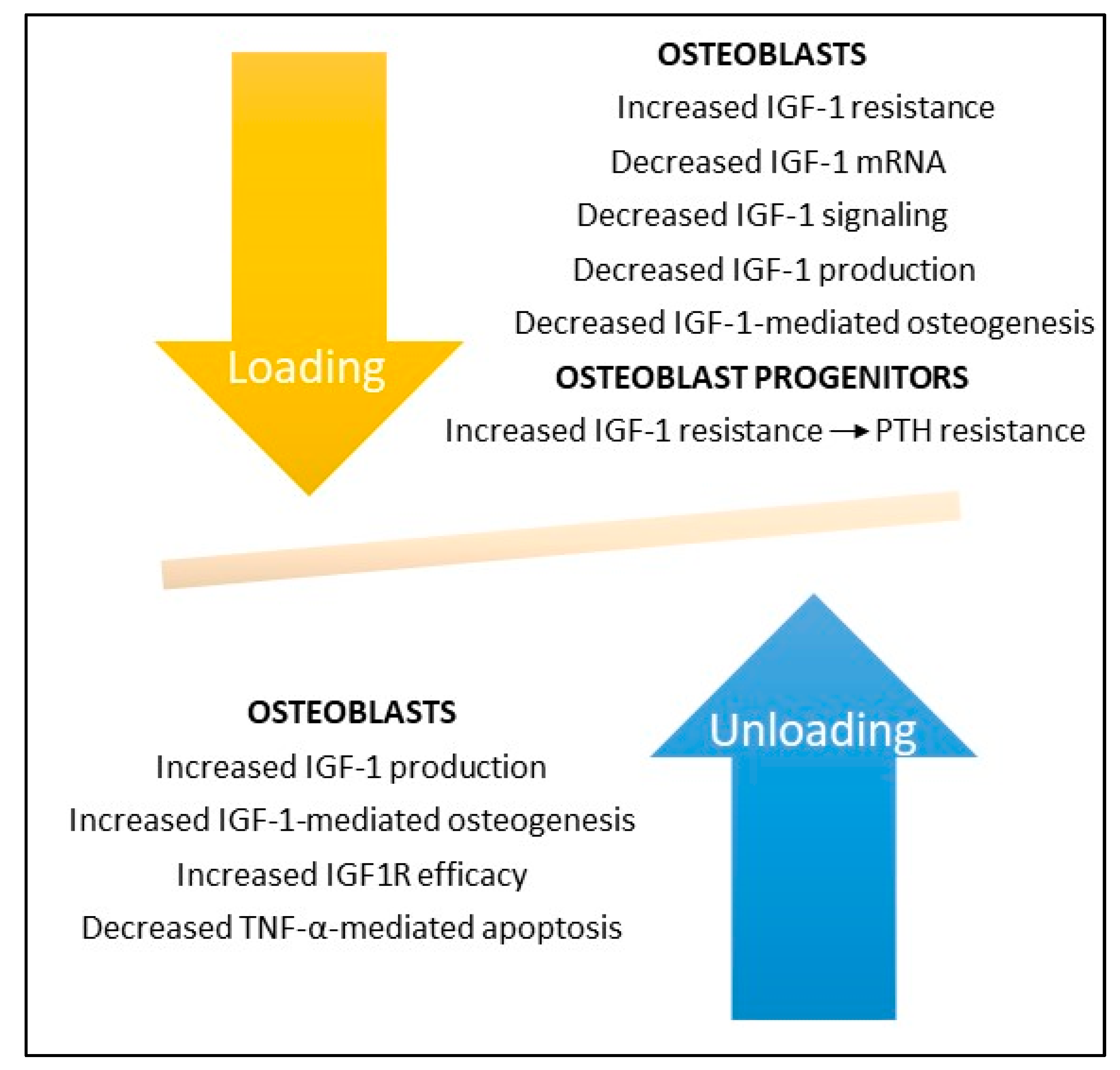
Studies during spaceflight and in bone unloading experiments on Earth have shown that bone unloading suppresses IGF-1 production (see Figure 4). Kumei et al. found that spaceflight decreased mRNA levels of IGF-1 in cultures of rat osteoblasts as compared to ground controls; they also found that microgravity completely suppressed mRNA expression of the insulin receptor substrate-1, a post-receptor signaling molecule of IGF-1 [53]. Sakata and associates found that skeletal unloading induces resistance to IGF-1 by inhibiting the activation of IGF-signaling pathways including the expression of alphaVbeta3 integrin [54]. And Triplett et al. found that mechanical loading by fluid shear stress enhanced IGF-1R signaling in osteoblasts whereas mechanical unloading inhibited its signaling [51]. Kostenuik and associates found that skeletal unloading caused resistance of osteoprogenitor cells to parathyroid hormone due to the development of resistance to IGF-1 [48].
Hind limb suspension in rats has been shown to inhibit IGF1R signaling pathways with consequent reduction in the number and differentiation of osteoblast progenitors and reduced bone formation; responsiveness to IGF1R could be restored with reloading [54,55]. In contrast, unloading of IGF-1R knockout mice caused a permanent loss of bone synthesis, indicating that IGF1 signaling is necessary for bone formation following disuse or zero gravity [7,44].

10. Discussion

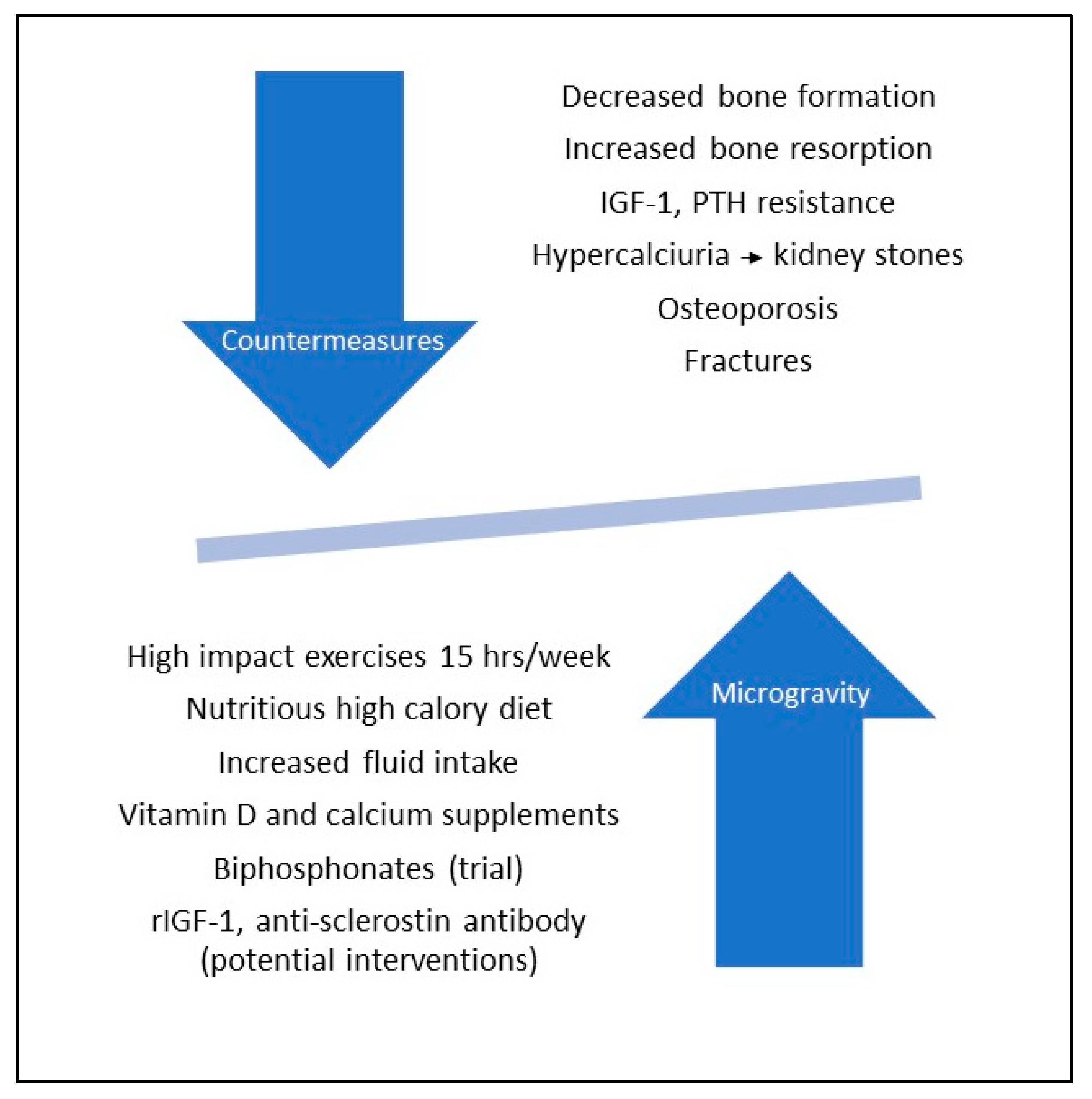
Many of the studies on bone homeostasis in space and in terrestrial models of microgravity are motivated by the need to find treatments capable of abrogating the bone loss that occurs despite NASA’s implementation of exercise and dietary regimens designed to maintain bone homeostasis. Although most recommendations are non-specific, a few have suggested specific targets to investigate. These include TRAIL [37], syncytin-A [38], sclerostin [26], and rIGF-1 [54,55,56]. I provide below a table (Table 2) outlining NASA’s current approach to maintain bone health during spaceflight [57,58,59,60,61] as well as a brief discussion of the aforementioned targets in ascending order of potential.

10.1. Tumor Necrosis Factor-Related Apoptosis-Inducing Ligand (TRAIL)

TRAIL is a cytokine of the TNF superfamily known for its ability to trigger apoptosis in tumor cells while being relatively safe towards normal cells. Binding to its cognate receptors, TRAIL-R1 or TRAIL-R2, induces the formation of a macromolecular complex that recruits caspase-8 and caspase-10 to initiate apoptosis [62,63]. Although TRAIL has been shown to trigger osteoclast apoptosis, in the opinion of the author it is unlikely that this cytokine could be ever be used to target only osteoclasts and titer its use in such a way that the subject does not develop osteopetrosis.

10.2. Syncytin-A

Syncytin-A is an intact retroviral envelope protein, the product of a retrovirus gene that was inserted into the human genome somewhere between 10 and 80 million years ago [64]. Syncytin-A is called a syncytin because of its membrane fusogenic capacity. In bone, syncytin A fuses the membranes of mononucleated pre-osteoclasts producing mature multinucleated osteoclasts. However, pre-osteoclast fusion is also mediated by CD47 and syncytin-A plays a critical role in the fusion of the placental syncytiotrophoblast [64,65,66]. In the opinion of the author, this makes syncytin an unlikely candidate to target with monoclonal antibody. More suited for this purpose and currently under widespread use are the biphosphonates, whose effects include osteoclast apoptosis. Potential complications of oral bisphosphonate therapy include esophagitis and, rarely, osteonecrosis of the jaw. When given intravenously, biphosphonates can cause a flu-like illness with fever, myalgias, and arthralgias [60].

10.3. Sclerostin

Sclerostin, a 22-kD glycoprotein secreted primarily by osteocytes, is a soluble inhibitor of canonical Wnt/β-catenin signaling pathways that direct mesenchymal stem cells towards the osteoblastic lineage. Sclerostin also enhances bone resorption by decreasing the production of osteoprotegerin, the soluble decoy receptor that inhibits osteoclastogenesis by binding RANKL. Thus, when present in high concentrations sclerostin causes decreased bone formation and increased bone resorption [67].
Scientists have recently developed a humanized monoclonal antibody directed against sclerostin (romosozumab) which was approved by the FDA for treatment of osteoporosis in 2019. Clinical trials have shown that monthly subcutaneous injections of romosozumab is effective in increasing bone formation and density and in decreasing bone resorption, results in keeping with the known effects of sclerostin on bone homeostasis [68]. However, there is some concern about potential cardiotoxicity of romosozumab, prompting the need for further clinical observations [69].

10.4. IGF-1

The potential role of rIGF-1 in maintaining skeletal mass in space was recognized by Tanaka and associates who found that the complex IGF-1 and its specific binding protein, IGFBP-3 stimulated bone turnover in an animal model flown in space [55]. Bateman and associates found that administration of IGF-1 to rodents flown for 10 days aboard the Space Shuttle increased their humerus periosteal bone formation by 37.6% [56].
In light of the central role that IGF-1 plays in all phases of bone formation and preservation, it is perhaps surprising that there are no published proof of principle trials assessing the efficacy of rIGF-1 in preventing bone loss in astronauts, particularly in light of the fact that rIGF-1 is available in an easy to use nasal spray and when used judiciously is generally well tolerated [70]. IGF-1 administration would have the added advantage of bolstering muscle growth and exerting a positive effect on immunoregulation. rIGF-1 has been used to treat patients with IGF-1 deficiency, adolescents with anorexia nervosa and low bone mass, and patients with diabetes mellitus, a disorder characterized by low circulating levels of IGF-1. The side effects of rIGF-1 are dose-related and may include extremity edema, jaw pain, arthralgias, myalgias, and hypoglycemia [71,72].
It is recognized that IGF1/IGF1Rt studies done under simulated conditions of microgravity do not necessarily equate with those done in the zero gravity of space. Simulated microgravity studies achieve at best a thousand-fold reduction in gravitational forces (≥1 × 10⁻³ g) whereas in space the force of gravity is a million-fold less than that experienced on Earth (1 × 10⁻⁶ g). As noted in this review, osteoblasts grown in space have extended cell shapes, fragmented and condensed nuclei, shortened microtubules, defective actin microfilaments, smaller and fewer focal adhesions, and even a complete implosion of the cytoskeleton. In contrast, osteoblasts grown in simulated microgravity are characterized more by disruption in functional than in structural aberrations. Thus, although rIGF-1 proved useful in reversing the recalcitrance of blastocytes to mechanostressors in simulated microgravity, the same may not be true in space.
Unfortunately, the adverse effects of microgravity on the skeletal system may persist for years postflight. In addition, astronauts experience a variety of other stressors, including social isolation, sleep deprivation, confinement, solar radiation, and alterations in circadian rhythms; on landing they experience the stress of the sudden readjustment to Earth’s gravity field [73].
In their approach to future space missions, scientists are faced with the need to carefully balance factors that enhance bone loss with those that have the potential to prevent or remit bone loss (see Figure 5).
The United States, Russia, and China are planning to establish colonies on our moon sometime in the 2030s. The force of gravity on the Moon is 1.6 m/s2 which is 16.7% of Earth’s gravity or ~1 × 10⁻² g, well within the range of simulated microgravity studies already performed. It would seem that rIFG-1 may hold greater promise as a therapy to abrogate hypogravity-related changes in bone homeostasis in astronauts destined to colonize the moon than those aboard a spaceship.

11. Conclusions

  • By tradition bone formation is divided into four phases: 1. Osteoclast differentiation; 2. Osteoclast-mediated bone resorption; 3. Osteoblast-mediated bone formation/mineralization; and 4. Osteoblast to osteocyte transformation and establishment of bone homeostasis.
  • Zero and modeled microgravity adversely affect all four phases of bone formation, increasing the maturation and the resorptive activities of osteoclasts, decreasing the differentiation, maturation, and bone forming abilities of osteoblasts, and causing apoptotic-mediated death of osteocytes with consequent dysregulation of bone homeostasis.
  • Microgravity-associated changes in osteoblast microstructure include extended cell shapes, fragmented and condensed nuclei, shortened microtubules, smaller and fewer focal adhesions, and even complete collapse of the cytoskeleton due to microfilament and/or microtubular failure. Osteocyte death is reflected by increases in lacunar vacancies and volumes and in the production of sclerostin, a mechanosensitive protein that inhibits bone formation and enhances bone resorption.
  • IGF-1 and its receptor IGF1R play critical roles in all phases of bone formation, promoting both radial and linear bone growth. Osteocyte and osteoblast responses to mechanostressors is IGF-1/IGF1R dependent and deletion of IGF-1 or IGF1R genes abates their responses to mechanical unloading and reloading. Treatment with recombinant IGF-1 (rIGF1) reverses these defects when IGF1R is intact, raising the prospect that rIGF-1 may prove useful in preventing bone loss associated with space travel or, more likely, in the microgravity of the Moon. Another potential candidate to prevent bone loss in space or on the Moon is romosozumab, a humanized antibody against sclerostin which has been approved by the FDA for treatment of osteoporosis.

Funding

This research received no external funding.

Acknowledgments

The author is grateful for the support of the Division of Academic Affairs in the preparation of this article.

Conflicts of Interest

The author declares no conflict of interest.

References

  1. Svitkina, T. The actin cytoskeleton and actin-based motility. Cold Spring Harb. Perspect. Biol. 2018, 10, a018267. [Google Scholar] [CrossRef] [Green Version]
  2. LaFlamme, S.E.; Mathew-Steiner, S.; Singh, N.; Colello-Borges, D.; Nieves, B. Integrin and microtubule crosstalk in the regulation of cellular processes. Cell Mol. Life Sci. 2018, 75, 4177–4185. [Google Scholar] [CrossRef] [PubMed]
  3. Burke, B. Chain reaction: LINC complexes and nuclear positioning. F1000Research 2019, 8, 136. [Google Scholar] [CrossRef] [PubMed]
  4. Martínez, P.T.; Navajas, P.L.; Lietha, D. FAK Structure and regulation by membrane interactions and force in focal adhesions. Biomolecules 2020, 10, 179. [Google Scholar] [CrossRef] [PubMed] [Green Version]
  5. Bérut, A.; Chauvet, H.; Legué, V.; Moulia, B.; Pouliquen, O.; Forterre, Y. Gravisensors in plant cells behave like an active granular liquid. Proc. Natl. Acad. Sci. USA 2018, 115, 5123–5128. [Google Scholar] [CrossRef] [Green Version]
  6. Howard, R.; Scheiner, A.; Cunningham, J.; Gatenby, R. Cytoplasmic convection currents and intracellular temperature gradients. PLoS Comput. Biol. 2019, 15. [Google Scholar] [CrossRef] [Green Version]
  7. Yakar, S.; Werner, H.; Rosen, C.J. Insulin-like growth factors: Actions on the skeleton. J. Mol. Endocrinol. 2018, 61, T115–T137. [Google Scholar] [CrossRef]
  8. Wittkowske, C.; Reilly, G.C.; Lacroix, D.; Perrault, C.M. In vitro bone cell models: Impact of fluid shear stress on bone formation. Front. Bioeng. Biotechnol. 2016, 4, 87. [Google Scholar] [CrossRef] [Green Version]
  9. Issack, P.S.; Helfet, D.L.; Lane, J.M. Role of Wnt signaling in bone remodeling and repair. HSS J. 2008, 4, 66–70. [Google Scholar] [CrossRef] [Green Version]
  10. Sakai, A. Spaceflight/bedrest immobilization and bone. Osteocyte as a sensor of mechanical stress and Wnt signal. Clin. Calcium. 2012, 22, 1829–1835. [Google Scholar]
  11. Aventaggiato, M.; Barreca, F.; Vernucci, E.; Bizzarri, M.; Ferretti, E.; Russo, M.A.; Tafani, M. Putative receptors for gravity sensing in mammalian cells: The effects of microgravity. Appl. Sci. 2020, 10, 2028. [Google Scholar] [CrossRef] [Green Version]
  12. Hillsley, M.V.; Frangos, J.A. Bone tissue engineering: Role of interstitial fluid flow. Biotechnol. Bioeng. 1994, 43, 573–581. [Google Scholar] [CrossRef] [PubMed]
  13. Liu, C.; Zhao, Y.; Cheung, W.-Y.; Gandhi, R.; Wang, L.; Lidan, Y. Effects of cyclic hydraulic pressure on osteocytes. Bone 2010, 46, 1449–1456. [Google Scholar] [CrossRef] [PubMed] [Green Version]
  14. Einhorn, T.A. The role of cyclooxygenase-2 in bone repair. Arthritis Res. Ther. 2003, 5, 5–7. [Google Scholar] [CrossRef]
  15. Young, S.R.L.; Hum, J.M.; Rodenberg, E.; Turner, C.H.; Pavalko, F.M. Non-overlapping functions for Pyk2 and FAK in osteoblasts during fluid stress-induced mechanotransduction. PLoS ONE 2011, 6, e16026. [Google Scholar] [CrossRef] [Green Version]
  16. Ban, Y.; Wu, Y.; Yu, T.; Geng, N.; Wang, Y.; Liu, X.; Gong, P. Response of osteoblasts to low fluid shear stress is time dependent. Tissue Cell 2011, 43, 311–317. [Google Scholar] [CrossRef]
  17. Robling, A.G.; Niziolek, P.J.; Baldridge, L.A.; Condon, K.W.; Allen, M.R.; Alam, I.; Mantila, S.M.; Gluhak-Heinrich, J.; Bellido, T.M.; Harris, S.E.; et al. Mechanical stimulation of bone in vivo reduces osteocyte expression of Sost/sclerostin. J. Biochem. 2008, 283, 5866–5875. [Google Scholar]
  18. Gerbaix, M.; Gnyubkin, V.; Farlay, D.; Olivier, C.; Ammann, P.; Courbon, G.; Laroche, N.; Genthial, R.; Follet, H.; Peyrin, F.; et al. One-month spaceflight compromises the bone microstructure, tissue-level mechanical properties, osteocyte survival and lacunae volume in mature mice skeletons. Sci. Rep. 2017, 7, 2659. [Google Scholar] [CrossRef]
  19. Rodionova, N.V.; Organov, V.S.; Zolotova, N.V. Ultrastructure changes in osteocytes in microgravity conditions. Adv. Space Res. 2002, 30, 765–770. [Google Scholar] [CrossRef]
  20. Carmeliet, G.; Bouillon, R. The effect of microgravity on morphology and gene expression of osteoblasts in vitro. FASEB J. 1999, 13, S129–S134. [Google Scholar] [CrossRef] [Green Version]
  21. Nabavi, N.; Khandani, A.; Camirand, A.; Harrison, R.E. Effects of microgravity on osteoclast bone resorption and on osteoblast cytoskeletal organization and adhesion. Bone 2011, 49, 965–974. [Google Scholar] [CrossRef] [PubMed]
  22. Hughs-Fulford, M. Function of the cytoskeleton in gravisensing during spaceflight. Adv. Space Res. 2003, 32, 1583–1593. [Google Scholar] [CrossRef]
  23. Hughs-Fulford, M.; Tjandrawinata, R.; Fitzgerald, J.; Gasuad, K.; Gilbertson, V. Effects of microgravity on osteoblast growth. Gravit. Space Biol. Bull. 1998, 11, 51–60. [Google Scholar]
  24. Guignandon, A.; Lafage-Proust, M.H.; Usson, Y.; Laroche, N.; Caillot-Augusseau, A.; Alexandre, C.; Vico, L. Cell cycling determines integrin-mediated adhesion in osteoblastic ROS 17/2.8 cells exposed to space-related conditions. FASEB J. 2001, 15, 2036–2038. [Google Scholar] [PubMed] [Green Version]
  25. Tamma, R.; Colaianni, G.; Camerino, C.; Di Benedetto, A.; Greco, G.; Strippoli, M.; Vergari, R.; Grano, A.; Mancini, L.; Mori, G.; et al. Microgravity during spaceflight directly affects in vitro osteoclastogenesis and bone resorption. FASEB J. 2009, 23, 2549–2554. [Google Scholar] [CrossRef]
  26. Lin, C.; Jiang, X.; Dai, Z.; Guo, X.; Weng, T.; Wang, J.; Li, Y.; Feng, G.; Gao, X.; He, L. Sclerostin mediates bone resorption response to mechanical unloading through antagonizing Wnt/beta-catenin signaling. J. Bone Miner. Res. 2009, 24, 1651–1661. [Google Scholar] [CrossRef]
  27. Saxena, R.; Pan, G.; McDonald, J.M. Osteoblast and osteoclast differentiation in modeled microgravity. Ann. N. Y. Acad. Sci. 2007, 1116, 494–498. [Google Scholar] [CrossRef]
  28. Ontiveros, C.; McCabe, L.R. Simulated microgravity suppresses osteoblast phenotype, Runx2 levels and AP-1 transactivation. J. Cell. Biochem. 2003, 88, 427–437. [Google Scholar] [CrossRef]
  29. Patel, M.J.; Liu, W.; Sykes, M.C.; Ward, N.E.; Risin, S.A.; Risin, D.; Jo, H. Identification of mechanosensitive genes in osteoblasts by comparative microarray studies using the rotating wall vessel and the random positioning machine. J. Cell. Biochem. 2007, 101, 587–599. [Google Scholar] [CrossRef]
  30. Shi, W.; Xie, Y.; He, J.; Zhou, J.; Gao, Y.; Wei, W.; Ding, N.; Ma, H.; Xian, C.J.; Chen, K.; et al. Microgravity induces inhibition of osteoblastic differentiation and mineralization through abrogating primary cilia. Sci. Rep. 2017, 7, 1866. [Google Scholar] [CrossRef]
  31. Sun, Z.; Li, Y.; Zhou, H.; Cai, M.; Liu, J.; Gao, S.; Yang, J.; Tong, L.; Wang, J.; Zhou, S.; et al. Simulated microgravity reduces intracellular-free calcium concentration by inhibiting calcium channels in primary mouse osteoblasts. J. Cell. Biochem. 2019, 120, 4009–4020. [Google Scholar] [CrossRef] [PubMed]
  32. Dai, Z.Q.; Wang, R.; Ling, S.K.; Wan, Y.M.; Li, Y.H. Simulated microgravity inhibits the proliferation and osteogenesis of rat bone marrow mesenchymal stem cells. Cell. Prolif. 2007, 40, 671–684. [Google Scholar] [CrossRef] [PubMed]
  33. Li, L.; Zhang, C.; Chen, J.L.; Hong, F.F.; Chen, P.; Wang, J.F. Effects of simulated microgravity on the expression profiles of RNA during osteogenic differentiation of human bone marrow mesenchymal stem cells. Cell. Prolif. 2019, 52, e12539. [Google Scholar] [CrossRef] [PubMed]
  34. Chen, Z.; Luo, Q.; Lin, C.; Song, G. Simulated microgravity inhibits osteogenic differentiation of mesenchymal stem cells through down regulating the transcriptional co-activator TAZ. Biochem. Biophys. Res. Commun. 2015, 468, 21–26. [Google Scholar] [CrossRef]
  35. Michaletti, A.; Gioia, M.; Tarantino, U.; Zolla, L. Effects of microgravity on osteoblast mitochondria: A proteomic and metabolomics profile. Sci. Rep. 2017, 7. [Google Scholar] [CrossRef]
  36. Sambandam, Y.; Townsend, M.T.; Pierce, J.J.; Lipman, C.M.; Haque, A.; Bateman, T.A.; Reddy, S.V. Microgravity control of autophagy modulates osteoclastogenesis. Bone 2014, 61, 125–131. [Google Scholar] [CrossRef] [Green Version]
  37. Sambandam, Y.; Baird, K.L.; Stroebel, M.; Kowai, E.; Balasubramanian, S.; Reddy, S.V. Microgravity induction of TRAIL expression in preosteoclast cells enhances osteoclast differentiation. Sci. Rep. 2016, 6, 25143. [Google Scholar] [CrossRef] [Green Version]
  38. Ethiraj, P.; Link, J.R.; Sinkway, J.M.; Brown, G.D.; Parler, W.A.; Reddy, S.V. Microgravity modulation of syncytin-A expression enhance osteoclast formation. J. Cell. Biochem. 2018, 119, 5696–5703. [Google Scholar] [CrossRef]
  39. Rucci, N.; Rufo, A.; Alamanou, M.; Teti, A. Modeled microgravity stimulates osteoclastogenesis and bone resorption by increasing osteoblast RANKL/OPG ratio. J. Cell. Biochem. 2007, 100, 464–473. [Google Scholar] [CrossRef]
  40. Saxena, R.; Pan, G.; Dohm, E.D.; McDonald, J.M. Modeled microgravity and hindlimb unloading sensitize osteoclast precursors to RANKL-mediated osteoclastogenesis. J. Bone Miner. Metab. 2011, 29, 111–122. [Google Scholar] [CrossRef] [Green Version]
  41. Li, J.; Choi, E.; Yu, H.; Bai, X.C. Structural basis of the activation of type 1 insulin-like growth factor receptor. Nat. Commun. 2019, 10, 4567. [Google Scholar] [CrossRef] [PubMed]
  42. Zhang, X.; Yu, D.; Sun, J.; Wang, Y.; Ouyang, O.; Wang, T. Visualization of ligand-bound ectodomain assembly in the full-length human IGF-1 receptor by cryo-EM single-particle analysis. Structure 2020, 28, 1–7. [Google Scholar] [CrossRef] [PubMed]
  43. Janssen, J.A. New insights from IGF-1R stimulating activity analysis: Pathological considerations. Cells 2020, 9, 862. [Google Scholar] [CrossRef] [PubMed] [Green Version]
  44. Yakar, S.; Rosen, C.J.; Beamer, W.G.; Ackert-Bicknell, C.L.; Wu, Y.; Liu, J.-L.; Ooi, G.T.; Setser, J.; Frystyk, J.; Boisclair, Y.R.; et al. Circulating levels of IGF-1 directly regulate bone growth and density. J. Clin. Invest. 2002, 110, 771–781. [Google Scholar] [CrossRef]
  45. Crane, J.L.; Cao, X. Function of matrix IGF-1 in coupling bone resorption and formation. J. Mol. Med. 2014, 92, 107–115. [Google Scholar] [CrossRef] [Green Version]
  46. Clarmatori, S.; Kiepe, D.; Haarmann, A.; Huegel, U.; Tonshoff, B. Signaling mechanisms leading to regulation of proliferation and differentiation of the mesenchymal chondrogenic cell line. J. Mol. Endocrinol. 2007, 38, 493–508. [Google Scholar] [CrossRef] [Green Version]
  47. Xian, L.; Wu, X.; Pang, L.; Lou, M.; Rosen, C.; Qiu, T.; Crane, J.; Frassica, F.; Zhang, L.; Rodriguez, J.P.; et al. Matrix IGF-1 regulates bone mass by activation of mTOR in mesenchymal stem cells. Nat. Med. 2012, 18, 1095–1101. [Google Scholar] [CrossRef] [Green Version]
  48. Kostenuik, P.J.; Harris, J.; Halloran, B.P.; Turner, R.T.; Morey-Holton, E.R.; Bikle, D.D. Skeletal unloading causes resistance of osteoprogenitor cells to parathyroid hormone and to insulin-like growth factor-1. J. Bone Miner. Res. 1999, 14, 21–31. [Google Scholar] [CrossRef]
  49. Wang, Y.; Nishida, S.; Elalieh, H.Z.; Long, R.K.; Halloran, B.P.; Bikle, D.D. Role of IGF-1 signaling in regulating osteoclastogenesis. J. Bone Min. Res. 2006, 21, 1350–1358. [Google Scholar] [CrossRef] [Green Version]
  50. Pavalko, F.M.; Gerard, R.L.; Ponik, S.M.; Gallagher, P.J.; Jin, Y.; Norvell, S.M. Fluid shear stress inhibits TNF-alpha-induced apoptosis in osteoblasts: A role for fluid shear stress-induced activation of PI3-kinase and inhibition of caspase-3. J. Cell. Physiol. 2003, 194, 194–205. [Google Scholar] [CrossRef]
  51. Triplett, J.W.; O’Riley, R.; Tekulve, K.; Norvell, S.M.; Pavalko, F.M. Mechanical loading by fluid shear stress enhances IGF-1 receptor signaling in osteoblasts in a PKCzeta-dependent manner. Mol. Cell. Biomech. 2007, 4, 13–25. [Google Scholar] [PubMed]
  52. Tian, F.; Wang, Y.; Bickle, D.D. IGF-1 signaling mediated cell-specific skeletal mechano-transduction. J. Orthop. Res. 2018, 36, 576–583. [Google Scholar] [CrossRef] [PubMed]
  53. Kumei, Y.; Nakamura, H.; Morita, S.; Akiyama, H.; Hirano, M.; Ohya, K.; Shinomiya, K.I.; Shimokawa, H. Space flight and insulin-like growth factor signaling in rat osteoblasts. Ann. N. Y. Acad. Sci. 2002, 973, 75–78. [Google Scholar] [CrossRef] [PubMed]
  54. Sakata, T.; Wang, Y.; Halloran, B.P.; Elalieh, H.Z.; Cao, J.; Bikle, D.D. Skeletal unloading induces resistance to insulin-like growth factor-1 (IGF-1) by inhibiting activation of the IGF-1 signaling pathways. J. Bone Miner. Res. 2004, 19, 436–446. [Google Scholar] [CrossRef] [PubMed]
  55. Tanaka, H.; Moriwake, T.; Matsuoka, Y.; Nakamura, T.; Seino, Y. Potential role of rhIGF-1/IGFBP-3 in maintaining skeletal mass in space. Bone 1998, 22, 145S–147S. [Google Scholar] [CrossRef]
  56. Bateman, T.A.; Zimmerman, R.J.; Ayers, R.A.; Ferguson, V.L.; Chapes, S.K.; Simske, S.J. Histomorphometric, physical, and mechanical effects of spaceflight and insulin-like growth factor on rat long bones. Bone 1998, 23, 527–535. [Google Scholar] [CrossRef]
  57. Iwamoto, J.; Takeda, T.; Sato, Y. Interventions to prevent bone loss in astronauts during space flight. Keio J. Med. 2005, 54, 55–59. [Google Scholar] [CrossRef] [Green Version]
  58. Cavanagh, P.R.; Licata, A.A.; Rice, A.J. Exercise and pharmacological countermeasures for bone loss during long-duration space flight. Gravit. Space Biol. Bull. 2005, 18, 39–58. [Google Scholar]
  59. Smith, S.M.; McCoy, T.; Gazda, D.; Morgan, J.L.L.; Heer, M.; Zwart, S.R. Space flight calcium: Implications for astronaut health, spaceflight operations, and Earth. Nutrients 2012, 4, 2047–2068. [Google Scholar] [CrossRef] [Green Version]
  60. Leblanc, A.; Matsumoto, T.; Jones, J.; Shapiro, J.; Lang, T.; Shackelford, L.; Smith, S.M.; Evans, H.; Spector, E.; Ploutz-Snyder, R.; et al. Biphosphonates as a supplement to exercise to protect bone during long-duration spaceflight. Osteoporos. Int. 2013, 24, 2105–2114. [Google Scholar] [CrossRef]
  61. NASA—Food For Space Flight. Available online: https:/www.nasa.gov/specials/apollo50thy/index.html (accessed on 12 June 2020).
  62. Micheau, O. Regulation of TNF-related apoptosis-inducing ligand signaling by glycosylation. Int. J. Mol. Sci. 2018, 19, 715. [Google Scholar] [CrossRef] [PubMed] [Green Version]
  63. Walczak, H. Biochemical analysis of the native TRAIL death-inducing signaling complex. Methods Mol. Biol. 2008, 414, 221–239. [Google Scholar] [PubMed]
  64. Dupressoir, A.; Lavialle, G.; Heidmann, T. From ancestral infectious retroviruses to bone fide cellular genes: Role of the captured syncytins in placentation. Placenta 2012, 33, 663–671. [Google Scholar] [CrossRef] [PubMed]
  65. Peng, X.; Pan, J.; Gong, R.; Liu, Y.; Kang, S.; Feng, H.; Qiu, G.; Guo, D.; Tien, P.; Xiao, G. Functional characterization of syncytin-A, a newly murine endogenous virus protein. Implication for its fusion mechanism. J. Biol. Chem. 2007, 282, 381–389. [Google Scholar] [CrossRef] [Green Version]
  66. Moller, A.M.; Delaissé, J.M.; Soe, K. Osteoclast fusion: Time-lapse reveals involvement of CD47 and syncytin-1 at different stages of nuclearity. J. Cell. Physiol. 2017, 232, 1396–1403. [Google Scholar] [CrossRef]
  67. De Maré, A.; D’Haese, P.C.; Verhulst, A. The role of sclerostin in bone and ectopic calcification. Int. J. Mol. Sci. 2020, 21, 3199. [Google Scholar] [CrossRef]
  68. Bandeira, L.; Lewiecki, E.M.; Bilezikian, J.P. Romosozumab for the treatment of osteoporosis. Expert Opin. Biol. Ther. 2017, 17, 255–263. [Google Scholar] [CrossRef]
  69. Shakeri, A.; Adanty, C. Romosozumab (sclerostin monoclonal antibody) for the treatment of osteoporosis in postmenopausal women: A review. J. Popul. Ther. Clin. Pharmacol. 2020, 27, e25–e31. [Google Scholar] [CrossRef] [Green Version]
  70. Liu, X.F.; Fawcett, J.R.; Thome, R.G.; DeFor, T.A.; Frey, W.H., 2nd. Intranasal administration of insulin-like growth factor-1 bypasses the blood-brain barrier and protects against focal cerebral ischemic damage. J. Neurol. Sci. 2001, 187, 91–97. [Google Scholar] [CrossRef]
  71. Misra, M.; McGrane, J.; Miller, K.K.; Goldstein, M.A.; Ebrahimi, S.; Weigel, T.; Klibanski, A. Effects of rhIGF-1 administration on surrogate markers of bone turnover in adolescents with anorexia nervosa. Bone 2009, 45, 493–498. [Google Scholar] [CrossRef] [Green Version]
  72. Mohamed-Ali, V.; Pinkney, J. Therapeutic potential of insulin-like growth factor-1 in patients with diabetes mellitus. Treat. Endocrinol. 2002, 1, 399–404. [Google Scholar] [CrossRef] [PubMed]
  73. Smith, J.K. IL-6 and the dysregulation of immune, bone, muscle, and metabolic homeostasis during spaceflight. npj Microgravity 2018, 4, 24. [Google Scholar] [CrossRef] [PubMed] [Green Version]
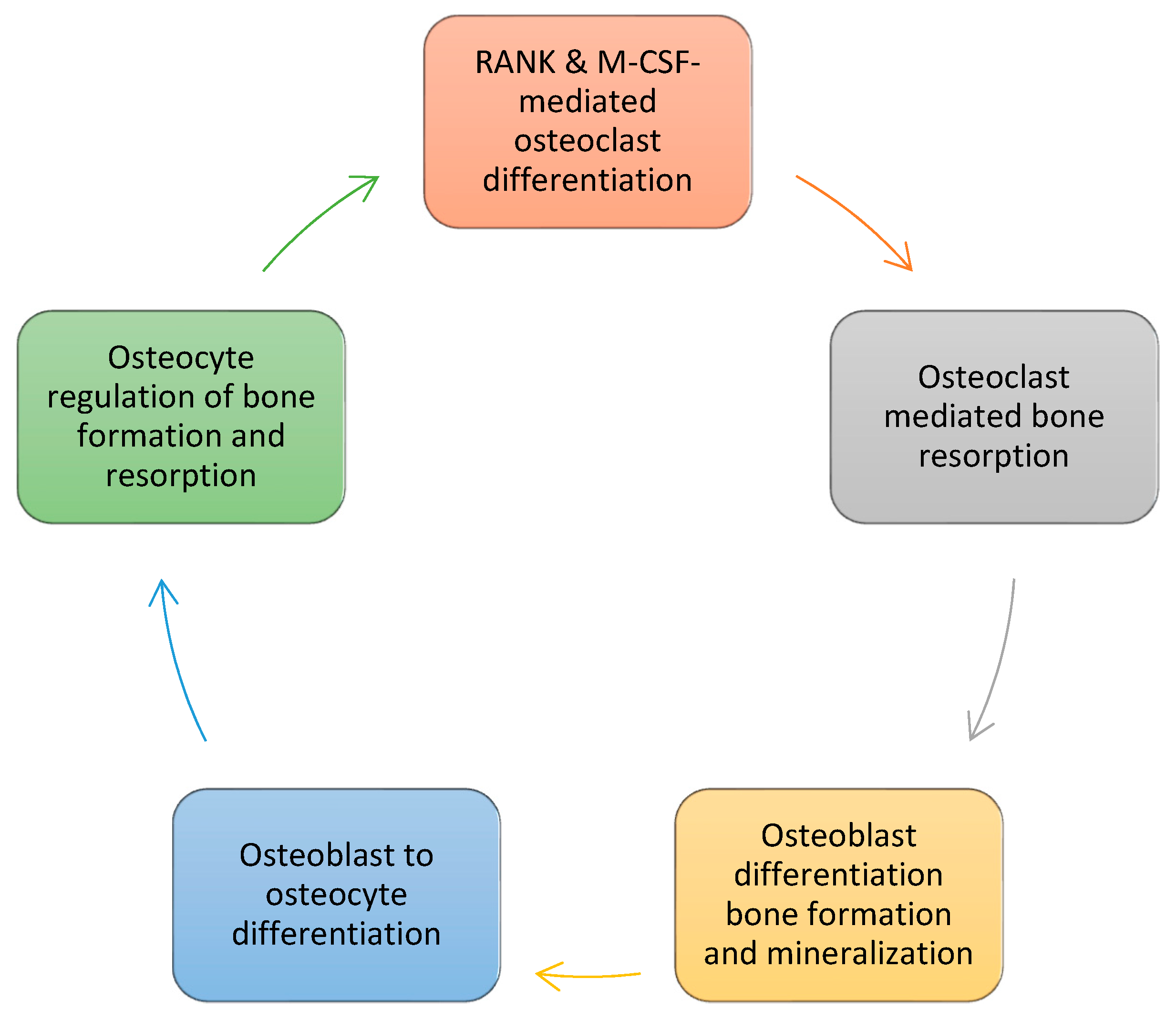
Figure 1. The cycle of bone formation. RANK and macrophage colony stimulating factor (M-CSF) signaling initiates osteoclast differentiation from hematopoietic stem cell precursors. Mature multilobed osteocytes resorb bone, releasing growth factors (IGF-1, TGF-β, and BMPs) that initiate osteoblast differentiation from mesenchymal stem cell precursors. Osteoblasts form and mineralize bone matrix, and eventually evolve into osteocytes—the master regulators of bone homeostasis.
Applsci 10 04433 g001
Figure 2. A photomicrograph of an osteocyte embedded in its lacuna. Evolved from osteoblasts entrapped in bone matrix, osteocytes form a multitude of dendritic extensions that communicate with osteoblasts, osteoclasts, and other osteocytes through a network of channels cut into the bone matrix. These dendritic extensions are sensitive to pressure changes associated with mechanical loading and unloading and allow osteocytes to regulate the balance between bone absorption and bone formation. Osteocytes undergo apoptosis in zero gravity, losing their control over bone homeostasis, and secrete sclerostin, a mechanosensitive protein that inhibits bone formation and enhances bone resorption (photograph by Steve Gschmeissner).
Applsci 10 04433 g002
Figure 3. The somatotropic axis. The hypothalamus produces growth hormone releasing hormone stimulating the pituitary to release growth hormone which acts on hepatocytes to produce insulin-like growth factor-1 and some of its binding proteins. Circulating IGF-1 reaches the bone matrix where it and some of its binding proteins are produced primarily by osteoblasts in an autocrine/paracrine manner. Released from its binding proteins by proteases, IGF-1 binds to its receptor IGF1R initiating its radial growth effects on osteoblasts and, with the aid of growth hormone, its linear growth effects on chondrocytes.
Applsci 10 04433 g003
Figure 4. Bone loading and unloading experiments demonstrate the key role IGF-1 and its receptor IGF1R play in bone formation. Bone unloading increases IGF-1 resistance and decreases IGF-1 production, signaling, and IGF-1-mediated bone formation by osteoblasts and enhances IGF-1 and PTH resistance in osteoblast mesenchymal stem cell precursors. In contrast, bone loading increases IGF-1 production and IGF1R efficiency and decreases TNF-α-mediated apoptosis in osteoblasts. Deletion of IGF-1 or IGF1R in rodents abrogates osteoblast responses to bone loading and unloading (not shown).
Applsci 10 04433 g004
Figure 5. A careful balancing is required to diminish factors that lead to bone loss with interventions designed to maintain bone health during spaceflight.
Applsci 10 04433 g005
Table 1. Effects of gravity on osteocyte, osteoblast, osteoclast and mesenchymal and hematopoietic stem cell precursors.
MechanostressorsG x 1G ≥ 10−3G = 10−6
Osteocytes *Fluid shear stress
Hydrostatic pressure [8,9,10,11,12,13,14]
Increased Wnt/beta-catenin, calcium, COX-2 signaling, RANKL/OPG mRNA ratios. Cytoskeletal reorganization [8,9,10,11,12,13,14]Increased Sost expression, sclerostin production. Decreased Wnt/beta-catenin signaling, bone formation [17,26]Apoptosis
Sclerostin release [18,19]
OsteoblastsBone loading via
FAK
Fluid shear stress [15,16].
Increased bone formation via enhanced expression of COX-2, c-Fos, osteopontin E2 [15,16]Decreased expression of mechanosensitive genes & markers of bone formation. Calcium channel inhibition [27,28,29,30,31,32,33,34] Loss of focal adhesions, FAK. F-actin micro-filament & microtubular failure with cytoskeleton implosion [20,21,22,23,24]
OsteoclastsAs per osteocytes & osteoblastsResorption inhibited by osteoblast production of OPG and by reduced production of sclerostin by osteocytes [17,26]Increased osteoclastogenesis and bone resorption secondary to increased RANKL/OPG ratios [36,37,38,39,40]Increased expression of genes involved in osteoclast maturation and bone resorption [25]
Mesenchymal stem cells Decreased osteoblast differentiation, maturation & ciliogenesis.
Increased adipogenesis [27,32,33,34]
Hematopoietic stem cells Enhanced maturation.
Increased expression of syncytin-A. Increased response to RANKL-mediated osteoclasto-genesis [36,37,38,39,40]
* Mechanosensors include the cytoskeleton, actin microfibers, microtubules, cilia, dendritic extensions and integrins in the extracellular matrix. COX-2, cyclooxygenase-2; FAK, focal adhesion kinases; OPG, osteoprotegerin; RANKL, receptor activator of nuclear factor kappa B ligand; Sost, gene encoding sclerostin.
Table 2. Measures currently taken to help prevent bone loss during spaceflight [57,58,59,60,61].
ExerciseNutritious High Calory DietVitamins/MineralsBone Sparing Agents
2.5 h/day x 6 days a week of combined aerobic & resistance exercisesCalories: 2700 to 3700/day * Frozen foods: Most entrees, vegetable and fruit items. Refrigerated foods: Fresh and fresh treated fruits, vegetables and dairy products.
Ambient foods: Thermostabilized, aseptic-fill, shelf-stable natural form foods and rehydrated beverages
Recommended daily allowance (RDA) of vitamins & minerals.
1000–1200 mg calcium & 800 IU vitamin D/day (Smith et al.)
† Biphosphonates
* Calorie requirements are determined by the National Research Council formula for basal energy expenditure (BEE). For women, BEE = 655 + (9.6 × W) + (1.7 × H) – (4.7 × A); for men BEE = 66 + (13.7 × W) + (5 × H)−(6.8 × A), where W = weight in kg, H = height in cm, and A = age in years. Astronauts choose their menu approximately 28 days preflight. † Joint studies between NASA and JAXA suggest that alendronate may be helpful in preventing bone loss during spaceflight but concerns about side effects remain. Despite the implementation of this exercise and dietary regimen, astronauts lose 1–1.5% of their bone mass for every month they spend in space.