Commentary

Tryptophan and Membrane Mobility as Conditioners and Brokers of Gut–Brain Axis in Depression

1
Department of Veterinary Medical Sciences, University of Bologna, Via Tolara di Sopra, 50, 40064 Ozzano dell’Emilia (Bologna), Italy
2
Department of Pharmaceutical Sciences, University of Perugia, Via Romana, 06126 Perugia, Italy
*
Author to whom correspondence should be addressed.
Appl. Sci. 2020, 10(14), 4933; https://doi.org/10.3390/app10144933
Submission received: 2 June 2020 / Revised: 16 July 2020 / Accepted: 16 July 2020 / Published: 17 July 2020

Abstract

:
The aim of this brief narrative commentary is to discuss some aspects involved in depression. It is increasingly evident that the phenomenon of mood disorders, despite its unequivocal genetic origin, slips into a multifactorial set of biochemical and molecular events that involve the whole organism. A vast literature has provided evidence that recognizes changes in serotonergic neurotransmission in the pathophysiology of depression. In addition, an increased arachidonic acid/omega-3 fatty acid ratio, which confers to mammalian cell membranes their fluidity, is associated with the depressive state. The combination of the excessive expression of kinurenine and the increased fluidity of the membrane has never been considered in the meaning of a simultaneous effect in the determinism of the depressive condition. Furthermore, various evidence supports the relationship between intestinal microbiota and depression and confirms alterations in the microbiota in depressive pathology.

1. Introduction

A paper published in The Lancet in 1969 about tryptophan stated, “Psychic depression may result from deficiency of brain serotonin. It is suggested that in depression the production of tryptophane pyrrolase by the liver is stimulated by raised blood-corticosteroid levels. As a result the metabolism of tryptophane is shunted away from serotonin production, and towards kynurenine production” [1].
In fact, this article highlighted how the neurotropic activity of kinurenine, by modifying the bioavailability of tryptophan, favored the reduction of serotonin (5-hydroxytryptamine, 5-HT), opening the door to states of anxiety, psychosis, and cognitive decline—conditions all associated with depression.
In 1973, other authors conducted an experiment in depressed and nondepressed subjects claiming that the data obtained did not support the increase in tryptophan metabolism toward the kinurenine pathway in depression [2]. Despite the contrast of these data, probably attributable to the experimental modalities, the evidence remains that, in the depressive condition, tryptophan triggers a decrease in 5-HT levels.
Over time, up to the present day, the concept of the centrality of tryptophan in the determinism of the reduction of 5-HT, due to the greater transformation of the metabolic pathway of tryptophan into kinurenine, has become increasingly popular [3]. In particular, stress and inflammation, as well as proinflammatory cytokines, can induce the activation of indoleamine 2,3-dioxygenase (IDO) (an enzyme involved in the catabolism of tryptophan to kynurenine), and higher levels of kynurenine have been linked with a depressive condition [4,5] (Figure 1). In addition, genetic variants of 5-HT receptors, as well as tryptophan hydroxylase, have been associated with a higher risk of depression [6]. Furthermore, proinflammatory cytokines increase monoamine reuptake by further reducing 5-HT levels [7,8,9].
In the 1970s and 1980s, a further element to the evaluation of the reduced presence of 5-HT in the depressive condition was added by some authors with reference to the similarity between neurons and platelets in relation to finding a reduced amount of 5-HT in the neurons and the platelets in the depressive condition [10,11,12,13,14,15,16,17].
In more recent years, the results obtained by other authors through blood platelet fatty acids, in the interpretation of an artificial neural network, have enabled the possibility of carrying out a precise diagnostic evaluation of the two main psychopathologies, namely major depression (MD) and bipolar disorder (BD) [18,19,20].
Whereas 5-HT is mostly known for its role in mood, anxiety, psychosis, or memory in the central nervous system (CNS), more than 95% of total body 5-HT is present in the periphery. In addition, the majority of peripheral 5-HT is stored in platelets. On the other hand, platelets do not synthesize 5-HT, but they adsorb it from plasma through the 5-HT transporter and release it during their activation [21]. Actually, the function of 5-HT in platelets is not clear yet; studies suggest that it is important for the serotonylation of the proteins necessary in platelet aggregation [22].
A recent study revealed platelet contributions to inflammation in reactions involving antigen–antibody complexes [23]. Furthermore, some studies have found a significant correlation between the abnormal membrane fluidity of platelets and the severity of dementia [24,25,26].
Platelets and neurons are unquestionably different cells, however, they share common characteristics in molecular organization and in protein composition [27]. In particular, various proteins are typically expressed in both neurons and platelets, and circulating platelets have been proposed as an alternative model to investigate neuronal dysfunctions and as an accessible peripheral biomarker to monitor the onset and the progression of neurological diseases. In this respect, some studies have reported platelet alterations in autism spectrum disorders [28].
The hypothesis that is formulated refers to the possibility of using the platelet as a diagnostic element in psychopathology [29] and in accordance with Heron et al. [30]. The focus is on the role of membrane fluidity in the management of 5-HT uptake. The cell membrane is known to live in a state of continuous mobility. Its fluidity is affected by the ratio of phospholipids to free cholesterol. In particular, long chain fatty acids, specifically arachidonic acid (AA) and docosahexaenoic acid (DHA), are integral components of neural membrane phospholipids and are extremely important for the maintenance of the optimum membrane fluidity of neurons [31]. There are several potential sources of AA in the brain, and an alteration in their components cannot only influence crucial intracellular and intercellular signaling but also alter many membrane physical properties, such as fluidity, phase transition temperature, bilayer thickness, and lateral domains, and therefore, the activity of membrane-dependent proteins [31,32]. Alterations in membrane proteins have been documented in depressed patients [33]. Walsh et al. reported an increase in the expression of the platelet adhesion receptor glycoprotein-Ib in depressed subjects [34]. In addition, Piletz et al. showed an increased P-selectin platelet content in depressed patients [35]. In particular, an excessive phospholipase-A 2 (PLA 2) activity could disrupt membrane fluidity, composition, and consequently, the activity of membrane-dependent proteins [33]. Membrane fluidity among depressed patients has been reported to be significantly involved, and brain levels of DHA have been shown to be reduced [36].
It has been hypothesized that membrane viscosity influences the reactivity of circulating platelets. The viscosity of the platelet membrane, the dislocation of the membrane components, and the distortion of the lipid–protein interactions could give rise to a signal mediated by an altered fluidity of the platelet membrane [37].
Therefore, the differential lipid fluidity of the membrane represents a crucial node of 5-HT signaling. Indeed, the differential accessibility of 5-HT receptors decreases with lipid fluidity [30]. Among the various subtypes of 5-HT receptors, the type-1A receptor serves as an important target in the development of therapeutic agents for neuropsychiatric disorders, including depression. In particular, it has been observed that the mobility of the 5-HT 1A receptor and the dynamics of the cell surface also depend on the receptor’s interaction with G proteins [38].
A correlation between platelet fatty acid composition in humans (isolated from venous and arterial blood) and pigs (isolated from venous blood and the brain) has been performed [39]. Experiments have been conducted on the transfer of AA between platelets and the brain, in pigs compared to humans, where it has not been possible to investigate the human brain [36,37,38]; these studies have shown, through a mathematical extrapolation of the data, the increased concentration of AA [39] as the reason for the increased fluidity of the membrane and the reduction in the accessibility of 5-HT, as is also reported by Green et al. [40]. In particular, the concentration of AA in brain tissue from various areas of an animal model of depression has been shown to be higher than in the corresponding brain areas of the controls [40].
Under conditions of an alteration in the fluidity of the membrane due to an increase in AA, as happens in the depressive state, 5-HT would not be captured by the platelets and, moreover, could contribute to bone and cardiovascular damage [31,41,42].
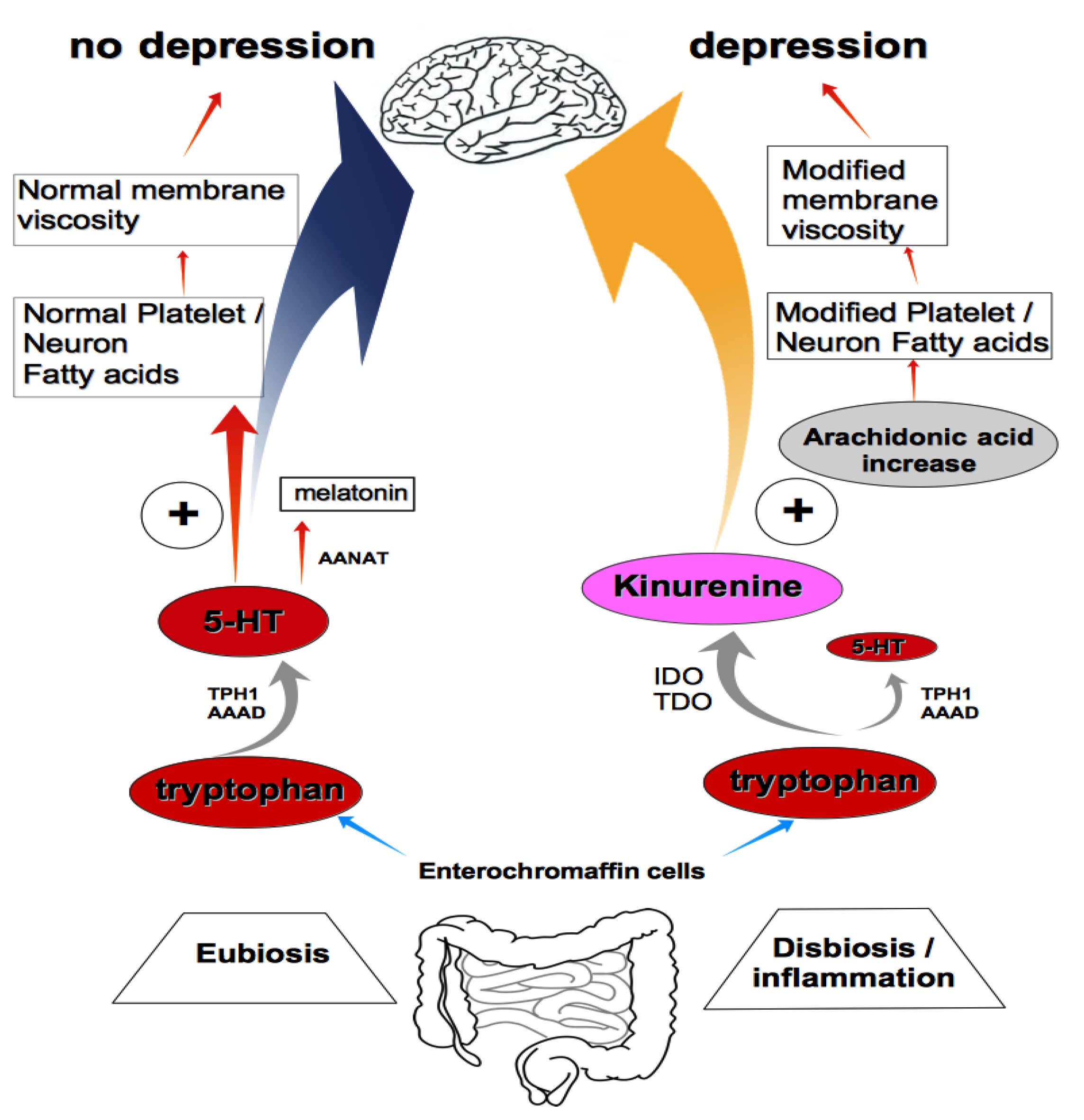
In conclusion, there could be two mechanisms that induce the reduction in 5-HT in psychopathological subjects, both centrally and peripherally: on the one hand, the excessive hyper- expression of the kinurenine pathway, and on the other, the substantial modification of the mobility of the membrane in the direction of increased fluidity due to an excessive concentration of AA [31] (Figure 2).
The combination of the excessive expression of kinurenine and the increased fluidity of the membrane has never been considered in the meaning of a simultaneous effect in the determinism of depressive conditions, such as MD and BD, and in stress.
This observation appears to be relevant for the treatment of depressive psychopathology as well as for further cardiovascular and bone damage [41,42].
Therefore, how to intervene?
Evidence supports the relationship between the microbiota and depression, confirmed by research conducted on alterations of the microbiota in depressive pathology [8,43,44,45]. In particular, recent studies have shown how and to what extent the intestinal microbiota is involved in the regulation of functional aspects of the brain and behavior, with particular regard to alterations in tryptophan metabolism and the serotonergic system. Research has been focused on the bidirectional communication between the brain and gut, looking at 5-HT as a molecular signaling factor in both the brain and the enteric nervous system, where 5-HT regulates functions such as secretion, vasodilation, peristalsis, and the perception of pain. The gut microbiota is a critical element of this axis and performs its function not only locally but on several levels. Alterations of 5-HT occur in disorders of the microbiota–gut–brain axis and in communication between the districts [8,46]. This axis explains the mood alterations in irritable bowel syndrome (IBS) and suggests that IBS pathogenesis is partly related to the dysfunctional control of 5-HT production by the gut microbiota. Furthermore, the development of IBS has been shown to be connected to tryptophan depletion. Mechanisms supporting the influence of the microbiota on the availability of tryptophan and therefore of 5-HT are possible through the enzymatic activities responsible for the degradation of tryptophan along the kinurenine pathway [47]. This pathway is involved in a reduction in the kinurenine/tryptophan ratio in germ-free mice (mice born and raised in the absence of microbiota). Furthermore, the alterations of IDO/tryptophan 2,3-dioxygenase (TDO) activity seem to have relevant clinical implications in various pathological conditions [48].
Interestingly, the conversion of tryptophan to 5-HT occurs predominantly in the intestine, in the enterochromaffin cells of the intestinal mucosa. As previously mentioned, the vast majority of 5-HT is not found in the CNS but in the gastrointestinal tract [49]. The levels of 5-HT vary depending on plasma tryptophan levels and on the current availability of tryptophan from nutrition [50].
Several species of enteric microorganisms, such as Lactobacillus, Bifidobacterium, Escherichia, Bacillus, and Saccharomyces, are able to produce neurotransmitters, including 5-HT. Unlike 5-HT, tryptophan produced by gut microbiota is permeable with respect to the blood–brain barrier and produces positive effects on mood through an increase in 5-HT levels in the brain [51]. In addition, gut microbiota can use tryptophan directly. Preclinical studies have reported direct and indirect mechanisms by which the gut microbiota is able to regulate tryptophan availability for kynurenine pathway metabolism, with downstream effects on CNS function [52].
Finally, impaired rapid eye movement (REM) sleep in depression is partially caused by the hyperactivation of REM-on cholinergic neurons. This is supported by the discovery that cholinergic stimulation leads to a more severe REM sleep inhibition in depressed patients compared to healthy controls [53]. Sleep disturbances are common in depressed subjects and also in subjects with dysbiosis and intestinal inflammation [54]. This is probably also attributable to an altered level of melatonin consequent to the altered level of its precursor, 5-HT. A recent study reports that total microbiome diversity was positively correlated with increased sleep efficiency and total sleep time and was negatively correlated with waking after sleep onset [55].

2. Conclusions

Membrane mobility changes the uptake of serotonin, which decreases in the case of increased fluidity and increases in the case of increased viscosity. The gut microbiota affects depression-like behavior through influencing brain tryptophan accessibility and the serotonergic system [56].
The overlapping of the two phenomena, a common finding in depressive psychopathology, helps to reduce the availability of serotonin and, therefore, its regulatory function on mood.
Furthermore, for the reasons previously analyzed and explained, inflammation, osteoporosis risk, and ischemic cardiovascular risk can also exist in psychopathology.
In this context, the microbiota–gut–brain axis, in the complexity of its interactions, appears crucial in governing our thoughts, actions, and weaknesses. For these reasons, we can hypothesize that “depression” should be understood as a biological and cultural synthesis of human existence.

Author Contributions

Conceptualization, M.C. and G.T.; writing—original draft preparation, M.C. and G.T.; writing—review and editing, G.T.; visualization, G.T. All authors have read and agreed to the published version of the manuscript.

Funding

This research received no external funding.

Conflicts of Interest

The authors declare no conflict of interest.

Abbreviations

AAarachidonic acid
BDbipolar disorder
CNScentral nervous system
DHAdocosahexaenoic acid
5-HT5-hydroxytryptamine, serotonin
IBSirritable bowel syndrome
IDOindoleamine 2,3-dioxygenase
MDmajor depression
REMrapid eye movement
TDOtryptophan 2,3-dioxygenase

References

  1. Lapin, I.P.; Oxenkrug, G.F. Intensification of the central serotoninergic processes as a possible determinant of the thymoleptic effect. Lancet 1969, 1, 32–39. [Google Scholar] [CrossRef]
  2. Frazer, A.; Pandey, G.N.; Mendels, J. Metabolism of Tryptophan in Depressive Disease. Arch. Gen. Psychiatry 1973, 29, 528–535. [Google Scholar] [CrossRef] [PubMed]
  3. Cowen, P.J.; Parry-Billings, M.; Newsholme, E.A. Decreased plasma tryptophan levels in major depression. J. Affect. Disord. 1989, 16, 27–31. [Google Scholar] [CrossRef]
  4. Gabbay, V.; Ely, B.A.; Babb, J.; Liebes, L. The possible role of the kynurenine pathway in anhedonia in adolescents. J. Neural. Transm. 2012, 119, 253–260. [Google Scholar] [CrossRef] [Green Version]
  5. Zhang, X.; Gainetdinov, R.R.; Beaulieu, J.M.; Sotnikova, T.D.; Burch, L.H.; Williams, R.B.; Schwartz, D.A.; Krishnan, R.R.; Caron, M.G. Loss-of-Function mutation in tryptophan hydroxylase-2 identified in unipolar major depression. Neuron 2005, 45, 11–16. [Google Scholar] [CrossRef] [Green Version]
  6. Lemonde, S.; Turecki, G.; Bakish, D.; Du, L.; Hrdina, P.D.; Bown, C.D.; Sequeira, A.; Kushwaha, N.; Morris, S.J.; Basak, A.; et al. Impaired repression at a 5-hydroxytryptamine 1A receptor gene polymorphism associated with major depression and suicide. J. Neurosci. 2003, 23, 8788–8799. [Google Scholar] [CrossRef] [Green Version]
  7. Hendriksen, E.; van Brgeijk, D.; Oosting, R.S.; Redegeld, F.A. Mast cells in neuroinflammation and brain disorders. Neurosci. Biobehav. Rev. 2017, 79, 119–133. [Google Scholar] [CrossRef]
  8. Traina, G. Mast cells in the brain- Old cells, new target. J. Integr. Neurosci. 2017, 16, S69–S83. [Google Scholar] [CrossRef]
  9. Traina, G. Mast cells in gut and brain and their potential role as an emerging therapeutic target for neural diseases. Front. Cell. Neurosci. 2019, 13, 345. [Google Scholar] [CrossRef]
  10. Takahashi, S. Reduction of Blood Platelet Serotonin Levels in Manic and Depressed Patients. Folia Psych. Neurol. Jap. 1976, 30, 476–486. [Google Scholar] [CrossRef]
  11. Stahl, S.M. The Human Platelet. A Diagnostic and Research Tool for the Study of Biogenic Amines in Psychiatric and Neurologic Disorders. Arch. Gen. Psychiatry 1977, 34, 509–516. [Google Scholar] [CrossRef] [PubMed]
  12. Pletscher, A.; Laubscher, A. Blood Platelets as Models for Neurons: Uses and Limitations. J. Neural Transm. Suppl. 1980, 16, 7–16. [Google Scholar]
  13. Da Prada, A.M.; Cesura, J.M.; Launay, J.G.; Richards, J.G. Platelets as a Model for Neurones? Cell. Mol. Life Sci. 1988, 44, 115–126. [Google Scholar] [CrossRef] [PubMed]
  14. Kim, H.L.; Plaisant, O.; Leboyer, M.; Gay, C.; Kamal, L.; Devynck, M.A.; Meyer, P. Reduction of Platelet Serotonin in Major Depression (Endogenous Depression). Comptes Rendus Seances Acad. Sci. III 1982, 295, 619–622. [Google Scholar]
  15. Musselman, D.L.; Tomer, A.; Manatunga, A.K.; Knight, B.T.; Porter, M.R.; Kasey, S.; Marzec, U.; Harker, L.A.; Nemeroff, C.B. Exaggerated Platelet Reactivity in Major Depression. Am. J. Psychiatry 1996, 153, 1313–1317. [Google Scholar]
  16. Camacho, A.; Dimsdale, J.E. Platelets and Psychiatry: Lessons Learned from Old and New Studies. Psychos. Med. 2000, 62, 326–336. [Google Scholar] [CrossRef] [Green Version]
  17. Plein, H.; Berk, M. The Platelet as a Peripheral Marker in Psychiatric Illness. Hum. Psychopharm. 2001, 16, 229–236. [Google Scholar] [CrossRef]
  18. Cocchi, M.; Tonello, L.S.; Tsaluchidu, S.; Puri, B.K. The use of Artificial Neural Networks to Study Fatty Acids in Neuropsychiatric Disorders. BMC Psychiatry 2008, 8, S3. [Google Scholar] [CrossRef] [Green Version]
  19. Cocchi, M.; Tonello, L. Bio Molecular Considerations in Major Depression and Ischemic Cardiovascular Disease. Cent. Nerv. Syst. Agents Med. Chem. 2010, 10, 97–107. [Google Scholar] [CrossRef]
  20. Benedetti, S.; Bucciarelli, S.; Canestrari, F.; Catalani, S.; Mandolini, S.; Marconi, V.; Mastrogiacomo, A.; Silvestri, R.; Tagliamonte, M.; Venanzini, R.; et al. Platelet’s Fatty Acids and Differential Diagnosis of Major Depression and Bipolar Disorder through the use of an Unsupervised Competitive- Learning Network Algorithm (SOM). Open J. Depress. 2014, 3, 52–73. [Google Scholar] [CrossRef] [Green Version]
  21. Mammadova-Bach, E.; Mauler, M.; Braun, A.; Duerschmied, D. Immuno-Thrombotic Effects of Platelet Serotonin; IntechOpen: London, UK, 2017. [Google Scholar]
  22. Walther, D.J.; Peter, J.U.; Winter, S.; Höltje, M.; Paulmann, N.; Grohmann, M.; Vowinckel, J.; Alamo-Bethencourt, V.; Wilhelm, C.S.; Ahnert-Hilger, G.; et al. Serotonylation of small GTPases is a signal transduction pathway that triggers platelet alpha-granule release. Cell 2003, 115, 851–862. [Google Scholar] [CrossRef] [Green Version]
  23. Cloutier, N.; Allaeys, I.; Marcoux, G.; Machlus, K.R.; Mailhot, B.; Zufferey, A.; Levesque, T.; Becker, Y.; Tessandier, N.; Melki, I.; et al. Platelets release pathogenic serotonin and return to circulation after immune complex-mediated sequestration. Proc. Nat. Acad. Sci. USA 2018, 115, E1550–E1559. [Google Scholar] [CrossRef] [Green Version]
  24. Zubenko, G.S.; Kopp, U.; Seto, T.; Firestone, L.L. Platelet Membrane Fluidity Individuals at Risk for Alzheimer’s Disease: A Comparison of Results From Fluorescence Spectroscopy and Electron Spin Resonance Spectroscopy. Psychopharmacology 1999, 145, 175–180. [Google Scholar] [CrossRef]
  25. Zubenko, G.S.; Cohen, B.M.; Reynolds, C.F.; Boller, F.; Malinakova, I.; Keefe, N. Platelet membrane fluidity in Alzheimer's disease and major depression. Am. J. Psychiatry 1987, 144, 860–868. [Google Scholar] [PubMed]
  26. van Rensburg, S.J.; Carstens, M.E.; Potocnik, F.C.V.; Aucamp, A.K.; Taljaard, J.J.F.; Koch, K.R. Membrane fluidity of platelets and erythrocytes in patients with Alzheimer's disease and the effect of small amounts of aluminium on platelet and erythrocyte membranes. Neurochem. Res. 1992, 17, 825–829. [Google Scholar] [CrossRef] [PubMed]
  27. Leiter, O.; Walker, T.L. Platelets in Neurodegenerative Conditions—Friend or Foe? Front. Immunol. 2020, 11, 747. [Google Scholar] [CrossRef]
  28. Padmakumar, M.; Van Raes, E.; Van Geet, C.; Freson, K. Blood platelet research in autism spectrum disorders: In search of biomarkers. Res. Pract. Thromb. Haemost. 2019, 3, 566–577. [Google Scholar] [CrossRef] [Green Version]
  29. Tonello, L.; Cocchi, M. The Cell Membrane: Is it a bridge from psychiatry to quantum consciousness? NeuroQuantology 2010, 1, 54–60. [Google Scholar] [CrossRef] [Green Version]
  30. Heron, D.S.; Shinitzky, M.; Hershkowitz, M.; Samuel, D. Lipid Fluidity Markedly Modulates the Binding of Serotonin to Mouse Brain Membranes. Proc. Natl. Acad. Sci. USA 1980, 77, 7463–7467. [Google Scholar] [CrossRef] [Green Version]
  31. Traina, G.; Cocchi, M. Mast Cells, Astrocytes, Arachidonic Acid: Do They Play a Role in Depression? Appl. Sci. 2020, 10, 3455. [Google Scholar] [CrossRef]
  32. Horrocks, L.A.; Farooqui, A.A. Docosahexaenoic acid in the diet: Its importance in maintenance and restoration of neural membrane function. Prostag. Leukotr. Essent. Fat. Acids 2004, 70, 361–372. [Google Scholar] [CrossRef] [PubMed]
  33. Hibbeln, J.R.; Palmer, J.W.; Davis, J.M. Are disturbances in lipid-protein interactions by phospholipase-A2 a predisposing factor in affective illness? Biol. Psychiatry 1989, 25, 945–961. [Google Scholar] [CrossRef]
  34. Walsh, M.T.; Dinan, T.G.; Condren, R.M.; Ryan, M.; Kenny, D. Depression is associated with an increase in the expression of the platelet adhesion receptor glycoprotein Ib. Life Sci. 2002, 70, 3155–3165. [Google Scholar] [CrossRef]
  35. Piletz, J.E.; Zhu, H.; Madakasira, S.; Pazzaglia, P.; DeVane, C.L.; Goldman, N.; Halaris, A. Elevated P-selectin on platelets in depression: Response to bupropion. J. Psychiatr. Res. 2000, 34, 397–404. [Google Scholar] [CrossRef]
  36. Müller, C.P.; Reichel, M.; Muhle, C.; Rhein, C. Brain membrane lipids in major depression and anxiety disorders. Bioch. Biophys. Acta 2015, 1851, 1052–1065. [Google Scholar] [CrossRef] [PubMed] [Green Version]
  37. Watala, C.; Golański, J.; Boncler, M.A.; Pietrucha, T.; Gwoździński, K. Membrane Lipid Fluidity of Blood Platelets: A Common Denominator That Underlies the Opposing Actions of Various Agents that Affect Platelet Activation in Whole Blood. Platelets 1998, 9, 315–327. [Google Scholar]
  38. Kalipatnapu, S.; Pucadyil, T.J.; Chattopadhyay, A. Membrane Organization and Dynamics of the Serotonin1A Receptor Monitored Using Fluorescence Microscopic Approaches. In Serotonin Receptors in Neurobiology; Chattopadhyay, A., Ed.; CRC Press/Taylor & Francis: Boca Raton, FL, USA, 2007; Chapter 3. [Google Scholar]
  39. Cocchi, M.; Tonello, L.; Amato, P.; De Lucia, A. Platelet and Brain Fatty acid transfer: Hypothesis on Arachidonic Acid and its relationship to Major Depression. J. Biol. Res. 2009, 82, 47–53. [Google Scholar] [CrossRef]
  40. Green, P.; Gispan-Herman, I.; Yadid, G. Increased Arachidonic Acid Concentration in the Brain of Flinders Sensitive Line Rats, an Animal Model of Epression. J. Lipid. Res. 2005, 46, 1093–1096. [Google Scholar] [CrossRef] [Green Version]
  41. Cocchi, M.; Tonello, L.; Lercker, G. Fatty acids, membrane viscosity, serotonin and ischemic heart disease. Lipids Health Dis. 2010, 9, 97. [Google Scholar] [CrossRef] [Green Version]
  42. Cocchi, M.; Tonello, L.; Gabrielli, F.; Pregnolato, M. Depression, osteoporosis, serotonin and cell membrane viscosity between biology and philosophical anthropology. Ann. Gen. Psychiatry 2011, 10, 9. [Google Scholar] [CrossRef] [Green Version]
  43. Bear, T.L.K.; Dalziel, J.E.; Coad, J.; Roy, N.C.; Butts, C.A.; Gopal, P.K. The Role of the Gut Microbiota in Dietary Interventions for Depression and Anxiety. Adv. Nutr. 2020, 11, 890–907. [Google Scholar] [CrossRef] [PubMed] [Green Version]
  44. Dinan, T.G.; Cryan, J.F. Melancholic microbes: A link between gut microbioma and depression? Neurogastroenterol. Motil. 2013, 25, 713–719. [Google Scholar] [CrossRef] [PubMed]
  45. Dinan, T.G.; Cryan, J.F. The impact of gut microbiota on brain and behaviour: Implications for psychiatry. Curr. Opin. Clin. Nutr. Metab. Care 2015, 18, 552–558. [Google Scholar] [CrossRef]
  46. Conte, C.; Sichetti, M.; Traina, G. Gut–Brain Axis: Focus on Neurodegeneration and Mast Cells. Appl. Sci. 2020, 10, 1828. [Google Scholar] [CrossRef] [Green Version]
  47. Gao, J.; Xu, K.; Liu, H.; Liu, G.; Bai, M.; Peng, C.; Li, T.; Yin, Y. Impact of the Gut Microbiota on Intestinal Immunity Mediated by Tryptophan Metabolism. Front. Cell. Infect. Microbiol. 2018, 8, 13. [Google Scholar] [CrossRef] [PubMed] [Green Version]
  48. Cervenka, I.; Agudelo, L.Z.; Ruas, J.L. Kynurenines: Tryptophan's Metabolites in Exercise, Inflammation, and Mental Health. Science 2017, 357, 9794. [Google Scholar] [CrossRef] [Green Version]
  49. Banskota, S.; Ghia, J.E.; Khan, W.I. Serotonin in the gut: Blessing or a curse. Biochimie 2019, 161, 56–64. [Google Scholar] [CrossRef]
  50. Jenkins, T.A.; Nguyen, J.C.D.; Polglaze, K.E.; Bertrand, P.P. Influence of tryptophan and serotonin on mood and cognition with a possible role of the gut-brain axis. Nutrients 2016, 8, 56. [Google Scholar] [CrossRef]
  51. Kaur, H.; Bose, C.; Mande, S.S. Tryptophan Metabolism by Gut Microbiome and Gut-Brain-Axis: An in silico Analysis. Front. Neurosci. 2019, 13, 1365. [Google Scholar] [CrossRef]
  52. Kennedy, P.J.; Cryan, J.F.; Dinan, T.G.; Clarke, G. Kynurenine Pathway Metabolism and the Microbiota-Gut-Brain Axis. Neuropharmacology 2017, 112, 399–412. [Google Scholar] [CrossRef]
  53. Berger, M.; Riemann, D.; Höchli, D.; Spiegel, R. The rapid-movement cholinergic sleep induction test of the eye with RS-86. Indicator of state or trait of depression? Arch. Gen. Psychiatry 1989, 46, 421–428. [Google Scholar] [CrossRef] [PubMed]
  54. Li, Y.; Hao, Y.; Fan, F.; Zhang, B. The Role of Microbiome in Insomnia, Circadian Disturbance and Depression. Front. Psychiatry 2018, 9, 669. [Google Scholar] [CrossRef] [PubMed] [Green Version]
  55. Smith, R.P.; Easson, C.; Lyle, S.M.; Kapoor, R.; Donnelly, C.P.; Davidson, E.J.; Parikh, E.; Lopez, J.V.; Tartar, J.L. Gut microbiome diversity is associated with sleep physiology in humans. PLoS ONE 2019, 14, e0222394. [Google Scholar] [CrossRef] [PubMed]
  56. Lukic, I.; Getselter, D.; Koren, O.; Elliott, E. Role of Tryptophan in Microbiota-Induced Depressive-Like Behavior: Evidence from Tryptophan Depletion Study. Front. Behav. Neurosci. 2019, 13, 123. [Google Scholar] [CrossRef] [Green Version]
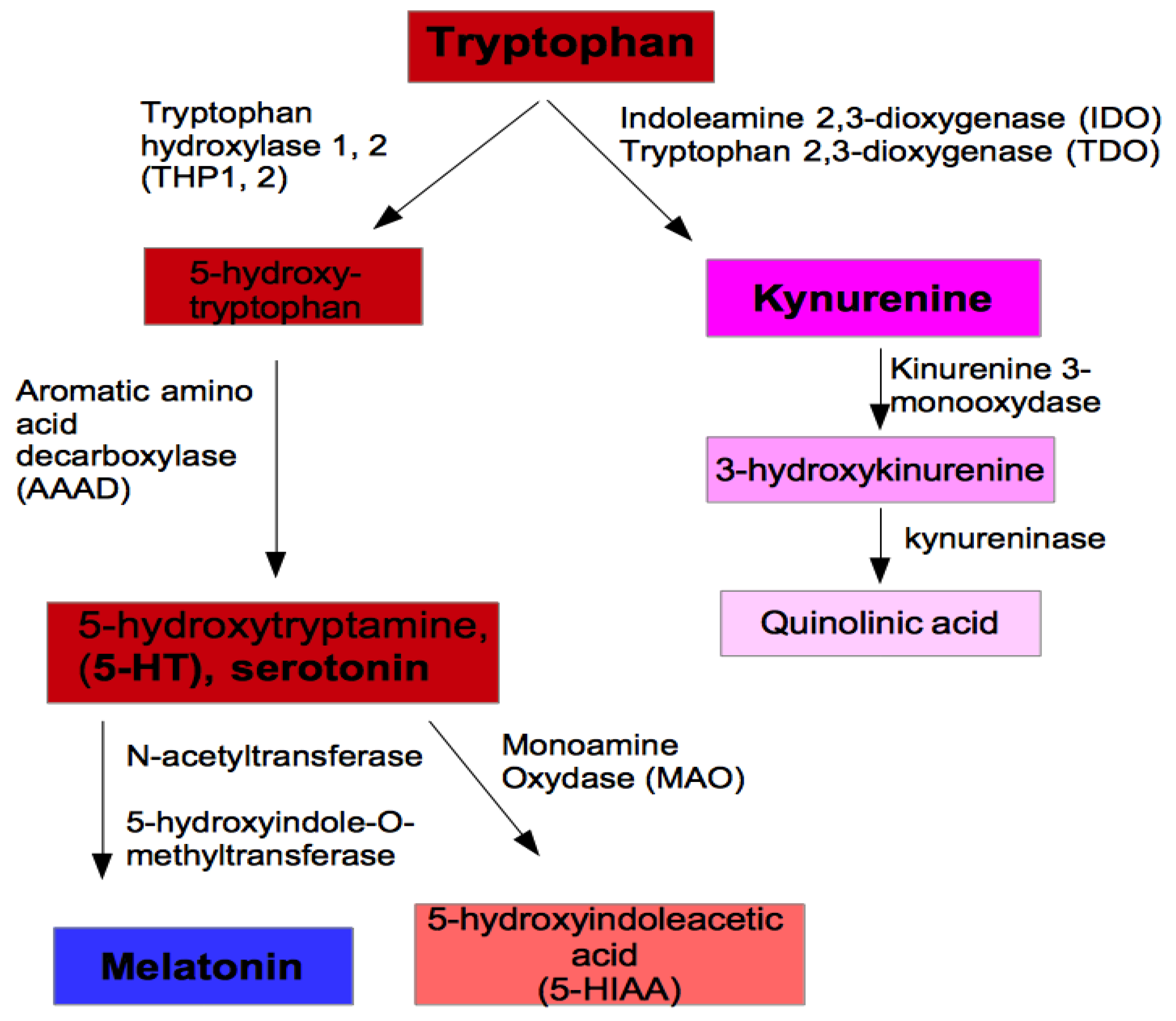
Figure 1. Metabolic pathways of tryptophan.
Applsci 10 04933 g001
Figure 2. Tryptophan pathway from gut to brain. On the left, a schematic description of the metabolic pathway of tryptophan from enterochromaffin cells to platelets is shown under normal conditions. To the right, the same hypothetical pathway is modified in conditions of intestinal inflammation or dysbiosis (loss of stability of gut microbiota) and under an alteration in the fluidity of the membrane due to the excessive presence of arachidonic acid. In this case, 5-HT would not be captured by platelets and would remain free, thus contributing to possible cardiovascular and bone damage. In addition, the enzymes IDO and TDO metabolize tryptophan to kinurenine and, furthermore, stop tryptophan from entering the brain. AAAD, aromatic L-amino acid decarboxylase; AANAT, N-acetyl transferase; IDO, indoleamine 2,3-dioxygenase; TDO, tryptophan 2,3-dioxygenase; 5-HT, 5-hydroxytryptamine, serotonin; TPH1, tryptophan hydroxylase 1.
Applsci 10 04933 g002