Article

Improvement of Bacterial Vaginosis by Oral Lactobacillus Supplement: A Randomized, Double-Blinded Trial

1
Department of Obstetrics and Gynecology, Kuo General Hospital, Tainan 70054, Taiwan
2
Research and Development Department, GenMont Biotech Incorporation, Tainan 74144, Taiwan
3
Department of Biomedical Sciences, Chung Shan Medical University, Taichung 40201, Taiwan
4
Department of Medical Research, Chung Shan Medical University Hospital, Taichung 40201, Taiwan
*
Author to whom correspondence should be addressed.
Contributed equally.
Appl. Sci. 2021, 11(3), 902; https://doi.org/10.3390/app11030902
Submission received: 16 December 2020 / Revised: 14 January 2021 / Accepted: 15 January 2021 / Published: 20 January 2021

Abstract

:

Featured Application

VGA-1, a combination formula of GMNL Lactobacillus strains, shows improvement effects in bacterial vaginosis (BV) patients and it can potentially be used for BV intervention as a single agent or in combination with the current antibiotics.

Abstract

Bacterial vaginosis (BV) is the most common vaginal infection globally, with a high recurrent rate after antibiotic treatment. Probiotics consumption is known to improve BV with different efficacy among species or strains. After in vitro selection of Lactobacillus strains with growth inhibition and preventing adhesion to HeLa cervical epithelial cells, a randomized and double-blinded trial of two Lactobacillus formula, namely, VGA-1 and VGA-2, in BV patients with Nugent scores of 4–10 was conducted. Among 37 subjects who completed the trial, we observed significantly decreased Nugent scores in both VGA-1 (n = 18) and VGA-2 (n = 19) consumption groups. VGA-1 consumption significantly improved vaginal discharge odor/color and itching at both 2-week and 4-week-consumption, but those only observed after a 4-week-consumption in the VGA-2 group. We also observed a tendency to reduce recurrent rates among enrolled participants after VGA-1 or VGA-2 consumption. The improvement effect of VGA-1/VGA-2 was associated with the significant reduction of interleukin-6 expression after 4-week-consumption and the restoration of normal vaginal microflora by quantitative polymerase chain reaction analysis. In conclusion, VGA-1 or VGA-2 displayed beneficial effects in BV patients, but the VGA-1 formula showed a better efficacy, potentially used for BV intervention.

Graphical Abstract

1. Introduction

Bacterial vaginosis (BV) is the most common vaginal infection in adult women of reproductive age, with an incidence rate of 15% to 50% [1]. Half of BV patients show no symptom; clinical symptoms of BV include a burning feeling during urination, itching around the outside of the vagina, and an increased vaginal pH (>4.5), abnormal color of vaginal discharge, and an unpleasant fishy odor [2]. The most widely used laboratory examination of BV is the Nugent scoring system (0 to 10), which is a Gram stain test for vaginal smears. Scores of 0 to 3, 4 to 6, or 7 to 10 are considered negative, intermediate, or indicative of BV, respectively [3]. The untreated BV increases the risk for infections of human immunodeficiency virus [4] or human papilloma virus infection [5], higher rates of premature birth [6], and low-birth-weight babies [7]. The change of vaginal microflora is a characteristic in BV [8]. Lactobacilli, including L. crispatus, L. gasseri, L. iners, and L. jensenii, are the most abundant normal flora in vaginas with the ability to secrete lactic acid to maintain the vaginal acidic environment, thus inhibiting the growth of pathogenic bacteria [9]. In addition, the productions of hydrogen peroxide (H2O2) and antimicrobial peptides by Lactobacilli are considered mechanisms in the suppression of pathogenic bacteria in vaginas [10]. The depletion of Lactobacilli together with the outgrowth of predominantly anaerobic microorganisms including Gardnerella vaginalis, Prevotella, Peptostreptococcus, and Bacteroides spp. are characteristics of BV [11]. The aerobic pathogenic bacteria in BV include Escherichia coli, Staphylococcus aureus, and group B Streptococcus [12]. Oral or intravaginal administration of antibiotics, such as metronidazole or clindamycin treatment, is the current medication for BV with a high cure rate; however, 20% to 75% of patients experience recurrence within 3 months [13]. This condition indicates the need to develop new agents for BV medication.
Given the unbalanced vaginal microflora in BV, probiotic consumption is used to ameliorate this disease through reconstruction of the vaginal microbial environment. Borges et al. proposed a criteria for the selection of probiotics used for BV intervention, including the adherence to vaginal epithelial cells, exclusion or reduction of the adherence of pathogenic bacteria, or persistent replication and production of antagonist substances such as lactic acid or H2O2 [14]. Martinez et al. conducted a randomized, double-blind, placebo-controlled trial of a probiotics formula consisting of L. rhamnosus GR-1 and L. reuteri RC-14 on BV for 60 days [15]. The participants exhibited a significantly increased percentage of restoration of normal vaginal microflora under microscopy examination [15]. Another BV trial of GR-1/RC-14 oral consumption for 28 days demonstrated a significantly high percentage of participants with normal vaginal microbiota by the Gram-stain Nugent score in the probiotic group [16]. In a meta-analysis of 10 blinded randomized controlled studies by Wang et al., the decreased Nugent score was found in subjects with probiotics-only therapy, but were not significant in those with probiotics–antibiotics combination therapy at a 30-day-consumption protocol [17]. In addition, the subgroup analysis revealed that the Nugent score significantly decreased in the mixed ethnic group but not in the white-dominant group [17]. Information about the effect of probiotics intervention in BV among Asian people is limited. Based on the ability for epithelial adherence, the exclusive ability of pathogenic bacteria on vaginal epithelial cells, and the inhibition of pathogenic bacteria-induced proinflammatory cytokine production in vaginal epithelial cells, we selected four GMNL Lactobacillus strains and designed two combination formula for them (called VGA-1 or VGA-2) and conducted a randomized, double-blinded trial on BV patients involving a 4-week-consumption of the treatment to compare their efficacy for BV improvement. The beneficial effects of VGA-1 or VGA-2 consumption were found including the significant reductions in Nugent scores, the color and odor of vaginal discharges, and itching, but VGA-1 displayed a better efficacy.

2. Materials and Methods

2.1. Bacterial Strains

The accuracy of L. rhamnosus GMNL-74 (also called GM-020®; China Center for Type Culture Collection (CCTCC) number: M203098; Bioresource Collection and Research Center (BCRC) number: BCRC910236), L. acidophilus GMNL-185 (CCTCC number: M2017764; BCRC number: BCRC910774), L. rhamnosus GMNL-680 (CCTCC number: M2017766; BCRC number: BCRC910775), and L. plantarum GMNL-682 (CCTCC number: M2017767; BCRC number: BCRC910776) was confirmed by 16S rRNA sequencing. L. rhamnosus GR-1 (ATCC 55826) and L. reuteri RC-14 (ATCC 55845) were purchased from the American Type Culture Collection (ATCC, Manassas, VA, USA). All Lactobacilli were cultured on de Man Rogosa and Sharpe (MRS) agar plates (BD Biosciences, Franklin Lakes, NJ, USA) at 37 °C for 72 h, under anaerobic conditions (BD GasPakTM 100 System, BD Biosciences, San Jose, CA, USA). A single colony of Lactobacilli was transferred to an MRS broth (BD Biosciences, Franklin Lakes, NJ, USA) and cultured at 37 °C for 24 h. Then, the activated culture were washed with phosphate buffered saline (PBS) and adjusted to appropriate concentrations for the in vitro experiments. Escherichia coli (BCRC 11634), Staphylococcus aureus (BCRC 10451), Candida albicans (BCRC 21538), Gardnerella vaginalis (BCRC 17040), and Streptococcus agalactiae (BCRC 10,787 and BCRC 10902) were purchased from BCRC, Hsinchu, Taiwan. E. coli (BCRC 11634) was cultured on Luria-Bertani (LB) agar plates (Merck, Darmstadt, Germany), S. aureus (BCRC 10451) and C. albicans (BCRC 21538) were cultured on tryptic soy agar (TSA) agar plates (BD Biosciences, San Jose, CA, USA), G. vaginalis (BCRC 17040) was cultured on Brain Heart Infusion (BHI) (BD Biosciences, San Jose, CA, USA) agar plates with 5% sheep blood, and S. agalactiae (BCRC 10787 and BCRC 10902) was cultured on TSA agar plates with 5% sheep blood at 37 °C under anaerobic conditions.

2.2. Human Cervical Epithelial Cell Line

HeLa cell line, the human cervical cancer cells, was obtained from the American Type Culture Collection (ATCC, Manassas, VA, USA) and cultured in Dulbecco’s Modified Eagle Medium (DMEM) medium (Thermo Fisher Scientific, Waltham, MA, USA), supplemented with 10% fetal bovine serum (FBS, Corning Life Sciences, Corning, NY, USA), 1% penicillin/streptomycin (PS, Thermo Fisher Scientific, Waltham, MA, USA) at 37 °C, in a humidified atmosphere containing 5% CO2.

2.3. Antibacterial Activity of Lactobacilli

The well diffusion assay was used to detect antibacterial activities of supernatants isolated from Lactobacilli, according to a previous report [18]. Briefly, 100 μL of 5 × 10⁸ cells/mL pathogenic bacteria suspended in PBS were swabbed on agar plates; 5 different pathogenic bacteria were individually used in the experiment including E. coli, S. aureus, G. vaginalis, and 2 strains of S. agalactiae. Then, 7 mm diameter wells were made with cork borers in the pathogen spread plates and. Next, 100 μL of cell-free supernatants isolated from respective activated Lactobacillus culture (2 × 10⁹ cells/mL) were loaded in the wells. After 48 h of incubation at 37 °C, the diameters of inhibition zones were recorded. The antibacterial activity of bioactive components produced by Lactobacilli was calculated using the formula: Arbitrary unit per mL (AU/mL) = (Diameter of the zone of clearance (mm) × 1000) ÷ Volume placed in the well (µL).

2.4. Adherence of Bacteria to HeLa Cells

A total of 4 × 10⁵ HeLa cells were seeded in 6-well plates and incubated at 37 °C for 24 h. After PBS washing, HeLa cells were incubated with 1 mL of Lactobacilli suspension at a concentration of 1.5 × 10⁹ cells/mL at 37 °C for 1 h, to allow the adhesion of Lactobacilli to cells. Six different Lactobacillus strains were individually used in the experiment including L. rhamnosus GMNL-74, L. acidophilus GMNL-185, L. rhamnosus GMNL-680, L. plantarum GMNL-682, L. rhamnosus GR-1, and L. reuteri RC-14. Then, the cells were washed twice with PBS to remove nonadhering bacteria. For quantitation of the adhering bacteria, the washed cells were lysed in PBS containing 1% triton × 100 and centrifuged at 2200 g for 5 min. After removing the supernatant, the pellet was resuspended and serially diluted in PBS and then the appropriate dilution was inoculated onto MRS agar plates, followed by 48-h incubation at 37 °C. After incubation, the colonies were counted to determine the adhering bacteria number.
To further examine the ability of Lactobacilli to block the adherence of pathogenic bacteria to cells, 3 different adhesion assays were performed, including exclusion assay, displacement assay, and competition assay [18]. The scheme of the experimental procedure is shown in Figure S1. Three different pathogens were individually used in these adhesion assays including G. vaginalis, S. agalactiae, and C. albicans. In the exclusion assay, the ability of Lactobacilli to interfere with the adherence of vaginal pathogenic bacteria to HeLa cells was examined. 4 × 10⁵ HeLa cells in 6-well plates were incubated with 1 mL of Lactobacilli suspension (1.5 × 10⁹/mL) at 37 °C for 1 h, to allow the adhesion of Lactobacilli to cells. Subsequently, the cells were washed twice with PBS to remove the nonadhering bacteria, and incubated again with 1 mL of bacterial pathogen suspension (1.5 × 10⁹/mL) at 37 °C for 1 h. Finally, the cells were washed, stained with Giemsa stain and examined under the microscope. In displacement assay, the ability of Lactobacilli to displace the already-adhered pathogenic bacteria on HeLa cells was examined. The experiment protocol was the same as the exclusion test, but the incubation sequence was reversed. HeLa cells were first incubated with pathogenic bacterial suspension and then incubated with Lactobacilli suspension. In a competitive assay, HeLa cells were simultaneously incubated with Lactobacilli and pathogenic bacteria suspensions at 37 °C for 1 h. For microscopy examinations, the cells were fixed with methanol, stained with Giemsa stain and the adherence of pathogenic bacteria was counted under the microscope (BX51, Olympus, Japan). The following formula was used to calculate the inhibition of pathogen adhesion to cells—inhibition rate (%) = [1 − (mean number of pathogens in Lactobacillus and pathogen co-treatment group/mean number of pathogens in pathogen treatment group)] × 100.

2.5. Clinical Study Protocol and Sample Collection

Forty-eight female subjects (aged between 22 and 53) diagnosed with bacterial vaginosis were initially enrolled in this randomized, double-blind study. This study was approved by the Institutional Review Board of Kuo General Hospital, Tainan, Taiwan (IRB No. A-16 K002, NCT 03116789 in ClinicalTrials.gov) and performed in accordance with the relevant guidelines and regulations. The inclusion criteria were as follows—Nugent score 4–10, age ranging from 20–55 years, normal menstruation cycle (18–35 days), without sexual intercourse within 72 h before each visit. The exclusion criteria were as follows—pregnancy, lactation, allergy of the probiotic product in this study, abnormal genital tract bleeding, congenital or acquired immunodeficiency, diabetes mellitus, mental disorders, cancers, applications of NuvaRing hormonal contraceptive vaginal ring, mechanical contraceptives (e.g., diaphragms, intrauterine contraceptive insert, except condom), and hormonal preparations (e.g., Vagifem, Ovestin and vaginal estrogens in reproductive period), mycotic vaginitis, any antibiotic and corticosteroid use during the trial or four weeks before the first visit, and participation in another clinical study. Subjects were informed of the purpose of the trial and gave their signed informed consents before participation. Upon diagnosis, the eligible subjects were randomly allocated to 2 groups: VGA-1 (n = 21, a combination of L. rhamnosus GMNL-74 and L. acidophilus GMNL-185 as 1 × 10¹⁰ CFU) and VGA-2 (n = 22, a combination of L. rhamnosus GMNL-680 and L. plantarum GMNL-682 as 1 × 10¹⁰ CFU), which received two oral capsules, once daily for 28 days. For allocation of the participants, the randomization sequence created using the SAS version 9.4 statistical software by Bestat Pharmaservices Corp. (Taipei, Taiwan) was used. The primary outcome of this clinical trial is the Nugent score. The study protocol is provided in the Supplementary Materials (Supplementary protocol) and the VGA-1 or VGA-2 supplements were prepared by GenMont Biotech Inc. (Tainan, Taiwan). The subjects were asked to return 2 weeks and 1 month later, for a gynecological examination to evaluate the outcome. At the first visit (baseline), the information of demographic characteristics, gynecological history, and current genital symptoms were collected. The subjects received probiotic capsules for the first 14 days at the first visit (baseline), returned their unused capsules and received probiotic capsules again for the next 14 days at 2 weeks (W2), and eventually gave back the unused capsules to principal investigators at 4 weeks (W4). Subject with poor medicine adherence (defined as less than 75%) were excluded from the study. BV recurrence of subjects after probiotic treatment, over the course of 3 months, was followed by reviewing the medical record or over the telephone.
During the trial, vaginal samples were collected at the first visit (baseline), 2 weeks (W2), and 1 month (W4) after oral consumptions of VGA-1 or VGA-2. At each visit, four dry swabs (COPAN Diagnosis Inc., Murrieta, CA, USA) from the vaginal wall were individually taken—the first sample was used for genomic DNA extraction to determine the vaginal microbiota, the second one was used for RNA extraction to evaluate vaginal inflammatory conditions, the third one was used for Gram staining to evaluate the vaginal flora, according to the Nugent score, and the last one was used for the BVBlue test to diagnose bacterial vaginosis. Samples of the first two vaginal swabs were immediately stored at −20 °C until assayed, while samples of the last two vaginal swabs were examined by the experienced microscopists at the Kuo General Hospital, within 2 h of collection. The swab collection sequence and the corresponding examination performed was based on the pretest results, as described in the Supplementary Information (Supplementary Table S1). Vaginal pH was measured using a pH-indicator strip (range of 0.0–6.0; Merck, Darmstadt, Germany). The method of clinical severity scores was modified according to Anderson et al. [19]. The score of color, odor, and secretion of vaginal discharge was evaluated by the principal investigators of the clinical trial, and the severity of vaginal itching was evaluated by participants. The color of vaginal discharge was scored from 0 to 4, with 0 being clear, 1 being white, 2 being yellow-white, 3 being yellow-green, and 4 being grey. The odor of vaginal discharge was scored from 0 to 4, with 0 being no odor, 1 being mild, 2 being moderate, 3 being severe, and 4 being very severe. The vaginal secretion was scored from 0 to 2, with 0 being small amount, 1 being moderate amount, and 2 being profuse amount. The vaginal itching was scored from 0 to 4, with 0 being no itch, 1 being mild, 2 being moderate, 3 being severe, and 4 being very severe.

2.6. RNA Extraction and Quantitative Reverse Transcription Polymerase Chain Reaction (qRT-PCR) Analysis of Inflammatory Cytokines

Total RNA of the obtained cell pellets from the vaginal swab was isolated using TRIzol reagent, according to the manufacturer’s instructions (Ambion Life Sciences, Thermo Fisher Scientific, Inc., Waltham, MA, USA). A total of 1 μg of RNA was reverse-transcribed to complementary DNA (cDNA) using MultiScribe™ Reverse Transcriptase (Thermo Fisher Scientific, Inc., Waltham, MA, USA). Expression of inflammatory cytokines was determined by qRT-PCR using the SYBR Green PCR Kit (QIAGEN, Hilden, Germany) and specific primers, at a final concentration of 0.6 μM. The sequences of specific primers targeting inflammatory cytokines (IL-1, IL-6, and TNF-α) were shown in the Supplementary Table S2. Human GAPDH was used as a housekeeping gene control for normalizing the mRNA levels of target genes. The fold changes of the inflammatory cytokines were calculated by the 2−ΔΔCt method for which the Ct values of samples collected at the first visit were set as the baseline.

2.7. DNA Extraction and Vaginal Microbiota Analysis

The vaginal swabs were suspended in 1 mL of sterile phosphate buffered saline (PBS) in the collect tube. After removing the swab with sterile forceps, 0.1 mL of the vaginal discharge was spread onto MRS agar plates (BD Biosciences, Franklin Lakes, NJ, USA) at 37 °C for 72 h, under anaerobic conditions (BD GasPakTM 100 System, BD Biosciences, San Jose, CA, USA) for the random amplified polymorphic DNA (RAPD)-PCR analysis. About 0.9 mL of the remaining vaginal discharge was centrifuged at 10,000 rpm for 5 min, and bacteria genomic DNA was extracted from the obtained pellets, using the DNeasy Blood and Tissue kit (QIAGEN, Hilden, Germany). DNA concentration was measured using a NanoDrop Lite spectrophotometer (Thermo Fisher Scientific, Waltham, MA, USA). qRT-PCR was used to determine the composition of the vaginal flora. All amplification reactions were performed in the presence of 10 ng purified genomic DNA, 0.6 μM specific primers targeting the vaginal flora, and Rotor-Gene SYBR Green PCR Kit (QIAGEN, Hilden, Germany) using a Rotor-Gene Q system (QIAGEN, Hilden, Germany). The primer sets are listed in Supplementary Table S3. 16S rRNA was used as the total bacteria level among samples. The changes of bacteria quantity were calculated using the 2−ΔΔCt method. For determination of colonization of the consumed Lactobacillus spp., the RAPD-PCR was used to distinguish bacteria strains by amplifying random segments of genomic DNA with arbitrary primers. RAPD-PCR was carried out in the presence of 20 ng purified genomic DNA, 0.8 μM arbitrary primers, and 2X SuperRed PCR Master Mix (BIOTOOLS CO., LTD., New Taipei City, Taiwan), using a Rotor-Gene Q system (QIAGEN, Hilden, Germany). The RAPD-PCR primers are listed in Supplementary Table S4. PCR thermal cycles were used as follows—initial DNA denaturation at 95 °C for 10 min; 35 cycles for 1 min at 93 °C, for 1 min at 36 °C, and for 1 min at 72 °C, and a final extension step at 72 °C for 7 min. After PCR amplification, the amplified DNA fragments were separated on 1.8% agarose gels, stained with ethidium bromide, and the different band patterns were observed under UV light.

2.8. Statistical Analysis

Data from in vitro experiments were expressed as the mean ± standard deviation (SD) from triplicate experiments and analyzed using one-way ANOVA, followed by Dunn’s multiple comparisons post-hoc test. For clinical data, continuous variables were analyzed by Wilcoxon matched-pairs signed rank test, and categorical variables were analyzed by chi-square test or Fisher’s exact test. Clinical symptoms were scored on the basis of symptom severity. The relationship between vaginal flora and Nugent score change was analyzed with Spearman’s rank correlation analysis. For all tests, a p value less than 0.05 was defined as a statistical significance and labeled as follows: * p < 0.05, ** p < 0.01, *** p < 0.001.

3. Results

3.1. Inhibition of Vaginal Pathogenic Bacteria by Different GMNL Lactobacillus Strains

To understand the suitability of Lactobacillus strains (L. rhamnosus GMNL-74, L. acidophilus GMNL-185, L. rhamnosus GMNL-680, L. plantarum GMNL-682) for clinical evaluation of BV, we first examined the growth inhibition ability of these Lactobacillus strains against vagina pathogenic bacteria, including E. coli, S. aureus, G. vaginalis, and S. agalactiae, by measuring the diameter of the inhibition zone (Table 1) and using L. rhamnosus GR-1 (GR-1) or L. reuteri RC-14 (RC-14)—which are well-recognized Lactobacillus strains with previously demonstrated beneficial effects of BV [15,16]—as controls. The results revealed that GMNL-680 or GMNL-682 displayed a greater inhibitory effect on S. aureus or S. agalactiae growth (Table 1), whereas GMNL-74 or GMNL-185 displayed a greater inhibitory effect on G. vaginalis (Table 1). GR-1 and RC-14 displayed a greater inhibitory effect on G. vaginalis but not on E. coli (Supplementary Table S5). RC-14 did not present any antibacterial activity against the two S. agalactiae strains (Supplementary Table S5). All four GMNL Lactobacillus strains showed comparable or better capabilities in comparison with the known strains of GR-1 or RC-14. Next, we examined the adherence ability of these Lactobacillus strains on the HeLa cervical epithelial cells, in accordance with a previous report [20]. The results revealed that GMNL-185 displayed the best adherent ability to HeLa cells, followed by GMNL-682 and GMNL-74 (Figure 1). GMNL-680 showed the lowest adherence to HeLa cells.
Next, we examined the effects of GMNL Lactobacillus strains on the adherence of three BV pathogenic bacteria, including G. vaginalis (Figure 2A–C, and Supplementary Results Figure S2) or S. agalactiae (Figure 2D–F, and Supplementary Results Figure S3), to the HeLa cervical epithelial cells with microscopy counting, after the Giemsa staining. The experimental protocols were further divided into three categories—exclusion, displacement, or competition (Figure S1). We observed that the GMNL-74 or GMNL-185 Lactobacillus strain displayed a similar effect on the inhibition of adherence of G. vaginalis, when compared to GR-1 or RC-14 under the exclusion method (Figure 2A). Whereas, the GMNL-680 or GMNL-682 Lactobacillus strain displayed a comparable adherence-inhibitory effect under the competition method (Figure 2C). The inhibitions of the adherence of S. agalactiae on HeLa cells for all four GMNL Lactobacillus strains were comparable to those of GR-1 or RC-14, under the exclusion (Figure 2D) or competition (Figure 2F) method. For displacemnet of S. agalactiae adherence to HeLa cells, all four GMNL Lactobacillus strains displayed inhibitory effects but GMNL-682 was the weakest one (Figure 2E). We further examined the effects of GMNL Lactobacillus in S. agalactiae-induced proinflammatory cytokines of the HeLa cells. The pre-incubation of GMNL Lactobacillus inhibited S. agalactiae-induced tumor necrosis factor (TNF)-α mRNA synthesis in HeLa cells (Figure S4). In summary, these four GMNL Lactobacillus strains displayed potentials for BV management, given their abilities in the growth inhibition of pathogenic bacteria and the adherence inhibition of pathogenic bacteria onto HeLa cells.

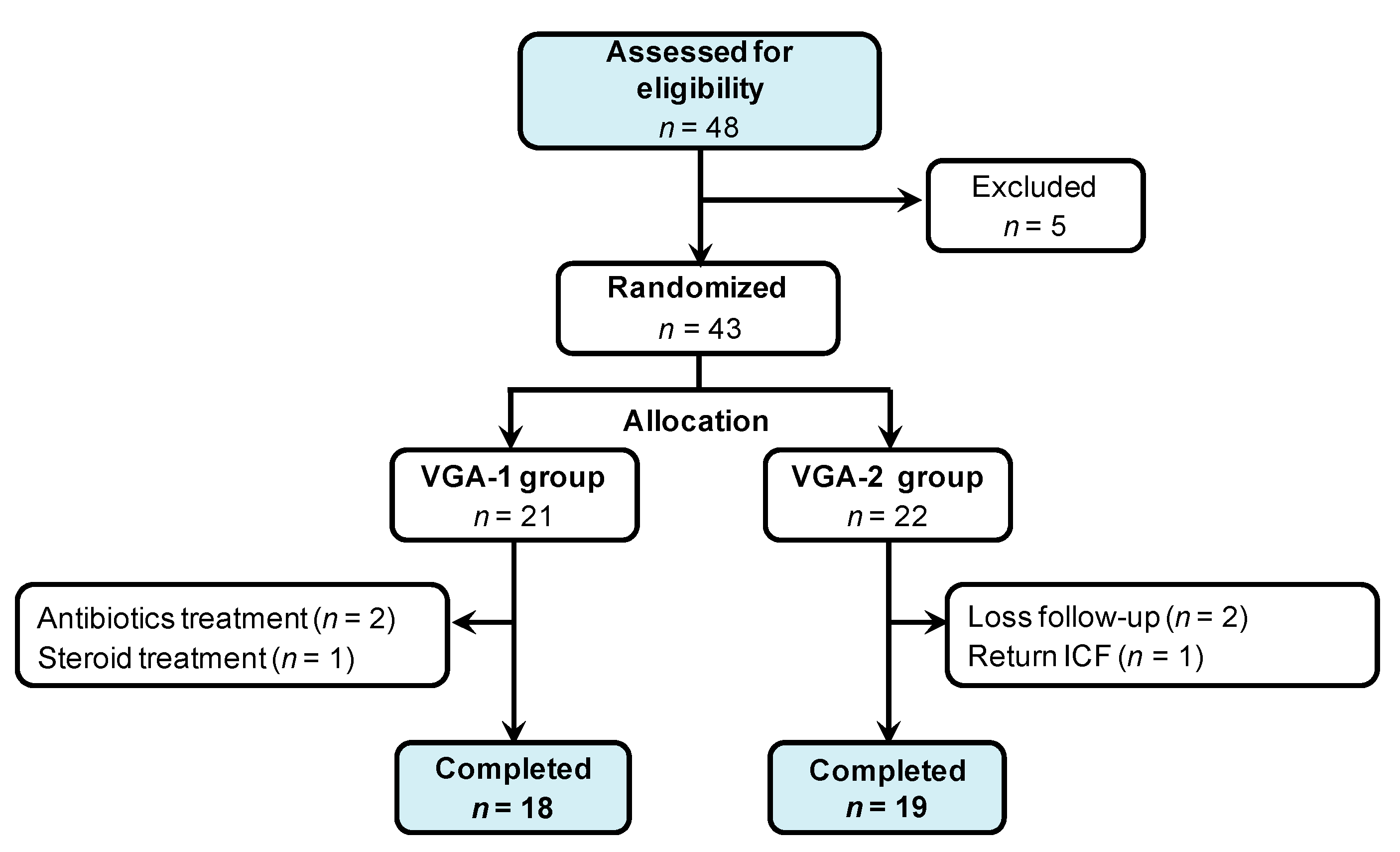
3.2. Randomized, Double-Blinded Trial of Two Formula of GMNL Lactobacillus Involving BV Patients

Next, we conducted a randomized, double-blinded trials to examine the effects of two Lactobacillus formula (VGA-1 which consists of GMNL-74 and GMNL-185 or VGA-2 which consists of GMNL-680 and GMNL-682) in BV patients, diagnosed by a Nugent score of 4–10. The recruitment and exclusion criteria are described in the Materials and Methods section, and the study design is shown in Figure 3. The 43 enrolled participants were randomly allocated into the VGA-1 (n = 21) or VGA-2 (n = 22) group, followed by daily oral consumption of 1 × 10¹⁰ CFU for 28 days. The participants underwent gynecological examination, including the Nugent score, BVBlue test, and vaginal pH determination, at week 2 and week 4. The genomic DNA and total RNA of vaginal swabs were also extracted to determine the vaginal microbiota and expression of inflammatory cytokines, respectively. A total of 6 enrolled participants were excluded or lost during follow-up, whereas the remaining 37 participants completed the trial. A total of 18 participants were subjected to VGA-1 consumption and 19 participants to VGA-2 consumption. Table 2 summarizes the characteristics of the 37 enrolled participants.

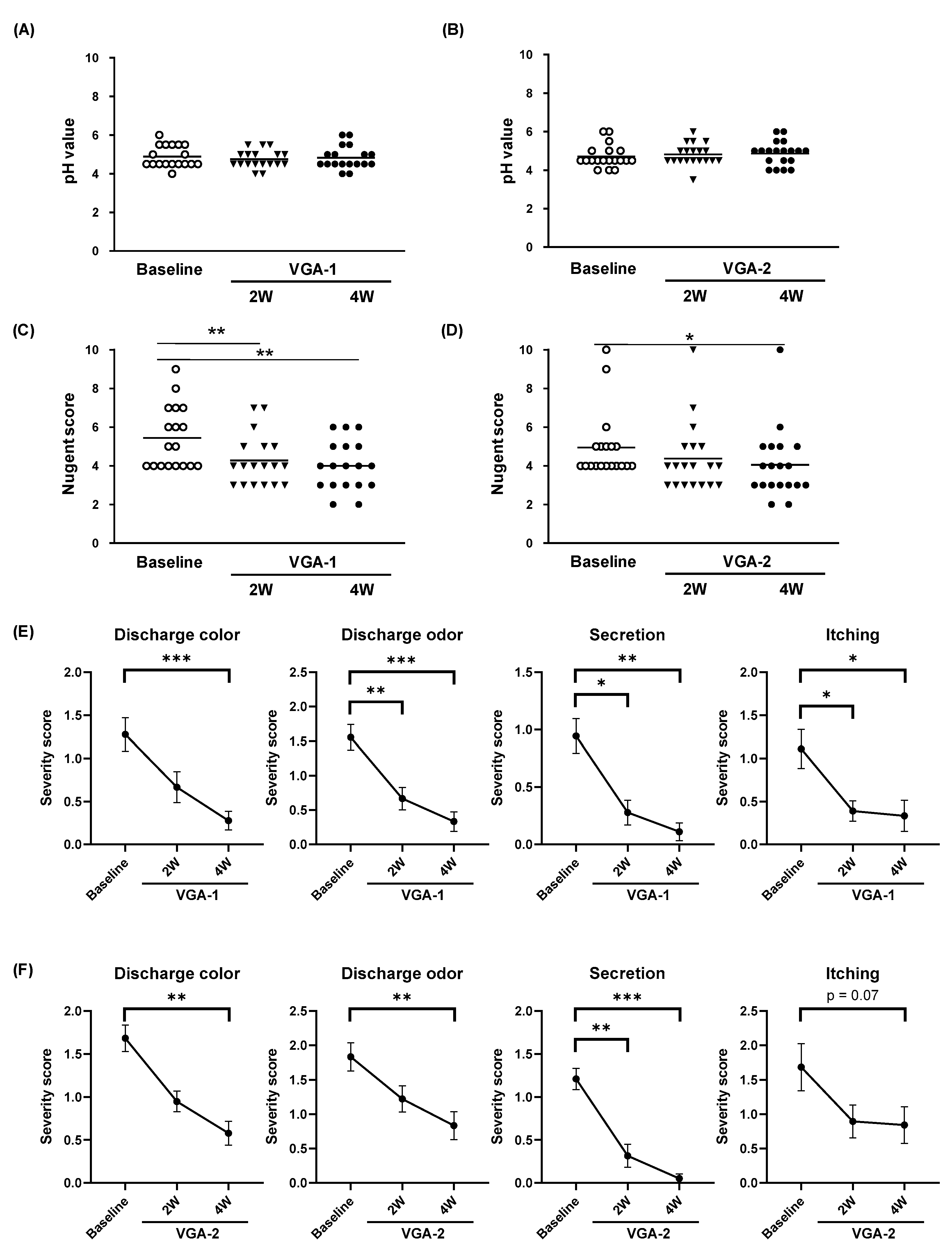
3.3. Effects of VGA-1 or VGA-2 Consumption on the Symptoms of BV Participants

The vagina is normally slightly acidic with a pH of 3.8–4.2, and a pH value greater than 4.5 is considered to be a sign of BV [9]. In comparison to the baseline, the vaginal pH values of VGA-1 (Figure 4A) or VGA-2 (Figure 4B) participants were unchanged at week 2 or week 4. The Nugent score is a Gram-stain scoring system for BV diagnosis [3]. We observed that the Nugent scores significantly decreased in VGA-1 (Figure 4C) and VGA-2 (Figure 4D) groups with the increase in consumption time, and the degree of decline was more evident in the VGA-1 group (Supplementary Table S6). The significantly improved color or odor of vaginal discharges with increasing consumption time were also observed in the VGA-1 (Figure 4E) and VGA-2 (Figure 4F) groups. The significant reduction of vaginal secretion in participants was also observed in the VGA-1 (Figure 4E) and VGA-2 (Figure 4F) groups. A reduced vaginal itching was observed in both groups, although that of the VGA-2 group did not reach a significant difference (Figure 4F, p = 0.07 for VGA-2 group). Given that the high recurrence rate of antibiotic treatment is the major problem of BV treatment [13], we further investigated the percentage and the interval time of recurrence among enrolled participants (Figure 5). The total recurrence percentage in the VGA-1 or VGA-2 group was reduced (from 44.4% to 16.7% in VGA-1 consumption group and 52.6% to 26.3% in VGA-2 consumption group), although it did not reach a statistical significance. Furthermore, the number of participants with an interval time for recurrence of less than three months reduced in VGA-1 and VGA-2 group (Figure 5). We also observed that one of eight or two of ten participants with BV history developed recurrence during three months’ follow-up, after 4-week-consumption of VGA-1 or VGA-2, respectively (Table 3). These data demonstrate that the daily consumption of VGA-1 or VGA-2 ameliorates BV symptoms, including the reduced Nugent score, vaginal discharge color/odor, and itching. It also probably reduced the recurrence percentage of BV women, albeit the results in the reduction of recurrence could not reveal a statistical significance.

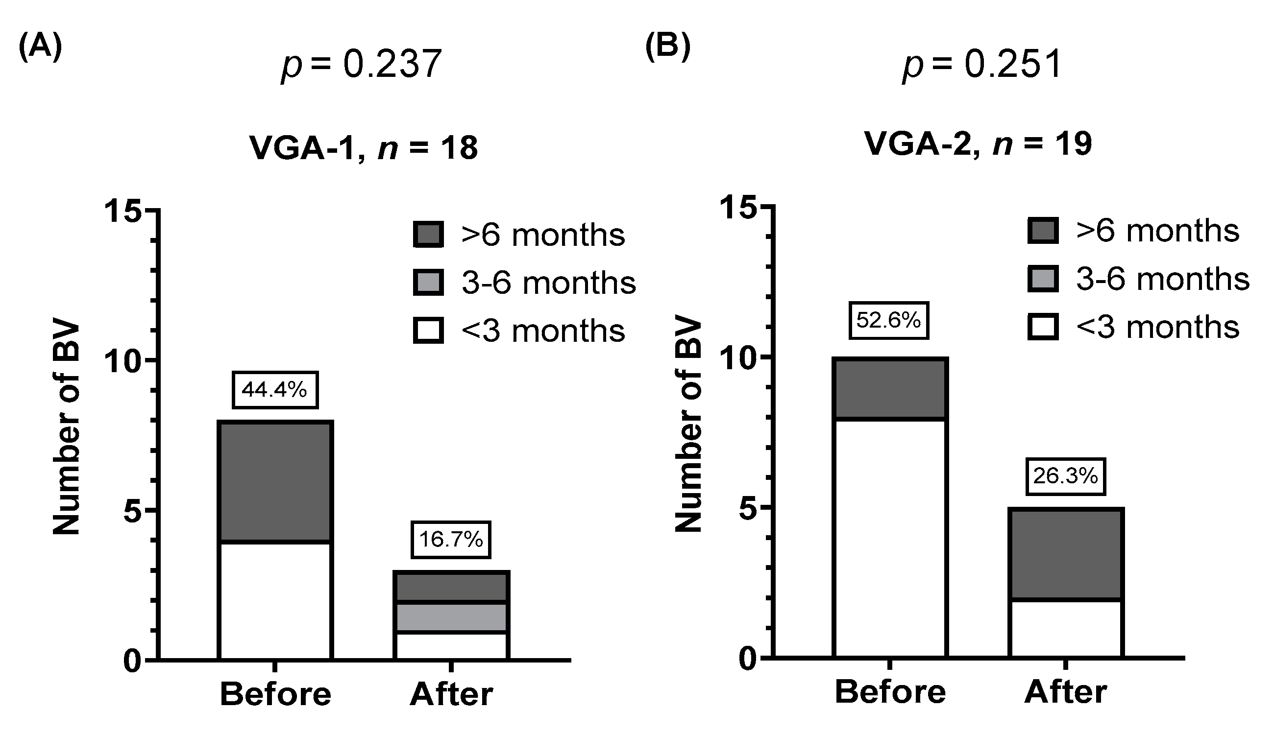
3.4. Colonization of GMNL Lactobacillus Strains among Participants of VGA-1 or VGA-2 Group

One hypothesis states that colonization on vaginal epithelium is one of the reason for the beneficial effects of probiotics consumption to BV [21]. To understand the colonization of GMNL Lactobacillus strains among participants of the VGA-1 or VGA-2 group, we spread the vaginal discharges onto MRS agar plates (Figure 6A) and cultured the Lactobacillus species, as described in the Materials and Methods section. Among the 37 enrolled participants, Lactobacillus colonies were found in No. S017 and No. S025 of the VGA-1 group. GMNL-74 or GMNL-185 Lactobacillus was confirmed by RAPD profiles, using arbitrary primers and PCR analysis (Figure 6B–E). From these data, the ameliorative effect of VGA-1 consumption in BV was suggested to be achieved mainly through a colonization independent mechanism.

3.5. Consumption of VGA-1 or VGA-2 Reduces interleukin (IL)-6 Expression in the Vaginal Specimens of BV Patients

Next, we examined the changes of pro-inflammatory cytokines, including IL-1β, IL-6, and TNF-α, in the vagina of BV patients, after consumption of VGA-1 or VGA-2. A qRT-PCR method was used to quantify the mRNA expression of these three pro-inflammatory cytokines in vaginal discharges. A decreased IL-6 expression was observed at week 4 samples of VGA-1 and VGA-2 consumption group (Figure 7, p = 0.012 for VGA-1 and p = 0.007 for VGA-2). The difference of IL-1β, IL-6, or TNF-α expression at week 2 was not significant compared to baseline or week 4, irrespective of the VGA-1 or VGA-2 group. The inhibition of TNF-α by GMNL Lactobacillus strains was also observed in HeLa cells after 1 h incubation of S. agalactiae (Figure S4). In addition, the four GMNL Lactobacillus strains displayed an induction effect to the IL-10 production in human peripheral blood mononuclear cells (Figure S5). These data suggest that the ameliorative effect of VGA-1 or VGA-2 consumption in BV could be mediated by their anti-inflammatory activity.

3.6. Changes in Vaginal Lactobacillus Species Composition in BV Patients after VGA-1 or VGA-2 Consumption
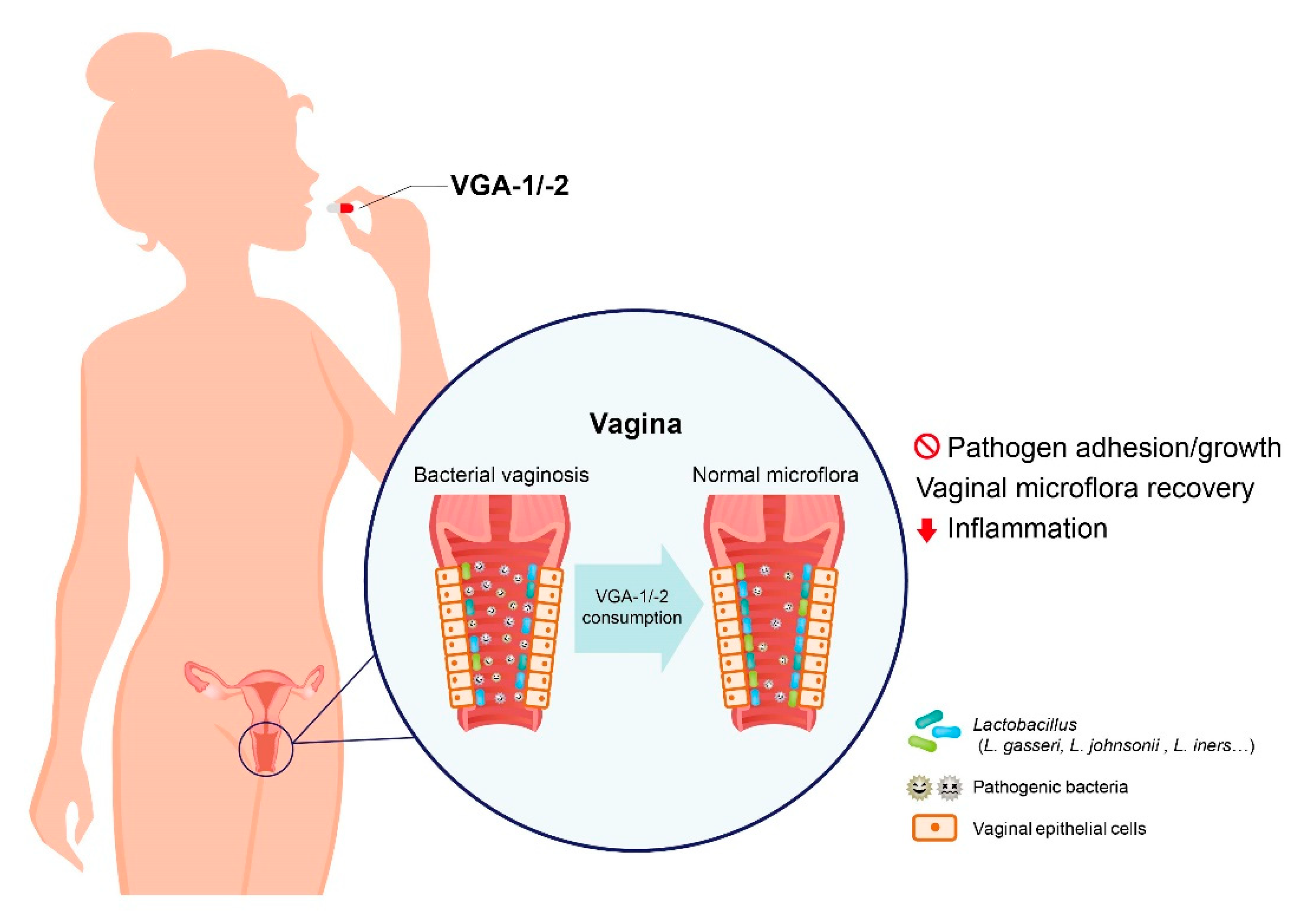
The decreased lactic acid or H2O2-produced Lactobacillus species in vagina was considered to be one of the mechanisms for BV [10]. To understand the alternations of vaginal Lactobacillus species composition among BV patients after VGA-1 or VGA-2 consumption, DNA was extracted from vaginal swabs at weeks 2 and 4, and the qPCR method was applied to quantify the changes in Lactobacillus species. As shown in Table 4, L. rhamnosus significantly increased by 2.2 ± 1.1-fold in the VGA-1 group (p = 0.0014), whereas L. crispatus significantly increased by 2.2 ± 1.1-fold in the VGA-2 group (p = 0.0397) at week 2. Significantly increased levels of L. gasseri and L. jensenii were observed in the VGA-1 consumption group (Table 4, 72.1 ± 39.5-fold for L. gasseri, p = 0.0426; 1.2 ± 0.3-fold for L. jensenii, p = 0.0426) at week 4. A significantly increased level of L. iners was found in the week-4 samples of the VGA-1 and VGA-2 consumption groups (Table 4, 43.8 ± 40.5-fold and p = 0.0041 in VGA-1 group; 1.8 ± 0.4-fold and p = 0.0388 in VGA-2 group). In addition, we observed a negative correlation between the Lactobacillus species and the Nugent scores of the VGA-1 or VGA-2 consumption group. In the VGA-1 group, the change of the Nugent score was significantly and negatively associated with the level of L. rhamnosus, L. acidophilus, or L. gasseri (Figure S6). A significant negative correlation between the Nugent score change and L. gasseri was observed in the VGA-2 consumption group (Figure S6). These data suggest that the ameliorative effects of VGA-1 or VGA-2 consumption in BV patients could be associated with increased vaginal Lactobacillus species.

4. Discussion

In the present study, we demonstrated the beneficial effects of four GMNL Lactobacillus strains in BV intervention through observations of the reduced Nugent score and other clinical symptoms, including vaginal itching and secretion, but not the reduction of vaginal pH. Hemalatha et al. showed that the mean vaginal pH in women with BV (pH 5.0) was higher than in those without BV (pH 4.6) [22]. Hemalatha et al. and Krauss-Silva et al. also demonstrated that vaginal pH (cutoff value: 4.5) was relatively sensitive but less specific for Nugent score-based BV detection [22,23]. Ya et al. showed that BV women who received probiotic capsules for 14 days had a lower incidence of BV recurrence at 2-month follow-up period, but a significantly reduced vaginal pH was observed at 11-month follow-up period [24]. Hence, we suggest that the reduction of vaginal pH under VGA-1/VGA-2 supplements might require a considerable time to achieve. In addition to BV, these four GMNL Lactobacillus strains might be used for the intervention of vaginal candidiasis, based on their capabilities for exclusion or competition in the adherence–inhibition of C. albicans to HeLa cells (Figure S7). The inhibitions of C. albicans adhesion on HeLa cells by all four GMNL Lactobacillus strains, using the exclusion method, were significantly stronger than that using GR-1 or RC-14 (Figure S7B), whereas all four GMNL Lactobacillus strains displayed the ability for inhibition of the adherence of C. albicans to HeLa cells, which is a comparable phenomenon to GR-1 or RC-14 in the displacement (Figure S7C) or competition (Figure S7D) method. The recurrence rates of BV among participants, after VGA-1 or VGA-2 consumption, showed reductions (Figure 5). Among the participants with BV history, the increased recurrent interval time of BV during follow-up was observed in the VGA-1 and VGA-2 groups (Supplementary Table S7; 75% (6/8) in VGA-1 group and 90% (9/10) in VGA-2 group). Amabebe et al. suggested that the recurrence of BV after treatment might result from the high densities of BV-associated bacteria in the rectum [25], which was observed in a study of bacterial loads in the vagina and rectum among pregnant women [26]. The intestinal tract serves as the primary reservoir for group B Streptococcus among women with group B Streptococcus infection, during pregnancy [27]. In our study, the inhibitory activity to common bacterial pathogens of the female gut or the genital tract, such as G. vaginalis, group B Streptococcus, E. coli, or S. aureus, was observed (Table 1), which might help in the control of intestinal bacterial pathogens and lead to the reduction of BV recurrence. Although the physiological reduction of vaginal Lactobacillus was found in menopausal women, it did not increase their BV risk [28]. Morison L. et al. found no significant changes in BV risk for the first six days of the menstrual cycle as compared to Day 14, but a significant increase for day 7–14 among African participants with a stable and high frequency of BV history was observed [29]. This meant that the menstrual cycle could be a factor to influence the risk of BV. Our trial only excluded pregnancy, and we did not include menstrual cycle status or menopause as a factor for the analysis, due to the lack of menstrual cycle records and the small sample size.
In our study, colonization of GMNL Lactobacillus strains was found in two participants in the VGA-1 group (Figure 6). In these cases, both participants were enrolled with a high initial Nugent score (score = 6 for S017 and score = 7 for S025). Significant increases in L. crispatus and L. gasseri were found in these cases, with the colonization of GMNL Lactobacillus strains in vagina (Supplementary Table S8, the fold changes of L. acidophilus, L. crispatus, or L. rhamnosus were 992.9/367.1/207.2 for S017 or 445.7/236.4/146.5 for S025, respectively) and their BV or intermediate vaginal flora shifting to normal flora after 4-week-comsuption of VGA-1. The low colonization frequency of GMNL Lactobacillus strains in our trial might be due to the insufficiency in oral consumption duration or dosage, low efficiency of the detection method, or a low Nugent score of the enrolled participants. Matrix-assisted laser desorption/ionization-time of flight–mass spectrometry (MALDI TOF-MS) was used for determination of the presence of orally consumed Lactobacilli in vagina. Yetfet et al. reported that the detection rate of L. rhamnosus GR-1 was 11% but without detectable L. reuteri RC-14, using MALDI TOF-MS in pregnant women at a high risk for preterm birth with normal vaginal flora (Nugent score ≤ 3), after a 2-month consumption [30]. The low detection rate of GMNL Lactobacillus strains in the vaginal swabs of our trial suggests the existence of other possibilities in addition to vaginal colonization of supplemented probiotics, such as immunomodulation or competitive inhibition of pathogens in the gastrointestinal tract. G. vaginalis infection might cause gut microbiota dysbiosis and inflammation through the upregulation of NF-κB, TNF-α, or myeloperoxidase in vivo. In a mouse study, the oral gavage of L. plantarum NK3 and B. longum NK49, which are anti-inflammatory probiotics, displayed inhibitory effects on NF-κB activation and TNF-α production and decreased G. vaginalis population in the vagina [31]. These findings suggest that the anti-inflammation activity of probiotics can be a mechanism for BV intervention. The induction of IL-10 and interferon (IFN)-γ through the four GMNL Lactobacillus strains was observed in healthy peripheral blood mononuclear cells (PMBCs) (Figure S5). Herny et al. previously observed that the treatment of IFN-γ rescued the S. aureus-induced severe injury of endothelial cells [32]. The IFN-γ production by neutrophils can facilitate bacterial clearance in a mouse model of bacterial pneumonia [33]. Although IFN-γ concentration in vaginal lavage fluid from BV women did not show significant changes in comparison with healthy women with vaginal pH less than 4.5, the IFN-γ level significantly decreased in the vaginal lavage fluid from healthy women with vaginal pH greater than 4.5 [34]. Thus, the induction of IFN-γ production from PMBCs through GMNL Lactobacillus strains might assist in the control of vaginal bacterial infection. Faure et al. noted a 100-fold decrease in IL-10 in pregnant women with adverse pregnancy outcome, in comparison with healthy controls [35]. The effects of VGA-1 or VGA-2 in pregnant women with BV should be studied in the future. IL-6 significantly increased in endocervical secretion of BV patients without human papilloma virus infection. Goepfert et al. noted that the 174 G > C polymorphism of IL-6 gene increased the risk for BV [36]. These reports suggest that IL-6 might serve as a pathogenic factor in BV. Manhanzva et al. recently observed that G. vaginalis induced IL-6 production in the VK2 endocervical cell line, and this phenomenon could be reduced by Lactobacillus isolates of women with optimal vaginal microflora [37]. Here, we observed the decreased IL-6 expression in vaginal swabs after 4-week-consupmtion of VGA-1 or VGA-2, which might reflect the status of normal-like vaginal microflora among participants.
The abundance of Lactobacillus species benefits vaginal health by preventing the colonizations of pathogenic bacteria with activity for lactic acid or H2O2 production [38,39]. The loss of diversity of Lactobacillus species is thought to be a biomarker for BV, and Lactobacillus profiling is used as a diagnosis tool for BV. Hütt et al. reported that the main composition of vaginal Lactobacillus strains included L. crispatus (56%), L. jensenii (26%), and L. gasseri (18%) [40]. All Lactobacillus isolates displayed antagonistic activity against G. vaginalis or E. coli and resistance to metronidazole [40], one of the current antibiotics used in BV treatment. Other reports indicated that a healthy vaginal microbiota predominantly consists of L. cripatus, L. jensenii, and L. iners [41,42]. In our study, increases in Lactobacillus species after 2- or 4-week consumption of VGA-1 or VGA-2 (including the increase in L. crispatus at week 2 for VGA-2 group), L. jensenii and L. gasseri at week 4 for VGA-1 group, and L. iners at week 4 in the VGA-1 and VGA-2 groups, were observed (Table 4). We also observed that the abundance of L. acidophilus in VGA-1 group was positively correlated with total Lactobacillus, L. gasseri, or L. crispatus (Figure S6). Thus, the consumption of VGA-1 or VGA-2 increased the abundance of vaginal Lactobacillus species to achieve beneficial effects to BV.
A limitation of this study was that this was not a placebo controlled trial. The main reason for not including a palcebo group was the medication rights of BV patients because they strongly suffer in vaginal itching and the strong fishy smell of vaginal secretion. In addition, probiotics interventions by oral consumption or vaginal implantation are widely considered to improve clinical symptoms of BV [43,44]. Martinez et al. conducted a trial to investigate GR-1/RC-14 and tinidazole’s efficacy combined with tinidazole single-agent treatment for 28 days. The Amsel test’s positive rate was decreased to 12.5% in a combination of antibiotics and probiotics group, whereas 50% of subjects in the placebo group remained positive in the Amsel test [16]. Vicariotto et al. conducted a trial of probiotics vaginal tablet in BV subjects, for 28 days or 56 days, and the results indicated that 58.3% of subjects with a probiotics vaginal tablet implantation, displayed a decreased Nugent score less than 4, whereas the reduction in Nugent score in the placebo group did not show a significant difference. Furthermore, the self-recovery time of subjects in the placebo group was more than 56 days in this trial [45]. These trials clearly demonstrate that probiotics interventions are beneficial to BV patients, which could be distinguished from placebo effects. The trials of medical interventions in BV without a placebo group are not uncommon, such as the trial of L. rhamnosus BMX 54 in BV subjects with concomitant HPV-infections [46] or vaginal lactoferrin administration in BV patients [47]. Based on these reasons, the goal of our trial was to compare the efficacy of two combinations of GNML Lactobacilli strains and the results indicated that the improvement effect of VGA-1 worked faster than VGA-2, as there was a reduction in vaginal discharge odor/color, secretion, and itching after 2-week-consumption (Figure 4E). The decreased Nugent score was observed in 66.7% or 77.8% of subjects in the VGA-1 group at 2-week or 4-week-consumption, respectively (Table S9). The decreased Nugent score was only observed in 52.6% or 57.9% of subjects in the VGA-2 group at 2-week or 4-week, respectively (Table S9). Although we could not rule out the spontaneous recovery of subjects in VGA-1 or VGA-2 consumption group, the differences in the efficiency in the improvement of clinical symptoms between the VGA-1 and VGA-2 group might reduce the concern of placebo effects. In the future, it is necessary to design a placebo controlled trial involving more subjects for VGA-1 to clearly prove the improvement effect in BV.

5. Conclusions

Our data demonstrated that four GMNL Lactobacillus strains (GMNL-74, -185, -680, and -682) inhibited the adherence of pathogenic bacteria on the surface of HeLa cells. A randomized, double-blind trial of oral consumptions of two formula of GMNL Lactobacillus strains of VGA-1 (GMNL-74 and GMNL-185) or VGA-2 (GMNL-680 and GMNL-682) displayed improvement effects on BV patients but VGA-1 displayed a better efficacy in the reductions in color/odor of vaginal discharges and vaginal secretions/itching. The prolonged time to relapse was also observed in both VGA-1 or VGA-2 consumption groups, and the beneficial effects were correlated with an increased abundance of Lactobacillus species and decreased IL-6 expression in vaginal swabs. We believe that the oral consumption of VGA-1 or VGA-2 can serve as a safe intervention for intermediate BV patients, with improvement of clinical symptoms, reduction of recurrence, and a prolonged relapse period, as presented in Figure 8, but it requires a placebo controlled trial involving more subjects for further validation. It also suggests that the combination of VGA-1 or VGA-2 with the currently used antibiotics indicates that BV patients can be considered in the future.

Supplementary Materials

The following are available online at https://www.mdpi.com/2076-3417/11/3/902/s1. Figure S1: Scheme of experimental procedure for the Lactobacillus strains on the adherence of pathogens to HeLa cervical epithelial cells. Figure S2: Effects of Lactobacillus strains on the adherence of Gardnerella vaginalis to HeLa cervical epithelial cells. Figure S3: Effects of Lactobacillus strains on Streptococcus agalactiae adherence to cervical epithelial cells. Figure S4: Inhibition of Lactobacillus strains on pathogenic bacteria-induced inflammation. Figure S5: Immune responses of human peripheral blood mononuclear cells to Lactobacillus strains. Figure S6: Correlations between Nugent score and vaginal flora. Figure S7: Effects of Lactobacillus strains on the adherence of Candida albicans to HeLa cervical epithelial cells. Table S1: Pretesting results of two vaginal swab specimens before starting the trial. Table S2: qRT-PCR primer sequence for inflammatory cytokines. Table S3: Species-specific primer sequence for PCR. Table S4: Arbitrary primer sequence for RAPD-PCR. Table S5: Growth inhibitory activity of Lactobacillus rhamnosus GR-1and Lactobacillus reuteri RC-14 against bacterial pathogens. Table S6: Characteristics of subjects during follow-up. Table S7: Interval time of bacterial vaginosis of subjects with BV history before and after probiotic intervention. Table S8: Lactobacillus contents of vaginal specimens isolated from BV patients S017 and S025 who received VGA-1 probiotics, at week 2 and week 4. Supplementary protocol: The study protocol of this randomized, double-blinded trial. Table S9. The number of BV subjects with decreased Nugent scores after VGA-1 or VGA-2 intervention.

Author Contributions

Conceptualization, I.-L.H., W.-H.T., and T.-C.L.; formal analysis, I.-L.H. and W.-H.T.; clinical investigation and data curation, T.-C.L., Y.-C.C., L.-C.K., and M.-S.H.; in vitro experiments, W.-L.Y. and S.-J.H.; trial monitoring, Y.-H.C.; writing—original draft preparation, I.-L.H., W.-H.T., and W.-W.C.; writing—review and editing, I.-L.H., W.-H.T., W.-W.C., and T.-C.L.; project administration, I.-L.H., W.-H.T., and T.-C.L.; funding acquisition, W.-H.T. All authors have read and agreed to the published version of the manuscript.

Funding

This research was funded by GenMont Biotech Incorporation (Tainan, Taiwan).

Institutional Review Board Statement

The study was conducted according to the guidelines of the Declaration of Helsinki, and approved by the Institutional Review Board of Kuo General Hospital, Tainan, Taiwan (IRB No. A-16 K002, August 2, 2016 of approval).

Informed Consent Statement

Informed consent was obtained from all subjects involved in the study.

Acknowledgments

We thank the Department of Laboratory Medicine in Kuo General Hospital for help with the physical examinations of vaginal discharges and Kai-Hung Wang (Department of Laboratory Medicine, Kuo General Hospital, Tainan, Taiwan) for his comments on the preparations of the manuscript draft.

Conflicts of Interest

This work was supported by GenMont Biotech Incorporation. I.-L.H., W.-H.T., W.-L.Y., Y.-H.C., and S.-J.H. are employed by GenMont Biotech Incorporation. T.-C.L., Y.-C.C., L.-C.K., M.-S.H., and W.-W.C. have no competing interest.

References

  1. Allsworth, J.E.; Peipert, J.F. Prevalence of bacterial vaginosis: 2001-2004 National Health and Nutrition Examination Survey data. Obstet. Gynecol. 2007, 109, 114–120. [Google Scholar] [CrossRef] [PubMed]
  2. Amsel, R.; Totten, P.A.; Spiegel, C.A.; Chen, K.C.; Eschenbach, D.; Holmes, K.K. Nonspecific vaginitis. Diagnostic criteria and microbial and epidemiologic associations. Am. J. Med. 1983, 74, 14–22. [Google Scholar] [CrossRef]
  3. Nugent, R.P.; Krohn, M.A.; Hillier, S.L. Reliability of diagnosing bacterial vaginosis is improved by a standardized method of gram stain interpretation. J. Clin. Microbiol. 1991, 29, 297–301. [Google Scholar] [CrossRef] [PubMed] [Green Version]
  4. Abdool Karim, S.S.; Baxter, C.; Passmore, J.S.; McKinnon, L.R.; Williams, B.L. The genital tract and rectal microbiomes: Their role in HIV susceptibility and prevention in women. J. Int. AIDS Soc. 2019, 22, e25300. [Google Scholar] [CrossRef] [PubMed] [Green Version]
  5. Gillet, E.; Meys, J.F.; Verstraelen, H.; Bosire, C.; De Sutter, P.; Temmerman, M.; Broeck, D.V. Bacterial vaginosis is associated with uterine cervical human papillomavirus infection: A meta-analysis. BMC Infect. Dis. 2011, 11, 10. [Google Scholar] [CrossRef] [Green Version]
  6. Peelen, M.J.; Luef, B.M.; Lamont, R.F.; de Milliano, I.; Jensen, J.S.; Limpens, J.; Hajenius, P.J.; Jorgensen, J.S.; Menon, R.; Group, P.B.W. The influence of the vaginal microbiota on preterm birth: A systematic review and recommendations for a minimum dataset for future research. Placenta 2019, 79, 30–39. [Google Scholar] [CrossRef] [Green Version]
  7. Hillier, S.L.; Nugent, R.P.; Eschenbach, D.A.; Krohn, M.A.; Gibbs, R.S.; Martin, D.H.; Cotch, M.F.; Edelman, R.; Pastorek, J.G., 2nd; Rao, A.V.; et al. Association between bacterial vaginosis and preterm delivery of a low-birth-weight infant. The Vaginal Infections and Prematurity Study Group. N. Engl. J. Med. 1995, 333, 1737–1742. [Google Scholar] [CrossRef]
  8. Lamont, R.F.; Sobel, J.D.; Akins, R.A.; Hassan, S.S.; Chaiworapongsa, T.; Kusanovic, J.P.; Romero, R. The vaginal microbiome: New information about genital tract flora using molecular based techniques. BJOG 2011, 118, 533–549. [Google Scholar] [CrossRef] [Green Version]
  9. Donati, L.; Di Vico, A.; Nucci, M.; Quagliozzi, L.; Spagnuolo, T.; Labianca, A.; Bracaglia, M.; Ianniello, F.; Caruso, A.; Paradisi, G. Vaginal microbial flora and outcome of pregnancy. Arch. Gynecol. Obstet. 2010, 281, 589–600. [Google Scholar] [CrossRef]
  10. Aroutcheva, A.; Gariti, D.; Simon, M.; Shott, S.; Faro, J.; Simoes, J.A.; Gurguis, A.; Faro, S. Defense factors of vaginal lactobacilli. Am. J. Obstet. Gynecol. 2001, 185, 375–379. [Google Scholar] [CrossRef]
  11. Livengood, C.H. Bacterial vaginosis: An overview for 2009. Rev. Obstet. Gynecol. 2009, 2, 28–37. [Google Scholar] [PubMed]
  12. Jahic, M.; Mulavdic, M.; Nurkic, J.; Jahic, E.; Nurkic, M. Clinical characteristics of aerobic vaginitis and its association to vaginal candidiasis, trichomonas vaginitis and bacterial vaginosis. Med. Arch. 2013, 67, 428–430. [Google Scholar] [CrossRef] [Green Version]
  13. Bradshaw, C.S.; Morton, A.N.; Hocking, J.; Garland, S.M.; Morris, M.B.; Moss, L.M.; Horvath, L.B.; Kuzevska, I.; Fairley, C.K. High recurrence rates of bacterial vaginosis over the course of 12 months after oral metronidazole therapy and factors associated with recurrence. J. Infect. Dis. 2006, 193, 1478–1486. [Google Scholar] [CrossRef] [PubMed] [Green Version]
  14. Borges, S.; Silva, J.; Teixeira, P. The role of lactobacilli and probiotics in maintaining vaginal health. Arch. Gynecol. Obstet. 2014, 289, 479–489. [Google Scholar] [CrossRef] [PubMed]
  15. Reid, G.; Charbonneau, D.; Erb, J.; Kochanowski, B.; Beuerman, D.; Poehner, R.; Bruce, A.W. Oral use of Lactobacillus rhamnosus GR-1 and L. fermentum RC-14 significantly alters vaginal flora: Randomized, placebo-controlled trial in 64 healthy women. FEMS Immunol. Med. Microbiol. 2003, 35, 131–134. [Google Scholar] [CrossRef] [Green Version]
  16. Martinez, R.C.; Franceschini, S.A.; Patta, M.C.; Quintana, S.M.; Gomes, B.C.; De Martinis, E.C.; Reid, G. Improved cure of bacterial vaginosis with single dose of tinidazole (2 g), Lactobacillus rhamnosus GR-1, and Lactobacillus reuteri RC-14: A randomized, double-blind, placebo-controlled trial. Can. J. Microbiol. 2009, 55, 133–138. [Google Scholar] [CrossRef]
  17. Wang, Z.; He, Y.; Zheng, Y. Probiotics for the Treatment of Bacterial Vaginosis: A Meta-Analysis. Int. J. Environ. Res. Public Health 2019, 16, 3859. [Google Scholar] [CrossRef] [Green Version]
  18. Tagg, J.R.; McGiven, A.R. Assay system for bacteriocins. Appl. Microbiol. 1971, 21, 943. [Google Scholar] [CrossRef]
  19. Anderson, M.R.; Klink, K.; Cohrssen, A. Evaluation of vaginal complaints. JAMA 2004, 291, 1368–1379. [Google Scholar] [CrossRef]
  20. Coman, M.M.; Verdenelli, M.C.; Cecchini, C.; Silvi, S.; Orpianesi, C.; Caspani, M.; Mondello, F.; Cresci, A. In vitro evaluation on HeLa cells of protective mechanisms of probiotic lactobacilli against Candida clinical isolates. J. Appl. Microbiol. 2015, 119, 1383–1390. [Google Scholar] [CrossRef] [Green Version]
  21. Reid, G.; Bruce, A.W.; Fraser, N.; Heinemann, C.; Owen, J.; Henning, B. Oral probiotics can resolve urogenital infections. FEMS Immunol. Med. Microbiol. 2001, 30, 49–52. [Google Scholar] [CrossRef] [PubMed]
  22. Hemalatha, R.; Ramalaxmi, B.A.; Swetha, E.; Balakrishna, N.; Mastromarino, P. Evaluation of vaginal pH for detection of bacterial vaginosis. Indian J. Med. Res. 2013, 138, 354–359. [Google Scholar] [PubMed]
  23. Krauss-Silva, L.; Almada-Horta, A.; Alves, M.B.; Camacho, K.G.; Moreira, M.E.; Braga, A. Basic vaginal pH, bacterial vaginosis and aerobic vaginitis: Prevalence in early pregnancy and risk of spontaneous preterm delivery, a prospective study in a low socioeconomic and multiethnic South American population. BMC Pregnancy Childbirth 2014, 14, 107. [Google Scholar] [CrossRef] [PubMed]
  24. Ya, W.; Reifer, C.; Miller, L.E. Efficacy of vaginal probiotic capsules for recurrent bacterial vaginosis: A double-blind, randomized, placebo-controlled study. Am. J. Obstet. Gynecol. 2010, 203, 120.e1–120.e6. [Google Scholar] [CrossRef] [PubMed]
  25. Amabebe, E.; Anumba, D.O.C. Female Gut and Genital Tract Microbiota-Induced Crosstalk and Differential Effects of Short-Chain Fatty Acids on Immune Sequelae. Front. Immunol. 2020, 11, 2184. [Google Scholar] [CrossRef]
  26. El Aila, N.A.; Tency, I.; Saerens, B.; De Backer, E.; Cools, P.; dos Santos Santiago, G.L.; Verstraelen, H.; Verhelst, R.; Temmerman, M.; Vaneechoutte, M. Strong correspondence in bacterial loads between the vagina and rectum of pregnant women. Res. Microbiol. 2011, 162, 506–513. [Google Scholar] [CrossRef]
  27. Dillon, H.C., Jr.; Gray, E.; Pass, M.A.; Gray, B.M. Anorectal and vaginal carriage of group B streptococci during pregnancy. J. Infect. Dis. 1982, 145, 794–799. [Google Scholar] [CrossRef]
  28. Cauci, S.; Driussi, S.; De Santo, D.; Penacchioni, P.; Iannicelli, T.; Lanzafame, P.; De Seta, F.; Quadrifoglio, F.; de Aloysio, D.; Guaschino, S. Prevalence of bacterial vaginosis and vaginal flora changes in peri- and postmenopausal women. J. Clin. Microbiol. 2002, 40, 2147–2152. [Google Scholar] [CrossRef] [Green Version]
  29. Morison, L.; Ekpo, G.; West, B.; Demba, E.; Mayaud, P.; Coleman, R.; Bailey, R.; Walraven, G. Bacterial vaginosis in relation to menstrual cycle, menstrual protection method, and sexual intercourse in rural Gambian women. Sex Transm. Infect. 2005, 81, 242–247. [Google Scholar] [CrossRef] [Green Version]
  30. Yefet, E.; Colodner, R.; Strauss, M.; Gam Ze Letova, Y.; Nachum, Z. A Randomized Controlled Open Label Crossover Trial to Study Vaginal Colonization of Orally Administered Lactobacillus Reuteri RC-14 and Rhamnosus GR-1 in Pregnant Women at High Risk for Preterm Labor. Nutrients 2020, 12, 1141. [Google Scholar] [CrossRef] [Green Version]
  31. Kim, D.E.; Kim, J.K.; Han, S.K.; Jang, S.E.; Han, M.J.; Kim, D.H. Lactobacillus plantarum NK3 and Bifidobacterium longum NK49 Alleviate Bacterial Vaginosis and Osteoporosis in Mice by Suppressing NF-kappaB-Linked TNF-alpha Expression. J. Med. Food 2019, 22, 1022–1031. [Google Scholar] [CrossRef]
  32. Beekhuizen, H.; van de Gevel, J.S. Gamma interferon confers resistance to infection with Staphylococcus aureus in human vascular endothelial cells by cooperative proinflammatory and enhanced intrinsic antibacterial activities. Infect. Immun. 2007, 75, 5615–5626. [Google Scholar] [CrossRef] [PubMed] [Green Version]
  33. Yamada, M.; Gomez, J.C.; Chugh, P.E.; Lowell, C.A.; Dinauer, M.C.; Dittmer, D.P.; Doerschuk, C.M. Interferon-gamma production by neutrophils during bacterial pneumonia in mice. Am. J. Respir. Crit. Care Med. 2011, 183, 1391–1401. [Google Scholar] [CrossRef] [PubMed] [Green Version]
  34. Fan, S.R.; Liu, X.P.; Liao, Q.P. Human defensins and cytokines in vaginal lavage fluid of women with bacterial vaginosis. Int. J. Gynaecol. Obstet. 2008, 103, 50–54. [Google Scholar] [CrossRef]
  35. Faure, E.; Faure, K.; Figeac, M.; Kipnis, E.; Grandjean, T.; Dubucquoi, S.; Villenet, C.; Grandbastien, B.; Brabant, G.; Subtil, D.; et al. Vaginal Mucosal Homeostatic Response May Determine Pregnancy Outcome in Women With Bacterial Vaginosis: A Pilot Study. Medicine (Baltimore) 2016, 95, e2668. [Google Scholar] [CrossRef] [PubMed]
  36. Goepfert, A.R.; Varner, M.; Ward, K.; Macpherson, C.; Klebanoff, M.; Goldenberg, R.L.; Mercer, B.; Meis, P.; Iams, J.; Moawad, A.; et al. Differences in inflammatory cytokine and Toll-like receptor genes and bacterial vaginosis in pregnancy. Am. J. Obstet. Gynecol. 2005, 193, 1478–1485. [Google Scholar] [CrossRef]
  37. Manhanzva, M.T.; Abrahams, A.G.; Gamieldien, H.; Froissart, R.; Jaspan, H.; Jaumdally, S.Z.; Barnabas, S.L.; Dabee, S.; Bekker, L.G.; Gray, G.; et al. Inflammatory and antimicrobial properties differ between vaginal Lactobacillus isolates from South African women with non-optimal versus optimal microbiota. Sci. Rep. 2020, 10, 6196. [Google Scholar] [CrossRef] [PubMed] [Green Version]
  38. Charlier, C.; Cretenet, M.; Even, S.; Le Loir, Y. Interactions between Staphylococcus aureus and lactic acid bacteria: An old story with new perspectives. Int. J. Food Microbiol. 2009, 131, 30–39. [Google Scholar] [CrossRef]
  39. Fredricks, D.N.; Fiedler, T.L.; Thomas, K.K.; Mitchell, C.M.; Marrazzo, J.M. Changes in vaginal bacterial concentrations with intravaginal metronidazole therapy for bacterial vaginosis as assessed by quantitative PCR. J. Clin. Microbiol. 2009, 47, 721–726. [Google Scholar] [CrossRef] [Green Version]
  40. Hutt, P.; Lapp, E.; Stsepetova, J.; Smidt, I.; Taelma, H.; Borovkova, N.; Oopkaup, H.; Ahelik, A.; Roop, T.; Hoidmets, D.; et al. Characterisation of probiotic properties in human vaginal lactobacilli strains. Microb. Ecol. Health Dis. 2016, 27, 30484. [Google Scholar] [CrossRef]
  41. Pavlova, S.I.; Kilic, A.O.; Kilic, S.S.; So, J.S.; Nader-Macias, M.E.; Simoes, J.A.; Tao, L. Genetic diversity of vaginal lactobacilli from women in different countries based on 16S rRNA gene sequences. J. Appl. Microbiol. 2002, 92, 451–459. [Google Scholar] [CrossRef] [PubMed]
  42. Zhou, X.; Bent, S.J.; Schneider, M.G.; Davis, C.C.; Islam, M.R.; Forney, L.J. Characterization of vaginal microbial communities in adult healthy women using cultivation-independent methods. Microbiology (Reading) 2004, 150, 2565–2573. [Google Scholar] [CrossRef] [PubMed] [Green Version]
  43. van de Wijgert, J.; Verwijs, M.C. Lactobacilli-containing vaginal probiotics to cure or prevent bacterial or fungal vaginal dysbiosis: A systematic review and recommendations for future trial designs. BJOG 2020, 127, 287–299. [Google Scholar] [CrossRef] [PubMed]
  44. Basavaprabhu, H.N.; Sonu, K.S.; Prabha, R. Mechanistic insights into the action of probiotics against bacterial vaginosis and its mediated preterm birth: An overview. Microb. Pathog. 2020, 141, 104029. [Google Scholar] [CrossRef] [PubMed]
  45. Vicariotto, F.; Mogna, L.; Del Piano, M. Effectiveness of the two microorganisms Lactobacillus fermentum LF15 and Lactobacillus plantarum LP01, formulated in slow-release vaginal tablets, in women affected by bacterial vaginosis: A pilot study. J. Clin. Gastroenterol. 2014, 48 (Suppl. 1), 106–112. [Google Scholar] [CrossRef]
  46. Palma, E.; Recine, N.; Domenici, L.; Giorgini, M.; Pierangeli, A.; Panici, P.B. Long-term Lactobacillus rhamnosus BMX 54 application to restore a balanced vaginal ecosystem: A promising solution against HPV-infection. BMC Infect. Dis. 2018, 18, 13. [Google Scholar] [CrossRef] [Green Version]
  47. Pino, A.; Giunta, G.; Randazzo, C.L.; Caruso, S.; Caggia, C.; Cianci, A. Bacterial biota of women with bacterial vaginosis treated with lactoferrin: An open prospective randomized trial. Microb. Ecol. Health Dis. 2017, 28, 1357417. [Google Scholar] [CrossRef] [Green Version]
Figure 1. Adherence of Lactobacillus strains to HeLa cervical epithelial cells. HeLa cells were incubated with the indicated Lactobacillus strains at 37 ℃ for 1 h. The unbound Lactobacilli were washed with PBS and then the cells were fixed, Giemsa stain was performed, and pictures were taken under the microscope. Black arrows indicated Lactobacili adhered to HeLa cells. (A) Microscopic images and (B) the quantitative data. Scale bars: 20 μm. L. rhamnosus GMNL-74 (GMNL-74), L. acidophilus GMNL-185 (GMNL-185), L. rhamnosus GMNL-680 (GMNL-680), L. plantarum GMNL-682 (GMNL-682), L. rhamnosus GR-1 (GR-1), and L. reuteri RC-14 (RC-14). Data represents the mean ± SD from three independent experiments. ** p < 0.01.
Applsci 11 00902 g001
Figure 2. The inhibition of Lactobacillus strains against the adherence of G. vaginalis or S. agalactiae to HeLa cervical epithelial cells. Lactobacillus strains were individually added to the HeLa cells before (called exclusion), during (called displacement), or after (called competition) exposure to G. vaginalis (AC) or S. agalactiae (DF). After washing out the non-adherent bacteria, cells were fixed, Giemsa stain was performed, and the adherent bacteria were counted under microscopy. Data represents the mean ± SD from three independent experiments and was analyzed with Dunn’s multiple comparisons test; the asterisks (*) represent p value < 0.05.
Applsci 11 00902 g002
Figure 3. Enrollment, randomization, and follow-up of the study patients. In this study, 48 subjects were enrolled, five subjects were excluded because of a Nugent score < 4 and candidal vaginitis; a total of 43 subjects were entered into this randomized clinical trials. In the VGA-1 group, three subjects were excluded because of antibiotics/steroid therapy. In the VGA-2 group, two subjects were lost during follow-up and 1 declined participation.
Applsci 11 00902 g003
Figure 4. The effects of VGA-1/VGA-2 supplementation on the clinical characteristics in BV patients. The changes in (A,B) vaginal pH value; (C,D) Nugent score; and (E,F) clinical symptom scores of VGA-1 (n = 18) and VGA-2 (n = 19) patients during the follow-up period. Asterisks (*) represent statistical significance (* p < 0.05, ** p < 0.01, *** p < 0.001) between two groups from the Wilcoxon matched-pairs signed rank test.
Applsci 11 00902 g004
Figure 5. BV recurrence rates of enrolled subjects before and after probiotic intervention. Stacked bar chart of BV recurrence occurred <3 month, 3–6 month, >6 month, respectively: (A) The VGA-1 group and (B) the VGA-2 group. The variables were analyzed using the chi-square test.
Applsci 11 00902 g005
Figure 6. RAPD profiles of Lactobacilli isolated from vaginal swabs of BV patients who received VGA-1 probiotics. (A) Scheme of isolating Lactobacillus strains from vaginal swabs. (BD) The vaginal discharge was spread onto the MRS agar plates for isolating Lactobacillus species. The genomic DNA extracted from the individual colonies grown on MRS agar plates were amplified with strain-specific primers (Lac P1-P4), respectively, followed by agarose gel electrophoresis. RAPD profiles of individual Lactobacillus strains isolated from BV patient No. S017 received VGA-1 probiotics at week 4 (B,C) or No. S025, who received VGA-1 probiotics at week 4 (D,E). Parental L. rhamnosus GMNL-74 and L. acidophilus GMNL-185 were used as the control strains for RAPD analysis.
Applsci 11 00902 g006
Figure 7. Inflammatory cytokines of vagina after VGA-1/VGA-2 treatment. The fold change (2−ΔΔCt) from baseline of inflammatory cytokines from vaginal specimens. Asterisks (*) represent statistical significance between baseline and week 4 (* p < 0.05; ** p < 0.01), which were calculated by the Wilcoxon signed-rank test.
Applsci 11 00902 g007
Figure 8. The main findings of this study. Four GMNL-Lactobacillus strains were selected by their activity to prevent the in vitro growth and adhesion to cervical epithelial cells of BV pathogenic bacteria. The oral consumption of VGA-1 or VGA-2, two GMNL-Lactobacillus combination formulas, in Taiwanese BV participants displayed beneficial effects in the improvements of clinical symptoms, which might be achieved by reducing proinflammatory cytokines and the recovery of microflora in vagina. VGA-1 or VGA-2 Lactobacillus formula could be potentially used for BV intervention.
Applsci 11 00902 g008
Table 1. Growth inhibitory activity of different Lactobacillus strains against bacterial pathogens.
Lactobacillus StrainAntimicrobial Activity of Probiotics (A.U./mL) a
E. coli (BCRC 11634)S. aureus (BCRC 10451)G. vaginalis (BCRC 17040)S. agalactiae (BCRC 10787)S. agalactiae (BCRC 10902)
L. rhamnosus GMNL-7419.85 ± 0.496.90 ± 2.69 d16.99 ± 1.1517.57 ± 1.03 c10.39 ± 1.73 c
L. acidophilus GMNL-18519.65 ± 4.3110.05 ± 1.0618.27 ± 3.02 c18.13 ± 0.7122.25 ± 0.76
L. rhamnosus GMNL-68018.33 ± 0.5058.77 ± 0.0913.87 ± 0.11 b25.87 ± 0.33 a30.95 ± 0.02 a
L. plantarum GMNL-68218.98 ± 0.1859.85 ± 0.40 a14.90 ± 0.1023.50 ± 0.1827.52 ± 0.06
Antimicrobial activity was presented as mean ± SD from three independent experiments. Arbitrary unit per mL (AU/mL) = Diameter of the zone of clearance (mm) * 1000/Volume taken in the well (uL). a represents statistical significance (p < 0.05) in comparison with GMNL-74, b represents statistical significance (p < 0.05) in comparison with GMNL-185, c represents statistical significance (p < 0.05) in comparison with GMNL-680, and d represents statistical significance (p < 0.05) in comparison with GMNL-682, against the same pathogen from Dunn’s multiple comparisons test.
Table 2. Subject characteristics at baseline.
CharacteristicsVGA-1, n = 18VGA-2, n = 19p Value
Age (y), median (range)35.5 (22–51)38 (28–53)0.34
<30, n (%)3 (17)1 (5)
≥30, n (%)15 (83)18 (95)
pH of secretion, mean ± SD4.89 ± 0.5574.71 ± 0.585
<4.5, n (%) 1 (6)3 (16)0.60
≥4.5, n (%) 17 (94)16 (84)
Nugent score, mean ± SD5.44 ± 1.6174.95 ± 1.715
4–6, n (%)13 (72)17 (89)0.23
≥7, n (%)5 (28) 2 (11)
History of BV before enrollment
Yes, n (%)8 (44)10 (53)0.87
No, n (%)10 (56)9 (47)
History of vaginal symptoms last episode if history
Urinary tract infection, n (%)2 (11)1 (5)0.85
Vaginitis, n (%)3 (17)5 (26)
Vulvitis, n (%)2 (11)2 (11)
No history, n (%)11 (61)11 (58)
History of treatment last episode if history
Antibiotics, n (%)1 (5)1 (5)0.97
Antifungal, n (%)5 (28)6 (32)
No treatment, n (%)12 (67)12 (63)
Values are given as numbers (percentages) of subjects at baseline. The variables were analyzed using the chi-square test and Fisher’s exact test.
Table 3. BV recurrence status of subjects with BV history after probiotic intervention.
VGA-1, n = 18VGA-2, n = 19
PatientBV recurrence after-intervention PatientBV recurrence after-intervention
BV history, n = 81 (12.5)BV history, n = 102 (20)
Values are given as numbers (percentages) of subjects after intervention.
Table 4. Vaginal flora of BV patients who received VGA-1 probiotics at week 2 and week 4.
VGA-1, n = 18VGA-2, n = 19
2W4W2W4W
2−ΔΔCtMean ± SEp-value (*)Mean ± SEp-value (*)p-value (#)Mean ± SEp-value (*)Mean ± SEp-value (*)p-value (#)
Lactobacilli24.7 ± 15.10.184512.6 ± 5.10.50940.97711.2 ± 0.30.74351.0 ± 0.10.75040.4084
L. plantarum13.5 ± 10.60.98512.8 ± 0.80.18450.50873.3 ± 1.20.28865.5 ± 1.90.50530.7774
L. rhamnosus2.2 ± 1.10.0014 **10.2 ± 8.60.51280.84571.3 ± 0.20.17712.5 ± 0.90.50570.8308
L. acidophilus205.0 ± 191.20.507929.1 ± 26.20.51280.88921.1 ± 0.20.17631.5 ± 0.30.17680.5452
L. crispatus106.4 ± 97.90.184516.5 ± 13.90.18450.97782.2 ± 1.10.0397 *1.2 ± 0.20.50020.5959
L. gasseri250.1 ± 222.3>0.999972.1 ± 39.50.0426 *0.23501.1 ± 0.20.70281.2 ± 0.20.46290.5121
L. jensenii4.5 ± 2.70.31781.2 ± 0.30.0426 *0.78041.1 ± 0.20.19311.2 ± 0.20.17680.9646
L. iners0.8 ± 0.10.0153 *43.8 ± 40.50.0041 **0.85991.2 ± 0.30.50001.8 ± 0.40.0388 **0.2579
The value of 2−ΔΔCt represents the fold change compared to the baseline of bacterial contents from vaginal specimens. Asterisks (*) represent statistical significance (*, p < 0.05; **, p < 0.01) compared to baseline, and Hashtags (#) represent statistical significance (#, p < 0.05) between week 2 and week 4 from the Wilcoxon signed-rank test.
Publisher’s Note: MDPI stays neutral with regard to jurisdictional claims in published maps and institutional affiliations.

