Article

Protective Action of Betulinic Acid on Cerebral Ischemia/Reperfusion Injury through Inflammation and Energy Metabolic Homeostasis

1
State Key Laboratory of Natural Medicines, China Pharmaceutical University, Nanjing 210009, China
2
Key Laboratory of Drug Metabolism and Pharmacokinetics, China Pharmaceutical University, Nanjing 210009, China
*
Author to whom correspondence should be addressed.
Appl. Sci. 2020, 10(7), 2578; https://doi.org/10.3390/app10072578
Submission received: 7 March 2020 / Revised: 2 April 2020 / Accepted: 7 April 2020 / Published: 9 April 2020
(This article belongs to the Section Applied Biosciences and Bioengineering)

Abstract

:
This work evaluated the protective effects of betulinic acid (BA) in vitro cerebral ischemia/reperfusion and provides clues about its pharmacological mechanism. A rat model of middle cerebral artery occlusion (MCAO) was established to investigate the effects of BA on cerebral ischemia. SHSY5Y cell injury was induced by oxygen–glucose deprivation and recovery (OGD/R) to further verify the action of BA in vitro. Our data show a significant improvement in infarct size, neurological score, and cerebral edema after BA treatment. Enzyme linked immunosorbent assay (ELISA) data show that BA inhibited interleukin-1β (IL-1β), interleukin-6 (IL-6), and tumor necrosis factor-α (TNF-α) in vivo and in vitro. Protein expression results show that BA down-regulated hypoxia-inducible factor-1α (HIF-1α), up-regulated adenosine monophosphate activated protein kinase (AMPK), peroxisome proliferative activated receptor (PPAR)-α, and PPAR-γ coactivator-1α (PGC-1α), and blocked phosphorylation of IκBα and nuclear factor kappa Bp65 (NF-κB-p65) in the brains of MCAO rats and OGD/R-stimulated SHSY5Y cells. The results reveal the potent effects of BA on cerebral ischemia, suggesting that HIF-1α might be a crucial therapeutic target to regulate energy metabolism and inflammation.

1. Introduction

The pathological background for stroke may either be ischemic or hemorrhagic disturbances of the cerebral blood circulation. In Caucasian populations and Asia countries approximately 60–80% of all strokes are ischemic. There are limited data sources on type of stroke from other parts of the world because of small sample sizes and being hospital-based [1,2]. The pathological mechanism of cell and tissue damage caused by cerebral ischemia is complex, involving energy depletion, calcium overload, production of inflammatory mediators, oxidative stress, and other pathogenic factors [3,4]. Cerebral ischemia could trigger a series of inflammatory responses; however, the energy metabolism pathway and related key enzymes, such as adenosine monophosphate-activated protein kinase (AMPK), perixisome proliferation-activated receptor alpha (PPARα), and peroxisome proliferator-activated receptor γcoactivator-1 alpha (PGC-1α), among others, have protective effects against damage caused by ischemia/reperfusion [5]. An increase in hypoxia inducible factor-1α (HIF-1α) was found to be closely related to ischemic neuron damage [6]. The relationship between hypoxia-inducible factor-1α (HIF-1α) expression and energy metabolism or inflammatory responses has not yet been clarified, and the regulation of HIF-1α expression is a potential question of neuroprotection research. Middle cerebral artery occlusion (MCAO) is an acknowledged animal stroke model of cerebral ischemia and can be also use to assess drug neuroprotection in vivo. Compared with other animal models, the MCAO model in rats has the advantages of a consistent location and range of cerebral infarction, good repeatability, and accurate simulation of pathogenesis in humans [7]. Additionally, a simple and effective oxygen glucose deprivation/reoxygenation (OGD/R) neural cells known as ischemia model in vitro can be successfully applied to simulate cerebral ischemia model in vivo.
Betulinic acid (BA) is a pentacyclic triterpenoid derived chemically from botulin that exerts biological actions, including anti-viral, anti-malarial, anti-microbial, and anti-cancer properties [8,9]. BA was found to exhibit protective effects on cardiovascular and renal ischemia/reperfusion damage [10,11]. However, the protection of BA against cerebral ischemia still remains unclear. In the present study, the protective action of BA on cerebral ischemia/reperfusion was investigated in order to elucidate its signal pathways. Clopidogrel, a platelet aggregation inhibitor, functions via inhibiting platelets and is widely acknowledged as a beneficial treatment in shock [12,13]. Based on these properties, we employed clopidogrel as comparative drug to obtain a comprehensive understanding of BA.

2. Materials and Methods

2.1. Reagent

Betulinic acid (BA) and clopidogrel were obtained from Nanjing Jiancheng Biological Technology Co., Ltd. (China). The structure of BA is shown in Figure 1. Interleukin-1 beta (IL-1β), IL-6, and tumor necrosis factor-alpha (TNF-α) enzyme-linked immunosorbent assay (ELISA) kits were provided by Shanghai Excel Biological Technology Co., Ltd. (Shanghai, China). All antibodies were provided by Cell Signaling Technology (Danvers, MA, USA). Other reagents were purchased from Beyotime Biological Technology Co., Ltd. (Shanghai, China).

2.2. Animals

Male Sprague–Dawley rats weighing 220 ± 20 g were purchased from the Shanghai laboratory animal center (Shanghai, China). All animals were kept under the same laboratory temperature (25 ± 2 °C) and lighting (12:12 h light/dark cycle) and were given free access to standard laboratory rations and tap water. All rats were allowed to acclimatize for seven days before experimentation. All studies were carried out in accordance with the guidelines of the Animal Care Council of the China Pharmaceutical University (201902003).

2.3. Middle Cerebral Artery Occlusion (MCAO)

The animals were randomly divided into the sham group, the MCAO group, the clopidogrel group (7.5 mg/kg), and the BA treatment (25, 50 mg/kg) group (n = 12 per group). The animals were given clopidogrel or BA dissolved in 0.5% sodium carboxyl methyl cellulose (CMC-Na) intragastrically once a day for one week prior to the ischemia induction.
On the seventh days, 2 h after the last drug treatments, the Sprague–Dawley rats were anesthetized by an intraperitoneal injection of 10% chloral hydrate (3.5 mL/kg) and subjected to a protocol of MCAO. Briefly, the internal carotid artery (ICA), right common carotid artery, and external carotid artery (ECA) of individual rats were surgically exposed. A monofilament nylon suture with silicon coated tip (Beijing Sunbio Biotech, Beijing, China) was inserted into the ICA through the ECA stump and gently advancing to the middle cerebral artery (MCA) to occlude the origin of MCA. After 2 h occlusion, reperfusion was induced by removal of the filament and then the skin was sutured. Simultaneously, the control animals received a sham surgery. The core body temperature of each rat was maintained at 37 ± 0.5 °C during the whole experiment with a thermostatically controlled infrared lamp. Sham-operated rats received the same surgical exposure procedures without occlusion of MCA. Finally, after 24 h occlusion (after 22 h reperfusion), the serum and brain were sampled, and then the protective effect of BA on ischemic/reperfusion injury was evaluated by different indexes.

2.4. Neurological Scores

Neurologic scores were determined at 24 h after the ischemia/reperfusion injury according to the following score system: No observable deficit = 0; forelimb flexion = 1; decreased resistance to contralateral push without circling = 2; reclination to the contralateral side = 3; and no spontaneous motor activity = 4.

2.5. Measurement of Brain Infarction and Cerebral Edema

After behavioral evaluation, blood was obtained by puncture of retinal venous. Animals were sacrificed by cervical dislocation and brain samples were obtained, kept on ice, and cut into 2-mm slices. The slices were incubated in a 2% 2,3,5-triphenyltetrazolium chloride (TTC) working solution at 37 °C for 30 min. The undamaged areas were indicated by a red color, and the infracted areas appeared white.
The whole brain were rapidly excised, and the wet weights were immediately recorded. Subsequently, the samples were dried at for 48 h at 60 °C to remove any moisture and determine the dry weight.

2.6. Oxygen–Glucose Deprivation and Recovery (OGD/R)

Human neuroblastoma SHSY5Y cells were obtained from China Pharmaceutical University. The cells were cultured in Dulbecco’s Modified Eagle’s medium/F12 (Gibco, Waltham, MA, USA) supplemented with 10% bovine serum, 100 IU/mL penicillin, and 100 IU/mL streptomycin in 5% CO2 at 37 °C. Cells were seeded in 96-well plates at 5 × 10⁴ cells/mL in 100 μL culture medium for 24 h prior to administration of the OGD/R model. The cells were incubated with BA at multiple concentrations (10, 20, 40, 80, 160, or 320 μM) and dissolved in bovine serum for 1 h. The cells were then transferred to an anaerobic chamber supplemented with 1% O2, 5% CO2, and 94% N2 for 4 h under glucose deprivation. After OGD/R challenge, cell viability was assayed using an 3-(4,5-Dimethylthiazol-2-yl)-2,5-diphenyltetrazolium bromide assay (MTT, Sigma, St.Louis, MO, USA).

2.7. MTT Assay

After re-oxygenation, the SHSY5Y cells in well were added to10 μL of MTT working solution (5 mg/mL/100 μL of medium) followed by incubation at 37 °C for 1 h. After the culture medium was removed, 150 μL of dimethyl sulfoxide (DMSO) was added to dissolve the formazan crystals. The absorbance was measured at 490 nm and the data were presented as the percentage of the control group MTT assay, according to the manufacturer’s instructions.

2.8. Biochemical Assay

The concentrations of IL-6, IL-1β, and TNF-α in supernatant and serum were assayed using ELISA kits. All test protocols were in strict accordance with the instructions of the ELISA kit manufacturers.

2.9. Immunohistochemistry Staining

For immunohistochemical staining, the brain tissues were stored in 4% paraformaldehyde for over 24 h at 4 °C. Coronal sections (5 μm thick) at the level of penumbral cortex were dewaxed and presented for immunohistochemical staining. The sections were then washed in xylene and hydrated in different concentrations of alcohol. The sections were collected and placed in 3% H2O2 in methanol at room temperature for 15 min to eliminate the endogenous peroxidase property, and then submerged in citrate buffer (0.01M, pH 6.0) at 95 °C for 20 min. The sections were washed with PBS for several times and pre-incubated in 1% bovine serum albumin for 45 min at room temperature. The sections were incubated with the primary antibody against HIF-1α (1 mg/mL) at 4 °C for overnight. Thereafter, the sections were incubated with polymer enhancer for 20 min and further reactivated with enzyme-labeled for 30 min anti-rabbit/mouse polymer. The sections were treated with 3,4-diaminobenzidine (Vector Laboratories Ltd. Peterborough, UK) and visualized under light microscope (Olympus BX50, Shinjuku City, Japan) in a blinded manner. The numbers of positive cells in different groups were obtained by counting the cells in six randomly selected microscopic fields, respectively. The percentage of positive cells in each field was calculated and an average percentage in all six fields was obtained.

2.10. Western Blotting

The brain samples and SH-SY5Y cells collected from each group were taken to detect the protein expressions in vivo or in vitro. First of all, the brain samples and SH-SY5Y cells were lysed in a radio immunoprecipitation assay (RIPA) buffer (Sigma, St.Louis, MO, USA). After centrifugation at 12,000 rpm for 10 min, the supernatants were harvested for the protein concentration measurement using a bicinchoninic acid assay. Aliquots of protein (40 μg/lane) were separated sodium dodecyl sulfate polyacrylamide gel electrophoresis (SDS-PAGE) and transferred to polyvinylidene fluoride membranes (Millipore Corporation, Boston, MA, USA). The membranes were blocked for 2 h in 5% nonfat dry milk-TBS-0.1% Tween 20. 2 h later, the blots were incubated with primary antibodies against HIF-1α (1 mg/mL), AMPK (1 mg/mL), PPARα (0.5 mg/mL), PGC-1α (1 mg/mL), IκBα (1 mg/mL), P-IκBα (1 mg/mL), NF-kBP65 (1 mg/mL), and P-NF-kBP65 (1 mg/mL) at 4 °C overnight followed by a 2 h incubation with a horseradish peroxidase-conjugated secondary anti-rabbit antibody at room temperature. The relative optical densities of specific proteins were visualized by an enhanced chemiluminescence (ECL) solution using Image Lab analysis program (Bio-Rad, USA).

2.11. Statistical Analysis

Data were expressed as means ±standard deviations. Data analyses were performed by one-way ANOVA with Tukey multiple comparison test.

3. Results

3.1. Effects of BA on Ischemia/Reperfusion Injury In Vivo

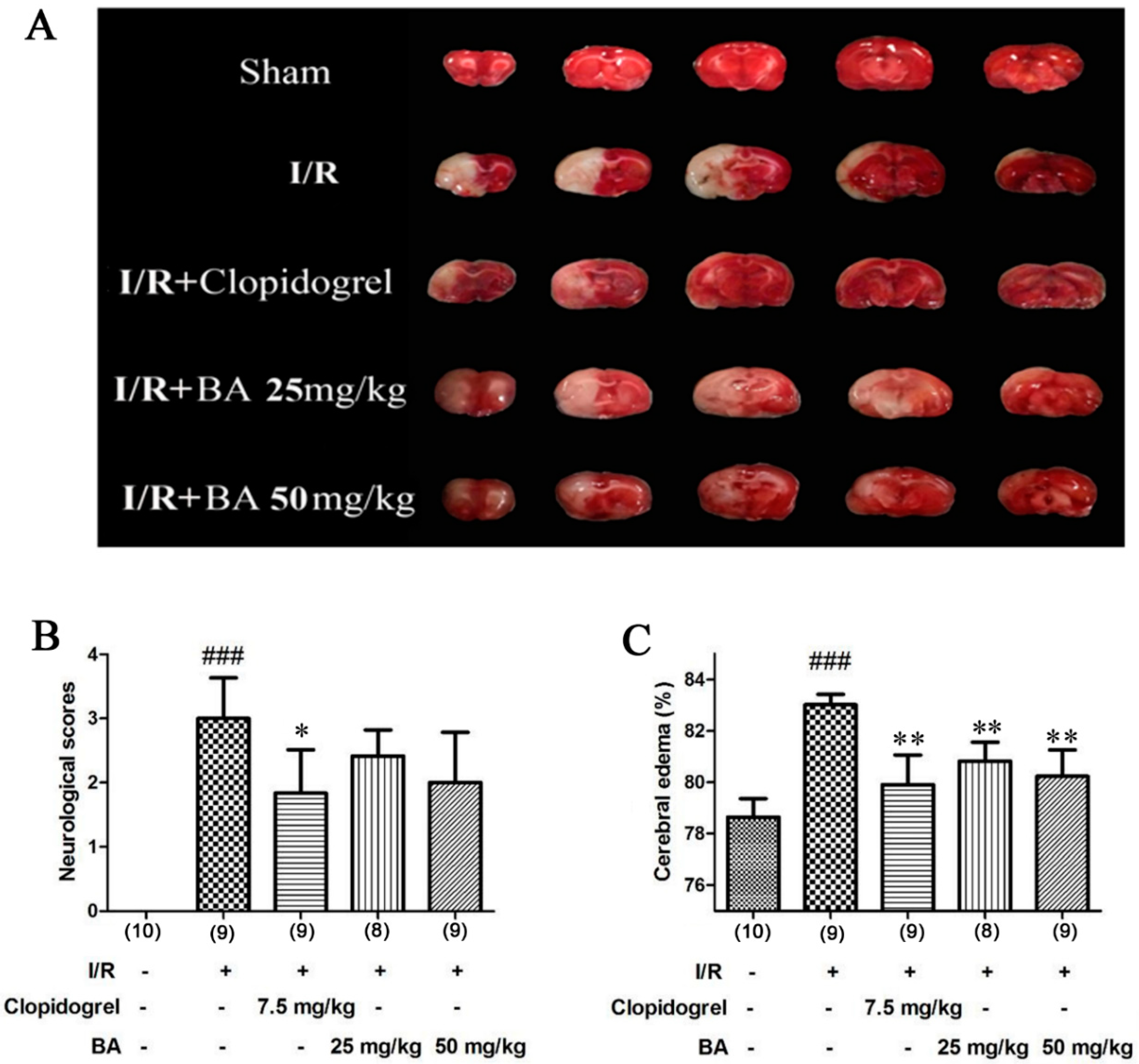
The protection of BA in the MCAO model was assessed using TTC staining, neurological behavior, and edema assays. Neurologic behavior evaluations were performed using a four-point scale. The successful ischemia/reperfusion model was confirmed by the infarct size, as observed via TTC staining. The infarct areas (white areas) in the model group were significantly increased, but the cerebral infarct areas were markedly decreased after treatment with BA and clopidogrel, suggesting that BA suppressed the cerebral infarction (Figure 2A). No observable neurological scores were present in the sham group (Figure 2B). The ischemia/reperfusion rats showed significant increases in neurologic score when compared to sham rats, which were partial reversed upon clopidogrel or BA treatment. Behavioral investigations indicated that BA intervention improved neurological function in successful ischemia/reperfusion models. Similar to the infarct, cerebral edema data showed that the average ratio of wet/dry weight after MCAO challenge was comparatively elevated. By contrast, the cerebral edema of the BA and clopidogrel groups significantly declined compared with the model group (Figure 2C). The beneficial effects of BA on MCAO-induced ischemia/reperfusion injury were observed under TTC staining, neurological behavior evaluations, and edema assays in vivo.

3.2. Effects of BA on Pro-inflammatory Cytokine

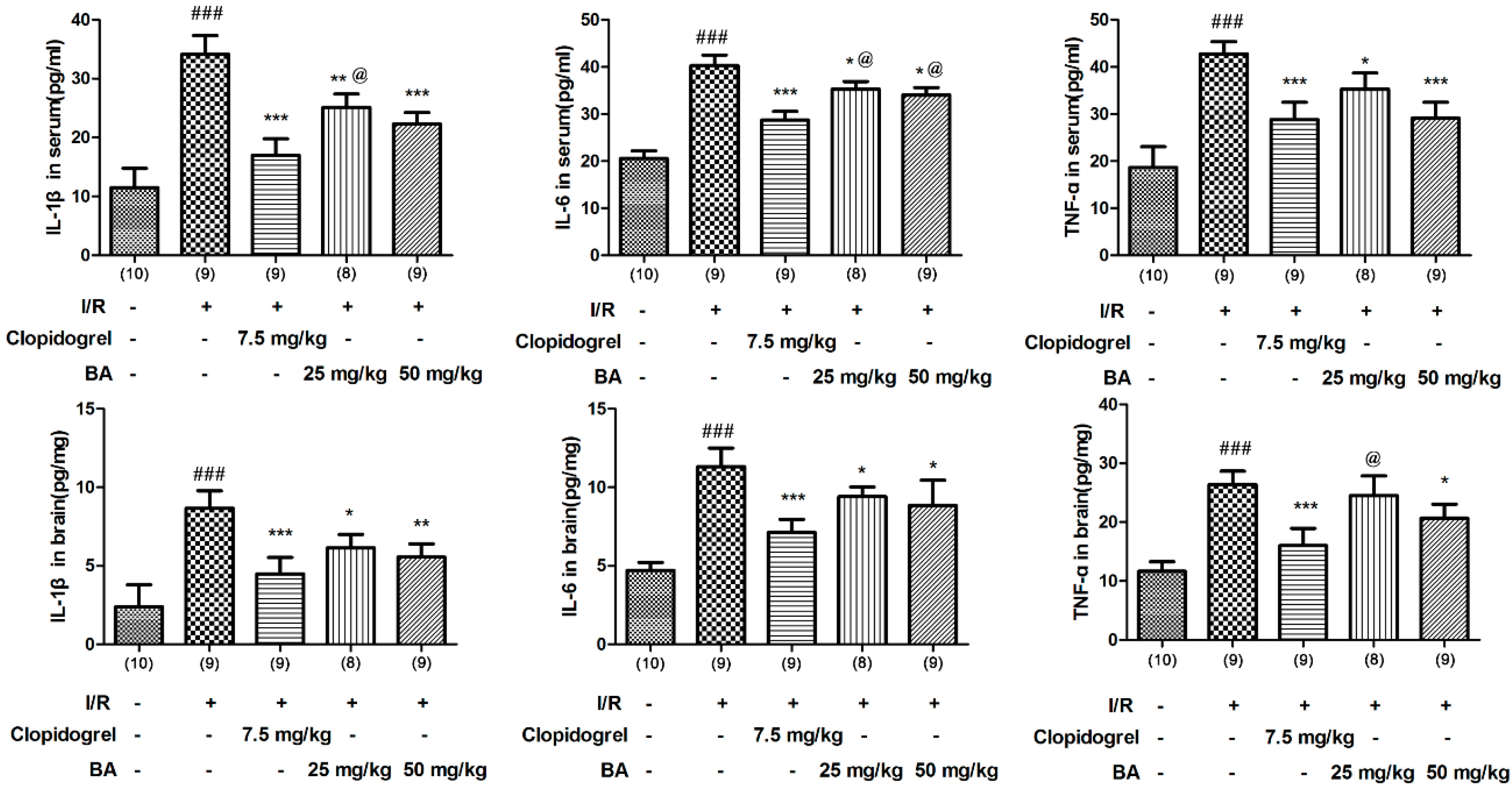
The inflammatory responses were examined by determining the values of IL-6, IL-1β, and TNF-α in rats and in SHSY5Y cell. In vivo, the results showed that MCAO surgery markedly increased IL-1β, IL-6, and TNF-α concentration in serum and brain of the treated rats compared to the sham rats. However, L-1β, IL-6, and TNF-α levels were reduced in MCAO rats after BA and clopidogrel treatment (Figure 3).
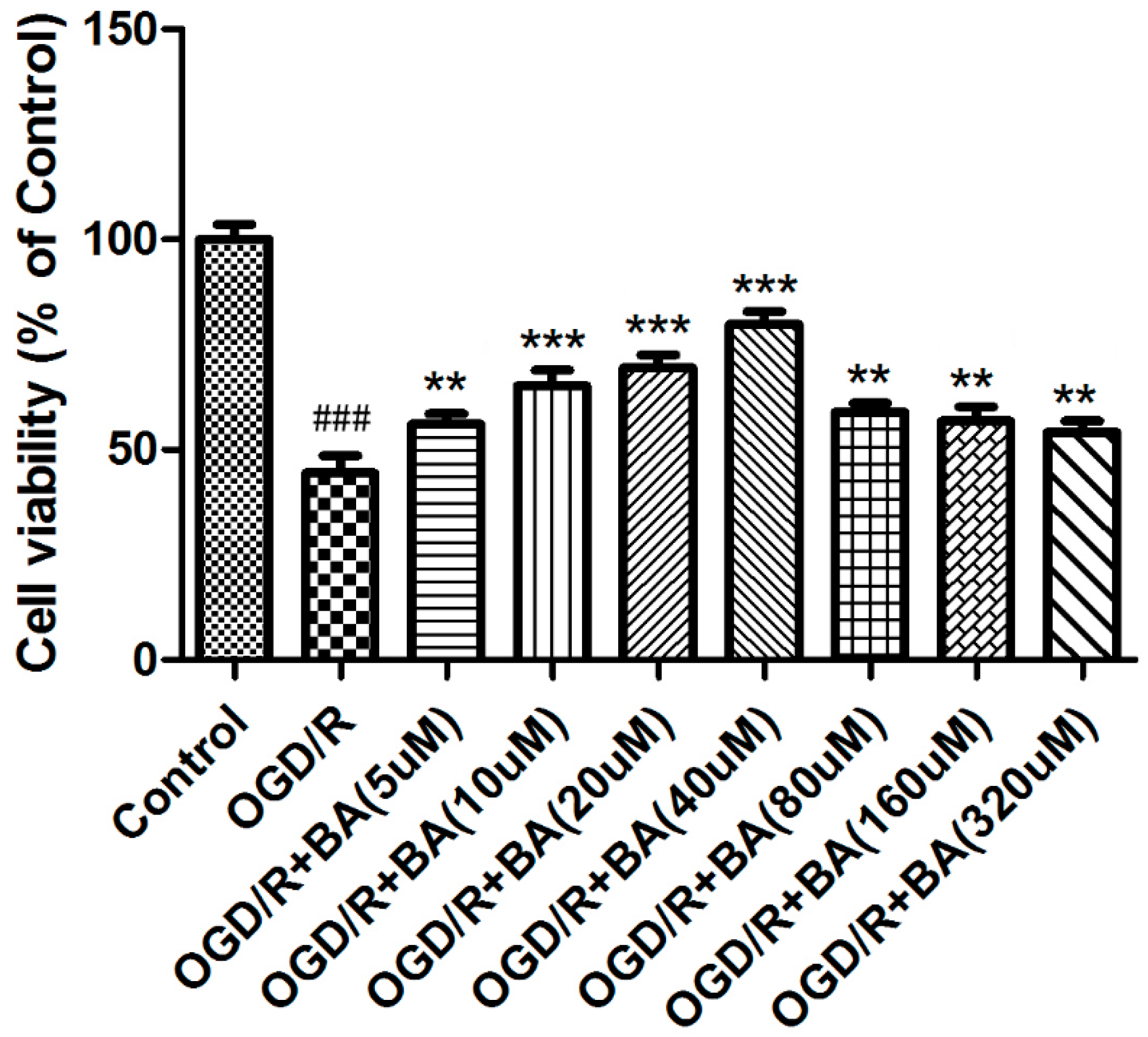
To investigate BA activity on the cell experiments, SHSY5Y cell was selected and cell viability was examined using MTT assays (Figure 4). Upon OGD/R induction, SHSY5Y cell proliferation was considerably reduced. Intervention with BA (5, 10, 20, and 40 μM) presented effective cell viability improvement under OGD/R (Figure 4). Intervention with BA (80, 160 and 320 µM) showed same viability as 10 µM, lower than that with BA at 20 uM and 40 uM. This may be due to BA’s cytotoxicity at high concentration (80, 160, and 320 µM).
After confirmation that the BA protection was associated with an anti-inflammatory response in rats, we investigated its effect on the OGD/R model (using concentrations of 10, 20, or 40 μM of BA) in vitro. Attenuated action of BA (10, 20, or 40 μM) was observed from the pro-inflammatory cytokine levels of the SHSY5Y cell that were exposed to OGD/R stimulation, which coincided with the altered pro-inflammatory cytokine production levels observed in rats (Figure 5).

3.3. Effects of BA on HIF-1α Signaling

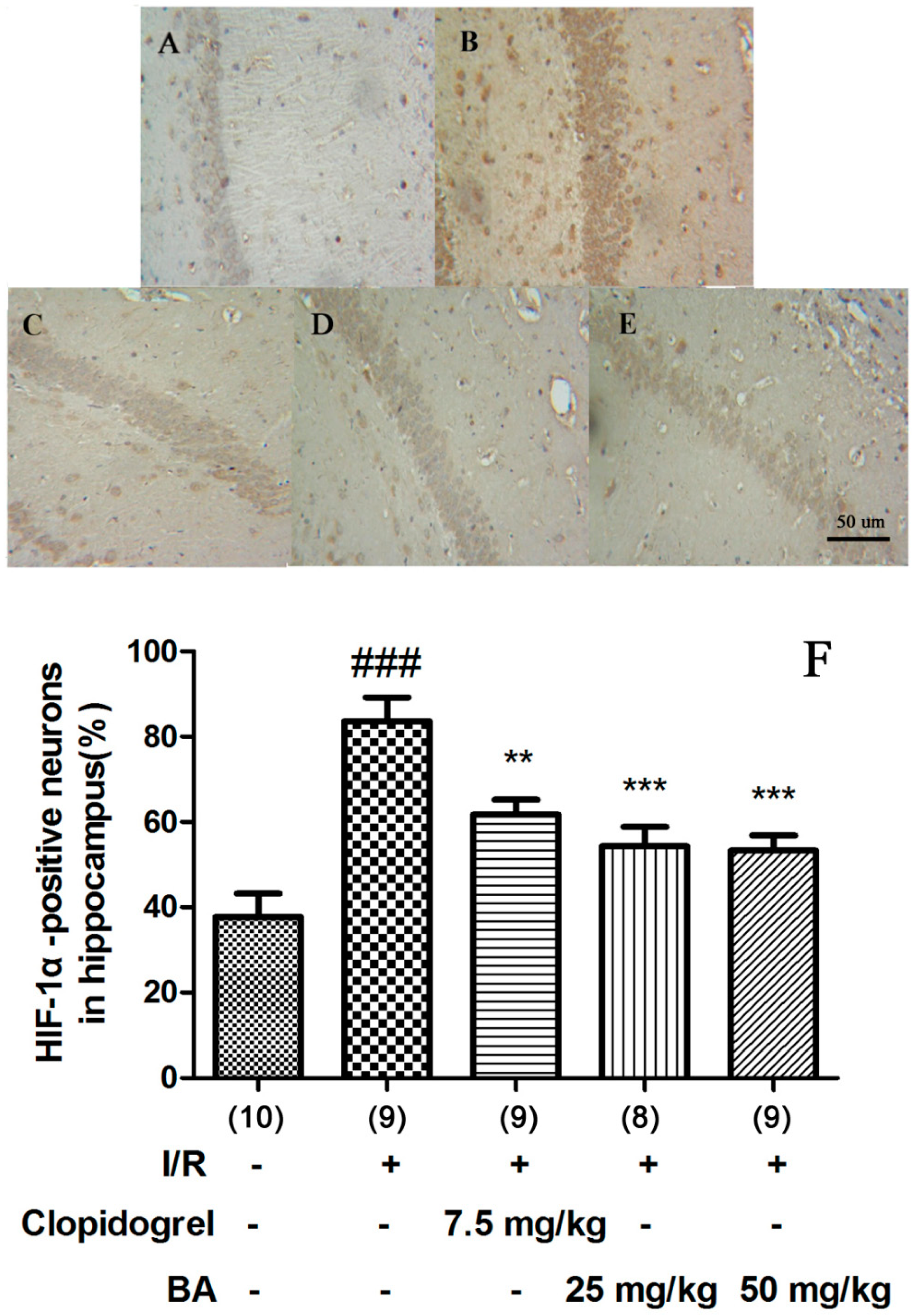
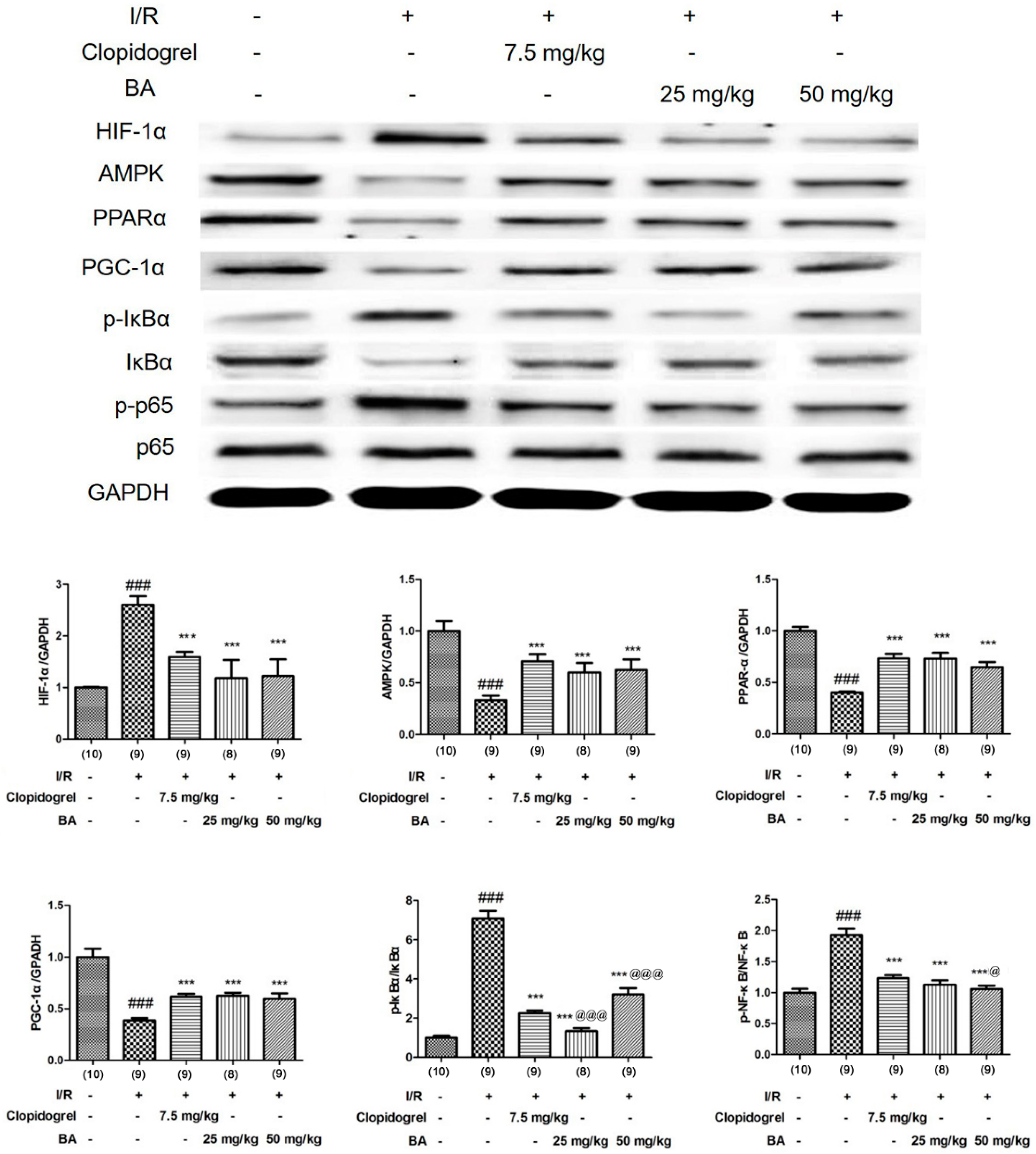
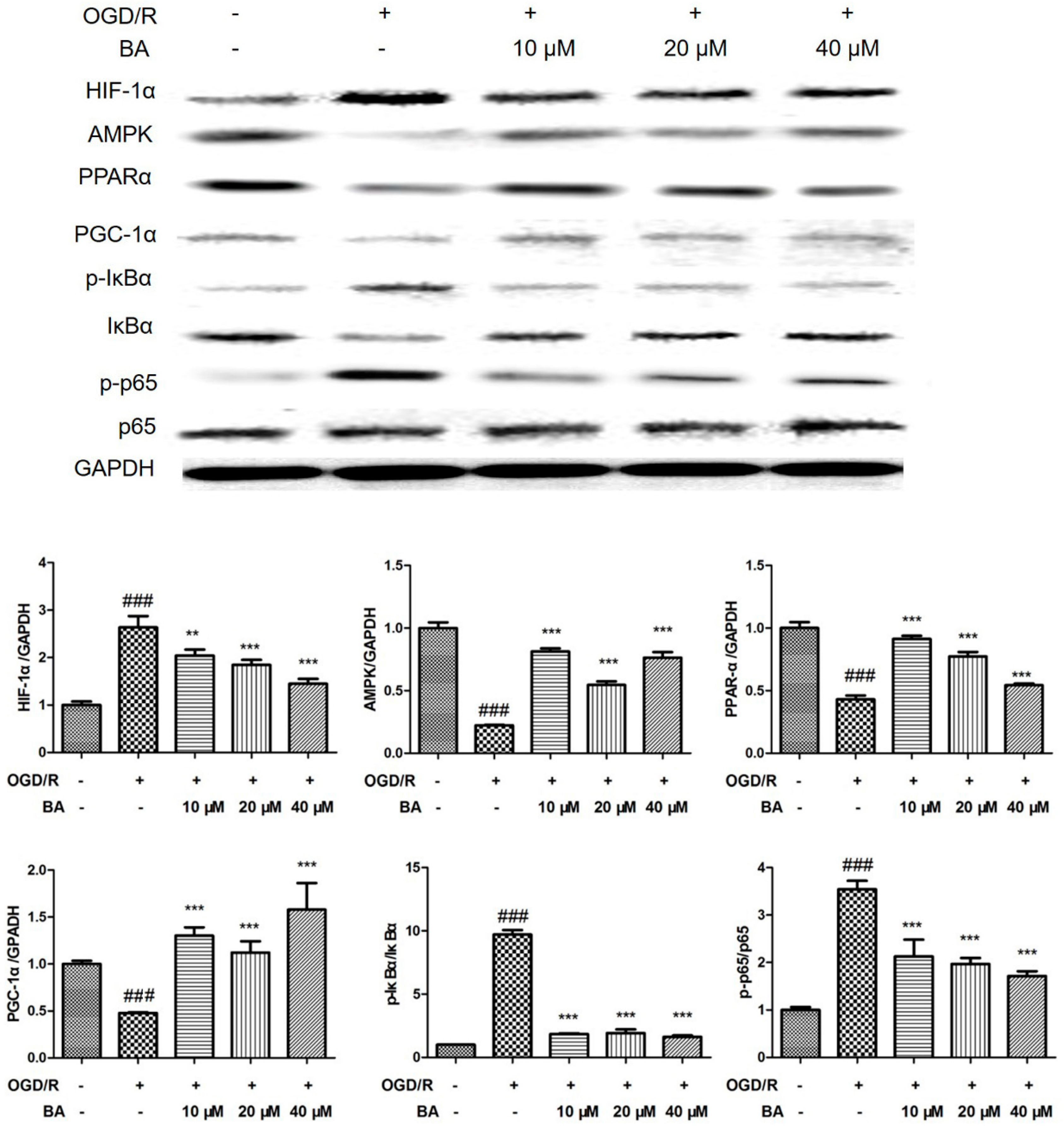
To explain the internal mechanism, the influence of HIF-1α/AMPK/PPARα/PGC- 1α/NF-κB signaling factors was closely monitored. In Figure 6, ischemia/reperfusion stimulation triggered the up-regulation of HIF-1α, which was confirmed by immunohistochemistry staining. Down-regulation of AMPK, PPAR-α, and PGC-1α and phosphorylation of IκBα, and NF-κBp65 were also observed in ischemia/reperfusion-induced rats (Figure 7). Similar changes appeared in vitro after OGD/R challenge (Figure 8), however, clopidogrel and BA reversed the up-regulation of HIF-1α and the down-regulation of AMPK, PPAR-α, and PGC-1α, and blocked the phosphorylation of IκBα and NF-κBp65 in the brain. The addition of BA also significantly inhibited the OGD/R-induced changes of the HIF-1α/AMPK/PPARα/PGC-1α/NF-κB signaling pathway according to a similar pattern as that seen in the animal results. These data suggested that the HIF-1α/AMPK/PPARα/PGC-1α/NF-κB signaling factors mediated inflammation, which may have been due to the beneficial actions of BA.

4. Discussion

Globally, stroke is a leading cause of mortality and disability and there are substantial economic costs for post-stroke care. Results from the 2015 iteration of the Global Burden of Diseases, Injuries, and Risk Factors Study (GBD) showed that although the age-standardised death rates and prevalence of stroke have decreased over time, the overall burden of stroke has remained high. The results provided here are the most up-to-date estimates of death, prevalence, incidence, and disability for overall stroke and the pathological types of ischaemic and haemorrhagic stroke [14,15].
As a serious and complex pathological process, ischemia injury involves ion imbalance, peroxidation stress, neuroexcitotoxicity, and inflammatory activation. These cascading waterfall effects eventually lead to necrosis and apoptosis of nerve cells and the irreversible loss of nerve function [16,17]. At present, the only effective treatment for cerebral ischemia in the medical community is thrombolysis. However, the window of time in which thrombolytic drugs can be used effectively limits their clinical applications [18]. Therefore, finding effective and reliable neuroprotective agents for stroke treatment is urgent. Clopidogrel, a platelet aggregation inhibitor, was reported to work as a stroke preventative drug in vivo by slowing platelet aggregation [19]. The present study examined infarct size, cerebral edema, neurological behavior, and inflammatory cytokine levels of MCAO rats and OGD/R-induced SHSY5Y cells after BA intervention. Additionally, expression of the HIF-1α/AMPK/PPARα/PGC-1α/NF-κB pathway was measured in animals and cells.
Ischemia/reperfusion leads to cell inflammation caused by infiltration of immune cells [20]. Similarly, the acute stress response induced by inflammation may have a damaging effect on nerve cells [21]. The expression of inflammatory factors is mediated by activated NF-κB, which is a key inflammatory regulator. After NF-κB activation, inflammatory transcription factors migrate to the nucleus of a cell, promote pro-inflammatory gene transcription and enhance the inflammatory response [22]. The over-expression of PPAR and the PPAR activator rosiglitazone play important anti-inflammatory roles by inhibiting the NF-κB signal pathway [23]. PPAR-γ coactivator-1α (PGC-1α) can also reduce the inflammatory effect by interfering with NF-κB [24,25].
HIF-1, a basic helix–loop–helix transcription factor, consists of two subunits, namely, HIF-1α and HIF-1β. Generally, HIF-1α is a nuclear protein with transcriptional activity with a wide range of target gene profiles, including nearly 100 target genes related to hypoxia adaptation, inflammatory development, and tumor growth [26]. In hypoxic conditions, HIF-1α degradation is inhibited, thereby increasing HIF-1α levels [27].
Ischemic stroke was previously thought to result from hypoxia, which is associated with a decreased energy supply. The decreased adenosine triphosphate concentration, leading to a diminished energy supply, contributes to neuronal damage in cerebral ischemic injury [28]. AMPK controls the whole process of energy metabolism in tissues and cells; animals deficient in AMPK show accelerated energy failure and increased infarct sizes. When AMPK is activated, it rapidly phosphorylates downstream targets, blocks the ATP consumption pathway, and promotes activation of the ATP synthesis pathway [29,30].
The association between HIF-1α and AMPK is realized via sirtuin1 (SIRT1), which combines HIF-1α and blocks the binding of p300 with HIF-1α [31]. Therefore, stimulated SIRT1 activity eventually inhibits HIF-1α-induced transcriptional activation during ischemia and hypoxia. On the other hand, SIRT1 was reported to dissociate from the DBC1 protein and be activated by AMPK, indicating that AMPK is a main activator of SIRT1 and can therefore inhibit HIF-1α [31,32].
PPARα and PGC-1α are activated directly by AMPK. PPARα increases protein expression of downstream events and consequently leads to NF-κB inhibition [33]. PGC-1 α is a transcription coactivator of PPAR, which regulates cytokine production and expression of adhesion molecules to reduce inflammation [34]. The AMPK/PPARα/PGC-1α signaling pathway was previously confirmed in hypoxia/re-oxygenation injury, with NF-κB being a key mediator of inflammation in the pathological mechanism control of many diseases and injuries. NF-κBp65 is activated by the phosphorylation of IκBα, which acts as an inhibitor of NF-κB-p65 [35]. In the present study, we found that BA decreased HIF-1α, p-IκBα, and p-NF-κB-p65 expression and increased the levels of AMPK, PPAR-α, and PGC-1α in the brains of MCAO rats and OGD/R cells.
Betulinic acid has a wide range of pharmacological activities, including anti-tumor, anti-HIV, and anti-inflammatory properties [36]. Betulinic acid was originally reported to possess specific cytotoxicity and biological activities toward many cancer cells, with its mechanism of action involving directly acting on mitochondria and inducing tumor cell apoptosis [37]. In previous studies, BA was proven to protect kidneys and the heart from ischemia/reperfusion injuries. BA inhibits the activation of inflammation and regulates the expression of TNF-α and other inflammatory factors from myocardial and renal ischemia/reperfusion injury [10,11]. In addition, BA exhibits mild anti-inflammatory effects, thereby inhibiting the inflammation of mice induced with carrageenan, phorbol, and bryostatin-1, but showing no inhibitory effect toward arachidonic acid-induced inflammation and neurogenic inflammation models [38,39]. Isoflurane-induced brain neuron injuries in rats were shown to be improved by BA in that oxygen electrodes and ultraviolet spectrophotometer demonstrated that mitochondrial respiratory levels and intracellular ATP contents improved after treatment [40]. This suggested that BA has effects on mitochondrial stability, energy metabolic homeostasis, and neuronal apoptosis.
In conclusion, the benefits of BA included relieving ischemia/reperfusion injury through HIF-1α/AMPK/PPARα/PGC-1α/NF-κB-mediated inflammation.

Author Contributions

Investigation, K.H.; Methodology, W.J.; Supervision, K.H.; Validation, W.J.; Writing (original draft), W.J.; Writing (review and editing), K.H. All authors have read and agreed to the published versionof the manuscript.

Funding

This research was supported by Natural Science Foundation of China (Grants 81773826), The Drug Innovation Major Project (Grants 2018ZX09711001), 111 Incubation Project (BC2018024).

Conflicts of Interest

The authors declare no conflict of interest.

References

  1. Turelson, T.; Begg, S.; Mathers, C. The global burden of cerebrovascular disease. Global Burden of Disease. 2006. Available online: https://www.who.int/healthinfo/statistics/bod_cerebrovasculardiseasestroke.pdf (accessed on 7 March 2020).
  2. Sudlow, C.L.M.; Warlow, C.P. Comparable studies of the incidence of stroke and its pathological types. Results from an international collaboration. Stroke 1997, 28, 491–499. [Google Scholar] [CrossRef] [PubMed]
  3. Hardie, D.G.; Scott, J.W.; Pan, D.A. Management of Cellular Energy by the AMP-Activated Protein Kinase System. FEBS Lett. 2003, 546, 113–120. [Google Scholar] [CrossRef]
  4. Wang, C.P.; Shi, Y.W.; Tang, M.; Zhang, X.C.; Gu, Y.; Liang, X.M.; Wang, Z.W.; Ding, F. Isoquercetin Ameliorates Cerebral Impairment in Focal Ischemia Through Anti-Oxidative, Anti-Inflammatory, and Anti-Apoptotic Effects in Primary Culture of Rat Hippocampal Neurons and Hippocampal CA1 Region of Rats. Mol. Neurobiol. 2017, 54, 2126–2142. [Google Scholar] [CrossRef]
  5. Blázquez, C.; Woods, A.; Ceballos, M.L.; Carling, D.; Guzman, M. The AMP-Activated Protein Kinase is Involved in the Regulation of Ketone Body Production by Astrocytes. J. Neurochem. 1999, 73, 1674–1682. [Google Scholar] [CrossRef] [PubMed] [Green Version]
  6. Koh, H.S.; Chang, C.Y.; Jeon, S.B.; Yoon, H.J.; Ahn, Y.H.; Kim, H.S.; Kim, I.H.; Jeon, S.H.; Johnson, R.S.; Park, E.J. The HIF-1/glial TIM-3 Axis Controls Inflammation-Associated Brain Damage under Hypoxia. Nat. Commun. 2015, 6, 6340. [Google Scholar] [CrossRef] [Green Version]
  7. Crupi, R.; Paola, R.; Esposito, E.; Cuzzocrea, S. Middle Cerebral Artery Occlusion by an Intraluminal Suture Method. Methods Mol. Biol. 2018, 1727, 393–401. [Google Scholar]
  8. Zhao, H.; Holmes, S.S.; Baker, G.A.; Challa, S.; Bose, H.S.; Song, Z. Ionic derivatives of betulinic acid as novel HIV-1 protease inhibitors. J. Enzyme Inhib. Med. Chem. 2012, 27, 715–721. [Google Scholar] [CrossRef] [Green Version]
  9. Liu, J.H.; Zhu, Z.F.; Tang, J.; Lin, Q.H.; Chen, L.; Sun, J.B. Design and Synthesis of NO-Releasing Betulinic Acid Derivatives as Potential Anticancer Agents. Anti-Cancer Agents Med. Chem. 2017, 17, 241–249. [Google Scholar] [CrossRef]
  10. Xia, A.; Xue, Z.; Li, Y.; Wang, W.; Xia, J.; Wei, T.; Cao, J.; Zhou, W. Cardioprotective Effect of Betulinic Acid on Myocardial Ischemia Reperfusion Injury in Rats. Evid. Based Complement. Altern. Med. 2014, 2014, 573745. [Google Scholar] [CrossRef] [Green Version]
  11. Ekşioğlu-Demiralp, E.; Kardaş, E.R.; Özgül, S.; Yağci, T.; Bilgin, H.; Sehirli, O.; Ercan, F.; Sener, G. Betulinic Acid Protects against Ischemia/Reperfusion- Induced Renal Damage and Inhibits Leukocyte Apoptosis. Phytother. Res. 2010, 24, 325–332. [Google Scholar] [CrossRef]
  12. Wang, D.; Yang, X.H.; Zhang, J.D.; Li, R.B.; Jia, M.; Cui, X.R. Compared Efficacy of Clopidogrel and Tcagrelor in Treating Acute Coronary Syndrome: A Meta-Analysis. BMC Cardiovasc. Disord. 2018, 8, 217. [Google Scholar]
  13. Xu, Z.; Gu, J.; Gao, M.; Du, N.; Liu, P.; Xu, X.; Wang, M.J.; Cao, X. Study on Antiplatelet Effect of A New Thiophenopyridine Platelets P2Y12 Receptor Antagonist DV-127. Thromb. Res. 2018, 170, 192–199. [Google Scholar] [CrossRef] [PubMed]
  14. Rajsic, S.; Gothe, H.; Borba, H.H.; Sroczynski, G.; Vujicic, J.; Toell, T.; Siebert, U. Economic burden of stroke: A systematic review on post-stroke care. Eur. J. Health Econ. 2019, 20, 107–134. [Google Scholar] [CrossRef] [PubMed]
  15. GBD 2015 Neurological Disorders Collaborator Group. Global, regional, and national burden of neurological disorders during 1990–2015: A systematic analysis for the Global Burden of Disease Study 2015. Lancet Neurol. 2017, 16, 877–897. [Google Scholar] [CrossRef] [Green Version]
  16. Rodrigo, R.; Fernández-Gajardo, R.; Gutiérrez, R.; Matamala, J.M.; Carrasco, R.; Miranda-Merchak, A.; Feuerhake, W. Oxidative Stress and Pathophysiology of Ischemic Stroke: Novel Therapeutic Opportunities. CNS Neurol. Disord. Drug Targets 2013, 12, 698–714. [Google Scholar] [CrossRef] [PubMed]
  17. Grysiewicz, R.A.; Thomas, K.; Pandey, D.K. Epidemiology of Ischemic and Hemorrhagic Stroke: Incidence, Prevalence, Mortality, and Risk Factors. Neurol. Clin. 2008, 26, 871–895. [Google Scholar] [CrossRef]
  18. Deb, P.; Sharma, S.; Hassan, K.M. Pathophysiologic Mechanisms of Acute Ischemic Stroke: An Overview with Emphasis on Therapeutic Significance beyond Thrombolysis. Pathophysiology 2010, 17, 197–218. [Google Scholar] [CrossRef]
  19. Pop, C.; Matei, C. Benefits and Risks of Anticoagulation in Acute Coronary Syndrome. Am. J. Ther. 2019, 26, e198–e207. [Google Scholar] [CrossRef]
  20. Eltzschig, H.K.; Eckle, T. Ischemia and Reperfusion from Mechanism to Translation. Nat. Med. 2011, 17, 1391–1401. [Google Scholar] [CrossRef] [Green Version]
  21. Kim, J.Y.; Park, J.; Chang, J.Y.; Kim, S.H.; Lee, J.E. Inflammation after Ischemic Stroke: The Role of Leukocytes and Glial Cells. Exp. Neurobiol. 2016, 25, 241–251. [Google Scholar] [CrossRef] [Green Version]
  22. Zhu, L.; Wei, T.; Gao, J.; Chang, X.; He, H.; Luo, F.; Zhou, R.; Ma, H.; Liu, Y.; Yan, T.H. The Cardioprotective Effect of Ssalidroside against Myocardial Ischemia Reperfusion Injury in Rats by Inhibiting Apoptosis and Inflammation. Apoptosis 2015, 20, 1433–1443. [Google Scholar] [CrossRef] [PubMed]
  23. Delerive, P.; Fruchart, J.C.; Staels, B. Peroxisome Proliferator Activated Receptors in Inflammation Control. J. Endocrinol. 2001, 169, 453–459. [Google Scholar] [CrossRef] [Green Version]
  24. St-Pierre, J.; Drori, S.; Uldry, M.; Silvaggi, J.M.; Rhee, J.; Jäger, S.; Handschin, C.; Zheng, K.; Lin, J.; Yang, W.; et al. Suppression of Reactive Oxygen Species and Neurodegeneration by the PGC-1 Transcriptional Coactivators. Cell 2006, 127, 397–408. [Google Scholar] [CrossRef] [PubMed] [Green Version]
  25. Eisele, P.S.; Furrer, R.; Beer, M.; Handschin, C. The PGC-1 Coactivators Promote an Anti-inflammatory Environment in Skeletal Muscle In Vivo. Biochem. Biophys. Res. Commun. 2015, 464, 92–97. [Google Scholar] [CrossRef] [Green Version]
  26. Ndubuizu, O.I.; Tsipis, C.P.; Li, A.; LaManna, J.C. Hypoxia-inducible Factor-1 (HIF-1)- Independent Microvascular Angiogenesis in the Aged Rat Brain. Brain Res. 2010, 1366, 101–109. [Google Scholar] [CrossRef] [PubMed] [Green Version]
  27. Arany, Z.; Foo, S.Y.; Ma, Y.; Ruas, J.L.; Bommi-Reddy, A.; Girnun, G.; Cooper, M.; Laznik, D.; Chinsomboon, J.; Rangwala, S.M.; et al. HIF-Independent Regulation of VEGF and Angiogenesis by the Transcriptional Coactivator PGC-1alpha. Nature 2008, 451, 1008–1012. [Google Scholar] [CrossRef] [PubMed] [Green Version]
  28. Zou, M.H.; Mu, Y. AMP-Activated Protein Kinase Activation as a Strategy for Protecting Vascular Endothelial Function. Clin. Exp. Pharmacol. Physiol. 2008, 35, 535–545. [Google Scholar] [CrossRef] [Green Version]
  29. Turnley, A.M.; Stapleton, D.; Mann, R.J.; Witters, L.A.; Kemp, B.E.; Bartlett, P.F. Cellular Distribution and Developmental Expression of AMP-Activated Protein Kinase Isoforms in Mouse Central Nervous System. J. Neurochem. 1999, 72, 1707–1716. [Google Scholar] [CrossRef]
  30. Zou, M.H.; Hou, X.Y.; Shi, C.M.; Nagata, D.; Walsh, K.; Cohen, R.A. Modulation by Peroxynitrite of Akt- and AMP-Activated Kinase-Dependent Ser1179 Phosphorylation of Endothelial Nitric Oxide Synthase. J. Biol. Chem. 2002, 277, 32552–32557. [Google Scholar] [CrossRef] [Green Version]
  31. Lim, J.H.; Lee, Y.M.; Chun, Y.S.; Chen, J.; Kim, J.E.; Park, J.W. Sirtuin 1 Modulates Cellular Responses to Hypoxia by Deacetylating Hypoxia-Inducible Factor 1α. Mol. Cell 2010, 38, 864–878. [Google Scholar] [CrossRef]
  32. Nakagawa, T.; Guarente, L. Sirtuins at a Glance. J. Cell Sci. 2011, 124, 833–838. [Google Scholar] [CrossRef] [PubMed] [Green Version]
  33. Berglund, E.D.; Kang, L.; Lee-Young, R.S.; Hasenour, C.M.; Lustig, D.G.; Lynes, S.E.; Donahue, E.P.; Swift, L.L.; Charron, M.J.; Wasserman, D.H. Glucagon and Lipid Interactions in the Regulation of Hepatic AMPK Signaling and Expression of PPARalpha and FGF21 Transcripts In Vivo. Am. J. Physiol. Endocrinol. Metab. 2010, 299, E607–E614. [Google Scholar] [CrossRef] [PubMed]
  34. He, X.; Liu, W.; Shi, M.; Yang, Z.; Zhang, X.; Gong, P. Docosahexaenoic Acid Attenuates LPS-Stimulated Inflammatory Response by Regulating the PPARγ/NF-κB Pathways in Primary Bovine Mammary Epithelial Cells. Res. Vet. Sci. 2017, 112, 7–12. [Google Scholar] [CrossRef] [PubMed]
  35. Alvarez-Guardia, D.; Palomer, X.; Coll, T.; Davidson, M.M.; Chan, T.O.; Feldman, A.M.; Laguna, J.C.; Vázquez-Carrera, M. The P65 Subunit of NF-kappaB Binds to PGC-Lalpha, Linking Inflammation and Metabolic Disturbances in Cardiac Cells. Cardiovasc. Res. 2010, 87, 449–458. [Google Scholar] [CrossRef] [Green Version]
  36. Yogeeswari, P.; Sriram, D. Betulinic Acid and Its Derivatives: A Review on Their Biological Properties. Curr. Med. Chem. 2005, 12, 657–666. [Google Scholar] [CrossRef]
  37. Kasperczyk, H.; La Ferla-Brühl, K.; Westhoff, M.A.; Behrend, L.; Zwacka, R.M.; Debatin, K.M.; Fulda, S. Betulinic Acid as New Activator of NF-κB: Molecular Nechanisms and Implications for Cancer Therapy. Oncogene 2005, 24, 6945–6956. [Google Scholar] [CrossRef] [Green Version]
  38. Huguet, A.; del Carmen Recio, M.; Maez, S.; Giner, R.; Ríos, J. Effect of Triterpenoids on the Inflammation Induced by Protein Kinase C Activators, Neuronally Acting Irritants and Other Agents. Eur. J. Pharmacol. 2000, 410, 69–81. [Google Scholar] [CrossRef]
  39. Costa, J.F.; Barbosa-Filho, J.M.; Maia, G.L.; Guimarães, E.T.; Meira, C.S.; Ribeiro-dos-Santos, R.; de Carvalho, L.C.; Soares, M.B. Potent Anti-Inflammatory Sctivity of Betulinic Scid Yreatment in A Model of Lethal Endotoxemia. Int. Immunopharmacol. 2014, 23, 469–474. [Google Scholar] [CrossRef] [Green Version]
  40. Erasso, D.M.; Camporesi, E.M.; Mangar, D.; Saporta, S. Effects of Isoflurane or Propofol on Postnatal Hippocampal Neurogenesis in Young and Aged Rats. Brain Res. 2013, 1530, 1–12. [Google Scholar] [CrossRef]
Figure 1. The chemical structure of betulinic acid (BA).
Applsci 10 02578 g001
Figure 2. The protection of BA against cerebral infarction (A), neurological score (B), and edema (C) compared with sham rats (### p < 0.001) and compared with I/R group (* p < 0.05 and ** p < 0.01). The Arabic number in brackets represent the numbers of animals in each group.
Applsci 10 02578 g002
Figure 3. The beneficial effects of BA on the level of inflammatory cytokines in serum and brain tissues compared with sham rats (### p < 0.001), compared with I/R group (* p < 0.05, ** p < 0.01, and *** p < 0.001) and compared with clopidogrel group (@ p < 0.05). The Arabic number in brackets represent the numbers of animals in each group.
Applsci 10 02578 g003
Figure 4. The influence of BA on oxygen–glucose deprivation and recovery (OGD/R)-induced SHSY5Y cells compared with control cells (### p < 0.001) and compared with OGD/R cells (** p < 0.01 and *** p < 0.001).
Applsci 10 02578 g004
Figure 5. The influence of BA on the level of inflammatory cytokines in OGD/R-induced SHSY5Y cells compared with control cells (### p < 0.001) and compared with OGD/R cells (** p < 0.01, and *** p < 0.001).
Applsci 10 02578 g005
Figure 6. The influence of BA on hypoxia-inducible factor-1α (HIF-1α) by immunohistochemical analysis. (A): sham group; (B): I/R group; (C): I/R+ clopidogrel group; (D): I/R+BA (25 mg/kg) group; (E): I/R+BA (50 mg/kg) group; (F): HIF-1α positive neurons in hippocampus. Compared with sham rats (### p < 0.001) and compared with I/R (** p < 0.01, *** p < 0.001). The Arabic number in brackets represent the numbers of animals in each group.
Applsci 10 02578 g006
Figure 7. The influence of BA on HIF-1α, adenosine monophosphate-activated protein kinase (AMPK), perixisome proliferation-activated receptor alpha (PPAR-α), peroxisome proliferator-activated receptor γcoactivator-1 alpha (PGC-1α), IκBα and NF-κBp65 in brain tissues compared with sham rats (### p < 0.001), compared with I/R group (*** p < 0.001) and compared with clopidogrel group (@ p < 0.05 and @@@ p < 0.001). The Arabic number in brackets represent the numbers of animals in each group.
Applsci 10 02578 g007
Figure 8. The influence of BA on HIF-1α, AMPK, PPAR-α, PGC-1α, IκBα and NF-κBp65 in OGD/R-induced SHSY5Y cells. Compared with control cells (### p < 0.001) and compared with OGD/R cells (** p < 0.01 and *** p < 0.001).
Applsci 10 02578 g008