Article

Design of a Sandwich Hierarchically Porous Membrane with Oxygen Supplement Function for Implantable Glucose Sensor

1
Key Laboratory of Biotechnology Science and Technology, Ministry of Education, College of Bioengineering, Chongqing University, Chongqing 400030, China
2
Chongqing Engineering Research Center of Medical Electronics Technology, Chongqing 400030, China
3
Key Laboratories for National Defense Science and Technology of Innovative Micro-Nano Devices and System Technology, Chongqing University, Chongqing 400030, China
*
Authors to whom correspondence should be addressed.
Appl. Sci. 2020, 10(8), 2848; https://doi.org/10.3390/app10082848
Submission received: 26 February 2020 / Revised: 7 April 2020 / Accepted: 16 April 2020 / Published: 20 April 2020

Abstract

:

Featured Application

A functional membrane was proposed to maintain a wide linear detection range, increase the sensitivity of the glucose sensor and reduce the difficulty of continuous glucose monitoring systems (CGMS) development. This CGMS system includes a glucose sensor, a data collector and a health management platform that can help people with diabetes maintaining euglycemia at an affordable cost in the future.

Abstract

This study aims to develop an oxygen regeneration layer sandwiched between multiple porous polyurethanes (PU) to improve the performance of implantable glucose sensors. Sensors were prepared by coating electrodes with platinum nanoparticles, Nafion, glucose oxidase and sandwich hierarchically porous membrane with an oxygen supplement function (SHPM-OS). The SHPM-OS consisted of a hierarchically porous structure synthesized by polyethylene glycol and PU and a catalase (Cat) layer that was coated between hierarchical membranes and used to balance the sensitivity and linearity of glucose sensors, as well as reduce the influence of oxygen deficiency during monitoring. Compared with the sensitivity and linearity of traditional non-porous (NO-P) sensors (35.95 nA/mM, 0.9987, respectively) and single porous (SGL-P) sensors (45.3 nA /mM, 0.9610, respectively), the sensitivity and linearity of the SHPM-OS sensor was 98.45 nA/mM and 0.9989, respectively, which was more sensitive with higher linearity. The sensor showed a response speed of five seconds and a relative sensitivity of 90% in the first 10 days and remained 78% on day 20. This sensor coated with SHPM-OS achieved rapid responses to changes of glucose concentration while maintaining high linearity for long monitoring times. Thus, it may reduce the difficulty of back-end hardware module development and assist with effective glucose self-management for people with diabetes.

1. Introduction

Diabetes mellitus [1] is one of the pandemics characterized by chronic hyperglycemia [2], which seriously threatens human physical and mental health and quality of life. Since no cure has been developed for this disease, maintaining euglycemia through diet, exercise and pharmacological management is a way to prevent diabetic complications [3,4,5]. Currently, continuous glucose monitoring systems (CGMS) are increasingly popular, since they can reflect fluctuation of blood glucose (BG), easily perceive asymptomatic hypoglycemia and relieve the pain of sticking fingers frequently for people with diabetes [2,6,7].
The minimally invasive implantable glucose sensor is a key module that affects the performance of CGMS [8]. The sensitivity, biocompatibility, stability and linear measuring range of the implantable sensors are important to the entire system [5]. Glucose oxidase (GOD) can be easily prepared, and has strong specificity to glucose, high activity and stability, which is widely applied in amperometric glucose sensor [9]. GOD catalyzes glucose into the gluconic acid and hydrogen peroxide (H2O2). Then the H2O2 undergoes an electrochemical reaction on the electrode, generating a response current. The reaction equations are displayed below:
glucose + O 2 + H 2 O   G O D   g l u c o n i c   a c i d + H 2 O 2 ,
H 2 O 2   + 0.65 v 2 H + + O 2   + 2 e
According to the chemical equation, the performance of such glucose sensors is limited by several interdependent aspects, including electrolysis of various endogenous and exogenous substances; deficient of oxygen at high glucose concentration; inflammation and sensor fibrosis caused by foreign body reaction; miniaturization of sensor system.
Minimizing the size of a device can result in less skin damage and reduce the extent of foreign body reaction. However, miniaturization of sensors has several issues, such as small sensing area, structural change of GOD and acceleration of the enzyme deactivation by direct immobilization, which make it difficult to continuously monitor BG fluctuations [10]. Some studies have optimized the efficiency of electron transfer by increasing the number of active sites and the catalytic ability of immobilized enzyme on sensor surface [11,12,13,14]. One of the most promising methods is to modify the surface of bare electrodes by metal nanomaterials with high superficial area, excellent biocompatibility and catalytic properties [13,14,15,16]. It has been confirmed that the electrical and catalytic properties of electrodes can be restructured by changing the surface morphology of nanoparticles through controlling different depositional conditions, like potential, reaction time and formulation of electrolyte [10,17].
Due to the higher concentration of glucose than oxygen in the interstitial fluid, the response and intensity of sensors is dependent on the oxygen saturation concentration rather than glucose concentration. Glucose sensors modified by metal nanomaterials show obviously higher oxygen consumption than classical sensors [6,12]. This modification also aggravates oxygen deficiency and further narrows down the linear monitoring range of CGMS [9,17]. Decreasing the glucose concentration and replenishing oxygen are also adopted to enlarge linear sensing range of implantable glucose sensors. Synthetic polymer membranes, such as PU, Nafion, polytetrafluoroethylene, polyethylene glycol (PEG), hydrogels and poly-L-lactic acid, have been confirmed to expand detection range and increase biocompatibility of glucose sensors [18,19,20]. Studies have shown that Nafion and hydrogel have the advantages of enhancing hydration, reducing immune cell adsorption and fibrosis, but also show defects such as more prone to expansion or mineralization [21,22,23,24,25,26]. Therefore, only PU and its modified polymers have been widely used as commercially restriction membranes for glucose transmission with improved linearity [27,28]. In addition, the excellent biocompatibility of PU can also optimize the stability of implantable sensors. However, PU provides a diffusion barrier to glucose and limits the outer diffusion rate of H2O2, which is correlated to sensor sensitivity [29]. Due to the strong turnover rate of Cat to H2O2, using Cat on ampere electrode can eliminate the accumulation of H2O2 in GOD and replenish oxygen, thereby ameliorating oxygen deficiency and ensuring the sufficient sensitivity to glucose [30,31,32]. However, immobilization of Cat on the same sensing plane restricts glucose reacting dose, and decreases GOD quantity [33]. Therefore, both of the two ways succeed at the cost of sensitivity decreasing. The decline of the signal-to-noise ratio also increases the difficulty of signal acquisition in following hardware circuit, and hinders the promotion and low-cost mass production of CGMS [5].
Single membrane is not enough to meet the multiple requirements of implantable glucose sensors [17,18]. Herein, the aim of this study is to introduce a novel simple method for preparing an implantable glucose sensor with high sensitivity, wide linear measurement range and good stability. SHPM-OS membrane consisting of PU, PEG and Cat sensing layer was designed. The modification of electrode surface would enlarge sensing area and improve sensor selectivity. The porous inner PU of SHPM-OS would accelerate the diffusion rate of oxygen and H2O2, thus improving response time. Cat immobilization in sandwich layer could convert H2O2 to oxygen, which would attenuate oxygen deficiency during glucose monitoring, and also prevent the accumulation of H2O2 around the glucose electrode. The porous inner and outer PU composited hierarchical structure would balance the sensitivity and linearity of glucose sensors. Moreover, directly depositing SHPM-OS membrane at room temperature would be helpful to maintain the high enzyme activity. The SHPM-OS and the AiTi health management platform (developed by our group) can form CGMS system, thus helping more users to achieve the effective glucose self-management.

2. Materials and Methods

2.1. Preparation of the Glucose Biosensor

The bare electrode was first polished with metallographic sandpapers and alumina powder to remove the oxide film, and then immersed in anhydrous ethanol and distilled water successively for ultrasonic cleaning. The electrode was then electrochemically cleaned in 1-M H2SO4 solution at a scan rate of 50 mV/s until the background current turned stable. The deposition of platinum nanoparticles was carried out in electrolyte solution composed of 2.5 mM H2PtCl6 and 0.5 M HCl at −0.18 V (vs. Ag/AgCl) for 300 s. The permselective inner film on platinum layer was prepared by dip-coating with 5% Nafion solution. The electrode was then kept in drying oven at 180 °C for 3 min.
After the temperature of the electrode was restored to the room temperature, the enzyme immobilization was achieved by incubating with a mixture (10 uL) of GOD (3 mg/mL), Bovine serum albumin (BSA, 10 mg/mL) and glutaraldehyde (GA, 2%) solution at a volume ratio of 5:5:1. The sensor was dried at room temperature for 2 h, followed by at least 2 h of immersion in phosphate buffer solution (PBS) in order to remove the unbound enzyme.
In order to prepare SHPM-OS structure, the 5% PU (w/v) and 4% PEG (w/v) were mixed at different ratios to prepare porous film with different pore sizes. PU/PEG mixtures were marked as the porous inner film and outer film based on the ratio of PEG. The porous inner film was constructed by slowly passing the electrode through a wire loop with film solution and dried for at least 2 h. The Cat enzyme layer was prepared by dipped coating the electrode using a composite solution of Cat, BSA and GA at a volume ratio of 5:5:1. The porous outer film was constructed with the same method as the porous inner film. The electrode was eventually soaked in 0.1 M PBS for at least 72 h to remove the solvents.

2.2. Glucose Sensor Performance Test

The three-electrode system was composed of the glucose sensor, Ag/AgCl and platinum electrode (working electrode, reference electrode and counter electrode). The sensors included the NO-P sensor only coated with PU membrane, SGL-P sensor coated with unilaminar PU/PEG film and SHPM-OS sensor. The evaluation was carried out by chronoamperometry of electrochemical workstation (CHI760E, Shanghai Chenhua Instruments Limited, China) at a potential of +0.65 V (vs. Ag/AgCl). At room temperature, the glucose titration experiments were performed in a 50 mL PBS solution, which was constantly and gently stirred. After background current was stabilized, high concentration of glucose (2.5 M) was dropwise added into PBS so that the glucose concentration in measurement solution was enhanced by 2 mM for each step until the final concentration reached 20 mM. The added amount of 2.5-M glucose solution for each step was calculated according to the solution dilution formula to ensure the stepwise increase of 2 mM. The response step curves of the sensors were obtained. The current corresponding to analyte concentration was measured at steady state (the plateau of step curve) and then the sensitivity and linearity of sensors were obtained by using multiple linear fitting between response current and glucose concentration.
To evaluate the stability of the SHPM-OS sensor, sensitivities were measured twice per week in PBS with the concentration of glucose in the range of 0–20 mM. The response speed of the glucose sensor was evaluated by adding the high concentration of glucose (2.5 M), which rapidly increased the glucose concentration to 10 mM in 50 mL PBS. Anti-interference experiment was performed by in sequence adding 4 mM glucose, 0.48 mM uric acid (UA), 0.11 mM ascorbic acid (AA), 0.17 mM acetamidophenol (AP) and 2 mM glucose [18,27]. And the selectivity of glucose biosensor against interference was evaluated by the relative rate of current change, which was defined as the ratio of the present current to the initial current. The surface morphology images of the glucose sensor were characterized by the field emission scanning electron microscope (SEM, ZEISS MERLIN Compact, Carl Zeiss AG, Germany), and the size of pores on porous membrane were measured by ImageJ (National Institutes of Health). After each test, the sensor was cleaned and stored in a 0.01-M PBS solution. The storage solution was changed twice a week. The sensitivity change of the sensor with storage time was also tested.

3. Results

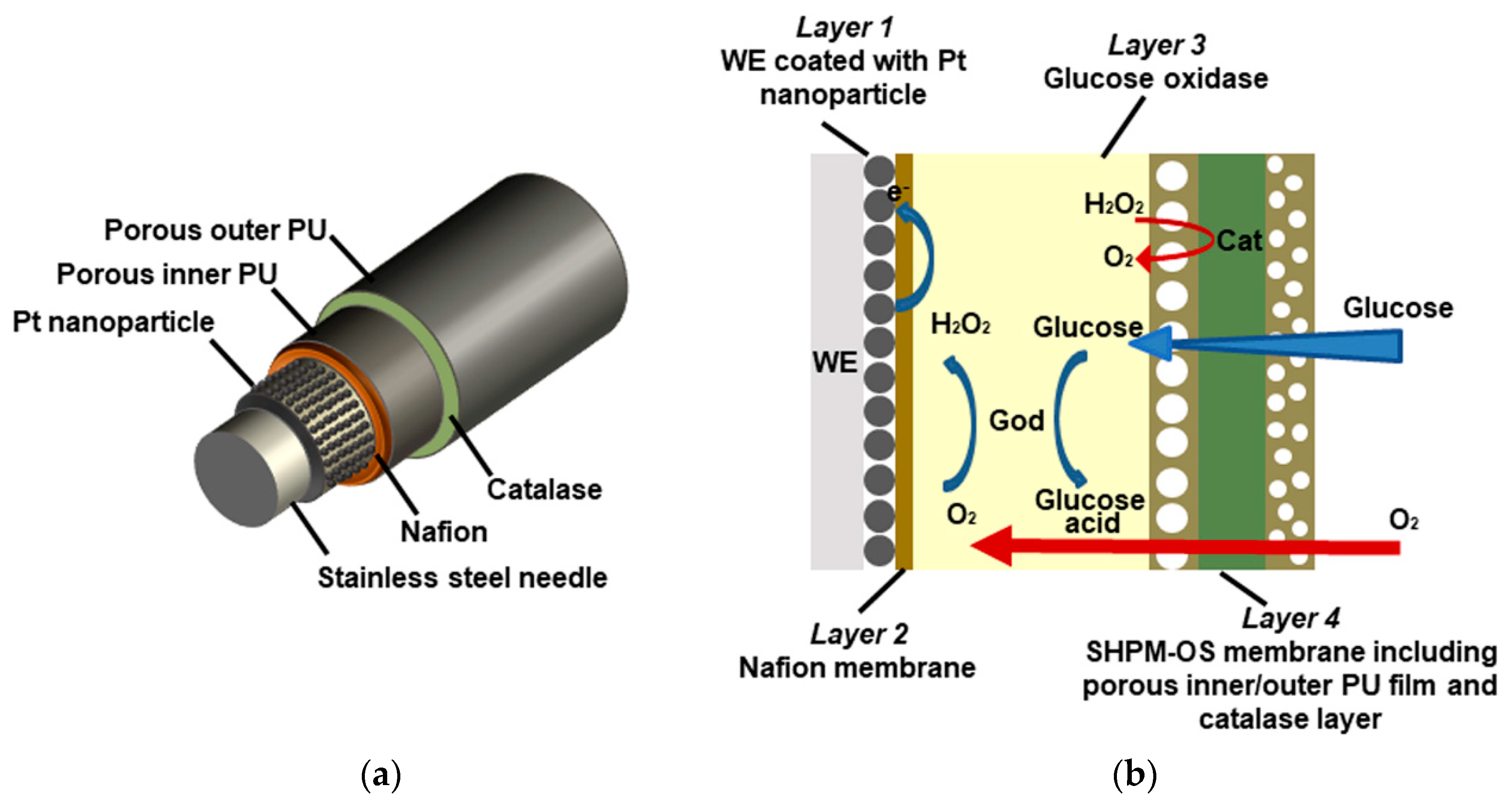
3.1. Principle and Design of the Glucose Sensor

The glucose sensor electrode was made by a stainless-steel acupuncture needle with a diameter of 0.3 mm (Figure 1a). Platinum nanoparticles were deposited on the needle to enhance electrical conductivity of electrode, catalyze H2O2 and enlarge the sensor surface area. Nafion permselective inner film was decorated on platinum layer to increase the quantity and activity of immobilized enzyme as well as to avoid the interference.
According to Figure 1b and chemical Equations (1) and (2), accumulation of H2O2 during the reaction leads to an inaccurate result and a narrow detection linear range. Hence, the SHPM-OS membrane was introduced. Porous inner PU with larger holes supported the Cat immobilization and accelerated the diffusion of H2O2 and oxygen. As shown in reaction (3), Cat in the sandwich layer decomposes H2O2 to replenish oxygen required for reaction real timely. Porous outer PU with smaller holes adjusted the concentration of glucose spreading to enzyme layer, which improved the sensitivity and linearity of the glucose sensor.
H 2 O 2   C a t 2 H + + O 2   + 2 e

3.2. Morphology of the New Sensor Electrode

The surface morphology of the new sensor electrode was evaluated by scanning electron microscope. As shown in Figure 2a, the size of the large pores on porous PU inner membrane was approximately (6.01 ± 0.925) μm. However, the size of the large pores on porous PU outer film was smaller with a diameter of (2.51 ± 0.638) um (Figure 2b).

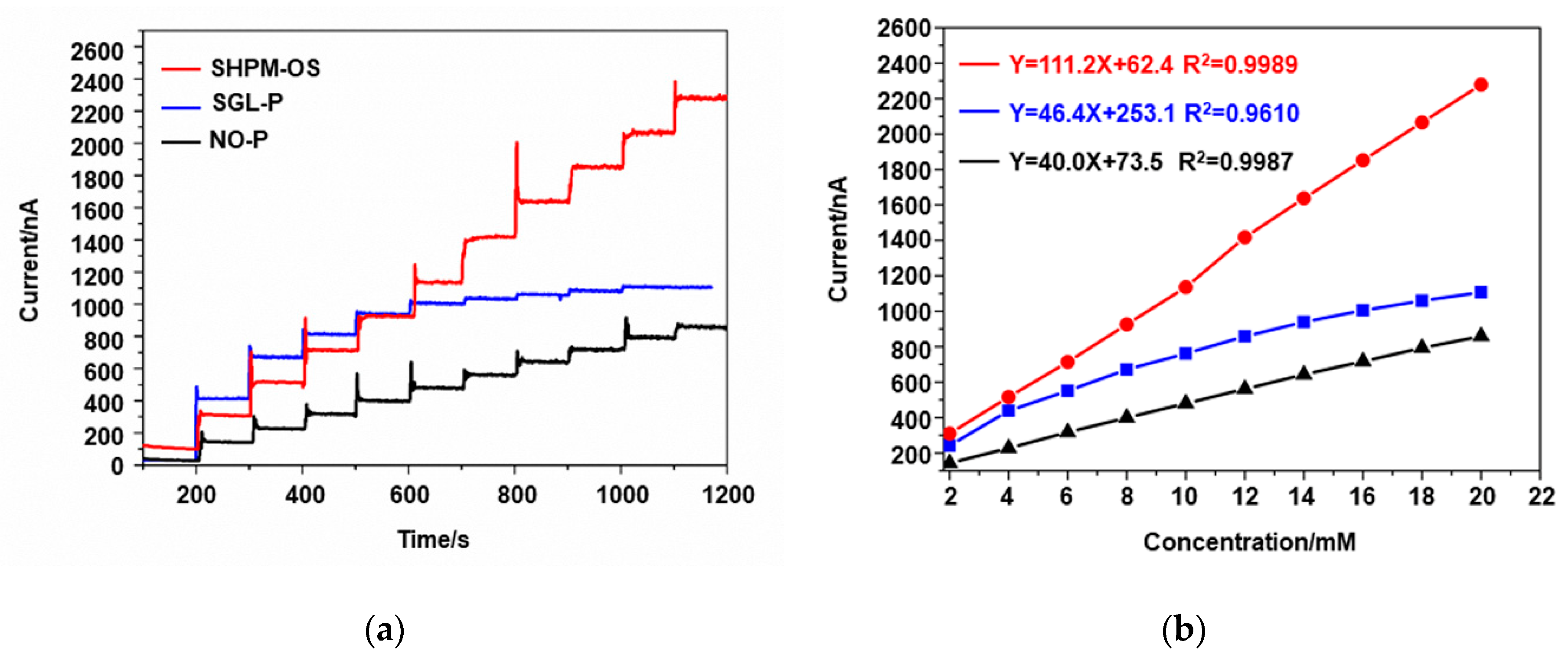
3.3. Linearity and Sensitivity of the Glucose Sensor

In order to evaluate and compare the performance of different sensors (the NO-P sensor, the SGL-P sensor and the SHPM-OS sensor), we performed chronoamperometry. Our results showed that the curve of the NO-P sensor was at the lowest position when the glucose concentration increased from 0 to 20 mM; the response current changed from 140 nA to 859 nA with the sensitivity of 35.95 nA/mM (Figure 3a), which is consistent with the sensors proposed before [10]. As the pore distribution on the PU surface expands, the limiting intensity of glucose transfer decreases. The current values of the SGL-P sensor increased from 200 nA to 1106.45 nA at the same glucose range and the corresponding sensitivity was 45.3 nA /mM, which was greater than the NO-P sensor (Figure 3a). However, when the glucose concentration was higher than 10 mM, the sensitivity of SGL-P rapidly decreased, and the R2 coefficient value decreased to 0.9610, which was lower than NO-P the (0.9987, Figure 3b). Compared to the NO-P and SGL-P sensors, the response value of the SHPM-OS sensor increased from 309 nA to 2278.32 nA with a higher sensitivity and linearity (98.45 nA/mM and 0.9989, respectively, Figure 3a). Although the current intensity was slightly smaller than the SGL-P sensor under low glucose concentration (0 to 10 mM), the sensitivity remained stable while the current values still increased gradually under high glucose concentration due to the constant replenishment of the oxygen.

3.4. Response Time of the Glucose Sensor

The time response curves of the SHPM-OS sensor were measured by rapidly increasing the glucose concentration. The response time of the SHPM-OS sensor decreased to 5s (Figure 4a), which is consistent with the sensor designed previously [34]. The selectivity of the SHPM-OS sensor against interference was evaluated by adding UA, AA and AP. As shown in Figure 4b, the relative rates of UA, AA and AP were 6%, 3% and 3%, respectively, which were small and negligible. The results demonstrate that the SHPM-OS sensor has high selectivity.

3.5. Long Time Stability of the Glucose Sensor

To evaluate the stability, the SHPM-OS sensor was further tested using glucose titration at intervals. As shown in Figure 5a, when the glucose concentration increased from 0 to 20 mM, all of the response curves showed obvious ladder shape and great linearity. The relative sensitivity was defined as the ratio of the present sensitivity to the initial sensitivity obtained at the beginning. The relative sensitivity of the SHPM-OS sensor remained at least 90% in the first 10 days (Figure 5b). Although the response slightly decreased after that, the relative sensitivity remained at approximately 78% of the original sensitivity on day 20 (Figure 5b), which is similar to the published data [10,17,20]. The results demonstrate that the SHPM-OS sensor could remain stabilization for a long time.

4. Discussion

To date, the biggest challenge of the implantable glucose sensors is how to improve the sensitivity while maintain good linearity, biocompatibility and lifetime [5,35]. There are several ways to solve this problem, including maximizing sensing surface of the electrode; reducing the negative effects of oxygen deficiency; balancing high sensitivity and wide linear detection range; increasing the signal to noise ratio; and, maintaining the stability of the sensor for a long time.
Generally, gold [11], platinum [36,37] and platinum–iridium [38,39] are often used as conventional glucose electrode bases, where GOD could be directly immobilized. However, these metals have poor hardness and have to be implanted through an auxiliary device, which makes it inconvenient for diabetic diagnosis. So some researches indicated that high hardness stainless-steel electrode optimized by metal nanoparticles can effectively expand sensing area, accelerate electron transfer [10,40] and be directly implanted by hand, improving the detection ability of immobilized GOD and minimizing the injury to body [41]. Moreover, the particles, such as size and density, could be easily adjusted by controlling reaction conditions. As a result, the sensing area is enlarged, the number of immobilized enzyme and active sites is increased and the electrical catalysis performance of electrode is optimized.
As indicated in previous studies, the ratio of glucose to oxygen was more than 100 in interstitial fluid, causing an oxygen deficiency during monitor and a narrow linear detection range [17,27,30]. The sensor modified by nanoparticles would aggravate oxygen deficiency [17]. It has been demonstrated that biocompatible films can regulate the ratio of oxygen-glucose concentration in the reaction process [10]. Moreover, influences of sensor coatings on glucose transmission depend on the thickness, uniformity of pores distribution, surface morphology and other factors. Currently, a variety of natural, semi-synthetic and synthetic materials, such as chitosan, PEG and polyvinyl acetate, have been applied to modify outer films to optimize the biocompatibility and monitoring performance of implantable glucose sensors [8]. Specially, PEG is a strong hydrophilic amphoteric ion and is a promising modification material for sensor coating. The PEG segments gather on the membrane surface and absorb a large mass of water, generating a lot of pores on the surface after adequate hydration and drying [18]. Hence, in this study, compared to the NO-P curve, the large size of pores on surface of the SGL-P sensor allowed more glucose to reach electrode, obtaining stronger response current at low concentration. However, the response current became unstable with the increase of glucose concentration. The linearity of sensor was also poor, probably due to the insufficient oxygen. Enhancing restriction of material transfer is an attractive membrane modification method to improve linearity of the glucose sensor, which however could decrease the sensitivity. For example, when the PVDF-Nafion glucose sensor was covered by nanoparticles with great restrictive effect, R2 increased from 0.776 to 0.9988, but the signal intensity decreased by 29% at the highest glucose concentration [17]. In previous studies, a PU/epoxy-enhanced (E-PU) membrane with smooth surface without holes was designed [27,42], which had a stronger effect on glucose restriction. When the concentration of epoxy reached 40%, the PU/E-PU could block over 70% glucose with a sensitivity of 63 nA/mM [27]. Particularly, the maximum linear detection range could expand to 40 mM with the increase of epoxy concentration, but the sensitivity decreased to 6 nA/mM [42].
Although the linear detection range could be enhanced by restricting glucose transmission [33], there are still difficulties to collect effective signal and to produce the sensors at low cost. Moreover, the successive accumulation of H2O2 in the reaction would result in electrode poisoning and the leakage would stimulate surrounding tissue [5,29,33]. An oxygen regeneration system prepared by immobilizing both GOD and Cat on sensing surface was proposed by Lucisano and Takashi et al. [30,31]. H2O2 can be rapidly decomposed, which generates oxygen when Cat is sufficient; the influence of oxygen deficiency can be partly relieved because the oxygen demand is halved. However, the sensing area of typical sensor electrode is limited, leading to the insufficient immobilized GOD when Cat is deposited on the same surface. Thus, the sensor sensitivity and lifetime need to be optimized [43]. Vaddiraju and Croce et al. [29,41] proposed an oxygen regeneration system, which immobilized GOD and Cat on different layers through layer-by-layer assembly approach and improved the sensitivity to 70 nA/mM. However, the function of signal outer membrane was simple, and it is difficult to obtain the optimal oxygen-glucose transport ratio. The SHPM-OS film proposed in this study provided a great strategy, which adjusted the morphology and distribution of pores with changed PEG concentration on different layers. Particularly, the pores of the porous PU inner film with a high proportion of PEG were large in diameter and clearly visible, which can enhance driving force for glucose and oxygen diffusion inward [41,44] and the speed of analyzing glucose change [45], thus shortening the response time of the glucose sensor. On the contrary, the pores of the porous PU outer membrane with less PEG were small and restricted glucose transmission, maintaining the high linearity of sensors. The response current had a linear change in the range of 0–20-mM glucose concentration (R2 was 0.9989). The comparison of different amperometric sensors for the glucose determination was shown in Table 1. The SHPM-OS sensor exhibited similar linear range as the other sensors in Table 1. However, it could continuously release oxygen from sandwich layer and thus, its sensitivity was increased by three times. However, compared with other commercial sensors, the linear range of SHPM-OS (0–20-mM) was relatively narrow. Thus, it is necessary to combine SHPM-OS with the AiTi health management platform (developed by our group) in the future, which would send an early warning to users, family members or private doctors when BG concentration exceeds 20 mM.
Since the sensitivity of the sensor will inevitably be reduced, CGMS needs to be manually and algorithmically calibrated at regular intervals to maintain the accuracy of the monitoring. The SHPM-OS sensor in this study maintained stable sensitivity within 20 days, which could relieve the pain of people with diabetes caused by finger-sticking. Moreover, a typical amperometric glucose sensor has to experience complicated immobilization procedure and degradation of enzyme, oxygen limitation and fibrosis [5,50]. Improving the loading enzyme capacity of the electrode [51,52,53,54], optimizing the immobilization methods [55] and enhancing enzyme activity [32,46] of GOD can slow down the degradation rate of the enzyme. The anti-inflammatory effect of nitric oxide and dexamethasone can increase the compatibility of tissue–sensor interface and improve the sensor lifetime [56,57,58,59], but the duration of anti-inflammatory medication is affected by the pore size of membrane. Hence, a coat with anti-inflammatory function can be added to SHPM-OS membrane structure in the future to reduce biofouling-related problems and the speed of electrode fibrosis [18]. The adjustment of the combination of hierarchically porous layers to prepare another novel implantable glucose sensor can be then developed, which may optimize biocompatibility and monitoring lifetime while maintain the existing sensitivity and linearity.

5. Conclusions

In conclusion, the proposed SHPM-OS implantable glucose sensor in this study has greater effects on enhancing the sensitivity while maintaining excellent linear detection range and stability. This SHPM-OS membrane replenishes sufficient oxygen by sandwich Cat layer and regulates the glucose–oxygen transport ratio by the hierarchical porous structure, which increases its sensitivity from 35.95 nA/mM to 98.45 nA/mM. Meanwhile, other performances, such as linearity, response time, selectivity and stability, are still good enough to monitor glucose change. Hence, this combination of hierarchical porous films and oxygen regeneration system is beneficial to the development of back-end monitoring hardware module and the dynamic BG monitoring system.

Author Contributions

Conceptualization—L.H. and X.P.; data curation—L.H. and X.P.; formal analysis—J.Z. and Z.J.; writing—original draft L.H.; writing—edit and review, X.P. and H.L. All authors have read and agreed to the published version of the manuscript.

Funding

This study was funded by the National Key Research and Development Program of China (Grant Number 2018YFC2000804), National Natural Science Foundation of China (Grant Number 81671850) and the Fundamental Research Funds for the Central Universities (Grant Number 2019CDCG0014).

Conflicts of Interest

The authors declare no conflict of interest.

References

  1. van Hooijdonk, R.T.M.; Leopold, J.H.; Winters, T.; Binnekade, J.M.; Juffermans, N.P.; Horn, J.; Fischer, J.C.; van Dongen-Lases, E.C.; Schultz, M.J. Point accuracy and reliability of an interstitial continuous glucose-monitoring device in critically ill patients: A prospective study. Crit. Care 2015, 19, 34. [Google Scholar] [CrossRef] [PubMed] [Green Version]
  2. Thomas, F.; Signal, M.; Chase, J.G. Using Continuous Glucose Monitoring Data and Detrended Fluctuation Analysis to Determine Patient Condition: A Review. J. Diabetes Sci. Technol. 2015, 9, 1327–1335. [Google Scholar] [CrossRef] [PubMed]
  3. van Enter, B.J.; Von, H.E. Challenges and perspectives in continuous glucose monitoring. Chem. Commun. 2018, 54, 5032. [Google Scholar] [CrossRef]
  4. Cappon, G.; Acciaroli, G.; Vettoretti, M.; Facchinett, A.; Sparacino, G. Wearable continuous glucose monitoring sensors: A revolution in diabetes treatment. Electronics 2017, 6, 65. [Google Scholar] [CrossRef] [Green Version]
  5. Scholten, K.; Meng, E. A review of implantable biosensors for closed-loop glucose control and other drug delivery applications. Int. J. Pharm. 2018, 544, 319–334. [Google Scholar] [CrossRef]
  6. van Beers, C.A.J.; Kleijer, S.J.; Serné, E.H.; Geelhoed-Duijvestijn, P.H.; Snoek, F.J.; Kramer, M.H.; Diamant, M. Design and rationale of the in control trial: The effects of real-time continuous glucose monitoring on glycemia and quality of life in patients with type 1 diabetes mellitus and impaired awareness of hypoglycemia. BMC Endocr. Disord. 2015, 15, 42. [Google Scholar] [CrossRef] [Green Version]
  7. Heo, Y.J.; Kim, S.-H. Toward Long-Term Implantable Glucose Biosensors for Clinical Use. Appl. Sci. 2019, 9, 2518. [Google Scholar] [CrossRef] [Green Version]
  8. Chen, C.; Zhao, X.L.; Li, Z.H.; Zhu, Z.G.; Qian, S.H.; Flewitt, A.J. Current and Emerging Technology for Continuous Glucose Monitoring. Sensors 2017, 17, 182. [Google Scholar] [CrossRef] [Green Version]
  9. Zhai, D.; Liu, B.; Shi, Y.; Pan, L.; Wang, Y.; Li, W.; Zhang, R.; Yu, G. Highly Sensitive Glucose Sensor Based on Pt Nanoparticle/Polyaniline Hydrogel Heterostructures. ACS Nano 2013, 7, 3540–3546. [Google Scholar] [CrossRef]
  10. Fang, Y.; Wang, S.; Liu, Y.; Xu, Z.; Zhang, K.; Guo, Y. Development of Cu nanoflowers modified the flexible needle-type microelectrode and its application in continuous monitoring glucose in vivo. Biosens. Bioelectron. 2018, 110, 44–51. [Google Scholar] [CrossRef]
  11. Abdulbari, H.A.; Basheer, E.A.M. Electrochemical Biosensors: Electrode Development, Materials, Design, and Fabrication. Chembioeng Rev. 2017, 4, 92–105. [Google Scholar] [CrossRef] [Green Version]
  12. Witkowska, N.E.; Kundys, M.; Jeleń, P.S.; Jönsson-Niedziółka, M. Electrochemical Glucose Sensing: Is There Still Room for Improvement? Anal. Chem. 2016, 88, 11271–11282. [Google Scholar] [CrossRef] [PubMed]
  13. Zulkifli, Z.A.; Ridhuan, N.S.; Nor, N.M.; Zakaria, N.D.; Razak, K. The effect of gold nanoparticles modified electrode on the glucose sensing performance. In Proceedings of the International Conference of Global Network for Innovative Technology, Penang, Malaysia, 27–29 January 2016; 2017. [Google Scholar]
  14. Stine, K.J. Biosensor Applications of Electrodeposited Nanostructures. Appl. Sci. 2019, 9, 797. [Google Scholar] [CrossRef] [Green Version]
  15. Xu, G.; Adeloju, S.B.; Wu, Y.; Zhang, X. Modification of polypyrrole nanowires array with platinum nanoparticles and glucose oxidase for fabrication of a novel glucose biosensor. Anal. Chim. Acta 2012, 755, 100–107. [Google Scholar] [CrossRef] [PubMed]
  16. German, N.; Ramanavicius, A.; Ramanaviciene, A. Amperometric Glucose Biosensor Based on Electrochemically Deposited Gold Nanoparticles Covered by Polypyrrole. Electroanalysis 2017, 29, 1267–1277. [Google Scholar] [CrossRef]
  17. Chen, D.; Wang, C.; Chen, W.; Chen, Y.; Zhang, X. PVDF-Nafion nanomembranes coated microneedles for in vivo transcutaneous implantable glucose sensing. Biosens. Bioelectron. 2015, 74, 1047–1052. [Google Scholar] [CrossRef]
  18. Lopes, I.C.; Zebda, A.; Vadgama, P. New directions in membrane designs for biosensors. Curr. Opin. Electrochem. 2018, 12, 107–112. [Google Scholar] [CrossRef]
  19. Wang, N.; Burugapalli, K.; Song, W.; Halls, J.; Moussy, F.; Ray, A.; Zheng, Y. Electrospun fibro-porous polyurethane coatings for implantable glucose biosensors. Biomaterials 2013, 34, 888–901. [Google Scholar] [CrossRef] [Green Version]
  20. Yang, W.; Xue, H.; Carr, L.R.; Wang, J.; Jiang, S. Zwitterionic poly(carboxybetaine) hydrogels for glucose biosensors in complex media. Biosens. Bioelectron. 2011, 26, 2454–2459. [Google Scholar] [CrossRef]
  21. Koh, A.; Nichols, S.P.; Schoenfisch, M.H. Glucose sensor membranes for mitigating the foreign body response. J. Diabetes Sci. Technol. 2011, 5, 1052–1059. [Google Scholar] [CrossRef] [Green Version]
  22. Mercado, R.C.; Moussy, F. In vitro and in vivo mineralization of Nafion membrane used for implantable glucose sensors. Biosens. Bioelectron. 1998, 13, 133–145. [Google Scholar] [CrossRef]
  23. Moattisirat, D.; Poitout, V.; Thome, V.; Gangnerau, M.N.; Zhang, Y.; Hu, Y.; Wilson, G.S.; Lemonnier, F.; Klein, J.C.; Reach, G. Reduction of acetaminophen interference in glucose sensors by a composite Nafion membrane—Demonstration in rats and man. Diabetologia 1994, 37, 610–616. [Google Scholar] [CrossRef]
  24. Brazell, M.P.; Kasser, R.J.; Renner, K.J.; Feng, J.; Moghaddam, B.; Adams, R.N. electrocoating carbon-fiber microelectrodes with Nafion improves selectivity for electroactive neurotransmitters. J. Neurosci. Methods 1987, 22, 167–172. [Google Scholar] [CrossRef]
  25. Yu, B.; Wang, C.; Ju, Y.; West, L.; Harmon, J.; Moussy, Y.; Moussy, F. Use of hydrogel coating to improve the performance of implanted glucose sensors. Biosens. Bioelectron. 2008, 23, 1278–1284. [Google Scholar] [CrossRef] [PubMed]
  26. Wang, C.; Yu, B.; Knudsen, B.; Harmon, J.; Moussy, F.; Moussy, Y. Synthesis and performance of novel hydrogels coatings for implantable glucose sensors. Biomacromolecules 2008, 9, 561–567. [Google Scholar] [CrossRef]
  27. Fang, L.; Liang, B.; Yang, G.; Hu, Y.; Zhu, Q.; Ye, X. A needle-type glucose biosensor based on PANI nanofibers and PU/E-PU membrane for long-term invasive continuous monitoring. Biosens. Bioelectron. 2017, 97, 196–202. [Google Scholar] [CrossRef]
  28. Jiang, L.; Koinkar, P.; Fuchiwaki, Y.; Yasuzawa, M. A fine pointed glucose oxidase immobilized electrode for low-invasive amperometric glucose monitoring. Biosens. Bioelectron. 2016, 86, 90–94. [Google Scholar]
  29. Vaddiraju, S.; Burgess, D.J.; Jain, F.C.; Papadimitrakopoulos, F. The role of H2O2 outer diffusion on the performance of implantable glucose sensors. Biosens. Bioelectron. 2009, 24, 1557–1562. [Google Scholar] [CrossRef] [Green Version]
  30. Kuwahara, T.; Ogawa, K.; Sumita, D.; Kondo, M.; Shimomura, M. Amperometric glucose sensing with polyaniline/poly(acrylic acid) composite film bearing glucose oxidase and catalase based on competitive oxygen consumption reactions. J. Electroanal. Chem. 2018, 811, 62–67. [Google Scholar] [CrossRef]
  31. Lucisano, J.; Routh, T.; Lin, J.; Gough, D. Glucose Monitoring in Individuals with Diabetes using a Long-Term Implanted Sensor/Telemetry System and Model. IEEE Trans. Bio-Med. Eng. 2017, 64, 1982–1993. [Google Scholar] [CrossRef] [Green Version]
  32. Vaddiraju, S.; Legassey, A.; Wang, Y.; Qiang, L.; Burgess, D.; Jain, F.; Papadimitrakopoulos, F. Design and fabrication of a high-performance electrochemical glucose sensor. J. Diabetes Sci. Technol. 2011, 5, 1044–1051. [Google Scholar] [CrossRef] [PubMed] [Green Version]
  33. Vaddiraju, S.; Legassey, A.; Qiang, L.; Wang, Y.; Burgess, D.J.; Papadimitrakopoulos, F. Enhancing the sensitivity of needle-implantable electrochemical glucose sensors via surface rebuilding. J. Diabetes Sci. Technol. 2013, 7, 441–451. [Google Scholar] [CrossRef] [PubMed] [Green Version]
  34. Kim, Y.-J.; Saviers, K.R.; Fisher, T.S.; Irazoqui, P.P. Continuous glucose monitoring with a flexible biosensor and wireless data acquisition system. Sens. Actuators B-Chem. 2018, 275, 237–243. [Google Scholar] [CrossRef]
  35. Yosefi, G.; Mirzakuchaki, S.; Raissi, F. New Programmable CMOS Fuzzifier and C2V Circuits Applicable in FLC Chip for Signal Processing of MEMS Glucose Sensors. Appl. Sci. 2015, 5, 402–414. [Google Scholar] [CrossRef]
  36. Ward, W.K.; Jansen, L.B.; Anderson, E.; Reach, G.; Klein, J.C.; Wilson, G.S. A new amperometric glucose microsensor: In vitro and short-term in vivo evaluation. Biosens. Bioelectron. 2002, 17, 181–189. [Google Scholar] [CrossRef]
  37. Chen, X.H.; Matsumoto, N.; Hu, Y.; Wilson, G. Electrochemically mediated electrodeposition/electropolymerization to yield a glucose microbiosensor with improved characteristics. Anal. Chem. 2002, 74, 368–372. [Google Scholar] [CrossRef]
  38. Bindra, D.S.; Zhang, Y.; Wilson, G.S.; Sternberg, R.; Thévenot, D.R.; Moatti, D.; Reach, G. Design and in vitro studies of a needle-type glucose sensor for subcutaneous monitoring. Anal. Chem. 1991, 63, 1692. [Google Scholar] [CrossRef] [Green Version]
  39. Yu, B.Z.; Moussy, Y.; Moussy, F. Coil-type implantable glucose biosensor with excess enzyme loading. Front. Biosci. Landmark 2005, 10, 512–520. [Google Scholar] [CrossRef] [Green Version]
  40. Guo, M.; Fang, H.; Wang, R.; Yang, Z.; Xu, X. Electrodeposition of chitosan-glucose oxidase biocomposite onto Pt–Pb nanoparticles modified stainless steel needle electrode for amperometric glucose biosensor. J. Mater. Sci. Mater. Med. 2011, 22, 1985–1992. [Google Scholar] [CrossRef]
  41. Croce, R.A., Jr.; Vaddiraju, S.; Kondo, J.; Wang, Y.; Zuo, L.; Zhu, K.; Lslam, S.k.; Burgess, D.J.; Papadimitrakopoulos, F.; Jain, F.C. A miniaturized transcutaneous system for continuous glucose monitoring. Biomed. Microdevices 2013, 15, 151–160. [Google Scholar]
  42. Yu, B.Z.; Long, N.; Moussy, Y.; Moussy, F. A long-term flexible minimally-invasive implantable glucose biosensor based on an epoxy-enhanced polyurethane membrane. Biosens. Bioelectron. 2006, 21, 2275–2282. [Google Scholar] [CrossRef] [PubMed]
  43. Koh, A.; Lu, Y.; Schoenfisch, M.H. Fabrication of Nitric Oxide-Releasing Porous Polyurethane Membranes Coated Needle-Type Implantable Glucose Biosensors. Anal. Chem. 2013, 85, 10488–10494. [Google Scholar] [CrossRef] [PubMed] [Green Version]
  44. Guo, T.; Gao, J.; Qin, X.; Zhang, X.; Xue, H. A Novel Glucose Biosensor Based on Hierarchically Porous Block Copolymer Film. Polymers 2018, 10, 723. [Google Scholar] [CrossRef] [PubMed] [Green Version]
  45. Narayan, R.J.; Jin, C.; Menegazzo, N.; Mizaikoff, B.; Gerhardt, R.; Andara, M.; Agarwal, A.; Shih, C.C.; Shih, C.M.; Lin, S.J.; et al. Nanoporous hard carbon membranes for medical applications. J. Nanosci. Nanotechnol. 2007, 7, 1486–1493. [Google Scholar] [CrossRef] [PubMed]
  46. Fang, L.; Liang, B.; Yang, G.; Hu, Y.; Zhu, Q.; Ye, X. Study of glucose biosensor lifetime improvement in 37 °C serum based on PANI enzyme immobilization and PLGA biodegradable membrane. Biosens. Bioelectron. 2014, 56, 91–96. [Google Scholar] [CrossRef]
  47. Fang, L.; Liu, B.; Liu, L.; Li, Y.; Huang, K.; Zhang, Q. Direct electrochemistry of glucose oxidase immobilized on Au nanoparticles-functionalized 3D hierarchically ZnO nanostructures and its application to bioelectrochemical glucose sensor. Sens. Actuators B Chem. 2016, 222, 1096–1102. [Google Scholar] [CrossRef]
  48. Edagawa, K.; Fuchiwaki, Y.; Yasuzawa, M. In Vivo Evaluation of Fine Needle Amperometric Glucose Sensors Implanted in Rabbit’s Blood Vessel. J. Electrochem. Soc. 2014, 161, B3111–B3115. [Google Scholar] [CrossRef]
  49. Kim, K.B.; Lee, W.C.; Cho, C.H.; Park, D.S.; Cho, S.J.; Shim, Y.B. Continuous glucose monitoring using a microneedle array sensor coupled with a wireless signal transmitter. Sens. Actuators B Chem. 2019, 281, 14–21. [Google Scholar] [CrossRef]
  50. Liu, G.-S.; Kong, Y.; Wang, Y.; Luo, Y.; Fan, X.; Xie, X.; Yang, B.-R.; Wu, M.X. Microneedles for transdermal diagnostics: Recent advances and new horizons. Biomaterials 2020, 232, 119740. [Google Scholar] [CrossRef]
  51. Zeng, X.; Zhang, Y.; Du, X.; Li, Y.; Tang, W. A highly sensitive glucose sensor based on a gold nanoparticles/polyaniline/multi-walled carbon nanotubes composite modified glassy carbon electrode. New J. Chem. 2018, 42, 11944–11953. [Google Scholar] [CrossRef]
  52. Zdarta, J.; Meyer, A.S.; Jesionowski, T.; Pinelo, M. A General Overview of Support Materials for Enzyme Immobilization: Characteristics, Properties, Practical Utility. Catalysts 2018, 8, 92. [Google Scholar] [CrossRef] [Green Version]
  53. Semenova, D.; Silina, Y.E.; Koch, M.; Micheli, L.; Zubov, A.; Gernaey, K.V. Sensors for biosensors: A novel tandem monitoring in a droplet towards efficient screening of robust design and optimal operating conditions. Analyst 2019, 144, 2511–2522. [Google Scholar] [CrossRef] [PubMed]
  54. Fang, Y.; Zhang, D.; Guo, Y.; Guo, Y.M.; Chen, Q. Simple one-pot preparation of chitosan-reduced graphene oxide-Au nanoparticles hybrids for glucose sensing. Sens. Actuators B Chem. 2015, 221, 265–272. [Google Scholar] [CrossRef]
  55. Donahue, C.E.T.; Miller, D.R., Jr.; Beger, T.W.; Johann, T.W.; Keithley, R.B. Improved formation of electrically-deposited enzyme-embedded chitosan coatings onto carbon fiber microelectrodes. Anal. Methods 2018, 10, 1565–1576. [Google Scholar] [CrossRef]
  56. Vallejoheligon, S.G.; Brown, N.L.; Reichert, W.M.; Klitzman, B. Porous, Dexamethasone-Loaded Polyurethane Coatings Extend Performance Window of Implantable Glucose Sensors in vivo. Acta Biomater. 2016, 30, 106–115. [Google Scholar] [CrossRef] [Green Version]
  57. Vallejo-Heligon, S.G.; Klitzman, B.; Reichert, W.M. Characterization of porous, dexamethasone-releasing polyurethane coatings for glucose sensors. Acta Biomater. 2014, 10, 4629–4638. [Google Scholar] [CrossRef] [Green Version]
  58. Soto, R.J.; Privett, B.J.; Schoenfisch, M.H. In vivo analytical performance of nitric oxide-releasing glucose biosensors. Anal. Chem. 2014, 86, 7141–7149. [Google Scholar] [CrossRef] [Green Version]
  59. de la Oliva, N.; del Valle, J.; Delgado-Martinez, I.; Mueller, M.; Stieglitz, T.; Navarro, X. Long-Term Functionality of Transversal Intraneural Electrodes Is Improved by Dexamethasone Treatment. IEEE Trans. Neural Syst. Rehabil. Eng. 2019, 27, 457–464. [Google Scholar] [CrossRef] [Green Version]
Figure 1. Schematic diagram of the SHPM-OS glucose sensor. (a) functional layers of glucose electrode. The needle is modified by Pt nanoparticles, Nafion, GOD and SHPM-OS membranes, which consists of porous inner PU with large-sized pores, Cat and porous outer PU with small-sized pores. (b) schematic diagram of electrochemical reaction and element transfer on different layers. Glucose transmission was regulated by both outer and inner membranes and then glucose reacts in the GOD layer to produce electrons and H2O2, which was catalyzed by Cat to replenish the oxygen.
Applsci 10 02848 g001
Figure 2. SEM images of hierarchically porous PU membranes covered on the SHPM-OS glucose sensor. (a) SEM image of porous inner PU with the large-sized pores (6.01 ± 0.925) μm. (b) SEM image of porous outer PU with the small-sized pores (2.51 ± 0.638) μm.
Applsci 10 02848 g002
Figure 3. Performance comparison of NO-P, SGL-P and SHPM-OS sensors. The line in red is the response of the SHPM-OS sensor; the others are the SGL-P (blue) and NO-P sensor (black), respectively. (a) current response of three sensors versus glucose concentration. (b) linear fitting curves of three sensors. The fitting equation is displayed next to the corresponding line.
Applsci 10 02848 g003
Figure 4. Response speed and anti-interference results of the SHPM-OS sensor. (a) current-time response of the SHPM-OS sensor to single high concentration (10 mM) injection of glucose into stirring phosphate buffer solution (PBS). (b) SHPM-OS sensor response to sequential glucose and common interferences. Concentrations of the additions in the measuring PBS include 4 mM glucose, 0.48-mM UA, 0.11-mM AA, 0.17-mM AP and 2-mM glucose.
Applsci 10 02848 g004
Figure 5. Stability of the relative sensitivity of the SHPM-OS sensor. (a) comparison of the amperometric response results of the SHPM-OS sensors versus glucose concentration at different intervals. (b) histogram of relative sensitivity of the SHPM-OS sensors restored in PBS.
Applsci 10 02848 g005
Table 1. Comparison of different amperometric sensors for the glucose determination.
Sensor StructureLinear Range mMCorrelation Coefficient R2Reference
Pt/Cu-NFs/Nafion/GOD/PU0–200.99[10]
Pt/PANI/GOD/PU/E-PU0–200.9996[27]
Pt-Ir/Pt/GOD0–210.9986[28]
Au/Pt-Nps/PANI/GOD/PU/PLGA0–140.998[46]
SUS/Pt/PANI/GOD/PVDF/Nafion0–200.9988[17]
GCE/Au-ZnO/GOD1–20 [47]
Pt-Ir/Nafion/GOD/PU/
PDMS/Heparin
0–22.40.98[48]
AuMN/pTCA-GOx/
Nafion
0.05–200.99[49]
SUS/Pt-Nps/Nafion/
GOD/SHPM-OS
0–200.9988This study

