Article

Ultrasound-Guided Detection and Segmentation of Photoacoustic Signals from Bone Tissue In Vivo

1
School of Electronic and Optical Engineering, Nanjing University of Science and Technology, Nanjing 210094, China
2
Institution of Acoustics, School of Physics Science and Engineering, Tongji University, Shanghai 200092, China
3
School of Electronic Science and Engineering, Nanjing University, Nanjing 210023, China
4
Academy for Engineering and Technology, Fudan University, Shanghai 200433, China
5
Department of Electronic Engineering, Fudan University, Shanghai 200433, China
*
Authors to whom correspondence should be addressed.
These authors contributed equally.
Appl. Sci. 2021, 11(1), 19; https://doi.org/10.3390/app11010019
Submission received: 28 October 2020 / Revised: 15 December 2020 / Accepted: 17 December 2020 / Published: 22 December 2020
(This article belongs to the Special Issue Photoacoustic Tomography (PAT) II)

Abstract

:
Photoacoustic (PA) techniques provide optical absorption contrast and spatial information at an ultrasound resolution in deep biological tissues. Among the greatest challenges encountered in the PA examination of bone is the analysis of trabecular bone, which holds key chemical and physical information required for bone health assessments. Ultrasound detection is naturally registered with PA detection; therefore, in this study, we propose ultrasound guidance for the PA detection of trabecular bone. We perform both numerical simulations and an in vivo experiment on a human subject to investigate the possibility of ultrasound-guided detection and segmentation of photoacoustic signals from bone tissue in vivo in a non-invasive manner. The results obtained from the simulation and in vivo experiment suggest that the ultrasound-guided PA method can distinguish PA signals from trabecular and cortical bones as well as from the overlying soft tissue. Considering that the PA technique is non-ionizing and non-invasive, it holds potential for clinical bone health assessment.

1. Introduction

Osteoporosis is a highly prevalent disease, affecting approximately 40% of women and 20% of men older than 50 years [1,2]. A failure to detect osteoporosis and delayed treatment may result in irreversible bone loss, particularly in trabecular bone, which is strongly related to the process of osteoporosis. The detection of changes in the quality and quantity of trabecular bone is crucial for predicting and diagnosing osteoporosis. Currently, the standard diagnosis of osteoporosis is primarily based on the bone quantity assessed using dual-energy X-ray absorptiometry (DEXA), which is a radiography-based technique. However, DEXA has limited sensitivity for monitoring chemical or molecular changes in the bone. Further, the bone mineral density provided by DEXA, bone microarchitecture, and material compositional characteristics, which cannot be assessed by radiographic imaging techniques, are significant for bone health [3]. Magnetic resonance imaging (MRI) may distinguish the changes in marrow lipid content and microarchitecture in osteoporotic bones [2,4,5]. However, the high cost and complexity of MRI have prevented its standardization as the primary diagnostic procedure. In contrast, quantitative ultrasound (QUS) techniques provide a practical and low-cost substitute for DEXA, and they have already been used in clinical instrumentation [6,7,8,9]. QUS assessments of the bone structure and strength are mainly based on two parameters: the speed of sound (SOS) and broadband ultrasound attenuation through the bone [10,11,12,13]. However, the specificity of QUS is limited for detecting bone diseases with pathogenesis involving both microarchitectural and chemical changes.
Optical techniques are more sensitive than ultrasound (US)-based methods to molecular and functional changes in the bone [14,15,16,17]. However, conventional optical-based techniques are affected by limited spatial information owing to the overwhelming optical scattering in biological tissues, particularly in bone tissue. The emerging biomedical photoacoustic (PA) techniques can be used to determine the highly sensitive optical absorption contrast of deep biological tissues [18,19,20]. Therefore, PA techniques, utilizing both light and acoustics, may be combined with US to address the aforementioned limitation.
Laser-induced PA sensing is based on the detection of light-induced ultrasonic signals that are less scattered in biological tissues than light [18,21,22,23,24]. Therefore, PA signal detection is naturally registered with the US method. PA sensing, while utilizing the acoustic resolution, can assess more spatial information in deep tissues than conventional optical-based techniques. PA signals also provide chemical information, similar to the optical method. To the best of our knowledge, the PA technique, as an adjunct to the established US system, has already demonstrated its potential in several preclinical and clinical applications [18,19,22,25,26,27,28,29]. Recent studies have indicated that the PA signal from the bone can provide critical chemical information, in addition to physical information [25,26,27,28,30,31,32], which is important for predicting and diagnosing osteoporosis. However, most of all the previous studies that focused on the assessment of trabecular bone using PA techniques are based on ex vivo bone specimens or small animal models. The possible challenges of in vivo applications for human bone assessment, including PA signal generation and propagation modes in trabecular bone, were not considered. For example, cancellous bone, which has a high percentage of trabecular bone, consists of a mineralized, porous trabecular network embedded in the bone marrow with filament-like trabeculae. The specific structure of the bone makes the generation and propagation of the PA signal more complicated than in soft tissue. The PA wave propagation in the bone and segmentation of the PA signal from the bone are not fully understood, because of the structural complexity and inhomogeneity of the bone. Previous studies reported that the bone can support two kinds of longitudinal waves that travel at different velocities, including fast wave and slow wave. The fast wave is associated with the movement of fluid (blood and marrow) in phase with the solid (mineralized trabeculae), and the slow wave is associated with the movement of fluid out of phase with the solid [10]. Apart from the longitudinal wave, there is a Lamb wave, which travels across the cortical bone at high speed. Therefore, isolating the PA signal of the trabecular portion from the complicated PA signal is a key challenge that must be addressed for the PA bone assessment technique adopted in clinical research.
In this study, we propose a US-guided PA bone assessment method for locating and segmenting the PA signal spatial information of the human calcaneus bone, which can be used to distinguish the trabecular bone signal from other signals (soft tissue, cortical bone and their echoes). Two aspects have been demonstrated in this study. First, we studied the feasibility of the detection and segmentation of the PA signal generated from different bone tissues using the US-guided PA bone assessment method based on a two-dimensional (2D) numerical simulation. Second, by conducting an experiment on a volunteer, we investigated the feasibility of detecting and distinguishing the PA signal of the trabecular bone in vivo using the US-guided method.

2. Materials and Methods

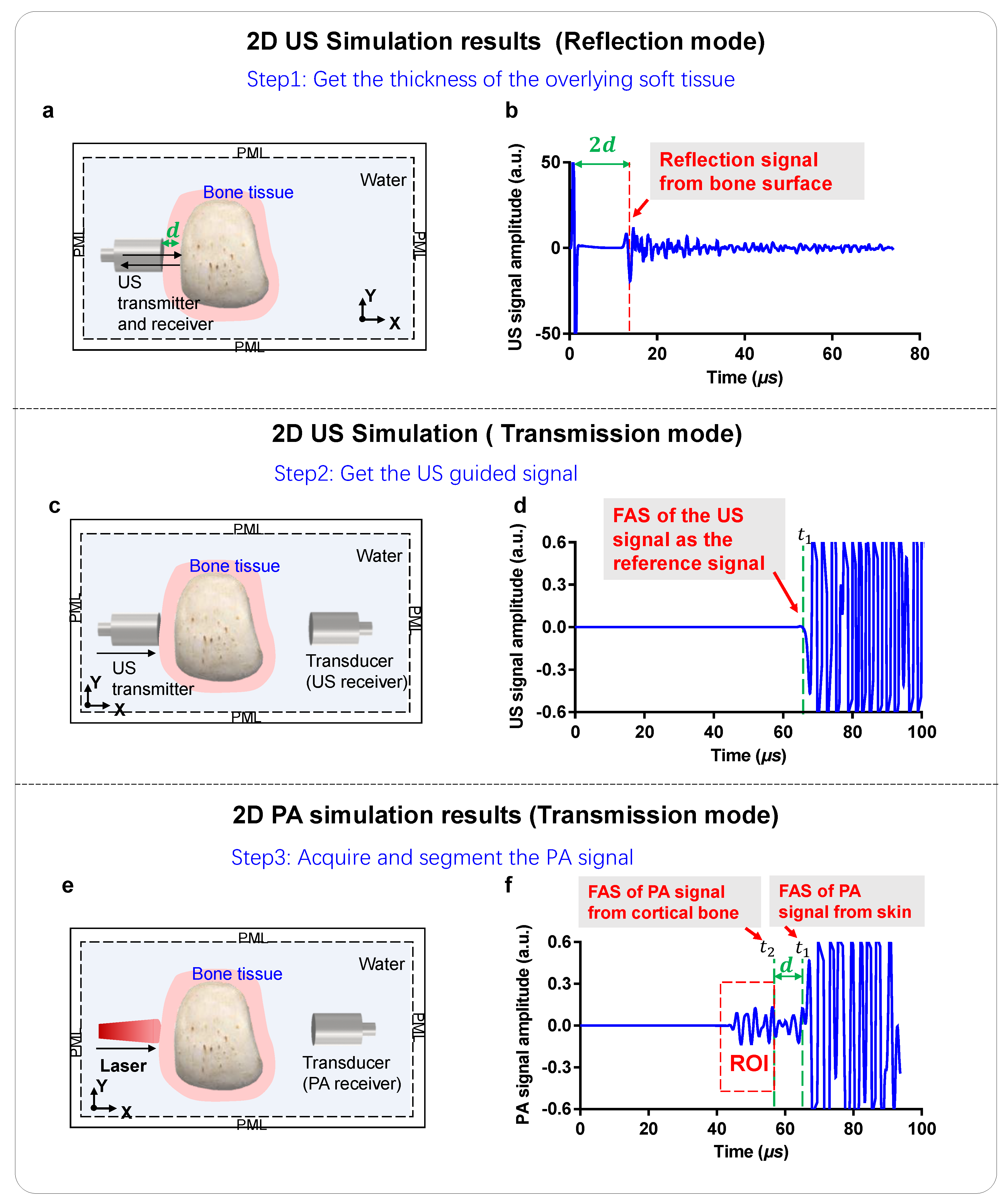
2.1. 2D Numerical US and PA Simulation Models

To investigate the feasibility of ultrasonic guidance for PA transmission-mode bone detection, we performed a 2D numerical simulation for US and PA signal propagation in the bone. The flow chart of the US and PA simulations is presented in Figure 1a.
In this study, both the US and PA simulations used the binarized micro-CT image of the calcaneus bone specimens from cadaver donors as the bone model. The bone specimens were degassed and immersed in distilled water, and micro-CT scanner (Skyscan 1076, Skyscan, Antwerp, Belgium) imaging was used for the microstructure measurement. In the simulations, we also added 8-mm-thick soft tissue at each side to cover the bone entirely, as shown in Figure 1b,c. In the 2D US numerical simulation models, it had both a transmission mode and reflection mode, as shown in Figure 1b. The reflection mode is used to calculate the thickness of the overlying soft tissue, whereas the transmission mode is used to find the first arrival signal (FAS) as the guidance signal. In the US transmission mode, the transmitting and receiving transducers were placed at different sides of the bone tissue. While in the US reflection mode, the transmission transducer also receives the ultrasound signal after the ultrasound source was excited. For the simulation of the PA signal measurements, it was in the transmission mode only, as shown in Figure 1c. For the 2D simulation of the PA mode, the light source and transducer were on opposite sides of the bone. The PML represents the perfectly matched layer. The parameters and properties of the related materials used in the simulation are listed in Table 1.

2.2. 2D Simulations of US Signal Propagation

A 2D simulation model of an elastic wave was developed based on the finite-difference time-domain (FDTD) method [33,34]. For the excitation condition, the Gaussian-modulated sine pulse defined in Equation (1) was used as the ultrasound pressure source.
p t = A · e x p ( t d 2 2 s 2 ) · s i n 2 π f t
where A is the amplitude, d is the delay of the pulse, s is the steepness of the function, and f is the central frequency. In our simulation, the parameters were defined as follows: A = 1, d = 1 μ s , and s = 1 μ s , and the center frequency f was set at 0.5 MHz. Figure 1b shows the geometry of the FDTD simulation model for ultrasound transmission measurement. The total area of the simulation region was approximately 50 × 100 mm2, with the bone model placed at x = 25 mm . Both the transmitting and receiving transducers were flat lines with a length of 25.4 mm. The transmitting transducer was placed at x = 17 mm, which is near the surface of bone tissue, and the ultrasonic pressure source was given at the corresponding points on the transmitting surface. The simulated ultrasonic waves propagate through the whole bone in the x direction. The transmission wave propagation through the whole bone was received by another ultrasonic transducer at another side of the bone tissue. The transmission signal that propagated through the entire bone was obtained by averaging the pressures at the corresponding points on the entire surface of the receiver transducer placed at x = 75 mm. The signal propagation lasted for 100 μs in the simulation, which allowed it to travel through and beyond the specified domain.

2.3. 2D Simulations of PA Signal Generation and Propagation

In the PA mode, the laser illuminated the bone and was absorbed by the bone tissue; it was converted into heat and thermal expansion to generate the PA pressure. In response to a heat source, H r , t , the pressure, p r , t at position r and time t in an acoustically homogeneous medium obeys the following wave equation (ignoring the thermal diffusion and kinematic viscosity) [21,35]:
2 p r , t 1 c 2 2 t 2 p r , t = β C p t H r , t ,
where H r , t is a heating function defined as the thermal energy converted at spatial position r and time t by the electromagnetic (EM) radiation per unit volume per unit time. β is the isobaric volume expansion coefficient in K−1, and C p is the isobaric specific heat in J (K kg)−1, and c is the acoustic speed. Under both thermal and stress confinement conditions, the heating time can be treated as a delta function, i.e.,
H ( r , t ) μ a r Φ r δ t ,
and the initial pressure p 0 of the absorber at location r after absorbing the light energy can be calculated by
p 0 ( r ) = c 2 β C p μ a r Φ r .
Here, μ a and Φ are the absorption coefficient and the optical radiation fluence rate, respectively. We assume that biological tissue can be regarded as “macro-homogeneous” [36]. The Φ r can be expressed as [36,37]
Φ r = 1 4 π D r r exp μ e f f r r
where μ e f f = 3 μ a μ a + μ s is the effective attenuation coefficient,   μ a is the optical absorption coefficient, μ s is the reduced scattering coefficient, parameter D is the diffusion coefficient given as D = 1 / 3 μ s , and r is the spatial position of the source.
In the simulation, the optical radiation fluence Φ in bone was simulated by using the NIRFAST-MATLAB 9.1 toolbox (Dartmouth College, University of Birmingham and Kitware Inc., Hanover, NH, USA) based on the finite element method (FEM) [38,39]. The   μ a and μ s were set as 0.08 cm−1 and 14.00 cm−1 for bone tissue [14], and 0.12 cm−1 and 14.8 cm−1 for soft tissue [40] in this study. In the simulation of PA signal generation and propagation, since none of β and C p affected the profile of the PA signal waveform, they were set at 1 in the simulation. In addition, the Gaussian-modulated sine pulse defined in Equation (1) was used as the PA pressure source for each light absorber in the simulation. Then, the excitation condition of the PA source p c r 0 , t   as a function of time can be expressed as
p c r 0 , t = · μ a r 0 · Φ r 0 A · e x p ( t d 2 2 s 2 ) s i n 2 π f t
The simulation of PA propagation in the bone is the same as that of US propagation in the bone based on the FDTD method [33,34]. The signal propagation lasted for 100 μs in the simulation, which allowed it to travel through the whole bone and beyond the specified domain.

2.4. Experimental Setup

For this in vivo study, the experiments were conducted on a volunteer. The written informed consent was received from the participant prior to inclusion in the study. The US and PA bone measurement systems used in this study are presented in Figure 2a,b. The US measurements can work in both the transmission and reflection mode, while the PA measurements operate in the transmission mode only. For the US measurement, the transmitting transducer and the receiving transducer were on the opposite sides of the heel. Both the transmitting transducer and the receiving transducer (V301 Olympus, 1 inch diameter) were unfocused and operated at a low central frequency of 0.5 MHz, which facilitated ultrasonic penetration of the bone. The transmitting transducer was driven by a pulse and receiver (5072PR, Olympus, Tokyo, Japan) with US pulses. The US waves penetrated the whole bone and were received by the receiving transducer. Those two transducers were coaxially aligned, which makes the US waves propagate along the same pathway in the bone as the PA signals of the bone. To measure the overlying soft tissue thickness and FAS, the US measurement was switched between the reflection and transmission mode, respectively. The US signals passed through the signal acquisition system and were recorded after averaging 150 pulses by the oscilloscope (Tektronix MSO54), as shown in Figure 2a.
For the transmission-mode PA measurement, the transmitting transducer is replaced by an optical fiber bundle for light illumination. Hence, as shown in Figure 2b, the light illumination and the US detection are performed on opposite sides of the heel, with the transducer and the laser beam coaxially aligned. The light was guided by the fiber bundle received by an Nd:YAG laser-pumped OPO laser (Phocus MOBILE, OPOTEK Inc., Carlsbad, CA, USA). This laser system has a repetition rate of 10 Hz and a pulse width of 5 ns at a wavelength of 690 nm. The pulse energy of the laser was maintained at less than 60 mJ and illuminated a circular area on the skin surface, with a diameter of 2 cm. Consequently, the light fluence was less than 19.1 mJ/cm2, which was less than the American National Standards Institute (ANSI, Washington, DC, USA) safety limit of 20 mJ/cm2 [29]. The PA signals were received by the transducer and passed through to the signal acquisition system. The PA signals were pre-amplified by 46 dB (sa-220f5, NF Inc., Wheaton, IL, USA) and amplified by 40 dB with a pulser and receiver (5072PR, Olympus). The PA signals, after a total amplification of 86 dB, were averaged over 150 laser pulses before recording by the oscilloscope (Tektronix MSO54).

3. Results and Discussion

The results of the US and PA wave propagation simulations are presented in Figure 3 and Figure 4, respectively. Figure 3a–c provide overviews of the wave propagated in the bone in the US mode at 1 µs, 10 µs, and 20 µs, respectively. In the US mode, as shown in Figure 3, the acoustic source was from the US transducer at the left side of the bone, which generated US wave propagation through the bone with attenuation and reflection. The reflection US wave was received by the transducer placed at the left side of bone, which can be used to measure the distance between the bone and transducer. The attenuated US wave that penetrated the whole bone was received by another transducer placed contralaterally at another side of the bone tissue. For the PA mode, the simulation results of the light distribution are shown in Figure 4a. The light distribution employed in this study was then normalized and converted into the PA initial pressure using Equations (3) and (4). Figure 4b,c provide overviews of the waves propagated in the bone in the PA mode at 1 µs and 10 µs, respectively. The PA signal from the trabecular bone (TB) and the soft tissue (ST) are marked in red and cyan, respectively.
The following can be observed from the PA simulation results presented in Figure 3 and Figure 4: (1) for the high optical attenuation in the bone, the overlying soft tissue and the cortical bone on the right side (near the transducer) had a very low PA signal; (2) the TB signal arrived at the transducer first because it was the closest to the transducer; and (3) the overlying soft tissue and the cortical bone signals near the laser side were propagated along two paths. One group of signals, the Lamb waves, propagated along the cortical bone surface. The 2D numerical simulation results are provided in the Supplementary Materials. The other signal had free-space propagation, which accounted for a large proportion of the signals. The Lamb waves constituted a small proportion; however, they propagated along the cortical bone, where the SOS was approximately 4000 m/s, which was higher than that of the trabecular portion. From the simulation, we confirmed that the Lamb wave generated by the cortical bone arrived after the compression wave generated by the TB. This was due to the propagation path of the Lamb waves; their propagation along the bone surface is longer than that of the compression waves, which propagate inside the bone. The whole PA signal could be segmented into two parts from the time domain. The first comprises the TB, which is the range of interesting (ROI), whereas the other part comprises the mixed PA signal from the overlying soft tissue, cortical bone, and their echoes. However, the distinctions of the PA signals from the TB and other parts are not ascertained owing to the complicated propagation modes of the laser-induced US signal in the bone. This causes an overlap of the PA signals from different parts in the time domain. The US transmission mode can help distinguish the PA signal of the trabecular portions from the others.
The setups and typical US signal profiles are shown in Figure 5, where the left column shows the setups for the US reflection mode, US transmission mode, and PA transmission mode, respectively. The three setups in Figure 5a,c,e are similar, whereas the differences are the excitation source and the receiver. The corresponding signals are shown in Figure 5b,d,f, respectively. In Figure 5b, the dashed line indicates the time of the first received reflection signal from the bone surface; d is the thickness of the overlying soft tissue. This time multiplied by the speed of sound, c also represents twice the distance between the transducer and the bone surface, as marked by 2d in Figure 5b. In Figure 5d, the dashed line indicates the time t1 of onset of the FAS of the US signal, which is also the demarcation point that we would define in this case. The typical PA signals received by the transducer are shown in Figure 5f.
To segment the signal in Figure 5f, we reasonably assume that the PA signal of the cortical bone and the transmitted ultrasonic signal have the same propagation path. Our objective is to exclude the cortical bone signals and all the signals arriving after them, so that we could distinguish the PA signal of the trabecular portions from the others. First, by using the US reflection mode, we can get the thickness d of the overlying soft tissue and get the FAS of the skin. Second, in the US transmission mode, the US transducer in the left side was closed to the skin, so that the time-of-flight t1 of the FAS is equal to the FAS of skin in the PA transmission mode, as shown in Figure 5f. With the known FAS (t1) from the skin and the thickness of the overlying soft tissue d, the FAS from the cortical bone can be calculated as t2 = t1d/c. Since the cortical bone of calcaneus bone is very thin, the thickness of the cortical bone is ignored. Then, the signal in the region of interest (ROI), which is generated by the trabecular bone, can be segmented by the time-of-flight t2. As a result, we successfully localized the ROI of the PA signal to ensure that it only came from the trabecular bone.
To validate the simulation results, we conducted an in vivo study on a female volunteer. The PA and US signals were acquired using the three setups illustrated in Figure 6a,c,e and the signal profiles are presented in Figure 6b,d,f, respectively. Figure 6f shows a typical PA signal obtained from the heel of the volunteer. Here, the PA signal mainly originated from two portions: first is the TB, and second is the overlying soft tissue layer and the cortical bone. This was similar in the simulation, as we had done some processing to segment the PA signal. The US signal in Figure 6b provided the distance d to calculate the skin thickness near the laser side. The US signal in Figure 6d still provided the t1 time to locate the skin position near the laser side. Then we can exclude the overlying soft tissue layer and the cortical bone signals: the distance that the sound traveled in the skin had to be subtracted so we have t2 = t1d/c. Therefore, we marked the ROI with the red dashed box in Figure 6f, indicating the signal from the trabecular bone.

4. Discussion and Conclusions

We highlighted a PA-based method, which is naturally co-registered with the transmission/reflection US methods, for segmenting the PA signal acquired from human calcaneus bone. This method is more accurate for determining the signal generated by the trabecular bone from other parts (overlying soft tissue and cortical bone), which is critical in PA signal analysis. Based on the US signal, the PA signal can be divided into two parts: (a) signals from the trabecular bone and (b) signals from the overlying soft tissue and cortical bone, which is more important for bone assessments. The obtained results indicate that US can provide structural information at a minimal additional cost. The setup used in this study may serve as a practical prototype for future trials on humans.
The presented study also had some limitations. First, in this study, we only tested one subject. In the future, additional in vivo data should be collected and analyzed on large group of subjects. Second, the numerical simulation was conducted on 2D models, which would be improved to 3D models in future work. Third, this work did not provide the quantified assessment based on the PA signal of trabecular bone. Since it contains rich information with bone health, we will compare the PA signal of trabecular bone at different wavelengths and frequencies for the bone with different bone mineral densities (BMDs) in future work. Despite these limitations, this study successfully proved the feasibility of using the US-guided PA techniques to segment the PA signal acquired from bone tissue in vivo.
For a clinical study, photoacoustic imaging is a potential way to implement the US-guided photoacoustic bone assessment method. For the photoacoustic imaging of bone, two of the most important problems are (1) the segmentation of the PA signal from the different layers and (2) the compensation for the large attenuation of the PA signal in the bone. We have proved the segmentation information can be obtained by the US method. Meanwhile, it can also provide the ultrasound parameters, such as the speed of sound and broadband ultrasound attenuation, which can be used for the compensation of the PA signal. Besides photoacoustic imaging, photoacoustic sensing has a greater potential to be a quicker way for clinical application and translation, as shown in our published work recently [32]. However, in the published work, we did not take the different sizes of bone into account for each subject in the PA signal segmentation. By using the segmentation based on the US-guided method proposed in this study, the segmentation can be done automatically.
Unlike the established osteoporosis detection technologies based on X-ray or QUS, which focus mainly on the minerals in the bone, the PA technique described in this study has the potential to provide both physical and chemical bone information. However, segmentation of the PA signal, which is critical in PA signal analysis based on the time of flight without any guidance information, is not sufficiently accurate because of the structural complexity of the bone. Hence, the localization of the PA signal may require another modality. US measurement is usually performed in the transmission/reflection modes, targeting the human heel. US has unique advantages: it is non-ionizing and non-invasive, and it exhibits good penetration in the bone. Besides, it can provide many parameters that can be used for PA compensation. Furthermore, it is also highly compatible with the PA technique, and it can be used for its guidance. In the future, a PA-based bone assessment platform, in combination with QUS, must be developed to provide both microstructural and metabolic information of the bone, which is highly valuable for diagnosis and grading as well as treatment monitoring of osteoporosis and other bone diseases.

Supplementary Materials

The following are available online at https://www.mdpi.com/2076-3417/11/1/19/s1, Video S1: Photoacoustic propagation in the bone tissue.

Author Contributions

Conceptualization, T.F., Y.Z., J.Y., Q.C., C.L., D.T. and S.D.; methodology, T.F. and Y.Z.; software, T.F., Y.Z. and C.L.; validation, J.Y.; formal analysis, T.F. and Y.Z.; investigation, T.F. and Y.Z.; writing—original draft preparation, T.F. and Y.Z.; writing—review and editing, T.F., Y.Z., and J.Y.; visualization, T.F. and Y.Z.; supervision, J.Y. and Q.C.; project administration, J.Y. and Q.C.; funding acquisition, J.Y., Q.C., and D.T. All authors have read and agreed to the published version of the manuscript.

Funding

This work was funded by the National Natural Science Foundation of China (Grant Nos. 11704188, 11827808, 12034015, 11674249, 81702962), National Key Research and Development Program of China (No. 2017YFC0111400, 2016YFA0100800), and the China Postdoctoral Science Foundation (Grant No. 2019M651564).

Conflicts of Interest

The authors declare no conflict of interest.

References

  1. Harvey, N.; Dennison, E.; Cooper, C. Osteoporosis: Impact on health and economics. Nat. Rev. Rheumatol. 2010, 6, 99–105. [Google Scholar] [CrossRef] [PubMed]
  2. Griffith, J.F.; Yeung, D.K.W.; Antonio, G.E.; Lee, F.K.H.; Hong, A.W.L.; Wong, S.Y.S.; Lau, E.M.C.; Leung, P.C. Vertebral Bone Mineral Density, Marrow Perfusion, and Fat Content in Healthy Men and Men with Osteoporosis: Dynamic Contrast-enhanced MR Imaging and MR Spectroscopy. Radiology 2005, 236, 945–951. [Google Scholar] [CrossRef] [PubMed]
  3. Paschalis, E.P.; Gamsjaeger, S.; Klaushofer, K. Vibrational Spectroscopic Techniques to Assess Bone Quality. Osteoporos. Int. 2017, 28, 2275–2291. [Google Scholar] [CrossRef] [PubMed]
  4. Griffith, J.F.; Yeung, D.K.W.; Antonio, G.E.; Wong, S.Y.S.; Kwok, T.C.Y.; Woo, J.; Leung, P.C. Vertebral Marrow Fat Content and Diffusion and Perfusion Indexes in Women with Varying Bone Density: MR Evaluation. Radiology 2006, 241, 831–838. [Google Scholar] [CrossRef] [PubMed]
  5. Patsch, J.M.; Burghardt, A.J.; Kazakia, G.; Majumdar, S. Noninvasive Imaging of Bone Microarchitecture. Ann. N. Y. Acad. Sci. 2011, 77, 1240. [Google Scholar] [CrossRef] [Green Version]
  6. Kanis, J.A. Pocket Reference to Osteoporosis; Springer: Berlin/Heidelberg, Germany, 2019; pp. 11–20. [Google Scholar]
  7. Marcocci, C.; Saponaro, F. Multidisciplinary Approach to Osteoporosis: From Assessment to Treatment; Springer: Berlin/Heidelberg, Germany, 2018; pp. 45–57. [Google Scholar]
  8. Raum, K.; Leguerney, I.; Chandelier, F.; Bossy, E.; Talmant, M.; Saïed, A.; Peyrin, F.; Laugier, P. Bone Microstructure and Elastic Tissue Properties Are Reflected in QUS Axial Transmission Measurements. Ultrasound Med. Biol. 2005, 31, 1225–1235. [Google Scholar] [CrossRef]
  9. Laugier, P. Bone Quantitative Ultrasound; Springer: Berlin/Heidelberg, Germany, 2011; pp. 47–71. [Google Scholar]
  10. Wear, K.A. Mechanisms of Interaction of Ultrasound with Cancellous Bone: A Review. IEEE Trans. Ultrason. Ferroelectr. Freq. Control. 2019, 67, 454–482. [Google Scholar] [CrossRef]
  11. Töyräs, J.; Nieminen, M.; Kröger, H.; Jurvelin, J. Bone Mineral Density, Ultrasound Velocity, and Broadband Attenuation Predict Mechanical Properties of Trabecular Bone Differently. Bone 2002, 31, 503–507. [Google Scholar] [CrossRef]
  12. Liu, C.; Ta, D.; Fujita, F.; Hachiken, T.; Matsukawa, M.; Mizuno, K.; Wang, W. The Relationship between Ultrasonic Backscatter and Trabecular Anisotropic Microstructure in Cancellous Bone. J. Appl. Phys. 2014, 115, 64906. [Google Scholar] [CrossRef]
  13. Njeh, C.F.; Boivin, C.M.; Langton, C.M. The Role of Ultrasound in the Assessment of Osteoporosis: A Review. Osteoporos. Int. 1997, 7, 7–22. [Google Scholar] [CrossRef]
  14. Pifferi, A.; Torricelli, A.; Taroni, P.; Bassi, A.; Chikoidze, E.; Giambattistelli, E.; Cubeddu, R. Optical Biopsy of Bone Tissue: A Step toward the Diagnosis of Bone Pathologies. J. Biomed. Opt. 2004, 9, 474–481. [Google Scholar] [CrossRef] [PubMed]
  15. Schulmerich, M.V.; Dooley, K.A.; Morris, M.D.; Vanasse, T.M.; Goldstein, S.A. Transcutaneous Fiber Optic Raman Spectroscopy of Bone Using Annular Illumination and a Circular Array of Collection Fibers. J. Biomed. Opt. 2006, 11, 060502. [Google Scholar] [CrossRef] [Green Version]
  16. Morris, M.D.; Mandair, G.S. Raman Assessment of Bone Quality. Clin. Orthop. Relat. Res. 2010, 469, 2160–2169. [Google Scholar] [CrossRef] [PubMed] [Green Version]
  17. Draper, E.R.C.; Morris, M.D.; Camacho, N.P.; Matousek, P.; Towrie, M.; Parker, A.W.; Goodship, A.E. Novel Assessment of Bone Using Time-Resolved Transcutaneous Raman Spectroscopy. J. Bone Miner. Res. 2005, 20, 1968–1972. [Google Scholar] [CrossRef] [PubMed]
  18. Wang, X.; Pang, Y.; Ku, G.; Xie, X.; Stoica, G.; Wang, L.V. Noninvasive Laser-Induced Photoacoustic Tomography for Structural and Functional in Vivo Imaging of the Brain. Nat. Biotechnol. 2003, 21, 803–806. [Google Scholar] [CrossRef] [PubMed]
  19. Wang, L.V.; Hu, S. Photoacoustic Tomography: In Vivo Imaging from Organelles to Organs. Science 2012, 335, 1458–1462. [Google Scholar] [CrossRef] [Green Version]
  20. Zhu, Y.; Feng, T.; Cheng, Q.; Wang, X.; Du, S.; Sato, N.; Cheng, Q.; Singh, M.K.A. Towards Clinical Translation of LED-Based Photoacoustic Imaging: A Review. Sensors 2020, 20, 2484. [Google Scholar] [CrossRef]
  21. Xu, M.; Wang, L.V. Photoacoustic Imaging in Biomedicine. Rev. Sci. Instrum. 2006, 77, 041101. [Google Scholar] [CrossRef] [Green Version]
  22. Zhang, W.; Oraiqat, I.; Lei, H.; Carson, P.L.; El Naqa, I.; Wang, X. Dual-Modality X-Ray-Induced Radiation Acoustic and Ultrasound Imaging for Real-Time Monitoring of Radiotherapy. BME Front. 2020, 2020, 1–10. [Google Scholar] [CrossRef]
  23. Tian, C.; Pei, M.; Shen, K.; Liu, S.; Hu, Z.; Feng, T. Impact of System Factors on the Performance of Photoacoustic Tomography Scanners. Phys. Rev. Appl. 2020, 13, 014001. [Google Scholar] [CrossRef] [Green Version]
  24. Cox, B.T.; Laufer, J.G.; Arridge, S.R.; Beard, P.C. Quantitative Spectroscopic Photoacoustic Imaging: A Review. J. Biomed. Opt. 2012, 17, 0612021–06120222. [Google Scholar] [CrossRef] [PubMed] [Green Version]
  25. Lashkari, B.; Yang, L.; Mandelis, A. The Application of Backscattered Ultrasound and Photoacoustic Signals for Assessment of Bone Collagen and Mineral Contents. Quant. Imaging Med. Surg. 2015, 5, 46–56. [Google Scholar] [PubMed]
  26. Yang, L.; Lashkari, B.; Mandelis, A.; Tan, J.W.Y. Bone Composition Diagnostics: Photoacoustics Versus Ultrasound. Int. J. Thermophys. 2015, 36, 862–867. [Google Scholar] [CrossRef]
  27. Feng, T.; Perosky, J.E.; Kozloff, K.M.; Xu, G.; Cheng, Q.; Du, S.; Cheng, Q.; Deng, C.X.; Wang, X. Characterization of Bone Microstructure Using Photoacoustic Spectrum Analysis. Opt. Express 2015, 23, 25217–25224. [Google Scholar] [CrossRef] [PubMed] [Green Version]
  28. Feng, T.; Kozloff, K.M.; Tian, C.; Perosky, J.E.; Hsiao, Y.-S.; Du, S.; Cheng, Q.; Deng, C.X.; Wang, X. Bone Assessment via Thermal Photo-Acoustic Measurements. Opt. Lett. 2015, 40, 1721–1724. [Google Scholar] [CrossRef] [Green Version]
  29. Tian, C.; Zhang, W.; Mordovanakis, A.; Wang, X.; Paulus, Y.M. Noninvasive Chorioretinal Imaging in Living Rabbits Using Integrated Photoacoustic Microscopy and Optical Coherence Tomography. Opt. Express 2017, 25, 15947–15955. [Google Scholar] [CrossRef]
  30. Steinberg, I.; Shiloh, L.; Gannot, G.; Eyal, A. First-in-Human Study of Bone Pathologies Using Low-Cost and Compact Dual-Wavelength Photoacoustic System. IEEE J. Sel. Top. Quantum Electron. 2019, 25, 1–8. [Google Scholar] [CrossRef]
  31. Zhou, X.; Jin, Z.; Feng, T.; Cheng, Q.; Wang, X.; Ding, Y.; Zhan, H.; Yuan, J. Bone Mineral Density Value Evaluation Based on Photoacoustic Spectral Analysis Combined with Deep Learning Method. Chin. Opt. Lett. 2020, 18, 041701. [Google Scholar] [CrossRef]
  32. Feng, T.; Zhu, Y.; Morris, R.; Kozloff, K.M.; Wang, X. Functional Photoacoustic and Ultrasonic Assessment of Osteoporosis: A Clinical Feasibility Study. BME Front. 2020, 2020, 1–15. [Google Scholar] [CrossRef]
  33. Liu, C.; Ta, D.; Hu, B.; Le, L.H.; Wang, W. The Analysis and Compensation of Cortical Thickness Effect on Ultrasonic Backscatter Signals in Cancellous Bone. J. Appl. Phys. 2014, 116, 124903. [Google Scholar] [CrossRef]
  34. Liu, C.; Han, H.; Ta, D.; Wang, W. Effect of Selected Signals of Interest on Ultrasonic Backscattering Measurement in Cancellous Bones. Sci. China Ser. G Phys. Mech. Astron. 2013, 56, 1310–1316. [Google Scholar] [CrossRef]
  35. Haltmeier, M.; Scherzer, O.; Burgholzer, P.; Paltauf, G. Thermoacoustic Computed Tomography with Large Planar Receivers. Inverse Probl. 2004, 20, 1663–1673. [Google Scholar] [CrossRef]
  36. Kim, M.; Jeng, G.-S.; O’Donnell, M.; Pelivanov, I. Correction of Wavelength-Dependent Laser Fluence in Swept-Beam Spectroscopic Photoacoustic Imaging with a Hand-Held Probe. Photoacoustics 2020, 19, 100192. [Google Scholar] [CrossRef] [PubMed]
  37. Li, C.; Wang, L.V. Photoacoustic Tomography and Sensing in Biomedicine. Phys. Med. Biol. 2009, 54, R59–R97. [Google Scholar] [CrossRef] [PubMed]
  38. Jermyn, M.; Ghadyani, H.; Mastanduno, M.A.; Turner, W.; Davis, S.C.; Dehghani, H.; Pogue, B.W. Fast Segmentation and High-Quality Three-Dimensional Volume Mesh Creation from Medical Images for Diffuse Optical Tomography. J. Biomed. Opt. 2013, 18, 086007. [Google Scholar] [CrossRef] [PubMed] [Green Version]
  39. Dehghani, H.; Eames, M.E.; Yalavarthy, P.K.; Davis, S.C.; Srinivasan, S.; Carpenter, C.M.; Pogue, B.W.; Paulsen, K.D. Near Infrared Optical Tomography Using NIRFAST: Algorithm for Numerical Model and Image Reconstruction. Commun. Numer. Methods Eng. 2008, 25, 711–732. [Google Scholar] [CrossRef]
  40. Jacques, S.L. Optical Properties of Biological tissues: A review. Phys. Med. Biol. 2013, 58, R37. [Google Scholar] [CrossRef]
Figure 1. The ultrasound (US) and photoacoustic (PA) 2D simulation system. (a) Flow chart of the US and PA 2D simulation process. (b) Schematic diagram of the US mode. (c) Schematic diagram of the PA mode.
Applsci 11 00019 g001
Figure 2. Experimental setup. (a) US and (b) PA measurement system for calcaneus bone in vivo.
Applsci 11 00019 g002
Figure 3. Ultrasound simulation results. (a) Simulation results at t = 1 µs in the US mode. (b) Simulation results at t = 10 µs in the US mode. (c) Simulation results at t = 20 µs in the US mode. TT, transmitting transducer; RT, receiving transducer.
Applsci 11 00019 g003
Figure 4. Photoacoustic simulation results. (a) Simulation results of light distribution in bone. (b) Simulation results at t = 1 µs in the PA mode. The red and cyan colors indicate the signals from TB and ST, respectively, which represent the initial pressures. (c) Simulation results at t = 10 µs in the PA mode. The compression wave and Lamb wave are marked by the arrows. TB, trabecular bone; ST, soft tissue; RT, receiving transducer.
Applsci 11 00019 g004
Figure 5. Simulation results of the US and PA signals generated by bone. The bone in the figure is aligned at the position where the signal originated. (a) Setup of the US reflection mode simulation; d is the distance between the transducer and bone surface, which can be considered as the thickness of the overlying soft tissue. The transducer placed at the left side of the bone is used as both the US transmitter and receiver. (b) Simulation results of the US reflection signal received by the ultrasound transducer in the reflection mode. (c) Setup of the 2D numerical simulation for the US transmission mode. (d) Simulation results of US signal received by the ultrasound transducer in the transmission mode. t1 is the time of the FAS of the US signal, which is the US guidance signal. (e) Setup of the 2D numerical simulation for the PA transmission mode. (f) Simulation results of PA signal received by the ultrasound transducer in the transmission mode. t2 = t1d/c. The dashed box indicates the region of interest (ROI). FAS: first arriving signal.
Applsci 11 00019 g005
Figure 6. Results of the PA and US in vivo experiments from a human volunteer. (a) Schematic diagram of the US-guided system in the reflection mode. (b) US signal from bone obtained in vivo using the US transmission mode. (c) Schematic diagram of the US system in the transmission mode. (d) US signal from bone obtained in vivo using the US transmission mode. (e) Schematic diagram of the PA system in the transmission mode. (f) PA signal from bone obtained in vivo using the PA transmission mode. The markers had a similar meaning in Figure 5.
Applsci 11 00019 g006
Table 1. Properties of the related materials.
Bone MineralBone Marrow
Density (kg/m3)19601000
First Lamé coefficient (GPa)14.82
Second Lamé coefficient (GPa)8.30
Longitudinal wave SOS (m/s)4002.61500
Shear wave SOS (m/s)2057.8N/A
Publisher’s Note: MDPI stays neutral with regard to jurisdictional claims in published maps and institutional affiliations.

