Article

Evaluation of Antioxidant and Anticorrosive Activities of Ceriops tagal Plant Extract

by
Md Shamsuzzaman
1,2,
Kathirvel Kalaiselvi
3 and
Mayakrishnan Prabakaran
2,*
1
Department of Pharmacy, Stamford University Bangladesh, Dhaka 1217, Bangladesh
2
Department of Crop Science, College of Sanghuh Life Science, Konkuk University, Seoul 05029, Korea
3
Department of Chemistry, J.K.K. Nataraja College of Arts and Science, Namakkal 638 183, Tamil Nadu, India
*
Author to whom correspondence should be addressed.
Appl. Sci. 2021, 11(21), 10150; https://doi.org/10.3390/app112110150
Submission received: 25 August 2021 / Revised: 22 October 2021 / Accepted: 23 October 2021 / Published: 29 October 2021
(This article belongs to the Special Issue Modern Materials and Methods of Mitigating Metal Corrosion)

Abstract

:
Mangroves are plants known for their various medicinal and economical values, and therefore are widely investigated for their phytochemical, antioxidant, antidiarrheal, and antimicrobial activities. In the present study, we analyze the antioxidant and anticorrosive properties of Ceriops tagal (C. tagal), a tropical and subtropical mangrove plant of the Rhizophoraceae family. The total phenolic content (TPC) and total flavonoid content (TFC) were found to be 101.52 and 35.71 mg/g, respectively. The extract (100 µg/mL) exhibited 83.88, 85, and 87% antioxidant property against 1,1-diphenyl-2-picrylhydrazyl (DPPH), nitric oxide, and hydrogen peroxide free radicals. In addition, 600 ppm of C. tagal extract showed 95% corrosion inhibition against 1 M HCl attack on mild steel at 303 ± 1 K, which declined over other concentrations and temperatures, where AAS produced 82% inhibition at 600 ppm. UV-visible spectroscopy analysis revealed the formation of an inhibitor metal complex. The elemental analysis provided the presence of 84.21, 9.01, and 6.37% of Fe, O, and C, respectively, in inhibited mild steel, whereas the same were 71.54, 22.1, and 4.34%, respectively, in uninhibited specimen, stressing the presence of protective film on the metal surface. Scanning electron microscopy (SEM) also showed some noteworthy changes in both uninhibited and inhibited mild steel, making C. tagal plant a better alternative than any other synthetic inhibitors. Further, the atomic force microscopy (AFM) surface topography analysis showed that 600 ppm of C. tagal extract significantly diminished corrosion on the surface of mild steel.

1. Introduction

Mangroves are woody halophytic plants covering about 160,000 km2 all over the world. Mangroves adapt to anoxic soil and salt stress conditions inhabiting several exclusive plant species that are limited to mangrove vegetation. Mangroves are prevalent in upper intertidal zones of brackish or saline water, coastal regions of Asia (India, Indonesia, Bangladesh, and Malaysia), Africa (Nigeria and Madagascar), Australia, Brazil, Mexico, the Caribbean Sea, and the Western Atlantic Ocean [1,2,3]. Mangroves plants are recognized as economically valuable as they are utilized in medicines, agriculture, firewood, charcoal, and wood productions [4]. They act as a key factor in blocking tsunami and cyclones, whereas mangrove wetlands serve as a breeding place for migratory birds and fishes [5]. Many mangrove plants have traditionally been used to treat plant, animal, and human pathogens. These are plants with significant secondary metabolites of remarkable global importance. Components, specifically, alkaloids, saponins, amino acids, phenols, flavonoids, polyunsaturated fatty acids, carbohydrates, quinines, terpenes, tannins, and steroids, have been identified in numerous mangroves [6]. To be precise, pentanoic and propanoic acids, acetamide, diethylhydroxylamine (DEHA), pyrrolidine (tetrahydropyrrole), and 4-thiadiazole derivatives (ethylene oxide and 2-dimethyl-propanesulfinyl sulfone) have been reported as food additives, anti-atherosclerotic, antioxidant, anti-ageing, antitumor, anti-cholinergic, antimicrobial, analgesic, and anti-inflammatory agents from Avicennia marina (Forssk.) Vierh [7].
Apart from this, plants such as Laguncularia racemosa L., metformin from Galega officinalis L., Avicennia germinans L., Bruguiera gymnorhiza L., Rhizophora mucronate, and Rhizophora mangle are often used as insecticides and protein kinase inhibitor, anticancer, anti-ulcer, anti-inflammatory, antidiabetic, antioxidant, analgesic, anti-cholinesterase, anti-hemorrhage, antimicrobial, and wound healing products [8,9,10]. In addition to the plants, microbes associated with mangroves, for instance, Codium fragile, halophilic bacteria, actinomycete, and Nocardia sp., have been recognized as powerful anticoagulant, polyketide synthases (resveratrol, erythromycin, and lovastatin), anti-infection, and 2-pyronone derivatives [11]. In addition to the biological activities, plant compounds have been established as excellent corrosion inhibitors against aggressive mediums (acid and alkaline solutions) to protect metals and alloys replacing other inorganic and synthetic inhibitors as they are expensive and have proven to be toxic to the environment and humans. Interest in green inhibitors has consistently increased since 1950, which has majorly focused on plants extract and other plant-derived compounds as corrosion inhibitors. To name a few, R-(+)-pulegone from pennyroyal oil (Mentha pulegium), Aloe vera, Mangifera indica, natural polymers (mimosa tannin and guar gum), model green inhibitors (tryptophan and glutamic acid), allyl propyl disulfide (garlic), alkaloid berberine (mustard seeds), monomtrene 1,8-cineole (Eucalyptus oil), amines and bioflavonoids (Garcinia kola), and flavonoids, amino acids, and pigments (calyx extract) have been identified as effective corrosion inhibitors [12,13]. There is evidence that explains the role of double or triple bonded heteroatoms and high-density electrons such as oxygen, sulfur, phosphorus, and nitrogen in organic compounds in inhibiting corrosion through their impressive adsorption tendency [14]. Reports have even mentioned the inhibition efficiency of sulfur atoms in cysteine (-SH) and methionine (-S-CH3) as major factors that increase the anticorrosive nature as compared with other amino acids without sulfur group [15].
Though many plants of tropical regions have been put forward as green inhibitors, mangroves are less known in the field of corrosion. Therefore, in this study, efforts were made to identify the capability of Ceriops tagal (C. tagal), a salt-tolerant mangrove tree, for its antioxidant and anticorrosion properties. C. tagal belongs to the family of Rhizophoraceae and is predominant in tropical or subtropical areas [16]; α-amyrin, lupeol, triterpenoids, tannins, triterpenes, phenylproponoids, dolabranes, and several other constituents isolated from C. decandra and C. tagal are widely accepted as antifungal, antitumor, anti-larval, antimalarial, antifeedant, antidiabetic, anti-hemorrhage, antifouling, and antifeedant compounds [17,18,19]. In 2018, various new components, such as shown in Figure 1 (tagalphenylpropanoidins A, tagalphenylpropanoidins B, and 2,3,6-trimethoxy-5-(1-propenyl) phenol), from stems and twigs of C. tagal were identified [20].
In addition to these, Cladosporium sp. HNWSW-1 and Cladosporium sp. JJM22, two endophytic fungi strains from C. tagal, have also been found to produce succinimide, ribofuranose phenol, and naphthalene derivatives [21,22]. In this study, efforts were made to find out the biological and anticorrosive effects of C. tagal. In the case of mild steel corrosion against 1 M HCl, weight loss measurements, UV-visible, atomic adsorption spectrometric (AAS), and scanning electron microscopy (SEM) energy dispersive spectroscopy (EDX) were performed. Additionally, the surface topography of mild steel was analyzed by atomic force microscopy (AFM) and scanning electron microscopy (SEM).

2. Materials and Methods

2.1. Metal Preparation

Mild steel with 0.091% C, 0.021% P, 0.022% S, 0.21% Mn, 0.39% Mg, and the remaining made of Fe was selected. The mild steel coupon of size 3 × 1 × 0.5 cm was reserved for weight loss and surface analyses [23]. The same composition of mild steel rod with 0.785 cm2 exposed area was used for the spectrophotometric analysis. The specimens were abraded using 1/0, 2/0, 3/0, and 4/0 emery sheets and washed with double distilled water. Acetone was utilized to degrease the specimens, and later they were dried and stored alongside desiccator to prevent moisture. The corrodent solution (1 M HCl) was prepared by dissolving analar grade hydrochloric acid in double distilled water. All experiments were repeated three times to ensure accurate results.

2.2. Plant Extract Preparation

The C. tagal leaves were collected from Tamil Nadu, India. They were washed in running tap water to remove sand, dust, and other unwanted materials [23], and then shade dried for a few days and made into fine powder. To 300 g of plant material, 5 L of methanol was added and left as such for 5 days at room temperature (RT) 303 ± 1 K. The solvent was changed every 24 h for thorough extraction. Whatman grade 1 filter paper with 11 μm (particle retention) pores was used to filter the mixture. A rotatory evaporator was used to evaporate excess solvent leaving pure extract, and we obtained 112.5 g of plant extract. To prepare the different concentrations (100, 200, 300, 400, 500, and 600 ppm) of plant extract, a known amount of extract was diluted in 1 M HCl, and this was used for corrosion studies.

2.3. Total Phenolic Content (TPC) and Total Flavonoid Content Analysis

The TPC estimation was performed using a previously described method with slight modification [24]. For TPC, 3.16 mL of distilled water was mixed with 40 µL of C. tagal extract. To this, 200 µL of Folin-Ciocalteu (FC) reagent and 600 µL of 20% sodium carbonate were added and kept at RT (303 ± 1 K) for 2 h. After incubation, absorbance was read at 765 nm in a Mecasys Optizen 2120 UV plus UV-spectrophotometer (Mecasys, Korea) against blank. The readings were calculated using gallic acid standard curve and the values (in mg/g) were expressed as gallic acid equivalent (GAE). For TFC, 0.5 mL of 100 mg/mL C. tagal extract, 0.1 mL of 1 M potassium acetate and 10% aluminum chloride, and 4.3 mL of distilled water were mixed together [25]. This mixture was incubated for 30 min at RT (303 ± 1 K) and absorbance was read at 415 nm using a Mecasys Optizen 2120 UV plus UV-spectrophotometer (Mecasys, Korea). The final values were expressed in quercetin equivalents.

2.4. Antioxidant Studies

The antioxidant activity of plant extract was carried out by examining the free radical scavenging (1,1-diphenyl-2-picrylhydrazyl (DPPH), nitric oxide, and hydrogen peroxide), reducing potential, and total antioxidant capacity assays, with slight modifications [26,27]. The final values were determined by using ascorbic acid as the standard for each of these assays.

2.4.1. DPPH Free-Radical Scavenging Assay

For the DPPH free-radical scavenging assay, 0.2 mL of 10–100 µg/mL C. tagal extract was mixed with 2 mL of 0.5 mM 1,1-diphenyl-2-picrylhydrazyl (DPPH) prepared in methanol. With this, a blank containing only methanol was prepared. These were kept in the dark for 30 min. Absorbance was measured for test samples against a blank at 517 nm and the percentage of free radical scavenged was calculated using the following equation:
Inhibition of DPPH radical (%) = (Acontrol − Asample)/Acontrol × 100
Here, control absorbance, i.e., DPPH without sample is denoted as Acontrol and sample with DPPH as Asample.

2.4.2. Nitric Oxide Inhibition

For the nitric oxide inhibition assay, 2 mL of 10 mM sodium nitroprusside, 0.5 mL of phosphate saline (pH 7.4), and 0.5 mL of (each concentration, 100–1000 μg/mL) C. tagal sample were put in a test tube and incubated at 298 ± 1 K for 2 h. After incubation, 0.5 mL of Griess reagent prepared using 2% phosphoric acid, 1% of sulfanilic acid, and 0.1% naphthylethylenediamine dichloride was mixed and incubated at RT (303 ± 1 K) for 30 min. The percentage of nitric oxide inhibited by plant extract was calculated using the following equation after measuring the absorbance at 546 nm:
Nitric oxide inhibited (%) = (Acontrol − Asample)/Acontrol × 100
Here, reaction solution leaving test sample is denoted as Acontrol and sample with reaction mixture as Asample.

2.4.3. Hydrogen Peroxide Free-Radical Scavenging

For hydrogen peroxide free-radical scavenging, 1 mL of 40 mM hydrogen peroxide was prepared using phosphate buffer (pH 7.4). To this, different concentrations (100–1000 µg/mL) of C. tagal extracts were mixed well. The same was followed without adding extract for preparing the blank. All of these prepared solutions, i.e., samples and blank were kept as such for 10 min. Hydrogen peroxide scavenging activity was determined by reading the absorbance at 230 nm and using the following equation:
Hydrogen peroxide scavenged (%) = (Acontrol − Asample)/Acontrol × 100
Here, control absorbance, i.e., phosphate buffer without hydrogen peroxide is denoted as Acontrol and sample with hydrogen peroxide as Asample.

2.4.4. Reducing Potential

In 1.0 mL (each concentration, 50–500 μg/mL) of C. tagal sample dissolved in distilled water, 2.5 mL of 0.2 M phosphate buffer (pH 6.6), and 2.5 mL of 1% (w/v) potassium ferricyanide were mixed together and incubated at 323 ± 1 K for 20 min. Then, 2.5 mL of 10% trichloroacetic acid was added and centrifuged at 3000 rpm for 10 min. From this, 2.5 mL of upper layer was dropped in 2.5 mL of distilled water and 0.5 mL of 0.1% (w/v) ferric chloride to measure the absorbance at 700 nm.

2.4.5. Phosphomolybdate Method

One milliliter of reagent containing 4 mM ammonium molybdate, 28 mM sodium phosphate, and 0.6 M sulfuric acid was mixed with 0.1 mL of 100 µg C. tagal sample. The mixture was kept tightly caped at 368 ± 1 K for 90 min in a boiling water bath. After incubation, it was thoroughly cooled to RT (303 ± 1 K) and read at 695 nm against the blank that consisted of solvent and reagent solution.
Total antioxidant capacity (%) = (Acontrol − Asample)/Acontrol × 100
Here, Acontrol represents a blank whereas Asample represents a sample with reagent.

2.5. Corrosion Inhibition

2.5.1. Weight Loss Method

For finding the exact condition preventing corrosion in mild steel against 1 M HCl, weight loss methods in two different parameters (concentration and temperature) were examined. The percentage of inhibition efficiency (η%) and surface coverage (θ) were calculated individually for both the parameters using the following equations [28,29,30]:
Inhibition efficiency (η%) = (W0 − Wi/W0) × 100
Surface   coverage   =   ( θ )   =   η % 100
where W0 and Wi are the weight loss of the mild steel in the absence and the presence of the inhibitor, respectively.

Effect of Concentration on Corrosion Inhibition

The percentage inhibition efficiency (η%) and surface coverage (θ) of the plant extract were recognized by the difference between the initial and after weight of mild steel immersed in corrodent solution. The mild steel specimens were immersed in 1 M HCl without and with different concentrations (100, 200, 300, 400, 500, and 600 ppm) of inhibitor at 303 ± 1 K for 3 h. Then, the specimens were washed thoroughly and weighed.

Effect of Temperature on Corrosion Inhibition

The effect of different temperatures (313, 323, 333, and 343 ± 1 K) on different concentrations (100, 200, 300, 400, 500, and 600 ppm) of C. tagal extract was tested for 3 h. The same method as in concentration was followed. The weight of each specimen was noted before and after immersion to calculate the corrosion inhibition.

2.5.2. Atomic Adsorption Spectrometric (AAS)

The concentration of Fe+ ions in corrodent solutions without and with green inhibitors were examined by AAS to provide a clear picture about the formation of protective films on the test specimen. The mild steel coupons of 0.785 cm2 size were kept incubated in 1 M HCl without and with different concentrations (100, 200, 300, 400, 500, and 600 ppm) of C. tagal extract for 3 h at 303 ± 1 K [31]. After incubation, the corrodent solutions of both uninhibited and inhibited samples were read for absorbance in an atomic absorption spectrometer (AAS, Model GB 908, Australia). The percentage inhibition efficiency was calculated using the following equation:
Inhibition efficiency (η%) = B − A/B × 100
where B and A are the amount of dissolved iron in the absence (B) and the presence (A) of the inhibitor, respectively.

2.5.3. UV-Visible Spectroscopy Analysis

The UV-visible spectroscopic analysis was performed to understand the complex formation of inhibitor solution. In the experiment, samples were scratched out from before and after treating the surface of mild steels with the optimum concentration of C. tagal extract. UV-visible spectra were observed in the range of wavelengths from 200 to 800 nm using a UV 3000+ (India).

2.5.4. Surface Analysis by SEM-EDX

The mild steel specimens were immersed in 1 M HCl without and with 600 ppm of plant extract and incubated for 3 h at 303 ± 1 K [32]. After incubation, the surfaces of both the specimens were screened by scanning electron microscopy (SEM) (JEOL Model, Coimbatore, India) coupled with an energy-dispersive X-ray spectrometer (EDX) (JEOL Model JSM-6390).

2.5.5. Atomic Force Microscopy (AFM)

The surface topography of the mild steel in 1 M HCl in the presence and the absence of 600 ppm inhibitor was assessed using an atomic force microscope (NTMDT, NTEGRA Prima, Russia). The specimens (1 × 1 cm2) in size were incubated in 1 M HCl (100 mL) and with C. tagal (600 ppm) inhibitor at 303 ± 1 K for 3 h, as described. After incubation, the mild steel specimens were washed with triple distilled water, dried thoroughly, and then the images were visualized to examine the morphological changes.

3. Results and Discussion
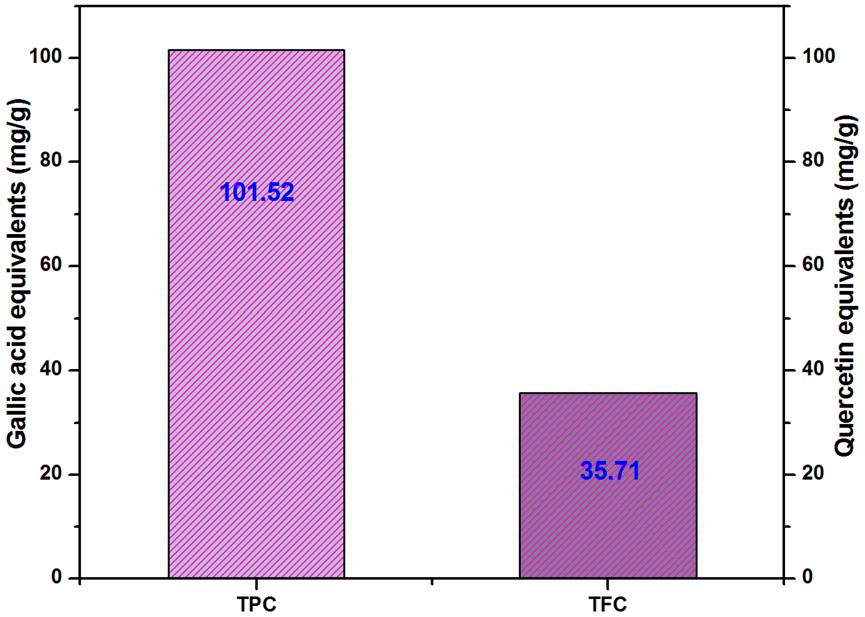
3.1. TPC-TFC

The presence of total phenols and flavonoids in plants is an indicator to use as a tool for solving several problems starting from the human body to industrial sectors. Polyphenols are the key ingredient for both antioxidant and pro-oxidant reactions [33,34]. For many years, the TPC and TFC of any plant have been approved as a benchmark for analyzing plant components for other economically beneficial roles. C. tagal plant extract had 101.52 and 35.71 mg/g of TPC and TFC (Figure 2), which further inspired checking the antioxidant and anticorrosive efficiency, as plants with significant amounts of TPC and TFC are excellent sources for inhibiting free radicals, pathogen, and corrosion [35,36,37].
The total phenol content and flavonoids, here, helps to gain a better understanding about various other crucial roles of C. tagal. In general, polyphenols from natural materials such as beverages, fruits, vegetables, and cereals are proven excellent sources for fighting against free radicals and diseases such as diabetes, neurodegenerative, and osteoporosis. These are compounds, either as intermediate or as precursor, are classified as phenols, flavonoids, and many more based on their structure and number of phenol rings [38]. In precise, phenols have a benzene ring attached to one or more hydroxyl groups, whereas flavonoids have two aromatic C6-C3-C6 ring structures assisting in specific and non-specific mechanisms with tremendous beneficial roles in human health [39].

3.2. Antioxidant Studies

The antioxidant studies of plant extracts provide a guide to identify the real role of active compounds in plants. The importance of antioxidant activity of plants has been a topic of interest for many years as it is either directly or indirectly connected to cures for many diseases such as neurodegenerative, endothelial dysfunction, inflammation, infection, and asthma [34,38,39]. The experiments on antioxidant activities of C. tagal were analyzed by DPPH, nitric oxide, hydrogen peroxide free radicals scavenging, total antioxidant capacity (Figure 3a–d), and reducing potential methods which are a few of the standard procedures.

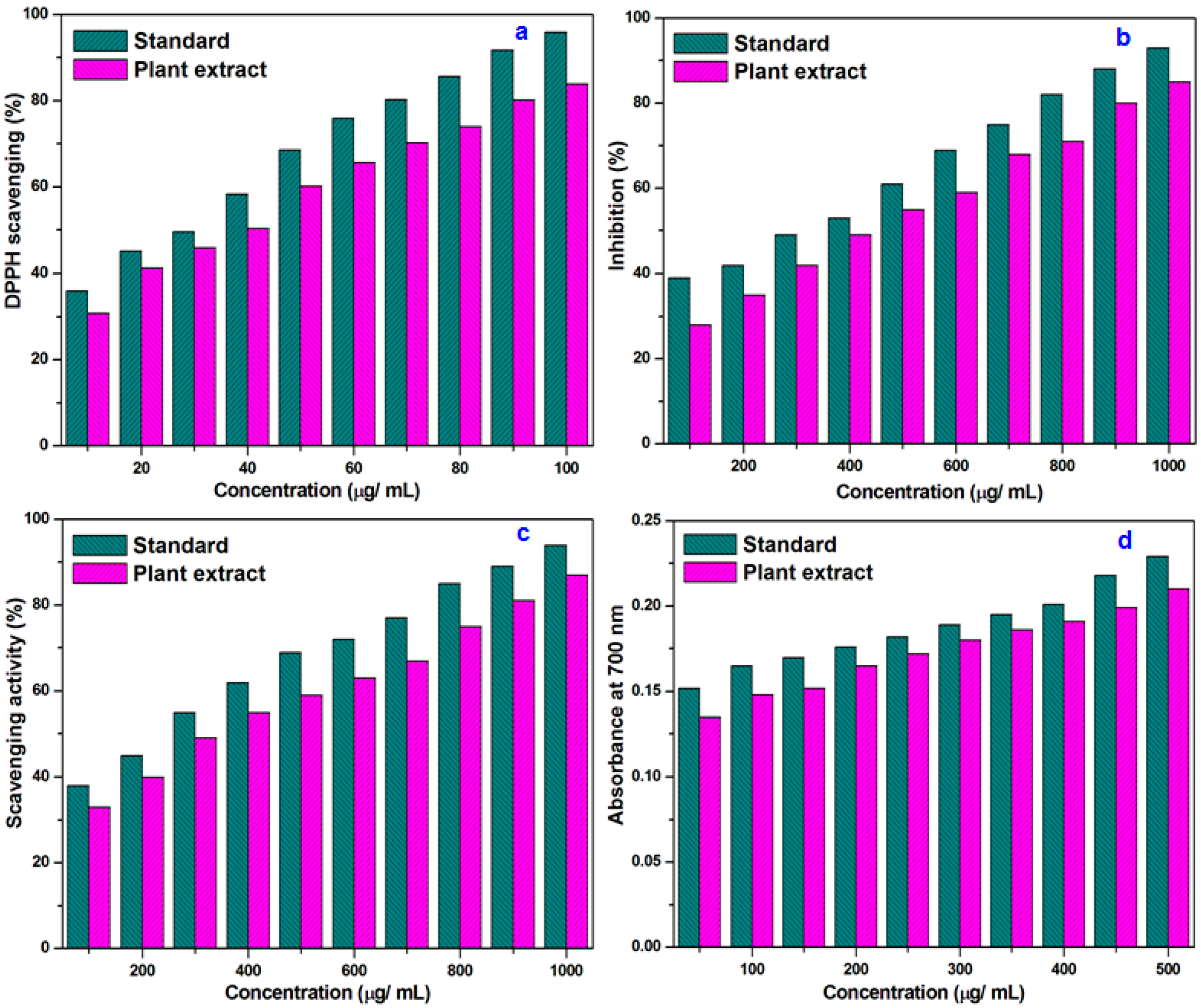
3.2.1. DPPH Free Radical Scavenging Assay

DPPH free radical is a stable compound which, when it receives hydrogen atoms from plant extract, changes its color from purple to yellow. This change is measured by a decrease in absorbance indicating an increase in percentage of free radical scavenged in a concentration dependent manner [40]. The results in Figure 3a, show the antioxidant potential of C. tagal extract in a concentration dependent manner ranging from 30.87 to 83.88% for 10–100 µg/mL, respectively. Various studies have also found that antioxidant activity is dependent on the structure of substrate molecules apart from concentration [41].

3.2.2. Nitric Oxide Inhibition

The nitric oxide inhibition (Figure 3b) of the plant extract corresponded to the DDPH assay with respect to concentration. The highest scavenging (85%) was found at 1000 µg/mL and the lowest (28%) was observed at 100 µg/mL, as mentioned in a previous study [42]. Generally, nitric oxides have unpaired electrons that readily react with some proteins and free radicals such as superoxide forming peroxynitrite anion that are linked to inflammation and carcinomas. The role of phenols, flavonoids, and saponins was unveiled when monitoring the mechanism behind scavenging free radicals. The factor behind this was attributed to the redox potential of plant metabolites transfering them as hydrogen donors, singlet oxygen quenchers, and reducing elements. Thus, this creates a correlation between the phenols (concentration, types, and structure) and antioxidant potential of those compounds to encounter free radicals [41,42,43].

3.2.3. Hydrogen Peroxide Free Radical Scavenging

The reduction of hydrogen peroxide was as per the concentration of the extract, as shown in Figure 3c. The highest scavenging was observed at 1000 µg/mL with 87% and the lowest was observed at 100 µg/mL with 33%. Overall, a gradual increase in radical scavenging was observed from least to highest concentration, similar to the DPPH and nitric oxide assays. The antioxidant (for instance phenols and flavonoids) present in the extract actively reduces the reactive oxygen species (hydrogen peroxide) into water by donating electrons in a dose dependent manner [27]. The present results also show the same pattern as several previous studies that reported on Helichrysum longifolium DC, Newbouldia laevis, and Zizyphus jujuba Miller [27,42,44].

3.2.4. Reducing Potential of Plant Extract

The reducing power assay that analyzed the reduction of Fe3+ to Fe2+ through electron transfer from antioxidants were found to be adequate. As shown in Figure 3d, the best reduction was found at 500 µg/mL in a concentration dependent manner when decreasing at a lower concentration, i.e., 50 µg/mL, reciprocating other antioxidant assays of C. tagal and earlier studies of Helichrysum longifolium DC and Limnophila aromatica. Thus, identifying the reducing power corroborates with the antioxidant nature of active compounds from plants [27,45,46].

3.2.5. Phosphomolybdate Method

The total antioxidant capacity performed by reading the reduction of Mo (IV) to Mo (V) was found to produce significant activity (789.54 mg/g) at given concentration of plant extract read at 659 nm. Usually, mangroves are specialized in producing a high number of antioxidants related to its external stress factors such as salinity, toxicity, drought, temperature, and nutrient deficiency. They also have a strong enzymatic system for defense, namely catalase, superoxide dismutase, and glutathione reductase to protect them from these stressful environments. Plants such as Lumnitzera racemosa, Rhizophora mucronatz scvatxe, Suaeda maritima, and Sonneratia caseolaris have been identified to produce phenols, terpenoids, saponins, steroids, and tannins as antioxidants [5]. These antioxidant capabilities have been studied by methods including DPPH, nitric oxide, and hydrogen peroxide scavenging activity, and phosphomolybdenum assays in Acanthus ilicifolius L., Rhizophora mucronata Lam., and Rhizophora apiculata Blume in leaf, root, stem, and bark [9,46].

3.3. Corrosion Inhibition

Corrosion inhibition by C. tagal extract was measured by various analyses focusing on weight loss at different concentrations and temperatures, difference in dissolved ions, functional groups, surfaces of specimens, and percentage composition. The best results were obtained with 3 h of immersion, therefore, we chose 3 h as the best immersion time.

3.3.1. Weight Loss Measurement

Effect of Concentration

The inhibition competency of different concentrations (100, 200, 300, 400, 500, and 600 ppm) of C. tagal extract was tested against mild steel in 1 M HCl at 303 ± 1 K. Maximum η% (95) was established by 600 ppm, from there the inhibition tended to decrease (49%) until 100 ppm, as presented in Table 1.
Several previous reports have reported the influence of inhibitor concentration in decreasing corrosion rate. Studies had emphasized the correlation between the surface coverage (θ), concentration, and inhibition efficiency (η%) which perfectly suits the action of C. tagal extract, as in Table 1 [47,48,49]. Here, the surface coverage (θ) values increased with an increase in concentration, as the higher concentration increased the strength of the protective layer, thereby, lifting the inhibition efficiency of the extract at 303 ± 1 K [50,51].

Effect of Temperature

C. tagal extract was tested against 1 M HCl with different temperatures (313, 323, 333, and 343 ± 1 K) and concentrations (100, 200, 300, 400, 500, and 600 ppm) for 3 h. Table 2 reveals the effect of change in concentration and temperature affecting the η% at a large scale.
This test shows the inability of a high concentration (600 ppm) in inhibiting corrosion (54%) at 343 ± 1 K. Although concentration helped to increase the inhibition efficiency and to decrease corrosion rate, a gradual fall in surface coverage (θ) and η% were observed at 313, 323, 333, and 343 ± 1 K. A change in η% according to the rise in temperature has been observed in plants such as Mentha pulegium, Zenthoxylum alatum, Jojoba oil, Aster koraiensis, and Cryptostegia grandiflora due to decreased adsorption and increased desorption of inhibitor along with an increase in hydrogen evolution [23,25].

3.3.2. Atomic Adsorption Spectrometric (AAS)

The concentration of Fe+ ions in the corrodent solution of both types are presented in Table 3.
Maximum inhibition (81.74%) was found at 600 ppm which was reduced to 28% at 100 ppm. These results showcase the presence of a protective layer on mild steel immersed in C. tagal extract with varying concentrations of Fe+ ions in the corrodent solution based on concentration. Variations in the amounts of ions in both the solutions (without and with plant extract) were due to the presence of a protective film which barred the corrosion to about 82% (8.25 mg/I of mild steel corrodent) [52]. The inhibition was concentration dependent, since a decrease in η% was observed at lower concentrations ranging from 28 to 72% for 200–500 ppm. The change was due to the acid attack which had occurred by larger exposure of the mild steel surface. Usually organic compounds with sulfur, nitrogen, and oxygen atoms are linked to aromatic rings forming a conjugate system in order to provide electronegative space for interacting with the metal surface to shield them from corrosive environment. This type of shielding has been observed in biomass (Pseudomonas chlororaphis) and plants (Senna cana, Zanthoxylum syncarpum Tull., Dimorphandra gardneriana Tul., and Mangifera indica L.) via their aminophenol derivatives and heterocyclic compounds (phenols, alkaloids, anthocyanidins, and saponins) being adsorbed onto the metal surface decreasing the chance for oxidation and preventing corrosion [53,54].

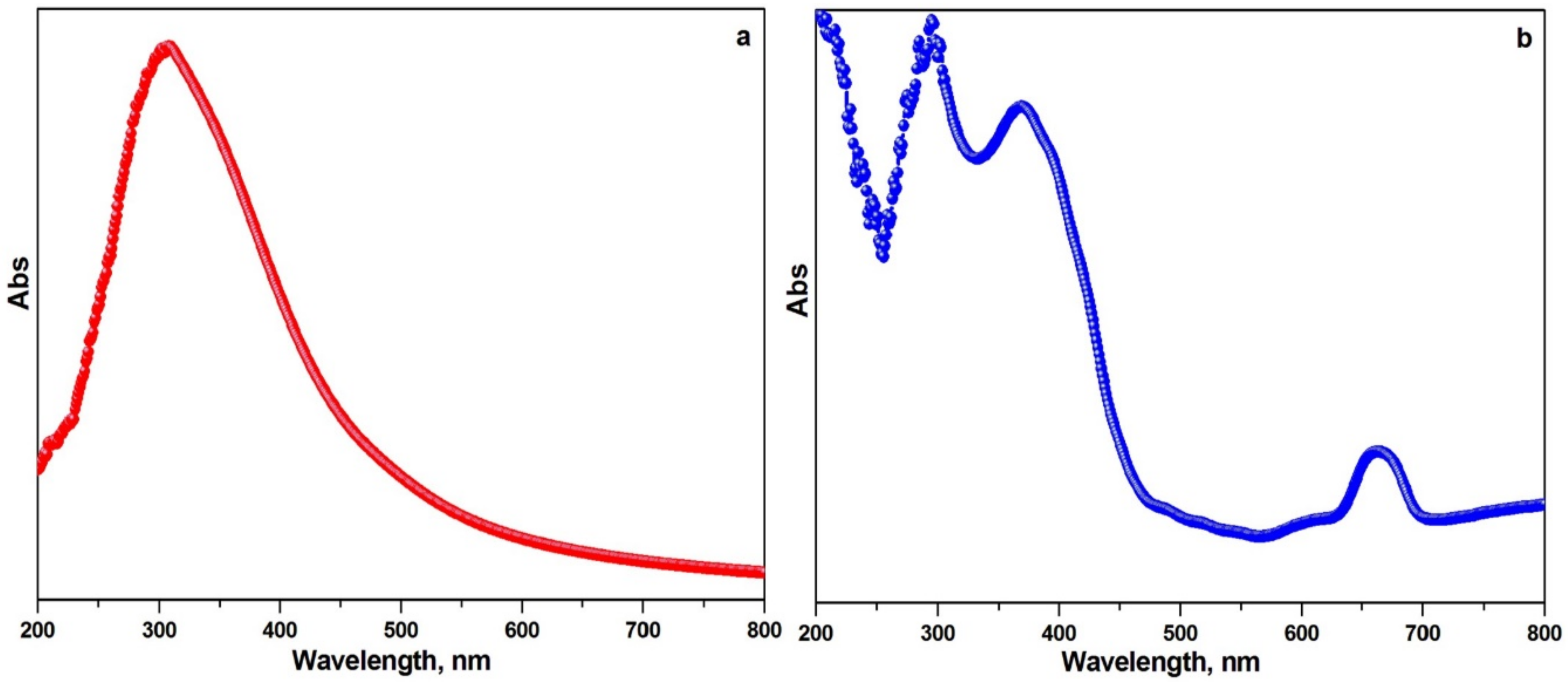
3.3.3. UV-Visible Spectroscopy Analysis

The anticorrosion effect of 1 M HCl with the presence and the absence of C. tagal (600 ppm) inhibitor treatment on mild steel was determined and shown in Figure 4. The change in absorbance maximum clearly shows the formation of an inhibitor metal complex. The absorption spectra of the pure C. tagal extract before immersion reveal maximum absorption at 320 nm (Figure 4a), corresponding to π-π* and n-π* transition for carbonyl C=O and N–H groups, respectively. The variation in the absorption maximum of treated C. tagal (600 ppm) inhibitor solution (Figure 4b) displayed shifted absorption regions to 285 and 360 nm. These estimations reveal the significant change in the position of the absorption maximum after immersion of mild steel. Hence, the change in the absorption maximum position showed the formation of the complex between the 1 M HCl and the (600 ppm) C. tagal inhibitor solution.

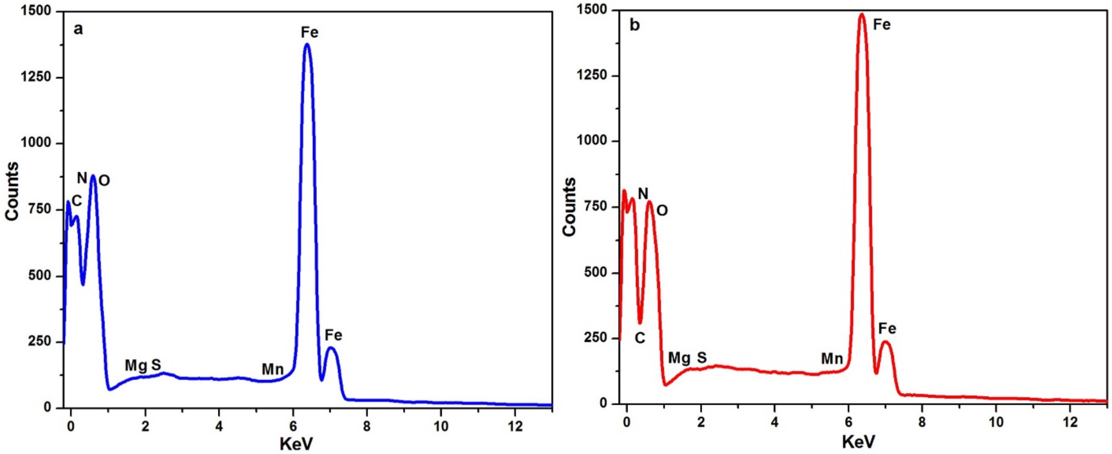
3.3.4. Surface Analysis by SEM-EDX
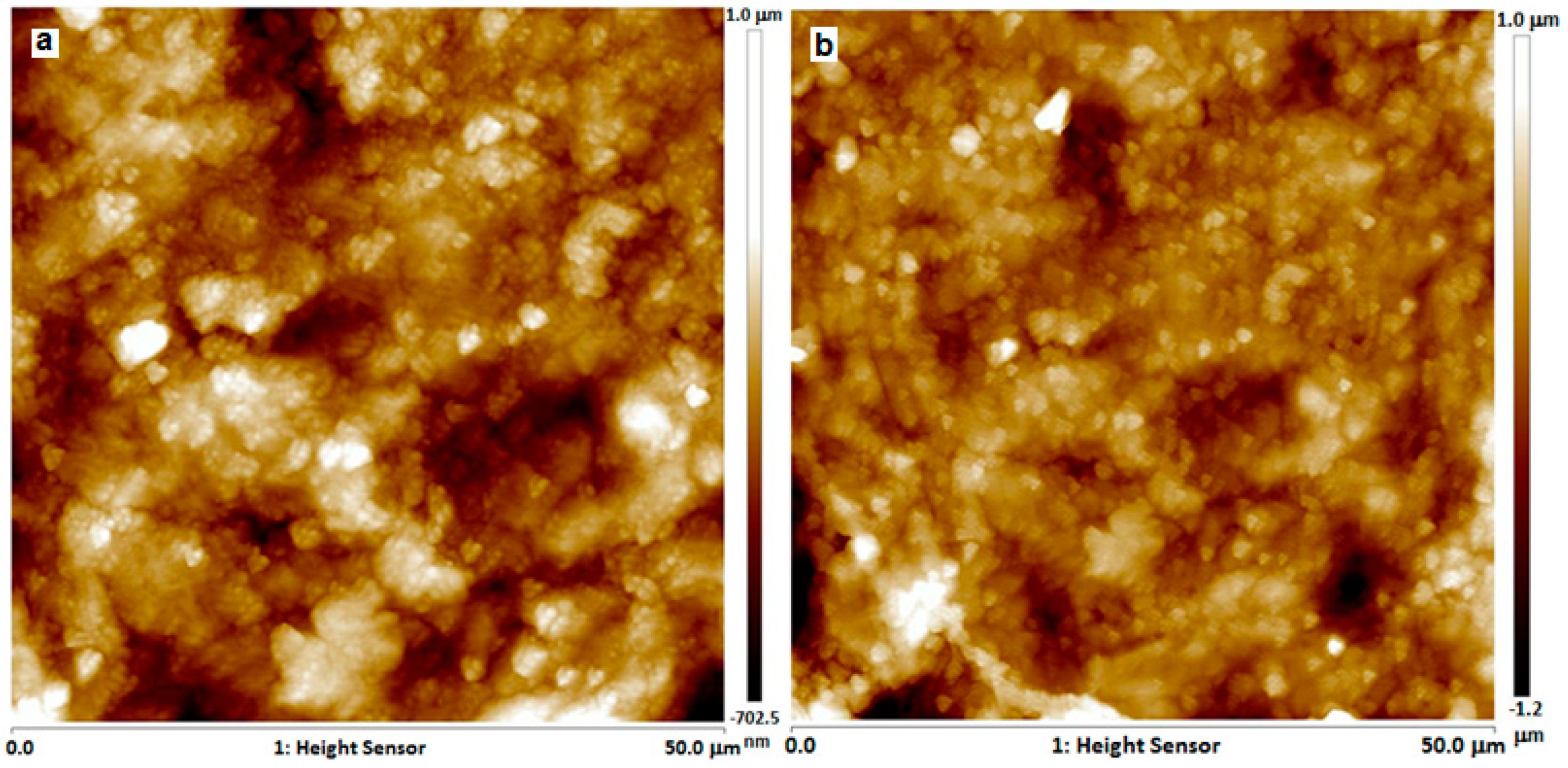
Figure 5a,b, representing the SEM images of mild steel without and with 600 ppm of C. tagal extract in 1 M HCl, is yet another observation made that supports enhancing the role of C. tagal extract on corrosion inhibition.
The roughness, large pores, cracks, and pits on the surface of mild steel, shown in Figure 5a, indicate an aggressive acid attack causing corrosion, whereas the smooth and even surface (Figure 5b) are due to the protective film formed by plant extract. In addition to these results, numerous studies on mild steel have demonstrated the possibility of plant metabolites for protecting metal surface from corrosion [55,56,57]. The differences in the elemental composition in both uninhibited and inhibited samples were clearly identified in EDX. Figure 6a,b shows the variation in the percentage composition of elements in blank and inhibited that provides proof of the action of C. tagal active compounds on mild steel surface [58].
Secondary metabolites such as phenols, tannins, carbohydrates, and proteins are known for their shielding effects on metals and alloys, which is the bases for the reduction in corrosion rate by preventing oxidation [58]. It is clear that the differences in Table 4 and Figure 6a, with 71.54% Fe, 22.1% O, 4.34% C, and 1.51% Mn, and Figure 6b, with 84.21% Fe, 9.01% O, 6.37% C, and 0.07% Mn, represents the acid attack depleting iron atoms by oxidizing the mild steel. The latter with a lower percentage of oxygen and higher percentage of iron atoms shows a decrease in acid attack due to the protective film of plant extract [59,60]. This assures that the decreased corrosion rate might be due to the active ingredient in plant extract.

3.3.5. AFM

AFM is a potent tool for determining the surface topography of the polished mild steel exposed to (1 M) HCl along with and without green inhibitor. As shown in Figure 7, the two-dimensional (2D) surface morphology of mild steel treated with 1 M HCl reveals a severe crack in the surface (Figure 7a) and 1 M HCl with 600 ppm inhibitor significantly diminished the surface crack (Figure 7b) [61,62,63]. The average roughness (251 nm) of the mild steel before the exposure to acidic solution were noticed and, in the presence of the optimal concentration of C. tagal plant extract, the reduced average roughness was determined (65 nm), which confirmed the anticorrosion efficiency of the adsorbed inhibitor molecules produced by the protective film [64,65,66].

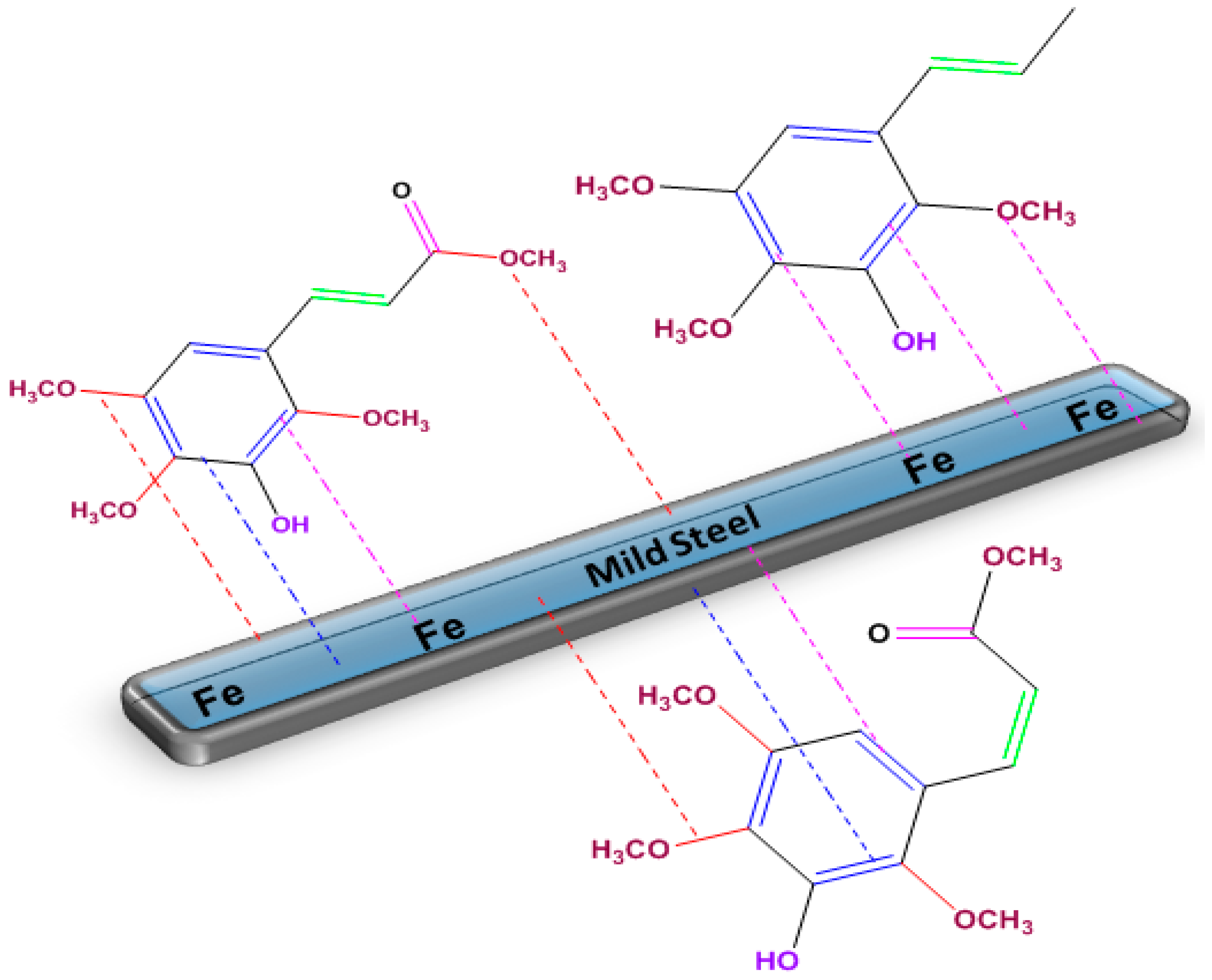
3.3.6. Corrosion Mechanism

The most relevant mechanism behind the inhibition of corrosion by plant extract against mild steel in 1 M HCl is represented in Figure 8.
They are a few possible ways that components in any plant extract interact and interrupt corrosion. The active metabolites adsorbed on the surface of metal are affected by the structure of inhibitors, the metal’s surface and nature, and the type of corrosive medium used [67]. Organic compounds such as phenols, flavonoids, amino acids, dye, etc. are known green inhibitors as they are non-toxic, go easy, biodegradable, and cost effective. Experiments on metals in aggressive solutes such as HCl and H2SO4 have successfully found several new plant-based materials as corrosion inhibitors, as these compounds pose heteroatoms such as O, S, N, and P which are oxidized on the metal surface or adsorbed in the metal forming protective layer [60]. Apparently, the inhibition mechanism is strongly influenced by the choice of electrolyte, nature of metal, structure and concentration of inhibitors, temperature, and pH. In the case of electrolyte, variations in inhibitor behavior (lawsonia extract) were observed against corrosion of nickel and C-steel in neutral, acid, and alkaline mediums. On the one hand, nitrogen and sulfur containing compounds, alkaloids, and aldehydes influence acid media; on the other hand, chromate, nitrite, and phosphate act on neutral media [12,33]. The green inhibitors are heterocyclic with functional polar groups (-OH, -CH3, -OCH3, -NO2, and -COOH). The functional polar groups along conjugated π-electrons with double/triple bonds aid in adsorption, thereby, blocking cathodic and anodic reaction results in corrosion inhibition [56,68,69,70,71].

3.3.7. Corrosion Comparison

It is evident that C. tagal extract is competent with other known green inhibitors, as it exhibits the strongest inhibition against mild steel corrosion in 1 M HCl. As in Table 5, plant extracts such as Aster koraiensis (90.53%) [23], Cryptostegia grandiflora (87.54%) [25], sweet melon peel (91.59%) [30], Desmodium triflorum (92.99%), Polycarpaea corymbose (91.78%) [32], Citrullus lanatus (91%) [57], Lawsonia inermis (92.06%) [62], Lepidagathis keralensis (92.73%) [63], Mangifera indica (92%) [64], Mentha pulegium (80%) [65], and Petroselium Sativum (92.39%) [66] were overcome by the acceptable results of C. tagal extract.

4. Conclusions

  • This study mainly focuses on emphasizing the eminent role of the incredible mangrove plant C. tagal in preventing oxidation and corrosion.
  • The amount of TPC and TFC highlights the need to focus on other active ingredient in C. tagal to learn more about the plant in the future.
  • The scavenging activity on DPPH, nitric oxide, and hydrogen peroxide portraits the performance of C. tagal as a successful antioxidant.
  • The inhibition efficiency of the plants is more evident in weight loss, ASS, UV-visible, SEM-EDX, and AFM for corrosion inhibition.
  • Although there is no direct correlation between the antioxidant and anticorrosive activities, similar studies on several other plants and their components have highlighted the role of secondary metabolites in both cases. Thus, the C. tagal plant can be taken for further research on metal inhibition and other pharmaceutical applications.

Author Contributions

Conceptualization, methodology, data curation, writing—original draft preparation, M.S.; conceptualization, resources, visualization, writing—original draft preparation, K.K.; conceptualization, software, investigation, data curation, writing—original draft preparation, writing—review and editing, supervision, project administration, M.P. All authors have read and agreed to the published version of the manuscript.

Funding

This research received no external funding.

Institutional Review Board Statement

Not applicable.

Informed Consent Statement

Not applicable.

Data Availability Statement

Not applicable.

Acknowledgments

This paper was supported by the KU Research Professor Program of Konkuk University.

Conflicts of Interest

The authors declare no conflict of interest.

References

  1. Hossain, M.; Nuruddin, A. Soil and Mangrove: A Review. J. Environ. Sci. Technol. 2016, 9, 198–207. [Google Scholar] [CrossRef] [Green Version]
  2. Basha, S.K.C. An overview on global mangroves distribution. Indian J. Geo-Mar. Sci. 2018, 47, 766–772. [Google Scholar]
  3. Ricklefs, R.E.; Latham, R.E. Global Patterns of Diversity in Mangrove Floras. Species Diversity in Ecological Communities: Historical and Geographical Perspectives; University of Chicago Press: Chicago, IL, USA, 1993; Chapter 20; pp. 215–229. [Google Scholar]
  4. Khafagi, I.; Gab-Alla, A.; Salama, W.; Fouda, M. Biological activities and phytochemical constituents of the gray mangrove Avicennia marina (Forssk.) Vierh. Egypt. J. Biol. 2003, 5, 62–69. [Google Scholar]
  5. Eswaraiah, G.; Peele, K.A.; Krupanidhi, S.; Kumar, R.B.; Venkateswarulu, T. Studies on phytochemical, antioxidant, antimicrobial analysis and separation of bioactive leads of leaf extract from the selected mangroves. J. King Saud Univ.-Sci. 2019, 32, 842–847. [Google Scholar] [CrossRef]
  6. Cruz, S.M.; Marroquín, N.; Alvarez, L.E.; Chang, D.E.; Cáceres, A. Evaluation of mangrove (Rhizophora mangle L.) products as coloring, antimicrobial and antioxidant agents. Int. J. Phytocosmet. Nat. Ingred. 2015, 2, 12. [Google Scholar] [CrossRef] [Green Version]
  7. Prabhu, V.V.; Guruvayoorappan, C. Phytochemical screening of methanolic extract of mangrove Avicennia marina (Forssk.) Vierh. Der Pharm. Sin. 2012, 3, 64–70. [Google Scholar]
  8. Glasenapp, Y.; Korth, I.; Nguyen, X.V.; Papenbrock, J. Sustainable use of mangroves as sources of valuable medicinal compounds: Species identification, propagation and secondary metabolite composition. S. Afr. J. Bot. 2019, 121, 317–328. [Google Scholar] [CrossRef]
  9. Bibi, S.N.; Fawzi, M.M.; Gokhan, Z.; Rajesh, J.; Nadeem, N.; Kannan, R.; Albuquerque, R.D.D.G.; Pandian, S.K. Ethnopharmacology, phytochemistry, and global distribution of mangroves-A comprehensive review. Mar. Drugs 2019, 17, 231. [Google Scholar] [CrossRef] [PubMed] [Green Version]
  10. Rahim, A.A.; Rocca, E.; Steinmetz, J.; Kassim, M.J.; Ibrahim, M.S.; Osman, H. Antioxidant activities of mangrove Rhizophora apiculata bark extracts. Food Chem. 2008, 107, 200–207. [Google Scholar] [CrossRef]
  11. Thatoi, H.; Behera, B.C.; Mishra, R.R.; Dutta, S.K. Biodiversity and biotechnological potential of microorganisms from mangrove ecosystems: A review. Ann. Microbiol. 2012, 63, 1–19. [Google Scholar] [CrossRef]
  12. Kesavan, D.; Gopiraman, M.; Sulochana, N. Green inhibitors for corrosion of metals: A review. Chem. Sci. Rev. Lett. 2012, 1, 1–8. [Google Scholar]
  13. Chigondo, M.; Chigondo, F. Recent Natural Corrosion Inhibitors for Mild Steel: An Overview. J. Chem. 2016, 2016, 6208937. [Google Scholar] [CrossRef] [Green Version]
  14. Khan, G.; Md, K.; Newaz, S.; Basirun, W.J.; Ali, H.B.M.; Faraj, F.L.; Khan, G.M. Application of natural product extracts as green corrosion inhibitors for metals and alloys in acid pickling processes—A review. Int. J. Electrochem. Sci. 2015, 10, 6120–6134. [Google Scholar]
  15. Fateh, A.; Aliofkhazraei, M.; Rezvanian, A. Review of corrosive environments for copper and its corrosion inhibitors. Arab. J. Chem. 2020, 13, 481–544. [Google Scholar] [CrossRef]
  16. Xiao, X.; Hong, Y.; Xia, W.; Feng, S.; Zhou, X.; Fu, X.; Zang, J.; Xiao, Y.; Niu, X.; Li, C.; et al. Transcriptome Analysis of Ceriops tagal in Saline Environments Using RNA-Sequencing. PLoS ONE 2016, 11, e0167551. [Google Scholar] [CrossRef] [Green Version]
  17. Kumar, V.A.; Ammani, K.; Siddhardha, B.; Sreedhar, U.; Kumar, G.A. Differential biological activities of the solvent extracts of Ceriops decandra (Griff.) and their phytochemical investigations. J. Pharm. Res. 2013, 7, 654–660. [Google Scholar] [CrossRef]
  18. Ni, S.-J.; Li, J.; Li, M.-Y. Two New Dolabrane Diterpenes from the Chinese Mangrove Ceriops tagal. Chem. Biodivers. 2018, 15, e1700563. [Google Scholar] [CrossRef] [PubMed]
  19. Yang, Y.; Zhang, Y.; Liu, D.; Li-Weber, M.; Shao, B.; Lin, W. Dolabrane-type diterpenes from the mangrove plant Ceriops tagal with antitumor activities. Fitoterapia 2015, 103, 277–282. [Google Scholar] [CrossRef] [PubMed]
  20. Ni, S.-J.; Li, J.; Li, M.-Y. Two new phenylpropanoids from the Chinese mangrove Ceriops tagal. Nat. Prod. Res. 2017, 32, 1676–1681. [Google Scholar] [CrossRef]
  21. Wang, P.; Cui, Y.; Cai, C.; Chen, H.; Dai, Y.; Chen, P.; Kong, F.; Yuan, J.; Song, X.; Mei, W.; et al. Two New Succinimide Derivatives Cladosporitins A and B from the Mangrove-derived Fungus Cladosporium sp. HNWSW-1. Mar. Drugs 2018, 17, 4. [Google Scholar] [CrossRef] [Green Version]
  22. Wu, J.-T.; Zheng, C.-J.; Zhang, B.; Zhou, X.-M.; Zhou, Q.; Chen, G.-Y.; Zeng, Z.-E.; Xie, J.-L.; Han, C.-R.; Lyu, J.-X. Two new secondary metabolites from a mangrove-derived fungus Cladosporium sp. JJM22. Nat. Prod. Res. 2018, 33, 34–40. [Google Scholar] [CrossRef]
  23. Prabakaran, M.; Kim, S.-H.; Mugila, N.; Hemapriya, V.; Parameswari, K.; Chitra, S.; Chung, I.-M. Aster koraiensis as nontoxic corrosion inhibitor for mild steel in sulfuric acid. J. Ind. Eng. Chem. 2017, 52, 235–242. [Google Scholar] [CrossRef]
  24. Helen, L.Y.S.; Rahim, A.A.; Saad, B.; Saleh, M.I.; Raj, P.B. Aquilaria crassna leaves extracts-a green corrosion inhibitor for mild steel in 1 M HCl medium. Int. J. Electrochem. Sci. 2014, 9, 830–846. [Google Scholar]
  25. Prabakaran, M.; Kim, S.-H.; Hemapriya, V.; Chung, I.-M. Evaluation of polyphenol composition and anti-corrosion properties of Cryptostegia grandiflora plant extract on mild steel in acidic medium. J. Ind. Eng. Chem. 2016, 37, 47–56. [Google Scholar] [CrossRef]
  26. Alam, N.; Bristi, N.J. Rafiquzzaman Review on in vivo and in vitro methods evaluation of antioxidant activity. Saudi Pharm. J. 2012, 21, 143–152. [Google Scholar] [CrossRef] [Green Version]
  27. Aiyegoro, O.A.; Okoh, A.I. Preliminary phytochemical screening and In vitro antioxidant activities of the aqueous extract of Helichrysum longifolium DC. BMC Complement. Altern. Med. 2010, 10, 21. [Google Scholar] [CrossRef] [PubMed] [Green Version]
  28. Malathy, R.; Prabakaran, M.; Kalaiselvi, K.; Chung, I.M.; Kim, S.H. Comparative polyphenol composition, antioxidant and anticorrosion properties in various parts of panax ginseng extracted in different solvents. Appl. Sci. 2021, 11, 93. [Google Scholar] [CrossRef]
  29. Hemapriya, V.; Chung, I.-M.; Kim, S.-H.; Prabakaran, M. Inhibitory effect of biowaste on copper corrosion in 1 M HCl solution. Mater. Today Commun. 2021, 27, 102249. [Google Scholar] [CrossRef]
  30. Saeed, M.; Saleem, M.; Usmani, S.; Malik, I.A.; Al-Shammari, F.A.; Deen, K.M. Corrosion inhibition of mild steel in 1 M HCl by sweet melon peel extract. J. King Saud Univ. Sci. 2019, 31, 1344–1351. [Google Scholar] [CrossRef]
  31. Fadare, O.; Okoronkwo, A.E.; Olasehinde, E.F. Assessment of anti-corrosion potentials of extract of Ficus asperifolia -Miq (Moraceae) on mild steel in acidic medium. Afr. J. Pure Appl. Chem. 2016, 10, 8–22. [Google Scholar] [CrossRef]
  32. Sakunthala, P.; Vivekananthan, S.S.; Gopiraman, M.; Sulochana, N.; Vincent, A.R. Spectroscopic investigations of physicochemical interactions on Umoren mild steel in an acidic medium by environmentally friendly green inhibitors. J. Surfactants Deterg. 2013, 16, 251–263. [Google Scholar] [CrossRef]
  33. Rani, B.E.A.; Basu, B.B.J. Green Inhibitors for Corrosion Protection of Metals and Alloys: An Overview. Int. J. Corros. 2011, 2012, 380217. [Google Scholar] [CrossRef]
  34. Lipinski, B. Hydroxyl Radical and Its Scavengers in Health and Disease. Oxidative Med. Cell. Longev. 2011, 2011, 809696. [Google Scholar] [CrossRef] [Green Version]
  35. Kumar, T.; Jain, V. Appraisal of total phenol, flavonoid contents, and antioxidant potential of folkloric Lannea coromandelica using in vitro and in vivo assays. Scientifica 2015, 2015, 203679. [Google Scholar] [CrossRef] [Green Version]
  36. Chaouche, T.M.; Haddouchi, F.; Ksouri, R.; Atik-Bekkara, F. Evaluation of antioxidant activity of hydromethanolic extracts of some medicinal species from South Algeria. J. Chin. Med. Assoc. 2014, 77, 302–307. [Google Scholar] [CrossRef] [PubMed] [Green Version]
  37. Zhang, J.; Song, Y.; Su, H.; Zhang, L.; Chen, G.; Zhao, J. Investigation of Diospyros Kaki L. f husk extracts as corrosion inhibitors and bactericide in oil field. Chem. Central J. 2013, 7, 109. [Google Scholar] [CrossRef] [PubMed] [Green Version]
  38. Pandey, K.B.; Rizvi, S.I. Plant Polyphenols as Dietary Antioxidants in Human Health and Disease. Oxidative Med. Cell. Longev. 2009, 2, 270–278. [Google Scholar] [CrossRef] [PubMed] [Green Version]
  39. Fraga, C.G.; Galleano, M.; Verstraeten, S.; Oteiza, P.I. Basic biochemical mechanisms behind the health benefits of polyphenols. Mol. Asp. Med. 2010, 31, 435–445. [Google Scholar] [CrossRef]
  40. Gangwar, M.; Gautam, M.K.; Sharma, A.K.; Tripathi, Y.B.; Goel, R.K.; Nath, G. Antioxidant Capacity and Radical Scavenging Effect of Polyphenol RichMallotus philippenensisFruit Extract on Human Erythrocytes: AnIn VitroStudy. Sci. World J. 2014, 2014, 279451. [Google Scholar] [CrossRef] [Green Version]
  41. Liang, N.; Kitts, D.D. Antioxidant Property of Coffee Components: Assessment of Methods that Define Mechanisms of Action. Molecules 2014, 19, 19180–19208. [Google Scholar] [CrossRef] [Green Version]
  42. Habu, J.B.; Ibeh, B.O. In vitro antioxidant capacity and free radical scavenging evaluation of active metabolite constituents of Newbouldia laevis ethanolic leaf extract. Biol. Res. 2015, 48, 16. [Google Scholar] [CrossRef] [PubMed] [Green Version]
  43. Boora, F.; Chirisa, E.; Mukanganyama, S. Evaluation of Nitrite Radical Scavenging Properties of Selected Zimbabwean Plant Extracts and Their Phytoconstituents. J. Food Process. 2014, 2014, 918018. [Google Scholar] [CrossRef] [Green Version]
  44. Zhao, H.-X.; Zhang, H.-S.; Yang, S.-F. Phenolic compounds and its antioxidant activities in ethanolic extracts from seven cultivars of Chinese jujube. Food Sci. Hum. Wellness 2014, 3, 183–190. [Google Scholar] [CrossRef] [Green Version]
  45. Do, Q.D.; Angkawijaya, A.E.; Tran-Nguyen, P.L.; Huynh, L.H.; Soetaredjo, F.E.; Ismadji, S.; Ju, Y.H. Effect of extraction solvent on total phenol content, total flavonoid content, and antioxidant activity of Limnophila aromatica. J. Food Drug. Anal. 2014, 22, 296–302. [Google Scholar] [CrossRef] [PubMed] [Green Version]
  46. Prabakaran, M.; Kim, S.-H.; Sasireka, A.; Chandrasekaran, M.; Chung, I.-M. Polyphenol composition and antimicrobial activity of various solvent extracts from different plant parts of Moringa oleifera. Food Biosci. 2018, 26, 23–29. [Google Scholar] [CrossRef]
  47. Chung, I.-M.; Malathy, R.; Priyadharshini, R.; Hemapriya, V.; Kim, S.-H.; Prabakaran, M. Inhibition of mild steel corrosion using Magnolia kobus extract in sulphuric acid medium. Mater. Today Commun. 2020, 25, 101687. [Google Scholar] [CrossRef]
  48. Manokarana, G.; Prabakaran, M. Evaluation of antioxidant and anticorrosion activities of Ligularia fischeri plant extract. Chem. Sci. Eng. Res. 2019, 1, 16–24. [Google Scholar] [CrossRef]
  49. Chung, I.-M.; Kim, S.-H.; Prabakaran, M. Evaluation of Phytochemical, Polyphenol Composition and Anti-corrosion Capacity of Cucumis anguria L. Leaf Extract on Metal Surface in Sulfuric Acid Medium. Prot. Met. Phys. Chem. Surf. 2020, 56, 214–224. [Google Scholar] [CrossRef]
  50. Nahlé, A.; Abu-Abdoun, I.; Abdel-Rahman, I. Effect of Temperature on the Corrosion Inhibition of Trans-4-Hydroxy-4′-Stilbazole on Mild Steel in HCl Solution. Int. J. Corros. 2012, 2012, 380329. [Google Scholar] [CrossRef] [Green Version]
  51. Labjar, N.; Bentiss, F.; Lebrini, M.; Jama, C.; El Hajjaji, S. Study of Temperature Effect on the Corrosion Inhibition of C38 Carbon Steel Using Amino-tris(Methylenephosphonic) Acid in Hydrochloric Acid Solution. Int. J. Corros. 2011, 2011, 548528. [Google Scholar] [CrossRef] [Green Version]
  52. Umoren, S.; Eduok, U.; Solomon, M.; Udoh, A. Corrosion inhibition by leaves and stem extracts of Sida acuta for mild steel in 1M H2SO4 solutions investigated by chemical and spectroscopic techniques. Arab. J. Chem. 2016, 9, S209–S224. [Google Scholar] [CrossRef] [Green Version]
  53. Júnior, J.M.F.; Silva, M.G.D.; Monteiro, J.A.; Barros, A.D.S.; Falcão, M.J.C.; Morais, S.M.D. Evaluation of antioxidant activity and inhibition of corrosion by brazilian plant extracts and constituents. Int. J. Electrochem. Sci. 2016, 11, 3862–3875. [Google Scholar] [CrossRef]
  54. Hassan, H.M. Utility of bacterial biomass as antioxidant, anticorrosive, and antifriction additives for lubricating oils. Petrol Sci. Technol. 2011, 29, 2086–2094. [Google Scholar] [CrossRef]
  55. Prabakaran, M.; Kim, S.-H.; Kalaiselvi, K.; Hemapriya, V.; Chung, I.-M. Highly efficient Ligularia fischeri green extract for the protection against corrosion of mild steel in acidic medium: Electrochemical and spectroscopic investigations. J. Taiwan Inst. Chem. Eng. 2016, 59, 553–562. [Google Scholar] [CrossRef]
  56. Prabakaran, M.; Kim, S.-H.; Hemapriya, V.; Chung, I.-M. Tragia plukenetii extract as an eco-friendly inhibitor for mild steel corrosion in HCl 1 M acidic medium. Res. Chem. Intermed. 2015, 42, 3703–3719. [Google Scholar] [CrossRef]
  57. Dehghani, A.; Bahlakeh, G.; Ramezanzadeh, B.; Ramezanzadeh, M. A combined experimental and theoretical study of green corrosion inhibition of mild steel in HCl solution by aqueous Citrullus lanatus fruit (CLF) extract. J. Mol. Liq. 2019, 279, 603–624. [Google Scholar] [CrossRef]
  58. Taheri, S.; Delgado, G.P.; Agbaje, O.B.A.; Giri, P.; Clark, S.M. Corrosion inhibitory effects of mullite in concrete exposed to sulfuric acid attack. Corros. Mater. Degrad. 2020, 1, 282–295. [Google Scholar] [CrossRef]
  59. Baran, E.; Cakir, A.; Yazici, B. Inhibitory effect of Gentiana olivieri extracts on the corrosion of mild steel in 0.5 M HCl: Electrochemical and phytochemical evaluation. Arab. J. Chem. 2016, 12, 4303–4319. [Google Scholar] [CrossRef] [Green Version]
  60. Patni, N.; Agarwal, S.; Shah, P. Greener Approach towards Corrosion Inhibition. Chin. J. Eng. 2013, 2013, 784186. [Google Scholar] [CrossRef] [Green Version]
  61. Chitra, S.; Chung, I.-M.; Kim, S.-H.; Prabakaran, M. A study on anticorrosive property of phenolic components from Pachysandra terminalis against low carbon steel corrosion in acidic medium. Pigment Resin Technol. 2019, 48, 389–396. [Google Scholar] [CrossRef]
  62. Chung, I.-M.; Hemapriya, V.; Kim, S.-H.; Ponnusamy, K.; Arunadevi, N.; Chitra, S.; Prabakaran, M.; Gopiraman, M. Liriope platyphylla extract as a green inhibitor for mild steel corrosion in sulfuric acid medium. Chem. Eng. Commun. 2019, 208, 72–88. [Google Scholar] [CrossRef]
  63. Mahalakshmi, D.; Unnisa, C.B.N.; Hemapriya, V.; Subramaniam, E.P.; Roopan, S.M.; Chitra, S.; Chung, I.M.; Kim, S.H.; Prabakaran, M. Anticorrosive potential of ethanol extract of Delonix elata for mild steel in 0.5 M H2SO4—A green approach, Bulg. Chem. Commun. 2019, 51, 31–37. [Google Scholar]
  64. Chung, I.-M.; Malathy, R.; Kim, S.-H.; Kalaiselvi, K.; Prabakaran, M.; Gopiraman, M. Ecofriendly green inhibitor from Hemerocallis fulva against aluminum corrosion in sulphuric acid medium. J. Adhes. Sci. Technol. 2020, 34, 1483–1506. [Google Scholar] [CrossRef]
  65. Hemapriya, V.; Prabakaran, M.; Chitra, S.; Swathika, M.; Kim, S.-H.; Chung, I.-M. Utilization of biowaste as an eco-friendly biodegradable corrosion inhibitor for mild steel in 1 mol/L HCl solution. Arab. J. Chem. 2020, 13, 8684–8696. [Google Scholar] [CrossRef]
  66. Anitha, R.; Unnisa, C.B.N.; Hemapriya, V.; Roopan, S.M.; Chitra, S.; Chung, I.-M.; Kim, S.-H.; Mayakrishnan, P. Anti-corrosive potential of Cyperus rotundus as a viable corrosion inhibitor for mild steel in sulphuric acid. Pigment Resin Technol. 2020, 49, 295–304. [Google Scholar] [CrossRef]
  67. Al-Amiery, A.A.; Ahmed, M.H.O.; Abdullah, T.A.; Gaaz, T.S.; Kadhum, A.A.H. Electrochemical studies of novel corrosion inhibitor for mild steel in 1 M hydrochloric acid. Results Phys. 2018, 9, 978–981. [Google Scholar] [CrossRef]
  68. Chung, I.-M.; Kim, S.-H.; Hemapriya, V.; Kalaiselvi, K.; Prabakaran, M. Inhibition behavior of Tragia involucrata L. phenolic compounds against acidic medium corrosion in low carbon steel surface. Chin. J. Chem. Eng. 2018, 27, 717–725. [Google Scholar] [CrossRef]
  69. Chung, I.-M.; Hemapriya, V.; Ponnusamy, K.; Arunadevi, N.; Chitra, S.; Youn, C.-H.; Kim, S.-H.; Prabakaran, M. Assessment of Low Carbon Steel Corrosion Inhibition by Eco-Friendly Green Chaenomeles sinensis Extract in Acid Medium. J. Electrochem. Sci. Technol. 2018, 9, 238–249. [Google Scholar] [CrossRef] [Green Version]
  70. Prabakaran, M.; Kim, S.H.; Hemapriya, V.; Gopiraman, M.; Kim, I.S.; Chung, I.M. Rhus verniciflua as a green corrosion inhibitor for mild steel in 1 M H2SO4. RSC Adv. 2016, 6, 57144–57153. [Google Scholar] [CrossRef]
  71. Anitha, R.; Chitra, S.; Hemapriya, V.; Chung, I.-M.; Kim, S.-H.; Prabakaran, M. Implications of eco-addition inhibitor to mitigate corrosion in reinforced steel embedded in concrete. Constr. Build. Mater. 2019, 213, 246–256. [Google Scholar] [CrossRef]
  72. Ostovari, A.; Hoseinieh, S.M.; Peikari, M.P.; Shadizadeh, S.R.; Hashemi, J. Corrosion inhibition of mild steel in 1 M HCl solution by henna extract: A comparative study of the inhibition by henna and its constituents (Lawsone, Gallic acid, alpha-D-Glucose and Tannic acid). Corros. Sci. 2009, 51, 1935–1949. [Google Scholar] [CrossRef]
  73. Leena, P.; Hukuman, N.H.Z.; Biju, A.R.; Jisha, M. Studies on methanolic extract of Lepidagathis keralensis as green corrosion inhibitor for mild steel in 1M HCl. J. Electrochem. Sci. Technol. 2019, 10, 231–243. [Google Scholar]
  74. Ramezanzadeh, M.; Bahlakeh, G.; Sanaei, Z.; Ramezanzadeh, B. Corrosion inhibition of mild steel in 1 M HCl solution by ethanolic extract of eco-friendly Mangifera indica (mango) leaves: Electrochemical, molecular dynamics, Monte Carlo and ab initio study. Appl. Surf. Sci. 2019, 463, 1058–1077. [Google Scholar] [CrossRef]
  75. Bouyanzer, A.; Hammouti, B.; Majidi, L. Pennyroyal oil from Mentha pulegium as corrosion inhibitor for steel in 1M HCl. Mater. Lett. 2006, 60, 2840–2843. [Google Scholar] [CrossRef]
  76. Benarioua, M.; Mihi, A.; Bouzeghaia, N.; Naoun, M. Mild steel corrosion inhibition by Parsley (Petroselium Sativum) extract in acidic media. Egypt. J. Pet. 2019, 28, 155–159. [Google Scholar] [CrossRef]
Figure 1. Chemical statures of: (a) Tagalphenylpropanoidins A; (b) tagalphenylpropanoidins B; (c) 2,3,6-trimethoxy-5-(1-propenyl) phenol.
Applsci 11 10150 g001
Figure 2. TPC and TFC of C. tagal plant extract.
Applsci 11 10150 g002
Figure 3. Antioxidant activities of C. tagal: (a) DPPH free-radical scavenging; (b) nitric oxide inhibition; (c) hydrogen peroxide free-radical scavenging; (d) reducing power.
Applsci 11 10150 g003
Figure 4. UV-visible spectrum of: (a) Pure C. tagal extract; (b) inhibited C. tagal.
Applsci 11 10150 g004
Figure 5. SEM photographs of mild steel dipped in: (a) 1 M HCl; (b) 1 M HCl with 600 ppm of inhibitor.
Applsci 11 10150 g005
Figure 6. EDX spectra of mild steel dipped in: (a) 1 M HCl; (b) 1 M HCl with 600 ppm of inhibitor.
Applsci 11 10150 g006
Figure 7. 2D images of mild steel: (a) 1 M HCl; (b) 1 M HCl with 600 ppm of inhibitor.
Applsci 11 10150 g007
Figure 8. An illustration of possible corrosion mechanism of C. tagal extract.
Applsci 11 10150 g008
Table 1. % IE for various concentrations (Conc.) of the C. tagal extracts for corrosion of mild steel in 1 M HCl obtained by weight loss measurements at room temperature (303 ± 1 K).
Conc. (ppm)W (mg·cm−2)θη%σ a
Blank0.0989---
1000.05010.49490.03
2000.04320.56560.02
3000.03880.61610.01
4000.02980.70700.02
5000.01020.90900.03
6000.00550.95950.01
a σ is the standard deviation.
Table 2. The η% values for various concentrations (Conc.) and temperatures of C. tagal extract for mild steel corrosion in 1 M HCl by weight loss measurements.
T (±1 K)Conc. (ppm)W (mg·cm−2)θη%σ a
Blank0.1251---
1000.07020.44440.02
2000.06330.49490.04
3133000.05040.60600.03
4000.04130.67670.03
5000.03310.74740.02
6000.02090.83830.03
Blank0.1410---
1000.08570.39390.02
2000.07580.46460.01
3233000.07010.50500.03
4000.05980.58580.01
5000.05030.64640.02
6000.03280.77770.02
Blank0.1787---
1000.12210.32320.02
2000.11510.36360.03
3333000.10200.43430.01
4000.08520.52520.01
5000.07570.58580.02
6000.06290.65650.02
Blank0.2002---
1000.14850.26260.02
3432000.13020.35350.03
3000.12210.39390.01
4000.11120.44440.02
5000.10210.49490.03
6000.09210.54540.02
a σ is the standard deviation.
Table 3. AAS study for different concentrations of C. tagal extract in 1 M HCl (303 ± 1 K).
Conc. (ppm)Amount of Mild Steel Corrodant (mg/I)η (%)σ a
Blank45.214--
10032.546280.01
20028.541370.03
30025.299440.02
40020.548550.04
50012.571720.02
60008.254820.02
a σ is the standard deviation.
Table 4. Percentage compositions of elements present on the mild steel surface.
ElementsComposition (Atomic%)
BlankInhibited
C4.346.37
Fe71.5484.21
O22.109.01
S0.120.11
N0.180.12
Mn1.510.07
Mg0.210.11
Table 5. Comparison of C. tagal (present) green inhibitor over other green inhibitors.
S. NoGreen Inhibitorη (%)
1.Aster koraiensis90.53 [23]
2.Ceriops tagal#95
3.Cryptostegia grandiflora87.54 [25]
4.Sweet melon peel91.59 [30]
5.Desmodium triflorum92.99 [32]
6.Polycarpaea corymbosa91.78 [32]
7.Citrullus lanatus91.00 [57]
8.Lepidagathis keralensis92.06 [72]
9.Mangifera indica92.73 [73]
10.Mangifera indica92.00 [74]
11.Mentha pulegium80.00 [75]
12.Petroselium Sativum92.39 [76]
# Present work.
Publisher’s Note: MDPI stays neutral with regard to jurisdictional claims in published maps and institutional affiliations.

