1. Introduction
Low back pain (LBP) in the equestrian athlete negatively impacts performance by acting as a distractor that alters the coordination dynamics of the horse and rider [
1]. Most authors agree that LBP is the most common chronic injury experienced by equestrian athletes [
1,
2,
3,
4,
5], and it has been linked to poor postural control [
6] and lack of balance, stability and alignment of the skeleton at the pelvic level [
7]. Dąbek et al. (2015) reported a significantly higher incidence of back pain in amateur riders when compared to advanced riders [
8]. On the other hand, high volumes and repetitive motions have previously been correlated to the high prevalence rates of LBP in the athletic population [
9]. This contradiction may be explained by the more harmonic riding technique of the advanced riders, and one could conclude that a very important factor influencing the prevalence of LBP in equestrian sports is the level of expertise, i.e., it is skill-dependent. Understanding the biomechanical differences between advanced and novice equestrianism is therefore of major interest.
In the last decade, several publications have compared the interactions in the horse-rider system of advanced and novice riders at a mechanical level by looking at joint angles [
10] and inertial data [
11,
12]. It is known that the pelvis of a horse rider acts as a communication channel, physically interacting with the horse’s behaviour [
11], and great differences can be observed between advanced and novice riders [
11,
12]. Lagarde et al. (2005) showed that advanced riders achieved a higher level of synchronicity and coordination with the horse’s movement when compared to novice riders [
13]. Coordination dynamics, also known as harmony [
14], are strongly related to the postural control of the rider [
15], which greatly depends on the coordination and neuromuscular awareness of the core muscles [
16]. Publications analysing biological differences between advanced and novice riders are sparse [
16,
17] and rarely consider mechanics and biology simultaneously [
18].
This study aims to describe how the rider’s neuromuscular system (NMS) copes with external mechanical strain when exposed to the postural perturbations elicited by the horse in motion, based on inertial and surface electromyographical (sEMG) measures. Both parameters were described and correlated, then compared between advanced and novice riders to portray the skill-dependent differences of the interactions between the horse and rider.
We hypothesized that advanced riders would show a greater ability to attenuate the shockwave generated by the horse’s movement by having a higher overall sEMG level and earlier muscle activation.
2. Materials and Methods
2.1. Participants
Six novice riders (age 24 ± 7 years; training 2–4 h/week, 10 ± 8 years of experience), and nine advanced riders (age 31 ± 5 years; training >35 h/week, >17 ± 9 years of experience) from the Spanish Classical School of Riding (Lipica) volunteered to take part in this study. All participants were informed of the study’s procedure and signed an informed consent. The National Medical Ethics Committee (Ministry of Health, Ljubljana) gave full approval to the project according to the declaration of Helsinki (approval number: 0120-346/2018/3).
Four horses (age 15 ± 3 years) of similar morphology were selected for the trials. All horses showed self-carriage capacities and a calm and steady gait. A veterinarian checked the animals and approved each horse to take part in the study.
2.2. Experimental Protocol
A cross-sectional, single visit study design was used. All measurements were performed in an indoor arena and the study lasted <1.5 h. After obtaining informed consent from the participants, sEMG sensors (Delsys Trigno, Natick, MA, USA; 2017) were placed on the riders. For sEMG normalization purposes, subjects were asked to perform a set of 3 maximal voluntary contractions (MVC) for each muscle. Each MVC was performed following the instructions described elsewhere [
19]. After a standardized warm-up of the horse and rider, participants were asked to perform a 30 m straight line on the horse and were encouraged to ride it with minimal use of riding aids. Each subject repeated this procedure until 60 horse-strides were recorded at walk, trot and canter. To prevent any potential injury of the horse, the three gait conditions were performed in a random balanced order. Riders were asked to perform rising trot (a two beat, medium speed gait) and collected canter (a three beat, medium speed gait) always on the right hand. If the horse was disunited or the performed leg changed, the whole 30 m repetition was excluded from analysis and the trial repeated. Upon completion, cool down of the horse with loose reigns at walk was performed. Additional information of horse gait characteristics can be found elsewhere [
15].
2.3. Horse Set-Up
An all-purpose, 18″ saddle (Zaldi Olympic, Zaldi, Salamanca, Spain) was used in all trials. The saddle was fitted with a 0° seat angle following the standardized agreements proposed elsewhere [
20]. Stirrup length was established by asking the riders to match their knee with the knee roll of the saddle and adjusting the length that would elicit a knee angle as close as possible to 90°. All horses were ridden with a simple snaffle bit that would fit their mouth. Noseband pressure was checked with a commercially available noseband pressure gauge.
A closed, neoprene horse boot equipped with an inertial measuring unit was installed on the front inside leg. Another boot was installed on the opposing leg for the horse to feel the same amount of compression in both legs, in order to avoid imbalances of the gait.
2.4. Surface Electromyography (sEMG)
A wireless sEMG recording system (Delsys Trigno, Natick, MA, USA; 2017) was used to record sEMG activity of erector spinae (ES) multifidus (MF), erector spinae (ES), abdominal external oblique (EO), biceps femoris (BF), vastus lateralis (VL) and gastrocnemius lateralis (GL) muscles on both sides of the rider’s body. The electrodes, placed using a self-adhesive interface and impregnated with conductive cream, were located according to the recommendations of Surface EMG for Non-Invasive Assessment of Muscles [
21]. Skin was previously shaved, cleaned with alcohol and abraded to get an impedance of <5 kΩ [
22]. The raw sEMG signals were pre-amplified (gain 300) with a built-in amplifier and recorded at a sampling rate of 1100 Hz.
2.5. Inertial Measures
Using inertial measuring units (IMU) (Delsys Trigno, Natick, MA, USA), inertial data were continuously recorded at the level of the 4th thoracic vertebrae (T4), at the level of the 1st lumbar vertebrae (L1), on the rider’s helmet (occipital region), on the cantle of the saddle and on the horse’s right forelimb. For ergonomic purposes, all IMUs matched the
X-axis aligned with the gravity on the standing rider and the still horse, and the
Y-axis corresponded to the horizontal anatomical axis (
Figure 1). The raw signal was amplified (gain: accelerometer, 2000; gyroscope, 16; magnetoscope, 10) and recorded at a sampling rate of 150 Hz (accelerometer and gyroscope) and 75 Hz (magnetoscope).
2.6. Data Analysis
All signals were processed ex situ using EMGWorks 4. (Natick, USA). sEMG signals were rectified and bandpass filtered with a 10/500 Hz, second-order, zero-lag Chebyshev filter [
16]. sEMG amplitude was obtained from an RMS linear envelope (0.02-sec window; 0.01-sec window overlap) and normalized to the peak sEMG recorded during the corresponding MVC [
16].
All signals corresponding to non-discarded horse strides were concatenated, time normalized (0 to 100 arbitrary units) and averaged to obtain a representative cycle for each rider at each gait that was comparable across both subjects and gaits. To select starting and ending points to determine one full horse stride, IMU data from the right forelimb of the horse were filtered with a built-in impact filter (EMGWorks 4, Natick, USA), and an epoch was set at the initiation of each impact.
Shock attenuation (SA) was calculated between accelerations at T4:L1, Head:Saddle, T4:saddle and L1:saddle. Such attenuations were measured using a transfer function [
23] given in decibels (dB) as:
ACChigh and ACClow represent, respectively, the power spectral densities of the accelerations recorded by the highest and lowest accelerometers with regards to the vertical plane. The lowest accelerometer’s data are placed at the denominator so that negative values represent attenuation, whereas positive values indicate an increase in signal strength.
2.7. Statistical Analysis
All statistical analyses were performed with IBM Corp. SPSS 23 (Armonk, NY, USA), MATLAB R2020b (Natick, Massachusets) and MS Excel 16.24 (Redmond, WA, USA). SA was analysed with a two-way ANOVA (group*gait). sEMG amplitude of both groups was compared using a one-way ANOVA. The Shapiro Wilk test of normality revealed a normal distribution of SA and acceleration data (
p > 0.05) but a non-normal distribution of mean sEMG amplitudes. A logarithmic transformation was applied to such variables. A cross-correlation between SA and sEMG signals was calculated for each subject of both groups at each gait, in order to calculate phase shifts between mechanical strain and neuromuscular response. Peak cross-correlation coefficients were calculated and normalized (0–1), and the time lag at which they occurred was identified. Coefficients > 0.80 were considered high [
24]. To gain a deeper insight into the direction of the correlations, a cumulative integral of the signals was calculated via the trapezoidal method.
Values are reported as mean ± standard deviation. A Kruskal–Wallis test was performed to identify significant differences between both peak coefficients and the time lag at which they occurred.
3. Results
3.1. Shock Attenuation
A two-way ANOVA was conducted that examined the effect of proficiency level and gait on shock attenuation. T4:L1 and T4:Saddle appeared to be lower (i.e., more efficient) in the advanced group (F (1,38) > 5.023,
p < 0.03 and gaits, F (2,39) > 8.808,
p < 0.01). Due to the lack of interaction between groups in the remaining shock attenuation waves, further analyses were only conducted on T4:L1 and T4:Saddle.
Figure 2 displays the overall SA for each group at each gait.
3.2. Electromyographic Activity
A two-way ANOVA was conducted that examined the effect of proficiency level and electromyographic activity. Advanced riders showed a statistically significantly higher overall sEMG amplitude compared to novice riders (F (1,488) > 9.80, p < 0.02).
3.3. Cross-Correlation between Electromyographic Activity and Shock Attenuation
Cross-correlation analysis between sEMG activity and SA was conducted and peak correlation coefficients were calculated.
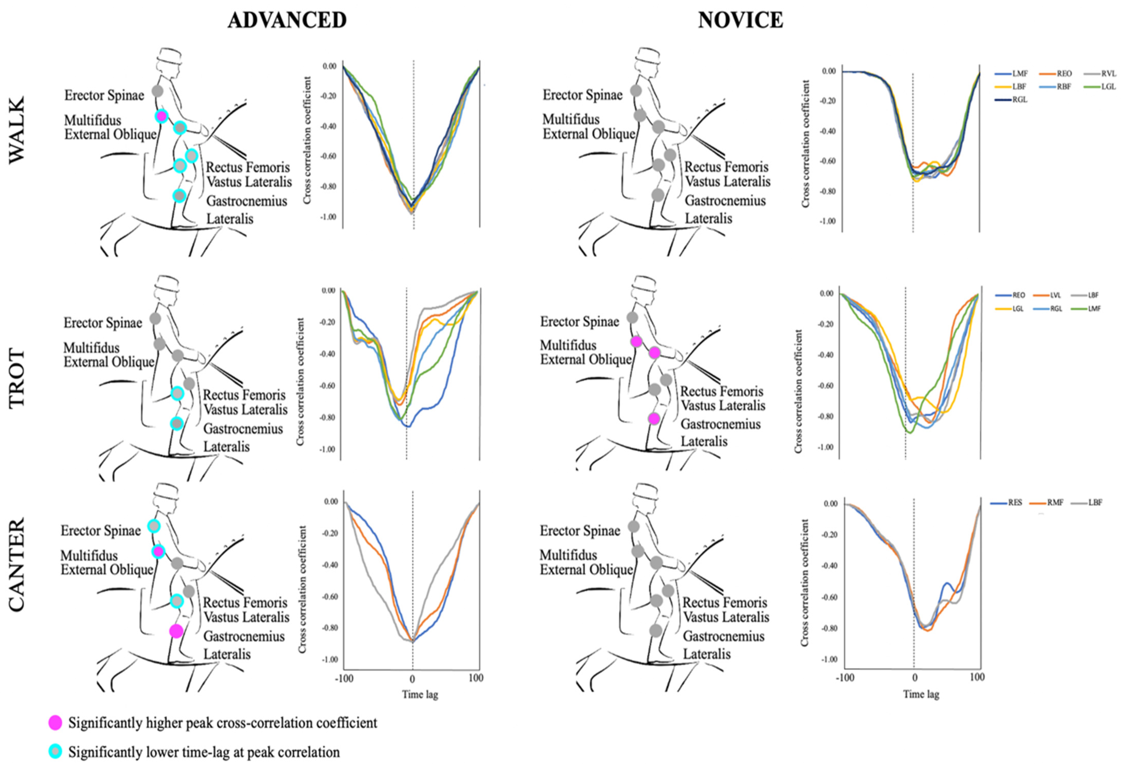
Figure 3 shows the correlation between each muscle and T4:saddle and T4:L1 shock attenuation waves for both advanced and novice riders. To gain a deeper insight into the direction of the correlations, a cumulative integral of each of the signals was calculated via the trapezoidal method. In all cases, as the sEMG signal increased, SA waves decreased.
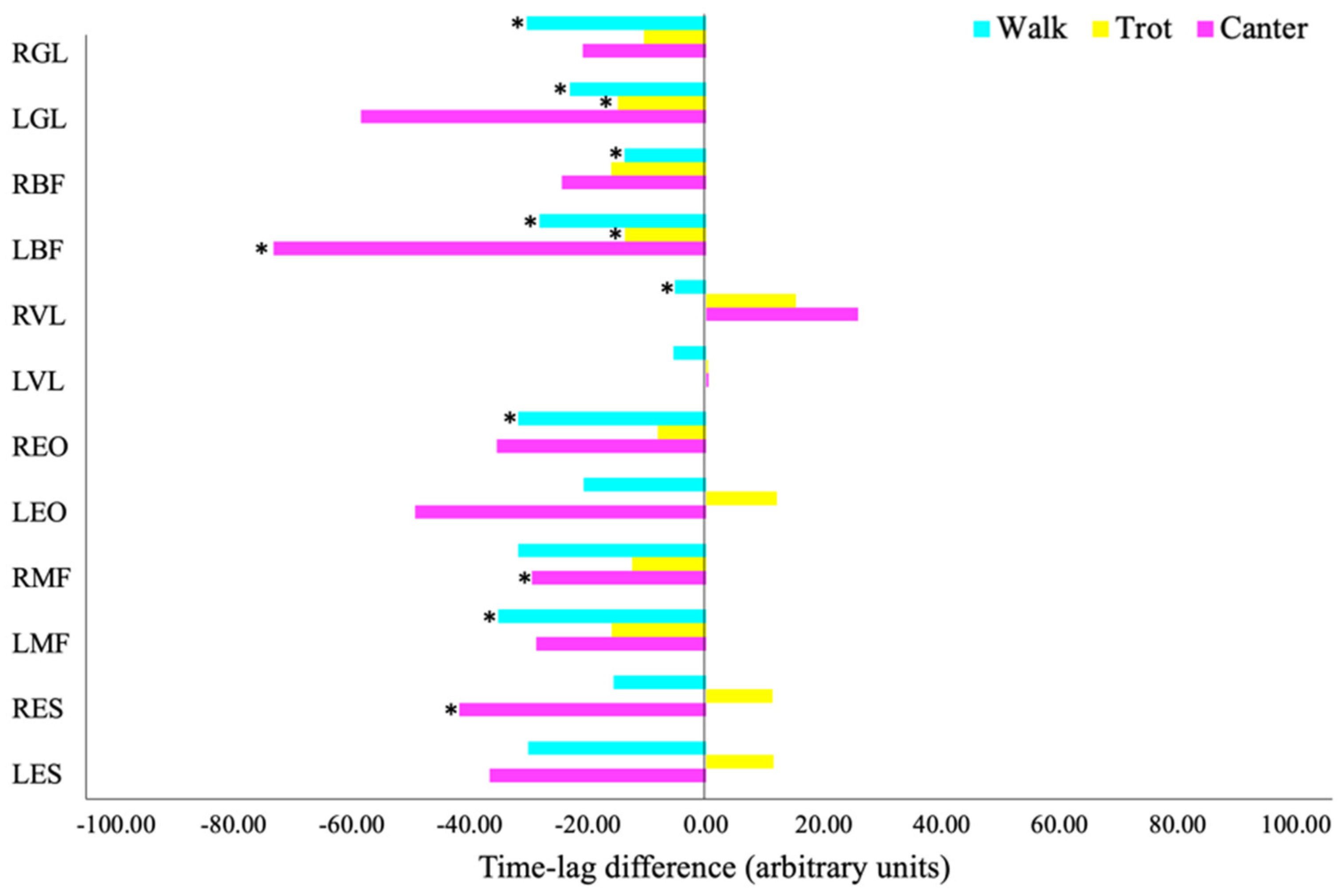
The time lag at which peak correlation occurred was also identified and the difference between the advanced and novice group (advanced–novice) was calculated. Further analyses with Kruskal–Wallis test revealed significant differences between groups in the peak correlation coefficients and the time lag at peak correlation between sEMG signals and T4:saddle SA waves but not between sEMG signals and T4:L1 SA waves.
Figure 4 summarizes the differences in time lag of the peak correlations with each muscle, at each gait and for each group. Significant differences revealed by the Kruskal–Wallis test have been marked (
p < 0.05).
4. Discussion
This study has investigated the shock attenuation behaviour at the back of horse riders at walk, posting trot and canter. The relevance of neuromuscular activity of the rider’s trunk muscles and its interaction with such attenuation has also been analysed. Novice riders and advanced riders have been compared to understand how proficiency level affects the biomechanics of horse riding. We initially hypothesised that advanced riders would show better shock attenuation capacities by activating their muscles earlier and to a higher degree. While this hypothesis was generally met, some exceptions were found. This study suggests that advanced riders do have a greater ability to absorb shockwaves when compared to novice riders, yet the mechanisms through which they attenuate such forces are more strongly related to the timing of muscle activation rather than the level of activation.
Overall, the results suggest that both groups are actively using their muscles to attenuate the shock produced by the horse in motion. However, sEMG levels are not as high as seen in other athletic activities such as cycling [
24] or running [
25], nor did the trials elicit substantial neuromuscular fatigue [
26]. These findings are consistent with the moderate rider’s sEMG activity reported by previous authors [
17,
18,
27].
SA at the levels of T5:L4 and T5:saddle are more efficient (i.e., lower) in advanced riders than novice riders. SA waves were associated with sEMG signals, revealing minimal differences between the groups in terms of peak correlation, but substantial and consistent differences in time lag.
Figure 5 is a schematical representation of the between-groups significantly different peak correlations, accompanied by a representative curve of the corresponding cross-correlations. It also highlights the correlations that were observed significantly earlier. This study demonstrates that the most relevant difference between novice and advanced riders regarding the relationship between sEMG activity and SA lies in the timing and not the level of correlation. Advanced riders were able to attenuate shockwaves by activating their muscles earlier than novice riders. The latter finding is particularly relevant since anticipation plays a major role in the management of impact forces. Mizrahi et al. (2000) demonstrated that eccentric contractions during running impair the ability to dissipate shock propagation, hence pre-activation (i.e., anticipation) is crucial to avoid such type of contractions [
28]. This finding is supported by the proposed idea that pre-activation of the human leg musculature is crucial in the management of heel strike reaction forces while running [
29].
Several publications have shown that modifying saddle characteristics and fit can effectively reduce spinal loading of the rider [
30,
31,
32,
33]. Yet this paper demonstrates that not only saddle configuration, but physical fitness and level of expertise can considerably reduce shock propagation through the spine.
It should be acknowledged that this study is limited by the nature of equestrian sports, which involve the participation of an animal over which we have a limited control, and repeatability of measurements will be inevitably limited. Furthermore, the small sample group who took part in the study is not representative of the highly scattered riding population, and the use of different horses might have affected the results. However, we chose the four most consistent and similar horses over a sample of 200 highly trained horses, in order to keep variability to a minimum while preserving the animal welfare (a horse cannot be ridden an infinite number of times), and all trials were conducted in a quiet indoor arena with minimal distractions for the horse.
5. Conclusions
As previously stated, amateur riders suffer higher incidence rates of LBP than advanced riders, even though the latter spend substantially more time on the horse. To our knowledge, this is the first study to demonstrate that not only can advanced riders substantially reduce impact forces on their spine compared to novice riders, but also that this is achieved by an early activation and a higher overall muscle tone of their muscles. These findings could have great practical implications, as we now know that working on early reaction and pre-activation of the core muscles not only potentially increases riding performance, but also decreases the spinal loading of the rider. Such work may include, for instance, plyometric exercises and proprioceptive training in the riders’ calendar. Future research is needed to identify the most effective exercises that will be able to teach horse riders the ability to anticipate horse-related impacts.
Author Contributions
Conceptualization, M.E.G. and N.Š.; methodology, M.E.G. and N.Š.; software, M.E.G.; validation, M.E.G. and N.Š.; formal analysis, M.E.G.; investigation, M.E.G.; resources, N.Š.; data curation, M.E.G.; writing—original draft preparation, M.E.G.; writing—review and editing, N.Š.; visualization, M.E.G. and N.Š.; supervision, N.Š.; project administration, N.Š.; funding acquisition, N.Š. All authors have read and agreed to the published version of the manuscript.
Funding
The Slovenian Research Agency provided author NS with support in the form of salary through the programme ‘Kinesiology of monostructural, polystructural and conventional sports’ [P5-0147 (B)] and the project TELASI-PREVENT [L5-1845] (Body asymmetries as a risk factor in musculoskeletal injury development: studying aetiological mechanisms and designing corrective interventions for primary and tertiary preventive care). The funders did not have any additional role in the study design, data collection and analysis, decision to publish, or preparation of the manuscript.
Institutional Review Board Statement
The National Medical Ethics Committee (Ministry of Health, Ljubljana) gave full approval to the project according to the declaration of Helsinki (approval number: 0120-346/2018/3).
Informed Consent Statement
Informed consent was obtained from all subjects involved in the study.
Data Availability Statement
The data is available upon request to corresponding author’s email.
Conflicts of Interest
The authors declare no conflict of interest.
References
- Lewis, V.; Kennerley, R. A preliminary study to investigate the prevalence of pain in elite dressage riders during competition in the United Kingdom. Comp. Exerc. Physiol. 2017, 13, 259–263. [Google Scholar] [CrossRef]
- Cejudo, A.; Ginés-Díaz, A.; Rodríguez-Ferrán, O.; Santonja-Medina, F.; De Baranda, P.S. Trunk Lateral Flexor Endurance and Body Fat: Predictive Risk Factors for Low Back Pain in Child Equestrian Athletes. Children 2020, 7, 172. [Google Scholar] [CrossRef]
- Feucht, C.L.; Patel, D.R. Analgesics and Anti-inflammatory Medications in Sports: Use and Abuse. Pediatr. Clin. N. Am. 2010, 57, 751–774. [Google Scholar] [CrossRef]
- Greve, L.; Dyson, S. The horse–saddle–rider interaction. Vet. J. 2013, 195, 275–281. [Google Scholar] [CrossRef]
- Kraft, C.N.; Pennekamp, P.H.; Becker, U.; Young, M.; Diedrich, O.; Lüring, C.; Von Falkenhausen, M. Magnetic Resonance Imaging Findings of the Lumbar Spine in Elite Horseback Riders. Am. J. Sports Med. 2009, 37, 2205–2213. [Google Scholar] [CrossRef]
- Hobbs, S.J.; Baxter, J.; Broom, L.; Rossell, L.-A.; Sinclair, J.; Clayton, H.M. Posture, Flexibility and Grip Strength in Horse Riders. J. Hum. Kinet. 2014, 42, 113–125. [Google Scholar] [CrossRef] [Green Version]
- Nevison, C.M.; Timmis, M.A. The effect of physiotherapy intervention to the pelvic region of experienced riders on seated postural stability and the symmetry of pressure distribution to the saddle: A preliminary study. J. Vet. Behav. 2013, 8, 261–264. [Google Scholar] [CrossRef]
- Dąbek, J.; Koczy, B.; Piotrkowicz, J. [Horse riding as a form of recreation and professional sport taking into account the spine mobility of riders—A preliminary results]. Polski Merkur. Lek. Organ Polskiego Towar. Lek. 2015, 39, 297–304. [Google Scholar]
- Zemková, E.; Kováčiková, Z.; Zapletalová, L. Is There a Relationship Between Workload and Occurrence of Back Pain and Back Injuries in Athletes? Front. Physiol. 2020, 11, 894. [Google Scholar] [CrossRef]
- Eckardt, F.; Witte, K. Kinematic Analysis of the Rider According to Different Skill Levels in Sitting Trot and Canter. J. Equine Vet. Sci. 2016, 39, 51–57. [Google Scholar] [CrossRef]
- Münz, A.; Eckardt, F.; Witte, K. Horse–rider interaction in dressage riding. Hum. Mov. Sci. 2014, 33, 227–237. [Google Scholar] [CrossRef]
- Peham, C.; Licka, T.; Kapaun, M.; Scheidl, M. A new method to quantify harmony of the horse-rider system in dressage. Sports Eng. 2001, 4, 95–101. [Google Scholar] [CrossRef]
- Lagarde, J.; Peham, C.; Licka, T.; Kelso, J.A.S. Coordination Dynamics of the Horse-Rider System. J. Mot. Behav. 2005, 37, 418–424. [Google Scholar] [CrossRef] [Green Version]
- Eckardt, F.; Witte, K. Horse–Rider Interaction: A New Method Based on Inertial Measurement Units. J. Equine Vet. Sci. 2017, 55, 1–8. [Google Scholar] [CrossRef]
- Hobbs, S.J.; George, L.S.; Reed, J.; Stockley, R.; Thetford, C.; Sinclair, J.; Williams, J.; Nankervis, K.; Clayton, H.M. A scoping review of determinants of performance in dressage. PeerJ 2020, 8, e9022. [Google Scholar] [CrossRef]
- González, M.E.; Šarabon, N. Muscle modes of the equestrian rider at walk, rising trot and canter. PLoS ONE 2020, 15, e0237727. [Google Scholar] [CrossRef]
- Clayton, H.; Terada, K.; Mullineaux, D.R.; Lanovaz, J.; Kato, K. Electromyographic analysis of the rider’s muscles at trot. Equine Comp. Exerc. Physiol. 2004, 1, 193–198. [Google Scholar] [CrossRef]
- Terada, K. Comparison of Head Movement and EMG Activity of Muscles between Advanced and Novice Horseback Riders at Different Gaits. J. Equine Sci. 2000, 11, 83–90. [Google Scholar] [CrossRef] [Green Version]
- Vera-Garcia, F.J.; Moreside, J.M.; McGill, S.M. MVC techniques to normalize trunk muscle EMG in healthy women. J. Electromyogr. Kinesiol. 2010, 20, 10–16. [Google Scholar] [CrossRef]
- Dyson, S.; Carson, S.; Fisher, M. Saddle Fitting, Recognising an Ill-fitting Saddle and the Consequences of an Ill-fitting Saddle to Horse and Rider. Equine Vet. Educ. 2015, 27, 533–543. [Google Scholar] [CrossRef]
- Hermens, H.J.; Freriks, B.; Merletti, R.; Stegeman, D.; Blok, J.; Rau, G.; Disselhorst-Klug, C.; Hägg, G. European Recommendations for Surface ElectroMyoGraphy; Roessingh Research and Development: Enschede, The Netherlands, 1999. [Google Scholar]
- Frère, J.; Hug, F. Between-subject variability of muscle synergies during a complex motor skill. Front. Comput. Neurosci. 2012, 6, 99. [Google Scholar] [CrossRef] [Green Version]
- Castillo, E.R.; Lieberman, D.E. Shock attenuation in the human lumbar spine during walking and running. J. Exp. Biol. 2018, 221, jeb177949. [Google Scholar] [CrossRef] [Green Version]
- Sarabon, N.; Fonda, B.; Markovic, G. Change of muscle activation patterns in uphill cycling of varying slope. Graefe’s Arch. Clin. Exp. Ophthalmol. 2011, 112, 2615–2623. [Google Scholar] [CrossRef]
- Gazendam, M.G.J.; Hof, A.L. Averaged EMG profiles in jogging and running at different speeds. Gait Posture 2007, 25, 604–614. [Google Scholar] [CrossRef]
- Thomas, K.; Elmeua, M.; Howatson, G.; Goodall, S. Intensity-Dependent Contribution of Neuromuscular Fatigue after Constant-Load Cycling. Med. Sci. Sports Exerc. 2016, 48, 1751–1760. [Google Scholar] [CrossRef]
- Guillaume, J.F.; Laroche, D.; Babault, N. Kinematics and electromyographic activity of horse riders during various cross-country jumps in equestrian. Sports Biomech. 2019, 1–13. [Google Scholar] [CrossRef]
- Mizrahi, J.; Verbitsky, O.; Isakov, E. Shock accelerations and attenuation in downhill and level running. Clin. Biomech. 2000, 15, 15–20. [Google Scholar] [CrossRef]
- Nigg, B.; Stefanyshyn, D.; Cole, G.; Stergiou, P.; Miller, J. The effect of material characteristics of shoe soles on muscle activation and energy aspects during running. J. Biomech. 2003, 36, 569–575. [Google Scholar] [CrossRef]
- Gandy, E.A.; Bondi, A.; Hogg, R.; Pigott, T.M.C. A preliminary investigation of the use of inertial sensing technology for the measurement of hip rotation asymmetry in horse riders. Sports Technol. 2014, 7, 79–88. [Google Scholar] [CrossRef]
- Kotschwar, A.B.; Baltacis, A.; Peham, C. The effects of different saddle pads on forces and pressure distribution beneath a fitting saddle. Equine Vet. J. 2010, 42, 114–118. [Google Scholar] [CrossRef]
- Roost, L.; Ellis, A.D.; Morris, C.; Bondi, A.; Gandy, E.A.; Harris, P.; Dyson, S. The effects of rider size and saddle fit for horse and rider on forces and pressure distribution under saddles: A pilot study. Equine Vet. Educ. 2020, 32, 151–161. [Google Scholar] [CrossRef]
- Stapley, E.D.; Stutzman, B.E.; Manfredi, J.M. The Effect of Stirrup Iron Style on Normal Forces and Rider Position. J. Equine Vet. Sci. 2020, 94, 103203. [Google Scholar] [CrossRef] [PubMed]
| Publisher’s Note: MDPI stays neutral with regard to jurisdictional claims in published maps and institutional affiliations. |
© 2021 by the authors. Licensee MDPI, Basel, Switzerland. This article is an open access article distributed under the terms and conditions of the Creative Commons Attribution (CC BY) license (http://creativecommons.org/licenses/by/4.0/).


