Review

Maturation of Anterior Cruciate Ligament Graft—Possibilities of Surgical Enhancement: What Do We Know So Far?

1
Artromedical Orthopaedic Clinic, Chrobrego 24, 97-400 Belchatow, Poland
2
ORTIM Orthopaedic Clinic, Mlynowa 17, 15-404 Bialystok, Poland
3
Orthopedic and Trauma Department, Veteran’s Memorial Teaching Hospital in Lodz, Medical University of Lodz, Zeromskiego 113, 90-549 Lodz, Poland
4
Twin Cities Orthopedics, 4010 West 65th Street, Edina, MN 55435, USA
*
Author to whom correspondence should be addressed.
Appl. Sci. 2021, 11(8), 3597; https://doi.org/10.3390/app11083597
Submission received: 11 February 2021 / Revised: 14 April 2021 / Accepted: 15 April 2021 / Published: 16 April 2021
(This article belongs to the Special Issue Multicriterial Assessment of Ligament Healing in the Knee Joint)

Abstract

:
The purpose of this study is to review the surgical methods of enhancing anterior cruciate ligament (ACL) graft maturation. Several methods of ACL maturation enhancement were identified through research of the literature available in the PubMed database. ACL remnant preservation was the most extensively investigated technique. ACL reconstruction with a pedunculated hamstring graft provides superior revascularization of the graft along with higher mechanical strength. The usage of a graft enveloped with a periosteum was proposed to enhance the tendon-bone unit formation, and consequently, to prevent the bone tunnel widening. The muscle tissue on the graft is a potential source of stem cells. However, an excessive amount may weaken whole graft strength despite its enhanced remodeling. Similarly, amniotic tissue may augment the ACL reconstruction with stem cells and growth factors. Despite the existence of several surgical techniques that utilize amnion, the outcomes of these augmentation methods are lacking. Lastly, the intra-articular transplantation of the synovium on the surface of an ACL was proposed to augment the graft with synovial tissue and blood vessels. In conclusion, diverse approaches are being developed in order to enhance the maturation of an ACL reconstruction graft. Although these approaches have their foundation in on well-established scientific research, their outcomes are still equivocal. Clinical trials of high quality are needed to evaluate their utility in clinical practice.

1. Introduction

Recent decades have witnessed progress in the management of anterior cruciate ligament (ACL) tears. The introduction of anatomic reconstruction techniques along with the development of solid methods of graft fixation have significantly improved clinical outcomes. However, with the improvement in the results of ACL reconstruction surgery, the expectations of patients have grown as well. Constant demand is placed on a faster postoperative recovery and return to sport, especially in professional athletes, where underperformance and financial consequences are important. Moreover, despite the favorable outcomes, still up to 9% of reconstructed ACLs fail at 25 years of follow-up, with young, active patients being especially at risk [1].
These factors have laid the groundwork for increased interest in methods of ACL graft maturation enhancement. It is postulated that the enhancement of ACL ligamentization and integration may decrease the failure rate, increase stability, and improve proprioception and functional outcomes [2]. Recently, the application of growth factors, platelet-rich plasma and bone marrow aspirate, commonly referred as “ortho-biologics”, has gained attention as a modality to overcome the limitations of conventional ACL reconstructions [3]. Although the evidence supporting their use is constantly growing, their influence on the outcomes of ACL reconstruction remains a matter of debate. Besides these augmentative techniques, several other surgical methods have been proposed to boost ACL graft maturation. The purpose of this narrative review is to provide an insight into the scientific background of alternative “biosurgical” methods for ACL graft maturation enhancement, outline their premises and report their clinical outcomes.

2. Materials and Methods

Studies regarding the surgical methods for graft maturation enhancement in the setting of ACL reconstruction were included in this study. The use of orthobiologics, due to the numerous reviews regarding their use, was considered to be beyond the scope of this review. Studies regarding ACL reconstruction with artificial grafts were excluded from the study.
A primary search was performed in the PubMed database. To screen for surgical methods of ACL maturation enhancement, the searched item was “(anterior cruciate ligament reconstruction) AND ((enhancement) OR (augmentation))”. The search was performed without limitation regarding the year of publication, language or study design. After the screening of abstracts, followed by consultation between the authors, six methods of surgical enhancement of ACL maturation enhancement were identified, namely:
  • Preservation of remnants of a native ACL;
  • Preservation of hamstring tibial attachment during an ACL reconstruction with a hamstring autograft;
  • Enveloping of a graft with a periosteum;
  • Preservation of muscle tissue on a harvested graft;
  • Augmentation of a graft with an amniotic tissue;
  • The usage of an intra-articular synovial graft.
The secondary search aimed to collect the data regarding the scientific basis of the abovementioned methods and to summarize their outcomes. The secondary search was performed in the PubMed database without restriction regarding the year of publication or study design; only the studies in English were included. The search items were as follows:
  • (anterior cruciate ligament reconstruction) AND (remnant preservation);
  • (anterior cruciate ligament reconstruction) AND (attached hamstring graft);
  • (anterior cruciate ligament reconstruction) AND (periosteum);
  • (anterior cruciate ligament reconstruction) AND (muscle remnant);
  • (anterior cruciate ligament reconstruction) AND ((amnion) OR (amniotic tissue));
  • (anterior cruciate ligament reconstruction) AND (synovialization).
The references of included studies were screened to search for additional data sources. The data extracted from each article included the authors of the manuscript, year of publication, study design, type of the intervention, length of the follow-up and the key findings.

3. Remnant Preservation

The preservation of an ACL remnants was probably the most extensively investigated method of ACL reconstruction enhancement over the last decades (Figure 1). The evidence regarding outcomes of remnant-preserving procedures is summarized in Table 1.
The potential benefits of remnant retaining include increased stability, preservation of the proprioception, enhanced ligamentization and revascularization and prevention of the tunnel enlargement [2]. These factors laid the foundation for the rapid development of this trend in ACL reconstruction [2]. Nonetheless, certain drawbacks were also expected, including more demanding surgical techniques, range of motion (ROM) deficits or greater incidence of cyclops lesions [2]. Indeed, with growing evidence from clinical trials, these assumptions are being verified. To our knowledge, four meta-analyses have been published regarding the benefits of ACL remnant preservation in recent years [4,5,6,7]. All of them have highlighted the significantly higher Lysholm score in the remnant-preserved reconstruction groups. On the contrary, the International Knee Documentation Committee (IKDC) score failed to prove any significant difference between the remnant-preserving and remnant-sacrificing group. Regarding postoperative knee stability, neither the Lachman test nor Pivot shift test has shown significant differences between these groups in all of the studies. On the other hand, the results of the side-to-side analysis performed with KT-1000/2000 were equivocal; the results of Ma et al. [4], Wang et al. [6] and Wang et al. [7] favored the remnant-preservation group, while the results of Tie et al. [5] demonstrated no significant difference between both groups. Although the loss of ROM and the incidence of cyclops lesions were speculated to be more prevalent in the remnant-preserving reconstruction, the above-mentioned analyses proved that occurrence of these complications does not differ significantly between remnant-preserving and remnant-sacrificing procedures [4,5,6,7].
Although remnant-preserving procedures were designed to enhance the proprioception of the reconstructed ACL, the evidence supporting this property is still lacking. Of two studies that examined proprioception with reproduction of passive positioning (RPP) [8,9], only one proved that the remnant-preservation group outperformed the remnant-sacrificing group. On the other hand, Lee et al. [10] proved significantly better outcomes in the remnant-preserved group in RPP, threshold to detection of passive motion and single-legged hop tests. However, it is difficult to generalize these results to all remnant-preserving reconstructions as Lee at al. used remnant-preserving ACL reconstruction combined with preservation of the tibial attachment of a hamstring autograft.
The influence of remnant preservation on graft revascularization was assessed by magnetic resonance imaging (MRI) by means of signal/noise quotient (SNQ). Gohil et al. found the remnant-preserving group to have higher SNQ than the remnant-sacrificing group at 2 months of follow-up and lower SNQ at 6 months of follow-up [11]. Similarly, Lee et al. found the remnant-preserving group to have higher SNQ than the remnant-sacrificing group at 2–4 months of follow-up and lower SNQ at 12–18 months of follow-up [12]. Both authors concluded that revascularization of a graft proceeds more rapidly in the remnant-preserving reconstructions. Additionally, in the study by Ahn et al., the SNQ in the remnant-preserving group was not significantly lower than in the remnant-sacrificing group at 6.3 months of follow-up [13]. Regarding the progress of ligamentization, the meta-analysis of Wang et al. found superior results for remnant-preserving procedures [7]. On the other hand, the analysis of randomized controlled trials showed no significant difference, while analysis of cohort studies favored the remnant-preserving group. Furthermore, remnant preservation during the single-bundle reconstruction was found to prevent tunnel enlargement with the assessment of the tunnel diameter on plain radiographs [14,15]. However, in regard to double-bundle reconstructions assessed with computed tomography, Naraoka et al. [16] discounted any protective effect of remnant preservation on the incidence of tunnel enlargement, whereas Yanagisawa et al. [17] found remnant preservation to lower the incidence of tunnel enlargement. Additionally, Masuda et al. [18] found remnant preservation prevented tunnel enlargement, but only in the anteromedial femoral tunnel.

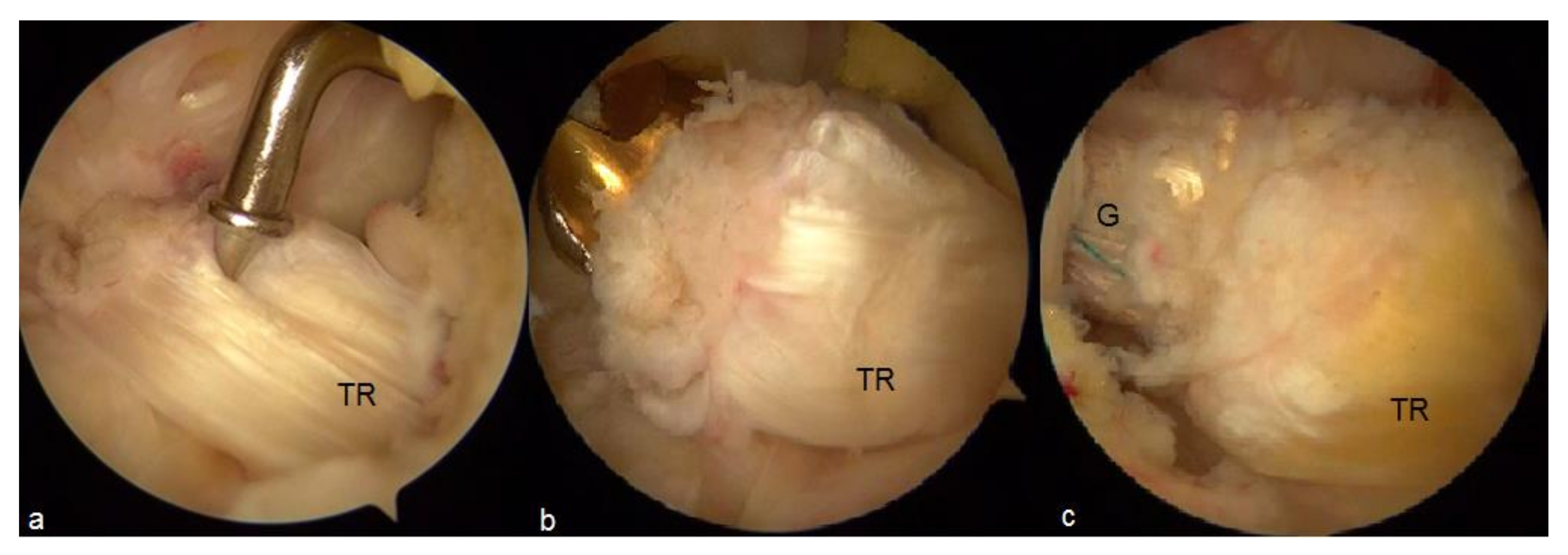
4. Preservation of Hamstrings Tibial Insertion

The evidence regarding the outcomes of pedunculated hamstring procedures is summarized in Table 2.
While reconstructing an ACL with the hamstring autograft, the benefits emerging from its tibial attachment preservation are two-fold—Biological and mechanical. First, Zaffagnini et al. [19] reported in their anatomic study that pes anserinus tendons are consistently well innervated and vascularized along their course. These authors suggest that preservation of this supply may be of clinical benefit. Experimental ACL reconstructions performed in an animal model revealed that the preservation of hamstrings tibial attachment preserved the viability of the graft and enabled the bypass of the stage of avascular necrosis during graft remodeling [20,21]. Second, the cadaver study conducted by Bahlau et al. [22] demonstrated the significantly higher pull-out strength of the pedunculated hamstring graft fixed with a tibial screw compared to the free hamstring graft, with a 65% higher load to failure in the pedunculated graft group. The load to failure in the pedunculated hamstring group without additional tibial fixation was 33% higher than in the free graft group, although the difference did not reach statistical significance.
A review published in 2015 regarding the preservation of hamstring tibial insertion pointed out the great heterogeneity of available surgical techniques as well as fixation methods, which precluded authors from drawing a definitive conclusion about the superiority of insertion-preserving or insertion-sacrificing procedure over each other [23]. However, the results from reported case series were satisfactory and comparable with conventional ACL reconstructions. Since then, several studies have evaluated the functional and biomechanical outcomes of hamstring insertion-preserving procedures. Gupta et al. compared the ACL reconstruction with pedunculated hamstring autograft and the free hamstring autograft after 2 years of follow-up [24]. The results of their single-blinded randomized trial reported significantly higher Cincinnati knee scores, greater improvement in the Tegner activity scale and lower side-to-side difference in KT-1000 testing in favor of the pedunculated hamstring group. Nonetheless, despite the significant difference in functional outcomes, no clinically significant difference regarding these scoring systems were found. Zhang et al. compared the outcomes between a single-bundle ACL reconstruction with a free hamstring autograft and so-called functional double-bundle ACL reconstruction, which included preservation of the hamstring tibial insertion and independent fixation of semitendinosus and gracilis grafts at different tensions in the single tibial tunnel [25]. The functional outcomes including the Tegner Activity Scale, IKDC score, Knee Injury and Osteoarthritis Outcome score and Lysholm score of the functional double-bundle ACL reconstruction group were better than the outcomes of the control group. Although the translational stability was similar, the rotational stability was significantly better in the functional double-bundle ACL reconstruction group. Recently, the prospective study of Bahlau et al. reported excellent functional outcomes in IKDC and Lysholm scores for an ACL reconstruction with pedunculated hamstring autograft [26]. Moreover, the postoperative stability and the postoperative recovery of hamstring and quadricep strength were comparable with conventional ACL reconstructions.
Considering the influence of hamstring attachment preservation on the progress of ACL graft maturation, the abovementioned scientific observations have found increasing support in the data from clinical studies. Ruffilli et al. demonstrated significantly better intra-articular morphology of pedunculated graft than free graft in the MRI at the 6-month follow-up [27]. On the contrary, the integration of the graft in the tibial tunnel did not differ significantly between both groups. Similarly, a pedunculated hamstring ”over-the-top” reconstruction combined with lateral plasty showed significantly better intra-articular features of the graft compared to the conventional hamstring reconstructions [28]. Moreover, the authors also confirmed significantly lower tibial tunnel enlargement in the pedunculated group. Liu et al. analyzed the course of the graft remodeling in consecutive MRIs and found that the pedunculated hamstring grafts maintained more stable and lower signal intensity than free grafts within 2 years of follow-up [29]. Given that the peak of signal intensity is caused by the early stage of graft avascular necrosis, the pedunculated hamstring graft seems to bypass this stage of remodeling and skip the phase of vascular supply disruption.

5. Periosteum-Enveloped Graft

The tendon graft-bone tunnel junction is a critical element of the whole ACL reconstruct, prone to high pull-out forces and subsequent ACL reconstruction graft failure [30]. Two forms of osteotendinous junctions have been described: fibrous, where a tendon attaches to the bone directly via the fibrous connective tissue, and fibrocartilaginous, where a fibrocartilage and a calcified fibrocartilage are interposed between a tendon and a bone [31]. The fibrocartilaginous enthesis is considered to better disperse the stress concentration; hence, the re-creation of this form of enthesis at the tendon graft-bone tunnel junction during ACL reconstruction may lead to its superior durability [32].
Periosteum is known for its osteogenic and chondrogenic properties, and therefore, was speculated to strengthen the incorporation of the graft in the bone tunnel. This idea was investigated in several animal studies, where a tendon, enveloped in periosteum, was transplanted into the bone tunnel [33,34,35]. All of them confirmed the osteochondral differentiation of periosteum-derived stem cells and the formation of the fibrocartilage at the tendon–bone interface, similarly as in the fibrocartilaginous enthesis. Moreover, all studies also proved the significantly higher pull-out strength of the periosteum-enveloped graft compared to the non-enveloped control group [33,34,35].
Based on these findings, a technique of periosteum-enhanced ACL reconstruction was proposed [36]. The authors harvested periosteum from the medial side of tibia during the hamstring graft preparation, then wrapped and sutured it to the parts of the graft that were subsequently localized in the tibial and femoral tunnel (Figure 2 and Figure 3). The follow-up evaluation of patients treated with this technique revealed significant improvement in the Lysholm score and the normal or nearly normal results of the majority of patients regarding the IKDC score 2–3 years postoperatively [37]. Tunnel enlargement over 1 mm was found in 5% of femoral and 6% of tibial tunnels. Further evaluation performed at2–7 years of follow-up supported these results [38].
Robert et al. prospectively compared the outcomes of the conventional hamstring graft reconstruction and reconstruction with periosteum enveloped around the graft near the femoral tunnel entrance [39]. The periosteum-enhanced reconstruction presented significantly lower femoral tunnel enlargement on plain radiographs throughout the follow-up. These authors concluded that a periosteal flap both enhances the creation of osteotendinous junction and also acts as a seal, preventing the synovial fluid inflow to the tunnel in the early postoperative period.

6. Preservation of Muscle Tissue on Tendon Graft

The residual muscle tissue is routinely scraped from the hamstring tendon during the graft preparation. However, these remnants contain a variety of potent stem cells to enhance graft maturation. Ćuti et al. analyzed the samples from hamstring tendons and found pericytes to be more numerous in muscle tissue than in tendon [40]. Moreover, the muscle-derived stem cells expressed higher activity of early bone differentiation markers like alkaline phosphatase and bone sialoprotein, while tendon-derived stem cells presented higher levels of late bone differentiation markers such as dental-matrix-protein 1 and osteocalcin. The authors concluded that muscle- and tendon-derived stem cells may act synergistically and sequentially, enhancing subsequent stages of the tendon–bone unit formation.
In an in vitro experiment conducted by Ghebes et al., co-culturing myoblasts with hamstring tendon-derived cells upregulated the expression of tendon/ligament differentiation markers and increased the synthesis of collagen units [41]. Moreover, authors analyzed the influence of muscle-secreted factors on the ACL reconstruction in the rat model. The results were equivocal—The tibial tunnel closure did not show any significant differences between the experimental and control groups, whereas the femoral tunnel closure was significantly improved in the reconstructions enhanced with the muscle-derived serum.
Sun et al. compared ACL reconstruction with the semi-tendon/semi-muscle graft (SSG) and the tendon-only graft in the rabbit model [42]. Although the diameter of the SSG gradually decreased during the postoperative period, the healing and incorporation of the graft were more evident than in the tendon-only group. The vascularity and cellularity of the SSG were significantly greater than in tendon-only graft reconstructions. Nonetheless, the biomechanical testing revealed the significantly lower ultimate failure load, yield load and elongation at failure in the SSG group. Authors concluded that even though muscle tissue enhances biological incorporation of the graft, excessive amount of muscle remnants may weaken the whole graft construct.

7. Amnion

Amniotic tissue is a potent source of growth factors and stem cells. Not only does it possess anti-inflammatory and antimicrobial properties, but it also acts as a scaffold that enhances tissue regeneration and healing processes [43]. Its application in knee osteoarthritis, plantar fasciitis and tendon repair has already been reported [44].
Despite the potential of amniotic tissue to enhance the biological healing processes, there is a paucity of literature concerning its application in ACL surgery. In an in vitro study, Li et al. proved that amnion-derived mesenchymal stem cells possess the potential to differentiate into ACL fibroblasts, and as a consequence, may enhance the results of an ACL reconstruction [45]. To date, two surgical techniques of ACL reconstruction utilizing the amniotic membrane have been proposed. Woodall et al. wrapped the amniotic membrane around a central part of the graft, leaving the femoral and tibial tunnel parts intact [46]. Lavender et al. proposed wrapping the graft with amniotic membrane combined with intra-tunnel injections of bone marrow and a suture tape augmentation [47]. However, the outcomes of these techniques were not reported.

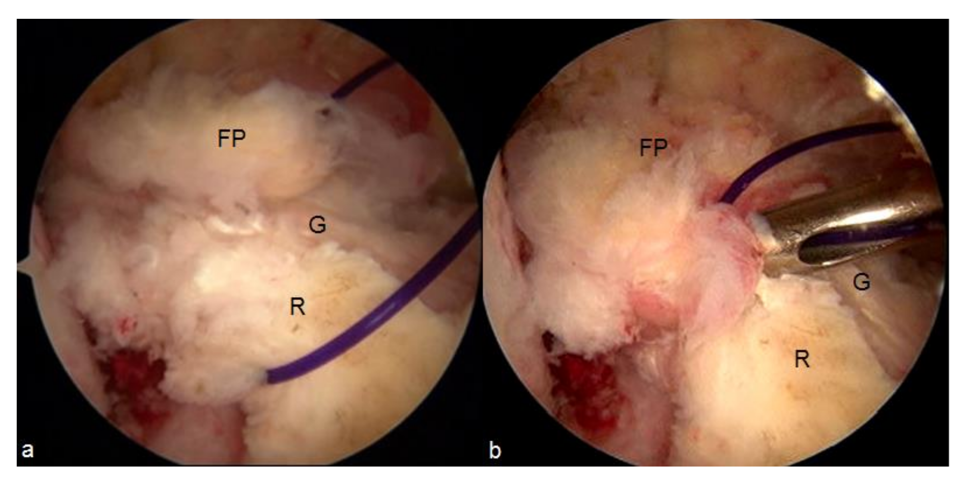
8. Intra-Articular Synovial Grafts

The ACL vasculature of the ligament–bone interface is scarce, and therefore, its blood supply is derived mostly through the subsynovial vessels originating from the medial genicular artery [48]. For this reason, the revascularization of a reconstructed ACL, and the graft remodeling, accordingly, is strictly dependent on the restoration of the graft synovial coverage [49]. This hypothesis was confirmed by the results of clinical trials, which reported that a greater amount of synovial coverage was associated with better graft survival rate, greater stability and better functional outcomes [50,51,52].
The first attempt to restore the synovial coverage of the ACL was proposed by Scapinelli in his anatomical study [48]. The author pointed out the abundant vascular network and rich synovial coverage of the ligamentum mucosum (infrapatellar plica) and proposed to suture it to a reconstructed ACL in order to boost its resynovialization. Nonetheless, no outcomes of this method were reported.
Recently, Malinowski et al. proposed a novel technique of an ACL synovialization enhancement [53]. In this method the fat pad covering the anterior part of the posterior cruciate ligament (PCL) is dissected, leaving its distal attachment intact, and sutured to the reconstructed or repaired ACL (Figure 4). Such a pedunculated synovial graft is supposed to supply the central part of an ACL with synovial cells and blood vessels, which, due to its poor vasculature, is especially prone to be torn (Figure 5). However, similarly to previous studies, the outcomes have not been yet reported.

9. Conclusions

Several emerging techniques for ACL reconstruction surgical enhancement have been reported in the literature. ACL remnant preservation was the most extensively investigated technique, but its beneficial effect differs depending on the assessed outcomes. ACL reconstruction with a pedunculated hamstring graft provides superior revascularization of the graft along with its higher mechanical strength. The usage of a graft enveloped with a periosteum is proposed to enhance the tendon–bone unit formation, and consequently, to prevent the bone tunnel widening. The muscle tissue on the graft is a potential source of stem cells. However, an excessive amount may weaken whole graft strength despite its enhanced remodeling. Similarly, amniotic tissue may augment the ACL reconstruction with stem cells and growth factors. Despite several surgical techniques that utilize amnion, the outcomes of these augmentation methods are lacking. Similarly, although the intra-articular transplantation of the synovium on the surface of an ACL were proposed to augment the graft with synovial tissue and blood vessels, the results of this technique have not been reported so far.
Despite the fact that the analyzed methods are easily applicable in routine practice, the evidence supporting their use is still lacking. The underlying premises, derived from the results of basic scientific studies, are promising. However, the majority of presented techniques have not been evaluated in clinical trials so far. Even so, the heterogeneity related to patient characteristics, surgical technique, graft type and fixation method precludes one from drawing definitive conclusions regarding their clinical utility. Specific randomized controlled trials, isolating the influence of the enhancement methods, are urgently needed to characterize their outcomes and describe the indications for their use.

Author Contributions

Conceptualization, M.E. and K.M.; methodology, M.E.; investigation, M.E., M.M. and A.G.; writing—Original draft preparation, M.E., M.M. and A.G.; writing—Review and editing, K.M. and R.F.L.; supervision, K.M. and R.F.L. All authors have read and agreed to the published version of the manuscript.

Funding

This research received no external funding.

Institutional Review Board Statement

Not applicable.

Informed Consent Statement

Not applicable.

Data Availability Statement

Data sharing not applicable.

Conflicts of Interest

Michał Ebisz, Adrian Góralczyk and Marcin Mostowy report no conflicts of interest. Robert F. LaPrade serves as a Consultant and receive royalties from Arthrex, Ossur and Smith and Nephew and serves on the Editorial Boards of AJSM, JEO and KSSTA. Konrad Malinowski serves as a Zimmer Biomet Consultant, PTArtro Board.

References

  1. Sanders, T.L.; Pareek, A.; Hewett, T.E.; Levy, B.A.; Dahm, D.L.; Stuart, M.J.; Krych, A.J. Long-term rate of graft failure after ACL reconstruction: A geographic population cohort analysis. Knee Surg. Sports Traumatol. Arthrosc. 2017, 25, 222–228. [Google Scholar] [CrossRef] [PubMed]
  2. Muneta, T.; Koga, H. Anterior cruciate ligament remnant and its values for preservation. Asia-Pac. J. Sports Med. Arthrosc. Rehabil. Technol. 2017, 7, 1–9. [Google Scholar] [CrossRef] [Green Version]
  3. Chahla, J.; Kennedy, M.I.; Aman, Z.S.; LaPrade, R.F. Ortho-Biologics for Ligament Repair and Reconstruction. Clin. Sports Med. 2019, 38, 97–107. [Google Scholar] [CrossRef] [PubMed]
  4. Ma, T.; Zeng, C.; Pan, J.; Zhao, C.; Fang, H.; Cai, D. Remnant preservation in anterior cruciate ligament reconstruction versus standard techniques: A meta-analysis of randomized controlled trials. J. Sports Med. Phys. Fit. 2017, 57, 1014–1022. [Google Scholar]
  5. Tie, K.; Chen, L.; Hu, D.; Wang, H. The difference in clinical outcome of single-bundle anterior cruciate ligament reconstructions with and without remnant preservation: A meta-analysis. Knee 2016, 23, 566–574. [Google Scholar] [CrossRef] [PubMed]
  6. Wang, H.-D.; Wang, F.-S.; Gao, S.-J.; Zhang, Y.-Z. Remnant preservation technique versus standard technique for anterior cruciate ligament reconstruction: A meta-analysis of randomized controlled trials. J. Orthop. Surg. Res. 2018, 13, 231. [Google Scholar] [CrossRef] [PubMed] [Green Version]
  7. Wang, H.; Liu, Z.; Li, Y.; Peng, Y.; Xu, W.; Hu, N.; Huang, W. Is Remnant Preservation in Anterior Cruciate Ligament Reconstruction Superior to the Standard Technique? A Systematic Review and Meta-Analysis. BioMed Res. Int. 2019, 2019, 1–15. [Google Scholar] [CrossRef] [Green Version]
  8. Hong, L.; Li, X.; Zhang, H.; Liu, X.; Zhang, J.; Shen, J.W.; Feng, H. Anterior Cruciate Ligament Reconstruction with Remnant Preservation: A prospective, randomized controlled study. Am. J. Sports Med. 2012, 40, 2747–2755. [Google Scholar] [CrossRef] [PubMed]
  9. Adachi, N.; Ochi, M.; Uchio, Y.; Sumen, Y. Anterior cruciate ligament augmentation under arthroscopy. A minimum 2-year follow-up in 40 patients. Arch. Orthop. Trauma Surg. 2000, 120, 128–133. [Google Scholar] [CrossRef]
  10. Lee, B.-I.; Kwon, S.-W.; Kim, J.-B.; Choi, H.-S.; Min, K.-D. Comparison of Clinical Results According to Amount of Preserved Remnant in Arthroscopic Anterior Cruciate Ligament Reconstruction Using Quadrupled Hamstring Graft. Arthrosc. J. Arthrosc. Relat. Surg. 2008, 24, 560–568. [Google Scholar] [CrossRef]
  11. Gohil, S.; Annear, P.O.; Breidahl, W. Anterior cruciate ligament reconstruction using autologous double hamstrings: A comparison of standardversusminimal debridement techniques using MRI to assess revascularisation. A randomised prospective study with a one-year follow-up. J. Bone Jt. Surg. Br. Vol. 2007, 89, 1165–1171. [Google Scholar] [CrossRef] [Green Version]
  12. Lee, B.I.; Kim, B.M.; Kho, D.H.; Kwon, S.W.; Kim, H.J.; Hwang, H.R. Does the tibial remnant of the anterior cruciate ligament promote ligamentization? Knee 2016, 23, 1133–1142. [Google Scholar] [CrossRef] [PubMed]
  13. Ahn, J.H.; Lee, S.H.; Choi, S.H.; Lim, T.K. Magnetic Resonance Imaging Evaluation of Anterior Cruciate Ligament Reconstruction Using Quadrupled Hamstring Tendon Autografts: Comparison of remnant bundle preservation and standard technique. Am. J. Sports Med. 2010, 38, 1768–1777. [Google Scholar] [CrossRef]
  14. Zhang, Q.; Zhang, S.; Cao, X.; Liu, L.; Liu, Y.; Li, R. The effect of remnant preservation on tibial tunnel enlargement in ACL reconstruction with hamstring autograft: A prospective randomized controlled trial. Knee Surg. Sports Traumatol. Arthrosc. 2014, 22, 166–173. [Google Scholar] [CrossRef] [PubMed]
  15. Demirağ, B.; Ermutlu, C.; Aydemir, F.; Durak, K. A comparison of clinical outcome of augmentation and standard reconstruction techniques for partial anterior cruciate ligament tears. Jt. Dis. Relat. Surg. 2012, 23, 140–144. [Google Scholar]
  16. Naraoka, T.; Kimura, Y.; Tsuda, E.; Yamamoto, Y.; Ishibashi, Y. Does Remnant Preservation Influence Tibial Tunnel Enlargement or Graft-to-Bone Integration After Double-Bundle Anterior Cruciate Ligament Reconstruction Using Hamstring Autografts and Suspensory Fixation? A Computed Tomography and Magnetic Resonance Imaging Evaluation. Orthop. J. Sports Med. 2018, 6, 2325967118790238. [Google Scholar] [CrossRef] [Green Version]
  17. Yanagisawa, S.; Kimura, M.; Hagiwara, K.; Ogoshi, A.; Nakagawa, T.; Shiozawa, H.; Ohsawa, T.; Chikuda, H. The remnant preservation technique reduces the amount of bone tunnel enlargement following anterior cruciate ligament reconstruction. Knee Surg. Sports Traumatol. Arthrosc. 2018, 26, 491–499. [Google Scholar] [CrossRef]
  18. Masuda, T.; Kondo, E.; Onodera, J.; Kitamura, N.; Inoue, M.; Nakamura, E.; Yagi, T.; Iwasaki, N.; Yasuda, K. Effects of Remnant Tissue Preservation on Tunnel Enlargement After Anatomic Double-Bundle Anterior Cruciate Ligament Reconstruction Using the Hamstring Tendon. Orthop. J. Sports Med. 2018, 6, 2325967118811293. [Google Scholar] [CrossRef] [Green Version]
  19. Zaffagnini, S.; Golanò, P.; Farinas, O.; de Pasquale, V.; Strocchi, R.; Cortecchia, S.; Marcacci, M.; Visani, A. Vascularity and neuroreceptors of the pes anserinus: Anatomic Study. Clin. Anat. 2002, 16, 19–24. [Google Scholar] [CrossRef] [PubMed]
  20. Papachristou, G.; Nikolaou, V.; Efstathopoulos, N.; Sourlas, J.; Lazarettos, J.; Frangia, K.; Papalois, A. ACL reconstruction with semitendinosus tendon autograft without detachment of its tibial insertion: A histologic study in a rabbit model. Knee Surg. Sports Traumatol. Arthrosc. 2007, 15, 1175–1180. [Google Scholar] [CrossRef]
  21. Liu, S.; Sun, Y.; Wan, F.; Ding, Z.; Chen, S.; Chen, J. Advantages of an Attached Semitendinosus Tendon Graft in Anterior Cruciate Ligament Reconstruction in a Rabbit Model. Am. J. Sports Med. 2018, 46, 3227–3236. [Google Scholar] [CrossRef] [PubMed]
  22. Bahlau, D.; Clavert, P.; Favreau, H.; Ollivier, M.; Lustig, S.; Bonnomet, F.; Ehlinger, M. Mechanical advantage of preserving the hamstring tibial insertion for anterior cruciate ligament reconstruction—A cadaver study. Orthop. Traumatol. Surg. Res. 2019, 105, 89–93. [Google Scholar] [CrossRef]
  23. Ruffilli, A.; Traina, F.; Evangelisti, G.; Borghi, R.; Perna, F.; Faldini, C. Preservation of hamstring tibial insertion in anterior cruciate ligament reconstruction: A review of the current literature. Musculoskelet. Surg. 2015, 99, 87–92. [Google Scholar] [CrossRef]
  24. Gupta, R.; Bahadur, R.; Malhotra, A.; Masih, G.D.; Sood, M.; Gupta, P.; Mathur, V.K. Outcome of Hamstring Autograft With Preserved Insertions Compared With Free Hamstring Autograft in Anterior Cruciate Ligament Surgery at 2-Year Follow-up. Arthrosc. J. Arthrosc. Relat. Surg. 2017, 33, 2208–2216. [Google Scholar] [CrossRef]
  25. Zhang, Q.; Yang, Y.; Li, J.; Zhang, H.; Fu, Y.; Wang, Y. Functional double-bundle anterior cruciate ligament reconstruction using hamstring tendon autografts with preserved insertions is an effective treatment for tibiofemoral instability. Knee Surg. Sports Traumatol. Arthrosc. 2019, 27, 3471–3480. [Google Scholar] [CrossRef] [PubMed]
  26. Bahlau, D.; Favreau, H.; Eichler, D.; Lustig, S.; Bonnomet, F.; Ehlinger, M. Clinical, functional, and isokinetic study of a prospective series of anterior cruciate ligament ligamentoplasty with pedicular hamstrings. Int. Orthop. 2019, 43, 2557–2562. [Google Scholar] [CrossRef] [PubMed]
  27. Ruffilli, A.; Pagliazzi, G.; Ferranti, E.; Busacca, M.; Capannelli, D.; Buda, R. Hamstring graft tibial insertion preservation versus detachment in anterior cruciate ligament reconstruction: A prospective randomized comparative study. Eur. J. Orthop. Surg. Traumatol. 2016, 26, 657–664. [Google Scholar] [CrossRef]
  28. Grassi, A.; Casali, M.; Macchiarola, L.; Lucidi, G.A.; Cucurnia, I.; Filardo, G.; Lopomo, N.F.; Zaffagnini, S. Hamstring grafts for anterior cruciate ligament reconstruction show better magnetic resonance features when tibial insertion is preserved. Knee Surg. Sports Traumatol. Arthrosc. 2021, 29, 507–518. [Google Scholar] [CrossRef] [PubMed]
  29. Liu, S.; Li, H.; Tao, H.; Sun, Y.; Chen, S.; Chen, J. A Randomized Clinical Trial to Evaluate Attached Hamstring Anterior Cruciate Ligament Graft Maturity with Magnetic Resonance Imaging. Am. J. Sports Med. 2018, 46, 1143–1149. [Google Scholar] [CrossRef]
  30. Goradia, V.K.; Rochat, M.C.; Grana, W.A.; Egle, D.M. Strength of ACL reconstructions using semitendinosus tendon grafts. J. Okla. State Med. Assoc. 1998, 91, 275–277. [Google Scholar]
  31. Benjamin, M.; Toumi, H.; Ralphs, J.R.; Bydder, G.; Best, T.M.; Milz, S. Where tendons and ligaments meet bone: Attachment sites (‘entheses’) in relation to exercise and/or mechanical load. J. Anat. 2006, 208, 471–490. [Google Scholar] [CrossRef] [PubMed]
  32. Huang, B.; Pathria, M.; Tadros, A. Muscle-Tendon-Enthesis Unit. Semin. Musculoskelet. Radiol. 2018, 22, 263–274. [Google Scholar] [CrossRef] [Green Version]
  33. Chen, C.-H.; Chen, W.-J.; Shih, C.-H.; Yang, C.-Y.; Liu, S.-J.; Lin, P.-Y. Enveloping the tendon graft with periosteum to enhance tendon-bone healing in a bone tunnel: A biomechanical and histologic study in rabbits. Arthrosc. J. Arthrosc. Relat. Surg. 2003, 19, 290–296. [Google Scholar] [CrossRef] [PubMed]
  34. Kyung, H.-S.; Kim, S.-Y.; Oh, C.-W.; Kim, S.-J. Tendon-to-bone tunnel healing in a rabbit model: The effect of periosteum augmentation at the tendon-to-bone interface. Knee Surg. Sports Traumatol. Arthrosc. 2003, 11, 9–15. [Google Scholar] [CrossRef]
  35. Youn, I.; Jones, D.G.; Andrews, P.J.; Cook, M.P.; Suh, J.-K.F. Periosteal Augmentation of a Tendon Graft Improves Tendon Healing in the Bone Tunnel. Clin. Orthop. Relat. Res. 2004, 419, 223–231. [Google Scholar] [CrossRef] [PubMed]
  36. Chen, C.H.; Chen, W.J.; Shih, C.H. Enveloping of periosteum on the Hamstring Tendon graft in anterior cruciate ligament reconstruction. Arthroscopy 2002, 18, 27E. [Google Scholar] [CrossRef]
  37. Chen, C.-H.; Chen, W.-J.; Shih, C.-H.; Chou, S.-W. Arthroscopic anterior cruciate ligament reconstruction with periosteum-enveloping hamstring tendon graft. Knee Surg. Sports Traumatol. Arthrosc. 2004, 12, 398–405. [Google Scholar] [CrossRef]
  38. Chen, C.-H.; Chang, C.-H.; Su, C.-I.; Wang, K.-C.; Liu, H.-T.; Yu, C.-M.; Wong, C.-B.; Wang, I.-C. Arthroscopic Single-Bundle Anterior Cruciate Ligament Reconstruction with Periosteum-Enveloping Hamstring Tendon Graft: Clinical Outcome at 2 to 7 Years. Arthrosc. J. Arthrosc. Relat. Surg. 2010, 26, 907–917. [Google Scholar] [CrossRef] [PubMed]
  39. Robert, H.; Es-Sayeh, J. The role of periosteal flap in the prevention of femoral widening in anterior cruciate ligament reconstruction using hamstring tendons. Knee Surg. Sports Traumatol. Arthrosc. 2003, 12, 30–35. [Google Scholar] [CrossRef] [PubMed]
  40. Ćuti, T.; Antunović, M.; Marijanović, I.; Ivković, A.; Vukasović, A.; Matić, I.; Pećina, M.; Hudetz, D. Capacity of muscle derived stem cells and pericytes to promote tendon graft integration and ligamentization following anterior cruciate ligament reconstruction. Int. Orthop. 2017, 41, 1189–1198. [Google Scholar] [CrossRef]
  41. Ghebes, C.A.; Groen, N.; Cheuk, Y.C.; Fu, S.C.; Fernandes, H.M.; Saris, D.B. Muscle-Secreted Factors Improve Anterior Cruciate Ligament Graft Healing: An In Vitro and In Vivo Analysis. Tissue Eng. Part A 2018, 24, 322–334. [Google Scholar] [CrossRef] [PubMed]
  42. Sun, L.; Hou, C.; Wu, B.; Tian, M.; Zhou, X. Effect of muscle preserved on tendon graft on intra-articular healing in anterior cruciate ligament reconstruction. Knee Surg. Sports Traumatol. Arthrosc. 2012, 21, 1862–1868. [Google Scholar] [CrossRef] [PubMed]
  43. Riboh, J.C.; Saltzman, B.M.; Yanke, A.B.; Cole, B.J. Human Amniotic Membrane-Derived Products in Sports Medicine: Basic Science, Early Results, and Potential Clinical Applications. Am. J. Sports Med. 2016, 44, 2425–2434. [Google Scholar] [CrossRef]
  44. Heckmann, N.; Auran, R.; Mirzayan, R. Application of Amniotic Tissue in Orthopedic Surgery. Am. J. Orthop. 2016, 45, e421–e425. [Google Scholar]
  45. Li, Y.; Liu, Z.; Jin, Y.; Zhu, X.; Wang, S.; Yang, J.; Ren, Y.; Fu, Q.; Xiong, H.; Zou, G.; et al. Differentiation of Human Amniotic Mesenchymal Stem Cells into Human Anterior Cruciate Ligament Fibroblast Cells by In Vitro Coculture. BioMed Res. Int. 2017, 2017, 1–15. [Google Scholar] [CrossRef] [PubMed] [Green Version]
  46. Woodall, B.M.; Elena, N.; Gamboa, J.T.; Shin, E.C.; Pathare, N.; McGahan, P.J.; Chen, J.L. Anterior Cruciate Ligament Reconstruction with Amnion Biological Augmentation. Arthrosc. Tech. 2018, 7, e355–e360. [Google Scholar] [CrossRef] [Green Version]
  47. Lavender, C.; Bishop, C. The Fertilized Anterior Cruciate Ligament: An All-Inside Anterior Cruciate Ligament Reconstruction Augmented with Amnion, Bone Marrow Concentrate, and a Suture Tape. Arthrosc. Tech. 2019, 8, e555–e559. [Google Scholar] [CrossRef] [Green Version]
  48. Scapinelli, R. Vascular anatomy of the human cruciate ligaments and surrounding structures. Clin. Anat. 1997, 10, 151–162. [Google Scholar] [CrossRef]
  49. Unterhauser, F.N.; Bail, H.J.; Höher, J.; Haas, N.P.; Weiler, A. Endoligamentous Revascularization of an Anterior Cruciate Ligament Graft. Clin. Orthop. Relat. Res. 2003, 414, 276–288. [Google Scholar] [CrossRef]
  50. Ateschrang, A.; Schreiner, A.J.; Ahmad, S.S.; Schröter, S.; Hirschmann, M.T.; Körner, D.; Kohl, S.; Stöckle, U.; Ahrend, M.-D. Improved results of ACL primary repair in one-part tears with intact synovial coverage. Knee Surg. Sports Traumatol. Arthrosc. 2019, 27, 37–43. [Google Scholar] [CrossRef]
  51. Noh, J.H.; Yang, B.G.; Roh, Y.H.; Lee, J.S. Synovialization on second-look arthroscopy after anterior cruciate ligament reconstruction using Achilles allograft in active young men. Knee Surg. Sports Traumatol. Arthrosc. 2011, 19, 1843–1850. [Google Scholar] [CrossRef] [PubMed]
  52. Kondo, E.; Yasuda, K. Second-Look Arthroscopic Evaluations of Anatomic Double-Bundle Anterior Cruciate Ligament Reconstruction: Relation with Postoperative Knee Stability. Arthrosc. J. Arthrosc. Relat. Surg. 2007, 23, 1198–1209. [Google Scholar] [CrossRef] [PubMed]
  53. Malinowski, K.; Ebisz, M.; Góralczyk, A.; Laprade, R.F.; Hermanowicz, K. Synovialization and Revascularization Enhancement in Repaired and Reconstructed ACL: PCL Fat Pad Transfer Technique. Arthrosc. Tech. 2020, 9, e1559–e1563. [Google Scholar] [CrossRef] [PubMed]
Figure 1. Reconstruction with preservation of the distal anterior cruciate ligament (ACL) remnant: (a) Tibial guide placed in the middle of a tibial remnant (TR); (b) Drilling of the tibial tunnel through the tibial remnant; (c) ACL graft (G) passing through the distal remnant of a native ACL.
Applsci 11 03597 g001
Figure 2. Harvesting of a semitendinosus tendon graft with an adjacent periosteum: (a) Exposed distal semitendinosus (ST) attachment with an adjacent periosteal flap (PF) (marked violet); (b) Harvested semitendinosus tendon graft with an adjacent periosteal flap.
Applsci 11 03597 g002
Figure 3. Preparation of a periosteal flap to enhance the femoral part of a graft: (a) Harvesting of the periosteum below the pes anserinus attachment; (b) Free periosteal flap; (c) Femoral part of an anterior cruciate ligament graft (G) augmented with a free periosteal flap (PF).
Applsci 11 03597 g003
Figure 4. Synovialization enhancement technique with a pedunculated posterior cruciate ligament (PCL) fat pad: (a) Pedunculated fat pad (FP) sutured to the anterior cruciate ligament (ACL) graft (G) with an PDS suture; (b) Central part of a graft covered with a fat pad; (c) Formation of a hematoma on a surface of the graft after tourniquet release.
Applsci 11 03597 g004
Figure 5. The PCL fat pad technique combined with the distal anterior cruciate ligament (ACL) remnant preservation: (a) The fat pad (FP) and the distal remnant of a native ACL (R) adjacent to the ACL graft (G); (b) Entire ACL graft covered with the fat pad and the distal remnant of a native ACL.
Applsci 11 03597 g005
Table 1. Summary of available literature concerning remnant-preserving ACL reconstructions (Abbreviations: IKDC—International Knee Documentation Committee; ROM—range of motion; ACLR—anterior cruciate ligament reconstruction; SNQ—signal/noise quotient).
AuthorYear of PublicationStudy DesignInterventionMean
Follow-Up
Outcomes
Ma et al. [4]2017Meta-analysis--Significantly higher Lysholm score, better arthrometer results and lower tibial tunnel enlargement in remnant-preserving reconstruction.
No statistically significant difference regarding IKDC grade, IKDC score, Lachman test, Pivot-shift test, range of motion (ROM), and the incidence of the cyclops lesion.
Tie et al. [5]2016Meta-analysis--Significantly lower incidence of tibial tunnel enlargement in remnant-preserving reconstructions.
No statistically significant difference regarding arthrometer results, Lachman test, Pivot-shift test, IKDC score and Lysholm score.
Wang et al. [6]2018Meta-analysis--Significantly higher Lysholm score and better arthrometer results in remnant-preserving reconstructions.
No statistically significant difference regarding IKDC score, complications, Pivot-shift test and Lachman test.
Wang et al. [7]2019Meta-analysis--Significantly better arthrometer results and Lysholm score in remnant-preserving group.
No statistically significant difference regarding graft maturation, complications, IKDC score, Lachman test and pivot-shift test.
Hong et al. [8]2012Randomized controlled trial Single-bundle ACLR with a 4-strand allograft.25.7 monthsNo statistically significant difference regarding Lysholm score, IKDC grade, Lachman test, Pivot-shift test, arthrometer results, graft synovial coverage and passive angle reproduction test.
Adachi et al. [9]2000Retrospective cohort studySingle-bundle ACLR with a hamstring autograft or fascia lata allograft.3.2 years Significantly better arthrometer results and passive angle reproduction test in favor of remnant-preserving group.
No statistically significant difference regarding Lysholm score and Gillquist score.
Lee et al. [10]2008Case series
Comparison of patients with over 20% of preserved remnant (I) and those with less than 20% of preserved remnant (II)
Single-bundle ACLR with a pedunculated hamstring autograft35.1 monthsSignificantly better threshold to detection of passive motion at 30° and reproduction of passive positioning at 15° and 30° in the I group.
Gohil et al. [11]2007Randomized controlled trialSingle-bundle ACLR with a hamstring autograft12 monthsThe remnant-preserving group had higher SNQ than the remnant-sacrificing group at 2 months of follow-up and lower SNQ at 6 months of follow-up.
Lee et al.
[12]
2016Retrospective cohort studySingle-bundle ACLR with a hamstring autograft18 monthsThe remnant-preserving group had higher SNQ than the remnant-sacrificing group at 2-4 months of follow-up and lower SNQ at 12-18 months of follow-up.
Ahn et al. [13]2010Retrospective cohort studySingle-bundle ACLR with a hamstring autograft6.3 monthsThe SNQ did not differ significantly between the both groups.
Zhang et al. [14]2014Randomized controlled trial
Tunnel diameter evaluated with plain radiographs
Single-bundle ACLR with a hamstring autograft24.5 monthsSignificantly lower incidence of tibial tunnel enlargement in the remnant-preserving group.
Demirağ et al. [15]
2012Randomized controlled trial
Tunnel diameter evaluated with plain radiographs
Single-bundle ACLR with a hamstring autograft24.3 months
Significantly lower tibial and femoral tunnel widening in the remnant-preserving group.
Naraoka et al. [16]2018Prospective cohort study
Tunnel diameter evaluated with computed tomography
Double-bundle ACLR with hamstring autograft1 yearSignificantly greater posterolateral tibial tunnel enlargement in the remnant-preserved group 1 year after ACLR.
Yanagisawa et al. [17]2018Prospective cohort study
Tunnel diameter evaluated with computed tomography
Double-bundle ACLR with hamstring autograft11.7-13.8 monthsSignificantly lower incidence of femoral and tibial anteromedial tunnel enlargement in the remnant-preserving group.
Masuda et al. [18]2018Prospective cohort study
Tunnel diameter evaluated with computed tomography
Double-bundle ACLR with hamstring autograft1 yearSignificantly lower incidence of femoral anteromedial tunnel enlargement in the remnant-preserving group.
Table 2. Summary of available literature concerning ACL reconstructions with pedunculated hamstring graft (Abbreviations: IKDC—International Knee Documentation Committee; ACLR—Anterior cruciate ligament reconstruction; SNQ—Signal/noise quotient; MRI—Magnetic resonance imaging).
Author.Year of PublicationStudy DesignInterventionMean
Follow-Up
Key Findings
Papachristou et al. [20]2007Animal studyACLR with pedunculated hamstring autograft-Preservation of hamstring tibial insertion allowed to bypass the stage of graft necrosis.
Liu et al. [21]2018Animal studyACLR with pedunculated hamstring autograft
-Preservation of hamstring tibial insertion allowed to bypass the stage of graft necrosis.
Restoration of fibrocartilaginous bone-tendon interface in the pedunculated hamstring group.
Significantly higher histologic scores of the tendon-bone interface, failure load and stiffness as well as smaller bone tunnel area and larger bone volume/total volume in the pedunculated hamstring group.
Bahlau et al. [22]2019Cadaver studyGroup 1: Hamstring autograft with intact tibial insertion without interference screw;
Group 2: Hamstring autograft with intact tibial insertion and interference screw;
Group 3: free hamstring autograft fixed with an interference screw
-The load at failure was 33% higher in group 1 than group 3.
The load at failure of group 2 was 25% higher than group 1 and 65% higher than group 3.
Gupta et al. [24]2017Randomized controlled trialACLR with pedunculated hamstring autograft24 monthsSignificantly better arthrometer results, higher Cincinnati knee score and lower difference between the preinjury and post-surgery Tegner level of sports activity in pedunculated hamstring group.
Zhang et al. [25]2019Randomized controlled trialFunctional double bundle ACL reconstruction with pedunculated hamstring graft vs. single-bundle ACL reconstruction with free hamstring autograft28.2 monthsSignificantly better Tegner Activity Scale score, IKDC, KOOS and Lysholm Knee Scoring Scale in the functional double bundle reconstruction group.
Lower Pivot-shift grade in the functional double bundle reconstruction group.
Bahlau et al. [26]2019Case seriesACLR with pedunculated hamstring autograft30 monthsLysholm score 95/100 and IKDC score 91/100 at the final follow up.
2 mm difference of the anteroposterior laxity compared to the contralateral knee at the final follow up.
Significant decrease of the quadriceps and flexor muscles strength deficit during the follow up.
Ruffilli et al. [27]2016Randomized controlled trialACLR with pedunculated hamstring autograft24 monthsIKDC score above 90 in the both groups at the final follow-up.
Better intra-articular morphology of the pedunculated graft at the final MRI.
No differences in graft integration between the both groups.
Grassi et al. [28]2020Randomized controlled trialOver-the-top ACLR with pedunculated hamstring graft18 monthsLess liquid within the graft, smaller tunnel diameter and lower SNQ of the intra-tunnel graft in the pedunculated graft group in the follow up MRIs.
Liu et al. [29]2018Randomized Controlled TrialACLR with pedunculated hamstring autograft24 monthsLower and stable SNQ in the pedunculated hamstring group in the follow up MRIs.
Significantly lower SNQ values in the study group versus the control group at 6 and 12 months of the follow up.
Publisher’s Note: MDPI stays neutral with regard to jurisdictional claims in published maps and institutional affiliations.

