Article

Spectrophotometric Analysis of 3D Printed and Conventional Denture Base Resin after Immersion in Different Colouring Agents—An In Vitro Study

by
Mariya Dimitrova
1,*,†,
Saverio Capodiferro
2,*,†,
Angelina Vlahova
1,3,
Rada Kazakova
1,3,
Stoyan Kazakov
4,
Giuseppe Barile
2,* and
Massimo Corsalini
2
1
Department of Prosthetic Dentistry, Faculty of Dental Medicine, Medical University–Plovdiv, 4000 Plovdiv, Bulgaria
2
Department of Interdisciplinary Medicine, ‘Aldo Moro’, University of Bari, 70100 Bari, Italy
3
CAD/CAM Center of Dental Medicine, Research Institute, Medical University–Plovdiv, 4000 Plovdiv, Bulgaria
4
Oral Surgeon, Private Dental Practice–Sofia, 1000 Sofia, Bulgaria
*
Authors to whom correspondence should be addressed.
These Authors equally contributed to the paper as co-first Authors.
Appl. Sci. 2022, 12(24), 12560; https://doi.org/10.3390/app122412560
Submission received: 17 November 2022 / Revised: 4 December 2022 / Accepted: 6 December 2022 / Published: 8 December 2022
(This article belongs to the Special Issue Clinical Applications for Dentistry and Oral Health, 2nd Volume)

Abstract

:
Three-dimensional printed denture base resins are relatively new materials, and their properties need to be thoroughly investigated to assess whether they can be used clinically. The aim of the current study was to evaluate the colour stability of 3D printed and conventional denture base resins after immersion in different staining solutions. A total of 200 specimens were manufactured from two types of materials: 3D printed dental resin NextDent Denture 3D+ (NextDent, 3D Systems, the Netherlands) and heat-polymerized PMMA Vertex (3D Systems, the Netherlands), which were immersed in four types of colourants—artificial saliva, coffee, red wine and coke (n = 25). For measuring the colour changes (CIE-L*a*b* system), a SpectroShade Micro spectrophotometer (SpectroShade, Oxnard, CA, USA) was used. After seven days (T1), 14 days (T2) and 21 days (T3), the mean ∆E values were calculated and compared by the Bonferonni post hoc test. The data were processed using the statistical software SPSS 26. The level of significance for rejecting the null hypothesis was fixed at p < 0.05. The highest mean values for ∆E were found for both types of dental resin in red wine, and the lowest mean values for ∆E were found for 3D printed specimens in artificial saliva. The 3D printed denture base resin demonstrated better colour stability than the conventional acrylic materials. The staining effect correlated with the immersion time, with the red wine and coke having the strongest chromogenic impact and the period with the highest colour changes being 21 days.

1. Introduction

Replacing defects in dental rows has been done for thousands of years. In the Middle Ages, when dentures were fitted, they were hand-carved and fixed with silk threads. Fixation of the teeth created difficulties, as the upper and lower dentures were held by means of steel springs [1]. Many of the famous people of the time, such as the president of the USA George Washington, had periodontal disease and used removable prosthetic restorations. The deficiency of suitable materials for removable restorations was one of the main reasons for the complicated treatment of edentulous patients [2].
After the introduction of acrylic plastics, in the 1930s, for the first time, entire prostheses were made from pressed polymerized plates [3]. In 1935, the company “Kultzer” synthesized the first factory-produced acrylic plastic, which consisted of two components: powder (polymer) and liquid (monomer) for the needs of dentistry, known by its trade name “Palladon”. Three years later, the first self-polymerizing plastic was created, for which the polymerization process was activated using tertiary amines [4].
Since then, PMMA plastic has dominated the field of removable prosthetic construction. The advantages of this material are favorable physical and aesthetic characteristics, availability of the material and relatively low cost. The relative ease with which PMMA can be processed, adjusted or repaired is another preferable characteristic [5].
Dental resin is a high-molecular-weight mixture of natural or synthetic origin that consists of a number of monomeric particles creating macromolecules (polymers) [6]. Monomers, on the other hand, are single simple molecules, which are attached to chains of polymers. The synthesis of macromolecules is obtained by a reaction of polymerization. It is divided into two types—condensation and addition [7]. Condensation polymerization is characterized by the fact that the molecules of the monomer are joined into a polymer molecule. It has a different empirical composition than the monomer, releasing a low-molecular-weight by-product—water [8]. Addition polymerization results in a polymer that is made up of multiple repetitions of the monomer molecule. Thus, the molecular weight of the polymer is equal to the sum of the molecular weights of the participating monomers [9].
Removable prostheses are still a preferred treatment option for edentulous people by dental specialists. These prosthodontic restorations may be manufactured by the conventional method, using heat-polymerized polymer polymethyl methacrylate (PMMA), injection molding or the CAD /CAM method—using subtractive or additive manufacturing [10]. Three-dimensional printing technology or additive manufacturing (AM) is a widely applied method. It is based on stereolithography (SLA), and the objects are printed by the 3D printer layer by layer [11]. This method is used for fixed and removable prosthodontic restorations, orthodontic aligners, surgical guides and implants [12,13].
All types of dental resins have relatively good optical and mechanical qualities and easy processing. They have satisfactory dimensional properties and resistance to function in the conditions of the oral cavity [14]. However, these types of dental materials also have a number of shortcomings, such as staining from different food ingredients and beverages after a certain period of time. The water sorption and the colouring effect of different components may cause irreversible changes in a removable prosthesis [15]. As a consequence, discomfort during chewing and degradation of the aesthetic appearance may occur, which after a certain period of time may lead to the patient’s dissatisfaction with prosthetic treatment [16].
Due to a number of contraindications of a general and local nature, when other methods of treatment are not possible, this type of material for removable dentures will continue to be dentists’ first choice. Other main reasons are the affordable price, simple technology and high social acceptance [17].
The increasing awareness of dentists regarding the optical changes of dental resins used for removable dentures in the oral cavity could help them to solve specific clinical situations in their dental practice [18]. The need for prosthetic treatment to restore the defects of the dental rows with removable dentures determines the actuality of the topic about the colour stability of the denture base resins [19].
Dental resins for removable dentures exhibit certain colour changes over time due to different staining factors. They are in constant interaction with oral fluids and different kinds of food and beverages [20,21]. According to a number of studies, 3D printed dental resins have better optical stability than PMMA (polymethyl methacrylate) resins for removable prosthodontics [22,23,24,25]. The optical quality of the denture base resins is a property of paramount importance because it ensures the aesthetical appearance and patients’ comfort [26].
In comparison with the conventional heat-polymerized PMMA for removable dentures, 3D printed dental resins have a number of advantages. They include simplified laboratory protocol, time effectiveness and more efficient planning of the prosthetic restoration [27,28,29]. The shortcomings of both types of dental materials include discolouration over time, which affects negative the aesthetics and the long-term success of the prosthetic treatment [30,31].
Colour determination is possible via different visual and instrumental methods. The visual method consists of a clinical evaluation performed by the dental practitioner using different colour shade guides [32]. In comparison, the instrumental techniques are more accurate and exclude the possibility of subjective errors. They include colourimeters and spectrophotometers, which detect the colour values in accordance with the CIE L*a*b* colouring system. The CIE L*a*b* system defines the colour space with both chromatic value and saturation of the L*a*b* coordinates: L* measures colour bleaching (a value of 100 corresponds to perfect white and 0 to black); a* measures colour in the red (a* > 0) and green (a* < 0) dimensions; and b* measures colour in the yellow (b* > 0) and blue (b* < 0) dimensions [33].
Delta E is defined as the difference between two colours in an L*a*b* colour space, which is referred to as a mathematical formula. This information is significant for the evaluation of the distinct values. The CIE L*a*b* formula is based on the Euclidian distance, which represents the distance between two points in a three-dimensional colour space [34].
In addition, the vision is more susceptible to particular areas of colour and less susceptible to others. This is a fact that the formula does not take into consideration. The deficiencies of the eyes were compensated using further colour equations [35]. Colours in a highly saturated area are determined less significant during the evaluation than colours along the gray axis. This is the area where the vision is most sensitive. In the case that the delta E value of 1 is visible to the untrained eye, this does not apply for the highly saturated region but it is true for the gray axis [36].
Spectrophotometry is a type of method that is explained by the theory of electromagnetic spectroscopy. This process is applied with the quantitative measurement of the reflection of a material as a function of the wavelength [37]. The spectrophotometers are used for the quantitative evaluation of molecules. They are based on the amount of light that is absorbed by a variety of compounds. Spectrophotometers measure the intensity of a light beam at different wavelengths and have the ability to obtain wide swaths of the electromagnetic spectrum [38]. Significant features of spectrophotometers include the spectral bandwidth. This is the scale of colours that can be transferred through the test sample, the percentage of sample transmission, the logarithmic span of sample absorption, and sometimes a percentage of reflectance assessment. Spectrophotometers can also be designed to estimate the diffusivity on any of the listed light spans that usually enfold around 200–2500 nm applying different adjustments. Within this extent of light, calibrations are necessary using standards that vary in type depending on the wavelength of the photometric resolution [39].
The aim of this in vitro study was to investigate and compare the colour changes occurring in these two types of denture materials in various staining agents for the selected immersion periods.

2. Materials and Methods

For the purpose of the current investigation, 200 samples were prepared in the shape of a parallelepiped. They were manufactured with dimensions of 20 mm by 20 mm in width and length and 3 mm in cross-sectional diameter. The shape and size of the test specimens were designed according to the predetermined criteria. Non-parametric software (Free CAD Version 0.19) was applied and a .STL file was created for this purpose (Figure 1).
The parameters of the experimental bodies of the two types of resin for removable dentures were determined using the software program G*Power Version 3.1 (Universität Düsseldorf, Düsseldorf, Germany). G*Power is a tool for calculating statistical power analyses for various tests. G*Power can also be used to calculate effect sizes and graphically display the results of power analyses.
The evaluation of the sample size and the power analysis were implemented using the following components: effect size, power (1-β), significance level (α) and type of statistical method. A priori analysis is a statistical test that is used for the determination of the sample size and is usually performed before the investigation of the research has been planned. Thus, it is used to define the sample size N needed to evaluate the effect size, the desired α level and the power level (1-β).
Once the effect size (in this case the sizes of the test samples) is calculated, the procedure for determination of the sample size is very simple. For measuring the sample size of a statistical test, the software of G*Power gives information about the effect size conventions of “small”, “medium” and “large,” based on the recommendations of Cohen. Thus, it evaluates conventional effect size values that are different for different tests.
  • Two-sample or independent t-test
A two-sample t-test (also known as an independent t-test or Student’s t-test) is a statistical test that differentiates the means of two independent samples. The null hypothesis (H0) proposes that the difference in group means is equal to 0, and the alternative hypothesis (H1) states that the difference in group means is non-0.
For the purposes of our research, an a priori analysis was performed with a pre-set p-value of 0.05 and a power of 0.95 with a two-group balanced design. The applied t-test determined a total of 176 test specimens that were required from both types of materials (n = 88 per group) with dimensions of 20 mm × 20 mm × 3 mm to satisfy the aforementioned parameters.
The null hypothesis states that there would be no colour changes in the selected two groups, while the alternative hypothesis proposes that there will be a significant staining effect on the tested specimens. Two groups of specimens, 100 pieces of each type of dental resin, were manufactured. The first group of experimental samples was made from Vertex BasiQ 20 (Vertex Dental, 3D Systems, Soesterberg, The Netherlands) heat-polymerizing acrylic by a conventional flasking method. The second group of experimental bodies was made by the 3D printing method from dental resin for removable dentures of NextDent Denture 3D+ (NextDent, 3D Systems, Soesterberg, The Netherlands) [40] (Table 1).
The wax prototype of the experimental body with the predetermined shape and dimensions was printed using a 3D printer. Castable Wax (Formlabs, Somerville, MA, USA) is used to print wax prototypes of prosthetic structures, which are then packaged and cast [41]. In this way, considerable time is saved in the laboratory protocol, and high accuracy and precision of the part are ensured (Figure 2).
For the production of the experimental bodies of thermosetting plastic, the polymer was pre-weighed and combined with the monomer in a weight ratio of 2:1. The polymer and monomer were mixed in a clean porcelain vessel at room temperature as indicated in the manufacturer’s instructions. After about 15 min, the working phase (dough) was reached, in which the plastic was placed in the cuvettes and clamped in a hydraulic press (Sirio P400, Sirio Dental, Meldola, Italy) under a pressure of 100 kg/cm2 and slowly relaxed until the final drop in the pressure. This was followed by placing the sample in a water bath for 20 min at 1000 °C to complete the heat polymerization process. After the completion of the polymerization cycle and slow cooling to room temperature, the surface of the test samples was treated with 70% ethyl alcohol and then dried.
The samples in the second group of were made of dental resin for denture bases by the 3D printing method using a pre-generated .STL file from the software program. The .STL file format, which substitutes the geometry of an object in the shape of triangles, is a standard for sending three-dimensional information to 3D printers. The dental resin NextDent 3D Denture + is available in 5 colours, and for the purpose of the study, we chose “Light pink” as the colour of the experimental bodies.
The technology was implemented using NextDent’s 3D printer (NextDent 5100 DLP 3D Printer, 3D Systems, Budel, The Netherlands). Dental resin is a photopolymerizable plastic based on polymethyl methacrylate and is in the form of a liquid. It was placed in the tray at the bottom of the printer. Once the file was imported and the 3D images of the trial bodies were positioned, the process in the 3D printer software could be started. The resin specimens were printed at a 50 µm layer thickness. and the exposure time for this process was 95 min.
After printing, the finished experimental bodies were placed on a special platform at the top of the printer. Samples were subject to additional processing—cleaning of the unpolymerized material from the surface by placing them in a tub of isopropyl alcohol for 10 min. Isopropyl alcohol (IPA-2-propanol or rubbing alcohol) is a transparent, strong cleaning medium used for various 3D printing materials. For 3D printed elements, the process usually takes six minutes, and the IPA is dissolved in distilled water to a ratio of 70% isopropyl alcohol:30% distilled water. After cleaning, the test pieces were placed for another 45 min in glycerin in the final polymerization oven to react the remaining monomers.
From each group, we divided the experimental samples into 4 subgroups (n = 25), which we immersed in four different types of staining solutions—artificial saliva, coffee, Coca-Cola and red wine. The artificial saliva was produced in accordance with a preliminary recipe by a chemist. The coffee was prepared in agreement with the manufacturer’s instructions (Nescafe, Nestle, Vevey, Switzerland): First, 1.8 g coffee powder was mixed with 200 mL water. Then, it was placed after cooling in a glass container. Red wine (Mezzek Merlot, Katarzyna Estate, Svilengrad, Bulgaria) and coke (Coca-Cola, Coca-Cola HBC) were purchased and also placed in identical glass containers. The solutions were 200 mL each and were replaced with new ones every day. The study was conducted at room temperature, and changes in the colour of the materials were recorded on day 7, day 14 and day 21 by spectrophotometric analysis. For this purpose, a SpectroShade Micro (SpectroShade, Oxnard, CA, USA) spectrophotometer was applied [42]. Before each measurement with the apparatus, the experimental samples were washed with distilled water. Excess water on the surfaces of the samples was eliminated with a paper towel and allowed to dry.
The colour changes of the test bodies were determined by the Commission Internationale de l’Eclairege L*a*b* (CIELab) system using visible light [22]. The CIE L*a*b* values were measured for each test specimen three times to eliminate the possibility of an error and to calculate the average value for each test body.
In agreement with the ISO/TR-28642:2016 standard, reported ∆E values that are ≤1.2 are accepted as the lower sensitivity threshold, and ∆E values between 1.2 and 2.7 are considered clinically acceptable [43]. Any ∆E values that are above 2.7 are not clinically acceptable. The obtained results were recorded in tables and analysed using the Bonferroni post hoc test (IBM SPSS 26 statistical package).

3. Results

The results obtained are summarized in Table 2 and Table 3; the mean and the standard deviation of the colour changes (∆E) of two types of denture base resin after immersion for three periods of time (7, 14, 21 days) in four different types of staining solutions (artificial saliva, coke, red wine, coffee) were evaluated.
The Bonferroni post hoc test was used to evaluate the interaction effect of the type of denture base resin, immersion time and type of solution on the colour stability. ∆E of all denture base materials was affected by the immersion period. The staining agent, especially combined with the period of time, significantly affected the values of ∆E for all types of specimens (Table 3). The selected interval of confidence was 95%.
The results in Figure 3 represent the interrelation between the type of material and observed time period, showing the lowest values for the first week for both of the tested materials and the highest values of colour changes for Vertex for the duration of three weeks.
Figure 4 represents the interaction between the staining agent and type of tested material. The optical changes for red wine, coffee and coke gradually increased for the observed period of three weeks, as they were the highest after the 21st day. The discolouration effect of the artificial saliva between the selected periods of time was not remarkable for both groups of specimens. The mean values for NextDent were lower in comparison with Vertex BasiQ 20 for all four types of staining agents. The 3D printed resin showed better colour stability for the three observed periods of time.

4. Discussion

In the current study, the colour changes of 3D printed and heat-cured PMMA denture base resins were investigated. They were immersed in four staining agents for three periods of time—7, 14 and 21 days. The results demonstrated that the different duration affected the ∆E of all tested specimens. Furthermore, the type of staining agent interacted extensively with the selected time periods. Therefore, the null hypothesis that there would be no major colour difference in any of the tested materials irrespective of the staining medium and duration of immersion can be rejected.
Our survey results concur with the findings of other authors [44,45,46]. Many researchers studied dental materials placed in different beverages, which suggests the necessity for standardization of the method. In agreement with the study of Alfouzan et al., the colour stability of 3D printed denture resins was better compared to conventional PMMA for the selected periods of time [47].
In accordance with the study of Faul et al., because a priori analysis provides a method of controlling type I and II errors for hypothesis testing, it is one of the best possible options for measuring sample size and power [48].
According to the research of Alp et al., after one week, all of the specimens had significant colour changes, with a steady increase for the other immersion periods [49]. The discolouration effect of the artificial saliva was investigated by a number of authors, and the results showed that there was a slight decrease in the delta E values with the progress of time [50]. This rate shows only a light degree of discolouration, which indicates a negligible change in the optical stability of the tested specimens.
Kerby et al. found that CAD/CAM fabricated resins for removable dentures are also responsive to hygroscopic dilation, which is caused by the two hydrophilic urethane units within their molecular compound, but less responsive than PMMA [51].
According to the survey of Hipolito et al., coffee and coke were the staining solutions demonstrating the highest colouring effect without any major differences [52]. However, our findings showed the highest values for staining for the red wine for all immersion periods in both of the tested groups, which was in accordance with the studies of Sarkis et al. and Gregorius et al. [53,54].
In contrast, in the study of Tango et al., no difference was found in the colour stability among the two groups of tested experimental polished specimens immersed in various solutions [55]. This may be due to the different conditions and exposure times in the individual studies.
According to the study of Zuo et al., the discolouration of different resins used in prosthetic dentistry after immersion in a variety of colouring solutions was higher than the clinically acceptable standard of ΔE 3.3. This is in contrast with the current investigation results showing that the two groups of dental resins are clinically acceptable. The result of the research of Zuo et al. could be due to the predisposition of high water absorption in light-activated denture base resins when compared to the other materials [56].
In the present study, a higher colour difference for the two types of denture base resins was found for the red wine group compared to other observed solutions. The most probable cause is that red wine contains anthocyanin, which is a water-soluble pigment and provides the grapes with their colour [57]. According to Paolone et al., the other possible reason for staining by red wine is ethanol, due to the fact that it can plasticize the composite’s surface [15].
In accordance with Mousavi et al., water absorption is involved in the process of coffee colouring, while coke causes stains on the surface of the denture base only by adsorption [58]. Moreover, a variety of cleansing agents are capable of reducing the coloured areas to a great extent [59]. After 1 month of exposure of several resin materials to coke, discolouration was measured in an acceptable range for ΔE00. The higher value of discolouration after immersion in a coke solution is a result of the caramel colour, which is created by heated glucose in the presence of acrylic acid. After 2 weeks of observation of the test bodies in coffee (∆E00 = 2.39), tea (∆E00 = 2.7) and cola (∆E00 = 2.42), an unacceptable degree of colour change was determined [60]. On the other hand, the alcohol has a plasticizing effect on the organic matrix during the period of observation, creating a significant level of water absorption of the red pigments and a higher staining effect [61,62].
A period time of 1 month of exposure to the staining agents might be considered too long a duration for an experimental process. However, the maximum length of immersion time is usually four weeks in most studies in order to obtain a distinct staining effect. One of the disadvantages of this study might be related to the methodology regarding the staining procedure. Between the different time intervals, the test specimens were not subjected to any cleaning procedure, which might not objectively affect the clinical conditions. Thus, the results from the current investigation might also be helpful in evaluating the consequences of low-quality denture care and could lead to future research [63,64]. Another limitation of this study was the fact that no thermocycling was performed. The conditions used for the staining procedures were at room temperature, while coffee is generally drunk hot [65].

5. Conclusions

In the current study, the values of NextDent were slightly lower for all colouring agents, in comparison with Vertex BasiQ, showing the better colour stability of the 3D printed denture base resins.
  • The red wine and coke had the most significant impact.
  • The period with the highest colour changes was 21 days.
  • There was a significant interaction between the periods of observation and the type of staining solution, as both groups of specimens demonstrated changes in colour stability at T1 compared to T3.

Author Contributions

Conceptualization, M.D., R.K.; methodology, M.D., A.V. and R.K..; software, M.D. and M.C.; validation, A.V.; formal analysis, R.K. and M.C.; investigation, A.V.; resources, M.D.; data curation, R.K.; writing—original draft preparation, M.D.; writing—review and editing, G.B. and M.D.; visualization, S.K.; supervision, A.V.; project administration, S.C.; funding acquisition, S.C. All authors have read and agreed to the published version of the manuscript.

Funding

This research received no external funding.

Institutional Review Board Statement

Not applicable.

Informed Consent Statement

Not applicable.

Data Availability Statement

Not applicable.

Acknowledgments

We are grateful for the support of Medical University of Plovdiv and the CAD/CAM Center of Dental Medicine, Research Institute, Department of Prosthetic Dental Medicine, Faculty of Dental Medicine, Medical University of Plovdiv, Bulgaria.

Conflicts of Interest

The authors declare no conflict of interest.

Abbreviations

AMadditive manufacturing
CAD/CAMcomputer-aided design/computer-aided manufacturing
PMMApolymer polymethyl methacrylate
SLAstereolithography
3Dthree-dimensional

References

  1. Zampetti, P. Storia dell’implantologia. In Proceedings of the 1st National Congress of Multidisciplinary Implantology; Istituto Padre Monti: Saronno, Italy, 2004; p. 135. [Google Scholar]
  2. Apostolov, N. Role of Prosthetic Adhesives in Treatment with Goals Prostheses. Ph.D. Thesis, Sofia Medical University, Sofia, Bulgaria, 2015; pp. 28–30. [Google Scholar]
  3. Official Webpage of Artisticd. Available online: https://artisticd.com/dentures-throughout-history/ (accessed on 19 November 2022).
  4. Zafar, M.S. Prosthodontic Applications of Polymethyl Methacrylate (PMMA): An Update. Polymers 2020, 12, 2299. [Google Scholar] [CrossRef] [PubMed]
  5. Gandini, A.; Lacerda, T.M. Monomers and Macromolecular Materials from Renewable Resources: State of the Art and Perspectives. Molecules 2021, 27, 159. [Google Scholar] [CrossRef] [PubMed]
  6. Schneiderman, D.K.; Hillmyer, M.A. 50th Anniversary perspective: There is a great future in sustainable polymers. Macromolecules 2017, 50, 3733–3749. [Google Scholar] [CrossRef]
  7. John, G.; Nagarajan, S.; Vemula, P.K.; Silverman, J.R.; Pillai, C.K.S. Natural monomers: A mine for functional and sustainable materials–Occurrence, chemical modification and polymerization. Progr. Polym. Sci. 2019, 92, 158–209. [Google Scholar] [CrossRef]
  8. Scholten, B.V.; Moatsou, D.; Detrembleur, C.; Meier, M.A.R. Progress toward sustainable reversible deactivation radical polymerization. Macromol. Rapid Commun. 2020, 41, 2000266. [Google Scholar] [CrossRef] [PubMed]
  9. Yang, F.; Antonietti, M. The sleeping giant: A polymer view on humic matter in synthesis and applications. Progr. Polym. Sci. 2020, 100, 101182. [Google Scholar] [CrossRef]
  10. Anadioti, E.; Musharbash, L.; Blatz, M.B.; Papavasiliou, G.; Kamposiora, P. 3D printed complete removable dental prostheses: A narrative review. BMC Oral Health 2020, 20, 343. [Google Scholar] [CrossRef]
  11. Gruber, S.; Kamnoedboon, P.; Özcan, M.; Srinivasan, M. CAD/CAM complete denture resins: An in vitro evaluation of color stability. J Prosthodont. 2021, 30, 430–439. [Google Scholar] [CrossRef]
  12. Stoqnova, D.; Peev, S.; Sapundzhiev, N. 3D Printed Models Application In Training of Endoscopically Navigated Maxillary Sinus Floor Augmentation Procedure. IJSR 2022, 11, 329–333. [Google Scholar] [CrossRef]
  13. Oguz, S.; Mutluay, M.M.; Dogan, O.M.; Bek, B. Color Change Evaluation of Denture Soft Lining Materials in Coffee and Tea. Dent. Mater. J. 2007, 26, 209–216. [Google Scholar] [CrossRef]
  14. Chan, K.H.S.; Mai, Y.; Kim, H.; Tong, K.C.T.; Ng, D.; Hsiao, J.C.M. Review: Resin Composite Filling. Materials 2010, 3, 1228–1243. [Google Scholar] [CrossRef]
  15. Paolone, G.; Formiga, S.; De Palma, F.; Abbruzzese, L.; Chirico, L.; Scolavino, S.; Goracci, C.; Cantatore, G.; Vichi, A. Color stability of resin-based composites: Staining procedures with liquids-A narrative review. J. Esthet. Restor. Dent. 2022, 6, 865–887. [Google Scholar] [CrossRef] [PubMed]
  16. Zhekov, J.; SAlexandrov Hr Kisov EFirkova, S. Arutyunov-Possibilities of CAD/CAM technology in splinting of periodontally damaged teeth—Scientific papers of the Union of Scientists in Bulgaria-Plovdiv. Ser. D. Med. Pharm. Dent. 2020, 14, 331–335. [Google Scholar]
  17. Gogushev, K.; Abadjiev, M. Conventional vs Digital Impression Technique for Manufacturing of Three-unit Zirconia Bridges: Clinical Time Efficiency. J. IMAB 2021, 27, 3765–3771. [Google Scholar] [CrossRef]
  18. Papathanasiou, I.; Papavasiliou, G.; Kamposiora, P.; Zoidis, P. Effect of staining solutions on color stability, gloss and surface roughness of removable partial dental prosthetic polymers. J. Prosthodont. 2021, 31, 65–71. [Google Scholar] [CrossRef]
  19. Chuchulska, B.; Hristov, I.; Dochev, B.; Raychev, R. Changes in the Surface Texture of Thermoplastic (Monomer-Free) Dental Materials Due to Some Minor Alterations in the Laboratory Protocol-Preliminary Study. Materials 2022, 15, 6633. [Google Scholar] [CrossRef]
  20. Ragain, J.C. A review of color science in dentistry: Shade matching in the contemporary dental practice. J. Dent. Oral Disord. Ther. 2016, 4, 1–5. [Google Scholar] [CrossRef] [Green Version]
  21. Chuchulska, B.; Zlatev, S. Linear Dimensional Change and Ultimate Tensile Strength of Polyamide Materials for Denture Bases. Polymers 2021, 13, 3446. [Google Scholar] [CrossRef]
  22. Aschheim, K.W. Esthetic Dentistry: A Clinical Approach to Techniques and Materials; Elsevier Health Science: Amsterdam, The Netherlands, 2014. [Google Scholar]
  23. Dayan, C.; Guven, M.C.; Gencel, B.; Bural, C. A comparison of the color stability of conventional and CAD/CAM polymethyl methacrylate denture base materials. Acta Stomatol. Croat. 2019, 53, 158–167. [Google Scholar] [CrossRef]
  24. Kattadiyil, M.T.; Goodacre, C.J.; Baba, N.Z. CAD/CAM complete dentures: A review of two commercial fabrication systems. J. Calif. Dent. Assoc. 2013, 41, 407–416. [Google Scholar]
  25. Revilla-León, M.; Özcan, M. Additive manufacturing technologies used for processing polymers: Current status and potential application in prosthetic dentistry. J. Prosthodont. 2019, 28, 146–158. [Google Scholar] [CrossRef] [PubMed] [Green Version]
  26. Hada, T.; Kanazawa, M.; Iwaki, M.; Arakida, T.; Soeda, Y.; Katheng, A.; Otake, R.; Minakuchi, S. Effect of printing direction on the accuracy of 3D-printed dentures using stereolithography technology. Materials 2020, 13, e3405. [Google Scholar] [CrossRef] [PubMed]
  27. Koh, E.S.; Cha, H.S.; Kim, T.H.; Ahn, J.S.; Lee, J.H. Color stability of three dimensional-printed denture teeth exposed to various colorants. J Korean Acad. Prosthodont. 2020, 58, 1–6. [Google Scholar] [CrossRef] [Green Version]
  28. Todorov, R.; Peev, T.; Zlatev, S. Shade guides used in the dental practice. J. IMAB 2020, 26, 3168–3173. [Google Scholar] [CrossRef]
  29. Sarafianou, A.; Iosifidou, S.; Papadopoulos, T.; Eliades, G. Color Stability and Degree of Cure of Direct Composite Restoratives After Accelerated Aging. Oper. Dent. 2007, 32, 406–411. [Google Scholar] [CrossRef] [Green Version]
  30. Yılmaz, M.N.; Gul, P. Susceptibility to discoloration of dental restorative materials containing dimethacrylate resin after bleaching. Odontology 2022. [Google Scholar] [CrossRef]
  31. Dimitrova, M.; Corsalini, M.; Kazakova, R.; Vlahova, A.; Barile, G.; Dell’Olio, F.; Tomova, Z.; Kazakov, S.; Capodiferro, S. Color Stability Determination of CAD/CAM Milled and 3D Printed Acrylic Resins for Denture Bases: A Narrative Review. J. Compos. Sci. 2022, 6, 201. [Google Scholar] [CrossRef]
  32. Babikir, M.; Gilada, M.W.; Fahmy, F.; AIsmail, I.; Alhajj, M.N.; Fadul, A.; Elasyouti, A. Effect of Commonly Consumed Beverages on Color Stability of Polymethyl Methacrylate Denture Base Material. Compend. Contin. Educ. Dent. 2019, 40, e1–e8. [Google Scholar]
  33. Hristov, I.L. Modern Analysis of Soft Rebasing Materials and Ways to Deal with Their Shortcomings. Ph.D. Thesis, Faculty of Dental Medicine, Plovdiv, Bulgaria, 2017. [Google Scholar]
  34. Öztürk, C.; Çelik, E.; Özden, A.N. Influence of bleaching agents on the color change and translucency of resin matrix ceramics. J. Esthet. Restor. Dent. 2020, 32, 530–535. [Google Scholar] [CrossRef]
  35. Waldemarin, R.F.; Terra, P.C.; Pinto, L.R.; Faot, F.; Camacho, G.B. Color change in acrylic resin processed in three ways after immersion in water, cola, coffee, mate and wine. Acta Odontológica Latinoam. 2013, 26, 138–143. [Google Scholar]
  36. Sari, M.E.; Koyutürk, A.E.; Çankaya, S. Color effect of frequently used food and beverage consumption of children on different filling materials. Turk. Klin. J. Dent. Sci. 2011, 17, 140–146. [Google Scholar]
  37. Official Web Page of NextDent. Available online: https://nextdent.com/products/denture-3dplus/ (accessed on 10 September 2022).
  38. Official Web Page of SpectroShade. Available online: https://spectroshade.us/products/spectroshade-micro-ii?variant=30872332364 (accessed on 14 September 2022).
  39. Al Amri, M.D.; Labban, N.; Alhijji, S.; Alamri, H.; Iskandar, M.; Platt, J.A. In vitro evaluation of translucency and color stability of CAD/CAM polymer-infiltrated ceramic materials after accelerated aging. J. Prosthodont. 2021, 30, 318–328. [Google Scholar] [CrossRef]
  40. Elsaka, S.; Taibah, S.; Elnaghy, A. Effect of staining beverages and bleaching on optical properties of a CAD/CAM nanohybrid and nanoceramic restorative material. BMC Oral Health 2022, 22, 96. [Google Scholar] [CrossRef] [PubMed]
  41. Official Webpage of Formlabs. Available online: https://formlabs.com/uk/store/materials/castable-wax-40-resin/ (accessed on 19 November 2022).
  42. Da Costa, G.C.; Aras, M.A.; Chalakkal, P. Shade guide for the fabrication of acrylic denture based on mucosal colour. J. Clin. Diagn. Res. 2017, 11, ZD12–ZD13. [Google Scholar] [CrossRef]
  43. Official Web Page of ISO. Available online: https://www.iso.org/standard/69046.html (accessed on 28 August 2022).
  44. Catelan, A.; Briso, A.; Sundfeld, R.H.; Goiato, M.; dos Santos, P.H. Color stability of sealed composite resin restorative materials after ultraviolet artificial aging and immersion in staining solutions. J. Prosthet. Dent. 2011, 105, 236–241. [Google Scholar] [CrossRef] [PubMed]
  45. Singh, K.; Suvarna, S.; Agnihotri, Y.; Sahoo, S.; Kumar, P. Color stability of aesthetic restorative materials after exposure to commonly consumed beverages. Eur. J. Prosthodont. 2014, 2, 15–22. [Google Scholar] [CrossRef]
  46. Shin, J.W.; Kim, J.E.; Choi, Y.J.; Shin, S.H.; Nam, N.E.; Shim, J.S.; Lee, K.W. Evaluation of the Color Stability of 3D-Printed Crown and Bridge Materials against Various Sources of Discoloration: An In Vitro Study. Materials 2020, 13, 5359. [Google Scholar] [CrossRef]
  47. Dimitrova, M.; Chuchulska, B.; Zlatev, S.; Kazakova, R. Colour Stability of 3D-Printed and Prefabricated Denture Teeth after Immersion in Different Colouring Agents-An In Vitro Study. Polymers 2022, 14, 3125. [Google Scholar] [CrossRef]
  48. Faul, F.; Buchner, A. Power analysis for categorical methods. In Encyclopedia of Statistics in Behavioral Science; Everitt, B.S., Howell, D.C., Eds.; Wiley: Chichester, UK, 2005; pp. 1565–1570. [Google Scholar]
  49. Alp, G.; Johnston, W.M.; Yilmaz, B. Optical properties and surface roughness of prepolymerized poly(methyl methacrylate) denture base materials. J. Prosthet. Dent. 2018, 121, 347–352. [Google Scholar] [CrossRef]
  50. Mortazavi, H.; Baharvand, M.; Khodadoustan, A. Colors in tooth discoloration: A new classification and literature review. Int. J. Clin. Dent. 2014, 7, 17–28. [Google Scholar]
  51. Kerby, R.E.; Knobloch, L.A.; Schricker, S.; Gregg, B. Synthesis and evaluation of modified urethane dimethacrylate resins with reduced water sorption and solubility. Dent. Mater. 2009, 25, 302–313. [Google Scholar] [CrossRef] [PubMed]
  52. Hipólito, A.C.; Barão, V.A.; Faverani, L.P.; Ferreira, M.B.; Assunção, W.G. Color degradation of acrylic resin denture teeth as a function of liquid diet: Ultra-violet-visible reflection analysis. J. Biomed. Opt. 2013, 18, 105005. [Google Scholar] [CrossRef] [PubMed] [Green Version]
  53. Sarkis, E. Color change of some aesthetic dental materials: Effect of immersion solutions and finishing of their surfaces. Saudi Dent. J. 2012, 24, 85–89. [Google Scholar] [CrossRef] [PubMed] [Green Version]
  54. Gregorius, W.C.; Kattadiyil, M.T.; Goodacre, C.J.; Roggenkamp, C.L.; Powers, J.M.; Paravina, R.D. Effects of ageing and staining on color of acrylic resin denture teeth. J. Dent. 2012, 40, e47–e54. [Google Scholar] [CrossRef]
  55. Tango, R.N.; Todorović, A.; Stamenković, D.; Karasan, D.N.; Sailer, I.; Paravina, R.D. Effect of Staining and Aging on Translucency Parameter of CAD-CAM Materials. Acta Stomatol. Croat. 2021, 55, 2–9. [Google Scholar] [CrossRef]
  56. Zuo, W.; Feng, D.; Song, A.; Gong, H.; Zhu, S. Effects of organic-inorganic hybrid coating on the color stability of denture base resins. J. Prosthet. Dent. 2016, 115, 103–108. [Google Scholar] [CrossRef]
  57. Craig, R.G. Restorative Dental Materials, 12th ed.; The CV Mosby Co: St Louis, MO, USA, 2012; pp. 91–92, 125–162, 537–539. [Google Scholar]
  58. Mousavi, S.; Narimani, S.; Hekmatfar, S.; Jafari, K. Colour stability of various types of acrylic teeth exposed to coffee, tea and cola. J. Dent. Biomater. 2016, 3, 335. [Google Scholar]
  59. Imirzalioglu, P.; Karacaer, O.; Yilmaz, B.; Ozmen MSc, I. Color stability of denture acrylic resins and a soft lining material against tea, coffee, and nicotine. J. Prosthodont. 2010, 19, 118–124. [Google Scholar] [CrossRef]
  60. Barszczewska-Rybarek, I.M. Characterization of urethane-dimethacrylate derivatives as alternative monomers for the restorative composite matrix. Dent. Mater. 2014, 30, 1336–1344. [Google Scholar] [CrossRef]
  61. Soares-Geraldo, D.; Scaramucci, T.; Steagall, W., Jr.; Braga, S.R.; Sobral, M.A. Interaction between staining and degradation of a composite resin in contact with colored foods. Braz. Oral. Res. 2011, 25, 366–369. [Google Scholar] [CrossRef] [Green Version]
  62. Tanthanuch, S.; Kukiattrakoon, B.; Peerasukprasert, T.; Chanmanee, N.; Chaisomboonphun, P.; Rodklai, A. The effect of red and white wine on color changes of nanofilled and nanohybrid resin composites. Restor. Dent. Endod. 2016, 41, 130–136. [Google Scholar] [CrossRef] [PubMed] [Green Version]
  63. Koksal, T.; Dikbas, I. Color Stability of Different Denture Teeth Materials against Various Staining Agents. Dent. Mater. J. 2008, 27, 139–144. [Google Scholar] [CrossRef] [Green Version]
  64. Ertas, E.; Gueler, A.U.; Yuecel, A.C.; Köprülü, H.; Güler, E. Color Stability of Resin Composites after Immersion in Different Drinks. Dent. Mater. J. 2006, 25, 371–376. [Google Scholar] [CrossRef] [PubMed] [Green Version]
  65. Lu, H.; Roeder, L.B.; Lei, L.; Powers, J.M. Effect of Surface Roughness on Stain Resistance of Dental Resin Composites. J. Esthet. Restor. Dent. Off. Publ. Am. Acad. Esthet. Dent. 2005, 17, 102–108, Discussion 109. [Google Scholar] [CrossRef] [PubMed]
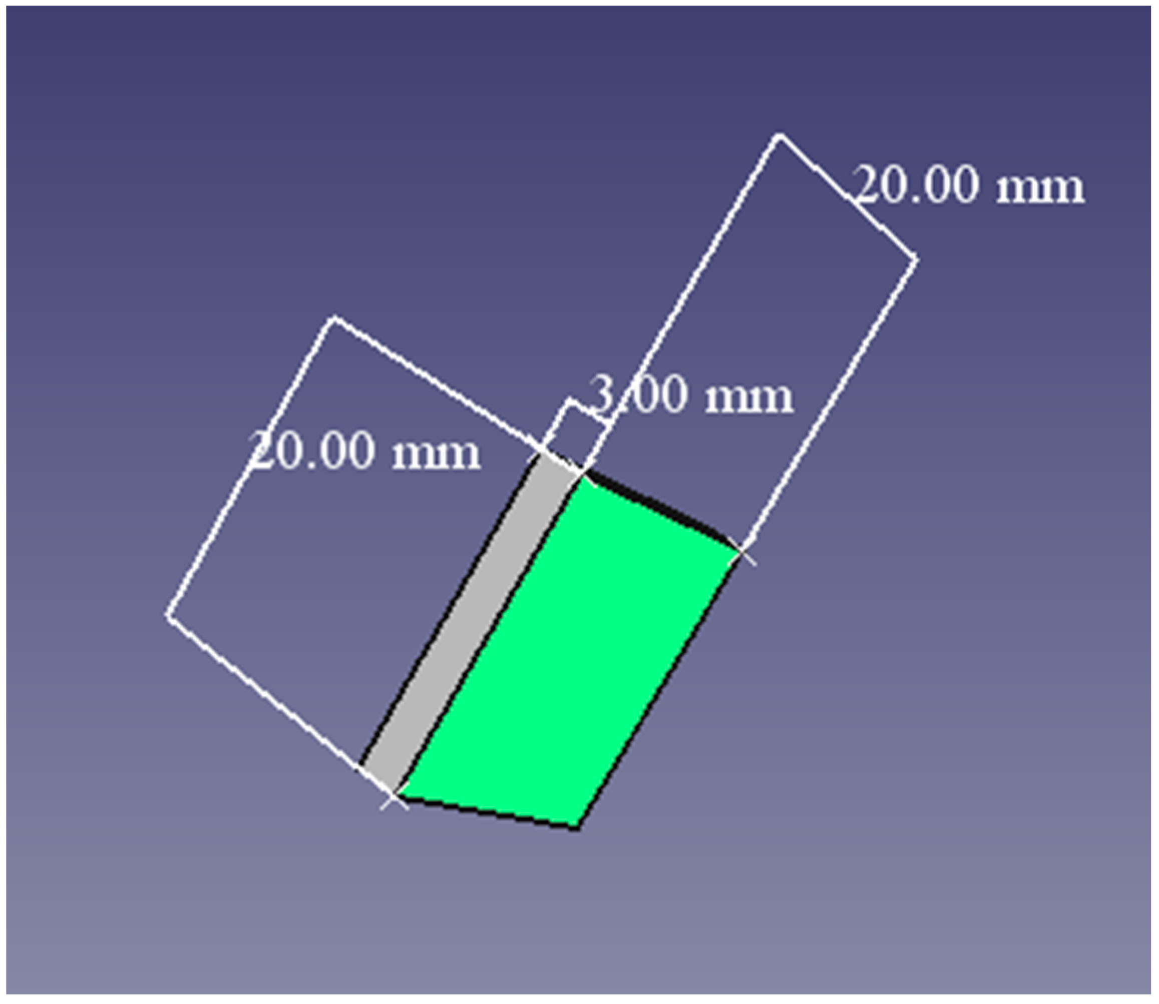
Figure 1. Graphic of the specimens’ design and dimensions.
Applsci 12 12560 g001
Figure 2. Process of printing the experimental samples in the 3D printer NextDent 5100 (NextDent, 3D Systems, Soesterberg, The Netherlands).
Applsci 12 12560 g002
Figure 3. Interactive plot for mean colour difference—interaction between type of material and immersion time, with thresholds of acceptability/perceptibility.
Applsci 12 12560 g003
Figure 4. Interactive plot for mean colour difference—interaction between type of material and staining medium, with thresholds of acceptability/perceptibility.
Applsci 12 12560 g004
Table 1. Types of materials used in the study.
ProductCompositionDetailManufacturer
NextDent Denture 3D+Methacrylate-based
photopolymerized
resin
3D printingNextDent, 3D Systems, Soesterberg, Netherlands
Vertex BasiQ 20PMMAHeat curingVertex Dental,
3D Systems, Soesterberg, Netherlands
Table 2. Scheme of the experiment organization—colour change values of the selected materials according to the different periods of time.
Type of MaterialPeriod of Time (T)Artificial Saliva
ΔE (Mean)
Coffee
ΔE (Mean)
Coca-Cola
ΔE (Mean)
Red Wine
ΔE (Mean)
SD
(Standard Deviation)
NextDent Denture 3D+7 days (T1)0.4982.1292.6763.0161.1160103
Vertex BasiQ0.5122.3282.8673.2681.2170435
NextDent Denture 3D+14 days (T2)0.4712.1942.7223.1631.1795063
Vertex BasiQ0.4842.5762.9663.5081.3227450
NextDent Denture 3D+21 days (T3)0.4622.2692.8753.3121.2535407
Vertex BasiQ 0.4572.7933.1583.8661.4764653
Table 3. Bonferroni post hoc test—multiple comparisons.
Multiple Comparisons
Dependent Variable; ΔE
Bonferroni Post-Hoc Test
(I) Period(J) PeriodMean Difference (I-J)Std. ErrorP95% Confidence Interval
Lower BoundUpper Bound
714−0.0987500.06857520.504−0.2808170.083317
21−0.237250 *0.06857520.009−0.419317−0.055183
1470.0987500.06857520.504−0.0833170.280817
21−0.1385000.06857520.178−0.3205670.043567
2170.237250 *0.06857520.0090.0551830.419317
140.1385000.06857520.178−0.0435670.320567
Based on observed means. The error term is Mean Square(Error) = 0.019. * The mean difference is significant at the 0.05 level.
Publisher’s Note: MDPI stays neutral with regard to jurisdictional claims in published maps and institutional affiliations.