Article

In Vivo and In Vitro Accuracy Analysis of Static Computer-Assisted Implant Surgery in an Edentulous Patient

1
Department of Oral and Maxillo-Facial Sciences, “Sapienza” University of Rome, Via Caserta 6, 00161 Rome, Italy
2
Private Practice, Via Roma 103, 84091 Battipaglia, Italy
*
Authors to whom correspondence should be addressed.
Appl. Sci. 2023, 13(2), 1185; https://doi.org/10.3390/app13021185
Submission received: 29 December 2022 / Revised: 9 January 2023 / Accepted: 12 January 2023 / Published: 16 January 2023
(This article belongs to the Special Issue Clinical Applications for Dentistry and Oral Health, 2nd Volume)

Abstract

:
Background: Recently, intraoral scanning (IOS) has been proposed as a new tool to evaluate the accuracy of static computer-assisted implant surgery (s-CAIS); however, further research is needed to improve the precision of IOS for full-arch impressions. The purpose of the study was to assess the accuracy of s-CAIS in an edentulous patient either in vivo or in vitro with two different evaluation techniques and to investigate if their results are comparable. Methods: A patient with terminal dentition was selected and four implants were placed using s-CAIS with a bone-supported stackable template. Segmentation used for designing a template was 3D printed, and then four implants were placed in the model following the same protocol as for s-CAIS. The model then underwent cone beam computed tomography (CBCT) and laboratory scanning to evaluate its accuracy. Data were uploaded to specific software, and accuracy values were automatically generated. Results: A statistical analysis was not attempted since all measurements were performed on the same patient and model. When descriptively comparing the accuracy of the two methods of treatment evaluation in the in vitro scenario, comparable results were obtained between IOS and CBCT, except for the angle. Conclusions: As the intraoral scanning procedure in fully edentulous patients is not yet clinically validated, utilizing CBCT can still be recommended for the accurate evaluation of computer-assisted implant placement.

1. Introduction

Digital workflow has been recently improved in dentistry, with the development of low-dose high-resolution cone beam computed tomography (CBCT), intraoral scanners (IOS), CAD/CAM software programs, 3D printers, and computer-assisted implant surgery (s-CAIS) systems [1,2]. Computer-assisted implant procedures are supposed to result in more precise implant positioning [3], and surgical templates might be fabricated in different ways with the same level of accuracy [4]. With the constant development of s-CAIS, several concerns have been raised about the reliability, accuracy, and precision of templates to replicate the planned implant position [5]. This is especially important in edentulous patients, where angulated implants should be placed in the exact planned position to allow the corresponding screw-retained abutments (SRA) axis to correspond to the prosthetically-oriented planning [6]. Although dental implants have proven to be reliable devices to replace missing teeth, they are, however, not free from mechanical or biological complications [7,8,9]. According to Bornstein et al. [10] and Tahmaseb et al. [11], mucosa-, tooth-, and mini-implant–supported templates demonstrate superior accuracy in implant placement to bone-supported guides. Carosi et al. state that computer-assisted flapless implant placement, using mucosa-supported templates in complete-arch restorations, can be considered a reliable and predictable treatment choice, despite the potential effects that a flapless approach could bring to the overall treatment [12]. Flapless and fully guided implant placement has the potential to maximize efficacy outcomes, and at the same time, minimize surgical invasiveness [13]. But in daily clinical practice, clinicians often face the necessity of bone reduction to place implants. Bone reduction may be required to level the alveolar crest to create the desired bone architecture, allowing for sufficient bone width for implant placement and ensuring adequate inter-arch restorative space [14]. Patients can rehabilitate full arches, even when bone reduction is mandatory because of a gummy simile or because of an irregular, or thin, bone crest [15]. The use of computer-guided surgery to rehabilitate edentulous arches by using four dental implants supporting a fixed screw-retained prosthesis could be a valid treatment option in the short and medium term [16]. When rehabilitating the edentulous jaw with four implants, the technique involves alveoloplasty before implant placement to provide space for the prosthetic components and to provide a platform on which dental implants can be placed in clinical situations where a knife-edge alveolar ridge is present [17]. As the aim of the present study was to evaluate the in vivo and in vitro accuracy of s-CAIS in an edentulous patient, the clinical situation was to be duplicated, and so the bone-supported template design was chosen. Tahmaseb et al., in the systematic review, state that several studies have provided information on the accuracy of s-CAIS, but a lack of homogeneity was found for accuracy assessment techniques and protocols: many different surgical factors and techniques were not standardized between the studies, which served to confound the true accuracy of guided surgery; there were many steps within the digital workflow itself, where there was a possibility of accumulating error, which also served to mask the real accuracy of the technique [5]. Most studies compare CBCT data before and after implant placement.
However, the lack of precision of CBCT machines represents a major issue. To date, around 279 CBCT devices exist in the market with different technical parameters, which makes it difficult to standardize scanning protocols [18]. According to Wanderley et al. [19], the visualization of implant dimensional alterations varied between different CBCT devices and scanning protocols with an increase in diameter ranging from 0.27 to 1.04 mm. Several factors relating to the patient and clinical situation, such as blooming, might tremendously affect the precision of CBCT scans. According to Pettersson et al. [20], the greatest errors were as a result of patient movements, therefore, alternative nonradiologic evaluation methods were proposed [21,22].
Bornstein et al. [10] recommended the use of digital impressions to evaluate the accuracy of computer-assisted implant placement. The use of a digital impression to evaluate the final position of an implant can avoid the need for a second CBCT [23]. In a dental practice, an accurate virtual model is vital for creating a template for precise dental implant placement and assessing treatment outcomes [24]. Hence, digital scanners obtain greater qualitative data compared to CBCT. Based on the study by Komuro et al. [25], it was noted that laboratory scanners had a mean shrinkage of 0.37–0.39%, while IOS and CBCT reported mean values of 0.9–1.4% and 1.8–6.9%, respectively. Hence, digital scanning demonstrates higher precision, seems to be less user-dependent, and reduces radiation exposure [26].
This study aimed to evaluate the in vivo and in vitro accuracy of s-CAIS in an edentulous patient with two different evaluation techniques, and investigate if both can be applied to edentulous patients.

2. Materials and Methods

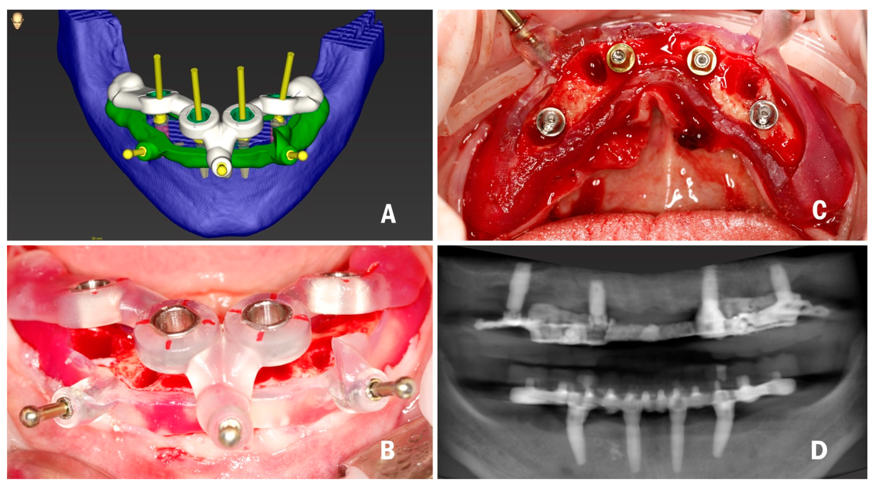
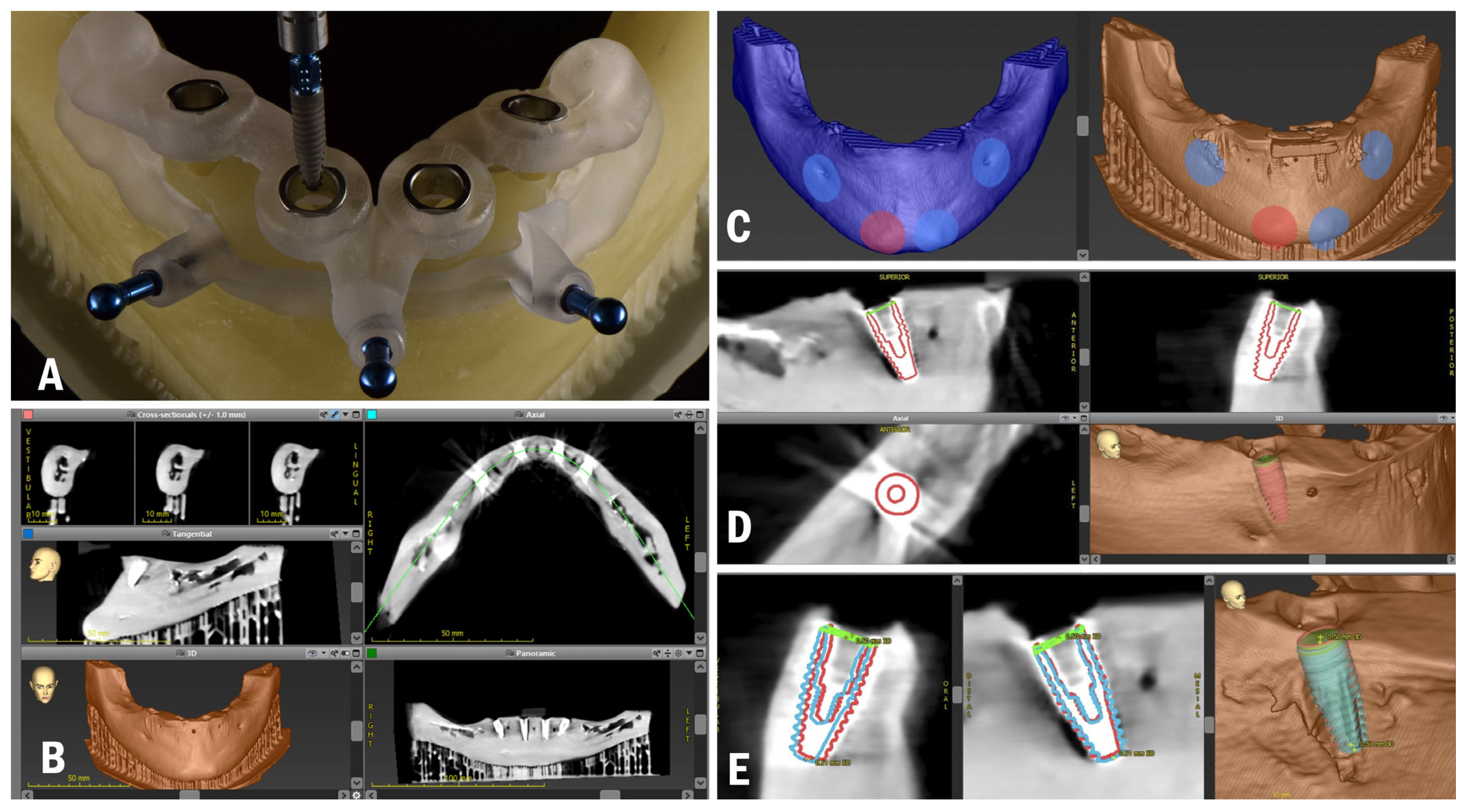
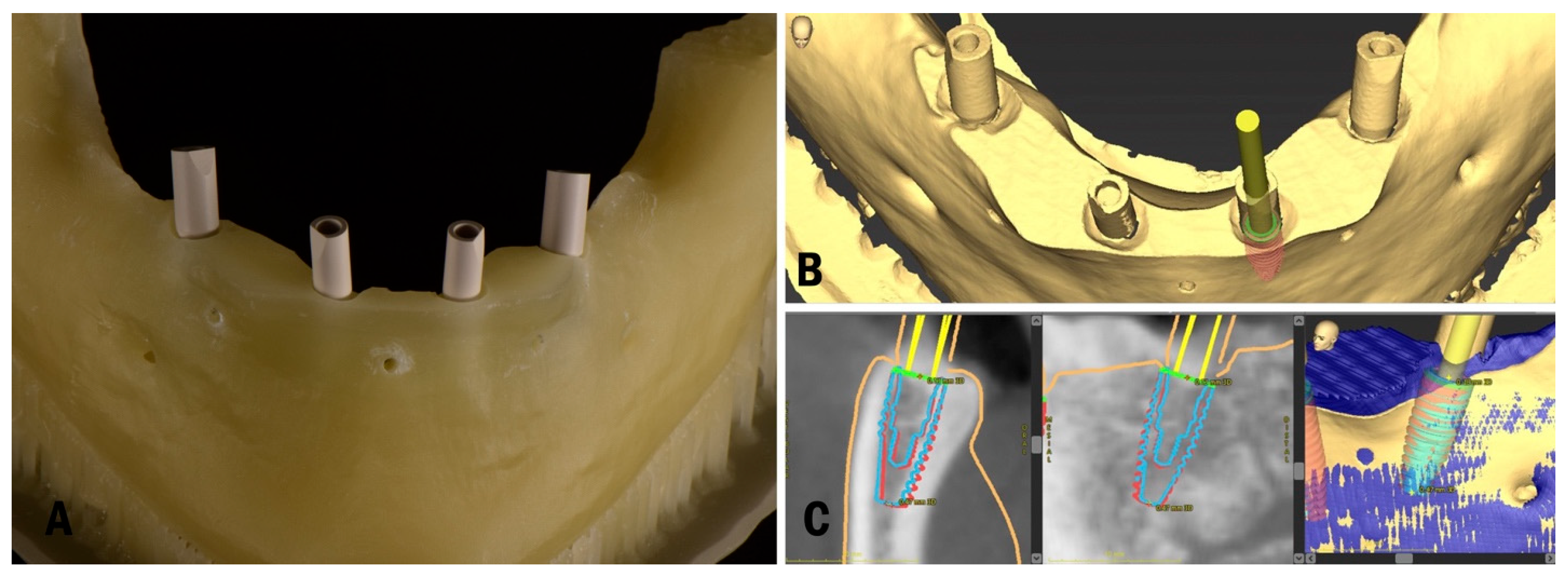
2.1. Planning and Surgery

To address the research purpose, the authors designed this study, which was conducted at the university clinic, and was approved by the Institution review board of the Department of Oral and Maxillo-Facial Sciences, “Sapienza” University of Rome (Ref: 038/2020). A 60-year-old male was selected to be included in this study, after attending the university clinic for implant treatment. The patient was in good health, displaying good oral hygiene (full mouth plaque score and full mouth bleeding score <25%), no need for guided bone regeneration procedures, an absence of uncontrolled systemic diseases, a nonsmoker, and no signs of acute local inflammation at the time of implant placement. He had a failing dentition (critical loss of attachment, nonrestorable teeth, fractured teeth, root caries) in the mandible, with four mobile incisors that could not be used as a support for a surgical template, which created difficulties for correct matching, and a very narrow ridge in the frontal area. These factors predetermined the stackable bone-supported surgical template design for this patient. Radiopaque 3D markers were attached to the existing prosthesis to also serve as a radiographic template prior to CBCT acquisition. The denture was further relined with soft material (GC Soft Liner) before the acquisition of the CBCT scan, thus avoiding possible prosthesis movement and for the precise transition of the actual soft tissue of the patient in the planning software. The first CBCT was performed on the patient with Green 16 (Vatech, Fort Lee, NJ, USA) in standard conditions (94  kVp, 8.0 mA, 360° rotation, 9 s, FOV: 100 x 85, voxel size: 200 μm) with the mouth closed, as separating the arches could cause radiological template movement. A protocol described by Storelli et al. [27] was implemented: after CBCT acquisition, an STL file of the denture was obtained using the laboratory scanner (Straumann 7 series lab scanner, Institut Straumann AG, Basel, Switzerland). Data obtained in digital imaging and communications in medicine (DICOM) format were then imported into a surgical planning software (coDiagnostiX, Institut Straumann AG). All of the acquisition parameters that could influence the accuracy of later steps in the generation of a virtual model, such as segmentation, as described by Shujaat et al. [28], were assessed. Data in standard tessellation language (STL) format were imported into the software to prosthetically orient implant positions. CBCT data of the patients in DICOM (Digital imaging and communications in medicine) format and STL files of radiographic templates of edentulous patients were matched in the surgical planning software. Four conical dental implants (BLT, Institut Straumann AG) with a sandblasted/long-grit/acid-etched surface (SLActive) were planned for prosthetically-oriented positions at 35, 32, 42, 45 sites: two 4.1 mm × 10 mm for the premolar sites and 3.3 mm × 12 mm for the incisor sites, in accordance with the Straumann Pro Arch protocol, which consists of dental implant and SRA (screw-retained abutment) placement with immediate loading. A bone-supported surgical template with a stackable design was chosen to increase the predictability of bone reduction [29], as the patient had a very narrow ridge coronally in the frontal area, which needed to be reduced to obtain the appropriate width for implant placement. The first part of the surgical template was bone-supported and was designed to guide bone reduction. It was designed on the base of the segmentation converted to the STL file. The template was stabilized to the bone with two lateral fixation pins. The second part of the surgical template was positioned on top of the bone reduction template and served in implant placement. Several techniques to connect two different surgical templates are described in the literature, such as the use of magnets [30]. In the present study, the authors describe a more cost- and time-efficient technique. The second part of the surgical template was fixed to the first template with the locks designed on both using the inspection window tool, and the third lateral fixation pin was stabilized either to the first part of the stent or to the bone (Figure 1A).
The virtual bone reduction was then performed in the software and a segmentation of the residual bone was created. To minimize factors relating to 3D printing that could affect accuracies, such as template-to-teeth offset or sleeve-to-template offset, the calibration matrix was 3D printed and the 3D printer and printing material were calibrated. The surgical template was then three-dimensionally (3D) printed.
One hour before surgery, prophylactic antibiotics, 2 gr of amoxicillin (Zimox, Pfizer), were given to the patient. The flap was raised, and the first template was positioned; osteotomies for pins were made and the template was fixed with lateral fixation pins. The bone reduction was performed with the piezoelectric instrument VarioSurg (NSK/Nakanishi, Japan). The second template for implant placement was positioned on top of the first one and fixed with the third pin. Osteotomies were performed through the surgical template following the protocol derived from the software (Figure 1B). Then, the dental implants were inserted by properly following the manufacturer’s instructions and the surgical insertion protocol. All implants reached a minimum insertion torque of 35 Ncm, so the SRA were tightened to the implants at 35 Ncm with a surgical motor with torque control (SurgicPro+, NSK/Nakanishi, Japan) (Figure 1C). Impressions were made with polyvinyl siloxane (Elite, Zhermack), the prosthesis was finalized in the laboratory, polished, and immediate loading was performed. The prosthesis was delivered to the patient after occlusion checking, tightening the screws at 15 Ncm, and sealing the screw access holes with polytetrafluoroethylene (PTFE) tape and a flowable composite resin. After 3 months, the temporary prosthesis was unscrewed for the first time to check implants osteointegration, which was uneventful and successful. The patient received a screw-retained titanium bar with resin veneering as the definitive prosthesis. The actual follow-up period is set for 3 years.

2.2. Time and Cost Analysis

According to the 6th EAO Consensus Conference, scientific evidence of the time and cost involved with computer-assisted implant planning and surgery protocols is rare, and data are reported as heterogenetic [3]. In the present study, the time and cost analyses are as follows:
Time analysis
  • Duration of planning (min) 30 min
  • Duration of surgery (min) 45 min
Cost analysis
  • Preoperative fee: cost for the CBCT (€120).
  • Surgical fee: cost for implants, their surgical installation, and the clinician’s fee (€1000 per implant).
  • Prosthetic fee: cost for all prosthetic components, their installation, the dental laboratory fee, and the clinician’s fee (€1500 for immediate loading and €4500 for definitive prosthesis).
  • Guide fee: cost of the specific surgical template (€400) and extra components (€20 per sleeve).

3. Results

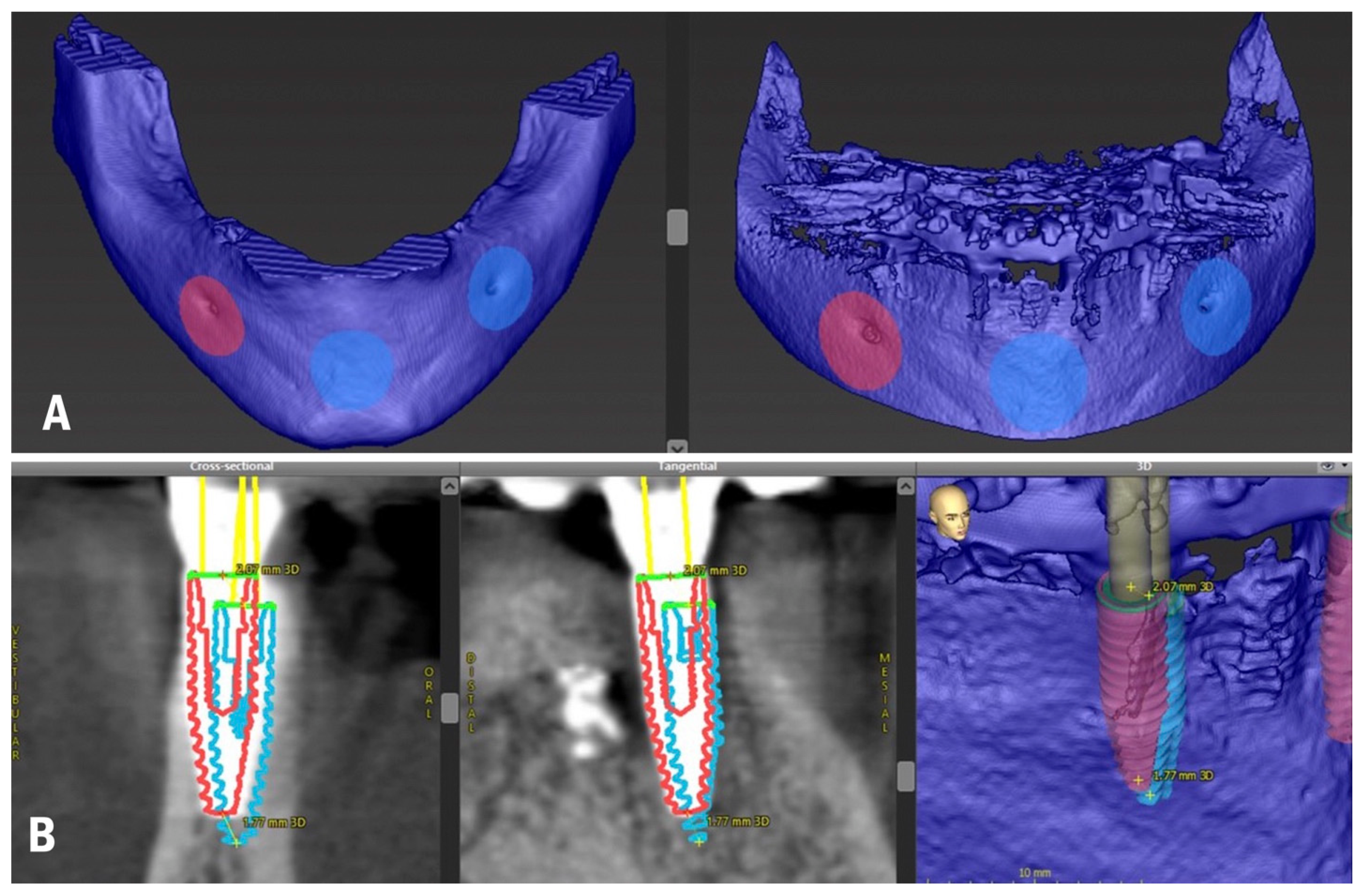
3.1. In Vivo Treatment Evaluation

For the in vivo evaluation of the DICOM-to-DICOM matching protocol, the accuracy assessment technique was applied, which consists of evaluating the matching of the planning dataset with the surgery results dataset. The patient received a second CBCT scan after the implant placement and delivery of the final prosthesis to perform an accuracy analysis (Figure 1D). The same CBCT device was used to obtain more qualitative data since the registration accuracy of implant planning software is significantly influenced by the preprocessing of imported data [31]. The area of interest (mandible) was segmented from the CBCT scan, which allowed for more qualitative matching. Data in DICOM format were inserted into the treatment evaluation tool of coDiagnostiX, and the matching of the preoperative and postoperative CBCT scans was performed (Figure 2A).
The limitation of the DICOM-to-DICOM protocol might be the discrepancy between the CBCT scans. To minimize this influence, the same CBCT device was used, and matching was performed in multiple areas with clearly visible anatomical structures following the recommended protocol of the treatment evaluation tool. Each implant was aligned in the preoperative and postoperative CBCT, and accuracy values were automatically generated by the software (Figure 2B). The 3D, distal, vestibular, and apical deviations were recorded on the coronal (base) and apical (tip) points of the implants, as well as angular deviations. The results are reported in Table 1.

3.2. In Vitro Treatment Evaluation

In the in vitro evaluation, it was possible to apply both DICOM-to-DICOM and DICOM-to-STL matching protocols of the accuracy assessment techniques. Segmentation used for designing the bone-supported template was converted into an STL file and 3D printed with a Phrozen Sonic 4K printer, utilizing a mixture of a radiopaque polymer Dental RO (HARZ Labs) (60%) and transparent polymer Dental Clear (HARZ Labs) (40%) to reproduce the mandible not fully radiopaque and of a similar bone density. Four implants, identical to those placed in vivo, were inserted into the 3D-printed model of the mandible. The same protocol for s-CAIS and the same surgical template was used (Figure 3A).
A CBCT of the model was then performed (Figure 3B) followed by its matching with the preoperative CBCT (Figure 3C) to obtain data for the accuracy analysis of implant placement. Each implant was aligned in the preoperative and postoperative CBCT (Figure 3D). Accuracy values were automatically generated by the software (Figure 3E) and the results are reported in Table 2.
Four scan bodies were tightened onto the implants inserted in the model (Figure 4A), and a lab scanner (Straumann 7 series lab scanner, Institut Straumann AG) was used to obtain data in STL format.
The STL file with the scan bodies was uploaded to the software and matched with the preoperative planning CBCT (Figure 4B). Each implant was matched in the preoperative CBCT and STL file by simply clicking on the top of the corresponding scan body. The matching was performed automatically compared to the CBCT method, otherwise, where it was performed manually by matching the anatomy of the implant, it became a user-dependent technique. Accuracy values were automatically generated by the software (Figure 4C) and the results are reported in Table 3.

4. Discussion

This study aimed to evaluate the in vivo and in vitro accuracy of s-CAIS in an edentulous patient using two different evaluation techniques and to investigate if both can be applied to edentulous patients.
When evaluating the accuracy of s-CAIS, the most assessed parameters are angular deviation, and deviation at the entry point and the apex of the implant. In the clinical part of the experiment, a maximum angular deviation of 5.3° was noticed at implant site 32, while in the in vitro CBCT evaluation, the maximum angle of deviation was 4° at implant site 42, and 5.1° at the same site with the STL assessment. Deviations between the planned and achieved implant positions should be expected because of inaccuracies in the various procedures during template design and manufacturing [32,33]. At the same time, the use of such an individualized template allows appropriate bone reduction for obtaining a predictable surgery and prosthetic stage [34].
The limitations of the study were related to the fact that during implant placement, several clinical factors can affect the accuracy of final treatment and some of them (different bone density guiding the osteotomy, different possible fit of a bone-supported template, stability, and fixation of the template) are missing in the in vitro part of the experiment. In this clinical report, the treatment evaluation was affected by the DICOM-to-DICOM matching procedure, as well as possible errors during CBCT acquisition. On the contrary, in the in vitro part of the study, dental implants seemed to be placed more accurately with the initial planning.
However, several inaccuracies might also occur in the in vitro analysis due to several factors relating to data acquisition and the subsequent 3D printing of the model. According to Zhou et al. [33], different types of errors might arise during image acquisition and data processing. There is a correlation between the dataset volume extracted from the CBCT and the CBCT resolution, as reported by Schnutenhaus [35]. Evaluating the results, the explanation for this is that when the CBCTs were created with a resolution of 0.2 voxels, a matching precision greater than 0.2 mm is not achievable. This error might have been doubled when printing the exported DICOM dataset of the mandible. Hence, volume shrinkage could be noticed at the matching stage. According to Shujaat et al. [36], there is also some inaccuracy due to 3D printing procedures. The 3D printer used in the present study had a precision of 35 µm, according to the manufacturer. Several parameters must be considered before choosing a 3D printer, such as the processing software, the type of 3D printer, mechanical characteristics, as well as properties of the printing material [37]. In fact, several factors were controlled as best as possible by standardizing the treatment planning and production parameters to increase the accuracy of the 3D printing so that they had a minor overall effect on the results.
The other issue to consider is the difference in bone density, which appeared to be denser than the real bone, and this was related to the use of radiopaque resin. In fact, adequate bone density was achieved by utilizing a mixture of radiopaque and transparent polymers. Additionally, when performing a CBCT scan of the printed model, a higher blooming effect was noticed around the implants compared to implants placed in the patient, which could also affect the implant matching procedure. Presently, no clinical evidence exists which relates to the optimal CBCT scanning settings for reducing the influence of metal artifacts [38].
IOS procedures today are widely integrated with s-CAIS. Implant rehabilitations planned utilizing intraoral digital impressions showed similar results when compared to conventional impressions and model scans. A digital impression may be a viable option for the rehabilitation of partially edentulous patients when computer-guided template-assisted implant placement is used [39]. The application of IOS in edentulous patients is currently still limited. Wismeijer et al. [40] stated that the accuracy of digital impressions is negatively influenced by an increase in the inter-implant distance in edentulous situations. When considering the accuracy of full-arch impressions for long-span prostheses, conventional impression methods remain the gold standard, since the IOS error rises with the increase in the edentulous scanning area [41]. Further research needs to be conducted to improve the precision and trueness of IOS for full-arch scanning [28]. For this reason, IOS was not used in the clinical part of the study and cannot be recommended yet for clinical use with edentulous patients. Furthermore, Komuro et al. [25] recommended the use of laboratory scanners as they provide more precise data compared to IOS. The STL file exported from the segmentation and STL file of the printed and scanned mandible with implants and scan body inserted were imported into Geomagic Studio 2013 (3D Systems, USA) (an open-source software for processing, editing, and comparing 3D triangular meshes) and aligned using the fine alignment algorithm (Figure 5).
As described by Nulty [42], this kind of software allows for the generation of a colorimetric map of the deviation across the surface of the STL mesh compared to the master STL, quantified at specific points. The colour map indicates deviation inward (blue) or outward (red), while green indicates minimal deviation. The same colour deviation scale was utilized to illustrate the minimum and maximum deviations for each comparison. The colour scale ranged from a maximum and minimum deviation of +200 (outward/red) to −200 μm (inward/blue). In the present study, the overall trueness was 27 μm. Thus, the accuracy of the 3D printing did not seem to significantly affect the results.
In a study comparing two treatment evaluation methods available using the same surgical planning software, Skjerven et al. [43] stated that accuracy measurements performed via IOS provided comparable results to those obtained by CBCT. However, in the abovementioned study, tooth-supported templates were used, which are more precise than mucosa- or bone-supported templates in edentulous patients [44], and IOS in partially edentulous patients in this study could be performed. Hence, single-unit crowns were manufactured based on IOS; multiple implants in fixed partial denture reconstructions were rehabilitated based on conventional impressions. To the best of the authors’ knowledge, there are no studies in the literature reporting data comparing IOS-to-DICOM and DICOM-to-DICOM treatment evaluation methods in edentulous patients.
Statistical analysis was not attempted since all measurements were performed with the same patient and model. However, when descriptively comparing the accuracy of the two methods of treatment evaluation in the in vitro scenario, we obtained comparable results, except for the angle.
For future research, it would be beneficial to access the data from the dynamic navigation of edentulous patients. Dynamic systems discussed in the Jung et al. [45] systematic review provided greater accuracy than static systems; this difference might be explained by the fact that static template-based systems are more often used clinically rather than in preclinical models, which have provided better accuracy. According to Wei et al. [46] the accuracy of dynamic computer-aided implant surgery reaches a clinically acceptable range and has potential for clinical usage, but more patient-centered outcomes and socioeconomic benefits should be reported. As for today, the scientific data on dynamic navigation in literature is scarce. Few studies are reporting on dynamic navigation and its application in edentulous patients [47]. Future research should be oriented towards utilizing dynamic navigation surgery in edentulism and reporting data for its accuracy.

5. Conclusions

When descriptively comparing the accuracy of the two methods of treatment evaluation in the in vitro scenario, comparable results were obtained between IOS and CBCT, except for the angle.
As intraoral scanning procedures in fully edentulous patients are not yet clinically validated, utilizing CBCT can still be recommended for the accurate evaluation of computer-assisted implant placement.

Author Contributions

Conceptualization, N.M.; methodology, N.M.; software, N.M. and V.S.; validation, G.D.C. and M.S.; formal analysis, B.D.M.; investigation, N.M.; resources, N.M.; data curation, N.M.; writing—original draft preparation, N.M.; writing—review and editing, P.P.; visualization, N.M.; supervision, A.P. and M.S.; project administration, M.S.; funding acquisition, N.M. All authors have read and agreed to the published version of the manuscript.

Funding

This research received no external funding.

Institutional Review Board Statement

All procedures performed in studies involving human participants were in accordance with the ethical standards of the institutional and/or national research committee and with the 1964 Helsinki declaration and its later amendments or comparable ethical standards. The study was approved by the Institution Review Board (IRB) of the Department of Oral and Maxillo-Facial Sciences, at “Sapienza” University of Rome (Ref. 038/2020).

Informed Consent Statement

The patient received detailed descriptions of the study protocol, signed the informed consent form, and gave written approval to publish his personal data.

Data Availability Statement

The datasets used and/or analyzed during the current study are available from the corresponding author upon reasonable request.

Acknowledgments

The authors thank Straumann for providing implants for the in vitro part of the study. The authors thank Sergey Yumatov for 3D printing support and Alexey Ryabov for data collection.

Conflicts of Interest

The authors declare no conflict of interest.

Abbreviations

s-CAIS: static computer-aided implant surgery; CAD/CAM: computer-aided design and computer-aided manufacturing; CBCT: cone beam computer tomography; STL: standard tessellation language; DICOM: digital imaging and communications in medicine; SRA: screw-retained abutments; SLActive: sandblasted/long-grit/acid-etched active; 3D: three-dimensional; IOS: intraoral scanning.

References

  1. Mangano, C.; Luongo, F.; Migliario, M.; Mortellaro, C.; Mangano, F.G. Combining Intraoral Scans, Cone Beam Computed Tomography and Face Scans: The Virtual Patient. J. Craniofacial Surg. 2018, 29, 2241–2246. [Google Scholar] [CrossRef] [PubMed]
  2. Papi, P.; Di Murro, B.; Penna, D.; Pompa, G. Digital prosthetic workflow during COVID-19 pandemic to limit infection risk in dental practice. Oral Dis. 2021, 27 (Suppl. S3), 723–726. [Google Scholar] [CrossRef] [PubMed]
  3. Jokstad, A.; Pjetursson, B.E.; Muhlemann, S.; Wismeijer, D.; Wolfart, S.; Fehmer, V.; Guth, J.F.; Holtzman, L.P.; Hammerle, C.H.F.; Makarov, N.; et al. Fabrication, workflow and delivery of reconstruction: Summary and consensus statements of group 4. The 6th EAO Consensus Conference 2021. Clin. Oral Implant. Res. 2021, 32 (Suppl. S21), 336–341. [Google Scholar] [CrossRef] [PubMed]
  4. Henprasert, P.; Dawson, D.V.; El-Kerdani, T.; Song, X.; Couso-Queiruga, E.; Holloway, J.A. Comparison of the Accuracy of Implant Position Using Surgical Guides Fabricated by Additive and Subtractive Techniques. J. Prosthodont. 2020, 29, 534–541. [Google Scholar] [CrossRef] [PubMed] [Green Version]
  5. Tahmaseb, A.; Wu, V.; Wismeijer, D.; Coucke, W.; Evans, C. The accuracy of static computer-aided implant surgery: A systematic review and meta-analysis. Clin. Oral Implant. Res. 2018, 29 (Suppl. S16), 416–435. [Google Scholar] [CrossRef] [Green Version]
  6. Makarov, N.; Pompa, G.; Papi, P. Computer-assisted implant placement and full-arch immediate loading with digitally prefabricated provisional prostheses without cast: A prospective pilot cohort study. Int. J. Implant. Dent. 2021, 7, 80. [Google Scholar] [CrossRef]
  7. Di Murro, B.; Papi, P.; Passarelli, P.C.; D’Addona, A.; Pompa, G. Attitude in Radiographic Post-Operative Assessment of Dental Implants among Italian Dentists: A Cross-Sectional Survey. Antibiotics 2020, 9, 234. [Google Scholar] [CrossRef]
  8. Papi, P.; Di Murro, B.; Pranno, N.; Bisogni, V.; Saracino, V.; Letizia, C.; Polimeni, A.; Pompa, G. Prevalence of peri-implant diseases among an Italian population of patients with metabolic syndrome: A cross-sectional study. J. Periodontol. 2019, 90, 1374–1382. [Google Scholar] [CrossRef]
  9. Di Murro, B.; Pranno, N.; Raco, A.; Pistilli, R.; Pompa, G.; Papi, P. Knowledge and Attitude towards Retrograde Peri-Implantitis among Italian Implantologists: A Cross-Sectional Survey. Int. J. Environ. Res. Public Health 2020, 17, 8356. [Google Scholar] [CrossRef]
  10. Bornstein, M.M.; Al-Nawas, B.; Kuchler, U.; Tahmaseb, A. Consensus statements and recommended clinical procedures regarding contemporary surgical and radiographic techniques in implant dentistry. Int. J. Oral Maxillofac. Implant. 2014, 29, 78–82. [Google Scholar] [CrossRef]
  11. Tahmaseb, A.; Wismeijer, D.; Coucke, W.; Derksen, W. Computer technology applications in surgical implant dentistry: A systematic review. Int. J. Oral Maxillofac. Implant. 2014, 29, 25–42. [Google Scholar] [CrossRef] [Green Version]
  12. Carosi, P.; Lorenzi, C.; Lio, F.; Cardelli, P.; Pinto, A.; Laureti, A.; Pozzi, A. Accuracy of Computer-Assisted Flapless Implant Placement by Means of Mucosa-Supported Templates in Complete-Arch Restorations: A Systematic Review. Materials 2022, 15, 1462. [Google Scholar] [CrossRef]
  13. Romandini, M.; Ruales-Carrera, E.; Sadilina, S.; Hammerle, C.H.F.; Sanz, M. Minimal invasiveness at dental implant placement: A systematic review with meta-analyses on flapless fully guided surgery. Periodontol. 2000 2022. [Google Scholar] [CrossRef]
  14. Mijiritsky, E.; Ben Zaken, H.; Shacham, M.; Cinar, I.C.; Tore, C.; Nagy, K.; Ganz, S.D. Variety of Surgical Guides and Protocols for Bone Reduction Prior to Implant Placement: A Narrative Review. Int. J. Environ. Res. Public Health 2021, 18, 2341. [Google Scholar] [CrossRef]
  15. Tonellini, G.; Saez Vigo, R.; Novelli, G. Double Guided Surgery in All-on-4((R)) Concept: When Ostectomy Is Needed. Int. J. Dent. 2018, 2018, 2672549. [Google Scholar] [CrossRef] [Green Version]
  16. Carosi, P.; Ferrigno, N.; Arcuri, C.; Laureti, M. Computer-Aided Surgery and Immediate Loading to Rehabilitate Complete Arch with Four Dental Implants and Fixed Screw-Retained Prosthesis Up to 4 Years in Function: A Retrospective Study. Int. J. Oral Maxillofac. Implant. 2021, 36, 1180–1187. [Google Scholar] [CrossRef]
  17. Faeghi Nejad, M.; Proussaefs, P.; Lozada, J. Combining guided alveolar ridge reduction and guided implant placement for all-on-4 surgery: A clinical report. J. Prosthet. Dent. 2016, 115, 662–667. [Google Scholar] [CrossRef]
  18. Gaeta-Araujo, H.; Alzoubi, T.; Vasconcelos, K.F.; Orhan, K.; Pauwels, R.; Casselman, J.W.; Jacobs, R. Cone beam computed tomography in dentomaxillofacial radiology: A two-decade overview. Dento Maxillo Facial Radiol. 2020, 49, 20200145. [Google Scholar] [CrossRef]
  19. Wanderley, V.A.; de Faria Vasconcelos, K.; Leite, A.F.; Pauwels, R.; Shujaat, S.; Jacobs, R.; Oliveira, M.L. Impact of the blooming artefact on dental implant dimensions in 13 cone-beam computed tomography devices. Int. J. Implant. Dent. 2021, 7, 67. [Google Scholar] [CrossRef]
  20. Pettersson, A.; Komiyama, A.; Hultin, M.; Nasstrom, K.; Klinge, B. Accuracy of virtually planned and template guided implant surgery on edentate patients. Clin. Implant. Dent. Relat. Res. 2012, 14, 527–537. [Google Scholar] [CrossRef]
  21. Schnutenhaus, S.; Edelmann, C.; Rudolph, H.; Luthardt, R.G. Retrospective study to determine the accuracy of template-guided implant placement using a novel nonradiologic evaluation method. Oral Surg. Oral Med. Oral Pathol. Oral Radiol. 2016, 121, e72–e79. [Google Scholar] [CrossRef] [PubMed]
  22. Nickenig, H.J.; Eitner, S. An alternative method to match planned and achieved positions of implants, after virtual planning using cone-beam CT data and surgical guide templates--a method reducing patient radiation exposure (part I). J. Cranio-Maxillofac. Surg. 2010, 38, 436–440. [Google Scholar] [CrossRef] [PubMed]
  23. Monaco, C.; Arena, A.; Corsaletti, L.; Santomauro, V.; Venezia, P.; Cavalcanti, R.; Di Fiore, A.; Zucchelli, G. 2D/3D accuracies of implant position after guided surgery using different surgical protocols: A retrospective study. J. Prosthodont. Res. 2020, 64, 424–430. [Google Scholar] [CrossRef] [PubMed]
  24. Vandenberghe, B. The digital patient—Imaging science in dentistry. J. Dent. 2018, 74 (Suppl. S1), S21–S26. [Google Scholar] [CrossRef] [PubMed]
  25. Komuro, A.; Yamada, Y.; Uesugi, S.; Terashima, H.; Kimura, M.; Kishimoto, H.; Iida, T.; Sakamoto, K.; Okuda, K.; Kusano, K.; et al. Accuracy and dimensional reproducibility by model scanning, intraoral scanning, and CBCT imaging for digital implant dentistry. Int. J. Implant. Dent. 2021, 7, 63. [Google Scholar] [CrossRef]
  26. Son, K.; Huang, M.Y.; Lee, K.B. A method to evaluate the accuracy of dental implant placement without postoperative radiography after computer-guided implant surgery: A dental technique. J. Prosthet. Dent. 2020, 123, 661–666. [Google Scholar] [CrossRef]
  27. Storelli, S.; Palandrani, G.; Amorfini, L.; Scanferla, M.; Ausenda, F.; Romeo, E. A digital workflow for computer-guided implant surgery integrating CBCT, model scanning, and CAD/CAM for a complete edentulism implant-supported prosthesis: A technique procedure. Int. J. Comput. Dent. 2020, 23, 269–279. [Google Scholar]
  28. Shujaat, S.; Bornstein, M.M.; Price, J.B.; Jacobs, R. Integration of imaging modalities in digital dental workflows—Possibilities, limitations, and potential future developments. Dento Maxillo Facial Radiol. 2021, 50, 20210268. [Google Scholar] [CrossRef]
  29. Yang, J.W.; Liu, Q.; Yue, Z.G.; Hou, J.X.; Afrashtehfar, K.I. Digital Workflow for Full-Arch Immediate Implant Placement Using a Stackable Surgical Guide Fabricated Using SLM Technology. J. Prosthodont. 2021, 30, 645–650. [Google Scholar] [CrossRef]
  30. Costa, A.J.M.; Teixeira Neto, A.D.; Burgoa, S.; Gutierrez, V.; Cortes, A.R.G. Fully Digital Workflow with Magnetically Connected Guides for Full-Arch Implant Rehabilitation Following Guided Alveolar Ridge Reduction. J. Prosthodont. 2020, 29, 272–276. [Google Scholar] [CrossRef]
  31. Flugge, T.; Derksen, W.; Te Poel, J.; Hassan, B.; Nelson, K.; Wismeijer, D. Registration of cone beam computed tomography data and intraoral surface scans—A prerequisite for guided implant surgery with CAD/CAM drilling guides. Clin. Oral Implant. Res. 2017, 28, 1113–1118. [Google Scholar] [CrossRef]
  32. Cassetta, M.; Di Mambro, A.; Giansanti, M.; Stefanelli, L.V.; Cavallini, C. The intrinsic error of a stereolithographic surgical template in implant guided surgery. Int. J. Oral Maxillofac. Surg. 2013, 42, 264–275. [Google Scholar] [CrossRef]
  33. Zhou, W.; Liu, Z.; Song, L.; Kuo, C.L.; Shafer, D.M. Clinical Factors Affecting the Accuracy of Guided Implant Surgery—A Systematic Review and Meta-analysis. J. Evid.-Based Dent. Pract. 2018, 18, 28–40. [Google Scholar] [CrossRef]
  34. Barteaux, L.; Daelemans, P.; Malevez, C. A surgical stent for the Branemark Novum bone reduction procedure. Clin. Implant. Dent. Relat. Res. 2004, 6, 210–221. [Google Scholar] [CrossRef]
  35. Schnutenhaus, S.; Groller, S.; Luthardt, R.G.; Rudolph, H. Accuracy of the match between cone beam computed tomography and model scan data in template-guided implant planning: A prospective controlled clinical study. Clin. Implant. Dent. Relat. Res. 2018, 20, 541–549. [Google Scholar] [CrossRef]
  36. Shujaat, S.; Shaheen, E.; Novillo, F.; Politis, C.; Jacobs, R. Accuracy of cone beam computed tomography-derived casts: A comparative study. J. Prosthet. Dent. 2021, 125, 95–102. [Google Scholar] [CrossRef]
  37. Meglioli, M.; Naveau, A.; Macaluso, G.M.; Catros, S. 3D printed bone models in oral and cranio-maxillofacial surgery: A systematic review. 3D Print. Med. 2020, 6, 30. [Google Scholar] [CrossRef]
  38. Nomura, Y.; Watanabe, H.; Manila, N.G.; Asai, S.; Kurabayashi, T. Evaluation of streak metal artifacts in cone beam computed tomography by using the Gumbel distribution: A phantom study. Oral Surg. Oral Med. Oral Pathol. Oral Radiol. 2021, 131, 494–502. [Google Scholar] [CrossRef]
  39. Tallarico, M.; Xhanari, E.; Kim, Y.J.; Cocchi, F.; Martinolli, M.; Alushi, A.; Baldoni, E.E.; Meloni, S.M. Accuracy of computer-assisted template-based implant placement using conventional impression and scan model or intraoral digital impression: A randomised controlled trial with 1 year of follow-up. Int. J. Oral Implantol. (Berl.) 2019, 12, 197–206. [Google Scholar]
  40. Wismeijer, D.; Joda, T.; Flugge, T.; Fokas, G.; Tahmaseb, A.; Bechelli, D.; Bohner, L.; Bornstein, M.; Burgoyne, A.; Caram, S.; et al. Group 5 ITI Consensus Report: Digital technologies. Clin. Oral Implant. Res. 2018, 29 (Suppl. S16), 436–442. [Google Scholar] [CrossRef] [Green Version]
  41. Suese, K. Progress in digital dentistry: The practical use of intraoral scanners. Dent. Mater. J. 2020, 39, 52–56. [Google Scholar] [CrossRef] [Green Version]
  42. Nulty, A.B. A Comparison of Full Arch Trueness and Precision of Nine Intra-Oral Digital Scanners and Four Lab Digital Scanners. Dent. J. 2021, 9, 75. [Google Scholar] [CrossRef] [PubMed]
  43. Skjerven, H.; Olsen-Bergem, H.; Ronold, H.J.; Riis, U.H.; Ellingsen, J.E. Comparison of postoperative intraoral scan versus cone beam computerised tomography to measure accuracy of guided implant placement—A prospective clinical study. Clin. Oral Implant. Res. 2019, 30, 531–541. [Google Scholar] [CrossRef] [PubMed]
  44. Arisan, V.; Karabuda, Z.C.; Ozdemir, T. Accuracy of two stereolithographic guide systems for computer-aided implant placement: A computed tomography-based clinical comparative study. J. Periodontol. 2010, 81, 43–51. [Google Scholar] [CrossRef] [PubMed]
  45. Jung, R.E.; Schneider, D.; Ganeles, J.; Wismeijer, D.; Zwahlen, M.; Hammerle, C.H.; Tahmaseb, A. Computer technology applications in surgical implant dentistry: A systematic review. Int. J. Oral Maxillofac. Implant. 2009, 24, 92–109. [Google Scholar]
  46. Wei, S.M.; Zhu, Y.; Wei, J.X.; Zhang, C.N.; Shi, J.Y.; Lai, H.C. Accuracy of dynamic navigation in implant surgery: A systematic review and meta-analysis. Clin. Oral Implant. Res. 2021, 32, 383–393. [Google Scholar] [CrossRef]
  47. Pozzi, A.; Hansson, L.; Carosi, P.; Arcuri, L. Dynamic navigation guided surgery and prosthetics for immediate loading of complete-arch restoration. J. Esthet. Restor. Dent. 2021, 33, 224–236. [Google Scholar] [CrossRef]
Figure 1. (A) Computer-assisted implant planning; (B) Implant placement surgery; (C) Implants and SRA in situ; and (D) Postoperative CBCT.
Applsci 13 01185 g001
Figure 2. (A) Planning and postoperative CBCT matching; (B) Accuracy analysis with postoperative CBCT (blue colour—planned implant position, red colour—actual implant position).
Applsci 13 01185 g002
Figure 3. (A) In vitro implant placement; (B) CBCT of a 3D-printed mandible with inserted implants; (C) Planning and 3D-printed model CBCT matching; (D) Matching of implants in planning and implants placed in vitro; and (E) Accuracy analysis with the CBCT of the model.
Applsci 13 01185 g003
Figure 4. (A) Scan bodies mounted on the implants placed in vitro; (B) Planning CBCT and postoperative STL matching; (C) Accuracy analysis between STL file and scan bodies.
Applsci 13 01185 g004
Figure 5. Original STL file exported from segmentation superimposed with the STL file of the printed and scanned mandible with implants and scan body inserted.
Applsci 13 01185 g005
Table 1. Accuracy analysis with postoperative CBCT.
Tooth PositionAngle3D Offset (Base)Distal (Base)Vestibular (Base)Apical (Base)3D Offset (Tip)Distal (Tip)Vestibular (Tip)Apical (Tip)
352.42.55−2.140.93−1.032.21−1.730.93−1.02
325.32.61−1.860.61−1.723.47−2.980.62−1.67
422.32.070.791.11−1.551.770.50.72−1.54
452.81.320.491.23−0.021.010.650.78−0.01
Table 2. Accuracy analysis with postoperative CBCT of the model.
Tooth PositionAngle3D Offset (Base)Distal (Base)Vestibular (Base)Apical (Base)3D Offset (Tip)Distal (Tip)Vestibular (Tip)Apical (Tip)
3500.220.2200.060.220.2200.06
321.60.790.780.130.090.810.78−0.190.1
4240.74−0.39−0.630.031.370−1.370.06
451.60.5−0.11−0.38−0.30.510.17−0.38−0.3
Table 3. Accuracy analysis with STL file of the printed and scanned mandible with implants and scan body inserted.
Tooth PositionAngle3D Offset (Base)Distal (Base)Vestibular (Base)Apical (Base)3D Offset (Tip)Distal (Tip)Vestibular (Tip)Apical (Tip)
353.10.13−0.070.080.060.47−0.02−0.460.08
323.20.710.170.380.570.830.55−0.180.59
425.10.62−0.350.060.511.10−0.950.55
454.10.4−0.180.080.350.640.43−0.280.38
Disclaimer/Publisher’s Note: The statements, opinions and data contained in all publications are solely those of the individual author(s) and contributor(s) and not of MDPI and/or the editor(s). MDPI and/or the editor(s) disclaim responsibility for any injury to people or property resulting from any ideas, methods, instructions or products referred to in the content.

