Article

Investigation of Antibacterial Activity of Carob-Mediated Calcium Hydroxide Nanoparticles against Different Aerobic and Anaerobic Bacteria

by
Hajar S. Alayed
1,2,
Sandhanasamy Devanesan
3,
Mohamad S. AlSalhi
3,*,
Mohammed G. Alkindi
4,
Osama G. Alghamdi
4 and
Nasser R. Alqhtani
5
1
Council of Joint Graduate Programs in Dentistry, College of Dentistry, King Saud University, Riyadh 11451, Saudi Arabia
2
Department of Oral and Maxillofacial Surgery, College of Dentistry, Qassim University, Buryadah 51452, Saudi Arabia
3
Research Chair in Laser Diagnosis of Cancer, Department of Physics and Astronomy, College of Science, King Saud University, Riyadh 11451, Saudi Arabia
4
Department of Oral and Maxillofacial Surgery, College of Dentistry, King Saud University, Riyadh 11451, Saudi Arabia
5
Department of Oral and Maxillofacial Surgery and Diagnostic Sciences, Prince Sattam Bin Abdulaziz University, Al-Kharj 16278, Saudi Arabia
*
Author to whom correspondence should be addressed.
Appl. Sci. 2022, 12(24), 12624; https://doi.org/10.3390/app122412624
Submission received: 21 November 2022 / Revised: 7 December 2022 / Accepted: 7 December 2022 / Published: 9 December 2022

Abstract

:
Carob-mediated calcium hydroxide nanoparticles (C-CaOH2 NPs) are a type of NPs, newly developed via a green synthesis method, that have demonstrated good in vitro biocompatibility. However, their antibacterial potential has not yet been explored. Both calcium hydroxide and carob are known for their antibacterial potency as bulk materials; thus, we hypothesized that C-CaOH2 NPs may exhibit promising antibacterial efficacy. This study aimed to examine the antibacterial activity of C-CaOH2 NPs against aerobic and anaerobic strains using zone of inhibition (ZOI), minimum inhibitory concentration (MIC), and minimum bactericidal concentration (MBC) tests. The results showed that the Gram-negative aerobic strains are more susceptible than the Gram-positive stains. The most susceptible bacteria were P. aeruginosa, E. coli, and S. aureus, in that order. Conversely, P. micra and E. faecalis were the least susceptible strains. The best recorded ZOIs were at 100 and 150 mg/mL concentrations in all bacteria, with the greatest diameter (11.7 ± 0.6 mm) exhibited in P. aeruginosa. Moreover, the MICs for aerobes were 3.12 mg/mL, except for E. faecalis (0.78 mg/mL) and K. pneumoniae (1.56 mg/mL). MBCs were 12.5 mg/mL for all except P. aeruginosa (3.12 mg/mL) and K. pneumoniae (6.25 mg/mL). The anaerobic strain P. micra exhibited the highest values for both MIC (15.62 mg/mL) and MBC (31.25 mg/mL). The current investigation revealed that C-CaOH2 NPs have intermediate and dose-dependent antibacterial activity that may have variable biomedical applications.

1. Introduction

Nanoparticles (NPs) have recently attracted a great deal of interest in various fields of biomedicine. This attraction is attributed to their superior physicochemical properties, sizable surface area, and greater biological activity compared to their bulk counterparts [1,2]. One compelling biological feature of NPs is their antibacterial potential. The exact antibacterial mechanism of NPs is still unclear [3]. However, some mechanisms have been reported: bacterial membrane disruption via electrostatic attachment and induced oxidative stresses triggered by free-radical formation within bacteria [4]. Thus, NPs present a promising solution in biomedical research to counteract infections and the continuously growing antibiotic resistance of microorganisms [3].
Green synthesis of nanomaterials has become the current trend in nanotechnology, providing a cleaner alternative to physical and chemical routes [5]. It provides an eco-friendly, economic, rapid, and scalable synthetic method [6]. Begic et al. reported that utilizing the green synthesis method to synthesize silver NPs using Ceratonia siliqua extract provided simplicity, rapidity, cost-effectiveness, and eco-friendliness compared to other methods [7]. Moreover, using certain plants containing active biological compounds in green synthesis could enhance or add to the properties of NPs (e.g., antibacterial activity) [8,9,10]. For instance, the green synthesis of silver NPs using various plant extracts has demonstrated enhanced antibacterial activity compared to commercially available silver and standard antibiotics [11,12]. In addition, Khan et al. reported that the green synthesis of chromium oxide NPs using Abutilon indicum leaf extract exhibited superior antibacterial potency compared to chemically synthesized NPs [13]. Furthermore, Vijayakmar et al. reported that zinc oxide NPs synthesized using Atalantia monophylla leaf extract had a better antibacterial effect than standard drugs [14]. Therefore, using plant sources in the green synthesis of NPs can produce superior antibacterial agents that are suitable for various biomedical applications [5,6].
Carob (Ceratonia siliqua L.; Alkharroub in Arabic) is a distinguished plant that has been used in various medical, pharmaceutical, and cosmetic applications [15]. It is an evergreen tree that belongs to the legume family (Fabaceae) and is well known for its rich nutritive value and its mineral and antioxidant contents [16]. Polyphenols are the primary phytochemicals in carob and are linked to its many capabilities, including its antibacterial capacity [15,16,17]. Over the years, several studies have investigated the antibacterial effect of carob and its different extracts and showed clear susceptibility of Gram-negative and Gram-positive strains [15,18,19,20]. In addition, recent studies have combined carob extracts with NPs (i.e., iron oxide and sliver) and reported a pronounced antibacterial activity compared to bulk materials [21,22]. This indicates that carob presents a valuable resource in the search for new antibacterial nano-agents.
Calcium hydroxide (CaOH2) has gained ample recognition in dentistry and medicine as an eco-friendly, biocompatible material with numerous applications, including intracanal medication, tissue regeneration, and antibacterial coatings of metal implants [23,24,25]. Its antibacterial activity is a valuable quality that stems from the dissolution of CaOH2 into calcium (Ca+) and hydroxyl (OH) ions [4]. This dissolution promotes a highly alkaline environment and inhibits enzymatic activities at the cell membrane, which are required for metabolism, growth, and proliferation of microorganisms [4]. Hence, CaOH2 has proven to have a wide range of antibacterial activity against different bacteria [4]. As nanoparticles, CaOH2 antibacterial efficacy is enhanced by the nanometer size, high surface area, and highly charged density of the nanoparticles that enable greater interaction and penetration of bacterial cells membranes [26,27,28]. Owing to these properties, studies have utilized CaOH2 NPs alone or combined with various materials to achieve enhanced efficacy against pathogens, especially the resistant strain E. faecalis [26,27,29]. Most recently, Harish et al. reported high antibacterial effectiveness of CaOH2 NPs against Gram-positive and Gram-negative bacteria [28].
Carob-mediated CaOH2 NPs (C-CaOH2 NPs) are a new type of NP developed by mediating carob aqueous extract in the green synthesis of CaOH2 NPs [30]. We reported their characterization and biocompatibility in a recent study [30]. They exhibited a hexagonal shape with a size ranging from 31.56 to 81.22 nm. In addition, X-ray diffraction analysis (XRD) showed high crystalline peaks at (100) 30.90, (111) 32.06, and (202) 60.52 of 2-theta degrees that were characteristic of CaOH2; and an amorphous peak at (220) 70.08 that may be linked to carob-CaOH2 interaction. Moreover, C-CaOH2 NPs demonstrated the presence of carbon (28.58%), oxygen (22.10%), magnesium (21.22%), zinc (19.95%), and calcium (8.15%) elemental contents using energy dispersive X-ray analysis (EDS) and displayed hydroxyl (OH), carboxyl (C-O-OH), and carbon (C-C) functional groups on the attenuated total reflection spectrum (ATR). The presence of OH groups is related to both CaOH2 dissolution by linking to H2O and carob structural phenolic OH groups [31,32]. Carboxyl and carbon groups were linked to carob polyphenol content (C-O-OH in phenolic acid and C-C in flavonoids) [17,31,33]. Finally, C-CaOH2 NPs exhibited good in vitro biocompatibility; however, their antibacterial potential has yet to be investigated. Hence, the aim of this study is to investigate the antibacterial activity of the green-synthesized C-CaOH2 NPs against different aerobic and anaerobic pathogens. This study is designed to provide preliminary information on the inhibitory and bactericidal activity of C-CaOH2 NPs. The hypothesis proposed is that the combination of carob and CaOH2 may yield NPs with efficient antibacterial properties. Therefore, C-CaOH2 NPs may provide a new eco-friendly antibacterial agent that can be used in various biomedical applications as, for example, coatings for metal implants to prevent site infections.

2. Materials and Methods

2.1. Chemicals and Reagents

Calcium hydroxide powder (Sigma–Aldrich®, Temecula, CA, USA), ethylene glycol (Sigma–Aldrich®, Temecula, CA, USA), dimethyl sulfoxide (Sigma–Aldrich®, Temecula, CA, USA), sodium hydroxide (Sigma–Aldrich®, Temecula, CA, USA), Mueller–Hinton Agar (Scharlau S.L, Barcelona, Spain), Mueller–Hinton broth (Oxoid Ltd., Basingstoke, UK), Brain Heart Infusion agar (HiMedia®, Mumbai, India), Brain Heart Infusion broth (HiMedia®, Mumbai, India), anaerobic packs (Thermo Scientific®, Waltham, MA, USA), 96-well microtiter U-bottom plates (NEST Scientific, Wuxi, China), and resazurin sodium salt (Loba Chemie, Mumbai, India).
The testing was performed using sterile Petri dishes and media, and all used tubes, pipettes, and instruments were autoclaved at 121 °C.

2.2. Synthesis of C-CaOH2 NPs and Preparation of Suspensions

C-CaOH2 NPs were green-synthesized using carob extract following our recently described method [30,34]. In brief, carob aqueous extract was prepared by adding 10 g of carob fine powder to 100 mL of distilled water, then filtered using Whattman No. 1 filter paper overnight. In a 99.9 °C ultrasonic water bath, 50 mL ethylene glycol was heated for 10 min, then 25 g of CaOH2 powder was added to it and stirred for 15 min. Next, 50 mL of carob aqueous extract was added to the mixture and stirred for 15 min, followed by 1 M (mol/L) of sodium hydroxide solution, and mixed for an extra 5 min. The mixture was then left to rest at room temperature under static conditions. Following this, it was centrifuged at 15,000 rpm for 30 min. Then, the supernatant was discarded and isopropanol was added to the pellet and centrifuged at 15,000 rpm for another 15 min; this was repeated three times. The collected nanocolloid sediments were allowed to dry at 40 °C for 24 h. To prepare C-CaOH2 NP suspensions, different amounts of C-CaOH2 NPs were mixed with dimethyl sulfoxide (DMSO), vortexed for 20 s, sonicated in an ultrasonic bath for 10 min, and then sterilized under ultraviolet light for 30 min before use.

2.3. Preparation of Bacterial Suspensions

Bacterial cultures, inoculum sizes, growth conditions, and tests specifications were adjusted in accordance with the standards recommended by the Clinical and Laboratory Standards Institute (CLSI) [35,36,37]. The C-CaOH2 NPs were tested against the following seven bacterial strains: three Gram-positive aerobes (Staphylococcus aureus ATCC®29213, Staphylococcus Epidermidis ATCC®12228, and Enterococcus Faecalis ATCC®29212); three Gram-negative aerobes (Escherichia coli ATCC®25922, Klebsiella Pneumoniae ATCC®700603, and Pseudomonas aeruginosa [clinical isolate]); and one Gram-positive anaerobic strain (Parvimonas micra [clinical isolate]). The clinical bacteria isolates were obtained from the Microbiology Laboratory, College of Dentistry Research Center, King Saud University, Riyad, Saudi Arabia. All procedures and culture handling were performed aseptically in a class II biological safety cabinet (SterilGARD® III Advance). All bacteria were initially cultured on Brain Heart Infusion agar (BHIA) and incubated in their suitable conditions (aerobic for 24 h; anaerobic for 48 h) at 37 °C. After incubation, two distinct bacterial colonies were inoculated into 3 mL of sterile Mueller–Hinton broth (MHB) and incubated at 37 °C. The final inoculum sizes were adjusted to 1 × 10⁸ CFU/mL (0.5 McFarland). The antibacterial activity of C-CaOH2 NPs was explored utilizing three essential in vitro assays: (1) agar well diffusion to assess inhibition zones (ZOI); (2) resazurin-based microdilution assay to determine the minimal inhibitory concentration (MIC); and (3) minimum bactericidal concentration (MBC). All tests were performed in triplicate for each bacterial strain.

2.4. Agar Well Diffusion ZOI Test

Petri dishes were prepared by pouring approximately 25 mL Mueller–Hinton agar (MHA) in each, then labeled accordingly. Each of the seven prepared final bacterial inoculums were spread homogeneously using sterile cotton swabs on MHA while rotating the plate 90 degrees after each application. Wells of 4 mm diameter were aseptically bored in each plate using a sterile cork-borer. Afterwards, 100 μL aliquots of C-CaOH2 NP solutions, at concentrations of 50, 75, 100, and 150 mg/mL, were loaded into each well. Ciprofloxacin (5 μL) was used as the positive control, whereas DMSO (100 μL) was used as negative control to verify that the antibacterial activity is not due to DMSO. Plates were left to settle at room temperature for 30 min to allow proper diffusion of C-CaOH2 NPs prior to being incubated at 37 °C. Lastly, zones of bacterial inhibition were measured (in mm) and the mean value was taken.

2.5. Resazurin-Based Broth Microdilution MIC Test

The assay was performed according to (CLSI) protocol with modification following the protocol reported by Elshikh et al. [38]. Two concentrations of C-CaOH2 NP stock solutions were prepared in DMSO: 75 mg/mL for aerobic strains and 500 mg/mL for the anaerobic bacteria. The bacterial inoculums were diluted 1:150 and adjusted to 0.5 McFarland. Each sterile 96-well U-bottom plate was labeled such that each row represented a bacterium while each column was composed of the following: (1) column 1 for broth sterility (C1); (2) column 2 for C-CaOH2 NP sterility (C2); (3) column 3 for antibiotic control (C3); (4) columns 4 to 11 were the serial dilution of C-CaOH2 NP samples (C4–C11); and (5) column 12 for growth control (C12). The final working volume of each well was 100 μL. The final C-CaOH2 NP concentrations tested after serial dilution were 50–0.39 mg/mL for aerobes and 250–1.95 mg/mL for anaerobes. Then, the bacterial inoculums were added to C3 (95 μL), C4–C11 (50 μL), and C12 (100 μL) and mixed well. Lastly, 5 μL of ciprofloxacin was added to C3 as positive control. All plates were incubated at 37 °C (24 h for aerobes and 48 h for anaerobes). Afterwards, 0.15 mg/mL of resazurin powder was dissolved in sterile phosphate-buffered saline to prepare a 0.015% resazurin solution. The solution was then vortexed and filter sterilized using a Millipore membrane 0.22 µm filter. After incubation, 30 μL/well of resazurin was added to all wells and plates were incubated for 4 h. The observed color change (blue: inhibition; pink: growth) was recorded. The MIC values were the lowest concentrations at which the color change started.

2.6. Minimum Bactericidal Concentrations (MBC)

The MBC test was performed after the MIC incubation period (before adding resazurin) by plating the contents of the 96-well plates into MHA. Four concentrations higher than the MIC value were cultured on MHA and incubated. The MBC values were determined by a visibly clear media indicating no colony growth.

2.7. Statistical Analysis

IBM SPSS statistics software (Version 26) was utilized for statistical analysis. The triplicate data means for the inhibition zones were statistically analyzed by Kruskal–Wallis analysis. The multiple comparisons in reference to control and between concentrations were carried out using the Mann–Whitney test. The significance value was set at p < 0.05.

3. Results

Table 1 summarizes the means and standard deviations of the ZOIs for each C-CaOH2 NP concentration against all strains. Overall, a positive activity was observed from all four C-CaOH2 NP concentrations that varied between bacteria. The diffusion of C-CaOH2 NP samples into the agar displayed a white halo bordering the clear distinct inhibition zones, whereas positive controls showed clear sizable zones (Figure 1). The Kruskal–Wallis mean rank analysis demonstrated statistical significance in all bacteria (p < 0.05). The largest recorded zone diameters were at the 150 mg/mL concentration in all tested strains. The inhibitory activity was slightly higher against Gram-negative strains compared to Gram-positive pathogens, as they showed larger ZOIs.
At the lowest concentration of 50 mg/mL, S. epidermidis showed the highest inhibition zones amongst the Gram-positive aerobes (8 ± 0.6 mm), followed by S. aureus (7 ± 0.6 mm). Furthermore, both bacteria showed nearly similar activity at 75 mg/mL (S. epidermidis 9 ± 0.0 mm; S. aureus 9 ± 1.0 mm). Conversely, E. faecalis showed no activity at 50 and 75 mg/mL. At 100 mg/mL, the zone of E. faecalis (7 ± 0.6 mm) was statistically different from lower concentrations, whereas S. aureus and S. epidermidis showed no significant change compared to 75 mg/mL. At 150 mg/mL, S. aureus showed the largest zones (11 ± 0.3 mm) among the Gram-positive aerobes, followed by S. epidermidis (10 ± 0.6 mm) and E. faecalis (9 ± 0.6 mm).
For the Gram-negative aerobes, P. aeruginosa exhibited the highest response at 75, 100, and 150 mg/mL concentrations (11 ± 0.0, 11 ± 0.3, and 12 ± 0.6 mm, respectively). On the other hand, the lowest activity observed among them was with K. pneumoniae. There was no difference in zone diameter between the 50 and 75 mg/mL concentrations in both K. pneumoniae (9 ± 0.0 mm) and E. coli (10 ± 0.0 mm). Moreover, both bacteria showed no significant change in zone size at 100 mg/mL concentration. At 150 mg/mL, the zones observed in E. coli (11 ± 0.3 mm) and K. pneumoniae (10 ± 0.6 mm) were the largest among the different concentrations.
The anaerobic pathogen P. micra showed low response to all concentrations. Both 50 and 75 mg/mL displayed equal zones (6 ± 0.6 mm), whereas slightly higher inhibition was observed at 100 and 150 mg/mL concentrations (7.5 ± 0.5 mm and 8 ± 0.6 mm, respectively).
The Mann–Whitney pair-wise comparisons of C-CaOH2 NPs with reference to control showed statistical significance in all pathogens (p < 0.05). When comparing the zones between the four concentrations, S. aureus demonstrated a significant statistical difference between 100 and 150 mg/mL (p < 0.043) (Figure 2). P. aeruginosa showed a statistically significant difference between 50 and 75 mg/mL (p < 0.025). Furthermore, there was a significant difference observed between 75 and 100 mg/mL in E. faecalis (p < 0.034). On the other hand, no statistically significant differences were found between concentrations in S. epidermidis, K. pneumoniae, E. coli, and P. micra.
The results for MIC and MBC are summarized in Table 2. For most of the aerobic pathogens, the minimal inhibitory effect was observed at the 1:16 dilution (3.12 mg/mL) except for E. faecalis (0.78 mg/mL) and K. pneumoniae (1.56 mg/mL) (Figure 3). All aerobes showed MBC values at 12.5 mg/mL except P. aeruginosa (3.12 mg/mL) and K. pneumoniae (6.25 mg/mL) (Figure 4). For P. micra, the MIC value that demonstrated minimal inhibition was higher than that of the aerobes at 15.62 mg/mL. Similarly, the MBC value that showed no growth was also high at 31.25 mg/mL.

4. Discussion

The findings of this study show varying activities of the C-CaOH2 NPs against the different aerobic and anaerobic strains tested. The selected bacterial strains in this study represent common pathogens associated with infections including surgical site, bloodstream, or dental infections [39,40,41]. The initial screening test conducted was the agar well diffusion test as the standard in vitro screening method for plant extract materials [42]. We found that P. aeruginosa was the most susceptible of all pathogens, not only among the Gram-negative aerobes. E. coli followed P. aeruginosa: they both exhibited the largest inhibition zones at all concentrations. On the other hand, K. pneumoniae was the least susceptible of the Gram-negative aerobes; however, its ZOIs were comparable to those of the Gram-positive aerobes. These findings coincide with the studies of Karthik et al. [27] and Harish et al. [28] that showed similar antibacterial efficacy of CaOH2 NPs against P. aeruginosa, E. coli, and K. pneumoniae. The Gram-positive aerobes S. aureus and S. epidermidis showed very similar results at 75 and 100 mg/mL; however, S. aureus was more susceptible at 150 mg/mL. Similar results have been reported for CaOH2 NPs, supporting a dose-dependent efficacy against S. aureus [28]. Conversely, E. faecalis was the only tested aerobe that exhibited no inhibition zones at lower concentrations. This can be related to E. Faecalis’s resistance to CaOH2 due to the buffering capacity of its cytoplasm that allows it to tolerate high pH levels, as reported [43,44]. Nevertheless, C-CaOH2 NPs exhibited efficacy against E. faecalis at 100 and 150 mg/mL concentrations. These results are supported by the study of Dianat et al., which reported similar efficacy of CaOH2 as nanoparticles against E. faecalis at 100 mg/mL concentrations [26].
Generally, we found that Gram-negative aerobes demonstrated greater susceptibility to C-CaOH2 NPs than the Gram-positive strains. This can be explained by differences in the architecture of cell wall components between Gram-positive and Gram-negative bacteria. It is well known that Gram-positive bacteria cell walls have a thicker peptidoglycan layer than the Gram-negative bacteria, which offers them more protection and hence explains the higher resistance to C-CaOH2 NPs [45,46]. Moreover, the thickness variations in the peptidoglycan layer within the Gram-positive strains can further explain the different responses among them [45,46]. For instance, the cell wall of S. epidermidis is reported to have approximately twice the peptidoglycan thickness of S. aureus, which could explain the lower antibacterial activity of C-CaOH2 NPs against them in this investigation [47,48]. Another reported variable that may have played a role in the different responses between the Gram-positive and Gram-negative bacteria is their cell wall shape. It has been reported that the rod-shaped P. aeruginosa and E. coli have greater surface area than the spherical-shaped S. aureus, which may have allowed higher C-CaOH2 NP interaction and antibacterial activity [28,46,49]. Furthermore, CaOH2 is more soluble in lipids than in water, which may imply a more favorable interaction with the Gram-negative bacterial outer cell membrane due to its high permeability and selectivity to hydrophilic and lipophilic compounds [43,50,51,52].
For the anaerobic bacteria, we found that C-CaOH2 NPs had little activity against P. micra, even at higher concentrations. Previous studies have reported that Gram-positive facultative anaerobes, such as P. micra, are resistant to CaOH2 as a bulk material [44]. Additionally, a study using agar well diffusion reported that CaOH2 produced no inhibition zones against P. micra [53]. In this study, C-CaOH2 NPs were proven to have inhibitory activity against P. micra despite the low activity.
In all bacteria, the antibacterial potency of C-CaOH2 NPs increased with increasing dosage. The highest susceptibilities in all tested strains were at 150 mg/mL concentration, and are considered intermediate susceptibilities as defined in the literature [54,55]. Additionally, the difference in the ZOIs between the maximum and minimum concentrations was statistically significant in all bacteria except for S. epidermidis and K. pneumoniae, for which the results were close. These findings are similar to those reported by Harish et al., which demonstrated a dose-dependent efficacy of CaOH2 NPs up to 100 mg/mL [28]. Although C-CaOH2 NPs demonstrated dose-dependent activity, the ZOIs exhibited no statistically significant difference between 75 and 100 mg/mL in all strains except in E. faecalis. This should be carefully considered when deciding the most effective dose with least toxic effect for future in vivo and clinical studies.
The MIC and MBC tests were used to further confirm the antibacterial potency of C-CaOH2 NPs. Resazurin-based MIC provides a colorimetric test to determine the lowest concentration where bacteria growth is visibly inhibited [38,56], whereas the MBC is used to determine the lowest concentration required to kill 99.9% of bacteria [56]. In this study, C-CaOH2 NPs exhibited high potency against tested strains, as the MIC value effective for aerobes was 3.12 mg/mL, with the exception of E. faecalis and K. pneumoniae, which required lower doses (0.78 mg/mL and 1.56 mg/mL, respectively). The MBC value for aerobes was 12.5 mg/mL for most strains, except for P. aeruginosa (3.12 mg/mL) and K. pneumoniae (6.25 mg/mL). Conversely, P. micra required higher effective concentrations, as the MIC was at 15.62 mg/mL and the MBC was at 31.25 mg/mL. These results agree with those reported in the literature about CaOH2 and carob respectively [19,26,57]. C-CaOH2 NPs can be considered bactericidal as the ratio between MBC and MIC is small [58].
C-CaOH2 NPs demonstrated biocompatible doses of up to 100 μg/mL in our recent work [30]. In this study, the ZOI activity of C-CaOH2 NPs was achieved at high doses (100 and 150 mg/mL), which can be argued to compromise biocompatibility. Dianat et al. reported a similar dose (100 mg/mL) of CaOH2 NPs and similar findings [26]. When compared to other biogenic NPs, C-CaOH2 NPs exhibited a dose-dependent effect similar to titanium oxide NPs. Albukhaty et al. investigated titanium oxide NPs and reported cell viability at 100 μg/mL [59]. They also found that the highest ZOI activity between the different concentrations (50, 100, and 200 mg/mL) was at 200 mg/mL against S. aureus and E. coli. Although the C-CaOH2 NPs’ MIC and MBC values were less than the ZOI doses, they were still higher than the 100 μg/mL biocompatible margin. This disparity between the biocompatible and inhibitory doses may influence the effective in vivo dose choice. Conversely, other biogenic NPs, such as zinc oxide and chitosan, require low doses to induce antibacterial effects. Alarfaj et al. assessed chitosan NPs’ antibacterial activity at different concentrations and observed superior activity at a 150 µg/mL concentration [60]. Moreover, zinc oxide NPs have been reported to have high antibacterial activity at low doses, ranging from 7.36 to 230 μg/mL [61,62,63]. Nevertheless, the current findings warrant future in vitro and in vivo investigations to optimize the results and address limitations.
The following possible mechanisms of C-CaOH2 NP activity are suggested based on the preexisting collective knowledge of NPs, CaOH2, and carob antibacterial effects, since their exact antibacterial activity has yet to be investigated. NPs are generally known for their small size and high surface area [2], which play roles in their antibacterial efficacy. In addition, they tend to bind electrostatically to bacterial cell membranes, leading to the loss of membrane integrity and death, as reported by Pelgrift and Friedman [64]. Karthik et al. reported that CaOH2 NP small size and greater surface area allowed higher interaction with bacteria and thus enhanced antibacterial activity [27]. We recently reported that C-CaOH2 NP size was less than 100 nm (31.56–81.22 nm) [30], which may have contributed to their antibacterial effect. Additionally, scanning electron microscope findings have shown hexagonal-shaped C-CaOH2 NPs [30], which may have influenced their effect. One study reported that the rough outer surface and corners of CaOH2 NPs can cause mechanical damage to bacterial membranes and therefore induce an antibacterial effect [27]. Furthermore, the dissolution of CaOH2 releases positively charged Ca2+ ions which have been reported to attach, penetrate, and damage negatively charged bacterial cell membranes [27]. Moreover, studies have reported that CaOH2 releases OH ions, which contribute to its high alkalinity and block bacterial cell functions, jeopardizing their survival [4,65]. Harish et al. recently reported that OH ions released from CaOH2 NPs maximized their antibacterial efficacy by causing damage to cell walls, proteins, and DNA [28]. In our previous study [30], we reported the presence of hydroxyl groups and Ca element in C-CaOH2 NPs, which were anticipated products of CaOH2. Hence, we assume that they may have played a role in C-CaOH2 NP activity. In addition, carob may have added to the effect via the binding of polyphenols present in carob to bacteria [66,67]. Phenolic acids and flavonoids polyphenols have been reported to develop complexes with bacterial cell walls, leading to the disruption of bacterial walls and intracellular functions, as well as impaired growth [17,33,68]. Our previous characterization study confirmed the presence of C-C and C-O-OH groups in C-CaOH2 NPs, which were linked to flavonoids and phenolic acid phytochemicals in carob [30]. Thus, their antibacterial role in C-CaOH2 NP activity may also be implied. Numerous studies have affirmed the inhibitory activity of carob extracts against different strains, including E. faecalis, S. aureus, S. epidermidis, P. aeruginosa, and E. coli [15,18,19], which is consistent with our findings. However, the synergistic effect of carob in C-CaOH2 NPs is uncertain, as the antibacterial activity of the C-CaOH2 NPs presented in this study appeared similar to that reported for other CaOH2 NPs. Importantly, the variations in C-CaOH2 NP activity and intermediate inhibition effect may have been influenced by the culture media buffering ability, carob extract type and acidity, and NP vehicle, which can influence the extent of dissolution and alkalinity of C-CaOH2 NPs that are crucial for their bactericidal potency [26,28,44,69,70,71].
The current study aimed to establish baseline information on C-CaOH2 NP antibacterial activity via standard in vitro assays. We concluded that within this study’s parameters, C-CaOH2 NPs demonstrated intermediate activity that was not superior to other CaOH2 NPs. Therefore, future studies are encouraged to investigate the exact antibacterial mechanism of C-CaOH2 NPs and their synergistic effect, and to address this study’s limitations, including the duration-dependency effect, the type of vehicle used to disperse C-CaOH2 NPs (e.g., aqueous or non-aqueous), and the type of carob extract used in synthesis (e.g., methanol or aqueous).

5. Conclusions

The present study findings provide preliminary information on the antibacterial activity of C-CaOH2 NPs against aerobic and anaerobic strains. The C-CaOH2 NPs demonstrated dose-dependent antibacterial efficacy against all tested pathogens. This antibacterial activity is considered intermediate compared to the control antibiotic. Moreover, the best recorded ZOIs were at 100 and 150 mg/mL concentrations in all bacteria. The exhibited inhibitory efficacy was better against the Gram-negative aerobes than the Gram-positive strains. Furthermore, the MIC and MBC values for aerobes indicate higher potency of C-CaOH2 NPs against them. Conversely, higher MIC and MBC values were required to induce inhibitory and bactericidal effects against the anaerobic strain (P. micra). Finally, the C-CaOH2 NPs intermediate activity was not superior when compared to other CaOH2 NPs reported in the literature. This study is the first to report the antibacterial potential of green-synthesized C-CaOH2 NPs. The C-CaOH2 NPs may serve as an antibacterial agent for biomedical applications, such as metal implants coatings and root canal medication. However, further investigations are required to address this study limitation and optimize the outcome.

Author Contributions

Conceptualization, H.S.A. and O.G.A.; methodology, H.S.A. and N.R.A.; validation, S.D.; formal analysis, H.S.A.; investigation, H.S.A. and S.D.; resources, M.S.A. and O.G.A.; data curation, H.S.A.; writing—original draft preparation, H.S.A.; writing—review and editing, M.G.A.; visualization, N.R.A.; supervision, M.G.A.; project administration, M.S.A. All authors have read and agreed to the published version of the manuscript.

Funding

This research received no external funding.

Institutional Review Board Statement

Not applicable.

Informed Consent Statement

Not applicable.

Data Availability Statement

Not applicable.

Conflicts of Interest

The authors declare no conflict of interest.

References

  1. Ciambelli, P.; La Guardia, G.; Vitale, L. Nanotechnology for green materials and processes. Stud. Surf. Sci. Catal. 2019, 179, 97–116. [Google Scholar] [CrossRef]
  2. Khan, I.; Saeed, K.; Khan, I. Nanoparticles: Properties, applications and toxicities. Arab. J. Chem. 2019, 12, 908–931. [Google Scholar] [CrossRef]
  3. Hajipour, M.J.; Fromm, K.M.; Ashkarran, A.A.; de Aberasturi, D.J.; de Larramendi, I.R.; Rojo, T.; Serpooshan, V.; Parak, W.J.; Mahmoudi, M. Antibacterial properties of nanoparticles. Trends Biotechnol. 2012, 30, 499–511. [Google Scholar] [CrossRef] [Green Version]
  4. Estrela, C.; Holland, R. Calcium hydroxide: Study based on scientific evidences. J. Appl. Oral Sci. 2003, 11, 269–282. [Google Scholar] [CrossRef] [Green Version]
  5. Ijaz, I.; Gilani, E.; Nazir, A.; Bukhari, A. Detail review on chemical, physical and green synthesis, classification, characterizations and applications of nanoparticles. Green Chem. Lett. Rev. 2020, 13, 59–81. [Google Scholar] [CrossRef]
  6. Devatha, C.P.; Thalla, A.K. Chapter 7—Green Synthesis of Nanomaterials. In Synthesis of Inorganic Nanomaterials; Mohan Bhagyaraj, S., Oluwafemi, O.S., Kalarikkal, N., Thomas, S., Eds.; Woodhead Publishing: Sawston, UK, 2018; pp. 169–184. [Google Scholar] [CrossRef]
  7. Beğiç, N.; Bener, M.; Apak, R. Development of a green synthesized silver nanoparticle-based antioxidant capacity method using carob extract. J. Nanostruct. Chem. 2021, 11, 381–394. [Google Scholar] [CrossRef]
  8. Meziani, S.; Oomah, B.D.; Zaidi, F.; Simon-Levert, A.; Bertrand, C.; Zaidi-Yahiaoui, R. Antibacterial activity of carob (Ceratonia siliqua L.) extracts against phytopathogenic bacteria Pectobacterium atrosepticum. Microb. Pathog. 2015, 78, 95–102. [Google Scholar] [CrossRef]
  9. Saleem, M.; Nazir, M.; Ali, M.S.; Hussain, H.; Lee, Y.S.; Riaz, N.; Jabbar, A. Antimicrobial natural products: An update on future antibiotic drug candidates. Nat. Prod. Rep. 2009, 27, 238–254. [Google Scholar] [CrossRef]
  10. Lewis, K.; Ausubel, F.M. Prospects for plant-derived antibacterials. Nat. Biotechnol. 2006, 24, 1504–1507. [Google Scholar] [CrossRef]
  11. Barabadi, H.; Mojab, F.; Vahidi, H.; Marashi, B.; Talank, N.; Hosseini, O.; Saravanan, M. Green synthesis, characterization, antibacterial and biofilm inhibitory activity of silver nanoparticles compared to commercial silver nanoparticles. Inorg. Chem. Commun. 2021, 129, 108647. [Google Scholar] [CrossRef]
  12. Salayová, A.; Bedlovičová, Z.; Daneu, N.; Baláž, M.; Lukáčová Bujňáková, Z.; Balážová, L.; Tkáčiková, L. Green synthesis of silver nanoparticles with antibacterial activity using various medicinal plant extracts: Morphology and antibacterial efficacy. Nanomaterials 2021, 11, 1005. [Google Scholar] [CrossRef] [PubMed]
  13. Khan, S.A.; Shahid, S.; Hanif, S.; Almoallim, H.S.; Alharbi, S.A.; Sellami, H. Green synthesis of chromium oxide nanoparticles for antibacterial, antioxidant anticancer, and biocompatibility activities. Int. J. Mol. Sci. 2021, 22, 502. [Google Scholar] [CrossRef] [PubMed]
  14. Vijayakumar, S.; Mahadevan, S.; Arulmozhi, P.; Sriram, S.; Praseetha, P.K. Green synthesis of zinc oxide nanoparticles using Atalantia monophylla leaf extracts: Characterization and antimicrobial analysis. Mater. Sci. Semicond. Process. 2018, 82, 39–45. [Google Scholar] [CrossRef]
  15. Fidan, H.; Mihaylova, D.; Petkova, N.; Sapoundzhieva, T.; Slavov, A.; Krastev, L. Determination of chemical composition, antibacterial and antioxidant properties of products obtained from carob and honey locust. Turk. J. Biochem. 2019, 44, 316–322. [Google Scholar] [CrossRef]
  16. Stavrou, I.J.; Christou, A.; Kapnissi-Christodoulou, C.P. Polyphenols in carobs: A review on their composition, antioxidant capacity and cytotoxic effects, and health impact. Food Chem. 2018, 269, 355–374. [Google Scholar] [CrossRef]
  17. Goulas, V.; Stylos, E.; Chatziathanasiadou, M.V.; Mavromoustakos, T.; Tzakos, A.G. Functional components of carob fruit: Linking the chemical and biological space. Int. J. Mol. Sci. 2016, 17, 1875. [Google Scholar] [CrossRef]
  18. Abdulkareem, R.S.; Al-Hayali, W.R.Y.; Ibrahim, I.I. Antimicrobial activity of Ceratonia silique L. extract against diarrheagenic E-coli. Syst. Rev. Pharm. 2020, 11, 2139–2141. [Google Scholar] [CrossRef]
  19. Al-Seeni, M.N. The antimicrobial, antioxidant, and in vivo hypoglycemic activities of the of the carob extract. IOSR J. Environ. Sci. Toxicol. Food Technol. 2017, 11, 24–29. [Google Scholar] [CrossRef]
  20. Abdul-Hussein, I.F. Study of the Effect of Carob (Ceratonia siliqua L.) Extract Activity as Antibiotic from UTI. Al-Qadisiyah J. Agric. Sci. 2018, 8, 6–12. [Google Scholar] [CrossRef]
  21. Awwad, A.M.; Salem, N.M.; Abdeen, A.O. Green synthesis of silver nanoparticles using carob leaf extract and its antibacterial activity. Int. J. Ind. Chem. 2013, 4, 29. [Google Scholar] [CrossRef] [Green Version]
  22. Aksu Demirezen, D.; Yılmaz, Ş.; Demirezen, Y.D.; Yıldız, Y.Ş. Green Synthesis of Iron Oxide Nanoparticles Using Ceratonia siliqua L. Aqueous Extract: Optimization, Characterization, Stabilization and Evaluation of its Antibacterial Activity against Gram-Positive and Gram-Negative Bacteria. SSRN Electron. J. 2022, 113, 849–861. [Google Scholar] [CrossRef]
  23. Garrocho-Rangel, A.; Escobar-García, D.M.; Gutiérrez-Sánchez, M.; Herrera-Badillo, D.; Carranco-Rodríguez, F.; Flores-Arriaga, J.C.; Pozos-Guillén, A. Calcium hydroxide/iodoform nanoparticles as an intracanal filling medication: Synthesis, characterization, and in vitro study using a bovine primary tooth model. Odontology 2021, 109, 687–695. [Google Scholar] [CrossRef] [PubMed]
  24. Zand, V.; Mokhtari, H.; Hasani, A.; Jabbari, G. Comparison of the Penetration Depth of Conventional and Nano-Particle Calcium Hydroxide into Dentinal Tubules. Iran. Endod. J. 2017, 12, 366–370. [Google Scholar] [CrossRef] [PubMed]
  25. Moseke, C.; Braun, W.; Ewald, A. Electrochemically Deposited Ca(OH)2 Coatings as a Bactericidal and Osteointegrative Modification of Ti Implants. Adv. Eng. Mater. 2009, 11, B1–B6. [Google Scholar] [CrossRef]
  26. Dianat, O.; Saedi, S.; Kazem, M.; Alam, M. Antimicrobial Activity of Nanoparticle Calcium Hydroxide against Enterococcus Faecalis: An In Vitro Study. Iran. Endod. J. 2014, 10, 39–43. [Google Scholar] [CrossRef]
  27. Karthik, K.; Dhanuskodi, S.; Gobinath, C.; Prabukumar, S.; Sivaramakrishnan, S. Dielectric and antibacterial studies of microwave assisted calcium hydroxide nanoparticles. J. Mater. Sci. Mater. Electron. 2017, 28, 16509–16518. [Google Scholar] [CrossRef]
  28. Harish; Kumari, S.; Parihar, J.; Akash; Kumari, J.; Kumar, L.; Debnath, M.; Kumar, V.; Mishra, R.K.; Gwag, J.S.; et al. Synthesis, Characterization, and Antibacterial Activity of Calcium Hydroxide Nanoparticles Against Gram-Positive and Gram-Negative Bacteria. ChemistrySelect 2022, 7, e202203094. [Google Scholar] [CrossRef]
  29. Hegazi, M.S.M.F.A.; Ali, M.M.; Hassan, R.E.S. Evaluation of Antimicrobial Effect of Conventional Calcium Hydroxide, Calcium Hydroxide Nanoparticle and Combined Calcium Hydroxide with Silver Nanoparticle as Intracanal Medication against Enterococcus Faecalis. Indian J. Public Health Res. Dev. 2019, 10, 2107. [Google Scholar] [CrossRef]
  30. Alayed, H.S.; Devanesan, S.; AlSalhi, M.S.; Alkindi, M.G.; Alghamdi, O.G.; Alkhalaf, R.I. Green synthesis of calcium hydroxide nanoparticles using carob fruit extract and evaluation of their cytotoxic activity. Appl. Nanosci. 2022, 12, 2511–2521. [Google Scholar] [CrossRef]
  31. Topală, C.M.; Tătaru, L.D.; Ducu, C. ATR-FTIR spectra fingerprinting of medicinal herbs extracts prepared using microwave extraction. Arab. J. Med. Aromat. Plants 2017, 3, 1–9. [Google Scholar]
  32. Chen, P.; Wang, Y.; He, S.; Wang, P.; Xu, Y.; Zhang, L. Green synthesis of spherical calcium hydroxide nanoparticles in the presence of tannic acid. Adv. Mater. Sci. Eng. 2020, 2020, 9501897. [Google Scholar] [CrossRef]
  33. Kumar, N.; Goel, N. Phenolic acids: Natural versatile molecules with promising therapeutic applications. Biotechnol. Rep. 2019, 24, e00370. [Google Scholar] [CrossRef]
  34. Alsalhi, M.S.; Devanesan, S.; Alkhalaf, R.I.S.; Allayed, H.A.; Alqhtani, N.R.R.; Alkindi, M.G.; Alghamdi, O.G.M. Calcium Hydroxide Nanoparticles Synthesized with Carob Pulp Extract. U.S. Patent US10780111B1, 22 September 2020. Available online: https://uspto.report/patent/grant/[10,780],111 (accessed on 22 September 2020).
  35. M02-A12; Performance Standards for Antimicrobial Disk Susceptibility Tests. Twelfth ed. Clinical & Laboratory Standards Institute: Wayne, PA, USA, 2015.
  36. M07; Methods for Dilution Antimicrobial Susceptibility Tests for Bacteria that Grow Aerobically. Eleventh ed. Clinical & Laboratory Standards Institute: Wayne, PA, USA, 2018.
  37. Barry, A.L.; Craig, W.A.; Nadler, H.; Reller, L.B.; Sanders, C.C.; Swenson, J.M. M26-A Methods for Determining Bactericidal Activity of Antimicrobial Agents; Approved Guideline; Clinical & Laboratory Standards Institute: Wayne, PA, USA, 1999; Volume 19.
  38. Elshikh, M.; Ahmed, S.; Funston, S.; Dunlop, P.; McGaw, M.; Marchant, R.; Banat, I.M. Resazurin-based 96-well plate microdilution method for the determination of minimum inhibitory concentration of biosurfactants. Biotechnol. Lett. 2016, 38, 1015–1019. [Google Scholar] [CrossRef] [PubMed] [Green Version]
  39. Krein, S.L.; Kowalski, C.P.; Hofer, T.P.; Saint, S. Preventing Hospital-Acquired Infections: A National Survey of Practices Reported by U.S. Hospitals in 2005 and 2009. J. Gen. Intern. Med. 2011, 27, 773–779. [Google Scholar] [CrossRef] [PubMed] [Green Version]
  40. Inweregbu, K.; Dave, J.; Pittard, A. Nosocomial infections. Contin. Educ. Anaesth. Crit. Care Pain 2005, 5, 14–17. [Google Scholar] [CrossRef]
  41. Sahrmann, P.; Gilli, F.; Wiedemeier, D.B.; Attin, T.; Schmidlin, P.R.; Karygianni, L. The Microbiome of Peri-Implantitis: A Systematic Review and Meta-Analysis. Microorganisms 2020, 8, 661. [Google Scholar] [CrossRef]
  42. Valgas, C.; De Souza, S.M.; Smânia, E.F.A.; Smânia, A., Jr. Screening methods to determine antibacterial activity of natural products. Braz. J. Microbiol. 2007, 38, 369–380. [Google Scholar] [CrossRef] [Green Version]
  43. Siqueira, J.F.; Lopes, H.P. Mechanisms of antimicrobial activity of calcium hydroxide: A critical review. Int. Endod. J. 1999, 32, 361–369. [Google Scholar] [CrossRef] [Green Version]
  44. De Gomes, B.P.F.A.; Ferraz, C.C.R.; Garrido, F.D.; Rosalen, P.R.; Zaia, A.A.; Batista Teixeira, F.; De Souza-Filho, F.J. Microbial susceptibility to calcium hydroxide pastes and their vehicles. J. Endod. 2002, 28, 758–761. [Google Scholar] [CrossRef]
  45. Beeby, M.; Gumbart, J.C.; Roux, B.; Jensen, G.J. Architecture and assembly of the Gram-positive cell wall. Mol. Microbiol. 2013, 88, 664–672. [Google Scholar] [CrossRef]
  46. Turner, R.D.; Vollmer, W.; Foster, S.J. Different walls for rods and balls: The diversity of peptidoglycan. Mol. Microbiol. 2014, 91, 862–874. [Google Scholar] [CrossRef] [PubMed] [Green Version]
  47. Sharif, S.; Singh, M.; Kim, S.J.; Schaefer, J. Staphylococcus aureus Peptidoglycan Tertiary Structure from Carbon-13 Spin Diffusion. J. Am. Chem. Soc. 2009, 131, 7023–7030. [Google Scholar] [CrossRef] [PubMed] [Green Version]
  48. Sanyal, D.; Greenwood, D. An electronmicroscope study of glycopeptide antibiotic-resistant strains of Staphylococcus epidermidis. J. Med. Microbiol. 1993, 39, 204–210. [Google Scholar] [CrossRef] [PubMed] [Green Version]
  49. Van Teeseling, M.C.F.; de Pedro, M.A.; Cava, F. Determinants of bacterial morphology: From fundamentals to possibilities for antimicrobial targeting. Front. Microbiol. 2017, 8, 1264. [Google Scholar] [CrossRef] [Green Version]
  50. Tavares, T.D.; Antunes, J.C.; Padrão, J.; Ribeiro, A.I.; Zille, A.; Amorim, M.T.P.; Ferreira, F.; Felgueiras, H.P. Activity of Specialized Biomolecules against Gram-Positive and Gram-Negative Bacteria. Antibiotics 2020, 9, 314. [Google Scholar] [CrossRef]
  51. Barani, M.; Zeeshan, M.; Kalantar-Neyestanaki, D.; Farooq, M.A.; Rahdar, A.; Jha, N.K.; Sargazi, S.; Gupta, P.K.; Thakur, V.K. Nanomaterials in the Management of Gram-Negative Bacterial Infections. Nanomaterials 2021, 11, 2535. [Google Scholar] [CrossRef]
  52. Yang, T.; Moreira, W.; Nyantakyi, S.A.; Chen, H.; Aziz, D.B.; Go, M.-L.; Dick, T. Amphiphilic Indole Derivatives as Antimycobacterial Agents: Structure–Activity Relationships and Membrane Targeting Properties. J. Med. Chem. 2017, 60, 2745–2763. [Google Scholar] [CrossRef]
  53. Athanassiadis, B.; Abbott, P.V.; George, N.; Walsh, L.J. An in vitro study of the antimicrobial activity of some endodontic medicaments and their bases using an agar well diffusion assay. Aust. Dent. J. 2009, 54, 141–146. [Google Scholar] [CrossRef]
  54. Reller, L.B.; Weinstein, M.; Jorgensen, J.H.; Ferraro, M.J. Antimicrobial Susceptibility Testing: A Review of General Principles and Contemporary Practices. Clin. Infect. Dis. 2009, 49, 1749–1755. [Google Scholar] [CrossRef]
  55. Pham, X.N.; Nguyen, H.T.; Pham, N.T. Green Synthesis and Antibacterial Activity of HAp@Ag Nanocomposite Using Centella asiatica (L.) Urban Extract and Eggshell. Int. J. Biomater. 2020, 2020, 8841221. [Google Scholar] [CrossRef]
  56. Balouiri, M.; Sadiki, M.; Ibnsouda, S.K. Methods for in vitro evaluating antimicrobial activity: A review. J. Pharm. Anal. 2015, 6, 71–79. [Google Scholar] [CrossRef] [PubMed] [Green Version]
  57. Putri, C.M.D.; Prisinda, D.; Malinda, Y. The MIC and MBC of calcium hydroxide medicament against bacteria that cause chronic periapical abscess in the vulnerable initial 7-days of endodontic treatment. Padjadjaran J. Dent. 2022, 34, 16–25. [Google Scholar] [CrossRef]
  58. Davis, J.L. Pharmacologic Principles. Equine Intern. Med. 2018, 4, 79–137. [Google Scholar] [CrossRef]
  59. Albukhaty, S.; Al-Bayati, L.; Al-Karagoly, H.; Al-Musawi, S. Preparation and characterization of titanium dioxide nanoparticles and in vitro investigation of their cytotoxicity and antibacterial activity against Staphylococcus aureus and Escherichia coli. Anim. Biotechnol. 2022, 33, 864–870. [Google Scholar] [CrossRef] [PubMed]
  60. Alarfaj, A.A. Preparation, characterization and antibacterial effect of chitosan nanoparticles against food spoilage bacteria. J. Pure Appl. Microbiol. 2019, 13, 1273–1278. [Google Scholar] [CrossRef] [Green Version]
  61. Gudkov, S.V.; Burmistrov, D.E.; Serov, D.A.; Rebezov, M.B.; Semenova, A.A.; Lisitsyn, A.B. A Mini Review of Antibacterial Properties of ZnO Nanoparticles. Front. Phys. 2021, 9, 641481. [Google Scholar] [CrossRef]
  62. Manzoor, U.; Siddique, S.; Ahmed, R.; Noreen, Z.; Bokhari, H.; Ahmad, I. Antibacterial, structural and optical characterization of mechano-chemically prepared ZnO nanoparticles. PLoS ONE 2016, 11, e0154704. [Google Scholar] [CrossRef] [Green Version]
  63. Azam, A.; Ahmed, A.S.; Oves, M.; Khan, M.S.; Habib, S.S.; Memic, A. Antimicrobial activity of metal oxide nanoparticles against Gram-positive and Gram-negative bacteria: A comparative study. Int. J. Nanomed. 2012, 7, 6003–6009. [Google Scholar] [CrossRef] [Green Version]
  64. Pelgrift, R.Y.; Friedman, A.J. Nanotechnology as a therapeutic tool to combat microbial resistance. Adv. Drug Deliv. Rev. 2013, 65, 1803–1815. [Google Scholar] [CrossRef]
  65. Godoy-Gallardo, M.; Eckhard, U.; Delgado, L.M.; De Roo Puente, Y.J.D.; Hoyos-Nogués, M.; Gil, F.J.; Perez, R.A. Antibacterial approaches in tissue engineering using metal ions and nanoparticles: From mechanisms to applications. Bioact. Mater. 2021, 6, 4470–4490. [Google Scholar] [CrossRef]
  66. Custódio, L.; Escapa, A.L.; Fernandes, E.; Fajardo, A.; Aligué, R.; Alberício, F.; Neng, N.; Nogueira, J.M.F.; Romano, A. Phytochemical Profile, Antioxidant and Cytotoxic Activities of the Carob Tree (Ceratonia siliqua L.) Germ Flour Extracts. Mater. Veg. 2011, 66, 78–84. [Google Scholar] [CrossRef] [PubMed]
  67. Ben Hsouna, A.; Trigui, M.; Ben Mansour, R.; Jarraya, R.M.; Damak, M.; Jaoua, S. Chemical composition, cytotoxicity effect and antimicrobial activity of Ceratonia siliqua essential oil with preservative effects against Listeria inoculated in minced beef meat. Int. J. Food Microbiol. 2011, 148, 66–72. [Google Scholar] [CrossRef] [PubMed]
  68. Farhadi, F.; Khameneh, B.; Iranshahi, M.; Iranshahy, M. Antibacterial activity of flavonoids and their structure–activity relationship: An update review. Phytoher. Res. 2019, 33, 13–40. [Google Scholar] [CrossRef] [PubMed] [Green Version]
  69. Yousif, A. Processing and characterization of carob powder. Food Chem. 2000, 69, 283–287. [Google Scholar] [CrossRef]
  70. Fulzele, P.; Baliga, S.; Thosar, N.; Pradhan, D. Evaluation of calcium ion, hydroxyl ion release and pH levels in various calcium hydroxide based intracanal medicaments: An in vitro study. Contemp. Clin. Dent. 2011, 2, 291–295. [Google Scholar] [CrossRef]
  71. Ben Ayache, S.; Reis, F.S.; Inês Dias, M.; Pereira, C.; Glamočlija, J.; Soković, M.; Saafi, E.B.; Ferreira, I.C.F.R.; Barros, L.; Achour, L. Chemical characterization of carob seeds (Ceratonia siliqua L.) and use of different extraction techniques to promote its bioactivity. Food Chem. 2021, 351, 129263. [Google Scholar] [CrossRef]
Figure 1. Agar well diffusion with the different C-CaOH2 NPs concentrations against aerobic and anaerobic strains. The diffusion of C-CaOH2 NPs displayed a white halo surrounding the inhibition zones. A: 50 mg/mL C-CaOH2 NPs concentration showed the least ZOI activity. The difference in ZOI between B: 75 mg/mL C-CaOH2 NPs concentration, and C: 100 mg/mL C-CaOH2 NPs concentration was not significant, however, dose-dependent increase in zones observed. D: 150 mg/mL C-CaOH2 NPs concentration showed the greatest ZOI. Compared to E: ciprofloxacin control, C-CaOH2 NPs exhibited intermediate ZOI activity.
Applsci 12 12624 g001
Figure 2. Pair-wise comparison between C-CaOH2 NPs concentrations for each bacterial strain. Only S. Aureus, P. Aeruginosa, and E. Faecalis showed statistical significance between concentrations (marked with an asterisk).
Applsci 12 12624 g002
Figure 3. MICs of C-CaOH2 NPs on the tested aerobic and anaerobic bacteria. BS: Broth sterility; NS: C-CaOH2 NPs sterility; PC: positive control Ciprofloxacin; wells from 50 to 0.3 and 250 to 1.9 are the two-fold serial dilutions of C-CaOH2 NPs; and NC: negative control.
Applsci 12 12624 g003
Figure 4. MBCs of C-CaOH2 NPs on the tested pathogens. In a counter clock-wise direction: +ve C: positive control; 50–1.56 and 250–7.81: C-CaOH2 NPs concentrations; and -ve C: negative control.
Applsci 12 12624 g004
Table 1. Means ± standard deviations of inhibition zones (in mm) for different concentrations of C-CaOH2 NPs.
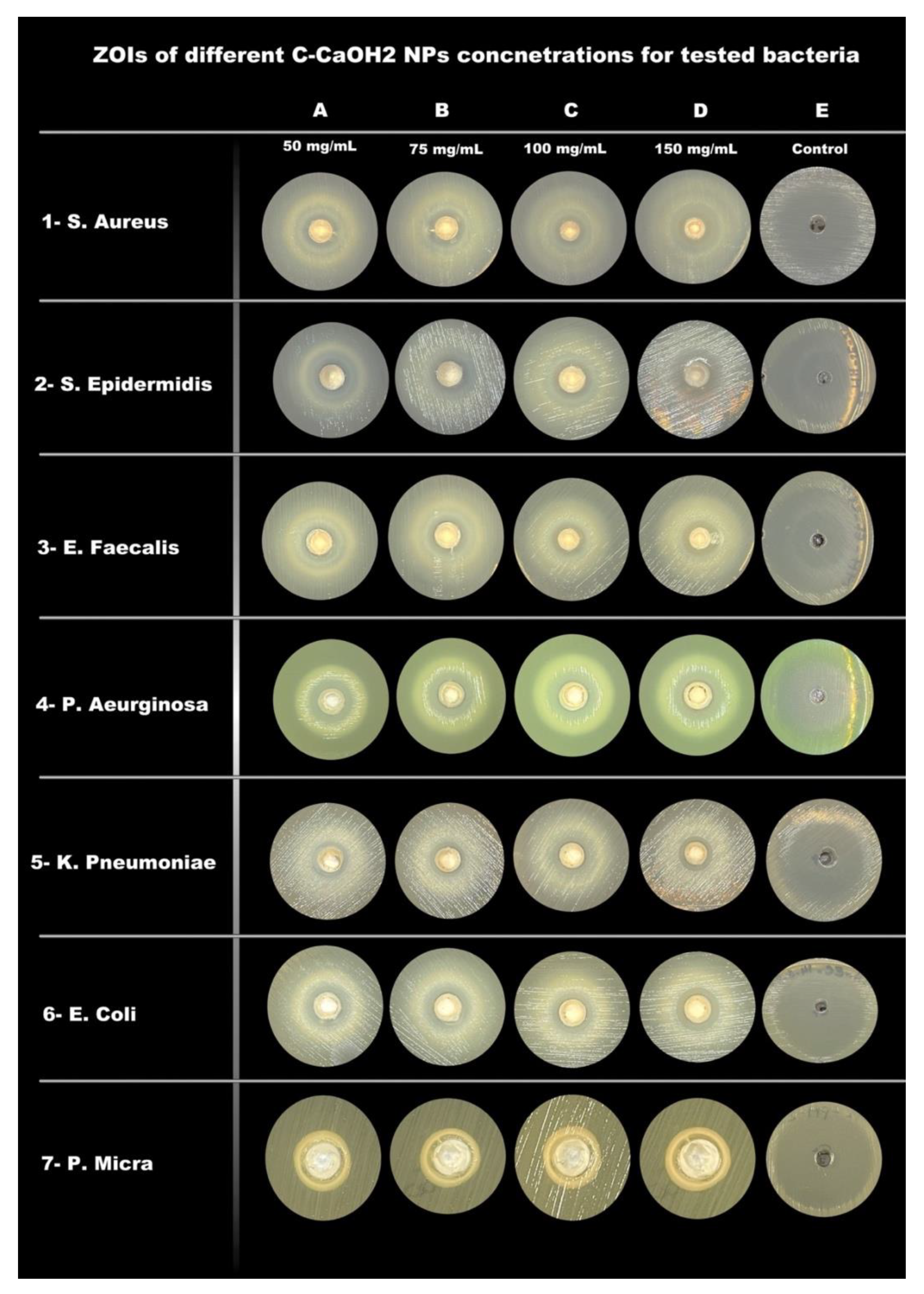
Mean ± Standard Deviation of Zones of Inhibition (ZOI) in mm
Bacterial Strains50 mg/mL75 mg/mL100 mg/mL150 mg/mLControl
S. Aureus7.3 ± 0.69 ± 19.3 ± 0.610.8 ± 0.326.7 ± 0.6
S. Epidermidis8.3 ± 0.69 ± 09.5 ± 0.59.7 ± 0.640.3 ± 0.6
E. Faecalis0 ± 00 ± 07.3 ± 0.68.7 ± 0.619.3 ± 0.6
P. Aeruginosa10 ± 011 ± 011.2 ± 0.311.7 ± 0.631.7 ± 0.6
K. Pneumoniae9 ± 09 ± 09.2 ± 0.39.7 ± 0.630.7 ± 0.6
E. Coli10 ± 010 ± 010.2 ± 0.310.8 ± 0.340.3 ± 0.6
P. Micra5.7 ± 0.66.3 ± 0.67.5 ± 0.57.7 ± 0.641 ± 1.0
Table 2. Minimum inhibitory concentrations (MIC) and Minimum bactericidal concentrations (MBC) of C-CaOH2 NPs against bacterial strains.
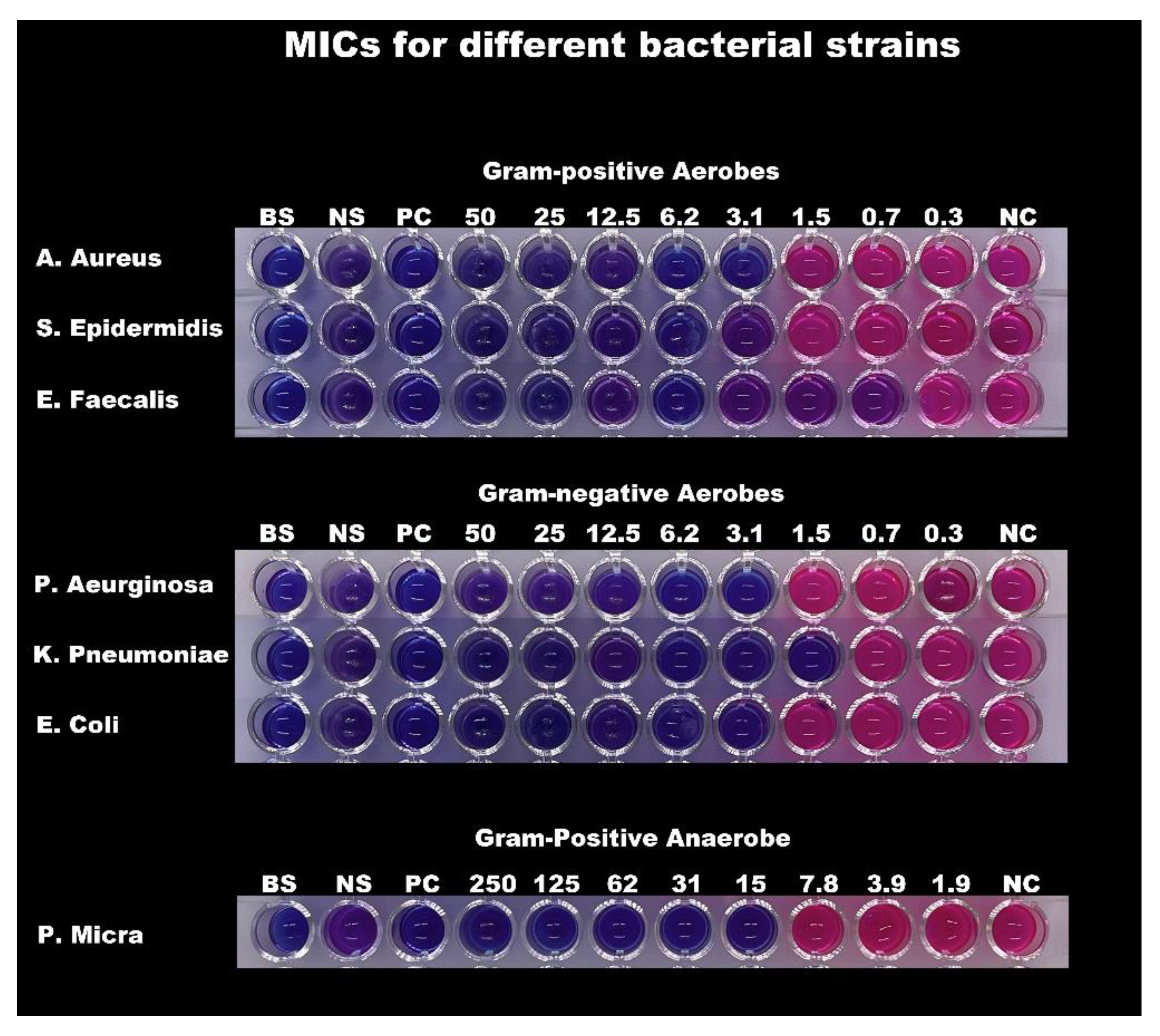
Minimal Inhibitory concentrations (MIC) and Minimal Bactericidal Concentrations (MBC)
Bacterial StrainsMIC (mg/mL)MBC (mg/mL)
S. Aureus3.1212.5
S. Epidermidis3.1212.5
E. Faecalis0.7812.5
P. Aeruginosa3.123.12
K. Pneumoniae1.566.25
E. Coli3.1212.5
P. Micra15.6231.25
Publisher’s Note: MDPI stays neutral with regard to jurisdictional claims in published maps and institutional affiliations.

