Article

High-Efficiency Adsorption of SARS-CoV-2 Spike 1 Protein by Plasma-Modified Porous Polymers

1
Department of Physiology, Shihezi University School of Medicine, Shihezi 832002, China
2
Key Laboratory of Materials Modification, Ministry of Education, Dalian University of Technology, Dalian 116024, China
3
The Key Laboratory of Xinjiang Endemic and Ethnic Diseases, Shihezi University Medical College, Shihezi 832002, China
4
Department of Physiology, Wuhan University School of Basic Medical Sciences, Wuhan 430070, China
5
Department of Physiology, Huazhong University of Science and Technology of Basic Medical Sciences, Wuhan 430070, China
*
Authors to whom correspondence should be addressed.
These authors contributed equally to this work.
Appl. Sci. 2022, 12(24), 12628; https://doi.org/10.3390/app122412628
Submission received: 17 November 2022 / Revised: 5 December 2022 / Accepted: 6 December 2022 / Published: 9 December 2022
(This article belongs to the Special Issue Plasma Technology and Its Applications)

Abstract

:
Under the background of the COVID-19 pandemic, this study reports an affordable and easily prepared porous material modified by nanosecond-pulsed discharge plasma, which can adsorb SARS-CoV-2 S1 protein efficiently. Both Western blotting and an enzyme-linked immunosorbent assay were used to detect the adsorption efficiency of SARS-CoV-2 S1 protein. The physical and chemical properties of the modified porous polymer were characterized by scanning electron microscopy, X-ray photoelectron spectroscopy and Fourier transform infrared spectroscopy. We found that the new type of porous polymer material presented an excellent performance on SARS-CoV-2 S1 protein adsorption, whose adsorption efficiency reached 99.99% in 1 min. Both the physical and chemical characterizations showed that the material has many fresh pores on the material surface and that the surface is implanted with polar functional groups (C−O, C=O, O−C=O and −NH), which gives the material a high chemisorption performance along with an enhanced physical adsorption performance. Notably, the material can be prepared at prices ranging in the tens of dollars per kilogram, which shows that it could have great applications for respiratory virus protection in global epidemic states.

1. Introduction

A sudden outbreak of coronavirus disease 2019 (COVID-19) caused by severe acute respiratory syndrome coronavirus (SARS-CoV-2) affected more than 600 million people worldwide and caused more than 6 million deaths, triggering extensive research activities aimed at preventing and treating coronavirus diseases [1,2]. At present, the global epidemic situation is still at a high level, and the novel coronavirus (SARS-CoV-2) continues to mutate. The ‘Omicron’ variant has significantly enhanced its transmission ability and increased its escape ability, and it has replaced other variants to become the absolutely dominant epidemic strain in the world [3]. Therefore, it is particularly necessary to develop purification technologies that can effectively block virus transmission and remove low levels of virus in indoor air and wastewater.
Adsorption is a method that uses adsorbents to accumulate and concentrate pollutants in air and water to remove them [4]. Virus adsorbents can use the attractive interaction between the adsorbent and the virus adsorbent to achieve virus separation and removal [5]. Previously reported virus adsorbents can be classified as inorganic adsorbents, inorganic-organic hybrid adsorbents and organic adsorbents [5,6]. Common inorganic adsorbents are clays (kaolinite, montmorillonite and bentonite) [6], which generally have a low adsorption efficiency for viruses. Cellulose nanofibers enriched with α-Fe2O3 nanoparticles are organic-inorganic hybrid materials with the ability to adsorb viruses [7]. The main organic adsorbents are polymers, such as polysaccharides (anionic polysaccharide sulfate and cationic chitosan) [8,9,10,11], and resins [12]. These virus adsorbents have different properties, but all of them show certain defects in practical applications, such as expensive equipment and operation costs, the difficulty of recycling adsorbents, and an ease in causing secondary pollution. Therefore, it is urgent to develop new virus adsorption materials with a high adsorption performance and low price, which can be applied to the isolation and removal of viruses worldwide.
At present, the research on the adsorption of SARS-CoV-2 focuses on its S protein, because the specific interaction between S protein and materials determines the applicability of virus adsorption materials [13]. However, due to the low expression of the protein, it cannot be obtained in large quantities. Thus, in recent years, multiscale modeling, an innovative computational technique, has been considered to investigate the interaction between proteins and materials, so as to explore the adsorption mechanism between viruses and materials [14]. For instance, Sahihi et al. [15] used all-atom molecular dynamics simulations to find that the adhesion of S1 protein over polystyrene was stronger than other materials and that adsorption was mainly driven by hydrophobicity and π-π interactions. De Luca et al. [13] predicted that the long-range interaction between S protein and the polymer surface is weaker than covalent or ionic bond energy by using a combination of molecular mechanics and dynamic simulation. Xin et al. [16] employed high-speed atomic force microscopy to monitor the adsorption of recombinant SARS-CoV-2 spike protein S1 on the surface of oxide materials, suggesting that the adsorption of S1 protein on the oxide surface was mainly due to electrostatic interaction. These simulations provide a theoretical basis for protein and virus adsorption, but more practical experiments are needed to verify the adsorption mechanism and apply it to virus adsorption and removal. Currently, macroporous polymer resin has been applied to adsorb pollutants because of its low cost, stable chemical structure, abundant pores and large specific surface area [17,18,19]. However, there are no oxygen-containing functional groups and microporous functional groups on the resin surface, which reduces the adsorption potential of the resin and hinders its practical application in virus removal [20]. Non-thermal plasma technology is a fast-processing and environmentally friendly modification method, which can greatly improve the adsorption performance of adsorbents [21,22]. In the process of non-thermal plasma discharge, numerous reactive substances (radicals, electrons and ions) are generated, which can interact with the material surface to form new functional groups and to enrich pores [23,24,25,26,27,28,29]. Thus, plasma modification can be applied to the surface modification of adsorbents to improve their adsorption performance.
Therefore, in this study, nanosecond pulsed discharge was used to modify macroporous polystyrene resin (MaPR) to enrich the number of pore sizes and surface functional groups and generate a modified porous polymer (MPP), in order to realize the effective adsorption of SARS-CoV-2 S1 protein. Western blotting, an enzyme-linked immunosorbent assay (ELISA), scanning electron microscopy (SEM) and Fourier-transform infrared spectrum (FTIR) were used to analyze the effects of plasma modification on the adsorption efficiency of SARS-CoV-2 S1 protein, morphological changes in the surface, the pore size and number, and surface functional groups of the porous polymers. Additionally, the interaction mechanism between plasma modification and the adsorbent was briefly analyzed. These results provide a research basis for the efficient removal of pathogenic microorganisms in indoor air and wastewater.

2. Materials and Methods

Chemicals and materials. MaPR with a particle size between 30–60 mesh and a surface area of approximately 300 m2 g−1 was purchased from Sigma-Aldrich (cat. no.9003-70-7). SARS-CoV-2 Spike Gycoprotein S1 (cat. no. ab273068) was purchased from Abcam. The MaPR was pretreated with anhydrous ethanol (Tianjin Fuyu Fine Chemical Co., Ltd., Tianjin, China).
Preparation of modified porous polymers. The MPPs in this study were obtained by nanosecond pulsed discharge plasma treatment. Firstly, 40 g of MaPR was soaked in anhydrous ethanol overnight for pretreatment. Subsequently, MaPR was washed continuously with distilled water until the pH of the soaking solution became neutral. MaPR was dried at 37 °C for 24 h. Then, 2 g of MaPR was placed into the plasma reactor and modified with nanosecond pulse discharge plasma under the condition of artificial air for 20 min. The pulse peak voltage was 30 kV, and the pulse repetition frequency was 120 Hz. The plasma modification device and detailed process are shown in the Supplementary Materials.
Adsorption experiments of SARS-CoV-2 S1 protein. S1 protein with a concentration of 1 mg/mL was diluted into 0.5 ug/mL or 0.1 ug/mL protein solution with sterile Phosphate Buffered Saline. A total of 0.1 g of MaPR or MPP was weighed and placed in sterile six-well plates, respectively. A total of 500 uL of protein solution was added to each well and then incubated for 1 min. The solution in each well was collected after incubation. The protein-adsorbed sample and the collected solution were further analyzed. The adsorption capacity of the S1 protein by resin was determined by measuring the consumption of protein in solution. The adsorption efficiency of the S1 protein was calculated as follows:
w (%) = (C0 − C1)/C0 × 100%
where w (%) represents the adsorption efficiency, C0 (mg/mL) represents the S1 protein concentration without adding resin, and C1 (mg/mL) represents the S1 protein concentration when adding 100 mg resin for 1 min.
Characterization. The surface morphology of the materials was scanned by scanning electron microscopy (SU8020, Hitachi) with a secondary electron beam and an accelerating voltage of 10 kV. The elemental composition and valence of the surface of the materials were analyzed by XPS (ESCALAB 250Xi, Thermo Fisher, Waltham, MA, USA). The spectrum data of C1s (284.6 eV) was obtained and then fitted with XPSPEAK4.1 software to analyze the oxygen functional groups. An FTIR spectroscopy spectrometer (EQUINOX55, Bruker) was used to measure the surface functional groups.
ELISA. Concentrations of the SARS-CoV-2 S1 protein after adsorption in each group were measured by a FastScanTM SARS-CoV-2 Spike protein ELISA Kit (Cell Signaling Technology, Inc., cat. no. 76349, Danvers, MA, USA) according to the manufacturer’s instructions. The detailed steps are shown in the Supplementary Materials.
Western blot. After adsorption, the protein solution of each group was collected for western blotting analysis. The proteins were separated using Sodium Dodecyl Sulfate-Polyacrylamide Gel Electrophoresis (SDS-PAGE, 8% gel) and transferred to polyvinylidene fluoride membranes (Invitrogen; Thermo Fisher Scientific, Inc., Waltham, MA, USA). After that, the membranes were blocked in 5% skim milk for 2 h to avoid nonspecific binding. Then, the membranes were incubated at 4 °C with anti-SARS-CoV-2 Spike Glycoprotein S1 antibody (1:1000 dilution, Abcam, cat. no. ab283942). On the next day, the membranes were incubated with HRP-labeled anti-rabbit IgG (1:10,000 dilution, Beijing Zhongshan Jinqiao Biotechnology Co., cat. no. ZB-5301, Beijing, China) at room temperature for 2 h. After exposure, the gray values of immunoreactive bands were analyzed by ImageJ software (v.1.8.0, National Institutes of Health, Bethesda, MD, USA).

3. Results and Discussion

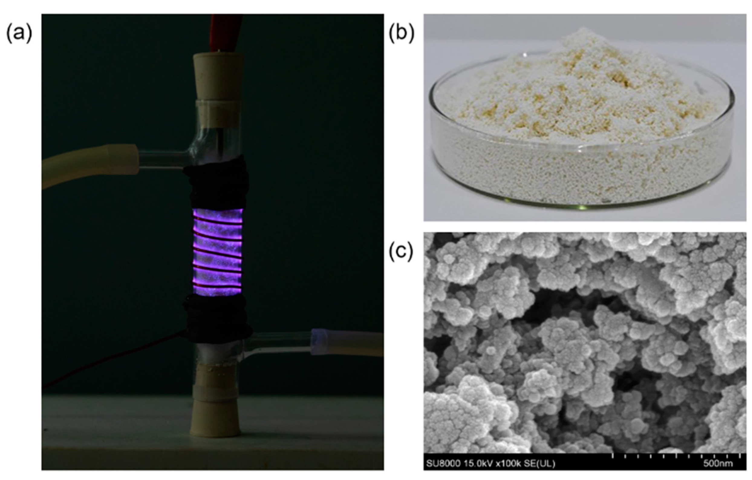
3.1. Microstructure and Functionalization of the Modified Porous Polymer

Figure 1 shows typical SEM images of the materials. After plasma treatment for 20 min, a mass of fresh pores and a regular pore network formed on the surface of the resin. The generated micropores and mesopores could facilitate the S1 protein adsorption process; thus, the new porous resin was named “modified porous polymer (MPP)” in this study.
FTIR and XPS spectroscopy were used to characterize the chemical structure of the MPP resins. The FTIR result of MPP is shown in Figure 2a; there is a weak band at ca. 1140 cm−1, which can be considered to show the existence of the C−O stretching vibrations, the band at ca. 1450 cm−1 represents the C−C skeletal vibration of the benzene ring, and the peaks around ca. 1610 cm−1 and 1710 cm−1 are attributed to C=O stretching vibrations of the ester groups or carboxylic groups [30,31,32]. The peak at 1630 cm−1 was attributed to N-H stretching. It has been suggested that an adsorbent surface implanted with functional groups similar to protein residues (−NH, O−C=O, C=O) can bind viral surface proteins more stably and effectively through the interaction between polar molecules. Thus, the oxygen-containing functional groups on the surface of MPP can greatly enhance the adsorption performance.
Figure 2b,c demonstrate the XPS result and the C1s spectrum of MPP. The C1s spectrum of MPP can be divided into four peaks with binding energies of 284.6, 285.7, 289.8 and 287.8 eV, representing C−C, C−O, O−C=O and C=O bonds in the benzene ring, respectively (Table 1). Carbon, oxygen and nitrogen were detected in MPP, with contents of 83.97%, 12.81% and 3.22%, respectively, and the O/C ratio was 3.98. Figure 2d shows the XPS spectra of N1s, which can be divided into Pyridinic−N, Amine/Pyrrolic−N, Graphitic−N and Oxidized−N, with binding energies of 398.5 eV, 399.8 eV, 401.2 eV and 403.1 eV, respectively [33,34,35]. In the total N atom of MPP, Graphitic−N accounts for 79.01%, and Amine/Pyrrolic−N accounts for 17.05%. These results are consistent with previous research [36]. We introduced oxygen-containing and nitrogen-containing functional groups into the MaPR through non-thermal plasma treatment to improve the adsorption performance.

3.2. Removal of SARS-CoV-2 S1 Protein

S1 protein is the outermost point of the SARS-CoV-2 envelope, participating in the specific binding between virions and biological surfaces, and it is the first contact point of non-specific adsorption on non-biological surfaces [37]. Therefore, the adsorption and inactivation of plasma-functionalized adsorbents on viruses can be focused on the interaction between functionalized adsorbents and proteins [16,38]. Thus, we used western blotting to specifically detect the adsorption of SARS-CoV-2 S1 protein by MaPR or MPP. As shown in Figure 3a,b, when the concentration of S1 protein solution was 0.5 μg/mL, compared with the MaPR, the content of S1 protein in the solution was significantly decreased after the plasma MPP adsorption for 1 min (P < 0.001, n = 3). Additionally, to investigate the adsorption stability of the absorbent on S1 protein after plasma discharge modification, MaPR or MPP was used to adsorb S1 protein solution (concentration of 5 μg/mL and 0.5 μg/mL) at different times. The experimental results showed that after adsorption with MPP, the amount of S1 protein in the solution was significantly reduced, and after adsorption for 1 min, 2 min, 4 min, 6 min, 8 min and 10 min, respectively, the amount of S1 protein in the solution remained at the same level, and there was no significant difference in the adsorption effect (Figure 3c–f). After prolonging the adsorption time, we found that the amount of S1 protein after adsorption with MPP was significantly reduced and that the amount of remaining S1 protein in the solution was also at a level within at least 4 h (Figure 3d,g,h). These results demonstrated that the adsorption effect of MPP on S1 protein was stable.

3.3. The Adsorption Efficiency of S1 Protein was Quantified by ELISA

ELISA was used to detect the concentration of residual S1 protein in the solution [39,40]. As shown in Figure 4a,b, the adsorption process of S1 protein in solution by MPP was rapid. When soaked in 0.5 μg/mL protein solution for 30 s and 1 min, the adsorption efficiency of MPP can reach 98.15% and 99.99%, respectively. When soaked in 0.1 μg/mL protein solution for 30 s and 1 min, the adsorption efficiency is 97.72% and 99.95%, respectively.
Further, to explore the reusability of the modified adsorbent, we used 0.1 g raw MaPR and 0.1 g MPP ten consecutive times (1 min each time). The results indicated that the adsorption efficiency of the raw resin and the modified resin decreased by different degrees during the repeated adsorption process. The adsorption efficiency of the raw MaPR decreased from 66.99% to 30.21%, while the adsorption efficiency of the MPP decreased from 95.95% to 63.37% (Figure 4c). In short, the MPP can be reused at least 10 times, and the adsorption efficiency can still remain above 60%.
The enhancement of the MPP adsorption efficiency on SARS-CoV-2 S1 protein may involve two key factors: the pore structure and chemical functional groups. During plasma discharge, electrons and charged particles accelerate in the electric field and bombard the material surface. As a result, many micropores and mesopores are created. On the other hand, depending on the ionization and dissociation process of plasma, many active species are generated, such as O, OH, O3 [41], which can destroy the original chemical bonds of resin and embed new oxygen-containing functional groups and nitrogen-containing functional groups (C−O, O−C=O, C=O, −NH).

4. Conclusions

S1 protein plays a key role in the interaction of viruses with the environment, and it is an indispensable target for adsorption research. In the current study, MPP, a kind of micro-mesoporous polymer adsorption material, was prepared by nanosecond-pulsed discharge plasma treatment and was applied to efficiently adsorb S1 protein of SARS-CoV-2, so as to be used in epidemic prevention and control. The material has a high adsorption efficiency for S1 protein. Western blotting and ELISA analysis were used to detect the adsorption ability of the MPP, and the results showed that the adsorption efficiency of the MPP for 0.5 μg/mL S1 protein solution is 99.99% after a 1 min immersion. The MPP can be reused at least 10 times, and the adsorption efficiency can still remain above 60%. It was found that the plasma treatment played a key role in the preparation of the MPP‘s adsorption performance and the improvement of the adsorption performance. SEM, FTIR and XPS spectroscopy were used to characterize the physical and chemical structures of the MPP, and the characterization revealed that plasma etching formed a large quantity of pores on the adsorbent’s surface. In addition, during plasma treatment, the implantation of oxygen-containing functional groups and nitrogen-containing functional groups (C−O, O−C=O, C=O, −NH) on adsorbent improves the adsorption capacity of SARS-CoV-2 S1 protein. As the preparation of the material is based on polystyrene divinylbenzene polymer, it has a low price and shows great potential for effectively removing pathogenic microorganisms with a low content in indoor air and wastewater, and it can be widely used for epidemic prevention and control.

Supplementary Materials

The following supporting information can be downloaded at: https://www.mdpi.com/article/10.3390/app122412628/s1, Figure S1: The experimental setup of plasma modification system.

Author Contributions

Conceptualization, J.S. and D.Y.; methodology, N.A. and S.L.; software, Q.X. and K.L.; investigation, N.A.; data curation, N.A.; writing—original draft preparation, N.A.; writing—review and editing, H.Y.; visualization, N.A. and S.L.; supervision, J.S. and D.Y.; project administration, D.Y.; funding acquisition, D.Y. All authors have read and agreed to the published version of the manuscript.

Funding

This research was funded by the National Natural Science Foundation of China, grant number 52077026, the Fundamental Research Funds for the Central Universities, grant number DUT21LK31, and the Finance Science and Technology Project of Xinjiang Production and Construction, grant number 2020AB019.

Institutional Review Board Statement

Not applicable.

Informed Consent Statement

Not applicable.

Data Availability Statement

The data presented in this study are available on request from the corresponding author.

Conflicts of Interest

The authors declare no conflict of interest.

References

  1. Hu, B.; Guo, H.; Zhou, P.; Shi, Z.L. Characteristics of SARS-CoV-2 and COVID-19. Nat. Rev. Microbiol. 2021, 19, 141–154. [Google Scholar] [CrossRef] [PubMed]
  2. WHO. WHO Coronavirus (COVID-19) Dashboard [Internet]. 2022. Available online: https://covid19.who.int/ (accessed on 10 November 2022).
  3. Tian, D.; Sun, Y.; Xu, H.; Ye, Q. The emergence and epidemic characteristics of the highly mutated SARS-CoV-2 Omicron variant. J. Med. Virol. 2022, 94, 2376–2383. [Google Scholar] [CrossRef] [PubMed]
  4. Sellaoui, L.; Badawi, M.; Monari, A.; Tatarchuk, T.; Jemli, S.; Dotto, G.L.; Bonilla-Petriciolet, A.; Chen, Z. Make it clean, make it safe: A review on virus elimination via adsorption. Chem. Eng. J. 2021, 412, 128682. [Google Scholar] [CrossRef]
  5. Wolski, K.; Nowakowska, M.; Pyrc, K.; Szczubiałka, K.; Ciejka, J. Biopolymeric nano/microspheres for selective and reversible adsorption of coronaviruses. Mater. Sci. Eng. C. Mater. Biol. Appl. 2017, 76, 735–742. [Google Scholar]
  6. Park, J.A.; Kang, J.K.; Kim, J.H.; Kim, S.B.; Yu, S.; Kim, T.H. Bacteriophage removal in various clay minerals and clay-amended soils. Environ. Eng. Res. 2015, 20, 133–140. [Google Scholar] [CrossRef] [Green Version]
  7. Ma, H.; Burger, C.; Hsiao, B.S.; Chu, B. Ultra-fine cellulose nanofibers: New nano-scale materials for water purification. J. Mater. Chem. 2011, 21, 7507–7510. [Google Scholar] [CrossRef]
  8. Junter, G.A.; Lebrun, L. Polysaccharide-based chromatographic adsorbents for virus purification and viral clearance. J. Pharm. Anal. 2020, 10, 291–312. [Google Scholar] [CrossRef] [PubMed]
  9. Imbert-Laurenceau, E.; Crepinior, J.; Crance, J.M.; Jouan, A.; Migonney, V. Polystyrene derivatives substituted with arginine interact with babanki (togaviridae) and kedougou (flaviviridae) viruses. J. Med. Virol. 2003, 69, 503–509. [Google Scholar] [CrossRef]
  10. Ogura, F.; Hayashi, K.; Lee, J.B.; Kanekiyo, K.; Hayashi, T. Evaluation of an edible blue-green alga, Aphanothece sacrum, for its inhibitory effect on replication of herpes simplex virus type 2 and influenza virus type A. Biosci. Biotechnol. Biochem. 2010, 74, 1687–1690. [Google Scholar] [CrossRef] [Green Version]
  11. Preeprame, S.; Hayashi, K.; Lee, J.B.; Sankawa, U.; Hayashi, T. A novel antivirally active fucan sulfate derived from an edible brown alga, Sargassum horneri. Chem. Pharm. Bull. 2001, 49, 484–485. [Google Scholar] [CrossRef] [Green Version]
  12. Pisharody, L.; Suresh, S.; Mukherji, S. Evaluation of adsorbents and eluents for application in virus concentration and adsorption-desorption isotherms for coliphages. Chem. Eng. J. 2021, 403, 126267. [Google Scholar] [CrossRef]
  13. De Luca, G.; Petrosino, F.; Luque Di Salvo, J.; Chakraborty, S.; Curcio, S. Advanced descriptors for long-range noncovalent interactions between SARS-CoV-2 spikes and polymer surfaces. Sep. Purif. Technol. 2021, 11, 120125. [Google Scholar] [CrossRef] [PubMed]
  14. Curcio, S.; Petrosino, F.; Morrone, M.; de Luca, G. Interactions between Proteins and the Membrane Surface in Multiscale Modeling of Organic Fouling. J. Chem. Inf. Model. 2018, 58, 1815–1827. [Google Scholar] [CrossRef] [PubMed]
  15. Sahihi, M.; Faraudo, J. Molecular Dynamics Simulations of Adsorption of SARS-CoV-2 Spike Protein on Polystyrene Surface. J. Chem. Inf. Model. 2022, 62, 3814–3824. [Google Scholar] [CrossRef]
  16. Xin, Y.; Grundmeier, G.; Keller, A. Adsorption of SARS-CoV-2 Spike Protein S1 at Oxide Surfaces Studied by High-Speed Atomic Force Microscopy. Adv. Nanobiomed. Res. 2021, 1, 2000024. [Google Scholar] [CrossRef]
  17. Freitas, P.A.M.; Iha, K.; Felinto, M.C.F.C.; Súarez-Iha, M.E.V. Adsorption of di-2-pyridyl ketone salicyloylhydrazone on Amberlite XAD-2 and XAD-7 resins: Characteristics and isotherms. J. Colloid Interface Sci. 2008, 323, 1–5. [Google Scholar] [CrossRef]
  18. Lv, C.; Yang, J.; Liu, R.; Lu, Q.; Ding, Y.; Zhang, J.; Deng, J. A comparative study on the adsorption and desorption characteristics of flavonoids from honey by six resins. Food Chem. 2018, 268, 424–430. [Google Scholar] [CrossRef]
  19. Nassar, R.A.; Browne, E.P.; Chen, J.; Kibonov, A.M. Removing human immunodeficiency virus (HIV) from human blood using immobilized heparin. Biotechnol. Lett. 2012, 34, 853–856. [Google Scholar] [CrossRef] [Green Version]
  20. Xu, Q.N.; Wang, H.L.; Zhou, X.F.; Liang, J.P.; Yuan, H.; Xu, Y.; Yang, D.Z. Micro-mesoporous polystyrene resins prepared by nanosecond pulsed discharge plasma for the efficient adsorption of PAHs. Sep. Purif. Technol. 2022, 301, 121829. [Google Scholar] [CrossRef]
  21. Yu, X.; Zhang, F.; Wang, N.; Hao, S.; Chu, W. Plasma-treated bimetallic Ni-Pt catalysts derived from hydrotalcites for the carbon dioxide reforming of methane. Catal. Lett. 2014, 144, 293–300. [Google Scholar] [CrossRef]
  22. Wang, H.; Xu, Q.; Zhou, X.; Liang, J.; Yuan, H.; Yang, D.; Wang, W. Highly efficient adsorptive removal of persistent organic pollutants using NDP-acid combined modified NaY zeolites. Chem. Eng. J. 2022, 431, 133858. [Google Scholar] [CrossRef]
  23. Qiu, J.; Wang, G.; Bao, Y.; Zeng, D.; Chen, Y. Effect of oxidative modification of coal tar pitch-based mesoporous activated carbon on the adsorption of benzothiophene and dibenzothiophene. Fuel Process. Technol. 2015, 129, 85–90. [Google Scholar] [CrossRef]
  24. Dou, S.; Tao, L.; Wang, R.L.; El Hankari, S.; Chen, R.; Wang, S.Y. Plasma-assisted synthesis and surface modification of electrode materials for renewable energy. Adv. Mater. 2018, 30, 1705850. [Google Scholar] [CrossRef] [PubMed]
  25. Duan, S.; Liu, X.; Wang, Y.; Meng, Y.; Alsaedi, A.; Hayat, T.; Li, J. Plasma surface modification of materials and their entrapment of water contaminant: A review. Plasma Processes Polym 2017, 14, 1600218. [Google Scholar] [CrossRef]
  26. Li, Y.; Yang, D.Z.; Qiao, J.J.; Zhang, L.; Wang, W.Z.; Zhao, Z.L.; Zhou, X.F.; Yuan, H.; Wang, W.C. The dynamic evolution and interaction with dielectric material of the discharge in packed bed reactor. Plasma Sources Sci. Technol. 2020, 29, 055004. [Google Scholar] [CrossRef]
  27. Wu, L.; Wan, W.; Shang, Z.; Gao, X.; Kobayashi, N.; Luo, G.; Li, Z. Surface modification of phosphoric acid activated carbon by using non-thermal plasma for enhancement of Cu(II) adsorption from aqueous solutions. Sep. Purif. Technol. 2018, 197, 156–169. [Google Scholar] [CrossRef]
  28. Zheng, X.Y.; Zheng, H.; Xiong, Z.; Zhao, R.; Liu, Y.; Zhao, C.; Zheng, C. Novel anionic polyacrylamide-modify-chitosan magnetic composite nanoparticles with excellent adsorption capacity for cationic dyes and pH-independent adsorption capability for metal ions. Chem. Eng. J. 2020, 392, 123706. [Google Scholar] [CrossRef]
  29. Xu, Q.N.; Yang, D.Z.; Wang, H.L.; Yuan, H.; Jia, Z.X.; Yang, H.; Zhou, X.F.; Wang, W.C.; Xu, Y.; Yan, E.Y. Enhancing the adsorption property of macroporous XAD-2 resin by nanosecond-pulsed discharge plasma modification. Plasma Processes Polym. 2021, 18, e2000117. [Google Scholar] [CrossRef]
  30. El Didamony, A.M.; Youssef, W.M.; Abdou, A.A. Modification of amberlite XAD-2 resin for U(VI) adsorption from egyptian crude phosphoric acid. Egypt. J. Pet. 2019, 28, 71–76. [Google Scholar] [CrossRef]
  31. Gradinaru, L.M.; Barbalata-Mandru, M.; Drobota, M.; Aflori, M.; Spiridon, M.; Gradisteanu Pircalabioru, G.; Bleotu, C.; Butnaru, M.; Vlad, S. Preparation and evaluation of nanofibrous hydroxypropyl cellulose and β-cyclodextrin polyurethane composite mats. Nanomaterials 2020, 10, 754. [Google Scholar] [CrossRef] [Green Version]
  32. Sun, S.; Guo, M.; Yi, X.; Zhang, Z. Preparation and characterization of a naphthalenemodified poly (aryl ether ketone) and its phase separation morphology with bismaleimide resin. Polym. Bull. 2017, 74, 1519–1533. [Google Scholar] [CrossRef]
  33. Kong, X.; Liu, Q.; Chen, D.; Chen, G. Identifying the active sites on N-doped graphene toward oxygen evolution reaction. Chem. Cat. Chem. 2017, 9, 846–852. [Google Scholar] [CrossRef]
  34. Mei, S.; Gu, J.; Ma, T.; Li, X.; Hu, Y.; Li, W.; Zhang, J.; Han, Y. N-doped activated carbon from used dyeing wastewater adsorbent as a metal-free catalyst for acetylene hydrochlorination. Chem. Eng. J. 2019, 371, 118–129. [Google Scholar] [CrossRef]
  35. Liu, L.; Yan, F.; Li, K.; Zhu, C.; Xie, Y.; Zhang, X.; Chen, Y. Ultrasmall FeNi3N particles with an exposed active (110) surface anchored on nitrogen-doped graphene for multifunctional electrocatalysts. J. Mater. Chem. A 2019, 7, 1083–1091. [Google Scholar] [CrossRef]
  36. Zhang, B.; Xu, P.; Qiu, Y.; Yu, Q.; Ma, J.; Wu, H.; Luo, G.; Xu, M.; Yao, H. Increasing oxygen functional groups of activated carbon with non-thermal plasma to enhance mercury removal efficiency for flue gases. Chem. Eng. J. 2015, 263, 1–8. [Google Scholar] [CrossRef]
  37. Walls, A.C.; Park, Y.J.; Tortorici, M.A.; Wall, A.; McGuire, A.T.; Veesler, D. Structure, function, and antigenicity of the SARS-CoV-2 spike glycoprotein. Cell 2020, 181, 281–292. [Google Scholar] [CrossRef] [PubMed]
  38. Dobrynin, D.; Polishchuk, I.; Portal, L.; Zlotver, I.; Sosnik, A.; Pokroy, B. Adsorption of SARS CoV-2 spike proteins on various functionalized surfaces correlates with the high transmissibility of Delta and Omicron variants. Mater. Today Bio. 2022, 14, 100265. [Google Scholar] [CrossRef]
  39. Krähling, V.; Halwe, S.; Rohde, C.; Becker, D.; Berghöfer, S.; Dahlke, C.; Eickmann, M.; Ercanoglu, M.S.; Gieselmann, L.; Herwig, A.; et al. Development and characterization of an indirect ELISA to detect SARS-CoV-2 spike protein-specific antibodies. J. Immunol. Methods 2021, 490, 112958. [Google Scholar] [CrossRef]
  40. Horn, M.P.; Jonsdottir, H.R.; Brigger, D.; Damonti, L.; Suter-Riniker, F.; Endrich, O.; Froehlich, T.K.; Fiedler, M.; Largiadèr, C.R.; Marschall, J.; et al. Serological testing for SARS-CoV-2 antibodies in clinical practice: A comparative diagnostic accuracy study. Allergy 2022, 77, 2090–2103. [Google Scholar] [CrossRef]
  41. Zeng, X.; Li, B.; Liu, R.; Li, X.; Zhu, T. Investigation of promotion effect of Cu doped MnO2 catalysts on ketone-type VOCs degradation in a one-stage plasma-catalysis system. Chem. Eng. J. 2020, 384, 123362. [Google Scholar] [CrossRef]
Figure 1. Preparation of MPP and its microstructure. (a) Plasma modification of MaPR; (b) MPP; (c) SEM images of MPP magnified to 500 nm.
Applsci 12 12628 g001
Figure 2. Functionalization of MPP. (a) FTIR spectrum of MPP; (b) XPS spectrum of MPP; (c) C1s XPS spectrum of MPP; and (d) N1s XPS spectrum of MPP.
Applsci 12 12628 g002
Figure 3. The expression of remaining SARS-CoV-2 S1 protein and the adsorption stability of MPP was analyzed by western blot. (a) Expression of SARS-CoV-2 S1 protein in solution after adsorption; (b) Gray value analysis of SARS-CoV-2 S1 protein band; (c) Expression of SARS-CoV-2 S1 protein in solution after MMP adsorption for 10 min; (d) Expression of SARS-CoV-2 S1 protein in solution after MMP adsorption for 4 h; (eh) Gray value analysis of SARS-CoV-2 S1 protein band.
Applsci 12 12628 g003
Figure 4. The adsorption efficiency of S1 protein was quantified by ELISA. (a) Concentration of SARS-CoV-2 S1 protein in solution after MPP adsorption for 30 s; (b) Concentration of SARS-CoV-2 S1 protein in solution after MPP adsorption for 1 min; (c) The efficiency of MPP resin at repeatedly adsorbing SARS-CoV-2 S1 protein.
Applsci 12 12628 g004
Table 1. Chemical contents and functional groups of C1s on MPP.
Chemical ContentsBinding Energy (eV)MPP (%)
C (at. %)284.683.97%
O (at. %)532.6412.81%
N (at. %)400.193.22%
O/C (%)3.98
C−C284.685.18%
C−O285.710.25%
O−C=O289.83.20%
C=O287.81.37%
Publisher’s Note: MDPI stays neutral with regard to jurisdictional claims in published maps and institutional affiliations.

