Case Report

Treatment Possibilities in Mandibular Defect Reconstruction Based on Ameloblastic Fibro-Odontoma Treatment—Does Small Bone Defects Heal without Bone Grafting?

1
Maxillo-Facial Surgery Ward, Sokołowski Hospital, Sokołowskiego 4, 58-309 Wałbrzych, Poland
2
Department of Anaesthesiology and Intensive Care, Sokołowski Hospital, Sokołowskiego 4, 58-309 Wałbrzych, Poland
3
Department of Biostructure and Animal Physiology, Wrocław University of Environmental and Life Sciences, Kożuchowska 1, 51-631 Wrocław, Poland
4
Department of Otolaryngology, Head and Neck Surgery, 4th Military Hospital in Wrocław, Weigla 5, 50-981 Wrocław, Poland
5
Department of Head and Neck Surgery, Otolaryngology Medical University in Wrocław, Borowska 213, 50-556 Wrocław, Poland
6
Department of Pediatric Dentistry and Preclinical Dentistry, Wrocław Medical University, Krakowska 26, 50-425 Wrocław, Poland
*
Authors to whom correspondence should be addressed.
Appl. Sci. 2022, 12(24), 12963; https://doi.org/10.3390/app122412963
Submission received: 27 November 2022 / Revised: 14 December 2022 / Accepted: 15 December 2022 / Published: 16 December 2022
(This article belongs to the Section Materials Science and Engineering)

Abstract

:
The occurrence and manifestation of each ameloblastic fibro-odontoma (AFO) is quite rare and uncommon. Mentioned odontogenic tumor classification had changed over the years; however, the treatment possibilities for this lesion remain the same. In most cases surgical enucleation is sufficient enough; however, bigger lesions might require bone curettage with sometimes performed bony ostectomy, which is a quite sufficient and effective method of treatment. In the presented case report, a panoramic radiological evaluation indicated an impacted molar tooth surrounded with mixed radiolucent/radiopaque areas. The 10-year-old Caucasian girl was scheduled for an incisional biopsy. The bone cavity in the mandible after tumor removal might be left for spontaneous healing or grafting techniques, depending on the shape and size of the defect. In the presented case report, the usage of PRF/iPRF in the mandibular bone cavity, and healing improved the overall final result.

1. Introduction

Ameloblastic fibroodontoma (AFO) is one of the odontogenic tumors (OT), present in the oral cavity and is considered one or rare lesion in the oral cavity, estimated 0.1–3.4% (0.3–2.3%) in some studies [1,2]. Since the WHO classification in 1992, some crucial changes in OT studies had changed [1,2]. AFO is common in children and young adults (one to two decades of life) and it might be related to odontogenesis. In almost all cases it grows slowly without any symptoms or other clinical manifestations and is mostly diagnosed on radiographs [2]. Santos et al. conclude that it is still unclear if AFO is an early stage of odontoma [3,4]. In both cases, impacted teeth at different stages of eruption might be found [5,6]. In the old World Health Organisation classification (2005), AFO was classified in the same group as ameloblastic fibroma (AF), ameloblastic fibrodentinoma (AFD), and others, characteristic of the presence of odontogenic epithelium with odontogenic ectomesenchyme with or without hard tissue formation [7,8]. Their shape, size, and advancement might vary greatly. Back in 2017, the new WHO classification changed AFO and AFD (ameloblastic fibrodentinoma) from two separate entities, since they represent the early stages of complex odontoma development (developing odontoma group) [9,10]. This fact was related to little clinical evidence of AOF, and AFD classifying them as independent entities. Similar changes influenced odontoameloblastoma (OA), since its more characteristic of an odontoma with coexisting ameloblastoma but with clinical signs as ameloblastoma [9,10]. The new classification divides OT and classifies them into epithelial, mesenchymal (ectomesenchymal), or mixed forms depending on which component of the tooth germ gives rise to the neoplasm [7,8,9,10].
Nowadays AFO is considered a benign mixed odontogenic lesion, most present in the mandible, and less commonly in the maxillary region. This origin is related to the presence of hard odontoid tissue with a great variety in the spectrum of developing odontoma [11,12,13]. In most cases, an asymptomatic swelling, followed by delayed teeth eruption with well-defined lesion borders remains the most typical symptom of this pathology [8,9,10,11,12]. More advanced lesions might rarely displace surrounding teeth and the lesion might exceed the cortical bone layer towards the soft tissues [8,9,10,11,12,13,14,15]. The inferior alveolar nerve is not involved in the lesion and its function loss is not common, similar to surrounding teeth resorption [8,9,10,11,12,13,14,15]. Bigger lesions might lead to facial disfigurement, mandibular asymmetry, bone swelling, and problems with biting and chewing. Malignant transformation is not common; however, long-term follow-up is necessary. Problems with tooth eruption might arise, or their displacement.
In radiological assessment, the lesion is mostly well-defined, unilocular, radiolucent mass with areas of radiopaque material, and teeth-like structures, with no, erupted teeth in the lesion cavity. Irregularity of size and shape of radiolucent areas might correspond with the growth phase of the lesion. Its well-defined margins with the presence of radiopaque areas might suggest its entity; however, other larger lesions might require a biopsy at first to establish the biology of the tumor before any final surgery will take place.
The current classification of AFO describes this lesion as of mixed origins of ameloblastic fibroma and a complex odontoma with ectodermal and mesenchymal components and odontogenic tissue such as enamel, dentin with the common presence of an ectopic impacted/retained teeth [7,8,9,10,11,12,13,14]. Some correlation with the scope of present different types of dental hard tissues is worth remembering [15,16]. This mixed benign neoplasm is also built of various dental hard tissues with different calcification stages. It seems that cone-beam computerized tomography might be very useful in the diagnosis of tumors and teeth-like masses [14,15,16,17,18]. The presented lesion is also quite often tooth eruption related. In most cases, ameloblastic cells are present with various fibrous tissues and parts of calcified masses. Additionally, types of extraosseous odontogenic tumors can occur. Those findings are very similar to our case, presenting combined radiolucent-radiopaque images on radiographs [19].
The most common occurrence of this lesion is the posterior mandible and the area of the third molar; however, other mandible areas might be also affected [2,3,4,5,6,7,8,9,10,11,12,13,14,15,16]. Buchner et al. performed a study on 114 cases of AFO worldwide [20]. The authors concluded that the mandible was involved in 65%, with 80% occurrence in the posterior part of the mandible and more than 92% was associated with impacted and/or unerupted teeth [21,22]. The maxillary location remains somehow rare [19,20,21,22,23].
Studies performed worldwide suggest that hybrid odontogenic tumors can be found [15,16,17,18,19,20]. AFO in most cases is a small lesion; however, cases with a great variety of shapes and sizes are describing occurrence both in the maxilla and mandible [8,16,17,18,19,20,21,22,23,24,25]. The recurrence rate is highly related to the surgical approach, localization, and structures involved in its composition [1,2,3,4,5,18,19,20,21,22,23,24,25].
Differential diagnostics should include pathologies with similar mixed radiological appearances such as calcifying epithelial odontogenic tumor (CEOT), calcifying odontogenic cyst (COC), immature complex odontoma, adenomatoid odontogenic tumor (AOT), ameloblastic fibrodentinoma (AFD) [7,8,9,10,11,12,13,14,15,16,17,18,19,20,21,22]. During clinical differentiation, bone swelling should be distinguished from odontogenic cysts (OC), eruption cysts, OKC (odontogenic keratocysts), cyst-related pathologies, and rarely with PHP, primary hyperparathyroidism tumors, namely Brown tumors related to the parathyroid gland adenomas or related lesions [8,9,10,11,12,13,14,15,16,17,18,19,20,21,22,23,24,25].
After the removal of each pathology from the bone, an empty intraosseous bony cavity is present. Not all cases require bone grafting or GBR (guided bone regeneration) procedures. A bone graft could be performed with calvarial, iliac, or fibula graft, while GBR is performed with autogenous and/or synthetic biomaterials. Recovery after regenerative interventions may be related to a period after surgery of delayed or complicated healing [26,27,28]. Shape, size, localization, and close proximity to an esthetic area in the oral cavity, along with the ability to support bone ridge/stable teeth stability in the oral cavity are considered the most important factors. In the presented paper, a rare case of AFO lesion and its treatment with blood cloths/PRF will be presented and discussed

2. Case Report Presentation

A 10-year-old Caucasian female was referred to with an asymptomatic right mandibular molar area lesion. The lesion was incidentally discovered by a dentist on a routine panoramic radiograph. The image consisted of a well-defined radiolucent area with various amounts of surrounding radiopaque areas in various shapes, and sizes, and with the presence of retained molar teeth. The presence of some irregular hard, radiopaque material included the differentiated diagnosis with PHP primary hyperparathyroidism or any other endocrine diseases but the findings were irrelevant. On further examination, the medical, social and family histories were unremarkable, as were the results of a review of systems and a physical examination. There was no history of local trauma or infection. Oral inspection revealed good oral hygiene. Examination revealed a slight swelling of the vestibule on the right side of the mandible, extending from the permanent first molar to the ramus of the mandible on the same side. The mucosa was unaffected and the swelling was bony-hard and there was no tenderness on palpation. The lower right permanent mandibular molar was non-mobile, non-tender, and vital. The right mandibular second permanent molar had not erupted in the oral cavity. There were no neurological deficiencies related to the trigeminal and facial nerves. Submandibular lymph nodes were nonpalpable and non-tender. Panoramic radiography exhibited an expansive, unilocular radiolucent, well-circumscribed lesion with scattered foci of calcified material which contained several radiopaque bodies of varying sizes and shapes (Figure 1 and Figure 2). The right lower first molar was associated with the lesion and had been displaced inferiorly to the caudal border of the mandibular body. CBCT scans revealed a mixed radiolucent-radiopaque lesion size of about 20 mm × 26 mm × 25 mm at the right angle of the mandible with features of bone expansion of the buccal cortical plate (Figure 3). The lingual plate remains untacked and without any tumor spread (Figure 4).
Decisions were made, to perform a surgical approach. No adjacent teeth required additional endodontic treatment, Before surgery patient was then scheduled for a dental hygienist in preparation for surgery. The surgical plan consisted of tumor removal, followed by bone curettage and if needed and guided bone regeneration-GBR or simple blood-cloth iPRF tumor cavity feeling because of their small size.
Under general anesthesia, and orotracheal intubation the right mandibular angle was exposed by vestibular mucoperiosteal incision. The surgical field was covered by sterile drapes. The oral cavity was rinsed with 0.1% CHX (chlorhexidine gluconate)(Eludril, Pierre-Fabre Oral care, France) solution for 30 s. Additionally, lips and skin surrounding the operating field were scrubbed with alcohol–ethanol 96% solution-AHD 1000 solution (MediLab, Lysoform, Berlin, Germany). Protective sterile Vaseline (Unilever, London, UK) cream was applied to the lips. Local injection of mixed 0.25% solution consisted of Lignocaine with Norepinephrine 2% (ampule, 2 mL, Polfa-Warszawa, Poland) with a mixture of Natrium Chlorate 0.9% (Natrium Chloratum 0.9% Fresenius KabiClear, Bad Homburg vor der Höhe, Germany) was used. A blade No 15c B (Swann Morton, WR Swann, Owlerton Grn, Hillsborough, Sheffield, England) was used for the incision. A modified USSO-unilateral sagittal-split osteotomy of the mandible approach incision was used. After elevating full-thickness mucoperiosteal flaps, the entire tumor and the retromandibular triangle of the mandible were exposed. The thin, buccally expanded cortical plate was removed and the lesion was removed by enucleation, followed by some bone curettage, and by removal of the impacted lower right molar, leaving the inferior alveolar nerve intact (Figure 5).
The cyst was not present. Tumor masses were colored light-red (strawberry-like) and were easily bleeding. After removing all calcified tooth-shaped structures with different hardness degrees impacted molar was removed. Fibrous tissues were present along various calcified bodies (Figure 6). The bony defect was filled with collagen sponges and blood-PRF feelings (platelet-rich fibrin-PRF, delivered from local laboratory from patients’ blood and gathered on a laboratory centrifuge-MPW-352, MPW Med Instruments, Warszawa, Poland). Since no pathological fractures were present no wire fixation, nor titanium mini plates were used. Because of a stable cortical lingual plate and good bone volume in the inferior mandibular margin of the mandible, a prophylactic titanium plate was not used.
Flaps were sutured onto the desired position with 4-0 Vicryl single sutures (Ethicon, Johnson & Johnson Medical N.V., Machelen, Belgium). No dressings nor rubber drains were used in the bony cavity). The surgery went uneventful, with standard antibiotics and anti-inflammatory drugs.
After discharge, the patient continued intra-oral antibiotics 1 g Amoxicillin (Sandoz Poland, Novartis Division-Basel, Switzerland), 200 mg Ibuprom (Ibuprofenum, USP PHARMACIA, Ziębicka 40, Wrocław, Poland). The oral cavity was rinsed with 0.1% CHX (chlorhexidine gluconate) (Eludril, Pierre-Fabre Oral care, France) solution three times per day after its mixture with mineral water 1:5.
Histological examination revealed a tumor composed of odontogenic epithelium areas embedded in a background of primitive connective tissue and dentin (Figure 7).
The follow-up examination was made two years after surgery, and the clinical and radiological appearance of the bone and surrounding soft tissue was normal. Panoramic radiography exhibited ossification at the original place of the lesion. Intraoral examination revealed complete healing without bone expansion.
Routine follow-ups took place every week for six weeks, then every month for six months. Now after 24 months post-op an overall good final surgical outcome was achieved (Figure 8 and Figure 9). Future controls were not enrolled because of the COVID-19 pandemic and changes in the Surgical Ward Localization.

3. Discussion

The occurrence of various OTs varies greatly. Their shape, size, location, and growth in time require not only some diagnostic steps but also great individualism in each case of bigger lesions. This situation is mostly associated with the scope of the surgical approach and the degree of excised structures. Small defects might heal quite well on their own, if surrounding bone walls are present, which enables a good final outcome from healing. Larger defects that might cause some defects in the bone anatomy, its function, and morphology might require one of the known regenerative procedures. Furthermore, the occurrence of both maxillary and mandible bones of different OT might result in a different approach and excision of tumor resection. In some cases, the scope of the first surgical approach might greatly influence later tumor reoccurrence or even malignant transformation reoccurring and not fully healed lesions [9,10,11,12,13,14,15,20,21,22,23,24,25,26,27,28,29].
The aggressiveness of AFO is still unclear; however, its malignant transformation is possible [22,23,24,25]. If present, a surgical removal should be performed; however, surgical margins and radicalism of the procedure still are discussable [29]. Authors confirm that because of its great similarity to other odontogenic tumors AFO can be easily misdiagnosed [30,31]. Mummolo et al. conclude that AFO can be easily misdiagnosed with AF (ameloblastic fibroma) or AFD (ameloblastic fibrodentinoma) [26]. However, today because of the new WHO classification of OT, the presence of AFO has been moved towards the odontoma section, which also resulted in changes in its treatment, such as enucleation with curettage, and rarely ostectomy in bigger lesions [20,21,22,23,24,25,26,27,28,29,30].
Treatment modalities are mostly related to conservative approaches such as enucleation and curettage since AFO is a non-aggressive lesion. Unlike AF (ameloblastic fibroma), which requires a more radical approach, AFO is not likely to suspect any malignant transformation towards AFS (ameloblastic fibrosarcoma); however, a rare possibility is also possible. This is yet another reason to separate AF from AFO/AFD and their potential malignant formation.
Treatment of choice is conservative, In well-defined lesions with clear borders, enucleation with or without curettage is enough. Destructive lesions without clear borders might even require surgical resection, marginectomies, or even in some extreme cases maxillectomies/mandibulectomies. Many papers describe possible approaches, with a great correspondence with the shape, size, location, and cortical bone loss, and are focused on [10,11,12,13,14,15,16,17,18,25,26,27,28,29,30,31,32,33,34,35,36,37]:
-
Conservative approach with enucleation alone
-
Conservative approach with enucleation and bone curettage
-
Surgical curettage with bony cavity ostectomy
-
Surgical marginectomy in bigger lesions with/without reconstruction;
Regardless of the type of uses treatment, re-occurrence is rare [22,23,24,25,26,29,30,31,32,33,34,35,36]. The main question should be focused on the type of possible restoration and defect support to ensure adequate healing and bone regeneration. Bigger lesions might require some bone grafting protocols to restore some bone, while smaller lesions might heal on their own. Fresh-frozen allogenic bone grafts or PRF with xenograft bone materials seem to have good healing potential in defects where the cortical walls tend to improve bone stability [35,36]. A recent meta-analysis by Xiang et al. on PRF usage after third molar surgery highlights that it reduces some postoperative complications, but does not prevent all of them; however, it improves pain relief and swelling after lower third molar removal [37]. On the other hand, PRF mixed with a commercial spongious bone substitute, as reported in two case reports by Zhou et al., grants good results. Authors suggest that PRF serves as a bone scaffold in four-wall bony defects alone, nevertheless, it also can be combined with a xenograft in three-wall bony defects and presents excellent biocompatibility and good soft and hard tissue healing [38]. Those findings seem to confirm the Authors’ approach after AFO removal to combine collagen sponges with PRF/Blood cloths to the well-defined defects surrounded with intact bony cortical walls [36,37,38,39,40,41]. It seems that PRF and its modification are sufficient enough to promote healing in small bone defects when adjacent bony walls are untacked for better healing promotion [38,39,40,41,42,43]. Those small bone defects are hard to describe in size; however, lesions not larger than 15 mm seem to have good healing potential.
Small bone cavities after cyst enucleation are left for spontaneous healing. PRF usage is quite beneficial in oral cavity defects. Bone healing with PRF, based on blood gyration and various degree of centrifuge functions improve bone healing [35,36,37,38,39,40,41,42]. When some small cyst cavities tend to heal on their own it might take some time, while PRF or GBR procedures enable that effect much faster with a low rate of complications. Bigger lesions might require bone from fresh-frozen allogenic banks or perhaps the usage of a secondary surgery side for harvesting the proper amount of bone to ensure more improved bone defect healing. Such harvesting side might include calvaria bone, tibia, iliac bone crest, mental area, external oblique ridge, mandibular ramus, retromolar area, and others [37,38,39,40,41,42,43]. In all cases, stable defect suturing ensures its integrity if the sutures are tight enough and enable suturing without tension. Bigger reconstruction might include bone stabilization with titanium mini plates, meshes, or other titanium devices to ensure their stability for good healing promotion [37,38,39,40]. GBR techniques are useful in larger defects or those lacking a good volume of bone, and the shape of and presence of cortical walls surrounding the defect in either the mandibular or maxillary bones.
Last but not least, AFO in the past was derived from AF tumors, which has also a great potential for malignisation. This fact requires special attention. In the past, an ameloblastic fibro-odontosarcoma could arise from AF. If diagnosed early an extended surgical approach might lead to a great functional and therapeutic result. A differential diagnosis between AF and AFS has a great impact on the therapy plan. AFS should be always differentiated between forms of ameloblastoma, odontogenic fibroma, ameloblastic fibrodentinoma, ameloblastic fibroma, and ameloblastic fibro-odontoma. In some cases, we report that there is a very thin histological barrier and a greater differential diagnosis is the key to a greater therapeutic outcome. Gilani et al. agree that differential diagnosis should consist of other odontogenic sarcomas, ameloblastic carcinosarcoma, and spindle cell carcinoma. Khalili et al. point out that only the mesenchymal component represents sarcomatous alterations. Chemotherapy after surgical excision is a good treatment method and should be also taken under consideration in young patients and young adults in order to minimize its recurrence. A study performed by Pontes et al. suggests that Ki67 AND Bcl-2 proteins in AFS could be useful markers [44,45,46,47,48,49,50,51].

4. Conclusions

The presence of AFO requires curettage at most. A more radical surgical, ostectomy or even resection should be used in very big lesions, and those require some sort of bone grafting to restore the missing bone. Small defects should heal on their own or can be successfully treated with PRF usage. The overall success rates for AFO treatment remain good. The shape, size, and localization of the lesion greatly influence the type of surgical approach and possible reconstruction methods. We also conclude that macroscopic findings related to various numbers of tooth-like odontogenic tissues removed from the cavity could be very good additional information in recognizing the type of tumor.

Author Contributions

Conceptualization, K.N. and M.Ł.; methodology, K.N., S.B. and M.M.-K.; software, M.J.; validation, K.N., E.P. and M.Ł.; formal analysis, K.N. and M.M.-K.; investigation, K.N.; resources, S.B., W.P. and M.D., data curation, M.J. and M.D.; writing—original draft preparation, K.N. and W.P., writing—review and editing, W.P., M.Ł. and M.M.-K., visualization, K.N.; supervision, M.D. and M.Ł.; project administration, M.J., M.D. and E.P.; funding acquisition, M.J., M.D. and M.Ł. All authors have read and agreed to the published version of the manuscript.

Funding

This work was financed by a subsidy from Wroclaw Medical University, number SUBZ.B180.22.091.

Institutional Review Board Statement

This manuscript is a case report in which the patient cannot be identified, and therefore, the requirement for obtaining informed consent from the patient was waived. The study was conducted in accordance with the Declaration of Helsinki.

Informed Consent Statement

Informed consent was obtained from the patient involved in the study.

Data Availability Statement

Availability of supporting data—the datasets used and/or analyzed during the current study are available from the corresponding author on reasonable request.

Acknowledgments

Authors would like to thank Piotr Ziółkowski for a great and phenomenal histopathological evaluation and microscopic photographs. Secondly, the Authors would like to thank the attending surgeon Wojciech Pawlak for a tremendous surgical result and patient treatment. Furthermore, the Authors would like to add many thanks to Jan Wnukiewicz (1945–2013) for the access to the centrifuge.

Conflicts of Interest

The authors declare no conflict of interest.

References

  1. Franco, A.; Riscala, S.; Kahoudji, M.; Croue, A. Endoscopic removal of a mandibular ameloblastic fibro-odontoma. Rev. Stomatol. Chir. Maxillofac. 2009, 110, 359–361. [Google Scholar] [CrossRef] [PubMed]
  2. Cavalcante, A.S.; Anbinder, A.L.; Costa, N.C.; Lima, J.R.; Carvalho, Y.R. Ameloblastic fibro-odontoma: A case report. Med. Oral Patol. Oral Cir. Bucal 2009, 14, 650–653. [Google Scholar] [CrossRef] [PubMed]
  3. Santos TD, S.; de Carvalho RW, F.; Avelar, R.L.; Frota, R.; Anjos, E.D. Ameloblastic fibro-odontoma in children: Report of 2 cases. J. Dent. Child. 2011, 78, 173–177. [Google Scholar]
  4. Nanda, K.D.; Marwaha, M. Ameloblastic fibro-odontoma masquerading as odontoma. Indian J. Dent. Res. 2011, 22, 616. [Google Scholar] [CrossRef] [PubMed]
  5. Boxberger, N.R.; Brannon, R.B.; Fowler, C.B. Ameloblastic fibro-odontoma: A clinicopathologic study of 12 cases. J. Clin. Pediatr. Dent. 2011, 35, 397–403. [Google Scholar] [CrossRef] [PubMed]
  6. Flaitz, C.M.; Hicks, J. Delayed tooth eruption associated with an ameloblastic fibro- odontoma. Pediatr. Dent. 2001, 23, 253–254. [Google Scholar]
  7. Wright, J.M.; Odell, E.W.; Speight, P.M.; Takata, T. Odontogenic tumors, WHO 2005: Where do we go from here? Head Neck Pathol. 2014, 8, 373–382. [Google Scholar] [CrossRef] [PubMed] [Green Version]
  8. Barnes, L.; Eveson, J.W.; Reichart, P.; Sidransky, D. World Health Organization Classification of Tumours: Pathology and Genetics, Head and Neck Tumours; IARC Press: Lyon, France, 2005. [Google Scholar]
  9. Wright, J.M.; Soluk Tekkesin, M. Odontogenic tumors: Where are we in 2017? J. Istanb. Univ. Fac. Dent. 2017, 51, 10–30. [Google Scholar] [CrossRef] [Green Version]
  10. El-Naggar Chan, J.K.C.; Grandis, J.R.; Takata, T.; Slootweg, P. (Eds.) WHO Classification of Head and Neck Tumours. Chapter 8: Odontogenic and Maxilofacial Bone Tumours, 4th ed.; IARC: Lyon, France, 2017; pp. 205–260. [Google Scholar]
  11. Soluk-Tekkesin, M.; Vered, M. Ameloblastic Fibro-Odontoma: At the Crossroad between “Developing Odontoma” and True Odontogenic Tumour. Head Neck Pathol. 2021, 15, 1202–1211. [Google Scholar] [CrossRef]
  12. Okui, T.; Ibaragi, S.; Shimo, T.; Takabatake, K.; Fujita, M.; Hassan, N.M.M.; Sasaki, A. Ameloblastic fibro-odontoma in a 9-year-old boy. Stomatol. Dis. Sci. 2018, 2, 7. [Google Scholar] [CrossRef] [Green Version]
  13. Sanjai, K.; Reddy, L.P.; Roopavathi, K.; Muniswamy, H.K. Ameloblastic Fibro-Odontoma: A Journey of Progression? J. Oral Maxillofac. Pathol. 2022, 26 (Suppl. S1), 40–45. [Google Scholar] [CrossRef] [PubMed]
  14. Gantala, R.; Gotoor, S.G.; Kumar, R.V.; Munisekhar, M.S. Ameloblastic fibro-odontoma. BMJ Case Rep. 2015, 2015, 2015209739. [Google Scholar] [CrossRef] [PubMed]
  15. Pillai, A.; Moghe, S.; Guru, K.N.; Nair, P.P. Large ameloblastic fibro-odontoma in an 18-year-old girl and review of literature. BMJ Case Rep. 2012, 2012, 007160. [Google Scholar] [CrossRef] [PubMed] [Green Version]
  16. Zeng, X.J.; Zhou, L.; Liao, Q.; Gong, H.H. A large ameloblastic fibro-odontoma in the mandible. Quant. Imaging Med. Surg. 2012, 2, 135–136. [Google Scholar] [PubMed]
  17. Lúcio, P.S.; Cavalcante, R.B.; Maia, R.N.; Santos, E.S.; Godoy, G.P. Aggressive ameloblastic fibro-odontoma assessment with CBCT and treatment. Eur. Arch. Paediatr. Dent. 2013, 14, 179–184. [Google Scholar] [CrossRef] [Green Version]
  18. Hunter, A.K.; Muller, S.; Kalathingal, S.M.; Burnham, M.A.; Moore, W.S. Evaluation of an ameloblastic fibro-odontoma with cone beam computed tomography. Tex. Dent. J. 2012, 129, 619–624. [Google Scholar]
  19. Damm, D.D. Pericoronal radiolucency with radiopacities of posterior mandible. Ameloblastic fibro-odontoma. Gen. Dent. 2008, 56, 585–587. [Google Scholar]
  20. Buchner, A.; Kaffe, I.; Vered, M. Clinical and radiological profile of ameloblastic fibro-odontoma: An update on an uncommon odontogenic tumor based on a critical analysis of 114 cases. Head Neck Pathol. 2013, 7, 54–63. [Google Scholar] [CrossRef]
  21. Kämmerer, P.W.; Wriedt, S.; Al-Nawas, B. Re: “Pontes et al. Report of four cases of ameloblastic fibro-odontoma in mandible and discussion of the literature about the treatment. J. Craniomaxillofac. Surg. 2012, 40, 59–63”. J. Craniomaxillofac. Surg. 2012, 40, 509–610. [Google Scholar]
  22. Reibel, J.; Grønbaek, A.B.; Poulsen, S. Peripheral ameloblastic fibro-odontoma or peripheral developing complex odontoma: Report of a case. Int. J. Paediatr. Dent. 2011, 21, 468–470. [Google Scholar] [CrossRef]
  23. Phillips, M.D.; Closmann, J.J.; Baus, M.R.; Torske, K.R.; Williams, S.B. Hybrid odontogenic tumor with features of ameloblastic fibro-odontoma, calcifying odontogenic cyst, and adenomatoid odontogenic tumor: A case report and review of the literature. J. Oral Maxillofac. Surg. 2010, 68, 470–474. [Google Scholar] [CrossRef] [PubMed]
  24. Govindrajan, S.; Muruganandhan, J.; Shamsudeen, S.; Kumar, N.; Ramasamy, M.; Prasad, S. Complex composite odontoma with characteristic histology. Case Rep. Dent. 2013, 2013, 157614. [Google Scholar] [CrossRef] [PubMed]
  25. Chrysomali, E.; Leventis, M.; Titsinides, S.; Kyriakopoulos, V.; Sklavounou, A. Odontogenic tumors. J. Craniofac. Surg. 2013, 24, 1521–1525. [Google Scholar] [CrossRef] [PubMed]
  26. Mummolo, S.; Marchetti, E.; Di Martino, S.; Scorzetti, L.; Marzo, G. Ameloblastic fibro-odontoma: A case report. Ann. Stomatol. 2010, 1, 11–13. [Google Scholar]
  27. Hegde, V.; Hemavathy, S. A massive ameloblastic fibro-odontoma of the maxilla. Indian J Dent Res. 2008, 19, 162–164. [Google Scholar] [CrossRef]
  28. Dolanmaz, D.; Pampu, A.A.; Kalayci, A.; Etöz, O.A.; Atici, S. An unusual size of ameloblastic fibro-odontoma. Dentomaxillofac. Radiol. 2008, 37, 179–182. [Google Scholar] [CrossRef]
  29. Oghli, A.A.; Scuto, I.; Ziegler, C.; Flechtenmacher, C.; Hofele, C. A large ameloblastic fibro-odontoma of the right mandible. Med. Oral Patol. Oral Cir. Bucal. 2007, 12, 34–37. [Google Scholar]
  30. Friedrich, R.E.; Siegert, J.; Donath, K.; Jäkel, K.T. Recurrent ameloblastic fibro-odontoma in a 10-year-old boy. J. Oral Maxillofac. Surg. 2001, 59, 1362–1366. [Google Scholar] [CrossRef]
  31. Furst, I.; Pharoah, M.; Phillips, J. Recurrence of an ameloblastic fibro-odontoma in a 9-year-old boy. J. Oral Maxillofac. Surg. 1999, 57, 620–623. [Google Scholar] [CrossRef]
  32. Gururaju, C.R.; Rathva, V.J.; Usha, C.; Sundaresh, K.J. A compound odontoma in the path of an erupting incisor. BMJ Case Rep. 2013, 2013, 2013200825. [Google Scholar] [CrossRef] [Green Version]
  33. D’Cruz, A.M.; Hegde, S.; Shetty, U.A. Large Complex Odontoma: A report of a rare entity. Sultan Qaboos Univ. Med. J. 2013, 13, 342–345. [Google Scholar] [CrossRef] [PubMed] [Green Version]
  34. Nascimento, J.E.; Araújo, L.D.; Almeida, L.Y.; De Paula, A.M.; Bonan, P.R. Ameloblastic fibro-odontoma: A conservative surgical approach. Med. Oral Patol. Oral Cir. Bucal 2009, 14, 654–657. [Google Scholar] [CrossRef] [PubMed] [Green Version]
  35. Zehani, A.; Kourda, N.; Landolsi, A.; Adouani, A.; Zermani, R.; Ben Jilani, S. Pediatric mandibular ameloblastic fibro-odontoma. Rev. Stomatol. Chir. Maxillofac. 2011, 112, 187–189. [Google Scholar] [CrossRef] [PubMed]
  36. Coutinho, L.F.; Amaral, J.B.D.; Santos, É.B.D.; Martinez, E.F.; Montalli, V.A.M.; Junqueira, J.L.C.; Araújo, V.C.; Napimoga, M.H. Presence of Cells in Fresh-Frozen Allogeneic Bone Grafts from Different Tissue Banks. Braz. Dent. J. 2017, 28, 152–157. [Google Scholar] [CrossRef] [PubMed] [Green Version]
  37. Xiang, X.; Shi, P.; Zhang, P.; Shen, J.; Kang, J. Impact of platelet-rich fibrin on mandibular third molar surgery recovery: A systematic review and meta-analysis. BMC Oral Health 2019, 19, 163. [Google Scholar] [CrossRef] [PubMed] [Green Version]
  38. Zhou, J.; Li, X.; Sun, X.; Qi, M.; Chi, M.; Yin, L.; Zhou, Y. Bone regeneration around immediate placed implant of molar teeth with autologous platelet-rich fibrin: Two case reports. Medicine 2018, 97, 13058. [Google Scholar] [CrossRef] [PubMed]
  39. Pietruszka, P.; Chruścicka, I.; Duś-Ilnicka, I.; Paradowska-Stolarz, A. PRP and PRF-Subgroups and Divisions When Used in Dentistry. J. Pers. Med. 2021, 11, 944. [Google Scholar] [CrossRef]
  40. Sharma, R.; Sharma, P.; Sharma, S.D.; Chhabra, N.; Gupta, A.; Shukla, D. Platelet-Rich Fibrin as an Aid to Soft- and Hard-Tissue Healing. J. Maxillofac. Oral Surg. 2021, 20, 496–501. [Google Scholar] [CrossRef]
  41. Bernabeu-Mira, J.C.; Soto-Peñaloza, D.; Peñarrocha-Oltra, S.; Diago, M.P. Regenerated Traumatic Bone Cyst with Platelet-Rich Fibrin in the Mandible: A Case Report. Clin. Adv. Periodontics 2021, 11, 33–38. [Google Scholar] [CrossRef]
  42. Elsalanty, M.E.; Genecov, D.G. Bone grafts in craniofacial surgery. Craniomaxillofac. Trauma Reconstr. 2009, 2, 125–134. [Google Scholar] [CrossRef] [Green Version]
  43. Sittitavornwong, S.; Gutta, R. Bone graft harvesting from regional sites. Oral Maxillofac. Surg. Clin. N. Am. 2010, 22, 317–330. [Google Scholar] [CrossRef] [PubMed]
  44. Wewel, J.; Narayana, N. Ameloblastic fibro-odontoma of the anterior mandible in a 22-month-old boy. Indian J. Dent. Res. 2010, 21, 618–620. [Google Scholar] [PubMed]
  45. de Souza Tolentino, E.; Centurion, B.S.; Lima, M.C.; Freitas-Faria, P.; Consolaro, A.; Sant’ana, E. Ameloblastic fibro-odontoma: A diagnostic challenge. Int. J. Dent. 2010, 2010, 104630. [Google Scholar] [PubMed]
  46. Sivapathasundharam, B.; Manikandhan, R.; Sivakumar, G.; George, T. Ameloblastic fibro odontoma. Indian J. Dent. Res. 2005, 16, 19–21. [Google Scholar] [PubMed]
  47. Gilani, S.M.; Raza, A.; Al-Khafaji, B.M. Ameloblastic fibrosarcoma: A rare malignant odontogenic tumor. Eur. Ann. Otorhinolaryngol. Head Neck Dis. 2014, 131, 53–56. [Google Scholar] [CrossRef] [Green Version]
  48. Khalili, M.; Shakib, P.A. Ameloblastic fibrosarcoma of the upper jaw: Report of a rare case with long-term follow-up. Dent. Res. J. 2013, 10, 112–115. [Google Scholar]
  49. Demoor-Goldschmidt, C.; Minard-Colin, V.; Cassagneau, E.; Supiot, S.; Oberlin, O.; D’hautuille, C.; Corradini, N. Ameloblastic fibrosarcoma of the mandible: Report of 2 chemosensitive pediatric cases. J. Pediatr. Hematol. Oncol. 2012, 34, 72–76. [Google Scholar] [CrossRef]
  50. Gupta, N.; Barwad, A.; Kumar, R.; Rijuneeta Vaiphei, K. Ameloblastic fibrosarcoma: A cytologist’s perspective. Diagn. Cytopathol. 2011, 39, 598–602. [Google Scholar] [CrossRef]
  51. Soluk-Tekkesin, M.; Wright, J.M. The World Health Organization Classification of Odontogenic Lesions: A Summary of the Changes of the 2022 (5th) Edition. Turk Patoloji Derg 2022, 38, 168–184. [Google Scholar]
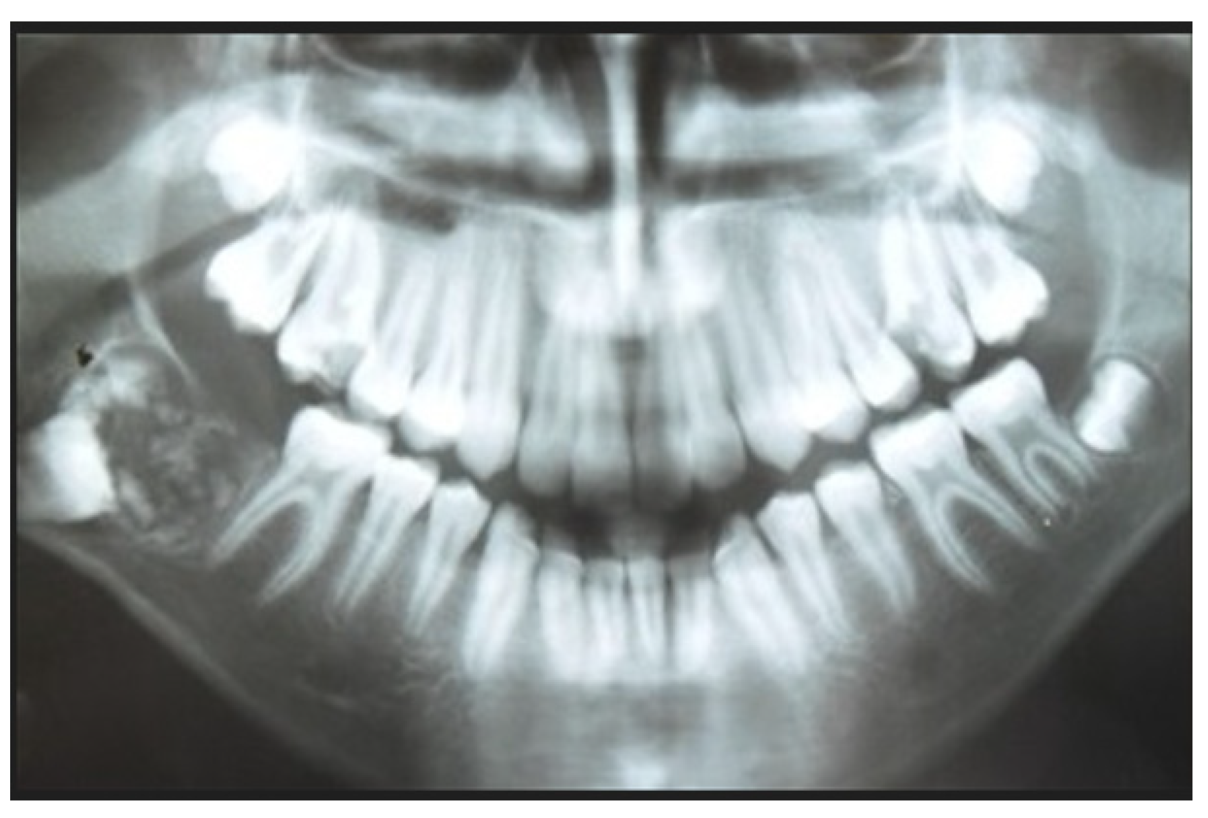
Figure 1. Preoperative panoramic radiography showed well-defined radiolucent region, which contained irregular radiopaque masses of varying sizes and shapes and deeply displaced tooth 47. This lesion occupied an area from the lower right first molar area to the right ramus. Courtesy of Dr. W. Pawlak.
Applsci 12 12963 g001
Figure 2. Enlarged visualization of the tumor mass in the right mandibular angle. Courtesy of Dr. W. Pawlak.
Applsci 12 12963 g002
Figure 3. CBCT axial projection of the bone. The image shows an expansile, well-circumscribed unilocular radiolucent lesion containing calcified masses compatible with odontogenic tissue and the unerupted right second molar (tooth 47). Courtesy of Dr. W. Pawlak.
Applsci 12 12963 g003
Figure 4. CBCT sagittal projection of the bone. Courtesy of Dr. W. Pawlak.
Applsci 12 12963 g004
Figure 5. Intraoperative photo of the bony defect after removal of the lesion and impacted tooth 47. Courtesy of Dr. W. Pawlak.
Applsci 12 12963 g005
Figure 6. Specimen–removed calcified bodies of varying shapes and sizes compatible with odontogenic tissue and the unerupted tooth 47 (black arrow). Courtesy of Dr. W. Pawlak.
Applsci 12 12963 g006
Figure 7. Histopathological examination of excised specimen. Round islands and solid areas of odontogenic epithelium in a background composed of primitive connective tissue. Hyaline-like material surrounding the islands is present. Dentin tissue is seen at the bottom right-hand corner of the figure. H-E staining, magn. 200×. Courtesy of Prof. Piotr Ziółkowski.
Applsci 12 12963 g007
Figure 8. Control panoramic radiograph and completely healed bone after treatment.
Applsci 12 12963 g008
Figure 9. Enlarged visualization of the entirely healed bony defect in the right mandibular angle. Courtesy of Dr. W. Pawlak.
Applsci 12 12963 g009
Publisher’s Note: MDPI stays neutral with regard to jurisdictional claims in published maps and institutional affiliations.