Article

Simulation and Experimental Study on the Responses of Subcellular Structures in Tumor Cells Induced by 5 ns Pulsed Electric Fields

State Key Laboratory of Power Transmission Equipment and System Security and New Technology, Chongqing University, Chongqing 400044, China
*
Author to whom correspondence should be addressed.
Appl. Sci. 2023, 13(14), 8142; https://doi.org/10.3390/app13148142
Submission received: 30 April 2023 / Revised: 3 July 2023 / Accepted: 7 July 2023 / Published: 13 July 2023

Abstract

:
In order to explore the bioelectric effect of 5 ns pulsed electric fields on tumor cells, a spherical single-cell multiphysics model was first established based on the finite element simulation platform. In consideration of the dielectric relaxation of the biological plasma membrane under the high-frequency electric fields, the electroporation and Maxwell stress tensors on the cell membrane and nuclear envelope were analyzed; secondly, taking MDA-MB-231 cells as the research object, combined with fluorescent probe technology, the state change and fluorescence dissipation of its subcellular structure exposed to pulse fields were studied. The results showed that 5 ns pulsed electric fields directly acted inside the cell, causing an electroporation effect and tensile stress on the nuclear envelope, destroying the integrity and order of the cytoskeleton, and damaging the functions of subcellular structures including endoplasmic reticulum, mitochondria, etc. This study provides theoretical and experimental evidence for the research and application of a high-voltage short pulse in the field of biomedical engineering.

1. Introduction

Cancer poses a grave global challenge, jeopardizing the lives and well-being of individuals across the globe. Recent data from the International Agency for Research on Cancer (IARC), a division of the World Health Organization, reveals alarming statistics: in 2020 alone, there were 19.29 million newly diagnosed cancer cases and a staggering 9.96 million cancer-related deaths worldwide [1]. Cancer has consistently remained one of the most intricate and challenging human diseases, often referred to as a ‘terminal illness’. Consequently, the advancement of cutting-edge medical technologies in tumor treatment has emerged as a pivotal area demanding urgent breakthroughs. Addressing this pressing need has become a primary direction for development, holding immense significance for the field of medicine.
Traditional methods for cancer treatment encompass surgical intervention, radiation therapy, and chemotherapy. Among them, surgery is currently the preferred approach for managing tumors, particularly in early stage cancer. However, as cancer progresses to advanced stages, surgical procedures only offer palliative benefits or life extension and do not eradicate the cancer. Furthermore, the trauma induced during tumor surgery may stimulate the growth and metastasis of cancerous tissue [2]. Radiation therapy involves the targeted application of radiation to eliminate tumor cells. However, this approach also affects surrounding healthy cells or tissues, leading to significant side effects. Hence, radiation therapy is typically combined with chemotherapy to enhance its effectiveness [3]. Chemotherapy utilizes chemical drugs to systemically treat tumors, exhibiting the therapeutic efficacy against both primary and metastatic lesions. Despite its significant treatment outcomes, the administration of high doses of drugs can cause harm to the body, with more effective treatments often accompanied by greater toxic side effects [4]. In recent years, ablation therapy has emerged as a rapidly evolving treatment modality, incorporating techniques such as radiofrequency ablation [5], microwave ablation [6], and cryoablation [7]. These methods offer promising alternatives to conventional treatments. The treatment method involves the application of various heat sources to the tumor region using minimally invasive treatment needles, leading to the destruction of adjacent tumor tissues. This approach exhibits minimal side effects and avoids drug resistance. However, when treating tissues near blood vessels, there is a possibility of “heat deposition” that may affect the treatment outcome and potentially result in sequelae. Clinical data reveal that, although traditional treatment methods can achieve an efficiency of up to 95% in treating early stage highly malignant tumors, they often encounter challenges such as relapses in the primary and surrounding areas or even distant metastases during later stages of treatment (after 3 months) resulting in treatment failure [8]. Immunotherapy [9] has the potential to enhance the anti-tumor immune response. It involves the use of drugs to activate or stimulate the immune system, enabling it to naturally attack tumor cells and prevent tumor cells from evading immune responses. However, the curative effect of immunotherapy varies, with only a subset of patients exhibiting a response. Consequently, predicting a patient’s response to treatment becomes challenging [10]. Additionally, in terms of safety, immunotherapy can trigger autoimmune side effects in some patients, leading to an immune response against healthy tissues [11].
With the advancements in bioelectromagnetics, electroporation therapy has found applications in cancer treatment. By applying pulsed electric fields to the cell membrane, unique effects are generated, which reduce the required drug dosage, enhance the treatment efficacy, and minimize the drug-related side effects on the body. This approach also preserves tissue functionality at the treatment site. The tumor treatment method utilizing pulsed electric fields mainly applies electric fields to the local lesion area through electrodes, disrupting the insulation properties of local tissue cell membranes, inducing the formation of micro-pores or electric pores, increasing membrane permeability and conductivity, disrupting the physiological balance inside and outside the cell, and ultimately leading to cell death [12]. High-intensity pulsed electric fields with microsecond durations (1–100 μs) can cause the irreversible perforation of cell membranes, leading to the collapse of transmembrane potential, disruption of cell membrane integrity, and release of intracellular contents, ultimately resulting in tumor cell death. This therapy is highly controllable, non-toxic, efficient, and has shown promising clinical efficacy [13]. However, the prolonged duration of pulse application can lead to strong muscle contractions in patients, which can be inconvenient during treatment. Consequently, sub-microsecond pulse electric fields (200–500 ns) have garnered increased attention, as their shorter pulse width acts simultaneously on intracellular and extracellular membranes, exploiting their potential to induce markedly different biological effects due to their pulse width being shorter than the cell membrane’s charging time [14]. This approach exhibits low side effects and high safety, showing excellent prospects for application. Researchers have conducted simulation studies on the perforation characteristics under the influence of pulse electric fields using molecular dynamics [15], finite element models [16], numerical models [17], and equivalent circuit models [18]. Moreover, experimental studies using techniques such as fluorescence probes [19], microscopy [20], and impedance measurement [21] have been carried out to analyze DNA damage [22], morphological changes [23], and changes in dielectric properties [24] induced by nanosecond pulses. These studies aim to elucidate the interaction mechanisms between electric fields and cells/tissues and preliminarily verify the safety and effectiveness of pulse electric fields in clinical tumor treatment, thereby promoting rapid development in the biomedical field. Further research has revealed that high-voltage nanosecond pulsed electric fields (nsPEFs) with pulse widths shortened to several nanoseconds have rich high-frequency components, which can selectively disrupt and perforate organelles without compromising the surface membrane integrity [25,26]. nsPEFs can also dissipate mitochondrial membrane potential [27], induce intracellular calcium waves [28], and damage chromosome telomeres [29]. Additionally, nsPEFs exert mechanical effects on subcellular structures in tumor cells, particularly affecting actin, microtubules, and intermediate filaments in the cell skeleton [30,31]. However, the mechanism of the force-electric response of nsPEFs on tumor cells remains unclear and necessitates further exploratory research in this field.
Therefore, this study aims to investigate the biophysical and electrical effects of nsPEFs on tumor cells through a combination of theoretical and experimental approaches. Firstly, a multiphysical field model is developed to simulate and analyze the electrical perforation and Maxwell stress experienced by cell membranes and nuclear membranes under nsPEFs in the presence of high-frequency electric fields. Secondly, to assess the impact of nsPEFs on subcellular structures, preliminary investigations are conducted on the bioelectrical responses of cell skeletons and subcellular structures using human breast cancer cells (MDA-MB-231) in cell experiments. Thirdly, the dose–effect relationship between nsPEFs and the bioelectrical responses of subcellular structures such as endoplasmic reticulum, mitochondria, and the cell nucleus is examined. The findings from this study hold significant scientific and medical value, providing important insights for the research and application of high-voltage nsPEFs in the field of biomedical engineering.
The subsequent sections of this article are structured as follows: Section 2 provides a detailed explanation of the proposed method. In Section 3, the simulation and experimental results are presented. Subsequently, Section 4 discusses the obtained experimental findings. Finally, Section 5 presents the concluding remarks of this study.

2. Materials and Methods

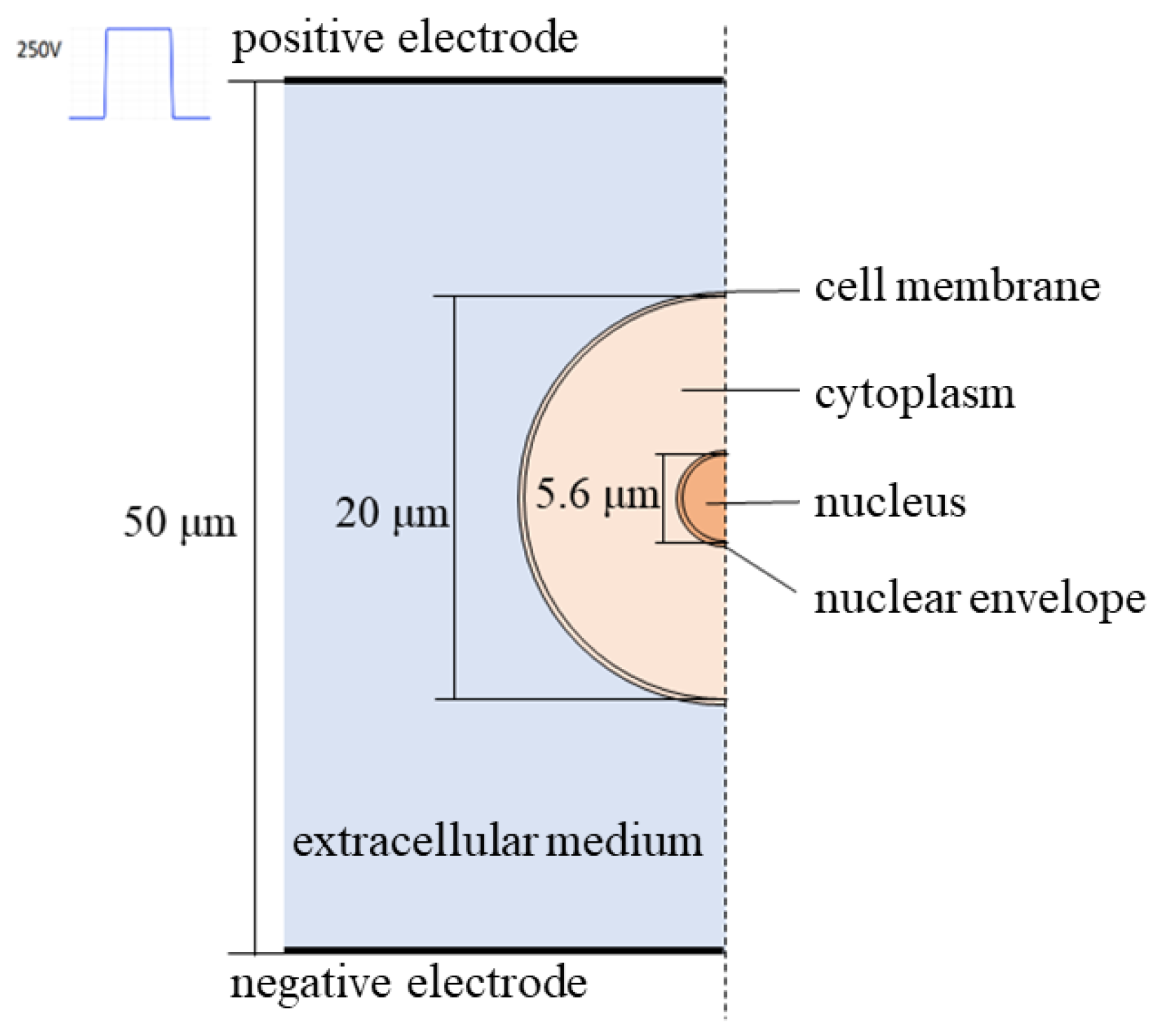
2.1. Establishment of Cell Mechatronic Model

In this study, a multiphysics model of a five-layered spherical cell was developed using COMSOL Multiphysics 5.6 software. To simplify the computational load in three dimensions, a two-dimensional axisymmetric calculation method was employed, taking advantage of the system’s symmetry. The calculation domain was a rectangular region measuring 25 μm × 50 μm, with the cell positioned at the center of the axis of symmetry, as depicted in Figure 1. The region outside the cell in the rectangular area represented the extracellular fluid, while the layers of the cell from the innermost to outermost were the nucleoplasm, nuclear membrane, cytoplasm, and cell membrane. A pulse source was connected to the upper boundary of the rectangle, while the lower boundary was grounded, generating nsPEFs within the calculation domain. The left and right boundaries were electrically insulated.
In this study, simulation parameters commonly used in both domestic and international research [32,33,34] were selected, ensuring their significant representativeness. To facilitate readers’ understanding, all the parameters are provided in Table 1. In order to accurately simulate the force-electric response of cells under nsPEFs, the finite element model utilized the asymptotic electroporation model to calculate the electroporation of the cell membrane and nuclear membrane [32]. Moreover, the second-order Debye dispersion model was employed to describe the dielectric relaxation phenomenon of the biomembrane under high-frequency electric fields [35]. Additionally, considering the viscoelastic properties of the biomembrane, the Maxwell stress tensor method was utilized to calculate the stress experienced by the biomembrane in the presence of nsPEFs.
The idea of the above modeling is as follows:
When an external electric field acts on the membrane structure of a cell, the membrane structure is polarized. When the transmembrane voltage Vm on the cell membrane and nuclear membrane reaches a specific threshold (~1 V) [36], the screening effect of the biomembrane is “pierced”, i.e., electroporation occurs. The occurrence of electroporation leads to an increase in the membrane’s electrical conductivity, followed by a decrease in the transmembrane voltage. When electroporation occurs, the rate of pore formation can be expressed as
d N d t = α e ( V m ( t ) V e p ) 2 ( 1 N N 0 e q ( V m ( t ) V e p ) 2 )
where N is the pore density on the membrane, N0 is the equilibrium pore density in the absence of applied electric field, Vep is the characteristic voltage of electroporation, α, q are constants. As the pore density increases, the membrane conductivity increases, and at this time, the transmembrane current density J(t) [34] is
J ( t ) = V m d σ ( t ) + ε ( t ) d V m t
In the equation, d is the thickness of the cell membrane, ε(t) is the dielectric constant of the membrane. σ(t) represents the electrical conductivity of the cell membrane, which can be composed of static conductivity and dynamic conductivity.
σ ( t ) = σ 0 + N ( t ) σ p π r p 2 K
In the equation, σ0 is the initial electrical conductivity of the cell membrane, σp is the conductivity of the pore, rp is the pore radius, and K is the voltage-dependent variable across the membrane [16].
When high-frequency electrical pulses are applied, due to the dielectric relaxation effect of the biomembrane, its dielectric constant significantly decreases [35], and the second-order Debye dispersion model describes the complex dielectric constant of the dielectric in the frequency domain.
ε ˜ = ε + i = 1 2 Δ ε i 1 + j ω τ i
In the equation, ε is the dielectric constant when the frequency tends towards infinity, Δεi and τi are the amplitude and relaxation time constants of the i-th order relaxation, respectively. The polarization vector P of the biomembrane is defined as follows:
P = ( ε ˜ ε 0 ) E
In the equation, ε0 is the permittivity of the vacuum, and E is the local electric field. The electric displacement vector D on the membrane is given by
D = ε 0 E + P
The electric field forces acting on each component of the cell can be described by the Maxwell stress tensor, and can be calculated by integrating the Maxwell stress tensor over the surface of each membrane. The equation is:
T = [ T x x T x y T x z T y x T y y T y z T z x T z y T z z ]
F = S T d A
T i j = ε 0 ( E i E j 1 2 δ i j E 2 )
where δij is the Kronecker symbol.
This paper assumes that the viscoelasticity of the membrane follows the generalized Maxwell model, which can be expressed as:
f ( t ) = 2 0 t Γ ( t τ ) ε ( t ) t d τ
Γ ( t ) = G 0 + G 1 e t / τ
In the equation, f(t) and ε(t) represent the total stress and strain, respectively; Γ(t) is the relaxation shear modulus function; G0 is the shear modulus of the elastic branch, and G1 is the unit shear modulus of the additional branch; τ is the relaxation time of the branch, defined as the ratio of viscosity η to shear modulus τ = η/G.

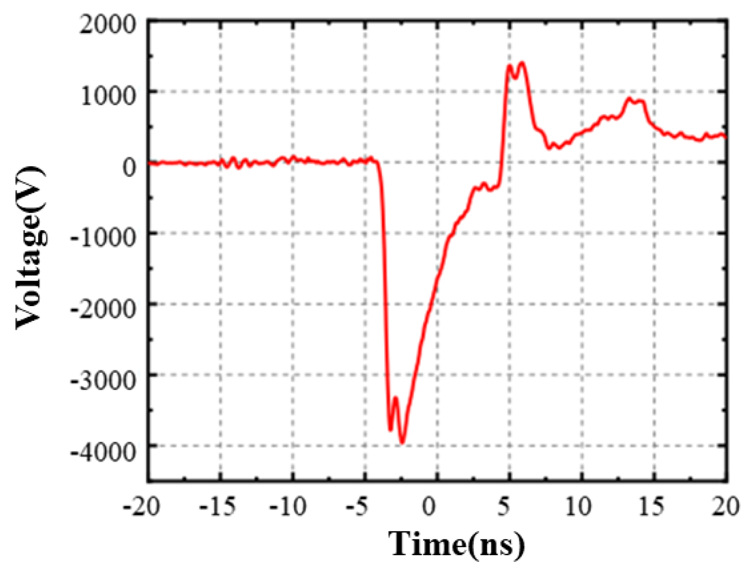
2.2. Experimental Device

The nanosecond pulse generator used for the experiment in this paper was developed within the laboratory. It boasts adjustable output pulse parameters ranging from 3 to 5 kV, and the pulse width is set at 5 ns. Figure 2 illustrates the experimental voltage waveform recorded by the oscilloscope during the experiment.
The experimental setup utilizes a custom-designed microelectrode configuration, as shown in Figure 3. The electrode spacing is 0.5 mm, and the overall length of the microelectrodes is 35 mm. They can be securely attached to a 6-well plate (Thermo Fisher Scientific, Waltham, MA, USA), tightly adhering to the bottom of the wells to apply pulsed electric fields with selected parameters to the adherent cells.
The nsPEFs generation system consists of a pulse generator, WavePro760Zi-A oscilloscope (LeCroy, Chestnut Ridge, Rockland County, NY, USA), high-voltage probe (LeCroy, USA), and a 6600 Pearson current coil (Pearson, Palo Alto, CA, USA). Real-time observation and imaging of the cell area under the electrode were performed using an inverted fluorescence microscope (Leica DMi8, Eggenstein-Leopoldshafen, Baden-Württemberg, Germany).

2.3. Cell Culture

The human breast cancer cell line MDA-MB-231 (purchased from the Cell Bank of Shanghai Institutes for Biological Sciences, Chinese Academy of Sciences) used in the experiment was cultured in DMEM (Dulbecco’s modified Eagle medium, GIBCO, Waltham, MA, USA) containing a combination of antibiotics (100 μg/mL penicillin and 100 μg/mL streptomycin, GIBCO, USA), and 10% inactivated fetal bovine serum (GIBCO, USA), and incubated in a humidified incubator (Thermo Fisher Scientific, USA) at 37 °C and 5% CO2.

2.4. Cell Fluorescence Experiment

Fixed-cell observation experiment: in the cytoskeleton observation experiment, the cell suspension was seeded in a 6-well plate and incubated for 24 h. The cells in the control group were not treated with the electric field; the cells in the experimental group were subjected to pulsed electric field through flat electrodes with a spacing of 0.5 mm. After the pulse was applied, each group waited for 0, 2, and 3 min, respectively. Add 4% paraformaldehyde (Solarbio, Beijing, China) to fix the cells, add 0.5% Triton X-100 (Solarbio, China) to increase the cell permeability, and add FITC-labeled phalloidin (Solarbio, China) to stain the cytoskeleton. Observe the fluorescence brightness and morphology of the cytoskeleton under an inverted fluorescence microscope.
Live cell real-time observation experiment: in the live-cell staining experiments for mitochondria, endoplasmic reticulum, and nucleus, cells were seeded in 6-well plates and incubated for 24 h. Mito-Tracker Red CMXRos (Beyotime, Nantong, China) was added to stain the mitochondria, ER-Tracker Green (Beyotime, China) was used to stain endoplasmic reticulum, and DAPI (Beyotime, China) was used to stain the nucleus. Customized microelectrodes were used to apply nsPEFs to the cells, and changes in the subcellular structures were observed in real-time under a fluorescence microscope. The degree of fluorescence quenching was used to characterize the degree of damage to each subcellular structure by nsPEFs. For the experimental group, one set of fluorescence images was captured after every 1000 pulses until all pulses were completed. For the control group, fluorescence images were taken at the same time intervals and frequency as the experimental group to ensure equal levels of fluorescence quenching across all groups.

2.5. Statistical Analysis

The experiment was analyzed using one-way ANOVA and t-test, and when the p value < 0.05 indicates a significant difference (*: p < 0.05; **: p < 0.01; ***: p < 0.001).

3. Results

3.1. Simulation Result

This study investigated the electroporation effect on cells under nsPEFs with a pulse width of 5 ns and an electric field strength of 50 kV/cm. The pore density on the cell membrane and nuclear membrane was analyzed, and the simulation results, as shown in Figure 4, indicated that the pore density on the nuclear membrane increased first under nsPEFs, and the pore density on the nuclear membrane was consistently higher than that on the cell membrane. The final average pore density on the nuclear membrane reached 7.12 × 10¹⁵ m−2, which was higher than the average pore density on the cell membrane of 3.5071 × 10¹⁵ m−2. The pore area ratio on the nuclear membrane (84%) was also higher than that on the cell membrane (77%). The electroporation pores were mainly distributed at both ends of the cell membrane and the nuclear membrane near the electrode. The maximum pore density on the nuclear membrane was 1.6245 × 10¹⁶ m−2, which was also higher than the maximum pore density on the cell membrane of 8.4693 × 10¹⁵ m−2.
Figure 5 shows the Maxwell stress on the cell membrane and nuclear membrane under nsPEFs. As can be seen from the figure, the force on the cell membrane and nuclear membrane corresponds to the spatial distribution of pore density, and the stress on the nuclear membrane is significantly greater than the electric field force on the cell membrane. The stress direction mainly focuses on the outward stretching of the nuclear membrane along the electric field direction, and the maximum value of the Maxwell stress in this direction reaches 2.6828 × 10⁴ N/m, inducing a strain trend that stretches the nucleus along the electric field direction.

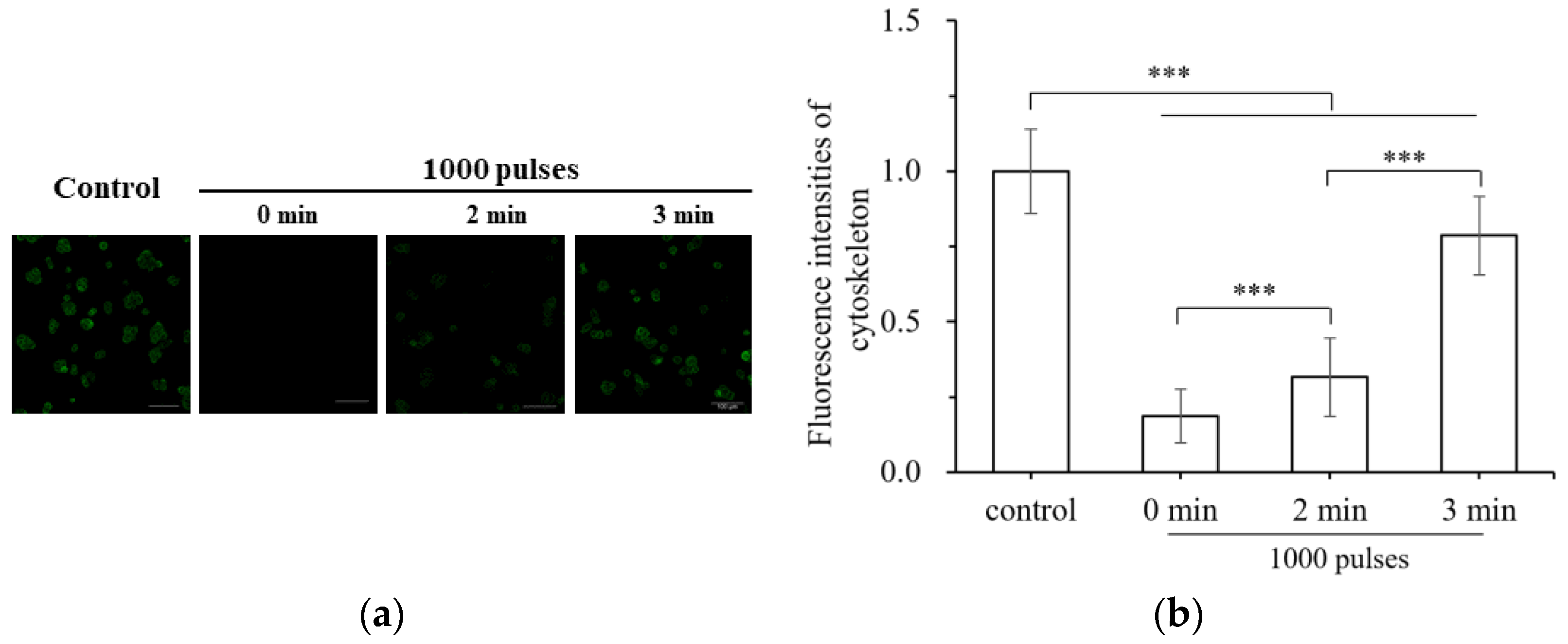
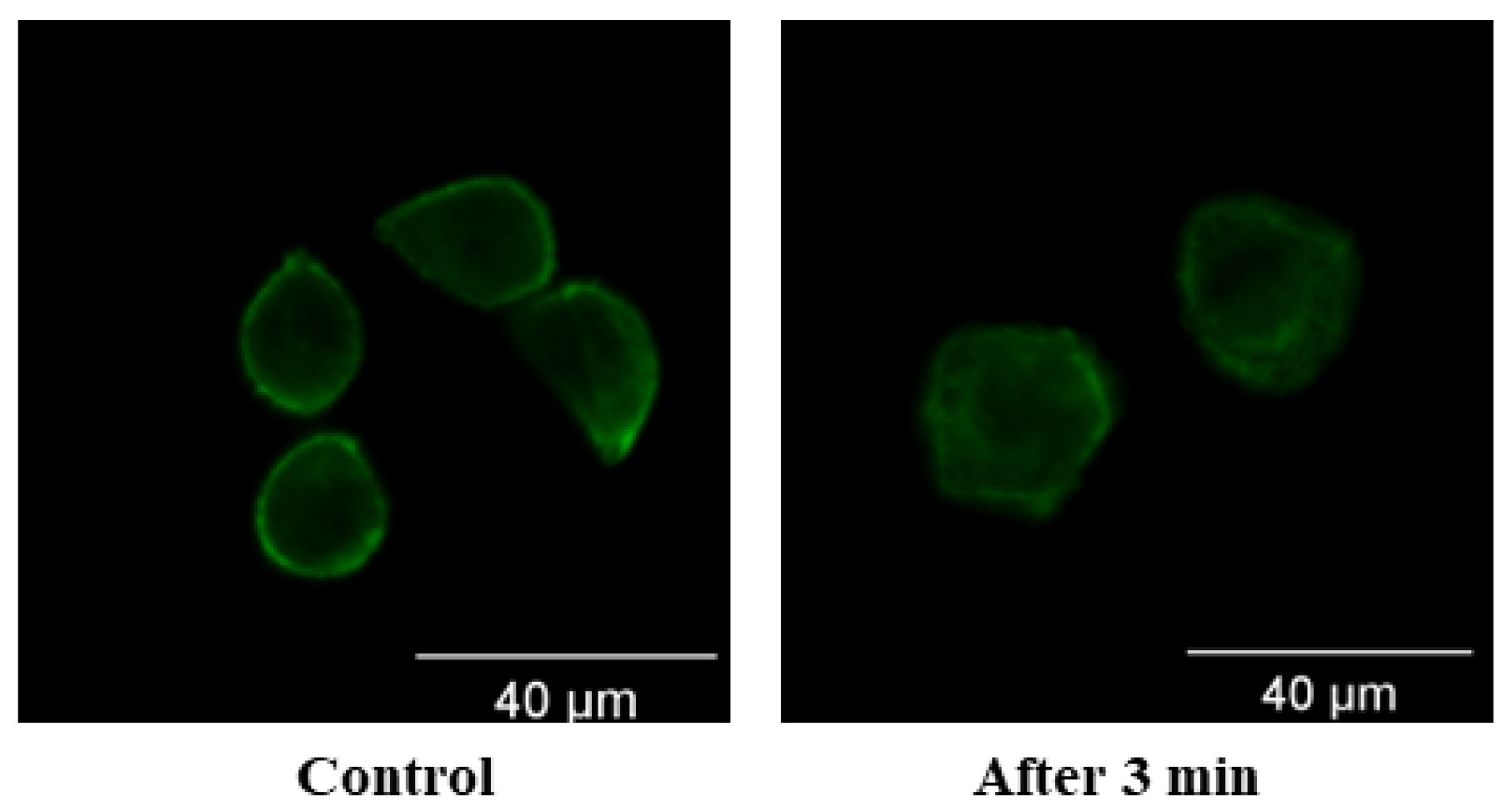
3.2. Damage of Subcellular Structure

The cytoskeleton, serving as a supporting structure for cell morphology and mechanical properties, also plays a crucial role in substance transport, signal transmission, and cellular metabolic processes [37]. In this study, MDA-MB-231 cells were exposed to 1000 nsPEFs at an electric field intensity of 80 kV/cm, and the fibrous actin (f-actin) within the cytoskeleton was stained for observation. The experiment revealed that, immediately after nsPEF treatment (0 min), minimal fluorescence was observed, indicating the disruption of cytoskeletal integrity. However, at 2 and 3 min, the fluorescence gradually reappeared, as depicted in Figure 6. Under the influence of the electric field, the actin within the cytoskeleton depolymerized into monomers, resulting in the disappearance of green fluorescence. Within a few minutes, the actin reassembled, and the green fluorescence reappeared, indicating the recovery and reorganization of the cytoskeletal structure.
However, it was observed that the cell skeleton did not fully recover to its original state following nsPEF treatment. To investigate the changes in the cell skeleton, the fluorescence images of the control group and the cell skeleton at 3 min after treatment were magnified for observation. The control group exhibited an orderly and tightly arranged cell skeleton, positioned along the inner side of the cell membrane, providing physical support for maintaining cell shape and facilitating stretching and compression. In contrast, the experimental group displayed a disordered and irregular cell skeleton at the cell boundary, indicating a failure in the support provided by the cell skeleton for cell morphology, as depicted in Figure 7. It is evident that, although the protein structure integrity of the cell skeleton had partially recovered within 3 min after the termination of electric field stimulation, its overall orderliness remained disrupted and the incomplete restoration of the cell skeleton was observed during the experiment.
Based on the previous simulation results, it is evident that nsPEFs have the ability to deeply penetrate the interiors of cells and exert their effects on biomembranes within the cells, leading to a more pronounced force-electric response. To further investigate this, fluorescent probe labeling observations were conducted on various cellular components, including the cell nucleus and organelles with membrane structures such as mitochondria and endoplasmic reticulum. The experimental procedure involved applying nsPEFs with a frequency of 100 Hz, a pulse width of 5 ns, and an electric field intensity of 100 kV/cm for a range of 2000–10,000 pulses. The fluorescence intensity of the aforementioned organelles gradually decreased during the process, as illustrated in Figure 8. The endoplasmic reticulum was labeled with green fluorescence, mitochondria with red fluorescence, and the nucleus with blue fluorescence. The extent of fluorescence decay observed in the figure positively correlated with the number of pulses applied, indicating a greater degree of structural damage to the nucleus, mitochondria, and endoplasmic reticulum with an increasing nsPEFs dose. Unlike traditional microsecond-pulsed electric fields that primarily induce the electroporation of the cell membrane, high-frequency pulsed electric fields have the ability to penetrate the cell membrane barrier and directly impact the membrane structures within the cell, resulting in damage to various subcellular structures and consequent alterations in cell function.
Subsequent analysis involved the quantification of the fluorescence intensity of the aforementioned organelles, as depicted in Figure 9. As the number of pulses increased, the fluorescence intensity of each organelle progressively decreased, indicating a more severe degree of organelle damage. Among the organelles, the endoplasmic reticulum (ER) exhibited the largest membrane structure area, accounting for approximately 50% of the total membrane area within the cell, and it was the most susceptible to the effects of nsPEFs. Consequently, the decrease in fluorescence intensity observed in the ER was the most prominent among the three organelles.
The fluorescence intensities of the organelles were analyzed following the exposure to pulse electric fields (nsPEFs) with electric field intensities of 70 kV/cm, 80 kV/cm, 90 kV/cm, and 100 kV/cm, as illustrated in Figure 10. It should be noted that the fluorescence intensity of the control group, which was not subjected to pulse electric fields, exhibited a slight decrease. This minor decrease can be attributed to the natural quenching of the fluorescence dye under the illumination of the fluorescence microscope lamp. However, the fluorescence attenuation observed in all experimental groups was significantly different from that of the control group (p < 0.001). As depicted in the figure, an increase in the electric field strength resulted in a gradual decrease in the fluorescence intensities of the mitochondria and endoplasmic reticulum, indicating an increased degree of organelle damage in response to higher applied pulse electric fields. However, it is important to note that the degree of damage to the cell nucleus was not found to be related to the strength of the electric field.

4. Discussion

Due to the low dielectric constant of the cell membrane, traditional microsecond-level pulse electric fields face difficulties in penetrating the “capacitance” of the cell membrane due to the dominance of low-frequency components. As a result, the primary target of pulse electric fields above the microsecond level is the cell membrane itself. By reducing the pulse width, it becomes possible to target the intracellular biomembranes, thereby enabling the regulation of subcellular structures. Investigating the mechanism behind the force-electric response of subcellular structures under high-frequency and high-voltage pulse electric fields can contribute to the advancement of pulse power technology in clinical and biomedical engineering fields. This research has the potential to address the current limitations of mainstream thermal tumor therapy, such as heat loss and poor prognosis [38].
Based on the finite element simulation results of the progressive electric perforation model, this study demonstrates that nsPEFs induce the electroporation of the nuclear membrane faster and at a significantly higher pore density compared to the cell membrane. This finding suggests a conversion of the pulsed electric field target from the cell membrane to the nuclear membrane. Previous simulation studies by Qiu H. et al. [34] have shown that a pulse width of 60 ns is more likely to cause the perforation of the inner and outer mitochondrial membranes compared to a 600 ns pulse width. Additionally, Rao X et al. [39] applied pulsed electric fields with pulse widths ranging from 30 ns to 100 μs to normal and cancerous skin cell models and found that nsPEFs with rich high-frequency components generate higher transmembrane voltages on the nuclear membrane. Equivalent circuit model analysis reveals that biological membranes can be considered as high-pass filters for pulsed electric fields due to their small dielectric constant and large equivalent capacitance. Consistent with the results of this study, nsPEFs contain rich high-frequency components that can penetrate the cell interior based on the cell membrane’s low permeability at high frequencies, in addition to inducing the electroporation of the cell membrane through high amplitude. In a study by Zaklit J. et al. [40], a pulsed electric field with an amplitude of 80 kV/cm and a pulse width of 5 ns was applied to a chromaffin cell model, which resulted in the electroporation of not only the cell membrane but also the endoplasmic reticulum membrane, secretory granule membrane, and outer mitochondrial membrane. However, Zaklit J. et al. hold the belief that the electroporation threshold of subcellular biomembranes is higher than that of the cell membrane, which contradicts the findings of this study and previous studies [41,42,43]. In addition to elucidating the mechanism of nsPEFs-induced subcellular structural damage through pore density, this study also calculated the Maxwell stress exerted on the cell membrane and nuclear membrane. The results indicate that the electric field force acting on the nuclear membrane is significantly greater than that on the cell membrane. From an energy perspective, the higher the transmembrane voltage across the cytoplasmic membrane, the greater the electric field force it experiences. Moreover, a higher energy level of the external electric field acting on the biomembrane makes it easier to surpass the electroporation energy barrier, leading to the electroporation and expansion of pore radius through the influence of the electric field force [44,45].
The cellular force-electric response to pulsed electric fields (PEFs) has been extensively studied as a crucial approach to investigate the different cell death pathways under electric field pulse stress. For instance, Chafai D. E. et al. [46] applied esPEF with a pulse width of 11 ns to cells and observed weakened microtubule polymerization, along with changes in zeta potential and microtubule structure. Similarly, Berghöfer T. et al. [47] treated tobacco BY-2 cells with esPEF of 10 ns pulse width, resulting in the depolymerization of cortical actin, the detachment of fibrous actin bundles from the cell periphery, and their contraction towards the nucleus. Cell migration and invasion are triggered by various chemotactic agents, which, upon binding to cell surface receptors, activate intracellular signaling pathways regulating actin cytoskeleton reorganization. Invasive protrusions or filopodia-like structures are generated by cancer cells to invade the extracellular matrix and migrate to distant sites, necessitating significant actin microfilament polymerization [48]. The depolymerization and geometric changes of cytoskeletal proteins under PEF were also confirmed in this study. Under nsPEF treatment, the anchoring between the actin layer and the plasma membrane was disrupted, resulting in their separation. Concurrently, the actin layer depolymerization and fibrous actin disruption increased the cell elasticity and aggravated the strain on various cellular components when subjected to force [49,50], akin to the effects of actin polymerization inhibitors [30].
In the experiment presented in this article, the fluorescence intensity of the cell nucleus significantly decreased following nsPEF treatment, indicating the successful penetration of the high-frequency pulsed electric field into the cell and its impact on the nucleus. This resulted in damage to the nuclear membrane and the disruption of the nuclear matrix. Researchers at the Center for Bioelectrics at Old Dominion University in the United States have employed pulsed electric fields with pulse widths ranging from 10 to 300 ns to treat cells and observed that the nuclear membrane becomes charged and forms electroporation pores. Furthermore, the pulsed electric field alters the structure of the nuclear protein complex, induces double-strand breaks in DNA, disrupts cell transcription mechanisms, and promotes cell apoptosis [51,52,53]. However, literature reports also suggest that nuclear membrane electroporation facilitates the entry of DNA plasmids into the nucleus, thereby enhancing the gene expression in cells [30]. Hence, it becomes essential to control nsPEF pulse parameters to strike a balance between cell killing by pulsed electric fields and gene transfection, which remains a significant scientific question for the further advancement of nsPEFs.
Mitochondria are vital organelles that play a crucial role in providing cellular energy through aerobic respiration. They are also involved in significant processes such as cell differentiation, signal transduction, and apoptosis regulation. The findings of this study demonstrate that treating MDA-MB-231 cells with nsPEFs resulted in the dissipation of mitochondrial membrane potential, the disruption of the double membrane structure, and release of cytochrome c from the mitochondria. This release of cytochrome c activated the apoptotic protein caspase, leading to apoptosis [27,54]. Additionally, the permeabilization of the mitochondrial membrane resulted in ATP depletion, which acts as a critical damage-associated molecular pattern (DAMP) and triggers immunogenic cell death, including necrosis [55,56]. The endoplasmic reticulum (ER) is a large membrane system responsible for protein and lipid synthesis required by the cell, as well as intracellular material transfer. When exposed to nsPEFs, the ER fluorescence dissipated, and the ER membrane underwent electroporation, leading to the release of calcium ions [57], ER stress, and subsequent cell apoptosis [58]. Furumoto Y et al. [58] conducted experiments using pulse electric fields with a pulse width of 14–70 ns and an amplitude of 20–100 kV/cm on Hela cells and mouse embryonic fibroblasts (MEFs), and their results indicated that higher frequency short pulse electric fields induced more significant ER stress. The osmotic pressure inside mitochondria and the ER is higher than that of the cytoplasmic environment. Therefore, the pulse electric field increases the permeability of the mitochondrial and ER membranes, leading to calcium ion release and the swelling of these organelles [56,59]. The physical connection between mitochondria and the ER is responsible for mediating various physiological functions, including autophagy, intracellular calcium dynamic balance, phospholipid synthesis and transport, and metabolic signal transduction. Pulse electric fields disrupt this physical connection, thereby inhibiting the aforementioned cellular functions [56].

5. Conclusions

Based on the simulation of a single-cell multi-physics field model of ball cells and combined with experiments at the multicellular level, this article explores the biophysical and electrical effects of subcellular structures under high-voltage nsPEFs (nanosecond pulsed electric field stimulation), and draws the following conclusions:
  • Due to the extremely short pulse duration and rich high-frequency components of high-voltage nsPEFs, they can pass through the cell membrane with large equivalent capacitance and reach the membrane structures inside the cell as the target site.
  • Under the action of high-voltage nsPEFs, the cell membrane structure undergoes electric perforation and is subjected to Maxwell stress. The stress on the nuclear membrane is greater than that on the cell membrane, which causes the nuclear membrane to stretch along the electric field direction.
  • High-voltage nsPEF stimulation causes fibrous actin bundles to depolymerize, increasing the degree of strain on cells after stress and damaging the integrity and order of the cell skeletons. The integrity of the cell skeleton will recover to some extent, but the order will not be restored after being disrupted.
  • High-voltage nsPEFs can damage the subcellular structures of tumor cells. Under the electric field, the nuclear membrane is perforated and damaged, affecting the nuclear matrix; the membrane potential of mitochondria is dissipated, the membrane structure is damaged, caspase apoptosis protein is activated, and ATP is depleted; endoplasmic reticulum perforation, calcium ion release, and endoplasmic reticulum stress occur.

Author Contributions

Conceptualization, C.Y. and X.M.; methodology, X.M. and Y.W.; software, X.M. and K.Q.; data curation, X.M. and Y.W.; writing—original draft preparation, X.M.; writing—review and editing, K.Q., C.Y. and S.D.; supervision, C.Y. and S.D. All authors have read and agreed to the published version of the manuscript.

Funding

This research was funded by the National Natural Science Foundation of China, grant number 52237010.

Institutional Review Board Statement

Not applicable.

Informed Consent Statement

Not applicable.

Data Availability Statement

Not applicable.

Conflicts of Interest

The authors declare no conflict of interest.

References

  1. Global Cancer Observatory. Available online: https://gco.iarc.fr/ (accessed on 13 February 2023).
  2. Friebel, E.; Kapolou, K.; Unger, S.; Tugues, S.; Neidert, M.C.; Becher, B. Single-Cell Mapping of Human Brain Cancer Reveals Tumor-Specific Instruction of Tissue-Invading Leukocytes. Cell 2020, 181, 1626. [Google Scholar] [CrossRef] [PubMed]
  3. Specht, L.; Yahalom, J.; Illidge, T.; Berthelsen, A.K.; Constine, L.S.; Eich, H.T.; Girinsky, T.; Hoppe, R.T.; Mauch, P.; Mikhaeel, N.G.; et al. Modern Radiation Therapy for Hodgkin Lymphoma: Field and Dose Guidelines from the International Lymphoma Radiation Oncology Group (ILROG). Int. J. Radiat. Oncol. Biol. Phys. 2013, 89, 854–862. [Google Scholar] [CrossRef] [PubMed]
  4. Bach, D.H.; Hong, J.Y.; Park, H.J.; Lee, S.K. The role of exosomes and miRNAs in drug-resistance of cancer cells. Int. J. Cancer 2017, 141, 220–230. [Google Scholar] [CrossRef] [PubMed] [Green Version]
  5. Wahl, D.R.; Matthew, H.; Tao, Y.; Pollom, E.L.; Caoili, E.M.; Schipper, T.L.; Feng, M. Outcomes After Stereotactic Body Radiotherapy or Radiofrequency Ablation for Hepatocellular Carcinoma. J. Clin. Oncol. 2016, 34, 452. [Google Scholar] [CrossRef] [PubMed] [Green Version]
  6. Lubner, M.G.; Brace, C.L.; Ziemlewicz, T.J.; Hinshaw, J.L.; Lee, F.T., Jr. Microwave ablation of hepatic malignancy. Semin. Interv. Radiol. 2013, 30, 56–66. [Google Scholar]
  7. Andrade, J.G.; Wells, G.A.; Deyell, M.W.; Bennett, M.; Essebag, V.; Champagne, J.; Roux, J.F.; Yung, D.; Skanes, A.; Khaykin, Y.; et al. Cryoablation or Drug Therapy for Initial Treatment of Atrial Fibrillation. Mass. Med. Soc. 2021, 384, 305–315. [Google Scholar] [CrossRef]
  8. Solyanik, G.I. Multifactorial nature of tumor drug resistance. Exp. Oncol. 2010, 32, 181–185. [Google Scholar]
  9. Rosenberg, S.A. IL-2: The first effective immunotherapy for human cancer. J. Immunol. 2014, 192, 5451–5458. [Google Scholar] [CrossRef] [Green Version]
  10. Maleki Vareki, S.; Garrigós, C.; Duran, I. Biomarkers of response to PD-1/PD-L1 inhibition. Crit. Rev. Oncol. Hematol. 2017, 116, 116–124. [Google Scholar] [CrossRef]
  11. Williams, A.D.; Payne, K.K.; Posey, A.D., Jr.; Hill, C.; Jose, C.G.; June, C.H.; Tchou, J. Immunotherapy for Breast Cancer: Current and Future Strategies. Curr. Surg. Rep. 2017, 5, 31. [Google Scholar] [CrossRef] [Green Version]
  12. Li, C.; Yao, C.; Sun, C.; Guo, F. Dependence on electric field intensities of cell biological effects induced by microsecond pulsed electric fields. IEEE Trans. Dielectr. Electr. Insul. 2012, 18, 2083–2088. [Google Scholar] [CrossRef]
  13. Onik, G.; Rubinsky, B. Irreversible electroporation: First patient experience focal therapy of prostate cancer. In Irreversible Electroporation; Springer: Berlin/Heidelberg, Germany, 2010; pp. 235–247. [Google Scholar]
  14. Yao, C.; Hu, X.; Mi, Y.; Li, C.; Sun, C. Window effect of pulsed electric field on biological cells. IEEE Trans. Dielectr. Electr. Insul. 2009, 16, 1259–1266. [Google Scholar] [CrossRef]
  15. Tang, J.; Yin, H.; Ma, J.; Bo, W.; Yang, Y.; Xu, J.; Liu, Y.; Gong, Y. Terahertz electric field-induced membrane electro-poration by molecular dynamics simulations. J. Membr. Biol. 2018, 251, 681–693. [Google Scholar] [CrossRef] [PubMed]
  16. Yao, C.; Liu, H.; Zhao, Y.; Mi, Y.; Dong, S.; Lv, Y. Analysis of dynamic processes in single-cell electroporation and their effects on parameter selection based on the finite-element model. IEEE Trans. Plasma Sci. 2017, 45, 889–900. [Google Scholar] [CrossRef]
  17. Pucihar, G.; Miklavcic, D.; Kotnik, T. A time-dependent numerical model of transmembrane voltage inducement and electroporation of irregularly shaped cells. IEEE Trans. Biomed. Eng. 2009, 56, 1491–1501. [Google Scholar] [CrossRef] [PubMed] [Green Version]
  18. Huang, Y.; Rubinsky, B. Microfabricated electroporation chip for single cell membrane permeabilization. Sens. Actuators A Phys. 2001, 89, 242–249. [Google Scholar] [CrossRef]
  19. Ivey, J.W.; Latouche, E.L.; Sano, M.B.; Rossmeisl, J.H.; Davalos, R.V.; Verbridge, S.S. Targeted cellular ablation based on the morphology of malignant cells. Sci. Rep. 2015, 5, 1757. [Google Scholar] [CrossRef] [Green Version]
  20. Nesin, O.M.; Pakhomova, O.N.; Xiao, S.; Pakhomov, A.G. Manipulation of cell volume and membrane pore comparison following single cell permeabilization with 60- and 600-ns electric pulses. Biochim. Biophys. Acta (BBA) Biomembr. 2011, 1808, 792–801. [Google Scholar] [CrossRef] [Green Version]
  21. Ivorra, A.; Rubinsky, B. In Vivo electrical impedance measurements during and after electroporation of rat liver. Bioelectrochemistry 2007, 70, 287–295. [Google Scholar] [CrossRef]
  22. Beebe, S.J.; Sain, N.M.; Ren, W. Induction of cell death mechanisms and apoptosis by nanosecond pulsed electric fields (nsPEFs). Cells 2013, 2, 136–162. [Google Scholar] [CrossRef] [Green Version]
  23. Pakhomov, A.G.; Xiao, S.; Pakhomova, O.N.; Semenov, I.; Kuipers, M.A.; Ibey, B.L. Disassembly of actin structures by nanosecond pulsed electric field is a downstream effect of cell swelling. Bioelectrochemistry 2014, 100, 88–95. [Google Scholar] [CrossRef] [PubMed] [Green Version]
  24. Zhuang, J.; Kolb, J.F. Time domain dielectric spectroscopy of nanosecond pulsed electric field induced changes in dielectric properties of pig whole blood. Bioelectrochemistry 2015, 103, 28–33. [Google Scholar] [CrossRef] [PubMed]
  25. Schoenbach, K.H.; Joshi, R.P.; Kolb, J.F.; Chen, N.; Stacey, M.; Blackmore, P.F.; Buescher, E.S.; Beebe, S.J. Ultrashort electrical pulses open a new gateway into biological cells. Proc. IEEE 2004, 92, 1122–1137. [Google Scholar] [CrossRef]
  26. Beebe, S.J.; Fox, P.M.; Rec, L.J.; Somers, K.; Stark, R.H.; Schoenbach, K.H. Nanosecond pulsed electric field (nsPEF) effects on cells and tissues: Apoptosis induction and tumor growth inhibition. IEEE Trans. Plasma Sci. 2002, 30, 286–292. [Google Scholar] [CrossRef]
  27. Beebe, S.J.; Chen, Y.J.; Sain, N.M.; Schoenbach, K.H.; Xiao, S. Transient features in nanosecond pulsed electric fields differentially modulate mitochondria and viability. PLoS ONE 2012, 7, e51349. [Google Scholar] [CrossRef]
  28. Vernier, P.T.; Sun, Y.; Marcu, L.; Salemi, S.; Craft, C.M.; Gundersen, M.A. Calcium bursts induced by nanosecond electric pulses. Biochem. Biophys. Res. Commun. 2003, 310, 286–295. [Google Scholar] [CrossRef]
  29. Stacey, M.; Fox, P.; Buescher, S.; Kolb, J. Nanosecond pulsed electric field induced cytoskeleton, nuclear membrane and telomere damage adversely impact cell survival. Bioelectrochemistry 2011, 82, 131–134. [Google Scholar] [CrossRef] [Green Version]
  30. Graybill, P.M.; Davalos, R.V. Cytoskeletal disruption after electroporation and its significance to pulsed electric field therapies. Cancers 2020, 12, 1132. [Google Scholar] [CrossRef]
  31. Haberkorn, I.; Siegenthaler, L.; Buchmann, L.; Neutsch, L.; Mathys, A. Enhancing single-cell bioconversion efficiency by harnessing nanosecond pulsed electric field processing. Biotechnol. Adv. 2021, 53, 107780. [Google Scholar] [CrossRef]
  32. Neu, J.C.; Krassowska, W. Asymptotic model of electroporation. Phys. Rev. E 1999, 59, 3471. [Google Scholar] [CrossRef]
  33. Guo, F.; Zhang, L.; Liu, X. Nonlinear dispersive cell model for microdosimetry of nanosecond pulsed electric fields. Sci. Rep. 2020, 10, 19456. [Google Scholar] [CrossRef] [PubMed]
  34. Qiu, H.; Xiao, S.; Joshi, R.P. Simulations of voltage transients across intracellular mitochondrial membranes due to nanosecond electrical pulses. IEEE Trans. Plasma Sci. 2014, 42, 3113–3120. [Google Scholar] [CrossRef]
  35. Guo, F.; Qian, K.; Deng, H.; Li, X. Multiphysics analysis of nsPEF induced electrodeformation in a dispersive cell model. Appl. Phys. Lett. 2021, 118, 083701. [Google Scholar] [CrossRef]
  36. Weaver, J.C. Electroporation of cells and tissues. IEEE Trans. Plasma Sci. 2000, 28, 24–33. [Google Scholar] [CrossRef] [Green Version]
  37. Das, K.; Prasad, R.; Roy, S.; Mukherjee, A.; Prosenjit, S. The protease activated receptor2 promotes Rab5a mediated generation of pro-metastatic microvesicles. Sci. Rep. 2018, 8, 7357. [Google Scholar] [CrossRef] [PubMed]
  38. Hsiao, C.Y.; Huang, K.W. Irreversible electroporation: A novel ultrasound-guided modality for non-thermal tumor ablation. J. Med. Ultrasound 2017, 25, 195–200. [Google Scholar] [CrossRef]
  39. Rao, X.; Chen, S.; Alfadhl, Y.; Chen, X.; Sun, L.; Yu, L.; Zhou, J. Pulse width and intensity effects of pulsed electric fields on cancerous and normal skin cells. Sci. Rep. 2022, 12, 18039. [Google Scholar] [CrossRef]
  40. Zaklit, J.; Craviso, G.L.; Leblanc, N.; Yang, L.; Vernier, P.T.; Chatterjee, I. Adrenal chromaffin cells exposed to 5-ns pulses require higher electric fields to porate intracellular membranes than the plasma membrane: An experimental and modeling study. J. Membr. Biol. 2017, 250, 535–552. [Google Scholar] [CrossRef]
  41. Retelj, L.; Pucihar, G.; Miklavčič, D. Electroporation of intracellular liposomes using nanosecond electric pulses—A theoretical study. IEEE Trans. Biomed. Eng. 2013, 60, 2624–2635. [Google Scholar] [CrossRef]
  42. Pakhomov, A.G.; Semenov, I.; Casciola, M.; Xiao, S. Neuronal excitation and permeabilization by 200-ns pulsed electric field: An optical membrane potential study with FluoVolt dye. Biochim. Biophys. Acta (BBA) Biomembr. 2017, 1859, 1273–1281. [Google Scholar] [CrossRef]
  43. Pakhomov, A.G.; Pakhomova, O.N. The interplay of excitation and electroporation in nanosecond pulse stimulation. Bioelectrochemistry 2020, 136, 107598. [Google Scholar] [CrossRef] [PubMed]
  44. Mi, Y.; Wu, X.; Xu, J.; Zheng, W.; Ma, C.; Chen, W.J.; Zhang, Q. Effect of the mechanical properties of the cell membrane on the transition energy barrier of electroporation. J. Appl. Phys. 2022, 131, 084701. [Google Scholar] [CrossRef]
  45. Yadollahpour, A.; Rezaee, Z. Electroporation as a new cancer treatment technique: A review on the mechanisms of action. Biomed. Pharmacol. J. 2015, 7, 53–65. [Google Scholar] [CrossRef]
  46. Chafai, D.E.; Sulimenko, V.; Havelka, D.; Kubínová, L.; Dráber, P.; Cifra, M. Reversible and Irreversible Modulation of Tubulin Self-Assembly by Intense Nanosecond Pulsed Electric Fields. Adv. Mater. 2019, 31, 1903636. [Google Scholar] [CrossRef] [PubMed] [Green Version]
  47. Berghöfer, T.; Eing, C.; Flickinger, B.; Hohenberger, P.; Wegner, L.H.; Frey, W.; Nick, P. Nanosecond electric pulses trigger actin responses in plant cells. Biochem. Biophys. Res. Commun. 2009, 387, 590–595. [Google Scholar] [CrossRef] [PubMed]
  48. Yamaguchi, H.; Condeelis, J. Regulation of the actin cytoskeleton in cancer cell migration and invasion. Biochim. Biophys. Acta Mol. Cell Res. 2007, 1773, 642–652. [Google Scholar] [CrossRef] [Green Version]
  49. Wong, P.K.; Tan, W.; Ho, C.M. Cell relaxation after electrodeformation: Effect of latrunculin A on cytoskeletal actin. J. Biomech. 2005, 38, 529–535. [Google Scholar] [CrossRef]
  50. Thompson, G.L.; Roth, C.; Tolstykh, G.; Kuipers, M. Role of cytoskeleton and elastic moduli in cellular response to nanosecond pulsed electric fields. In Terahertz and Ultrashort Electromagnetic Pulses for Biomedical Applications; SPIE: San Francisco, CA, USA, 2013. [Google Scholar]
  51. Chen, N.; Schoenbach, K.H.; Kolb, J.F.; Swanson, R.J.; Garner, A.L.; Yang, J.; Joshi, R.P.; Beebe, S.J. Leukemic cell intracellular responses to nanosecond electric fields. Biochem. Biophys. Res. Commun. 2004, 317, 421–427. [Google Scholar] [CrossRef]
  52. Chen, N.; Garner, A.L.; Chen, G.; Jing, Y.; Deng, Y.; Swanson, R.J.; Kolb, J.F.; Beebe, S.J.; Joshi, R.P.; Schoenbach, K.H. Nanosecond electric pulses penetrate the nucleus and enhance speckle formation. Biochem. Biophys. Res. Commun. 2007, 364, 220–225. [Google Scholar] [CrossRef] [Green Version]
  53. Chen, X.; Kolb, J.F.; Swanson, R.J.; Schoenbach, K.H.; Beebe, S.J. Apoptosis initiation and angiogenesis inhibition: Melanoma targets for nanosecond pulsed electric fields. Pigment Cell Melanoma Res. 2010, 23, 554–563. [Google Scholar] [CrossRef]
  54. Napotnik, T.B.; Polajžer, T.; Miklavčič, D. Cell death due to electroporation—A review. Bioelectrochemistry 2021, 141, 107871. [Google Scholar] [CrossRef] [PubMed]
  55. Nuccitelli, R.; McDaniel, A.; Anand, S.; Cha, J.; Mallon, Z.; Berridge, J.C.; Uecker, D. Nano-Pulse Stimulation is a physical modality that can trigger immunogenic tumor cell death. J. Immunother. Cancer 2017, 5, 32. [Google Scholar] [CrossRef] [PubMed] [Green Version]
  56. Guo, S.; Jing, Y.; Burcus, N.I.; Lassiter, B.P.; Tanaz, R.; Heller, R.; Beebe, S.J. Nano-pulse stimulation induces potent immune responses, eradicating local breast cancer while reducing distant metastases. Int. J. Cancer 2018, 142, 629–640. [Google Scholar] [CrossRef] [Green Version]
  57. Beebe, S.J.; Blackmore, P.F.; White, J.; Joshi, R.P.; Schoenbach, K.H. Nanosecond pulsed electric fields modulate cell function through intracellular signal transduction mechanisms. Physiol. Meas. 2004, 25, 1077. [Google Scholar] [CrossRef]
  58. Furumoto, Y.; Sato, D.; Teranishi, K.; Shimomura, N.; Hamada, Y.; Miyake, M.; Oyadomari, S. Activation of Endoplasmic Reticulum Stress Response by Applying of Nanosecond Pulsed Electric Fields for Medical Application. In Proceedings of the 2018 IEEE International Power Modulator and High Voltage Conference, Jack Lake Lodge, WY, USA, 3 June 2018. [Google Scholar]
  59. Taibi, A.; Perrin, M.L.; Albouys, J.; Jacques, J.; Yardin, C.; Durand-Fontanier, S.; Bardet, S.M. 10 ns PEFs induce a histological response linked to cell death and cytotoxic T-lymphocytes in an immunocompetent mouse model of peritoneal metastasis. Clin. Transl. Oncol. 2021, 23, 1220–1237. [Google Scholar] [CrossRef] [PubMed]
Figure 1. Spherical single-cell multiphysics model.
Applsci 13 08142 g001
Figure 2. The voltage waveform actually measured by the pulse generator during the experiment.
Applsci 13 08142 g002
Figure 3. The experimental electrodes. (a) Schematic diagram of the experimental setup. (b) The physical picture of the experimental electrode.
Applsci 13 08142 g003
Figure 4. Pore density in the inner and outer membranes of cells. (a) The dynamic process of the pore density of the cell membrane and nuclear envelope. (b) Changes in the average pore density on the cell membrane and nuclear membrane. (c) Changes in the maximum pore density on the cell membrane and nuclear membrane.
Applsci 13 08142 g004
Figure 5. Maxwell stress exerted on the cell membrane and nuclear membrane.
Applsci 13 08142 g005
Figure 6. Cytoskeleton of cells exposed to nsPEFs. (a) Changes in the fluorescence images of the cytoskeleton after the pulsed electric field. (b) Changes in the fluorescence intensity of the cytoskeleton after the pulsed electric field.
Applsci 13 08142 g006
Figure 7. Comparison of the cytoskeleton before and after nsPEFs.
Applsci 13 08142 g007
Figure 8. The fluorescence dissipation of cells exposed to nsPEFs. Changes in the fluorescence images of cells stained for endoplasmic reticulum, mitochondria, and nuclei after pulsed electric fields with different pulse numbers.
Applsci 13 08142 g008
Figure 9. Fluorescence intensities of three subcellular structures exposed to 100 kV/cm. (a) Fluorescence intensities of the nucleus exposed to 100 kV/cm. (b) Fluorescence intensities of mitochondria exposed to 100 kV/cm. (c) Fluorescence intensities of endoplasmic reticulum exposed to 100 kV/cm.
Applsci 13 08142 g009
Figure 10. Fluorescence intensity corresponding to three subcellular structures exposed to different electric field intensities and pulse numbers. (a) Fluorescence intensity corresponding to nucleus exposed to different electric field intensities and pulse numbers. (b) Fluorescence intensity corresponding to mitochondria exposed to different electric field intensities and pulse numbers. (c) Fluorescence intensity corresponding to endoplasmic reticulum exposed to different electric field intensities and pulse numbers.
Applsci 13 08142 g010
Table 1. Cell parameters used in calculation.
ParameterSimulation Calculation Value
Geometrical parameter/μmCell radius10
Cell membrane thickness0.004
Nuclear radius2.8
Nuclear membrane thickness0.004
Conductivity/(S·m−1)Extracellular fluid0.3
Cell membrane3.0 × 10−7
Cytoplasm1.3
Nuclear membrane3.0 × 10−5
Nucleoplasm1.3
Relative dielectric constantExtracellular fluid72
Cell membrane5
Cytoplasm72
Nuclear membrane5
Nucleoplasm72
Dispersion parameterFirst-order relaxation time τ1/s3.0 × 10−9
Second-order relaxation time τ2/s4.6 × 10−10
First-order relaxation amplitude Δε1/(F·m−1)2.3 × 10−11
Second-order relaxation amplitude Δε2/(F·m−1)7.4 × 10−12
High-frequency dielectric constant ε /(F·m−1)13.9 × 10−12
Electroporation parametersElectroporation parameters α/(m−2·s−1)1.0 × 109
Initial pore density N0/m−21.5 × 109
Characteristic voltage Uep/V0.258
Electroporation constant q2.46
Aperture rp/nm0.8
Microporous energy barrier w02.65
Disclaimer/Publisher’s Note: The statements, opinions and data contained in all publications are solely those of the individual author(s) and contributor(s) and not of MDPI and/or the editor(s). MDPI and/or the editor(s) disclaim responsibility for any injury to people or property resulting from any ideas, methods, instructions or products referred to in the content.

