Article

The Antibacterial Effect of the Films Coated with the Layers Based on Uncaria tomentosa and Formitopsis betulina Extracts and ZnO Nanoparticles and Their Influence on the Secondary Shelf-Life of Sliced Cooked Ham

by
Magdalena Ordon
1,
Weronika Burdajewicz
1,2,
Józef Sternal
2,
Marcin Okręglicki
2 and
Małgorzata Mizielińska
1,*
1
Center of Bioimmobilisation and Innovative Packaging Materials, Faculty of Food Sciences and Fisheries, West Pomeranian University of Technology Szczecin, Janickiego 35, 71-270 Szczecin, Poland
2
MarDruk Opakowania sp. z o.o., sp.k., Ul. Krakowska 83c, 34-120 Andrychów, Poland
*
Author to whom correspondence should be addressed.
Appl. Sci. 2023, 13(15), 8853; https://doi.org/10.3390/app13158853
Submission received: 27 June 2023 / Revised: 20 July 2023 / Accepted: 30 July 2023 / Published: 31 July 2023

Abstract

:
Microbial surface contamination of the cooked ham sliced by shop assistants has an influence on its potential secondary shelf-life. Active packaging may be the solution. The first goal of the work was to obtain coatings based on Formitopsis betulina and Uncaria tomentosa extracts (separately), with the addition of nanoparticles of ZnO (as a synergetic agent to increase plant extracts’ antimicrobial effectiveness), that could be active against selected bacterial strains. Another aim of the work was to determine the influence of obtained active layers on microbial purity and texture of the samples. The results of the study demonstrated that the layers containing F. betulina or U. tomentosa extracts with the addition of ZnO nanoparticles inhibited Staphylococcus aureus, Bacillus atrophaeus, and Escherichia coli growth completely and decreased the number of Pseudomonas syringae cells. Analyzing microbial purity of sliced cooked ham portions after storage, it was observed that the active packaging based on U. tomentosa extracts had a greater influence on the total count than the coatings containing F. betulina extracts and that both active packaging materials affected the microbial purity of the cooked ham. Analyzing the textural parameter changes after 48 h of storage, a decrease in the gumminess, chewiness and cohesiveness values was noted, but only for the ham portions stored in bags covered with the coating based on U. tomentosa. The effectiveness of the active packaging on textural parameters after 96 h of storage was not noticed.

1. Introduction

With the increasing demand for ready-to-eat food products and globalization, concerns for food quality and safety are expanding. Meat-based perishable food products are subjected to bacterial contamination causing undesirable reactions [1]. The sliced meat products are rich in fats, proteins, and some (added) carbohydrates that may be metabolized by spoilage microorganisms, resulting in off-odors, off-flavors, gas formation, discoloration, changes in texture, and slime-formation. These changes make the product unsuitable for consumption. Although cooking eliminates the majority of the microorganisms, products may be contaminated during handling operations after the cooking process, for instance, during packaging or slicing by shop assistants [2,3]. Microbial surface contamination of cooked meat products has an influence on the potential shelf-life stability, creating important food preservation challenges [2]. Vacuum and modified-atmosphere packaging (MAP) allows for an extension of the shelf-life period of meat-based products with minimal changes in the chemical and physical properties of the products, such as sliced cooked ham [2,3]. In order to inhibit the growth of microorganisms responsible for food spoilage in ready-to-eat sliced ham, bioactive compounds can be incorporated into product formulation, coated on its surface, or incorporated into the packaging material [1]. Cooked ham packaged in a modified atmosphere is a very popular product, subjected to abundant microbial contamination throughout its shelf life that can lead to deterioration of both textural properties and microbial purity [3,4]. Consumers are happy to purchase sliced ham packed in a vacuum or in MAP. However, many of them decide to buy cooked ham that is sliced in shops, and there can be situations where it is contaminated by the staff while being cut [3]. The assessment of appropriate secondary shelf life, defined as the time after package opening or after buying a meat product sliced in a butcher shop by a shop assistant during which the food product retains a required level of quality, is pivotal for reducing domestic food waste [4]. The need to produce safe and healthy meat-based products not based on chemical additives has resulted in the control of both spoilage and pathogenic microorganisms. This requirement has been led by consumer demand for safe food products while, in addition, leading to a huge decrease in processing, cutting out many preservatives, and an extension in shelf-life [5]. Active food packaging plays an important role in the preservation of the quality of food products and ensuring their safety [6]. Packaging materials covered with active coatings are designed to release active substances into the product and, by doing so, can extend the shelf life of the food while maintaining its safety and quality [6,7]. The fabrication of active films, which are based on a surface covering method, contains a number of biobased carriers and antibacterial substances, including those based on plant extracts to create active layers, that can result in an increase in both the primary or secondary shelf-life of foodstuffs [3,4,5].
Uncaria tomentosa, known as a cat’s claw, is a medicinal plant used as complementary medicine to treat many diseases. Extensive research carried out over many years has demonstrated its antibacterial, antiviral, anti-inflammatory, antioxidant, anticonceptive, and immunostimulant effects, as well as not being toxic [8,9]. Fomitopsis betulina is a plant known as a birch polypore. It is a medicinal and edible mushroom (when young). The effectiveness of F. betulina originates mostly from the triterpenoids, especially lanostane derivatives present in the fungus [9,10]. The results of the previous work [9] have shown that Fomitopsis betulina extract was unable to inhibit S. aureus growth. A lower than 1-log reduction in the number of bacterial cells was noted for 5% and 10% plant extracts. A greater than 2-log reduction in the number of this strain was only observed in the case of a 50% extract of F. betulina. It should be underlined that U. tomentosa extract had a much lower influence on the S. aureus cells than the F. betulina extract. When analyzing the effect of F. betulina and U. tomentosa extracts on B. subtilis cells, it was demonstrated that all of the extracts showed lower than a 2-log reduction but higher than a 1-log reduction in the number of bacterial cells. The results of the previous study have also confirmed that F. betulina and U. tomentosa extracts had no activity against E. coli cells [9]. An excellent example is an earlier work carried out by the authors [11], who analyzed the antibacterial effect of a methyl-hydroxy-propyl-cellulose (MHPC) active layer (G) containing 5% of geraniol in the coating carrier. The authors observed a 3-log reduction in the number of S. aureus cells, a 4-log reduction in the number of E. coli cells, and a 2-log reduction in the number of P. syringae cells. It should also be underlined that a characteristic aroma of geraniol in the active layer was noticeable. The findings of the previous work demonstrated that a decrease in the amount of geraniol (by 99.75%) in the MHPC carrier (G1) had a significant effect on the activity of the coating G1 against Gram-positive and Gram-negative strains. This coating slightly decreased the number of S. aureus strains (1 log). The G1 layer was found not to be active against E. coli and P. syringae cells; however, a lowering of the scent of the coating G1 compared to G was observed. The authors confirmed [12] that the coating containing 0.082 g of ZnO nanoparticles in 100 mL of MHPC carrier inhibited the growth of both Gram-positive and Gram-negative bacteria. The results of an earlier study [11] confirmed that the addition of the 0.041 g of nanoparticles (decreased amount) to the 100 mL of MHPC carrier containing 0.0125 g of geraniol (decreased amount compared to the MHPC containing 5% of geraniol) led to obtaining the active layer, which inhibited the growth of S. aureus cells. Additionally, a 4-log reduction in the number of E. coli and P. syringae cells was noticed.
Zinc oxide nanoparticles are widely used as antimicrobial agents. They have ideal properties such as biocompatibility, high safety, long-term efficiency, stability, and an uncomplicated production process, making them a proper option for active coatings/packaging applications. Additionally, the US Food and Drug Administration (FDA) has affirmed the safety of these nanoparticles [12,13]. The antibacterial activity of ZnO NPs against different bacterial strains such as Gram-negative Klebsiella sp., E. coli, and Gram-positive S. aureus, including its AMR variant (MRSA), vancomycin-resistant enterococci, and B. subtilis, was confirmed [12,13,14,15,16,17,18,19]. The various advantages of ZnO NPs may be attributed to their unique features, including their semiconducting behavior, high transparency in the visible light range, and high UV absorption capabilities. Ultraviolet radiation can lead to a degradation in the antimicrobial activity of packaging coverings by inactivation of the active agent introduced into the coating. The addition of an active agent which is resistant to UV in an active layer, or the addition of a substance with shielding properties, may prevent inactivation of the antimicrobial layer after UV irradiation [12,14,17,18,19].
As an assumption [11,12], it may be concluded that the addition of ZnO nanoparticles into the F. betulina or U. tomentosa (separately) extracts can lead to a synergistic effect between these plant extracts and nanoparticles, and as a result, it may increase their antibacterial activity. The coatings containing nano ZnO as an additive will be resistant to ultraviolet irradiation due to their UV-shielding properties; they will maintain the plant extract’s active compound effectiveness [12,17,18,19].
The first purpose of the work was to obtain coatings based on F. betulina and U. tomentosa extracts with the addition of a decreased number of nanoparticles of ZnO (to 0.021 g per 100 mL of coating carrier) that could be active against selected Gram-positive and Gram-negative bacterial strains. The ZnO nanoparticles were used as a synergetic agent to increase plant extracts’ antimicrobial activity and to prevent their inactivation when added to the coatings. Another aim of the study was to determine the influence of packaging covered with the obtained active layers on the microbial purity and texture of the sliced cooked ham.

2. Materials and Methods

2.1. Materials

The microorganisms used to verify antibacterial properties of coatings in this research were purchased from a collection from the Leibniz Institute DSMZ (Deutsche Sammlung von Mikroorganismen und Zellkulturen). There was a Bacillus atrophaeus DSM 675 IZT, a Staphylococcus aureus DSMZ 346, a Pseudomonas syringae van Hall 1902 DSM 21482, and an Escherichia coli DSMZ 498 strain.
Polypropylene films (A4, 20 μm) were obtained from a MarDruk company (Andrychów, Poland). The solvent dispersion 70 GU279686 (Hubergroup, Warsaw, Poland) was used as coating carrier (CC), as well as a Zinc Oxide AA 44899, (~70 nm) powder (Thermo Fisher GmbH, Kandel Germany), Uncaria tomentosa, and Fomitopsis betulina (Planteon, Borków Stary, Poland), which were used as antibacterial substances. The 99.8% ethanol (EUROCHEM BGD Sp. z o.o. Tarnów, Poland) was used as a plant solvent extract. Agar-agar, TSB (Triptic Soy Broth), TSA (Triptic Soy Agar), and MacConkey agar (Merck, Darmstadt, Germany) were used to determine the antimicrobial properties of all analyzed coatings. To carry out the microbial analysis of sliced, cooked ham pre- and post-storage, a number of chosen tests were performed. Samples were tested to verify the coliform bacteria count through the use of Violet Red Bile Glucose Agar (VRBG) (Merck KGAA, Darmstadt, Germany). As a verification of the total bacterial count and the S. aureus count of the sliced cooked ham, PPS (PPS: 0.85% m/v NaCl, 0.1% m/v peptone), PCA (BTL, Łódź, Poland) and Baird-Parker mediums (Merck KGAA, Darmstadt, Germany) were used. These mediums were made in accordance with instructions set by the manufacturer. All mediums, barring VRBG agar, were then weighed and later suspended in 1 L of distilled water; this was then autoclaved at 121 °C for 15 min. After being weighed, the VRBG agar was then suspended in 1 L of distilled water and heated to a resulting boiling temperature.

2.2. Extracts Preparation

The dry plants Uncaria tomentosa and Fomitopsis betulina were introduced (separately) into a TM6 Thermomix (VORWERK, Wrocław, Poland). The dry plants were ground to a powder (7600 rpm, 20 s). As next step, 100 g of each plant powder was introduced (separately) into 100 mL of 99.8% ethanol. The ethanol solutions containing the U. tomentosa and F. betulina powders were then introduced into a Microwave (Amica, Wronki, Poland) for 5 min at 70 °C. Next, the samples were introduced into a shaker (Ika, Staufen im Breisgau, Germany) and extracted for 1 h at 70 °C (150 rpm). The extraction process was carried out according to the methods described by the authors [9,20,21]. After the extraction, the plants were separated from the extracts using Büchner funnel, then the samples were filtered through a 0.2 μm filter and evaporated to obtain the dry mass of the samples 13.74% (F. betulina) and 21.72% (U. tomentosa). Then the extracts were used in the next stage of the experiments.

2.3. Coating Preparation

(1. Coating Zn) A total of 0.06 g of ZnO nanoparticles was introduced into 50 mL of ethanol. As a first step, the dispersion was mixed for 1 h using a magnetic stirrer (500 rpm, Ika, Warsaw, Poland). Next, it was sonicated for 30 min. (sonication parameters: amplitude: 20%; cycle: 0.5). A ZnO nanoparticle dispersion was introduced into the 50 mL of the coating carrier (CC) and mixed for 10 min using a magnetic stirrer (500 rpm).
(2. Coatings Ut and Fb) A total of 0.03 g of ZnO nanoparticles was introduced into 45 mL of ethanol. Initially, the dispersion was mixed for 1 h using a magnetic stirrer (500 rpm); the dispersion was then sonicated for 30 min (sonication parameters: amplitude: 20%; cycle: 0.5), while at the same time, the second nano ZnO dispersion was prepared as described above. Then 5 mL of U. tomentosa extract (Ut) was introduced into the first dispersion, and 5 mL of F. betulina (Fb) extract was introduced into the second dispersion. As next step of the experiment, the dispersions were mixed for 15 min using a magnetic stirrer (500 rpm). A 50 mL of the ZnO nanoparticle dispersions containing Ut and Fb extracts (separately) was introduced (separately) into the 50 mL of the coating carrier (CC) and mixed for 10 min using a magnetic stirrer (500 rpm).
Polypropylene (PP) films were coated using an Unicoater 409 (Erichsen, Hemer, Germany) at a temperature of 24 °C with a 40 μm diameter roller. The coatings were dried for 10 min at a temperature of 50 °C. PP films that were not coated were control samples (PP). Having been cut into square shapes (3 cm × 3 cm), the film samples were tested for their antibacterial properties.

2.4. Antibacterial Analysis

The antibacterial activity of the layers containing the Ut or Fb extracts with the addition of ZnO nanoparticles against Gram-negative bacteria: E. coli P. syringae and Gram-positive B. atrophaeus, S. aureus strains, compared to the non-coated PP film samples was performed according to the ASTM E 2180-01 standard [22]. As first step, agar slurry was prepared by dissolving 0.85 g of NaCl and 0.3 g of agar-agar in 100 mL of deionized water (one agar slurry was prepared for each analyzed microbial strain). Agar slurries were sterilized by autoclaving for 15 min at 121 °C and equilibrated at 45 °C. Then 1 mL of bacterial broth culture (1 × 10⁸ cells/mL; for each strain separately) was placed into 100 mL of the molten agar slurry to obtain the final concentration of microorganisms (1 × 10⁶ cells/mL). Square samples of the PP film (as control samples) and the samples covered with the UT and Fb coatings (test samples) were placed into sterile Petri dishes (separately). Then 1 mL of inoculated agar slurry was pipetted onto the control and test samples (in triplicates) to obtain a 1 mm film (in-depth). The samples were incubated for 24 h at 37 °C (E. coli, S. aureus, B. atrophaeus) and at 28 °C (P. syringae). The relative humidity within the incubator (Memmert GmbH, Schwabach, Germany) was 80%. After incubation, the samples were treated from Petri dishes to the sterile bags containing 100 mL of TSB and introduced into the BagMixer®400 VW (Interscience, Saint Nom la Bretêche, France). The samples were then mixed for 2 min. After mixing, serial dilutions of the initial TSB mediums containing control or test samples (separately) were performed. Each dilution was spread in duplicates on agar mediums as follows: (a) the dilutions of the samples containing B. atrophaeus and P. syringae were spread into TSA agar; (b) the dilutions of the samples containing S. aureus were spread into TSA agar containing 9% of NaCl; (c) the dilutions of the samples containing E. coli were spread into MacConkey agar. The plates were incubated for 48 h at 37 °C (E. coli, S. aureus, B. atrophaeus) and at 28 °C (P. syringae).

2.5. SEM Analysis of the Coatings

The PP films and PP coated with the active layers Zn (containing ZnO nanoparticles as active substance), Ut (containing U. tomentosa extract and ZnO nanoparticles as active agents), and Fb (containing F. betulina extract and ZnO nanoparticles as active compounds) were analyzed using a scanning electron microscope (SEM). As first step, the samples were placed on pin stubs and coated with a thin layer of gold in a sputter coater at room temperature (Quorum Technologies Q150R S, Laughton, East Sussex, UK). Microscopic analysis was carried out by the use of a Vega 3 LMU microscope (Tescan, Brno-Kohoutovice, Czech Republic), described earlier. These tests were of vital importance as they would clearly show if the PP films had been thoroughly and homogeneously coated with the active layers. Analysis took place at 24 °C through the use of a tungsten filament with an accelerating voltage of 10 kV being used to create SEM images of the non-active (uncoated) and active (covered) layers that had been put on PP foil. The specimens were then examined from above.

2.6. Bags and Spacers Preparation

PP film was covered with the active layers (Ut or Fb) containing plant extracts with the addition of ZnO nanoparticles (separately) from one side for the creation of bags while taken from both sides for the creation of spacers. A control sample (C) was created from PP film that was not coated with any layer. The films that included both the coated and non-coated films were then cut into pieces to prepare bags and for the creation of foil spacers which were square shaped. For the creation of the bags, both coated and non-coated films were welded together (HSE-3, RDM Test Equipment, Hertfordshire, Great Britain) in air conditions deemed normal. The welding parameters were set at pressure—4 kN; temperature—117 °C; time—4 s.

2.7. Packaging and Storage

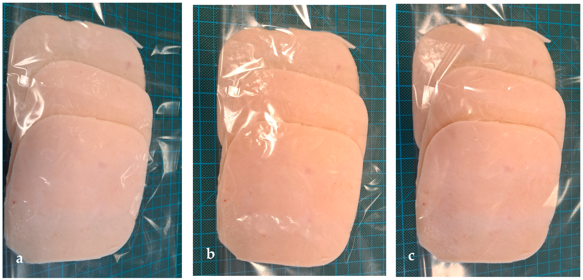
Two bars of cooked ham with the same expiration date (±2 days) were purchased in a local market (Szczecin, Poland). The producer suggested ham consumption within 2–3 days after package opening. The package was opened, and cooked ham was cut into 20 mm slices and 3 mm slices by a shop assistant; the portions were packed into polyethylene bags and immediately transported to the Center of Bioimmobilisation and Innovative Packaging Materials (CBIMO). The sliced ham was then aseptically introduced into bags. All of the cooked ham slices were separated by the use of a square spacer (Figure 1a) and placed into bags, which included (Figure 1b,c):
  • Six PP bags (C) (control samples), these sliced pieces of ham were separated by un-coated PP spacers;
  • Six PP bags coated with Ut coating, these sliced pieces of ham were separated by PP spacers, coated with the Ut layer on each side;
  • Six PP bags covered with Fb coating; these sliced pieces of ham were separated by PP spacers that were coated with the Fb layer on each side.
The precooked ham slices were placed in contact with the active layers on each side.
Both the active and non-active bags were welded and sealed (HSE-3, RDM Test Equipment, Hertfordshire, Great Britain) in air conditions deemed normal. Welding parameters were set at pressure—4 kN; temperature –117 °C; time—4 s.
The 18 bags (six of each packaging material) containing sliced cooked ham were then refrigerated at 5 °C. The 9 samples were later examined on 48 h (three bags of each packaging material) and 96 h of storage (three bags of each packaging material). The 20 mm slices were used for textural analysis. The 3 mm slices were used for dry mass analysis, microbiological analysis, SEM, and L*a*b* tests.

2.8. The Microbiological Analysis

Microbiological analysis was performed for sliced cooked ham before and after storage (in active and non-active packaging). For each microbiological analysis, 10 ± 0.1 g of individual cooked ham slices were aseptically introduced into a sterile stomacher bag in physiological saline peptone solution (PPS: 0.85% m/v NaCl; 0.1% m/v peptone). Then ham slices were homogenized in a Bag Mixer (Interscience, Saint-Nom-la-Brèteche, France) for 120 s, and appropriate decimal dilutions were prepared in PPS. The total count was determined according to PN-EN ISO 4833-2:2013-12 [23] as follows: serial dilutions of the initial, homogenized ham samples (in PPS) were performed; then the 0.1 mL of the initial inoculum and its dilutions were carefully, uniformly spread over the surface of the PCA medium (in duplicates, 90 mm Petri dishes). The plates were left for 15 min for the inoculum to be absorbed into the agar. Then the plates were inverted and introduced into the incubator (Memmert GmbH, Schwabach, Germany). The samples were then incubated for 72 h at 30 °C. After incubation, agar plates with fewer than 300 colonies were selected, and the colonies were counted. The S. aureus count analysis was performed according to PN-EN ISO 6888-1 [24] as follows: serial dilutions of the initial homogenized ham samples (in PPS) were performed; then the 1 mL of the initial inoculum and its dilutions were uniformly spread over the surface of the Baird-Parker agar medium (in duplicates, 90 mm Petri dishes). The plates were left for 15 min for the inoculum to be absorbed into the agar. Then the plates were incubated for 48 h at 37 °C. The total coliforms bacteria count was carried out according to PN-ISO 4832:2007 [25] as follows: serial dilutions of the initial homogenized ham samples were performed; then the 1 mL of the initial ham sample and its dilutions were pipetted from test tubes to the Petri dishes; VRBG medium was weighted, suspended in 1 L of distilled water, heated to a resulting boiling temperature (it was very important not to overheat the medium), and equilibrated at 45 °C. Then 15 mL of VRBG medium was pipetted to the Petri dishes containing the samples and their dilutions and mixed carefully. The mediums were left to solidify. As next step, 4 mL of VRBG medium was pipetted on the surface of each sample and left to solidify. The samples were incubated for 24 h at 37 °C.

2.9. The Textural Analysis

An analysis of the texture of the cooked ham (20 mm slices) took place in accordance with the PN-ISO 11036:1999 standard: “Sensory analysis. Methodology. Texture profiling” [26]. Zwick/Roell Z 2.5 was used (Wrocław, Poland). An analysis was carried out of the ham samples after delivery from a retail point and after our own storage tests.

2.10. Dry Mass Tests

The dry mass was noted in the case of the cooked ham both before being placed in bags and on 48 h and 96 h storage time. A Weight Dryer (Radwag, Warsaw, Poland) was used to ascertain dry mass. The samples from each bag were tested in duplicates.

2.11. L*a*b* Tests

The color of the cooked ham was determined through the average of 9 individual measurements from ham slice spots randomly selected by the use of a colorimeter (NR 20 XE, EnviSense) with other related data software. The measurement of color was carried out through an aperture (8 mm diameter) using a CIE L*a*b* color space which utilized a standard 10 observer and Illuminant D65. The selected parameters (that would account for the results) were ∆L (darkness and lightness difference) and ∆Elab (total color aberration). The EnviSense protocol was used to calculate parameters.

2.12. SEM Analysis of the Sliced Ham

Before being placed into bags and after 48 h and 96 h storage, an SEM analysis was carried out on the cooked ham. In addition, SEM micrographs were taken to visualize the cooked ham micro-texture. The samples of ham were set at 18 h at 4 °C (2% glutaraldehyde in a 0.1 M sodium cacodylate, pH 7.4). Further, the samples were washed with 0.1 M sodium cacodylate and later dehydrated in serial concentrations of ice-cold (–20 °C) methanol (20%, 40%, 60%, 80%, and 100%) at 2 h intervals. The samples of ham were then put in a Petri dish for 5 min. The food samples were then placed on pin stubs, and a thin layer of gold in a sputter coater was used to coat them at room temperature (Quorum Technologies Q150R S, Laughton, East Sussex, UK). The cooked ham samples were analyzed with a scanning electron microscope (SEM). The microscopic analysis of the samples was carried out through the use of a Vega 3 LMU microscope (Tescan, Brno-Kohoutovice, Czech Republic) already described above.

2.13. Statistical Analysis

The texture and microbiological analysis results were checked for statistical significance through the use of an analysis of variance (ANOVA) followed by an ANOVA test (one-way). Where p < 0.05, the values were seen as significantly different. All analyses were carried out through the use of GraphPad Prism 8 (GraphPad Software, Version 9, San Diego, CA, USA).

3. Results

3.1. Antibacterial Analysis

The results of the current work demonstrated that the coating (Zn) containing 0.06 g of ZnO nanoparticles in 100 mL of the coating carrier inhibited S. aureus, B. atrophaeus, and E. coli growth completely. The coatings Ut (containing 0.03 g of ZnO nanoparticles and 5% of U. tomentosa extract into 100 mL of the coating carrier) and Fb (containing 0.03 g of ZnO nanoparticles and 5% of F. betulina extract into 100 mL of the coating carrier) also had a bacteriolytic effect on these strains (Table 1). Unfortunately, neither Zn coating nor Ut and Fb layers inhibited the growth of P. syringae cells. It was noted that Zn coating was not effective against the P. syringae strain, but the number of these bacteria was decreased by Ut and Fb coatings. Statistical analysis confirmed that the changes in the number of bacterial cells were found to be significant (Figure 2).

3.2. SEM Analysis of the Coatings

It was shown through an SEM analysis that the PP film that had been used in the tests had a slightly rough surface of the uncovered film (Figure 3a. SEM images under magnification of 200× and 500×). As was emphasized in Figure 3b–d, active coatings (Zn, Ut, and Fb) had no effect on PP surface morphology. In addition, it was noted that PP film was homogenously and thoroughly coated with the active layers (covered foil samples micrographs reflected uncovered foil samples). Additionally, the Zn covering was not visible when compared to the Ut and Fb coatings. The covering of homogenous PP film with Zn, Ut, and Fb coatings can have an effect on the release of active agents on the whole layer surface of the food product. As was shown in the Figure 3d, convex, spherical particles were noted on the Fb layer. Additionally, as noted in Figure 3c, small pores were seen on the Ut coating.

3.3. Microbial Purity Analysis

It was shown that the number of mesophilic bacteria found in the sliced cooked ham kept in the non-active coating bags (C—control sample) increased markedly after 48 h (p < 0.001) and 96 h (p < 0.0001) of storage at 5 °C in air conditions (than when placed in comparison to sample “0”—pre-storage). Figure 4 shows that a Ut coating containing 5% of the U. tomentosa extract and decreased amount of ZnO nanoparticles had an impact on the increase in mesophilic bacterial cells. It decreased the number of living cells after 48 h of storage compared to the samples, which were stored in bags devoid of coatings. Additionally, a higher than 1 log increase in bacteria number was seen (compared to sample “0”—pre-storage) for the slices stored 48 h in bags covered with the Ut coating. Unfortunately, the Fb coating did not decrease the number of microorganisms compared to the “0” sample and to the samples stored in bags without any coatings. It would be too easy to advance that only the coating with Ut extract was able to be active against mesophilic bacteria and that Ut active layer was significantly more effective than the Fb layer. As emphasized below (Figure 2), the PP films coated with the Ut layer had a significant influence on the total count compared to the PP bags covered with the Fb coatings and to the number of mesophilic microorganisms detected from the samples stored in the bags devoid of active coatings after storage of 96 h. There was clear evidence that the number of bacterial cells in the case of sliced ham samples stored in PP bags and in PP bags that were coated with the active layers was higher (than compared to the “0” sample—pre-storage) after 96 h storage. A clear statistical analysis showed that any variances between the number of microorganisms stored for 96 h were found to be significant (p < 0.0001). From the point of view of microbiological analysis, a limit that would be acceptable for a total viable count of products such as sliced meat is 6–7 log10 CFU/g [27]. Nevertheless, it is of note that according to Grzybowski et al. [28], a microbial load lower than 3 log10 CFU/g is a satisfactory/acceptable number of mesophilic bacteria for products made up of sliced meat, and the 4 log10 CFU/g is generally accepted as a maximum total count that would be seen as acceptable for sliced ready-to-eat meat products. A higher number of bacteria than 4 log10 CFU/g is seen as an unacceptable microbial load in the case of consumable sliced cooked ham. This means that all samples stored in PP bags that did not contain active layers or in active bags that were fully covered with the Ut or Fb active coatings were not suitable to be consumed after 48 and 96 h of storage (Figure 4). It has to be underlined that two bars of cooked ham had the same expiration date (±2 days) on the day when they were purchased. Additionally, the ham was sliced by the shop assistant, who could contaminate the samples during slicing. It can be mentioned that the producer suggested the ham consumption within 2–3 days after the package opening. The results confirmed that the sliced ham was not acceptable for consumption after 48 h of their storage, but a lower increase in mesophilic microorganisms was noted for the samples stored in bags coated with the Ut layer. These findings suggest that the package covered with the more effective coating could decrease the number of bacteria to an acceptable level.
The results of the study confirmed that Ut and Fb coatings inhibited the growth of S. aureus, B. atrophaeus, and E. coli strains (Table 1) and decreased the number of P. syringae cells (Figure 2). According to Grzybowski et al. [28], the number of coliform bacteria found in 10 mg of sliced cooked ham has to be zero. In contrast, the number of cells of S. aureus in 1 g of this product can be 100 to be suitable for consumption. Results from this study showed that neither the coliforms nor S. aureus cells were detected in the “0” sample and in all of the analyzed sliced cooked ham stored in uncovered PP bags and in PP bags coated with the Ut and Fb layers. With this in mind, all of the samples that were analyzed were suitable for consumption even after 96 h of storage. In accordance with Grzybowski et al. [28], the number of Listeria monocytogenes cells must not be found in 1 g of the ready to eat sliced meat products, and Salmonella sp. should not be found in 25 g of sliced ham. The activity of Ut and Fb coatings against L. monocytogenes and against Salmonella sp. was not determined in this research. It is why the presence of these microorganisms in sliced cooked ham before and after storage was not analyzed as well.

3.4. The Textural Analysis

The results of this research have shown that springiness in the sliced cooked ham increased after 48 h storage in the uncovered PP films. After 96 h, a decrease in this parameter was noted. Modifying the bags with the active layers containing the F. betulina and ZnO nanoparticles working as active agents led to a slightly lower enlargement of the springiness of the sliced cooked ham after 48 h of storage and a slightly greater decrease after 96 h of storage than when compared to bags that were uncoated. It was observed that 48 h storage of the cooked ham samples in bags covered with the Ut coating had not any influence on springiness, but it caused an uptick in springiness when it was compared to the uncoated bags (Figure 5). The differences between these parameter values were seen as insignificant, later confirmed through statistical analysis (p > 0.05).
As was observed below (Figure 6), the gumminess of the sliced cooked ham packed in bags devoid of active layers insignificantly rose after a period of 48 h of storage. After 96 h, the gumminess was noted to have risen compared to sample “0”. In addition, this rise was seen to be higher when set in comparison to the sliced ham stored for more than 48 h. These differences were seen as insignificant and were confirmed by a detailed statistical analysis (p > 0.05). Similar results were obtained for packaging covered with the Fb coating. When analyzing the ham slices that were stored in packaging coated with the Ut active layer, it was observed that average gumminess decreased after 48 h of storage. After 96 h of storage, the gumminess of the sliced cooked ham kept in packaging with the Ut coating slightly increased when compared to that of the samples stored for 48 h and decreased compared to the “0” sample. The changes between the gumminess values were noted to be insignificant, again confirmed by statistical analysis (p > 0.05). Comparing the samples after 48 h of storage in coated PP packaging with a Ut layer with the samples stored for 96 h in these bags, the gumminess values of the sliced cooked ham were significantly lower. Contrary results were obtained for the sliced cooked ham after 48 h and 96 h of storage in bags coated with the Fb layers. The Fb coating did not influence gumminess values after 48 h of storage. Additionally, a slight, insignificant increase in this parameter in the case of samples kept for more than 96 h in packaging with an Fb layer was noted.
With regards to cohesiveness, it was seen that this particular value for the cooked sliced ham stored in uncoated PP bags increased after 48 h and after 96 h of storage. However, a rise in cohesiveness was seen to be less for samples stored for 96 h than for the slices stored for 48 h. These changes were insignificant and confirmed by statistical analysis (Figure 7). In the case of active packaging, a decrease in cohesiveness was noted in those samples stored for 48 h, but only in bags covered with Ut coating. After 96 h storage, an increase in the cohesiveness of the ham slices stored in these bags was observed. In addition, the differences in parameter values were found not to be significant. The average cohesiveness value, measured for the samples stored in bags with Fb layer, stored for 48 and for 96 h also increased, though this value was seen as insignificant.
Analyzing the decrease in the gumminess and cohesiveness values after short-term storage (48 h), it was demonstrated that a decrease in these two parameters was observed only for the samples stored in packaging coated with the Ut coating.
The results of the research also demonstrated that chewiness depended on the packaging material for 48 h of storage (Figure 8). Analyzing the chewiness of the sliced cooked ham slices, which were introduced into uncoated bags for 48 h, it was determined that these parameter values increased. Contrary results were obtained for the samples that were stored in bags covered with Ut and Fb coatings. The slight, insignificant increase in chewiness was seen in the slices kept in bags with a Fb layer. However, the Ut coating had no influence on the chewiness. After 96 h of being stored, a clear increase was noted as insignificant, this being confirmed by statistical analysis for sliced ham stored in all bags (coated or uncoated). However, a greater increase was noted for the samples that were introduced into the bags that were coated with Ut coating.

3.5. Dry Mass Analysis

The results of this research have uncovered that the dry mass of cooked ham slices was 63.84%. Storing ham samples in PP bags led to a rise in the dry mass of the ham slices to 64.95% after 48 h of being stored and to 70.55% after 96 h. It was shown that the dry mass of sliced cooked ham stored in bags coated with Ut and Fb layers was greater than the dry mass of those ham slices stored in bags that remained uncoated after 48 h and 96 h of being stored. The Ut coating led to a slightly lower increase in the dry mass of the sliced ham after 48 h of storage than the Fb layer. Contrary results were uncovered in samples kept for 96 h (Table 2).

3.6. L*a*b* Analysis

It was demonstrated in this work that ∆Elab had a great impact on the material used for packaging (Table 3). The ∆Elab of sliced cooked ham, which was introduced into the uncovered bags for 48 h, was higher than the ∆Elab of the samples that had been added to bags coated with Ut and Fb layers. This was contrary to PP films covered with a coating containing the F. betulina extract and ZnO nanoparticles; the ∆Elab for ham slices kept in packaging that had been coated with a layer that contained U. tomentosa and nano ZnO was lower. Similar results were also found on 96 h storage. The highest ∆Elab was noted in the case of samples of ham kept in bags that had not been coated. The lowest ∆Elab was noted for active packaging. It was also observed that ∆Elab was dependent on the material used for packaging. Tests on sliced cooked ham kept for 96 h showed that a lower ∆Elab value was obtained for ham slices added to packaging with Ut covering than those only with the layer of Fb.

3.7. SEM Analysis of the Sliced Ham

The microstructure of the sliced cooked ham before and after storage was analyzed using SEM. The cooked ham sample (Figure 9a) comprised a gel network including small and large fat particles entrapped in the protein network and unabsorbed protein structure. Although there were gullies on its surface, the overall surface of the sample was relatively smooth but not homogenous. The loose and porous structures with air bubbles (holes) were observed. Analyzing the influence of the storage on the microstructure of cooked ham, the samples were observed to have a gelatinous appearance (Figure 9b–g). After 48 h and 96 h of storage, a deterioration in myofibril led to an increase in water migration, which in turn led to a clearly visible irregular gully structure in the samples. The structure of the ham has become more compact (Figure 9b). However, a lower myofibril degradation was observed for the samples stored in active packaging than for the samples stored in uncoated bags. It was demonstrated that the surface of the slices of ham kept in bags of (active Ut and Fb) for a 96 h (Figure 9f,g) was less dense and compact than the microstructures of the samples stored in PP bags (Figure 9c), confirming water migration. In addition, small cracks were noted along the surface of the samples, as well as air bubbles (holes) visible on the sample surfaces of all items stored in the active packaging.

4. Discussion

The results of the current study demonstrated that the amount of the ZnO nanoparticles should be increased (up to 0.06 g in 100 mL of the coating carrier) to maintain the bacteriolytic effect of the coating against selected Gram-negative and Gram-positive bacterial strains. The results of the previous work [12] demonstrated that a methyl-hydroxy-propyl-cellulose (MHPC) coating containing ZnO nanoparticles (0.082 g in 100 mL of the coating carrier) inhibited the growth of S. aureus, E. coli, B. cereus, and P. aeruginosa. It is worth mentioning that accelerated irradiation of this coating did not influence its antibacterial properties due to the shielding properties of ZnO nanoparticles [12,13,14,15,16,17,18,19]. The decreased amount of the nanoparticles in MHPC coating (0.041 g in 100 mL of the coating carrier) led to obtaining the active layer that inhibited the growth of S. aureus but only decreased the number of E. coli and P. syringae cells, confirming that its antibacterial activity was lower [11]. The previous work [11] also indicated that the coating containing a decreased amount of ZnO (0.041 g/100 mL of the coating carrier) and decreased amount of geraniol was more active against S. aureus, E. coli, and P. syringae strains than the coatings containing a decreased amount of geraniol or decreased amount of nano ZnO, confirming the synergistic effect of these active agents. The analysis of the antibacterial properties of U. tomentosa and F. betulina showed that the activity of these two extracts was very low [9]. A lower than 1-log reduction in the number of S. aureus cells was noticed for 5% of extracts of described plants. While analyzing the antimicrobial properties of F. betulina extracts on B. subtilis cells, it was demonstrated that they demonstrated lower than a 2-log reduction but higher than a 1-log reduction in the number of living cells. U. tomentosa extract was found to be even less active. Unfortunately, both extracts were not effective against E. coli and P. syringae strains. The findings of the current work indicated that the coatings containing 5% of U. tomentosa or F. betulina extracts with the addition of a decreased amount of the nanoparticles of ZnO (0.03 g/100 mL of the coating carrier) demonstrated high antibacterial activity. The synergistic effect between active agents caused a complete inhibition of the growth of S. aureus, B. atropaheus, and E. coli strains and a reduction in the number of P. syringae cells. Additionally, SEM analysis confirmed that the PP film was homogenously and thoroughly covered with the active Ut and Fb layers. However, small pores were observed on the Ut coating, which might have led to a faster release of the active substances from this coating than from the Fb coating or which might have been caused by the release of the active compounds from the Ut layer. Comparing the coatings Ut and Fb, it may be suggested that the Ut active layers were able to display a greater influence on the growth of microorganisms than Fb coating, but for a shorter time.
The results led to the assumption that packaging materials coated with Ut and Fb layers could be used to preserve cooked ham during short-term storage in air conditions.
The described findings demonstrated that coatings containing 5% of the U. tomentosa or 5% of the F. betulina extracts with the addition of a decreased amount of nano ZnO did not inhibit the growth of mesophilic bacterial strains for a sliced cooked ham that was stored for 96 h, but decreased their number when compared to the ham stored in uncovered bags. Comparing the influence of active layers on the total count after 48 h, it may be mentioned that the most effective packaging material was the bags covered with the Ut coating. Comparing SEM micrographs of the “0” samples and ham slices after storage, it was observed that the most similar microstructures were the surfaces of the ham before storage and the ham slices stored in bags with Ut coating, confirming its effectiveness. The results of this study determined that the number of mesophilic microbes from ready-prepared, sliced cooked ham samples purchased from a local market (sample “0”) was almost 4 log10 CFU/g, which is seen as an unsuitable number of microorganisms for the sliced ham to be consumed [28]. The expiration date (±2 days) of the product could have been one of the reasons. Moreover, the producer suggested ham consumption within 2–3 days after package opening, meaning that a total count should be on the level acceptable for consumption after 48 h of ham storage. Unfortunately, according to Grzybowski et al. [28], ham samples were not acceptable for consumption after 2 days of storage. However, according to another author [29], an acceptable level of the total count of cooked ham products is generally set to 6 log10 CFU/g. It means that ham slices stored in uncoated and in active packaging were still acceptable to be consumed after 48 h of storage. While after 96 h of storage, the only suitable samples for consumption were the ham slices stored in bags covered with the Ut coating. It should be added that the ham was purchased in a local market and sliced by the shop assistant after the package opening. The high level of mesophilic bacteria observed for the sample “0” proved that the ham must have been contaminated during slicing. It may be predicted and even suggested that the number of bacteria in sample “0” was too high to be decreased by the active substances which were released from the layers. Considering the presence of pathogenic microorganisms, it should be emphasized that neither the coliform bacteria nor S. aureus cells were detected in the cooked ham slices after 48 h and 96 h of storage in PP bags coated with the Ut and Fb layer. From this point of view, these ham slices were still suitable for consumption even after 96 h of storage. In summary, the influence of Ut coating on the quality of sliced, cooked ham was noted, confirming that packaging covered with this active layer can be used for short-term storage. It may be predicted that if ham slices are not contaminated during slicing, the samples will be stored for a longer time than the time suggested by producers. Similar results were obtained in the previous study [2]. The bags and spacers coated with the layer containing 50% of the mixture of Glycyrrhiza L. and Scutelaria baicalensis extracts were proved to be the best packaging material for ready-prepared chicken sausages which were purchased from a local butcher’s shop and which were contaminated during slicing. This active packaging material preserved and maintained the quality of sausage slices after short-term storage (72 h) at 5 °C. Spampinato et al. [4] performed similar experiments. The authors purchased sliced cooked ham with an expiration date of ±3 days. According to the authors, the producer suggested ham consumption within 1–3 days after the package opening. It has to be underlined that the ham analyzed by the authors was not sliced by the shop assistant, but it was already sliced. The results of the authors’ study showed that the total count ranged between 5.8 and 9.0 log10 CFU/g at 0 days. Furthermore, after 4, 8, and 12 days of fridge storage (without active packaging), total aerobic bacteria significantly increased. Additionally, the absence of viable Salmonella sp. in the product after 12 days of storage was satisfying for the authors; nevertheless, the presence of other opportunistic pathogens, such as Klebsiella pneumoniae and E. coli, was noted. The experiments of the authors revealed that, at the opening of packages, sliced ham presented a high microbial load and quite rich microbiota. It can be explained by other authors who suggested [30] that it is possible that post-cooking contamination of cooked ham occurred during the packaging phases of manufacturing, with the bacteria occurring in the environment of the facility, entering into contact and being packaged with virtually sterile sliced cooked ham.
The results of the study led to the assumption that bags and spacers coated with a Ut layer, even if though not recommended for the long-term safe keeping of sliced cooked ham, may be used for short-term storage. Many consumers buy ready-to-eat sliced ham that is sliced by shop assistants in a local market. It has to be underlined that ham is very often contaminated during the slicing process. Therefore, a packaging material, such as PP film, paper, or biopolymer foil coated with a Ut layer, could be used to preserve sliced cooked ham during transport and short-term storage (48–96 h). The findings of this work were similar to the results presented by Shiji et al. [31], who introduced chicken sausages into active packaging for short-term storage. The authors proved that after four days of storage at 4 °C, the active pouches containing silver nanoparticles as active agents were more effective than polyethylene pouches. Similar results were obtained by Shahrampour et al. [32], who confirmed that the use of active packaging for sausages storage at 4 °C led to detect a lower number of mesophilic microorganisms than the total count isolated from the samples which were stored in the uncoated polyethylene packaging. Rüegg et al. [6] prepared polyethylene terephthalate (PET) foil covered with active coatings containing thyme and rosemary essential oils as active agents. The authors used antimicrobial packaging to preserve a sliced cooked chicken breast during short-term storage (6 days). As was shown by the authors, the active layers reduced the number of mesophilic bacteria compared to the uncovered foil, though only slightly. Furthermore, it was confirmed that the MAP packaging system had a greater effect on microorganisms than the antimicrobial coverings, which is why MAP is recommended for long-term storage.
The texture is one of the most important parameters determining the freshness and quality of cooked ham. A product that is too soft or is not cohesive enough can create doubts in the consumers as to its quality and freshness. All the forces holding the food products together are represented by cohesiveness. Additionally, gummy, tough, or stringy food product is not suitable for consumers as it presents excessively strong resistance during mastication [2]. The results of this study determined that the cohesiveness of sliced cooked ham increased for active and uncoated packaging materials (when compared to the sample “0” after 96 h of storage. It is tempting to deduce that the storage of ham slices in uncovered PP bags and in the bags covered with the active layer had a positive effect on the texture of the ham. The contrary findings were noted by Patiño et al. [33], who proved that cohesiveness tended to decrease during storage which may be caused by the interaction of proteins and fats with the rest of the components, affecting the texture of the sausages. Furthermore, Araújo et al. [34] suggested that to increase the parameter values of the sausage samples during storage, an additive such as collagen powder should be added. The results of the current work indicated that the springiness of the ham samples increased for all analyzed packaging materials (when compared to the sample “0”) after 48 h storage. Moreover, a decrease in this parameter was observed, except in the slices that were stored for 96 h in the bags coated with the Ut layer, confirming that the active coating containing U. tomentosa extract influenced the springiness improvement.
The present work indicated that, unfortunately, chewiness and gumminess increased in the samples stored in uncoated PP bags after 48 h and 96 h of storage, and this is a clear disadvantage. These findings were confirmed by SEM analysis, which showed a more compact microstructure of the cooked ham. Comparing the ham slices, which were stored in active packaging for 48 h, it has to be mentioned that the changes in chewiness values were not observed (sample “0”). In the case of gumminess, the decrease in this parameter was observed, but only for the samples stored in bags with a Ut layer. The bags covered with Fb coating did not influence the gumminess of the ham samples. This was backed up by a microbial purity analysis that showed that the bags coated with the Ut layer was the best packaging for the short-term (48 h) storage of the sliced cooked ham. It is also worth mentioning the disadvantage of the active bags with Ut and Fb layers; it was observed that water loss in sliced ham samples from these bags was higher than from uncoated bags. As was already suggested, the high water loss can release antibacterial substances from the active layers and thus could improve their activity. Analyzing the chewiness and gumminess for the ham slices stored in bags coated with Ut and Fb layers after 96 h of storage, the increase in these parameter values was noted. Simultaneously, the dry mass also increased. As was suggested by Zeraatpisheh et al. [35], when water loss increases, the meat proteins converge because of the formation of new crosslinks; consequently, the chewiness and gumminess of the samples are elevated. Gumminess is associated with food hardness; therefore, its increase is a clear disadvantage. Thus, in cooked ham stored in uncoated bags or in bags covered with a Fb layer with increased gumminess after storage, ham sample hardness may let to difficulties in swallowing [2]. While the ham stored in active packaging coated with Ut layer with decreased (after 48 h) or unchanged (after 96 h) gumminess, ham slices would still be acceptable for consumption.
The findings demonstrated in this work confirmed that the highest ∆Elab values were noticed for cooked ham portions stored in the uncoated bags and into bags covered with Fb coating for 48 h and 96 h. It was also indicated that the highest ∆L values were noted for these portions. This indicates that the ham samples taken from this package were the lightest. Furthermore, it should be mentioned that these parameter values increased for samples stored in all analyzed packaging materials. The results were confirmed by Azlin-Hasim et al. [36], who indicated that L* values increased in meat samples stored for 6 and 12 days in packaging containing Ag nanoparticles as antimicrobial substances.

5. Conclusions

Microbial contaminants which may be present in cooked ham originates from incorrect cooking of meat product, inappropriate sanitization practices, and/or recontamination during packaging, following the increase in microorganisms during the primary shelf-life. It is worth mentioning that the package opening may be the cause of a sudden variation of the headspace gas composition and a loss of protection. Furthermore, if the ham is sliced by the shop assistant, it may be contaminated during the process. All the facts make that the secondary shelf-life is remarkably shorter than the primary shelf-life. The only possibility to maintain the quality of cooked ham is to decrease the microbial load on the surface of the slices using active packaging during transportation and secondary storage.
The total count of the ham samples before storage was high, proving that the cooked ham was contaminated. The spacers and bags coated with the antimicrobial layer containing U. tomentosa extract and the ZnO nanoparticles were observed to be the best bags for ready-prepared, sliced cooked ham that was purchased from a local market. It was confirmed by microbial purity, textural, and SEM analysis. These antimicrobial bags may be used to maintain the freshness and quality of ham sliced by the shop assistant during the secondary shelf-life (48 h) at 5 °C.

Author Contributions

M.M., M.O. (Magdalena Ordon), M.O. (Marcin Okręglicki) and J.S. conceived and designed the experiments; M.O. (Magdalena Ordon) wrote the paper; M.O. (Magdalena Ordon) and W.B. performed the microbiological tests; M.M. and M.O. (Magdalena Ordon) analyzed the data; M.O. (Magdalena Ordon) and W.B. performed mechanical tests; M.O. (Magdalena Ordon) and M.M. analyzed the data; M.O. (Magdalena Ordon) and W.B. performed Lab tests; M.M. performed SEM analysis; M.O. (Magdalena Ordon) analyzed the data; M.O. (Magdalena Ordon) performed dry mass tests and analyzed the data; M.O. (Magdalena Ordon), W.B., M.O. (Marcin Okręglicki) and J.S. contributed and prepared reagents/materials; M.O. (Marcin Okręglicki) and J.S. contributed analysis tools; M.O. (Magdalena Ordon) performed statistical analysis and analyzed the data. All authors have read and agreed to the published version of the manuscript.

Funding

This research work was funded by the project POIR.01.01.01-00-0740/21: “Innovative, ecological, active packaging made of plastics, biodegradable and compostable films and paper intended for packaging food products with special, protective and functional properties”; co-financed by the European Union and the National Center for Research and Development.

Institutional Review Board Statement

Not applicable.

Informed Consent Statement

Not applicable.

Data Availability Statement

Not applicable.

Acknowledgments

The authors acknowledge Alicja Tarnowiecka-Kuca for a support given during mechanical tests.

Conflicts of Interest

The authors declare no conflict of interest.

References

  1. Nikmaram, N.; Budaraju, S.; Barba, F.J.; Lorenzo, J.M.; Cox, R.B.; Mallikarjunan, K.; Roohinejad, S. Application of plant extracts to improve the shelf-life, nutritional and health-related properties of ready-to-eat meat products. Meat Sci. 2018, 145, 245–255. [Google Scholar] [CrossRef]
  2. Ordon, M.; Burdajewicz, W.; Pitucha, J.; Tarnowiecka-Kuca, A.; Mizielińska, M. Influence of Active Packaging Covered with Coatings Containing Mixtures of Glycyrrhiza L. and Scutellaria baicalensis Extracts on the Microbial Purity and Texture of Sliced Chicken Sausages. Coatings 2023, 13, 795. [Google Scholar] [CrossRef]
  3. Duthoo, E.; Rasschaert, G.; Leroy, F.; Weckx, S.; Heyndrickx, M.; De Reu, K. The Microbiota of Modified-Atmosphere-Packaged Cooked Charcuterie Products throughout Their Shelf-Life Period, as Revealed by a Complementary Combination of Culture-Dependent and Culture-Independent Analysis. Microorganisms 2021, 9, 1223. [Google Scholar] [CrossRef]
  4. Spampinato, G.; Candeliere, F.; Amaretti, A.; Fabio, L.; Rossi, M.; Raimondi, S. Microbiota Survey of Sliced Cooked Ham During the Secondary Shelf Life. Front. Microbiol. 2022, 13, 842390. [Google Scholar] [CrossRef]
  5. de Niederhäusern, S.; Bondi, M.; Camellini, S.; Sabia, C.; Messi, P.; Iseppi, R. Plant Extracts for the Control of Listeria monocytogenes in Meat Products. Appl. Sci. 2021, 11, 10820. [Google Scholar] [CrossRef]
  6. Rüegg, N.; Teixeira, S.R.; Beck, B.M.; Monnard, F.W.; Menard, R.; Yildirim, S. Application of antimicrobial packaging based on modified calcium carbonate and EOs for RTE meat products. Food Packag. Shelf Life 2022, 34, 100982. [Google Scholar] [CrossRef]
  7. Mizielińska, M.; Bartkowiak, A. Innovations in food packaging materials. In Food Preservation and Packaging-Recent Process and Technological Advancements; Tumuluru, Y.S., Ed.; IntechOpen: London, UK, 2022. [Google Scholar] [CrossRef]
  8. Herrera, D.R.; Durand-Ramirez, J.E.; Falcăo, A.; Nogueira da Silva, E.J.L.; dos Santos, E.B.; Figueiredo de Almeida Gomes, B.P. Antimicrobial activity and substantivity of Uncaria tomentosa in infected root, canal dentin. Braz. Oral. Res. 2016, 30, 61–66. [Google Scholar] [CrossRef] [Green Version]
  9. Ordon, M.; Nawrotek, P.; Stachurska, X.; Schmidt, A.; Mizielińska, M. Mixtures of Scutellaria baicalensis and Glycyrrhiza L. Extracts as Antibacterial and Antiviral Agents in Active Coatings. Coatings 2021, 11, 1438. [Google Scholar] [CrossRef]
  10. Sofrenić, I.; Anđelković, B.; Todorović, N.; Stanojković, T.; Vujisić, L.; Novaković, M.; Milosavljević, S.; Tešević, V. Cytotoxic triterpenoids and triterpene sugar esters from the medicinal mushroom Fomitopsis betulina. Phytochemistry 2021, 181, 112580. [Google Scholar] [CrossRef] [PubMed]
  11. Mizielińska, M.; Nawrotek, P.; Stachurska, X.; Ordon, M.; Bartkowiak, A. Packaging Covered with Antiviral and Antibacterial Coatings Based on ZnO Nanoparticles Supplemented with Geraniol and Carvacrol. Int. J. Mol. Sci. 2021, 22, 1717. [Google Scholar] [CrossRef] [PubMed]
  12. Mizielińska, M.; Łopusiewicz, Ł.; Mężyńska, M.; Bartkowiak, A. The Influence of Accelerated UV-A and Q-SUN Irradiation on the Antimicrobial Properties of Coatings Containing ZnO Nanoparticles. Molecules 2017, 22, 1556. [Google Scholar] [CrossRef] [PubMed] [Green Version]
  13. Eskandari, F.; Mofidi, H.; Asheghi, B.; Mohammadi, F.; Gholami, A. Bringing resistance modulation to methicillin-resistant Staphylococcus aureus (MRSA) and vancomycin-resistant enterococci (VRE) strains using a quaternary ammonium compound coupled with zinc oxide nanoparticles. World J. Microbiol. Biotechnol. 2023, 39, 193. [Google Scholar] [CrossRef] [PubMed]
  14. Karimi, M.; Sadeghi, E.; Khosravi Bigdeli, S.; Zahedifar, M. Optical properties, singlet oxygen, and free radical production ability with different UV irradiations and antimicrobial inhibitors against various bacterial species of ZnO: Eu nanoparticles. Radiat. Phys. Chem. 2023, 212, 111132. [Google Scholar] [CrossRef]
  15. Wu, H.; Meng, Y.; Yu, M.; Yang, H. Modulating the antibacterial activity of ZnO/talc by balancing the monodispersity of ZnO nanoparticles. Appl. Clay Sci. 2023, 242, 107024. [Google Scholar] [CrossRef]
  16. Sougandhi, P.R.; Jena, S.K.; Brahmanand, P.S.; Tej, M.B.; Kadiyala, N.K.; Rani, T.S.; Rao, M.V.B. Multifunctional Bio-Nanocomposite Films of Carboxymethyl Cellulose and Pectin with Incorporated Zinc Oxide Nanoparticles. Lett. Appl. NanoBioSci. 2023, 12, 85. [Google Scholar]
  17. Nguyena, T.V.; Dao, P.H.; Khanh Linh Duong, K.L.; Duong, Q.H.; Vu, Q.T.; Nguyena, A.H.; Mac, V.P.; Lea, T.L. Effect of R-TiO2 and ZnO nanoparticles on the UV-shielding efficiency of water-borne acrylic coating. Prog. Org. Coat. 2017, 110, 114–121. [Google Scholar] [CrossRef]
  18. El-Feky, O.M.; Hassan, E.A.; Fadel, S.M.; Hassan, M.L. Use of ZnO nanoparticles for protecting oil paintings on paper support against dirt, fungal attack, and UV aging. J. Cult. Herit. 2014, 15, 165–172. [Google Scholar] [CrossRef]
  19. Kairyte, K.; Kadys, A.; Luksiene, Z. Antibacterial and antifungal activity of photoactivated ZnO nanoparticles in suspension. J. Photochem. Photobiol. Biol. 2013, 128, 78–84. [Google Scholar] [CrossRef]
  20. Kubra, I.R.; Kumar, D.; Rao, L.J.M. Effect of microwave-assisted extraction on the release of polyphenols from ginger (Zingiber officinale). Int. J. Food Sci. Tech. 2013, 48, 1828–1833. [Google Scholar] [CrossRef]
  21. Mandal, V.; Mohan, Y.; Hemalatha, S. Microwave Assisted Extraction—An Innovative and Promising Extraction Tool for Medicinal Plant Research. Pharmacogn. Rev. 2007, 1, 7–18. [Google Scholar]
  22. ASTM E 2180-07; Standard Test Method for Determining the Activity of Incorporated Antimicrobial Agent(s) in Polymeric or Hydrophobic Materials. ASTM: West Conshohocken, PA, USA, 2012.
  23. PN-EN ISO 4833-2:2013-12. Available online: https://sklep.pkn.pl/pn-en-iso-4833-2-2013-12p.html (accessed on 1 September 2022).
  24. PN-EN ISO 6888-1. Available online: https://sklep.pkn.pl/pn-en-iso-6888-1-2022-03e.html (accessed on 1 September 2022).
  25. PN-ISO 4832:2007. Available online: https://sklep.pkn.pl/pn-iso-4832-2007p.html (accessed on 1 September 2022).
  26. PN-ISO 11036:1999 Standard. Available online: http://sklep.pkn.pl/pn-iso-11036-1999p.html (accessed on 1 September 2022).
  27. EC. Commission Regulation No 1441/2007 of 5 December 2007 Amending Regulation (EC) No 2073/2005 on Microbiological Criteria for Foodstuffs. 2007. Available online: https://www.google.com/url?sa=i&rct=j&q=&esrc=s&source=web&cd=&ved=0CAIQw7AJahcKEwiA27a4kLaAAxUAAAAAHQAAAAAQAg&url=https%3A%2F%2Feur-lex.europa.eu%2FLexUriServ%2FLexUriServ.do%3Furi%3DOJ%3AL%3A2007%3A322%3A0012%3A0029%3AEN%3APDF&psig=AOvVaw1DPdQ5p_OWuHv8R7u9rKch&ust=1690795927928650&opi=89978449 (accessed on 1 September 2022).
  28. Grzybowski, J.; Reiss, J. Praktyczna Bakteriologia Lekarska i Sanitarna; Dom Wydawniczy Bellona: Warszawa, Polska, 2001; 346p. [Google Scholar]
  29. Vasilopoulos, C.; De Vuyst, L.; Leroy, F. Shelf-life Reduction as an Emerging Problem in Cooked Hams Underlines the Need for Improved Preservation Strategies. Crit. Rev. Food Sci. Nutr. 2013, 55, 1425–1443. [Google Scholar] [CrossRef] [PubMed]
  30. Raimondi, S.; Luciani, R.; Sirangelo, T.M.; Amaretti, A.; Leonardi, A.; Ulrici, A.; Foca, G.; D’Auria, G.; Moya, A.; Zuliani, V.; et al. Microbiota of sliced cooked ham packaged in modified atmosphere throughout the shelf life: Microbiota of sliced cooked ham in MAP. Int. J. Food Microbiol. 2019, 289, 200–208. [Google Scholar] [CrossRef] [PubMed]
  31. Shiji, M.; Snigdha, S.; Jyothis, M.; Radhakrishnan, E.K. Biodegradable and active nanocomposite pouches reinforced with silver nanoparticles for improved packaging of chicken sausages. Food Packag. Shelf Life 2019, 19, 155–166. [Google Scholar] [CrossRef]
  32. Shahrampour, D.; Razavi, S.; Sadeghi, A. Evaluation of green tea extract incorporated antimicrobial/antioxidant/biodegradable films based on polycaprolactone/polylactic acid and its application in cocktail sausage preservation. Food Meas. 2023, 17, 1058–1067. [Google Scholar] [CrossRef]
  33. Patiño, J.H.; Henríquez, L.E.; Restrepo, D.A.; Lantero, M.I.; García, M.A. Influence of polyamide composite casings with silver–zinc crystals on the quality of beef and chicken sausages during their storage. J. Food Sci. Technol. 2022, 59, 75–85. [Google Scholar] [CrossRef]
  34. Araújo, I.B.S.; Lima, D.A.S.; Pereira, S.F.; Paseto, R.P.; Madruga, M.S. Effect of storage time on the quality of chicken sausages produced with fat replacement by collagen gel extracted from chicken feet. Poultry Sci. 2021, 100, 1262–1272. [Google Scholar] [CrossRef]
  35. Zeraatpisheh, F.; Tabatabaei, Y.F.; Shahidi, F. Investigation of effect of cold plasma on microbial load and physicochemical properties of ready-to-eat sliced chicken sausage. J. Food Sci. Technol. 2022, 59, 3928–3937. [Google Scholar] [CrossRef]
  36. Azlin-Hasim, S.; Cruz-Romero, M.C.; Morris, M.A.; Cummins, E.; Kerry, J.P. Effects of a combination of antimicrobial silver low density polyethylene nanocomposite films and modified atmosphere packaging on the shelf life of chicken breast fillets. Food Packag. Shelf Life 2015, 4, 26–35. [Google Scholar] [CrossRef]
Figure 1. The sliced cooked ham: (a) in uncoated bag, separated by uncoated PP spacers; (b) in bag covered with the Ut coating, separated by PP spacers, coated with the Ut layer on each side; (c) in bag covered with the Fb coating, separated by PP spacers that were coated with the Fb layer on each side.
Applsci 13 08853 g001
Figure 2. The influence of coatings on P. syringae growth. PP—PP film; Zn—PP film covered with the coating containing 0.06 g ZnO nanoparticles in 100 mL carrier; Ut—PP film covered with the coating containing 0.03 g of ZnO nanoparticles and U. tomentosa extract in 100 mL carrier; Fb—PP film covered with the coating containing 0.03 g of ZnO nanoparticles and F. betulina extract in 100 mL carrier; One-way ANOVA; ****—p < 0.0001.
Applsci 13 08853 g002
Figure 3. (a) The uncoated PP film. (b) The PP film covered with the Zn coating. (c) The PP film covered with the Ut coating. (d) The PP film covered with the Fb coating.
Applsci 13 08853 g003aApplsci 13 08853 g003b
Figure 4. The total count of mesophilic bacteria detected from the sliced cooked ham stored for 48 and 96 h. Zero—sample “0”; (C) sliced pieces of ham, stored in PP bags, separated by uncoated PP spacers; (Ut) sliced pieces of ham, stored in PP bags covered with the Ut layer, separated by PP spacers coated with the Ut layer on each side; (Fb) sliced pieces of ham, stored in PP bags covered with the Fb layer, separated by PP spacers coated with the Fb layer on each side; One-way ANOVA ***—p < 0.001; ****—p < −0.0001; ns—p > 0.05.
Applsci 13 08853 g004
Figure 5. The springiness of the sliced cooked ham stored for 48 and 96 h. Zero—sample “0”; (C) sliced pieces of ham, stored in PP bags, separated by uncoated PP spacers; (Ut) sliced pieces of ham, stored in PP bags covered with the Ut layer, separated by PP spacers coated with the Ut layer on each side; (Fb) sliced pieces of ham, stored in PP bags covered with the Fb layer, separated by PP spacers coated with the Fb layer on each side; One-way ANOVA ns—p > 0.05.
Applsci 13 08853 g005
Figure 6. The gumminess of the sliced cooked ham stored for 48 and 96 h. Zero—sample “0”; (C) sliced pieces of ham, stored in PP bags, separated by uncoated PP spacers; (Ut) sliced pieces of ham, stored in PP bags covered with the Ut layer, separated by PP spacers coated with the Ut layer on each side; (Fb) sliced pieces of ham, stored in PP bags covered with the Fb layer, separated by PP spacers coated with the Fb layer on each side; One-way ANOVA **—p < 0.01; ns—p > 0.05.
Applsci 13 08853 g006
Figure 7. The cohesiveness of the sliced cooked ham stored for 48 and 96 h. Zero—sample “0”; (C) sliced pieces of ham, stored in PP bags, separated by uncoated PP spacers; (Ut) sliced pieces of ham, stored in PP bags covered with the Ut layer, separated by PP spacers coated with the Ut layer on each side; (Fb) sliced pieces of ham, stored in PP bags covered with the Fb layer, separated by PP spacers coated with the Fb layer on each side; One-way ANOVA **—p < 0.01; ns—p > 0.05.
Applsci 13 08853 g007
Figure 8. The chewiness of the sliced cooked ham stored for 48 and 96 h. Zero—sample “0”; (C) sliced pieces of ham, stored in PP bags, separated by uncoated PP spacers; (Ut) sliced pieces of ham, stored in PP bags covered with the Ut layer, separated by PP spacers coated with the Ut layer on each side; (Fb) sliced pieces of ham, stored in PP bags covered with the Fb layer, separated by PP spacers coated with the Fb layer on each side; One-way ANOVA; ns—p > 0.05.
Applsci 13 08853 g008
Figure 9. (a) Sliced cooked ham before storage (sample “0”). (b) Sliced pieces of ham stored for 48 h in uncoated PP bags, separated by uncovered PP spacers. (c) Sliced pieces of ham stored for 96 h in uncoated PP bags, separated by uncovered PP spacers. (d) Sliced pieces of ham, stored for 48 h in PP bags covered with the Ut layer, separated by PP spacers coated with the Ut layer on each side. (e) Sliced pieces of ham, stored for 96 h in PP bags covered with the Ut layer, separated by PP spacers coated with the Ut layer on each side. (f) Sliced pieces of ham, stored for 48 h in PP bags covered with the Fb layer, separated by PP spacers coated with the Fb layer on each side. (g) Sliced pieces of ham, stored for 96 h in PP bags covered with the Fb layer, separated by PP spacers coated with the Fb layer on each side.
Applsci 13 08853 g009aApplsci 13 08853 g009bApplsci 13 08853 g009c
Table 1. The influence of coatings on S. aureus, E. coli and B. syringae growth.
The SampleThe Number of Bacterial Cells [CFU/g]
S. aureusE. coliB. atrophaeus
PP4.05 × 106 ± 4.87 × 10 5 *9.05 × 107 ± 1.6 × 10 6 *7.73 × 105 ± 1.14 × 10 5 *
Zn0 ****0 ****0 ****
Ut0 ****0 ****0 ****
Fb0 ****0 ****0 ****
*—Standard Deviation; One-way ANOVA test: ****—p < 0.0001.
Table 2. The dry mass of sliced chicken sausage after 48 h and 96 h of storage.
Time [h]Dry Mass [%]
CUtFb
063.84 ± 0.00263.84 ± 0.00263.84 ± 0.002
4864.95 ± 0.09465.52 ± 0.03965.92 ± 0.015
9670.55 ± 0.02272.23 ± 0.01272.18 ± 0.005
(C) sliced pieces of ham, stored in PP bags, separated by uncoated PP spacers; (Ut) sliced pieces of ham, stored in PP bags covered with the Ut layer, separated by PP spacers coated with the Ut layer on each side; (Fb) sliced pieces of ham, stored in PP bags covered with the Fb layer, separated by PP spacers coated with the Fb layer on each side.
Table 3. The ΔElab of sliced chicken sausage after 72 h and 144 h of storage.
Time [h]CUtFb
ΔElab02.31 ± 0.192.31 ± 0.192.31 ± 0.19
ΔL2.25 ± 0.372.25 ± 0.372.25 ± 0.37
ΔElab483.40 ± 0.611.27 ± 0.152.19 ± 0.73
ΔL7.60 ± 1.060.37 ± 0.230.76 ± 0.50
ΔElab9612.03 ± 0.469.99 ± 0.3811.74 ± 0.21
ΔL11.99 ± 0.449.97 ± 0.3911.62 ± 0.26
(C) sliced pieces of ham, stored in PP bags, separated by uncoated PP spacers; (Ut) sliced pieces of ham, stored in PP bags covered with the Ut layer, separated by PP spacers coated with the Ut layer on each side; (Fb) sliced pieces of ham, stored in PP bags covered with the Fb layer, separated by PP spacers coated with the Fb layer on each side.
Disclaimer/Publisher’s Note: The statements, opinions and data contained in all publications are solely those of the individual author(s) and contributor(s) and not of MDPI and/or the editor(s). MDPI and/or the editor(s) disclaim responsibility for any injury to people or property resulting from any ideas, methods, instructions or products referred to in the content.

