Article

Ustilago maydis Yeast Mutant Produces Cytosolic Melanin by Tyrosine-Tyrosinase Activity with Stain Biosorption Capability

by
Cinthya Martínez-López
1,
Melissa Vázquez-Carrada
1,
Oscar Flores-Herrera
2,
Juan Pablo Pardo
2,
Dario Rafael Olicón-Hernández
1,* and
Guadalupe Guerra-Sánchez
1,*
1
Departamento de Micriobiología, Escuela Nacional de Ciencias Biológicas, Instituto Politécnico Nacional, Prolongación de Carpio y Plan de Ayala S/N, Casco de Sto Tomás, Alcaldía Miguel Hidalgo, Mexico City CP 11340, Mexico
2
Departamento de Bioquímica, Facultad de Medicina, Universidad Autónoma de México, Circuito Interior S/N, Ciudad Universitaria, Mexico City CP 04510, Mexico
*
Authors to whom correspondence should be addressed.
Appl. Sci. 2023, 13(20), 11288; https://doi.org/10.3390/app132011288
Submission received: 8 September 2023 / Revised: 11 October 2023 / Accepted: 13 October 2023 / Published: 14 October 2023

Abstract

:
Ustilago maydis is a biotrophic basidiomycete fungus that infects corn plants and works as an excellent phytopathogen model, facilitating numerous genetic transformations for studying the mechanisms of plant infection. A random mutation event in the mutant strains designed to investigate the physiological significance of two plasma membrane proton-ATPases in this model resulted in a pigmented phenotype strain. For this study, the FB2 strain and the ΔPMA1 mutant were chosen to assess the pigment, which was confirmed as melanin through thin-layer chromatography, UV, and IR spectrophotometry. The melanin was observed to accumulate in the cytosol, as evident from scanning and transmission electron microscopy, and did not interfere with normal cell growth in yeast extract peptone dextrose media or minimal media. Notably, the mutant exhibited a 25% higher melanin yield compared to wild-type cells. To analyze the melanin synthesis, the tyrosinase activity was measured in a phosphate buffer at pH 6.5. The enzyme demonstrated greater activity with tyrosine as a substrate than with L-3,4 dihydroxyphenylalanine, maintaining the same trend in ion preference. Both FB2 and ΔPMA1 mutant cells were subjected to biosorption experiments, revealing that the mutants with an excess of cytosolic melanin were capable of removing at least 50 ppm of methylene blue. In conclusion, U. maydis can accumulate melanin in the cytosol without adverse physiological effects and this presents biotechnological potential for dye removal.

1. Introduction

Ustilago maydis is a phytopathogenic biotrophic parasitic fungus that belongs to the Ustilaginacea family. U. maydis infects corn plants forming tumors, first as white and ending as a black mass, and because of this is named corn smut [1,2]. U. maydis has emerged as a prominent model microorganism for investigating the biology of plant-pathogenic basidiomycetes, a diverse collection of pathogens that are responsible for inducing severe plant diseases [3,4,5]. One of the notable advantages of working with U. maydis yeast is its amenability to genetic manipulation. This feature enables the creation of mutants, facilitating research into phytopathogenic processes and their interactions with host plants [6,7,8,9].
The dimorphic phenomenon is evident in Ustilago maydis. This fungus exhibits a yeast saprophytic form that thrives on artificial media [8]. However, these yeast-like cells cannot induce disease symptoms when applied to host plants in pure culture. To incite disease, a mating process involving two compatible yeasts (FB1 and FB2) is necessary, leading to the formation of the infective filament [10,11]. In the later stages of infection, this infective filament releases highly melanized sporidia, marking a transitional phase between the yeast and filamentous forms. Notably, both yeasts and filamentous fungi possess melanin within their cell walls [12]. Melanin serves various roles, including contributing to virulence, offering protection against biotic and abiotic stresses, and facilitating metal transport in a range of fungi [12,13,14,15]. Furthermore, melanin, as an integral component of microorganisms’ cell walls [16], plays a vital role in the maintenance U. maydis cells against environmental factors and contributes to a variety of processes, including pathogenesis [7,17,18,19,20].
These pigments can be generated through different pathways involving the oxidation and polymerization of phenolic and/or indolic compounds. Notably, melanins exhibit significant biological activities, including antioxidant properties and protection against photo and radiation damage, all while maintaining low toxicity levels. Furthermore, owing to their exceptional reactivity, melanins possess a remarkable capacity for adsorbing heavy metal ions, dyes, and toxic elements. The adsorption behavior is likely attributed to electrostatic interactions occurring between the contaminate and the negatively charged functional groups present in melanin as well as linked to the presence of hydrophilic centers within the melanin’s structural composition. The biosorption of contaminants such as dyes and heavy metals has emerged as a highly promising biotechnology for the removal of waste products from solutions and the recovery of substances. This approach has garnered considerable attention for several years due to its inherent simplicity, similarity to conventional ion exchange technology, apparent high efficiency, and the abundance of biomass and waste bio-products that can be readily harnessed for this purpose [21].
In pathogenic fungi of the Candida species, the melanization of the cell wall is considered a virulence factor [22,23]. In halophyte fungi, alterations occur in the cell wall and the lipid composition of the plasma membrane, including thickening of the cell wall with melanin and shifts in the phospholipid content [24]. In most instances, the fungal response to abiotic stress has been associated with cell wall melanization [25,26].
Melanin that accumulates in the cytosol is often referred to as “fungal melanosomes” and can sometimes be identified as small black vesicles through electron microscopy as they transit to the cell wall [27,28]. Typically, microbial melanin is produced through the conversion of either tyrosine/L-3,4 dihydroxyphenylalanine (L-DOPA; DOPA pathway) by tyrosinase or via the malonyl-coenzyme A pathway (DNH, Dihydroxynaphthalene pathway), involving different sets of enzymes, including polyketide synthase [29,30].
To elucidate the metabolic pathway for melanin synthesis in U. maydis, Reyes-Fernandez et al. described an unconventional melanin synthesis process involving polyketide synthase, monooxygenase, and versicolorin synthase using mutants lacking these enzymes. Some mutants displayed colorless phenotypes or produced yellowish cells [18]. Piña et al. reported a null mutant in the RNA binding protein UmRm75 in U. maydis, which resulted in an accumulation of eumelanin and hydrogen peroxide in the cytosol [17]. In both cases, the mutants were associated with the production and accumulation of melanin in the cytosol, although there was no clear link between the deletions of different genes and melanin synthesis.
In the process of generating null mutants of the two plasma membrane proton-ATPases (H+-ATPase, PMA1 or PMA2), the cells exhibited a pigmented phenotype initially. However, a new construction of these mutants resulted in null mutants without the pigmented phenotype [31]. Initially, the pigmented cells did not display any noticeable differences from the wild cells. This report aims to identify the chemical nature of the pigment that accumulates in the U. maydis mutant with the deleted PMA1 gene (ΔPMA1) and to pinpoint the point mutation introduced upstream and downstream of the ATPase gene through DNA sequencing. We also examined the distribution of the dark pigment in the cytosol, pigment yield, cell growth, and the activity of tyrosinase involved in melanin synthesis. Additionally, we studied the substrate preference and the effects of ions on enzyme activity. The capacity to remove the dye methylene blue was also assessed in both wild-type and mutant cells.

2. Materials and Methods

2.1. Strain Maintenance

The wild-type strain of U. maydis FB2 was sourced from the Centro de Investigación y de Estudios Avanzados of the Instituto Politécnico Nacional (CINVESTAV-IPN) and is registered in the National Center for Biotechnology Information (NCBI) database as ATCC 201384, featuring an a2b2 phenotype. The deletion mutants for the plasma membrane H+ ATPase were generated using this strain. The strains were cultured on yeast extract peptone dextrose agar plates (YPDA: 1% yeast extract, 0.25% peptone, and 1% dextrose, 2% agar) for 24 h at 29 °C. The yeasts were then inoculated in 25 mL of YPD and grown for 24 h at 29 °C with constant agitation at 125 rpm. The biomass was collected by centrifugation at 3000 rpm, and the cells were resuspended in 50% glycerol and aliquoted in 500 µL portions, which were stored at −70 °C. For specific experiments, the cells were inoculated in liquid YPD and incubated for 24, 48, or 72 h at 29 °C with agitation or using a minimal medium (MM) consisting of 1% glucose, 0.3% KNO3, and mineral salts.

2.2. Mutant Construction

2.2.1. Plasmid Construction for H⁺-ATPase Mutation

The Pma1 (um02581) gene was eliminated via homologous recombination using a hygromycin resistance cassette. This deletion was accomplished using the golden gate BsaI cloning system strategy developed by Terfrüchte et al. [32]. We utilized the commercial strain FB2 for this process and conducted in silico analysis using the U. maydis genome database MUMDB (MIPS Ustilago maydis database, http://mips.helmholtz-muenchen.de/genre/proj/ustilago accessed on 10 October 2020), along with Clone Manager Basic, Version number 9.4 software (© 2015 Scientific & Educational Software).
To generate flanking regions for the genes of interest, we performed a PCR, yielding approximately 1 Kb-sized fragments in each case. These fragments included cleavage sites for the restriction enzymes BsaI and SspI, positioned adjacent to the gene and at the ends, respectively. In a one-pot reaction, we constructed the transformation plasmid by combining the backbone sequence with the antibiotic resistance sequence and the PCR-generated flanks. Subsequently, the plasmids were replicated using the E. coli top 10 strain. The protoplasts of the wild-type yeast strain were transformed using heparin and polyethylene glycol. The mutant strains were verified through PCR, Southern blot analysis, and sequencing. Table 1 lists all the primers and plasmids used in this transformation.

2.2.2. Sequencing

The plasmid DNA was extracted according to the instructions of the NucleoSpin Plasmid EasyPure Mini kit (Macherey-Nagel) and sent to sequencing at Eurofilm with the primers MF502 (ACGACGTTGTAAAACGACGGCCAG) and MF721 (CCCGCAGGACATATCCAC) for the upstream flank, and MF168 (ACTAGATCCGATGATAAGCTG) and MF503 (TTCACACAGGAAACAGCTATGACC) for the downstream flank. The sequence was analyzed with the software Clone Manager Professional, 9.2 software (©Scientific & Educational Software).

2.3. Macroscopic and Microscopic Morphology of the Mutants

2.3.1. Macroscopic and Microscopic Analysis

An aliquot of 100 µL from the liquid culture (0.5 UDO mL−1) of each strain was seeded in YPD agar plates and incubated at 29 °C and monitored by 120 h. In the end, the colonies were recorded by visual observation. For the microscopic analysis, some aliquots of 50 µL were withdrawn at 24, 48, and 72 h and poured on a glass slide, and observed at 40× in optic microscopy for each sample.

2.3.2. Morphology Analysis of Mutant Strain by Electron Microscopy

The process of preparing the samples for scanning electron microscopy (SEM) and transmission electron microscopy (TEM) involved the following steps: The mutant strains and the FB2 wild-type control strain were cultured in accordance with previously established protocols in YPD or MM media for a duration of 24 h. Subsequently, the harvested cells were subjected to centrifugation and then washed twice with a 0.1 M PBS buffer solution at a pH of 7.0. To initiate fixation, a 2% glutaraldehyde solution was utilized for a period of 2 h, followed by the introduction of a 1% osmium tetroxide solution with an incubation time of 60 min. The yeast cells were systematically dehydrated through a series of ethanol solutions, ranging from 30% to 100%, with each solution being employed for 10 min intervals (100% ethanol was applied twice).
For the SEM analysis, the dehydrated yeast samples underwent a critical point drying process for 10 min at 5 °C. The resulting biological material was then placed in a metal container and coated with a thin layer of gold under vacuum conditions. A JEOL 5800LV scanning electron microscope was employed, operating at 15 kV. In the case of the TEM, following the ethanol dehydration process, two additional steps involved immersion in propylene oxide solution for 20 min each. Subsequently, the cells were mixed with various propylene oxide–Epon resin solutions at different ratios (2:1, 1:1, 1:3), with durations ranging from 3 to 12 h, depending on the specific ratio. Following this step, the cells were combined with pure Epon resin for 12 h (twice), and polymerization was allowed to occur for 18 h at 60 °C. Thin sections of 70 nm were prepared and contrasted using uranyl acetate and lead solutions, and finally, they were observed using a JEOL 1010 TEM operating at 60 kV [33].

2.4. Pigment Extraction and Quantification

The pigment extraction was conducted through a process involving cell lysis and autoclave heating. Both wild-type and ΔPMA1 cells were cultured for 24, 48, and 72 h in a mineral medium and subsequently rinsed with distilled water to remove the culture medium. The collected cells were then treated with 5 mL of 1N NaOH and 1% DMSO [34]. Vigorous agitation of the tubes was followed by autoclaving at 121 °C for 20 min. After autoclaving, the sample underwent centrifugation at 6000 rpm, and the supernatant was carefully decanted. To initiate acid digestion, 1.0 mL of concentrated HCl was added, and the mixture was incubated for 1 h, followed by evaporation at 60 °C for 24 h.
The resulting solid material was subsequently reconstituted in 1 mL of 1 N NaOH, and its absorbance was measured at 475 nm using a UV-visible spectrophotometer. To determine the pigment concentration in the solution, a standard curve was established using commercial melanin (Sigma-Aldrich, St. Louis, MO, USA, M8631). For pigment quantification, a 10 mL sample of the cell culture taken after 72 h was centrifuged at 6000 rpm, and the cells were washed with distilled water (3 times). The pellet was then weighed and subjected to digestion with an alkaline solution composed of 0.1 N NaOH and 10% DMSO, with glass bullets added in a 1:2 ratio (w/v) to facilitate mechanical disruption. The digestion process involved 1 min cycles of shaking and cooling, repeated for 10 cycles. Following this, the mixture was heated for 1 h at 80 °C and subsequently centrifuged for 10 min at 10,000 rpm at 4 °C. The resulting supernatant was used to measure the absorbance at 475 nm for pigment quantification and to determine the protein content using the Lowry method [35].

2.5. Identification of Melanin by Thin Layer Chromatography, UV, and IR Spectrophotometry

The pigment identification was conducted using thin-layer chromatography (TLC) on silica plates, employing a solvent mixture of n-butanol–acetic acid–water in the ratio of 70:20:10, respectively [36]. A portion of the isolated pigment was subjected to TLC for 1 h on a 5 × 10 cm plate that had been pre-saturated with the aforementioned solvent mixture. As a reference, commercial synthetic melanin from Sigma-Aldrich (St. Louis, MO, USA) was utilized. The silica plate was developed under a UV lamp. The pigment was then diluted in 200 µL of 1N NaOH and brought to a final volume of 1 mL with alkaline water at pH 8.0. All the samples were adjusted to a concentration of 30 µg/mL, and the UV-visible spectrum was recorded by scanning wavelengths ranging from 200 to 800 nm using a BioMate® 3S spectrophotometer. The obtained pigment spectrum was compared with that of the commercial synthetic melanin (Sigma-Aldrich, St. Louis, MO, USA), and the readings were plotted. The negative slope was subsequently calculated.
For the infrared spectrum analysis, the purified settled pigment was mixed with KBr (of infrared quality) in a 1:10 ratio and scanned within the range of 400 to 5000 cm−1. As a control, commercial synthetic melanin was employed.

2.6. Assay of Physiological Response

Growth Assay

The mutant cells and wild-type cells maintained in glycerol were streaked on YPD agar plates. After 24 h, the cells were inoculated in 10 mL of YPD media and cultivated by 24 h at 29 °C and 125 rpm stirring. An aliquot equivalent to 50 UDO/L was inoculated into 100 mL flasks and cultured until 72 h. Aliquots were withdrawn every six hours and growth was measured by optical density at 600 nm. For growth in the minimum media (MM), the cells from the YPD cultures were washed three times with distilled water and the OD was adjusted and inoculated in the same volume.

2.7. Assay of Tyrosinase Activity

2.7.1. Monophenolase and Diphenolase Activity

The inoculum was adjusted to 80 UDO/L (1 Optical Density Unit equivalent to 5 × 10⁵ cells/mL) of the wild strain (FB2) and the mutant (ΔPMA1) and added to a flask with 50 mL of rich YPD media. It was incubated at 28 °C with agitation for 24 h. The cultures were collected and centrifuged at 6000 rpm for 10 min to obtain the biomass, and washed with sterile distilled water, to remove the remains of the culture media. Subsequently, the same amount of glass beads (0.5 mm) and the lysis solution (v/v) (0.1 M phosphate buffer, pH: 7.0) were added; mechanical lysis was performed, shaking for 1 min and left to rest on ice for 1 min; and this was repeated for 10 cycles. The samples were then centrifuged at 12,000 rpm for 30 min at 4 °C to eliminate any cell debris. The resulting supernatant was carefully transferred into clean, labeled tubes and kept on ice for subsequent enzymatic activity determination. For the assessment of tyrosinase activity, the oxidation of 0.5 and 2 mM L-DOPA (for diphenol oxidase activity) or 0.5 and 2 mM L-tyrosine (for monophenol oxidase activity) was monitored. The increase in absorbance was recorded at 475 nm (εdopacromo = 3.4 mM−1 cm−1) in 0.1 M phosphate buffer at pH 6.0. In a reaction cuvette maintained at 37 °C, the respective substrate (L-DOPA or L-tyrosine) and 50 µL of the cell lysate supernatant were added. The reaction mixture was agitated, and readings were taken every 2 min until 20 min of reaction time had elapsed. The results were expressed in units of specific enzymatic activity (1 Usp = 1 µmol of dopachrome formed per minute/mg protein), and the protein content in the samples was determined using the Lowry method [35,37].

2.7.2. Effect of Ions on the Enzyme Activity

To investigate the impact of various ions on enzyme activity, we conducted experiments using the following salts: MgSO4, CuSO4, ZnSO4, CaCl2, and FeCl2, each at a concentration of 50 mM. In a reaction chamber containing 100 mM phosphate buffer at pH 6.0, we combined 100 µL of either substrate L-tyrosine or L-DOPA, each at a concentration of 5 mM, with 5 µL of the corresponding ion solution. To this mixture, we added 50 µL of the cell lysate containing the enzyme, bringing the final volume to 1 mL. The reaction mixture was thoroughly mixed and then incubated at 37 °C in a water bath. We recorded the absorbance at 475 nm at both zero time and after 10 min of the reaction. To determine the percentage of enzymatic activity, we used the control, which had no cofactors added, as the reference point set at 100% [38]. At the same time, control tubes were prepared containing the substrate (L-DOPA or L-tyrosine) and the ion but without the enzyme extract. These tubes were utilized to assess the oxidation of the substrate caused by the presence of the ion, and the resulting response was subtracted from the absorbance of the test tubes.

2.8. Biosorption of Methylene Blue with U. maydis Biomass

In the minimum media (50 mL), methylene blue dye was added to achieve the final absolute concentrations ranging from 10 to 90 ppm (equivalent to 10 to 90 µg of dye per 1 L of the total medium) and sterilized at 121 °C for 15 min at 15 lb. These culture media were inoculated with 80 UDO/L of U. maydis wild-type or the mutant strain and incubated at 28 °C with shaking (160 rpm). A flask without strains was used as control. Aliquots were removed at 24 and 48 h of incubation, filtrated through a Whatman 0.45 µm filter paper, and the absorbance of the filtrate was recorded at 668 nm. The biosorption percentage was measured by comparing the absorbance of the treatments to that of the control flask, using the following equation:
%   b i o s o r p t i o n = A b s   c o n t r o l A b s   t r a e t m e n t   A b s   c o n t r o l   100  
Methylene blue was chosen because it is a conventional model dye for initial biosorption experiments.

3. Results

3.1. Sequencing Analysis

To investigate the functions of the two U. maydis plasma membrane proton ATPases, we generated null mutants for these ATPases. An intriguing observation was the presence of pigment accumulation in these mutants. This raised questions, as previous studies on the deletion of genes encoding H+-ATPases in other organisms did not correlate with melanin accumulation. To delve deeper, we conducted DNA sequencing both upstream and downstream of the deletion site. The results unveiled several random mutations alongside the deletion of the target gene.
Notably, we identified a T-to-C mutation at position 2443 and an A-to-C mutation at 2467 (Figure 1A). Additionally, other mutations occurred at positions 5820, 5980, 6648, and 6769 (Figure 1B). Importantly, these point mutations were found in close proximity to the PMA1 of the H+ ATPase gene’s deletion site. To ensure the accurate deletion of the Pma1 gene, we meticulously performed the procedure again, resulting in a new strain devoid of these additional random mutations. Interestingly, this refined strain did not exhibit pigment accumulation, suggesting that this phenotype is indeed attributable to the additional mutations [30]. The mutant strain sequence was compared with the strain U. maydis 521 (NCBI:txid237631) gen UMAG 2581.

3.2. Morphological Characterization of ∆PMA1 Strain

It has been reported that the morphology of U. maydis yeasts and the production of secondary metabolites can be affected by certain types of mutations. In this way, we analyzed the growth of the cells in a solid YPD medium and studied the cell morphology using different microscopy techniques.

3.2.1. Growth on Solid Media and Optical Microscopy

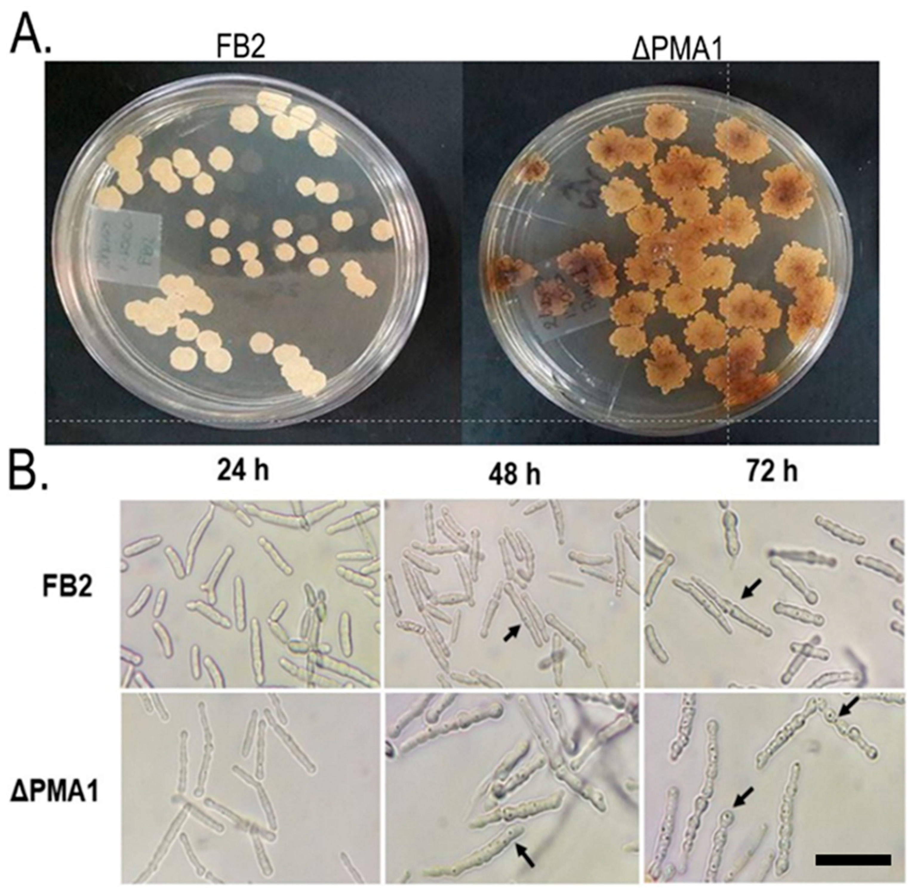
Because the synthesis of secondary metabolites is optimized during extended incubation periods, we conducted an investigation into the production of these metabolites in cell cultures on solid YPD agar for 96 h. As illustrated in Figure 2A, U. maydis yeast strains exhibited discernable differences in colony morphology. The FB2 wild-type strain displayed round colonies with slight rippling at the edges, whereas ΔPMA1 exhibited prominent ripples. However, the most notable observation was the brown coloration of the ΔPMA1 colonies, indicating the synthesis of a pigment. Figure 2B further highlights that ΔPMA1 cells exhibited a greater number of dark spots distributed throughout the cells compared to the wild-type strain.

3.2.2. Scanning and Electron Microscopy

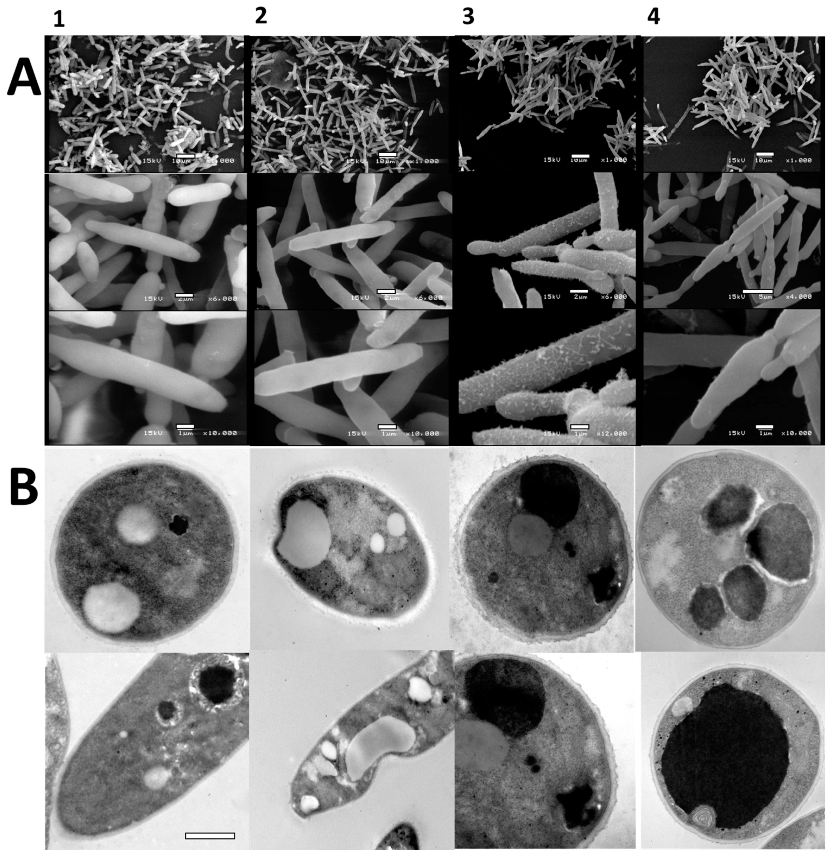
The scanning electron microscopy analysis revealed no significant differences in the surface characteristics between the wild-type and ΔPMA1 yeast cells (Figure 3A(1–4)). The distinctive elongated shape of the wild-type strain remained consistent in the mutant strain, irrespective of the culture medium used. In some instances, slight alterations in the cell wall, including peripheral aggregates, were observed (Figure 3A(3 and 4) sets), in contrast to the smooth surface of the FB2 cells. However, these minor surface changes did not impact the growth or physiological behavior of the mutant strains compared to the wild type.
Transmission electron microscopy (TEM) unveiled the presence of an electron-dense compound identified as melanin in both the wild-type and mutant strains. As depicted in Figure 3, it became evident that the proportion of melanin was lower in the wild-type strain (Figure 3B(1 and 2)) compared to the ΔPMA1 mutant strain (Figure 3B(3 and 4)). The wild type exhibited lipid bodies, albeit in limited numbers, while the ΔPMA1 strain (Figure 3B(3 and 4)) displayed a significantly higher amount of pigment in both growth media compared to the other cells.

3.3. Growth Analysis

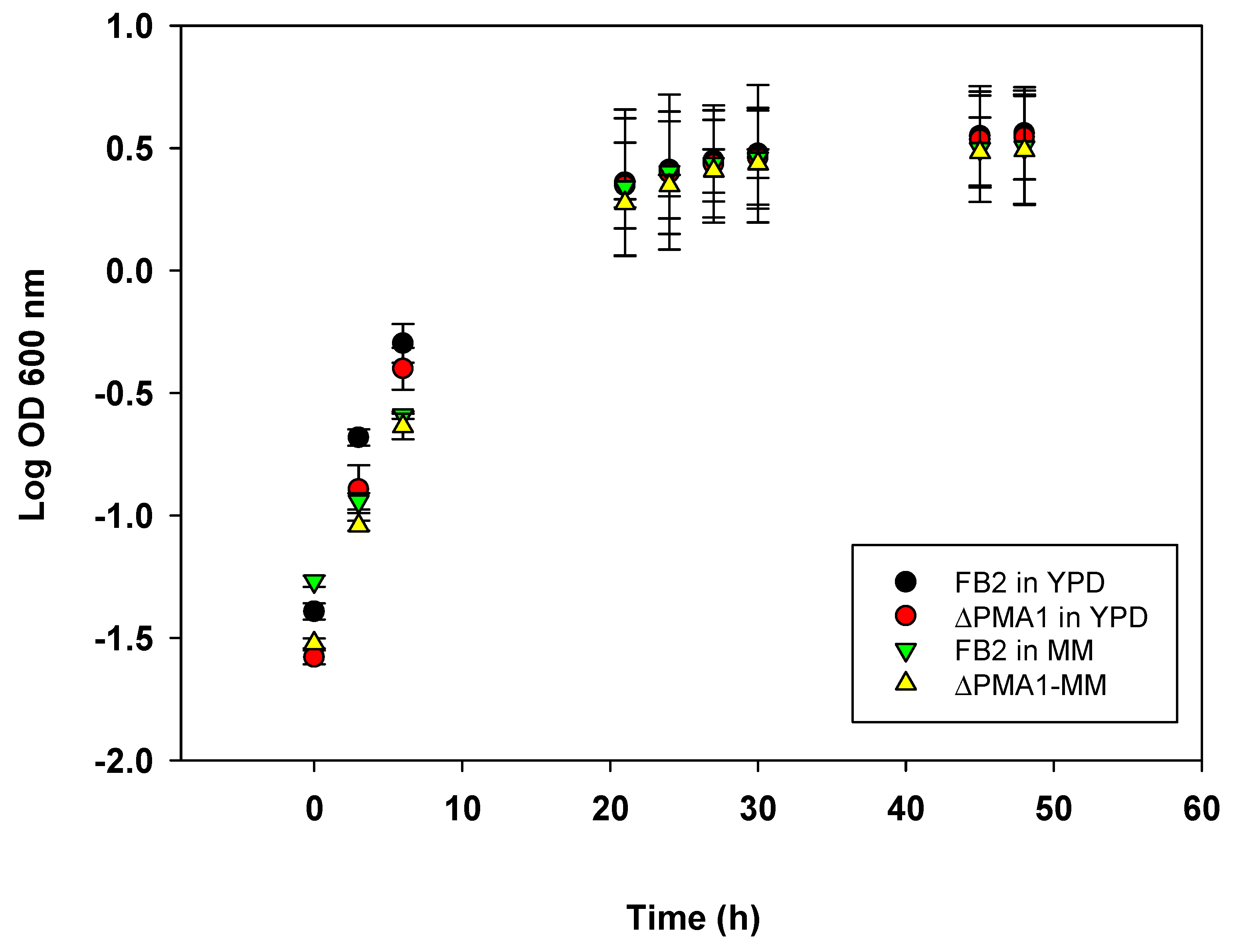
The cells were inoculated in YPD media, and their growth was monitored for 72 h. As shown in Figure 4, both strains reached the stationary phase at the same time and the growth was not affected by the deletion of the plasma membrane H+ ATPase gene and the associated additional mutations. The same occurred in the cells grown in MM. Table 2 shows that the duplication time of the mutant cells was not affected. Because cells in the stationary phase become more resistant to environmental stress and produce a larger number of secondary metabolites, we selected the stationary phase to study the accumulation of melanin.

3.4. Isolation and Identification of the Intracellular Pigment

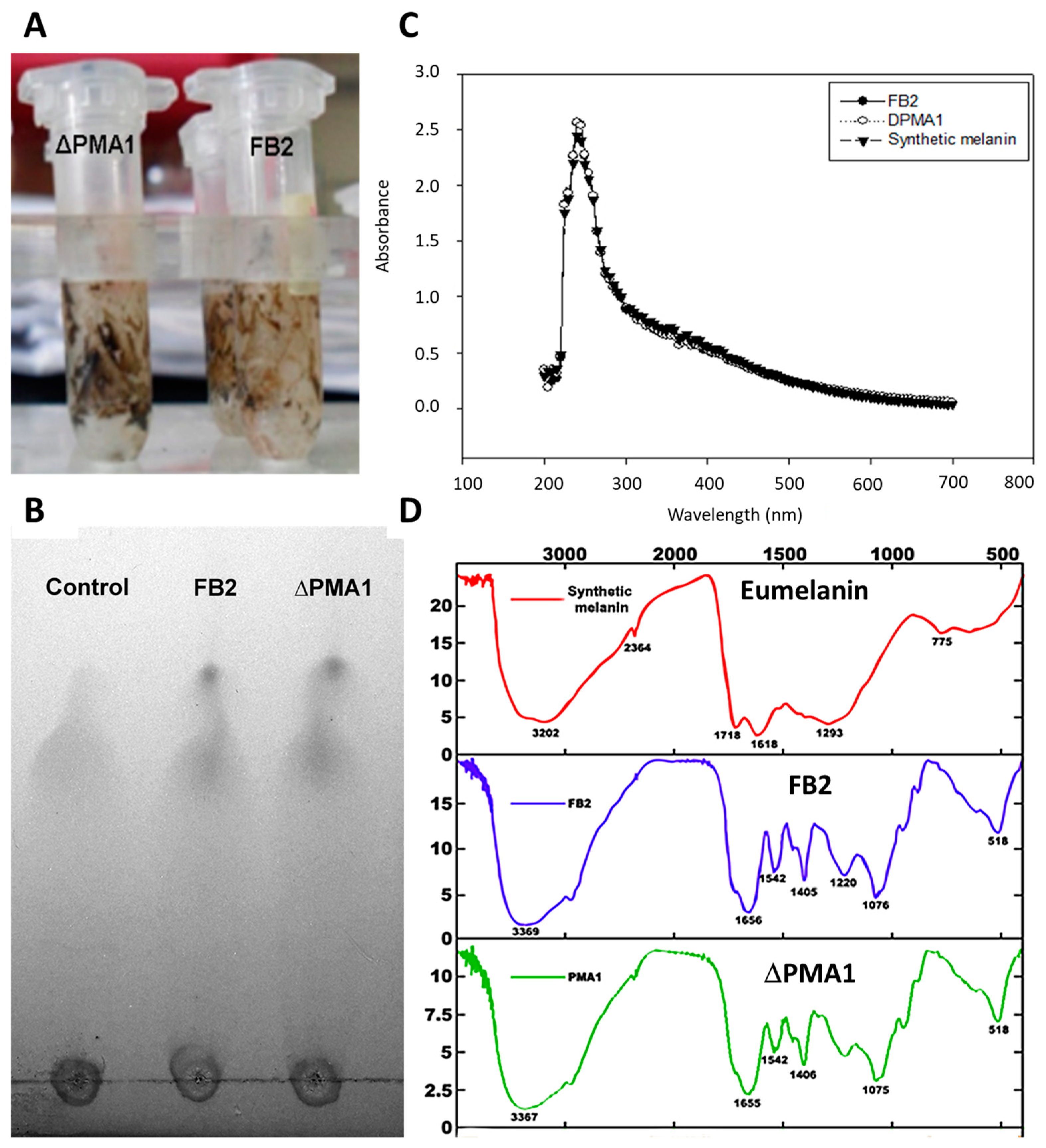
To determine the nature of the pigment, we conducted pigment isolation from both the wild-type and ΔPMA1 strains, followed by analysis using TLC, UV-Vis, and infrared spectrophotometry techniques. An initial visual inspection (Figure 5A) revealed that the pigments extracted from both strains appeared identical to the naked eye. Subsequently, we employed TLC chromatography (Figure 5B) with a melanin standard and the pigments isolated from the two strains. The congruent chromatographic patterns strongly indicated the presence of melanin in the preparations.
For a more comprehensive analysis of the pigment, we generated UV and IR spectra from two separate preparations. As illustrated in Figure 5C, the UV-Vis spectra of eumelanin and the two samples displayed considerable overlap, with all three spectra exhibiting an absorbance band peaking at 230 nm. This peak is a characteristic feature shared by commercial melanin and the fungal melanin found in Yarrowia lipolytica [39].
In Figure 5D, the IR spectrum displayed a broad absorption band spanning from 3200 to 3300 cm−1, signifying the presence of hydroxyl groups (O-H) and -NH bonds [34,40]. The peak observed at 1718 cm−1 can be attributed to the vibrations of C=O or COOH groups, while the peak at 1620 cm−1 corresponds to the vibrations of aromatic groups C=C and COO-. Additionally, a shoulder observed at 2300 cm−1 indicates N-H stretching. A comparative analysis of the infrared spectra indicated the presence of bands associated with phenolic groups (3200–3400 nm), further corroborating that the pigment is indeed melanin.

3.4.1. Content of Eumelanin in Wild-Type FB2 and ΔPMA1 Strains

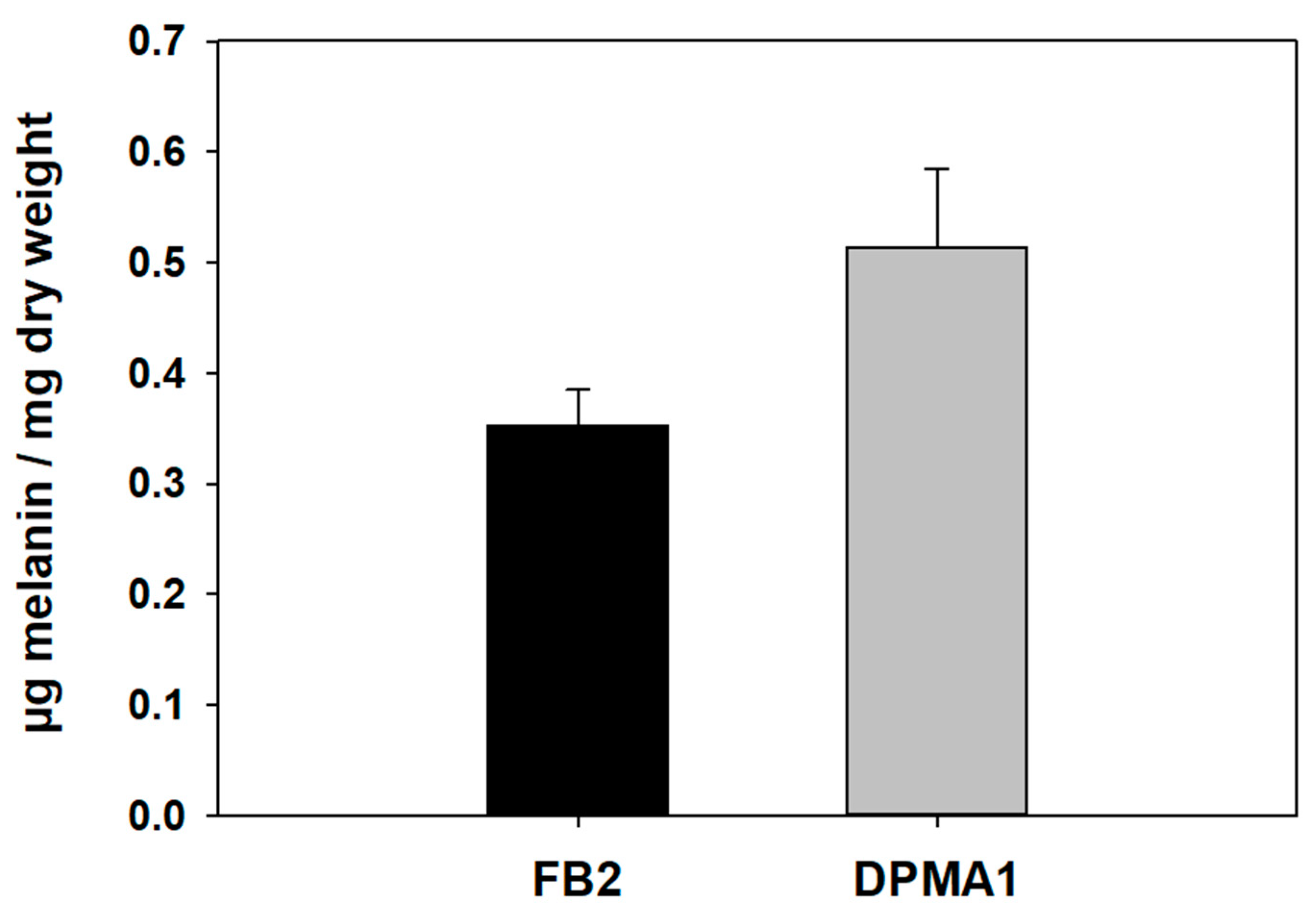
To measure the amount of eumelanin in the two strains, the pigment was extracted from cells following the protocol mentioned under the Materials and Methods section. With this protocol, the melanin from the cytosol and the cell wall was isolated. Figure 6 shows the ΔPMA1 and FB2 strain content of the melanin. The melanin content of the FB2 strain was approximately 30% lower than that found in the mutant. The data were subjected to the Student t-test (p < 0.05), and a significant difference was found between the wild-type FB2 strain and the ΔPMA1 mutant.

3.4.2. Assay of Tyrosinase Activity with L-Tyrosine and L-DOPA as Substrate

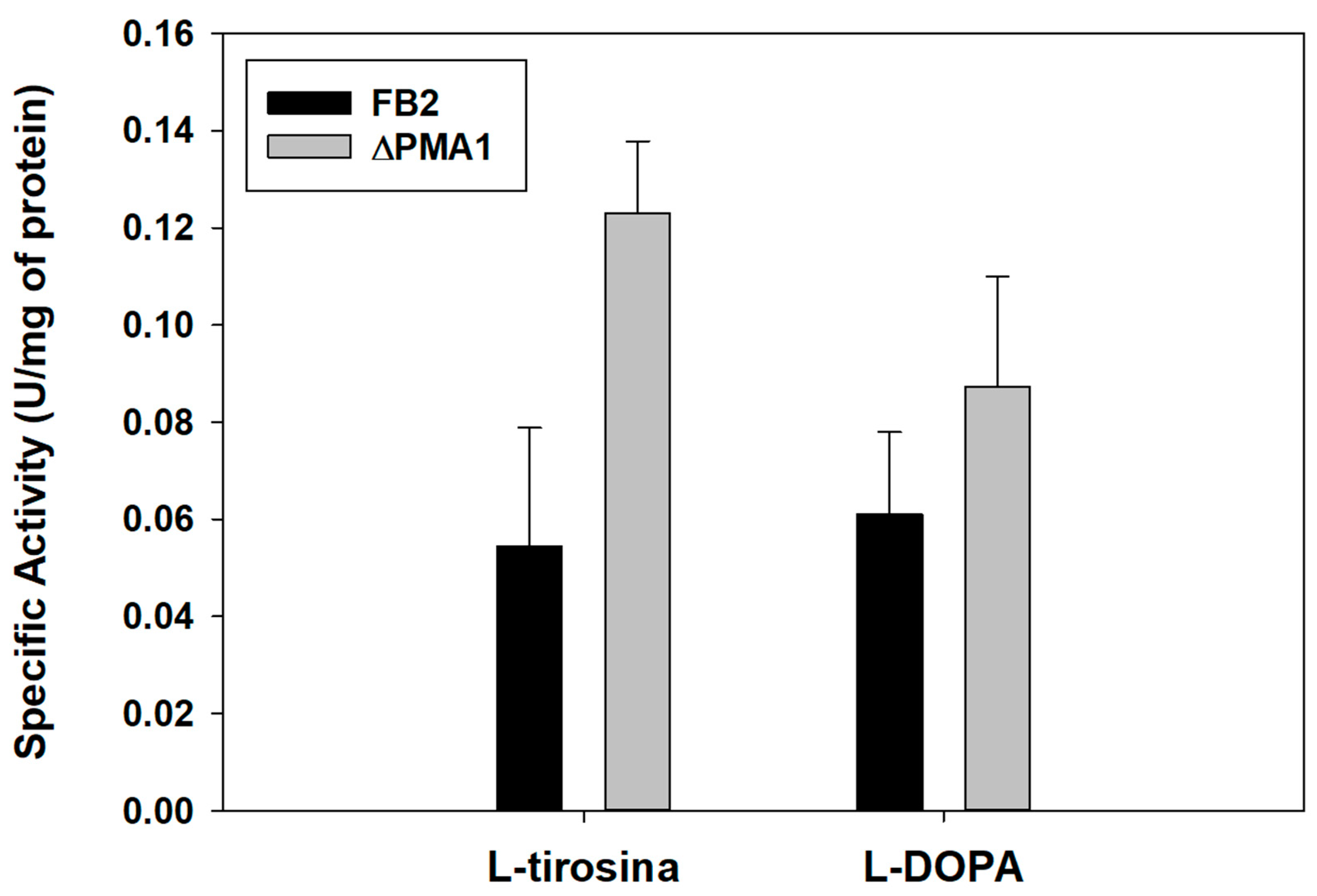
Since tyrosinase activity serves as an indirect indicator of melanin production, we conducted assays to measure the activity of this enzyme in the cell extracts obtained from both the wild-type and ΔPMA1 mutant strains. To maximize the pigment accumulation, we opted for a 72 h culture period. As depicted in Figure 7, the enzyme’s activity in the ΔPMA1 strain was approximately twice that observed in the wild-type cells. Upon subjecting the results to a Student t-test (p < 0.05), a significant difference was evident between the wild-type FB2 strain and the ΔPMA1 mutant strain. Consequently, these findings strongly suggest that the pigmentation observed in the ΔPMA1 mutant results from melanin accumulation, making this strain a promising candidate for pigment production purposes.

3.5. Tyrosinase Activity Assay with Metal Ions

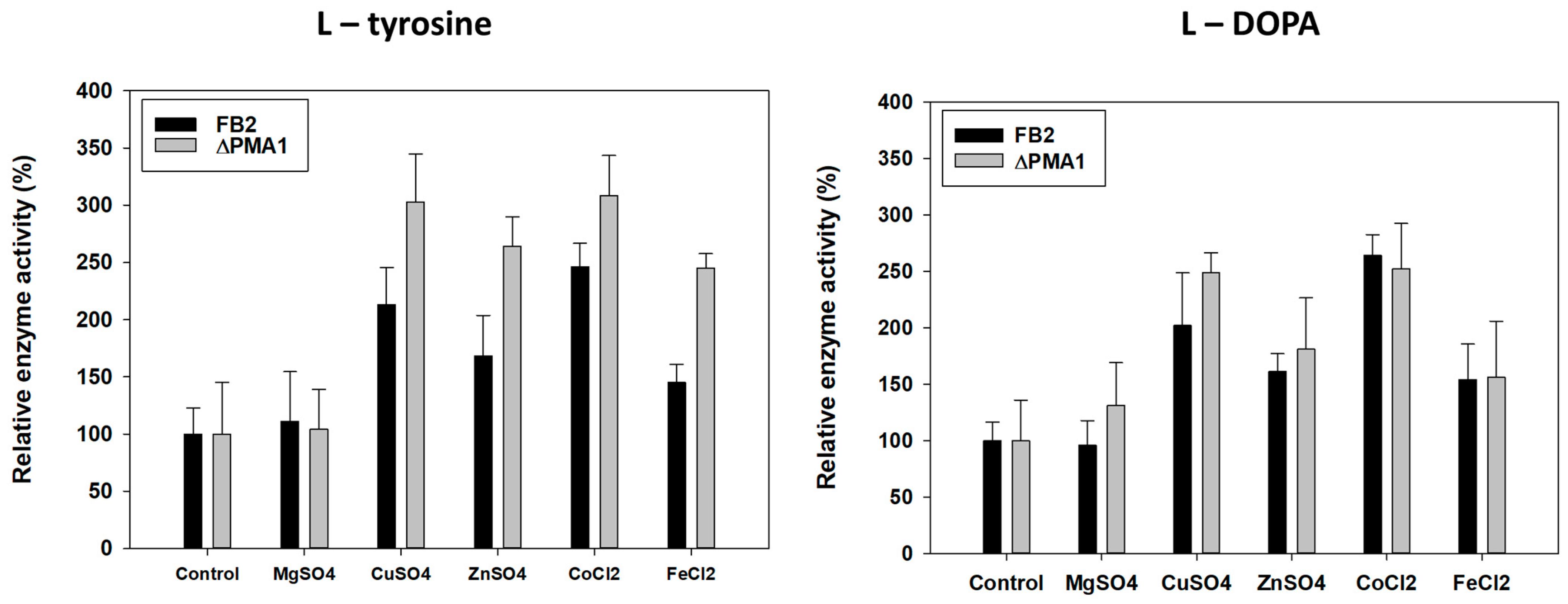
It is known that tyrosinase is a metalloenzyme, with a pair of copper ions in its active center [37,38]. Because this is the first time that the enzyme of U. maydis is studied, we tested other metals in addition to Cu2+; the effect of metal ions on the activity of tyrosinase was assayed using 50 mM of the following salts: MgSO4, CuSO4, ZnSO4, CoCl2, and FeCl2, with L-tyrosine or L-DOPA as substrate. As shown in Figure 8 the tyrosinase activity was higher in the presence of Cu > Co > Zn > Fe > Mg when tyrosine was the substrate and a similar tendency was obtained with L-DOPA but with smaller activity than with tyrosine. This results suggest that the monophenol oxidase activity of tyrosinase occurs also with Co2+ as a cofactor.

3.6. Removal of Methylene Blue from Water Solutions of FB2 and Mutant Cells

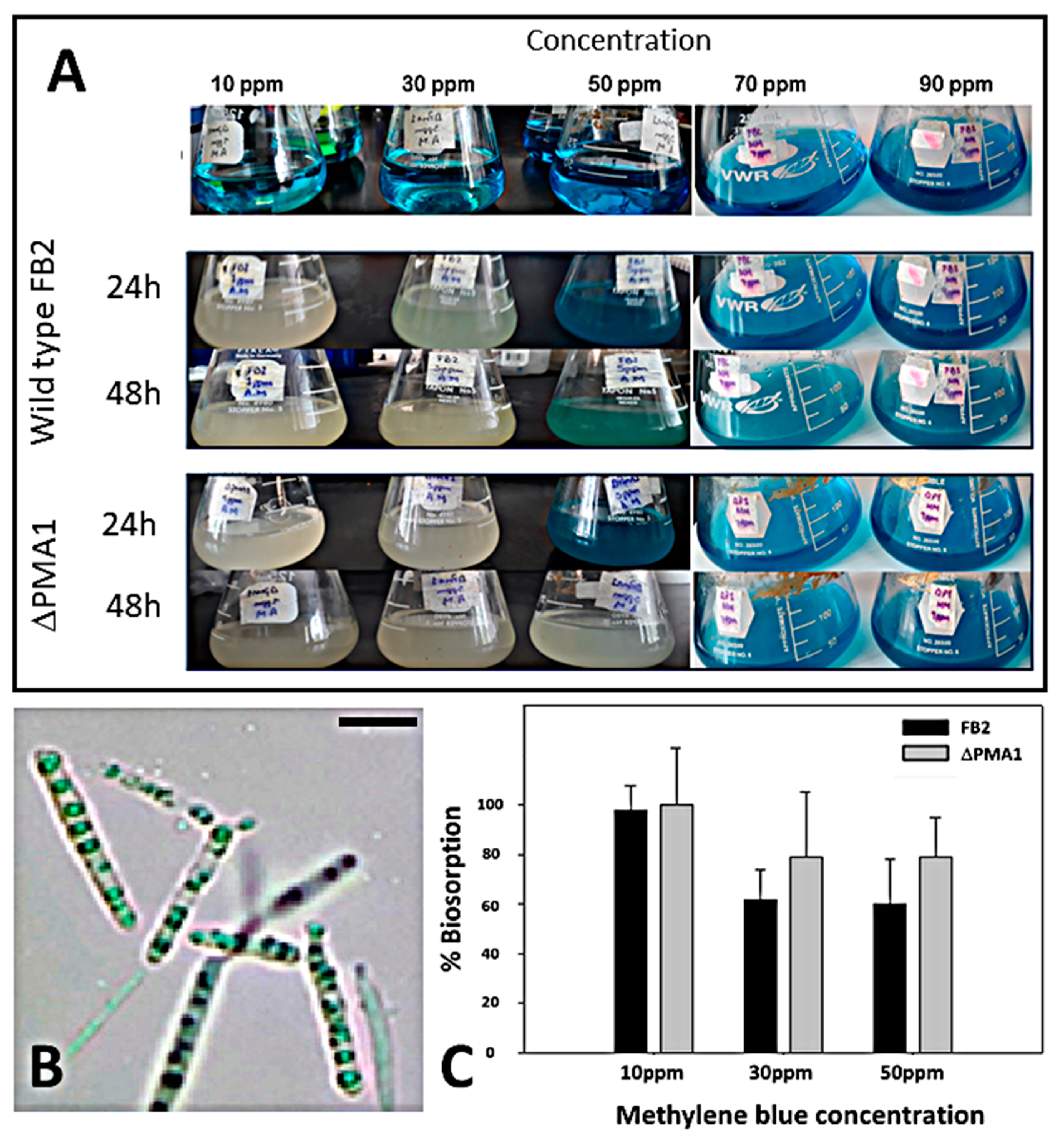
To test the capacity of the mutant and wild type cells to remove the dye methylene blue, the cells (0.5 g wet weight) were incubated in the presence of different concentrations of dye, in 50 mL of liquid YPD for 24 and 48 h with shaking to enhance the interaction of the cells with the dye. As shown in Figure 9, the ΔPMA1 was more efficient at removing the dye than the wild-type cells. At 48 h and a concentration of 10 ppm, the complete removal of the methylene blue was achieved by the ΔPMA1 strain. However, when the concentration of the dye was above 10 ppm, cells were less efficient in the removal of the stain. At 48 h of incubation, around 90% of the methylene blue was removed by the ΔPMA1 mutant, but only 60% by the FB2 strain. The cells with the adsorbed dye became all blue but released with centrifugation at 3000 rpm. To maintain the unstained solution, filtration with the Whatman paper was needed to obtain a clear liquid. The biosorption of methylene blue, as a conventional dye model, provides insight into the potential of the mutant strain to be used for removing other contaminants, using it as a reference point.

4. Discussion

During the construction of the ΔPMA1 mutant, extra point mutations were introduced in multiple sites downstream and upstream of the position of the PMA1 H+ ATPase gene, which resulted in a strain that accumulates melanin. A correct deletion of the Pma1 gene without the point mutations did not result in melanin accumulation, indicating that this response is due to random point mutations. The pigment characterization involved TLC chromatography, UV-vis, and infrared spectra. The comparison of the samples with the commercial synthetic melanin indicated that the pigment found in U. maydis was the same type.
The chemical characteristics of the U. maydis pigment agreed with other melanins described for microorganisms [30,40]. In animals, bacteria, and fungi it is common to find melanin (DOPA-melanin) and its precursor can be either tyrosine or L-DOPA. Fungal melanin is synthetized in internal vesicles that look like the mammalian melanosomes and is then transported to the cell wall where the melanin crosslinks with polysaccharides [41,42,43]. We detected small spots of melanin in the cytoplasm of the ΔPMA1mutant, and cells became brown after 72 h of growth on solid media, suggesting that the production of melanin may be a response to the stress caused by the additional point mutation, and the excess of melanin is trapped in the cytosol.
The content of melanin in fungi is variable [12]. In U. maydis, the saprophytic cells do not show melanogenesis. Normally, the spores are highly melanized, but once they become sporidia, the melanin is preferably located at the cell wall, presumably to protect the cell against environmental stresses. In this study, we measured the melanin content in wild-type FB2 and ΔPMA1 strains cultured for 72 h and found a higher content of melanin in the ΔPMA1 strain. To assess the basic behavior of the mutant in culture, cells were grown in YPD or MM media. Our results showed that the parent FB2 wild-type strain and ΔPMA1 mutant displayed similar growth and small differences in the duplication time. These results suggest that the accumulation of melanin did not affect the cell division processes and vegetative growth. Furthermore, deletion of the pma1 gene did not affect the glucose or oxygen consumption [31]. On the other hand, we noticed that the ΔPMA1 mutant was stable after long periods of storage at −80 °C.
Cytosolic melanin accumulation was observed in the cells containing a mutation of the UmRrm 75 gene, which encodes a protein with three RNA recognition motifs with glycine-rich regions. UmRrm 75 mutant cells showed morphological alterations, decreased mating, and reduced virulence [17]. The additional phenotype was the accumulation of melanin in the cytosol. The accumulated pigment was melanin, passing this phenotype to the modification of pH by the present mutation. In our study, upon deletion of the PMA1 gene, the mutant showed an accumulation of melanin. The H+ ATPase is an enzyme located at the plasma membrane that pumps protons outside the cell and generates a proton electrochemical potential across the membrane [44,45]. U. maydis contains two H+ ATPases genes, PMA1 and PMA2, and both are expressed in the wild type cells growing in the YPD or MM media [46]. In contrast, in the yeast Saccharomyces cerevisiae, only one gene (PMA1) is expressed [45]. In a recent publication about the U. maydis H+ ATPase, mutation of any of these genes did not affect the proton pumping or internal pH changes [31].
Fungal melanin biosynthesis hinges on two primary pathways: the 1,8-dihydroxy naphthalene intermediate (DHN-pathway) and the L-3,4-dihydroxyphenylalanine (L-DOPA pathway) [47,48]. Both pathways are widespread across bacterial and fungal kingdoms [40]. Typically, bacteria and basidiomycetous fungi prefer the L-DOPA pathway, while ascomycetous fungi, and even some non-microscopic counterparts like Tuber spp., favor the DHN-pathway for melanin production [34].
However, U. maydis sets itself apart by showcasing an unconventional melanin production pathway [18]. Researchers have identified a gene cluster associated with melanin production in this fungus. This cluster comprises five pivotal enzymes, including three polyketide synthases (pks3, pks4, and pks5), a cytochrome p540 monooxygenase, and a protein akin to versicolor synthase B (vbs1). The deletion of this gene cluster indicated that U. maydis employs an alternative mechanism for polyketide synthesis that ultimately leads to the formation of DHN-type melanin. This is in stark contrast to ascomycetes like Aspergillus flavus, A. niger, and A. tamarii, which primarily employ the L-DOPA pathway for melanin synthesis, or others like A. tubingensis and A. terreus, which opt for the DHN pathway [40]. On the other hand, Cryptococcus neoformans utilizes the L-DOPA pathway, akin to specific bacteria and mammals [49,50]. Generally, microbial melanin synthesis predominantly follows the L-DOPA pathway, utilizing tyrosine as a precursor. Most Ascomycota favor the DHN melanin pathway, while basidiomycetes produce L-DOPA melanin [50]. Interestingly, U. maydis’s ability to coexist with both melanin synthesis pathways, akin to Aspergillus spp., adds to the intrigue [40]. Our results underscore that a significant portion of the accumulated melanin in U. maydis originates from either tyrosine or L-DOPA as the substrate.
Tyrosinases (EC 1.14.18.1) have been isolated and characterized from plants, animals, and microorganisms [51]. The enzyme catalyzes two types of reactions: (a) as monophenolase, hydroxylating monophenols (e.g., L-tyrosine) to O-diphenols (e.g., L-DOPA), and (b) as diphenolase, oxidizing O-diphenols to O-DOPAquinone [51]. The subsequent reactions need two molecules of O-DOPAquinone to generate L-DOPA and dopachrome molecules. In most cases, the synthesis of melanin depends on the L-DOPA pathway, in which tyrosinase is one of the key enzymes [51]. To examine if the melanin biosynthesis in U. maydis follows this pathway, we measured the tyrosine activity in wild-type and mutant cells grown for 72 h in the crude extract. Our results indicated that the highest activity of tyrosinase was found in the ΔPMA1 strain, which also has the melanin accumulated with a preference for tyrosine substrate. This difference is obtained regardless of the culture medium (YPD or MM).
It has been reported that tyrosinase activity requires metal ions to reach activity [52,53]. We explored the effect of different ions (Mg2+, Cu2+, Zn2+, Co2+, and Fe2+). Remarkably, a noticeable difference in the tyrosinase activity was found when tyrosine or L-DOPA were used as a substrate. Metal ions also activate the tyrosinase activity in both substrates, but the higher activation was obtained with tyrosine Cu+2, Co2+, and Zn2+, which were found to be good cofactors for the U. maydis enzyme. The majority of the tyrosinases accept Cu2+ and Zn2+ as cofactors. However, the effect of Co2+ was unexpected because, for most of the tyrosinases, this metal ion does not function as an essential activator.
Finally, the melanized cells carrying the point mutation had a higher capacity for the removal of methylene blue. The dye was inside the cells, and the melanin may have been saturated. The use of a chitosan-based biosorbent with immobilized microfungal spores of U. maydis and U. digitariae for the removal of heavy metals has been reported [54]. In this study, the spores were fixed to chitosan and were found to have a better performance in the metal removal in U. digitariae than in U. maydis (higher r4motion 84% vs. 63%, respectively). Here, we showed that the melanized yeast can attain 50% removal of the dye without chitosan. The yeast can also be chitosan-immobilized and could display more efficient removal of dyes and even of metal ions. Finally, we found that U. maydis under genetic stress may accumulate melanin as an extra response without physiological changes, synthesizing melanin by the L-DOPA pathway and keeping it in the cytosol. The modified cells may offer biotechnological potential in the removal of dyes and metals [55].
In summary, the random point mutant strain ΔPMA1 of U. maydis synthesizes eumelanin within cytosolic melanosomes, a process related to the tyrosinase activity pathway. We studied the effects of metal ions on this enzyme activity, revealing higher activity with Cu2+ and Co2+. The accumulation of melanin does not significantly affect the physiology of the mutant strain when compared to the WT strain. In addition, the mutant strain exhibits biotechnological potential for dye bioremediation through interactions with melanin.

Author Contributions

Conceptualization, G.G.-S., C.M.-L. and D.R.O.-H.; methodology, C.M.-L., M.V.-C., D.R.O.-H. and G.G.-S.; software, M.V.-C.; validation, O.F.-H., J.P.P., D.R.O.-H. and G.G.-S.; formal analysis, D.R.O.-H., G.G.-S. and J.P.P.; investigation, G.G.-S. and C.M.-L.; resources, G.G.-S.; data curation, J.P.P. and O.F.-H.; writing—original draft preparation, C.M.-L., M.V.-C. and G.G.-S.; writing—review and editing, G.G.-S., J.P.P., O.F.-H. and D.R.O.-H.; visualization, G.G.-S.; supervision, G.G.-S. and D.R.O.-H.; project administration, G.G.-S.; funding acquisition, G.G.-S. and D.R.O.-H. All authors have read and agreed to the published version of the manuscript.

Funding

This research was funded by the Secretaria de Investigación y Posgrado (SIP) in the Intituto Politécnico Nacional, grant numbers 20190200, 20201123, 20210520, 20220487 and Conacyt 256520 to GGS and the Secretaria de Investigación y Posgrado (SIP) in the Intituto Politécnico Nacional, grant number 20230427 to DROH. Cinthya Martínez-López is a PhD student recipient of fellowships from CONACyT (CVU 737347) and BEIFI-IPN (A180142).

Institutional Review Board Statement

Not applicable.

Informed Consent Statement

Not applicable.

Data Availability Statement

Not applicable.

Acknowledgments

The group of authors would like to thank the Instituto Politécnico Nacional, ENCB-IPN, CONAHCyT (formerly called CONACyT), SIP-IPN, and BEIFI for the support provided. Also, thank you to the Microbiology group of the Institute of Microbiology of the University of Düsseldorf, Germany, especially Michael Feldbrügge, Silke Jankowski, Carl Haag and Ute Gengenbacher for the support in the mutant stain construction.

Conflicts of Interest

The authors declare no conflict of interest.

References

  1. Olicón-Hernández, D.R.; Araiza-Villanueva, M.G.; Pardo, J.P.; Aranda, E.; Guerra-Sánchez, G. New Insights of Ustilago maydis as Yeast Model for Genetic and Biotechnological Research: A Review. Curr. Microbiol. 2019, 76, 917–926. [Google Scholar] [CrossRef]
  2. Liebal, U.W.; Ullmann, L.; Lieven, C.; Kohl, P.; Wibberg, D.; Zambanini, T.; Blank, L.M. Ustilago maydis Metabolic Characterization and Growth Quantification with a Genome-Scale Metabolic Model. J. Fungi 2022, 8, 524. [Google Scholar] [CrossRef]
  3. Horst, R.J.; Doehlemann, G.; Wahl, R.; Hofmann, J.; Schmiedl, A.; Kahmann, R.; Kämper, J.; Voll, L.M. A model of Ustilago maydis leaf tumor metabolism. Plant Signal Behav. 2010, 5, 1446–1449. [Google Scholar] [CrossRef] [PubMed]
  4. Dean, R.; Van Kan, J.A.L.; Pretorius, Z.A.; Hammond-Kosack, K.E.; Di Pietro, A.; Spanu, P.D.; Rudd, J.J.; Dickman, M.; Kahmann, R.; Ellis, J.; et al. The Top 10 fungal pathogens in molecular plant pathology. Mol. Plant Pathol. 2012, 13, 414–430. [Google Scholar] [CrossRef]
  5. Paul, J.A.; Wallen, R.M.; Zhao, C.; Shi, T.; Perlin, M.H. Coordinate regulation of Ustilago maydis ammonium transporters and genes involved in mating and pathogenicity. Fungal Biol. 2018, 122, 639–650. [Google Scholar] [CrossRef] [PubMed]
  6. Lanver, D.; Tollot, M.; Schweizer, G.; Lo Presti, L.; Reissmann, S.; Ma, L.-S.; Schuster, M.; Tanaka, S.; Liang, L.; Ludwig, N.; et al. Ustilago maydis effectors and their impact on virulence. Nat. Rev. Microbiol. 2017, 15, 409–421. [Google Scholar] [CrossRef]
  7. Yu, C.; Qi, J.; Han, H.; Wang, P.; Liu, C. Progress in pathogenesis research of Ustilago maydis, and the metabolites involved along with their biosynthesis. Mol. Plant Pathol. 2023, 24, 495–509. [Google Scholar] [CrossRef] [PubMed]
  8. Heucken, N.; Tang, K.; Hüsemann, L.; Heßler, N.; Müntjes, K.; Feldbrügge, M.; Göhre, V.; Zurbriggen, M.D. Engineering and Implementation of Synthetic Molecular Tools in the Basidiomycete Fungus Ustilago maydis. J. Fungi 2023, 9, 480. [Google Scholar] [CrossRef]
  9. Djamei, A.; Depotter, J.; Saridis, G.; Prokchorchik, M.; Barghahn, S.; Silva, N.D.S.T.E.; Zuo, W.; Villamil, J.M.; Doehlemann, G. Modulation of Host Immunity and Development by Ustilago maydis. In Plant Relationships: Fungal-Plant Interactions; Scott, B., Mesarich, C., Eds.; Springer International Publishing: Cham, Switzerland, 2023; pp. 3–30. [Google Scholar] [CrossRef]
  10. Lin, J.-S.; Happel, P.; Kahmann, R. Nuclear status and leaf tumor formation in the Ustilago maydis–maize pathosystem. New Phytol. 2021, 231, 399–415. [Google Scholar] [CrossRef] [PubMed]
  11. Zou, K.; Li, Y.; Zhang, W.; Jia, Y.; Wang, Y.; Ma, Y.; Lv, X.; Xuan, Y.; Du, W. Early infection response of fungal biotroph Ustilago maydis in maize. Front. Plant Sci. 2022, 13, 970897. [Google Scholar] [CrossRef] [PubMed]
  12. Cordero, R.J.B.; Casadevall, A. Functions of fungal melanin beyond virulence. Fungal Biol. Rev. 2017, 31, 99–112. [Google Scholar] [CrossRef]
  13. El-Naggar, N.E.-A.; Saber, W.I.A. Natural Melanin: Current Trends, and Future Approaches, with Especial Reference to Microbial Source. Polymers 2022, 14, 1339. [Google Scholar] [CrossRef] [PubMed]
  14. Kyrmizi, I.; Ferreira, H.; Carvalho, A.; Figueroa, J.A.L.; Zarmpas, P.; Cunha, C.; Akoumianaki, T.; Stylianou, K.; Deepe, G.S.; Samonis, G.; et al. Calcium sequestration by fungal melanin inhibits calcium–calmodulin signalling to prevent LC3-associated phagocytosis. Nat. Microbiol. 2018, 3, 791–803. [Google Scholar] [CrossRef] [PubMed]
  15. Liu, R.; Meng, X.; Mo, C.; Wei, X.; Ma, A. Melanin of fungi: From classification to application. World J. Microbiol. Biotechnol. 2022, 38, 228. [Google Scholar] [CrossRef] [PubMed]
  16. Gow Neil, A.R.; Latge, J.-P.; Munro Carol, A. The Fungal Cell Wall: Structure, Biosynthesis, and Function. Microbiol. Spectr. 2017, 5, 10–1128. [Google Scholar] [CrossRef]
  17. Rodríguez-Piña, A.L.; Juárez-Montiel, M.; Hernández-Sánchez, I.E.; Rodríguez-Hernández, A.A.; Bautista, E.; Becerra-Flora, A.; López-Villegas, E.O.; Jiménez-Bremont, J.F. The Ustilago maydis null mutant strains of the RNA-binding protein UmRrm75 accumulate hydrogen peroxide and melanin. Sci. Rep. 2019, 9, 10813. [Google Scholar] [CrossRef]
  18. Reyes-Fernández Esmeralda, Z.; Shi, Y.-M.; Grün, P.; Bode Helge, B.; Bölker, M. An Unconventional Melanin Biosynthesis Pathway in Ustilago maydis. Appl. Environ. Microb. 2021, 87, e01510–e01520. [Google Scholar] [CrossRef]
  19. Fei, W.; Liu, Y. Biotrophic Fungal Pathogens: A Critical Overview. Appl. Biochem. Biotech. 2023, 195, 1–16. [Google Scholar] [CrossRef]
  20. Chethana, K.W.T.; Jayawardena, R.S.; Chen, Y.-J.; Konta, S.; Tibpromma, S.; Abeywickrama, P.D.; Gomdola, D.; Balasuriya, A.; Xu, J.; Lumyong, S.; et al. Diversity and Function of Appressoria. Pathogens 2021, 10, 746. [Google Scholar] [CrossRef]
  21. Rassabina, A.; Khabibrakhmanova, V.; Babaev, V.; Daminova, A.; Minibayeva, F. Melanins from the Lichens Lobaria pulmonaria and Lobaria retigera as Eco-Friendly Adsorbents of Synthetic Dyes. Int. J. Mol. Sci. 2022, 23, 15605. [Google Scholar] [CrossRef]
  22. Almeida-Paes, R.; Figueiredo-Carvalho, M.H.G.; da Silva, L.B.R.; Gerfen, G.; de S Araújo, G.R.; Frases, S.; Zancopé-Oliveira, R.M.; Nosanchuk, J.D. Candida glabrata produces a melanin-like pigment that protects against stress conditions encountered during parasitism. Future Microbiol. 2021, 16, 509–520. [Google Scholar] [CrossRef] [PubMed]
  23. García-Carnero, L.C.; Clavijo-Giraldo, D.M.; Gómez-Gaviria, M.; Lozoya-Pérez, N.E.; Tamez-Castrellón, A.K.; López-Ramírez, L.A.; Mora-Montes, H.M. Early Virulence Predictors during the Candida Species–Galleria mellonella Interaction. J. Fungi 2020, 6, 152. [Google Scholar] [CrossRef]
  24. Yuan, Z.; Druzhinina, I.S.; Gibbons, J.G.; Zhong, Z.; Van de Peer, Y.; Rodriguez, R.J.; Liu, Z.; Wang, X.; Wei, H.; Wu, Q.; et al. Divergence of a genomic island leads to the evolution of melanization in a halophyte root fungus. ISME J. 2021, 15, 3468–3479. [Google Scholar] [CrossRef]
  25. Chrissian, C.; Lin, C.P.-C.; Camacho, E.; Casadevall, A.; Neiman, A.M.; Stark, R.E. Unconventional Constituents and Shared Molecular Architecture of the Melanized Cell Wall of C. neoformans and Spore Wall of S. cerevisiae. J. Fungi 2020, 6, 329. [Google Scholar] [CrossRef]
  26. Szilágyi, M.; Anton, F.; Pócsi, I.; Emri, T. Autolytic enzymes are responsible for increased melanization of carbon stressed Aspergillus nidulans cultures. J. Basic. Microb. 2018, 58, 440–447. [Google Scholar] [CrossRef] [PubMed]
  27. Camacho, E.; Vij, R.; Chrissian, C.; Prados-Rosales, R.; Gil, D.; O’Meally, R.N.; Cordero, R.J.B.; Cole, R.N.; McCaffery, J.M.; Stark, R.E.; et al. The structural unit of melanin in the cell wall of the fungal pathogen Cryptococcus neoformans. J. Biol. Chem. 2019, 294, 10471–10489. [Google Scholar] [CrossRef]
  28. Smith, D.F.Q.; Casadevall, A. The Role of Melanin in Fungal Pathogenesis for Animal Hosts. In Fungal Physiology and Immunopathogenesis; Rodrigues, M.L., Ed.; Springer International Publishing: Cham, Switzerland, 2019; pp. 1–30. [Google Scholar] [CrossRef]
  29. Azumi, J.; Takeda, T.; Shimada, Y.; Aso, H.; Nakamura, T. The Organogermanium Compound THGP Suppresses Melanin Synthesis via Complex Formation with L-DOPA on Mushroom Tyrosinase and in B16 4A5 Melanoma Cells. Int. J. Mol. Sci. 2019, 20, 4785. [Google Scholar] [CrossRef]
  30. Pacelli, C.; Cassaro, A.; Maturilli, A.; Timperio, A.M.; Gevi, F.; Cavalazzi, B.; Stefan, M.; Ghica, D.; Onofri, S. Multidisciplinary characterization of melanin pigments from the black fungus Cryomyces antarcticus. Appl. Microbiol. Biot. 2020, 104, 6385–6395. [Google Scholar] [CrossRef]
  31. Vázquez-Carrada, M.; Feldbrügge, M.; Olicón-Hernández, D.R.; Guerra-Sánchez, G.; Pardo, J.P. Functional Analysis of the Plasma Membrane H+-ATPases of Ustilago maydis. J. Fungi 2022, 8, 550. [Google Scholar] [CrossRef]
  32. Terfrüchte, M.; Joehnk, B.; Fajardo-Somera, R.; Braus, G.H.; Riquelme, M.; Schipper, K.; Feldbrügge, M. Establishing a versatile Golden Gate cloning system for genetic engineering in fungi. Fungal Genet. Biol. 2014, 62, 1–10. [Google Scholar] [CrossRef] [PubMed]
  33. Olicón-Hernández, D.R.; Hernández-Lauzardo, A.N.; Pardo, J.P.; Peña, A.; Velázquez-del Valle, M.G.; Guerra-Sánchez, G. Influence of chitosan and its derivatives on cell development and physiology of Ustilago maydis. Int. J. Biol. Macromol. 2015, 79, 654–660. [Google Scholar] [CrossRef] [PubMed]
  34. Harki, E.; Talou, T.; Dargent, R. Purification, characterisation and analysis of melanin extracted from Tuber. melanosporum Vitt. Food Chem. 1997, 58, 69–73. [Google Scholar] [CrossRef]
  35. Lowry, O.; Rosebrough, N.; Farr, A.L.; Randall, R. Protein Measurement With The Folin Phenol Reagent. J. Biol. Chem. 1951, 193, 265–275. [Google Scholar] [CrossRef]
  36. Santiago, M.; Strobel, S. Chapter Twenty-Four—Thin Layer Chromatography. In Methods in Enzymology; Lorsch, J., Ed.; Academic Press: Cambridge, MA, USA, 2013; Volume 533, pp. 303–324. [Google Scholar]
  37. Goldfeder, M.; Kanteev, M.; Adir, N.; Fishman, A. Influencing the monophenolase/diphenolase activity ratio in tyrosinase. BBA—Proteins Proteom. 2013, 1834, 629–633. [Google Scholar] [CrossRef] [PubMed]
  38. Lv, Y.; Liang, Q.; Li, Y.; Li, X.; Liu, X.; Zhang, D.; Li, J. Effects of metal ions on activity and structure of phenoloxidase in Penaeus vannamei. Int. J. Biol. Macromol. 2021, 174, 207–215. [Google Scholar] [CrossRef] [PubMed]
  39. Carreira, A.; Ferreira, L.M.; Loureiro, V. Production of brown tyrosine pigments by the yeast Yarrowia lipolytica. J. Appl. Microbiol. 2001, 90, 372–379. [Google Scholar] [CrossRef]
  40. Singh, S.; Nimse, S.B.; Mathew, D.E.; Dhimmar, A.; Sahastrabudhe, H.; Gajjar, A.; Ghadge, V.A.; Kumar, P.; Shinde, P.B. Microbial melanin: Recent advances in biosynthesis, extraction, characterization, and applications. Biotechnol. Adv. 2021, 53, 107773. [Google Scholar] [CrossRef] [PubMed]
  41. Walker Claire, A.; Gómez Beatriz, L.; Mora-Montes Héctor, M.; Mackenzie Kevin, S.; Munro Carol, A.; Brown Alistair, J.P.; Gow Neil, A.R.; Kibbler Christopher, C.; Odds Frank, C. Melanin Externalization in Candida albicans Depends on Cell Wall Chitin Structures. Eukaryotic Cell 2010, 9, 1329–1342. [Google Scholar] [CrossRef]
  42. Zhan, F.; He, Y.; Zu, Y.; Li, T.; Zhao, Z. Characterization of melanin isolated from a dark septate endophyte (DSE), Exophiala pisciphila. World J. Microbiol. Biotechnol. 2011, 27, 2483–2489. [Google Scholar] [CrossRef]
  43. Eisenman, H.C.; Nosanchuk, J.D.; Webber, J.B.W.; Emerson, R.J.; Camesano, T.A.; Casadevall, A. Microstructure of Cell Wall-Associated Melanin in the Human Pathogenic Fungus Cryptococcus neoformans. Biochemistry 2005, 44, 3683–3693. [Google Scholar] [CrossRef] [PubMed]
  44. Schlesser, A.; Ulaszewski, S.; Ghislain, M.; Goffeau, A. A second transport ATPase gene in Saccharomyces cerevisiae. J. Biol. Chem. 1988, 263, 19480–19487. [Google Scholar] [CrossRef] [PubMed]
  45. Fernandes, A.R.; Sá-Correia, I. Transcription patterns of PMA1 and PMA2 genes and activity of plasma membrane H+-ATPase in Saccharomyces cerevisiae during diauxic growth and stationary phase. Yeast 2003, 20, 207–219. [Google Scholar] [CrossRef]
  46. Robles-Martínez, L.; Pardo, J.P.; Miranda, M.; Mendez, T.L.; Matus-Ortega, M.G.; Mendoza-Hernández, G.; Guerra-Sánchez, G. The basidiomycete Ustilago maydis has two plasma membrane H+-ATPases related to fungi and plants. J. Bioenerg. Biomembr. 2013, 45, 477–490. [Google Scholar] [CrossRef] [PubMed]
  47. Langfelder, K.; Streibel, M.; Jahn, B.; Haase, G.; Brakhage, A.A. Biosynthesis of fungal melanins and their importance for human pathogenic fungi. Fungal Genet. Biol. 2003, 38, 143–158. [Google Scholar] [CrossRef] [PubMed]
  48. Pal, A.K.; Gajjar, D.U.; Vasavada, A.R. DOPA and DHN pathway orchestrate melanin synthesis in Aspergillus species. Med. Mycol. 2014, 52, 10–18. [Google Scholar] [CrossRef]
  49. Williamson Peter, R.; Wakamatsu, K.; Ito, S. Melanin Biosynthesis in Cryptococcus neoformans. J. Bacteriol. 1998, 180, 1570–1572. [Google Scholar] [CrossRef]
  50. Toledo, A.V.; Franco, M.E.E.; Yanil Lopez, S.M.; Troncozo, M.I.; Saparrat, M.C.N.; Balatti, P.A. Melanins in fungi: Types, localization and putative biological roles. Physiol. Mol. Plant Pathol. 2017, 99, 2–6. [Google Scholar] [CrossRef]
  51. Kanteev, M.; Goldfeder, M.; Fishman, A. Structure–function correlations in tyrosinases. Protein Sci. 2015, 24, 1360–1369. [Google Scholar] [CrossRef]
  52. Fenoll, L.G.; Peñalver, M.J.; Rodríguez-López, J.N.; Varón, R.; García-Cánovas, F.; Tudela, J. Tyrosinase kinetics: Discrimination between two models to explain the oxidation mechanism of monophenol and diphenol substrates. Int. J. Biochem. Cell Biol. 2004, 36, 235–246. [Google Scholar] [CrossRef] [PubMed]
  53. Palumbo, A.; Misuraca, G.; D’Ischia, M.; Prota, G. Effect of metal ions on the kinetics of tyrosine oxidation catalysed by tyrosinase. Biochem. J. 1985, 228, 647–651. [Google Scholar] [CrossRef]
  54. Sargın, İ.; Arslan, G.; Kaya, M. Microfungal spores (Ustilago maydis and U. digitariae) immobilised chitosan microcapsules for heavy metal removal. Carbohyd Polym. 2016, 138, 201–209. [Google Scholar] [CrossRef] [PubMed]
  55. Mattoon, E.R.; Cordero, R.J.B.; Casadevall, A. Fungal Melanins and Applications in Healthcare, Bioremediation and Industry. J. Fungi 2021, 7, 488. [Google Scholar] [CrossRef] [PubMed]
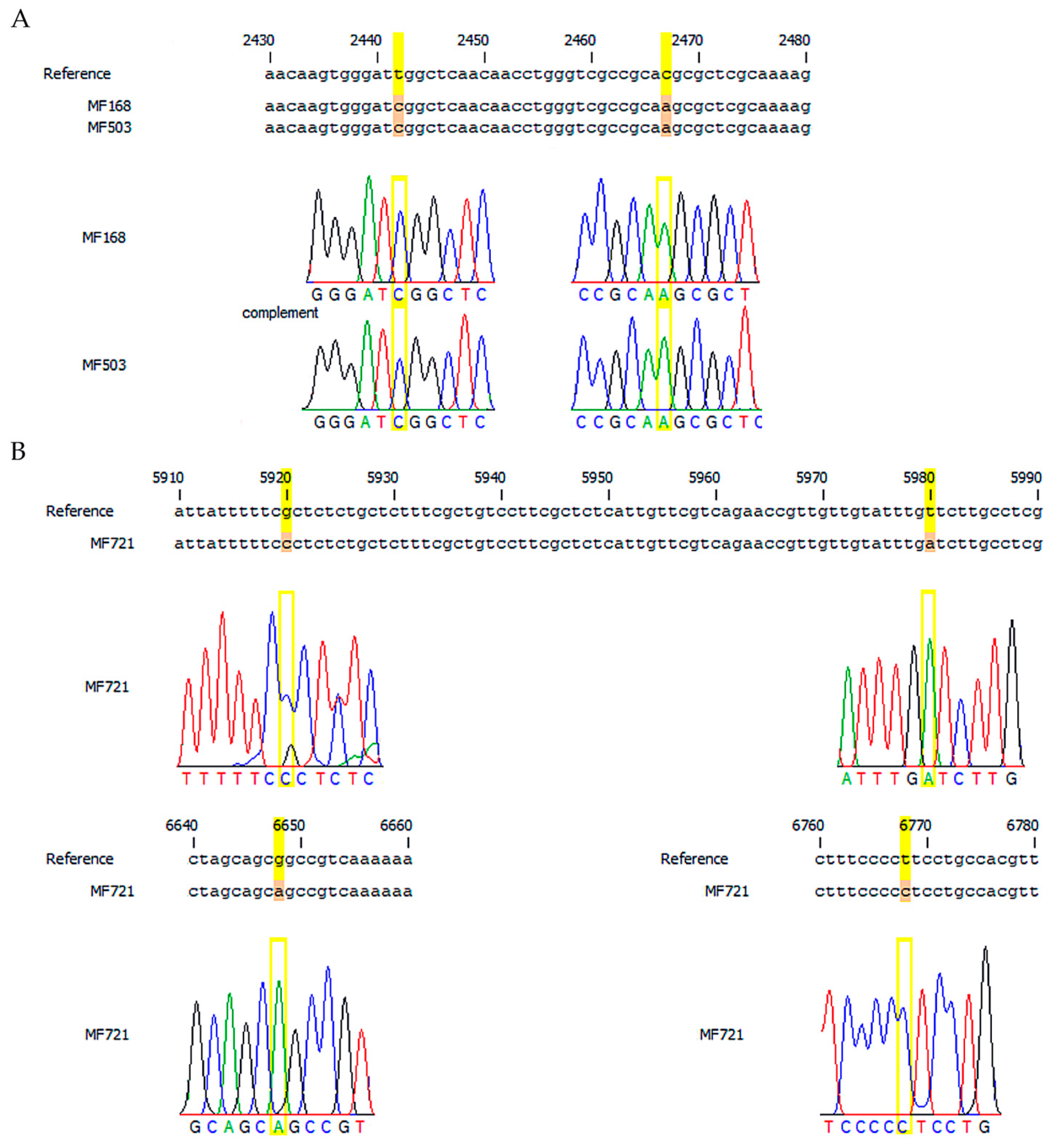
Figure 1. The sequence of U. maydis ΔPMA1 mutant strain. Point mutations are highlighted in yellow. (A), downstream flank. (B), upstream flank. The mutant strain sequence were compared with the strain U. maydis 521 (NCBI:txid237631) gen UMAG 2581.
Applsci 13 11288 g001
Figure 2. Comparison of cell morphology between U. maydis random mutant and wild-type. (A) YPD plates displaying the wild-type (right) and ΔPMA1 mutant (left) strains, with the mutant strain exhibiting higher pigment accumulation and rippling edges. (B) Micrographs of cell morphology captured using a light microscope. The microscopic view of U. maydis cells reveals distortions in the typical cigar shape and the presence of pigment accumulation spots (arrows), bar = 20 µm.
Applsci 13 11288 g002
Figure 3. Scanning and transmission electron microscopy analysis of wild-type and U. maydis mutants of plasma membrane H+-ATPase. (A): Scanning electron microscopy micrographs depicting the cell surface of mutants with a deletion of the H+-ATPase gene, grown in different culture media. A1: FB2 wild-type control in YPD medium; A2: FB2 wild-type control in MM medium; A3: ΔPMA1 in YPD medium; A4: ΔPMA1 in MM medium. The top row displays a panoramic view, while the lower rows correspond to various magnifications specified in each figure. Scale bars = 10 µm (upper row); 1–5 µm (middle row); and 1–2 µm (lower row), depending on the magnification. (B): Transmission electron microscopy images of cross-sections and longitudinal images of U. maydis cells and mutants, revealing electron-dense bodies predominantly present in ΔPMA1. B1 and B2: FB2 wild-type strain in YPD or MM; B3 and B4: ΔPMA1 in YPD or MM, respectively. Scale bar = 200 µm.
Applsci 13 11288 g003
Figure 4. Growth in YPD of the wild-type FB2, and mutant ∆PMA1 cells of U. maydis. Cells were cultured in YPD (FB2, black circle; ∆PMA1, red circle) and minimum media (FB2, inverted green triangle; ∆PMA1, yellow triangle) for 72 h. The growth of both strains was manipulated in the same way as mentioned in the Methods section.
Applsci 13 11288 g004
Figure 5. Identification of the intracellular pigment. (A) Pigment extracted from cells cultivated in YPD for 72 h. (B) Thin layer chromatography of the pigment isolated. (C) UV spectra of the isolated pigment and melanin standard with a 230 nm maximum absorption. (D) IR spectra of commercial melanin and the isolated pigment.
Applsci 13 11288 g005
Figure 6. Intracellular melanin content in U. maydis wild-type (FB2) and the mutant (ΔPMA1) cells. Cells were cultivated for 72 h with shaking, and 1 mL aliquot was taken to determine dry weight. At the end of the culture, cells were used to extract the melanin. The amount of melanin was determined by absorption at 470 nm. Statistical analysis was performed with the Student t-test.
Applsci 13 11288 g006
Figure 7. Tyrosinase-specific activity of the wild-type cells and the ΔPMA1 mutant carrying the punctual mutations. Crude extracts of FB2 and PMA mutant were assayed for tyrosinase activity using tyrosine or L-DOPA as substrate. Cells were cultured by 72 h at 125 rpm shaking and 28 °C.
Applsci 13 11288 g007
Figure 8. Influence of metal ions on tyrosinase enzyme activity using L-tyrosine and L-DOPA as substrates. Metal salts were used at a concentration of 50 mM for this enzymatic assay. The reaction solution consisted of 100 mM phosphate buffer with a pH of 6.0, and it included 100 µL of either L-tyrosine or L-DOPA as substrates, each at a concentration of 5 mM. The reaction was allowed to incubate for 10 min, and the resulting absorbance was measured.
Applsci 13 11288 g008
Figure 9. Dye removal by FB2 and ΔPMA1 cells. (A) Control of methylene blue solution. 90 ppm was the highest concentration to get 1.0 D.O; FB2 cells incubated by 24 and 48 h in the presence of different dye concentrations and ΔPMA1 cells incubated at 24 and 48 h in the presence of methylene blue. (B) Typical view of the cells during biosorption of the dye. Bar = 20 µm. (C) Graphical comparison of the residual absorbance after the incubation with the dye for 24 or 48 h.
Applsci 13 11288 g009
Table 1. Primers and plasmids were used to get H+ ATPase mutants.
PrimerSequence (5′-3′)Use
ΔPma1
UF FPGGTCTCGCCTGCAATATTCCCATTTCCATTTCCATTCFlank construction
UF RPGGTCTCCAGGCCGATGAAAGAAAAAAGACTACCGAGFlank construction
DF FPGGTCTCCGGCCATCCTCATTCTCGACGAAGTGATTCFlank construction
DF RPGGTCTCGCTGCAATATTCGACCTCTGAATCGCTAGCFlank construction
P1CGGTGTTGCCATGAACACCGATGGCCAGTGDiagnostic PCR
P2GAGGGCAACGGATTCGAGCTTCTTGGTCTTDiagnostic PCR
ΔPma2
UF FPGGTCTCGCCTGCAATATTCAACCTCTAAGACTCGCTTFlank construction
UF RPGGTCTCCAGGCCTCTGCCTCTTATCTTGCTCTCTTAGFlank construction
DF FPGGTCTCCGGCCGGGGAAACGTGGAGAAGGTCGCGAAAFlank construction
DF RPGGTCTCGCTGCAATATTACCACCCTGTGCCCTCTAGFlank construction
P1ACGCTTGACAATCTCGTACTTGTGCTCGGGGDiagnostic PCR
P2GAGGGCAACGGATTCGAGCTTCTTGGTCTTDiagnostic PCR
Plasmid
pUma2342Transforming plasmid to delete Pma1 gen
pUma2343Transforming plasmid to delete Pma2 gen
p1507Hyg resistance cassette
UF = Upstream flank. DF = Downstream flank. FP = Forward primer. RP = Reverse primer.
Table 2. Kinetics parameters of growth of wild strain FB2 and FB2-PMA1 mutant. Cells were cultured in YPD or mineral media for 72 h.
StrainCulture MediumGrowth Rate (µ)Generation Time (g)
FB2YPD0.297 ± 0.04 h−12.34 h
MM0.271 ± 0.08 h−12.55 h
∆PMA1YPD0.282 ± 0.09 h−12.45 h
MM0.247 ± 0.09 h−12.80 h
Disclaimer/Publisher’s Note: The statements, opinions and data contained in all publications are solely those of the individual author(s) and contributor(s) and not of MDPI and/or the editor(s). MDPI and/or the editor(s) disclaim responsibility for any injury to people or property resulting from any ideas, methods, instructions or products referred to in the content.