Abstract
Ginkgo biloba L. (Ginkgoacea) contains an abundance of beneficial compounds and has demonstrated positive clinical effects in the management of metabolic syndrome. Recent studies have emphasized its efficacy against type 2 diabetes mellitus (T2DM), including improvements in diabetic nephropathy and retinopathy. Particularly noteworthy are ginkgolic acid analogs, which have shown potential in combating T2DM by inhibiting protein tyrosine phosphatases (PTPs), facilitating glucose uptake, and influencing signaling pathways. In this study, we isolated six derivatives of ginkgolic acid from the MeOH extract of G. biloba leaves with the guidance of liquid chromatography–mass spectrometry (LC/MS). We determined the chemical structures of these isolated compounds as 2-hydroxy-6-(10′-hydroxypentadec-11′(E)-en-1-yl) benzoic acid (1), 2-hydroxy-6-(11′-hydroxypentadec-9′(E)-en-1-yl) benzoic acid (2), 2-hydroxy-6-tridecylbenzoic acid (3), 2-hydroxy-6-pentadecylbenzoic acid (4), 2-hydroxy-6-(12′-hydroxyheptadec-13′(E)-en-1-yl) benzoic acid (5), and 2-hydroxy-6-(11-hydroxyundecyl) benzoic acid (6) using NMR spectroscopic data and LC/MS analysis. To assess their potential for addressing T2DM, we subjected the isolated compounds (1–6) to tests measuring their inhibitory activity against six PTPs: PTPN11, PTPN2, PTP1B, DUSP9, PTPRS, and PTPN9. Among these compounds, compounds 3–5 displayed enzyme inhibition exceeding 90% against all six PTPs. In conclusion, ginkgolic acid derivatives, acting as PTP inhibitors relevant to insulin resistance, hold promise as potential therapeutic candidates for the prevention and treatment of T2DM.
1. Introduction
Type 2 diabetes mellitus (T2DM) is distinguished by its unique characteristics, encompassing disruptions in carbohydrate, lipid, and protein metabolism. This complex condition arises from compromised insulin secretion and resistance. T2DM holds particular significance due to its substantial prevalence, accounting for over 90% of all diabetes cases, in contrast to the less common type 1 diabetes mellitus (T1DM) [1]. T2DM is a chronic and intricate disorder that requires ongoing medical attention, self-management for the regulation of glucose levels, and blood pressure control to prevent both microvascular complications (including retinopathy, neuropathy, and nephropathy) and macrovascular complications (such as heart attacks and strokes) [2,3,4]. As of now, a definitive cure for T2DM remains elusive. However, available treatment approaches encompass lifestyle modifications, interventions for managing obesity, the administration of oral hypoglycemic agents, and insulin sensitizers, with metformin being the recommended first-line medication for obese patients [5]. Oral hypoglycemic agents are categorized based on their distinct mechanisms of action, which include inhibition of α-glucosidase and α-amylase in the digestive tract, influence on glucose uptake and transporter activity, enhancement of insulin secretion, promotion of pancreatic β cell proliferation, antioxidative properties to mitigate oxidative stress, and inhibitory activity of protein tyrosine phosphatase [6].
The insulin receptor necessitates the tyrosine autophosphorylation of three closely situated tyrosine residues within its kinase domain to facilitate subsequent insulin-induced glucose uptake. Recent efforts have been directed towards enhancing overall insulin action by mitigating the inhibitory effects on initial insulin responses. This is achieved by modulating the activity of cellular protein tyrosine phosphatases (PTPs), which serve as potential targets for therapeutic intervention [7]. Among these PTPs are PTP1B, PTPN2, PTPN11, PTPN9, PTPRS, and DUSP9, which collectively contribute to the induction of insulin resistance, making them attractive targets for treating T2DM [8]. PTP1B and PTPN9 function as negative regulators of insulin signaling pathways by dephosphorylating the insulin receptor. The absence of PTPN2 enhances insulin receptor activation in both hepatocytes and fibroblasts. PTPN11 serves as a negative regulator in the liver, and its deletion enhances hepatic insulin sensitivity [8,9]. Mice lacking PTPRS exhibit alterations in insulin sensitivity and glucose homeostasis [10]. DUSP9, located on the X chromosome, encodes a member of the mitogen-activated protein kinase phosphatase 4 family, playing a pivotal role in cell cycle regulation and the regulation of insulin action [11].
Ginkgo biloba (Ginkgoaceae) has played significant roles as a valuable plant for humans. Its history spans over 200 years, earning it the distinction of being a “living fossil”. G. biloba was introduced to Europe around 1730 and has since become a prominent tree in streets and parks. It has also found extensive use as a medicinal plant, particularly in China and Korea [12]. The leaves of G. biloba are traditionally employed in the treatment of diarrhea and infantile enteritis, while its seeds have been used to address various conditions such as ringworm sores, pox, dog bites, asthma, phlegm, and colds [13]. G. biloba contains a variety of beneficial compounds, including terpene trilactones (bilobalide), flavonoids (kaempferol, quercetin, and their glycosides), bioflavonoids (ginkgetin), proanthocyanidins (procyanidin), and alkylphenols (ginkgolic acids) [14]. Recent studies have indicated that extracts from G. biloba leaves exhibit a wide range of beneficial properties, including antiasthmatic, antioxidant, wound healing, radical-scavenging, neuroprotective, and metabolic syndrome management effects, which include weight loss, antidiabetic, and antihypertensive properties [12,15].
A recent study has highlighted the substantial efficacy of G. biloba against T2DM [16]. In diabetic nephropathy rats treated with G. biloba extract, notable improvements were observed, including significant reductions in fasting blood glucose levels, creatinine, blood urea nitrogen, urine protein levels, and oxidative stress levels when compared to diabetic nephropathy rats [17]. Additionally, a three-month regimen of orally administered G. biloba extract resulted in a significant reduction in malondialdehyde levels in erythrocyte membranes, decreased levels of fibrinogen, and improved blood viscosity and viscoelasticity. These improvements have the potential to enhance blood perfusion and retinal capillary blood flow rate in T2DM patients with retinopathy [18]. Of particular interest, ginkgolic acid (C13:0) and its analogs have been investigated for their anti-T2DM activity through the inhibition of PTPs. Our previous research has demonstrated that ginkgolic acid displayed inhibitory enzymatic activity against PTPN9 (Ki = 53 M) and DUSP9 (Ki = 2.5 M) in vitro, resulting in a notable enhancement of glucose uptake in C2C12 muscle cells and 3T3-L1 adipocytes [19]. Based on these findings, it is advisable to consider regular consumption of G. biloba as a potential exclusive remedy for T2DM treatment, with ginkgolic acid being regarded as a potential therapeutic candidate for managing T2DM [19].
As an ongoing aspect of our research for identifying bioactive natural metabolites from various natural sources [20,21,22], we investigated ginkgolic acid derivatives from the methanol extract of G. biloba leaves. The isolation process was facilitated by liquid chromatography-mass spectrometry (LC/MS), which led to the isolation of six ginkgolic acid derivatives (1–6). The structural elucidation of the isolated compounds was carried out utilizing data obtained from nuclear magnetic resonance (NMR) experiments and LC/MS analyses. In particular, the isolated components were subjected to evaluation for their inhibitory activity against PTPs, with the aim of assessing their potential in the treatment of T2DM. In this paper, we provide a detailed account of the procedures involved in the separation and identification of compounds 1–6, as well as the assessment of their inhibitory activity against PTPs.
2. Results
2.1. Isolation and Identification of Compounds 1–6
Dried G. biloba leaves were subjected to extraction using 80% aqueous methanol (MeOH). Following the extraction process, the extract was filtered to remove impurities and the resulting extract was then concentrated using a rotary evaporator, yielding a green crude extract. Solvent partitioning and continuous chromatography were employed to isolate bioactive compounds from the crude MeOH extract, guided by LC/MS analysis. Initially, we observed that a substantial portion of the hexane-soluble fraction comprised flavonoids, lipids, and ginkgolic acids. This observation was verified by comparing their mass and UV absorption spectra with data from our in-house libraries and previously reported information [23].
Our primary objective was to isolate characteristic compounds of G. biloba, known as ginkgolic acids, which possess various bioactivities, including antimicrobial, antitumor, anti-inflammatory, antiparasitic, and enzyme inhibitory properties [24]. With the guidance of LC/MS, we successfully isolated six ginkgolic acid derivatives (1–6) using high-performance liquid chromatography (HPLC) (Figure 1).
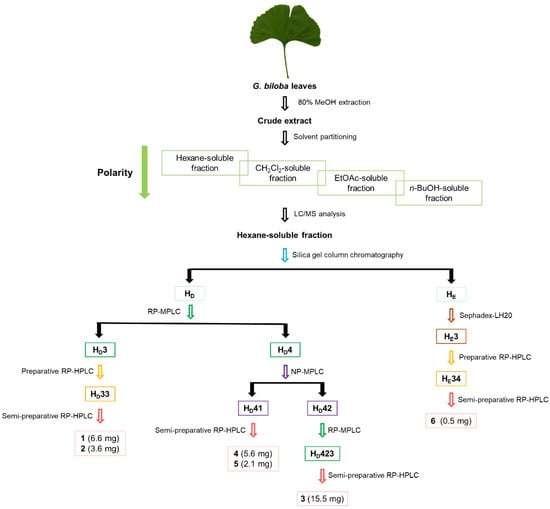
Figure 1.
Separation scheme of compounds (1–6) from the methanol extract of G. biloba leaves.
The identification of these isolated compounds was accomplished, and their structures were determined as follows: 2-hydroxy-6-(10′-hydroxypentadec-11′(E)-en-1-yl) benzoic acid (1) [25], 2-hydroxy-6-(11′-hydroxypentadec-9′(E)-en-1-yl) benzoic acid (2) [25], 2-hydroxy-6-tridecylbenzoic acid (3) [26], 2-hydroxy-6-pentadecylbenzoic acid (4) [27], 2-hydroxy-6-(12′-hydroxyheptadec-13′(E)-en-1-yl) benzoic acid (5) [25], and 2-hydroxy-6-(11-hydroxyundecyl) benzoic acid (6) [28] (Figure 2). All of these compounds were elucidated through a combination of NMR spectra comparison with reported data and LC/MS analysis (Figures S1–S18). Notably, compound 6 was isolated from G. biloba leaves for the first time. In the present study, compounds 1–6 were evaluated for their inhibitory activity against PTPs to assess their potential efficacy in the context of T2DM treatment.
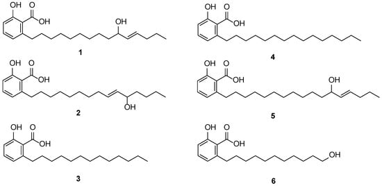
Figure 2.
Chemical structures of compounds 1–6.
2.2. Effects of Isolated Compounds 1–6 on the Inhibition of PTPs
PTPs play crucial roles in various cell signaling pathways, including those related to cell growth, metabolism, differentiation, proliferation, and survival, by regulating protein tyrosine phosphorylation [29,30,31,32,33,34,35,36]. Certain specific PTPs, such as PTP1B, PTPN2, PTPN9, PTPN11, DUSP9, and PTPRS, have been implicated in insulin resistance and are associated with the development of T2DM [37]. This suggests that these PTPs are attractive targets for drug development aimed at treating or preventing T2DM. Consequently, the identification of compounds capable of inhibiting the catalytic activity of PTPs relevant to insulin resistance represents a promising approach for T2DM treatment and prevention. In our study, we investigated whether the isolated components could inhibit the catalytic activity of specific PTPs associated with T2DM. To achieve this, several PTPs, including PTPN11, PTPN2, PTP1B, DUSP9, PTPRS, and PTPN9, were overexpressed and purified using a cobalt affinity resin. To evaluate the inhibitory activity of these PTPs against all isolated compounds (1–6), we measured the catalytic activities of the PTPs using 6,8-difluoro-4-methylumbelliferyl phosphate (DiFMUP) as a fluorogenic PTP substrate. Remarkably, three compounds (3, 4, and 5) exhibited enzyme inhibition exceeding 90% against all six PTPs (Table 1). Additionally, the half inhibitory concentration (IC50) values of compound 3 for PTP1B, PTPN9, and PTPN11 were evaluated as a sufficient quantity was isolated, which showed the IC50 values of 14.45, 7.84, and 11.18 µM, respectively (Figure 3). The compounds (1 and 2) demonstrated more than 70% enzymatic inhibition of PTPN11, PTPN2, PTP1B, and DUSP9 (Table 1). Compound 6 displayed more than 70% inhibitory activity against PTPN11, PTPN2, and PTP1B (Table 1). These findings indicate that the six compounds (1–6) effectively inhibited specific PTPs relevant to insulin resistance, making them potential therapeutic candidates for the prevention or treatment of T2DM.

Table 1.
Inhibition of PTPs by compounds 1–6 (50 μM).
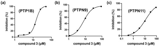
Figure 3.
IC50 values of compound 3 for PTP1B (a), PTPN9 (b), and PTPN11 (c) were estimated using a sigmoid curve against percent inhibition (%) for compound 3.
3. Discussion
PTPs such as PTPN11, PTPN2, PTP1B, DUSP9, PTPRS, and PTPN9 are known to be associated with T2DM, and the use of PTP inhibitors is considered an effective strategy for T2DM treatment [19]. In previous research, we demonstrated that several natural products have antidiabetic effects by inhibiting PTPs linked to insulin resistance [8]. Specifically, we showed that ginkgolic acid, acting as a dual-targeting inhibitor of PTPN9 and DUSP9, enhances glucose uptake in 3T3-L1 adipocytes and C2C12 muscle cells by activating the AMPK signaling pathway, which suggests that ginkgolic acid has potential as a therapeutic candidate for T2DM [19]. In our current study, we isolated ginkgolic acid derivatives and evaluated their inhibitory activity against specific PTPs, including PTP1B, PTPN2, PTPN9, PTPN11, DUSP9, and PTPRS, all associated with insulin resistance. When comparing the inhibition rates of these compounds, structural isomers 1 and 2, both featuring a hydroxy group, displayed similar inhibitory rates, which were lower than that of compound 4, having the same length of the aliphatic chain. From these results, we can infer that the length of the aliphatic chain and the presence of a hydroxy group in the chain are important factors influencing the PTP inhibitory activity of ginkgolic acid derivatives. It is anticipated that ginkgolic acids with longer aliphatic chains and the absence of a hydroxy group in the chain will demonstrate more potent inhibitory activity against PTPs. The findings support the idea that both compound 5, possessing a hydroxy group and a longer chain length than 1, and compound 3, with no hydroxy group and a shorter chain length than 1, exhibited potent inhibitory activity. These results underscore the importance of chain length concerning PTP inhibitory activity [19] and suggest that functional group substitutions within the chain can significantly impact this activity. To reiterate, our study showcases that these six compounds (1–6) effectively inhibit the catalytic activity of specific PTPs associated with insulin resistance, highlighting their potential as PTP inhibitors. Exploring whether these six compounds can enhance glucose uptake by inhibiting PTPs would be an intriguing direction for future research.
Natural product discovery for the treatment of diabetes is an area of growing interest in the field of medical research. The search for effective treatments for diabetes has led researchers to explore the vast reservoir of natural products found in plants, microorganisms, and other natural sources. This approach capitalizes on the diverse array of bioactive compounds present in nature, offering a rich pool of potential therapeutic agents. One noteworthy aspect of natural product discovery is its incorporation of traditional medicine systems. Ancient practices like ayurveda and traditional Chinese medicine have long utilized natural products for various ailments, including diabetes. These traditions provide valuable insights and starting points for modern scientific investigation. Natural products with antidiabetic properties often act through multiple mechanisms. These may include enhancing insulin sensitivity, reducing blood glucose levels, protecting pancreatic beta cells, and mitigating oxidative stress. Understanding these mechanisms is crucial for the development of effective treatments. Since some specific PTPs, such as PTPN2, PTPN9, PTPN11, PTP1B, PTPRS, and DUSP9, are associated with T2DM, the discovery of compounds that can inhibit these PTPs may be an effective strategy to treat or prevent T2DM [38]. We have previously identified some natural compounds with antidiabetic properties through the inhibition of PTPs: for example, chebulinic acid isolated from the fruits of Terminalia chebula acts as a dual inhibitor of PTPN9 and PTPN11 and exhibits antidiabetic properties in C2C12 muscle cells and 3T3-L1 adipocytes [19,39]. Our previous study has shown that ginkgetin, a biflavone from G. biloba leaves, could be a potential antiadipogenesis and antiobesity drug through STAT5-mediated PPARγ and C/EBPα regulation [39]. In addition, we previously demonstrated that terminalin isolated from African mango significantly increased glucose uptake in differentiated C2C12 muscle cells via inhibition of PTPN1, PTPN9, PTPN11, and PTPRS [38]. In line with these findings, we found that six compounds (1–6) from G. biloba leaves suppressed the catalytic activities of certain PTPs such as PTPN2, PTPN9, PTPN11, PTP1B, PTPRS, and DUSP9, suggesting the possibilities that these compounds may stimulate glucose uptake.
Recently, high-throughput screening and in vitro assays are integral to the discovery process. Promising compounds are rigorously tested in animal models and, if successful, advance to human clinical trials. This systematic approach ensures that only the most promising candidates progress toward potential clinical use. Safety is a paramount concern in natural product discovery. While these compounds offer potential therapeutic benefits, researchers must carefully assess their safety profiles and potential side effects. Striking the right balance between efficacy and safety is essential. Natural products can complement existing diabetes medications. They may be used alone or in combination with conventional treatments to enhance their effectiveness or reduce the risk of adverse effects. This approach offers a holistic perspective on diabetes management. Despite the promise of natural product discovery, challenges persist. These include issues related to sourcing, standardization of extracts, and the need for rigorous scientific validation. Ongoing research seeks to address these challenges, with the ultimate goal of identifying novel compounds and therapies. Numerous natural resources have shown promise in diabetes management, including bitter melon, cinnamon, fenugreek, ginseng, and berberine. These natural resources or bioactive compounds have sparked interest due to their potential to lower blood glucose levels and improve insulin sensitivity. Natural product discovery aligns with the principles of complementary and integrative medicine, offering patients a broader spectrum of treatment options. Integrating natural products into diabetes care plans reflects a patient-centered approach to healthcare. Thus, natural product discovery for diabetes treatment represents a dynamic and multidisciplinary field with substantial potential. By tapping into the power of nature and merging traditional knowledge with modern science, researchers aim to identify safe and effective therapies that can alleviate the burden of diabetes. As challenges are addressed and new discoveries are made, this approach holds promise for improving the lives of individuals living with diabetes and advancing the field of diabetes management.
4. Materials and Methods
4.1. Plant Material
G. biloba leaves were gathered from the arboretum located at Sungkyunkwan University, Suwon, Korea, during the month of August 2016. The identification of the plant was carried out by one of the authors (K. H. Kim). A voucher specimen of this material, designated as UHY-2016, has been duly archived in the herbarium of the School of Pharmacy at Sungkyunkwan University, Suwon, Republic of Korea.
4.2. Extraction and Isolation of Compounds 1–6
Dried G. biloba leaves (1.7 kg) were subjected to extraction using 80% aqueous methanol (MeOH, 3.0 L) over a duration of three days, followed by filtration. The resulting extract was concentrated using a rotary evaporator, yielding a green crude extract (293.8 g). This concentrated extract was then suspended in distilled water of 700 mL and subjected to solvent partitioning using hexane, dichloromethane (CH2Cl2), ethyl acetate (EtOAc), and n-butanol (n-BuOH), resulting in the isolation of four fractions: hexane-soluble (31.2 g), CH2Cl2-soluble (5.5 g), EtOAc-soluble (7.0 g), and n-BuOH-soluble (37.4 g).
The hexane-soluble fraction underwent further separation through normal-phase silica open column chromatography, employing a gradient solvent system of MeOH/CH2Cl2 (ranging from 1% to 100% MeOH). This process yielded five fractions denoted as HA–HF.
Fraction HD (5.6 g) was subsequently separated into four fractions (HD1–HD4) via reversed-phase MPLC using a gradient solvent system of MeOH/H2O (ranging from 65% to 75% MeOH). Subfraction HD3 (277.0 mg) was further fractionated using preparative reversed-phase HPLC, employing a gradient solvent system of MeOH/H2O (ranging from 92% to 100% MeOH), resulting in three fractions (HD31–HD33). Subfraction HD33 (68.2 mg) was purified via semi-preparative HPLC using an isocratic system (60% MeCN) to isolate compounds 1 (6.6 mg, tR = 32.2 min) and 2 (3.6 mg, tR = 35.0 min).
Fraction HD4 (3.1 g) was divided into three fractions (HD41–HD43) via normal-phase MPLC using a gradient solvent system of hexane/EtOAc (ranging from 0% to 100% EtOAc). Subfraction HD41 (73.1 mg) was further purified via semi-preparative HPLC with an isocratic system (69% MeCN) to isolate compounds 4 (5.6 mg, tR = 49.5 min) and 5 (2.1 mg, tR = 81.0 min). Subfraction HD42 (1.41 g) underwent further separation into five fractions, designated as HD421–HD425, utilizing reversed-phase MPLC with a gradient solvent system of MeOH/H2O (ranging from 70% to 100% MeOH). Subfraction HD423 (67.3 mg) was subsequently subjected to purification via semi-preparative HPLC, employing an isocratic system with 79% MeOH/H2O, resulting in the isolation of compound 3 (15.5 mg, tR = 70.2 min).
Fraction HE (4.3 g) was fractionated into four fractions (HE1–HE4) using Sephadex LH-20 column chromatography with MeOH. Subfraction HE3 (918 mg) underwent further separation through preparative reversed-phase HPLC, employing a gradient solvent system of MeOH/H2O (ranging from 40% to 90% MeOH), resulting in five fractions (HE31–HE35). Subfraction HE34 (13.0 mg) was purified via semi-preparative HPLC with an isocratic system (45% MeCN) to isolate compound 6 (0.5 mg, tR = 47.2 min).
4.3. Measurement of Catalytic Activity and Half Inhibitory Concentration (IC50) Values
The enzymatic activity of the purified PTPs was assessed using 6,8-difluoro-4-methylumbelliferyl phosphate (DiFMUP), a commonly utilized PTP substrate, as previously outlined [40]. In order to determine KM values, the following enzyme concentrations were employed: PTPN11 (2.0 nM), PTP1B (0.5 nM), PTPN9 (0.1 nM), DUSP9 (250 nM), PTPRS (0.2 nM), and PTPN2 (0.25 nM). These enzymes were introduced into a reaction buffer consisting of either 20 mM Bis-tris pH 6.0 (for PTPN11, PTP1B, and PTPN2) or 20 mM tris pH 7.0 (for PTPRS, PTPN9, and DUSP9), along with 2.5 mM dithiothreitol, 150 mM NaCl, and 0.01% Triton X-100, all containing DiFMUP, in 96-well plates. Fluorescence intensity was continuously monitored for a period of 10 min (excitation/emission wavelengths = 355/460 nm) using a VictorTM X4 multilabel plate reader (Perkin Elmer, Norwalk, CT, USA), and KM values were subsequently determined via Lineweaver–Burk plots. To assess the inhibitory activity of PTPs against ginkgolic acid derivatives (1–6), PTPs were introduced into solutions containing each compound (50 μM) within the reaction buffer, along with DiFMUP (2 × KM). To determine the half inhibitory concentration (IC50) values of compound 3 against PTP1B, PTPN9, and PTPN11, various concentrations of compound 3 were added to DiFMUP at a concentration of 2 × KM and mixed with these PTPs. To estimate the IC50 values, nonlinear regression analysis was performed using GraphPad Prism 5 software (GraphPad Software Inc., San Diego, CA, USA).
5. Conclusions
Ginkgolic acid derivatives (1–6) were isolated from G. biloba leaves, and their structures were fully elucidated through data interpretation from NMR spectra and ESIMS data, combined with LC/MS analysis. These isolated compounds were evaluated for their antidiabetic properties through an inhibitory test targeting PTPs. Notably, compounds 3–5 displayed enzyme inhibition exceeding 90% against PTPN11, PTPN2, PTP1B, DUSP9, PTPRS, and PTPN9. Additionally, compounds 1 and 2 exhibited significant inhibitory activity against PTPN11, PTPN2, PTP1B, and DUSP9. Our biological activity findings emphasize that both the side chain length and substitutions within the side chain of ginkgolic acid derivatives play a crucial role in predicting the inhibitory activity of PTPs. In summary, ginkgolic acid derivatives from G. biloba leaves, functioning as inhibitors of PTPs relevant to insulin resistance, may hold promise as therapeutic candidates for the prevention or treatment of T2DM.
Supplementary Materials
The following are available online at https://www.mdpi.com/article/10.3390/app132413220/s1: Figure S1: LC-UV chromatogram at 210 nm and UV spectrum of 1, Figure S2: Total ion chromatogram (TIC) and negative ion mode MS data of 1, Figure S3: 1H NMR spectrum of 1 (MeOD 850 MHz), Figure S4: LC-UV chromatogram at 210 nm and UV spectrum of 2, Figure S5: TIC and negative ion mode MS data of 2, Figure S6: 1H NMR spectrum of 2 (MeOD 850 MHz), Figure S7: LC-UV chromatogram at 254 nm and UV spectrum of 3, Figure S8: TIC and negative ion mode MS data of 3, Figure S9: 1H NMR spectrum of 3 (MeOD 850 MHz), Figure S10: LC-UV chromatogram at 254 nm and UV spectrum of 4, Figure S11: TIC and negative ion mode MS data of 4, Figure S12: 1H NMR spectrum of 4 (MeOD 850 MHz), Figure S13: LC-UV chromatogram at 254 nm and UV spectrum of 5, Figure S14: TIC and negative ion mode MS data of 5, Figure S15: 1H NMR spectrum of 5 (MeOD 850 MHz), Figure S16: LC-UV chromatogram at 210 nm and UV spectrum of 6, Figure S17: TIC and negative ion mode MS data of 6, Figure S18: 1H NMR spectrum of 6 (MeOD 850 MHz); general experimental procedures.
Author Contributions
Conceptualization, S.J.C. and K.H.K.; formal analysis, S.Y.J. and K.H.L.; investigation, S.Y.J., K.H.L., J.K.K., D.A., H.K. and S.-Y.Y.; writing—original draft preparation, S.Y.J. and S.-Y.Y.; writing—review and editing, S.Y.J. and K.H.K.; visualization, S.Y.J. and K.H.L.; supervision, S.J.C. and K.H.K.; project administration, K.H.K.; funding acquisition, K.H.K. All authors have read and agreed to the published version of the manuscript.
Funding
This work was supported by the Korea Environment Industry and Technology Institute (KEITI) through a project to make multiministerial national biological research resources more advanced funded by the Korea Ministry of Environment (MOE; 2021003420003).
Institutional Review Board Statement
Not applicable.
Informed Consent Statement
Not applicable.
Data Availability Statement
The data presented in this study are available on request from the corresponding author. The data are not publicly available due to privacy.
Conflicts of Interest
The authors declare no conflict of interest.
References
- DeFronzo, R.A.; Ferrannini, E.; Groop, L.; Henry, R.R.; Herman, W.H.; Holst, J.J.; Hu, F.B.; Kahn, C.R.; Raz, I.; Shulman, G.I. Type 2 diabetes mellitus. Nat. Rev. Dis. Primers 2015, 1, 1–22. [Google Scholar] [CrossRef]
- Giaccari, A.; Giorda, C.B.; Riccardi, G.; De Micheli, A.; Bruno, G.; Monge, L.; Frontoni, S. Comment on: Inzucchi et al. Management of hyperglycemia in type 2 diabetes: A patient-centered approach. Position statement of the American Diabetes Association (ADA) and the European Association for the Study of Diabetes (EASD). Diabetes Care 2012; 35: 1364–1379. Diabetes Care 2012, 35, e71. [Google Scholar]
- Handelsman, Y.; Bloomgarden, Z.T.; Grunberger, G.; Umpierrez, G.; Zimmerman, R.S.; Bailey, T.S.; Blonde, L.; Bray, G.A.; Cohen, A.J.; Dagogo-Jack, S. American Association of Clinical Endocrinologists and American College of Endocrinology–clinical practice guidelines for developing a diabetes mellitus comprehensive care plan–2015—Executive summary. Endocr. Pract. 2015, 21, 413–437. [Google Scholar] [CrossRef] [PubMed]
- Pozzilli, P.; David Leslie, R.; Chan, J.; De Fronzo, R.; Monnier, L.; Raz, I.; Del Prato, S. The A1C and ABCD of glycaemia management in type 2 diabetes: A physician’s personalized approach. Diabetes Metab. Res. Rev. 2010, 26, 239–244. [Google Scholar] [CrossRef] [PubMed]
- Olokoba, A.B.; Obateru, O.A.; Olokoba, L.B. Type 2 diabetes mellitus: A review of current trends. Oman Med. J. 2012, 27, 269. [Google Scholar] [CrossRef] [PubMed]
- Ríos, J.L.; Francini, F.; Schinella, G.R. Natural products for the treatment of type 2 diabetes mellitus. Planta Med. 2015, 81, 975–994. [Google Scholar] [CrossRef]
- Goldstein, B.J. Protein-tyrosine phosphatases: Emerging targets for therapeutic intervention in type 2 diabetes and related states of insulin resistance. J. Clin. Endocrinol. Metab. 2002, 87, 2474–2480. [Google Scholar] [CrossRef] [PubMed][Green Version]
- Yoon, S.Y.; Ahn, D.; Kim, J.k.; Seo, S.O.; Chung, S.J. Nepetin Acts as a Multi-Targeting inhibitor of protein tyrosine phosphatases relevant to insulin resistance. Chem. Biodivers. 2022, 19, e202100600. [Google Scholar] [CrossRef]
- Kaur, K.K.; Allahbadia, G.; Singh, M. Bioactive compounds within herbs and spices contributing to anti diabetic action in type2 diabetes mellitus (T2DM)-a short communication. Act. Sci. Nutr. Health 2020, 4, 88–92. [Google Scholar] [CrossRef]
- Chen, T.; Xu, J.; Liu, G.; Liu, H.; Chen, M.; Qin, Y.; Wu, W.; Xia, Y.; Ji, C.; Guo, X. Genetic variants in PTPRD and risk of gestational diabetes mellitus. Oncotarget 2016, 7, 76101. [Google Scholar] [CrossRef]
- Sun, X.; Yu, W.; Hu, C. Genetics of type 2 diabetes: Insights into the pathogenesis and its clinical application. Biomed Res. Int. 2014, 2014, 926713. [Google Scholar] [CrossRef]
- Singh, B.; Kaur, P.; Singh, R.; Ahuja, P. Biology and chemistry of Ginkgo biloba. Fitoterapia 2008, 79, 401–418. [Google Scholar] [CrossRef] [PubMed]
- Liu, Y.; Xin, H.; Zhang, Y.; Che, F.; Shen, N.; Cui, Y. Leaves, seeds and exocarp of Ginkgo biloba L. (Ginkgoaceae): A Comprehensive Review of Traditional Uses, phytochemistry, pharmacology, resource utilization and toxicity. J. Ethnopharmacol. 2022, 298, 115645. [Google Scholar] [CrossRef] [PubMed]
- Van Beek, T.A. Chemical analysis of Ginkgo biloba leaves and extracts. J. Chromatogr. A 2002, 967, 21–55. [Google Scholar] [CrossRef] [PubMed]
- Eisvand, F.; Razavi, B.M.; Hosseinzadeh, H. The effects of Ginkgo biloba on metabolic syndrome: A review. Phytother. Res. 2020, 34, 1798–1811. [Google Scholar] [CrossRef]
- Egbuna, C.; Awuchi, C.G.; Kushwaha, G.; Rudrapal, M.; Patrick-Iwuanyanwu, K.C.; Singh, O.; Odoh, U.E.; Khan, J.; Jeevanandam, J.; Kumarasamy, S. Bioactive compounds effective against type 2 diabetes mellitus: A systematic review. Curr. Top. Med. Chem. 2021, 21, 1067–1095. [Google Scholar] [CrossRef]
- Lu, Q.; Yin, X.-X.; Wang, J.-Y.; Gao, Y.-Y.; Pan, Y.-M. Effects of Ginkgo biloba on prevention of development of experimental diabetic nephropathy in rats. Acta Pharmacol. Sin. 2007, 28, 818–828. [Google Scholar] [CrossRef][Green Version]
- Huang, S.-Y.; Jeng, C.; Kao, S.-C.; Yu, J.J.-H.; Liu, D.-Z. Improved haemorrheological properties by Ginkgo biloba extract (Egb 761) in type 2 diabetes mellitus complicated with retinopathy. Clin. Nutr. 2004, 23, 615–621. [Google Scholar] [CrossRef]
- Yoon, S.-Y.; Lee, J.H.; Kwon, S.J.; Kang, H.J.; Chung, S.J. Ginkgolic acid as a dual-targeting inhibitor for protein tyrosine phosphatases relevant to insulin resistance. Bioorg. Chem. 2018, 81, 264–269. [Google Scholar] [CrossRef]
- Lee, B.S.; So, H.M.; Kim, S.; Kim, J.K.; Kim, J.C.; Kang, D.M.; Ahn, M.J.; Ko, Y.J.; Kim, K.H. Comparative evaluation of bioactive phytochemicals in Spinacia oleracea cultivated under greenhouse and open field conditions. Arch. Pharm. Res. 2022, 45, 795–805. [Google Scholar] [CrossRef]
- Yu, J.S.; Jeong, S.Y.; Li, C.S.; Oh, T.; Kwon, M.; Ahn, J.S.; Ko, S.K.; Ko, Y.J.; Cao, S.G.; Kim, K.H. New phenalenone derivatives from the Hawaiian volcanic soil-associated fungus Penicillium herquei FT729 and their inhibitory effects on indoleamine 2,3-dioxygenase 1 (IDO1). Arch. Pharm. Res. 2022, 45, 105–113. [Google Scholar] [CrossRef]
- Lee, K.H.; Kim, J.K.; Yu, J.S.; Jeong, S.Y.; Choi, J.H.; Kim, J.C.; Ko, Y.J.; Kim, S.H.; Kim, K.H. Ginkwanghols A and B, osteogenic coumaric acid-aliphatic alcohol hybrids from the leaves of Ginkgo biloba. Arch. Pharm. Res. 2021, 44, 514–524. [Google Scholar] [CrossRef]
- He, X.-G.; Bernart, M.W.; Nolan, G.S.; Lin, L.-Z.; Lindenmaier, M.P. High-performance liquid chromatography—Electrospray ionization-mass spectrometry study of ginkgolic acid in the leaves and fruits of the ginkgo tree (Ginkgo biloba). J. Chromatogr. Sci. 2000, 38, 169–173. [Google Scholar] [CrossRef] [PubMed]
- Boateng, I.D. A critical review of Ginkgolic acid in Ginkgo biloba leaves extract (EGb). Toxicity, technologies to remove the ginkgolic acids and its promising bioactivities. Food Funct. 2022, 13, 9226–9242. [Google Scholar] [CrossRef] [PubMed]
- Deguchi, J.; Hasegawa, Y.; Takagi, A.; Kutsukake, S.; Kono, M.; Hirasawa, Y.; Wong, C.P.; Kaneda, T.; Morita, H. Four new ginkgolic acids from Ginkgo biloba. Tetrahedron Lett. 2014, 55, 3788–3791. [Google Scholar] [CrossRef]
- Itokawa, H.; Totsuka, N.; Nakahara, K.; Takeya, K.; Lepoittevin, J.-P.; Asakawa, Y. Antitumor principles from Ginkgo biloba L. Chem. Pharm. Bull. 1987, 35, 3016–3020. [Google Scholar] [CrossRef] [PubMed]
- Pereira, J.M.; Severino, R.P.; Vieira, P.C.; Fernandes, J.B.; da Silva, M.F.G.; Zottis, A.; Andricopulo, A.D.; Oliva, G.; Corrêa, A.G. Anacardic acid derivatives as inhibitors of glyceraldehyde-3-phosphate dehydrogenase from Trypanosoma cruzi. Bioorg. Med. Chem. 2008, 16, 8889–8895. [Google Scholar] [CrossRef] [PubMed]
- Wapenaar, H.; Van Der Wouden, P.E.; Groves, M.R.; Rotili, D.; Mai, A.; Dekker, F.J. Enzyme kinetics and inhibition of histone acetyltransferase KAT8. Eur. J. Med. Chem. 2015, 105, 289–296. [Google Scholar] [CrossRef] [PubMed]
- He, R.-J.; Yu, Z.-H.; Zhang, R.-Y.; Zhang, Z.-Y. Protein tyrosine phosphatases as potential therapeutic targets. Acta Pharmacol. Sin. 2014, 35, 1227–1246. [Google Scholar] [CrossRef] [PubMed]
- Bollu, L.R.; Mazumdar, A.; Savage, M.I.; Brown, P.H. Molecular pathways: Targeting protein tyrosine phosphatases in cancer. Clin. Cancer Res. 2017, 23, 2136–2142. [Google Scholar] [CrossRef]
- Tautz, L.; Pellecchia, M.; Mustelin, T. Targeting the PTPome in human disease. Expert Opin. Ther. Targets 2006, 10, 157–177. [Google Scholar] [CrossRef] [PubMed]
- Vang, T.; Miletic, A.V.; Arimura, Y.; Tautz, L.; Rickert, R.C.; Mustelin, T. Protein tyrosine phosphatases in autoimmunity. Annu. Rev. Immunol. 2008, 26, 29–55. [Google Scholar] [CrossRef] [PubMed]
- Labbé, D.P.; Hardy, S.; Tremblay, M.L. Protein tyrosine phosphatases in cancer: Friends and foes! Prog. Mol. Biol. Transl. Sci. 2012, 106, 253–306. [Google Scholar] [PubMed]
- Goebel-Goody, S.M.; Baum, M.; Paspalas, C.D.; Fernandez, S.M.; Carty, N.C.; Kurup, P.; Lombroso, P.J. Therapeutic implications for striatal-enriched protein tyrosine phosphatase (STEP) in neuropsychiatric disorders. Pharmacol. Rev. 2012, 64, 65–87. [Google Scholar] [CrossRef] [PubMed]
- Zhang, Z.Y.; Dodd, G.T.; Tiganis, T. Protein Tyrosine Phosphatases in Hypothalamic Insulin and Leptin Signaling. Trends Pharmacol. Sci. 2015, 36, 661–674. [Google Scholar] [CrossRef] [PubMed]
- Menegatti, A.C.O. Targeting protein tyrosine phosphatases for the development of antivirulence agents: Yersinia spp. and Mycobacterium tuberculosis as prototypes. Biochim. Biophys. Acta Proteins Proteom. 2022, 1870, 140782. [Google Scholar] [CrossRef] [PubMed]
- Kennedy, B.P.; Ramachandran, C. Protein tyrosine phosphatase-1B in diabetes. Biochem. Pharmacol. 2000, 60, 877–883. [Google Scholar] [CrossRef] [PubMed]
- Yoon, S.-Y.; Kim, J.; Lee, B.S.; Baek, S.C.; Chung, S.J.; Kim, K.H. Terminalin from African mango (Irvingia gabonensis) stimulates glucose uptake through inhibition of protein tyrosine phosphatases. Biomolecules 2022, 12, 321. [Google Scholar] [CrossRef]
- Cho, Y.-L.; Park, J.-G.; Kang, H.J.; Kim, W.; Cho, M.J.; Jang, J.-H.; Kwon, M.-G.; Kim, S.; Lee, S.-H.; Lee, J. Ginkgetin, a biflavone from Ginkgo biloba leaves, prevents adipogenesis through STAT5-mediated PPARγ and C/EBPα regulation. Pharmacol. Res. 2019, 139, 325–336. [Google Scholar] [CrossRef]
- Baranowski, M.R.; Wu, J.; Han, Y.N.; Lambert, L.J.; Cosford, N.D.P.; Tautz, L. Protein Tyrosine Phosphatase Biochemical Inhibition Assays. Bio. Protoc. 2022, 12, e4510. [Google Scholar] [CrossRef]


Disclaimer/Publisher’s Note: The statements, opinions and data contained in all publications are solely those of the individual author(s) and contributor(s) and not of MDPI and/or the editor(s). MDPI and/or the editor(s) disclaim responsibility for any injury to people or property resulting from any ideas, methods, instructions or products referred to in the content. |
© 2023 by the authors. Licensee MDPI, Basel, Switzerland. This article is an open access article distributed under the terms and conditions of the Creative Commons Attribution (CC BY) license (https://creativecommons.org/licenses/by/4.0/).