Article

Characterization of Pterygomaxillary Suture Morphology: A CBCT Study

1
Department of Orthodontics, Sackler Faculty of Medicine, The Maurice and Gabriela Goldschleger School of Dental Medicine, Tel Aviv University, Tel Aviv 69978, Israel
2
Department of Oral and Maxillofacial Surgery, Goldschleger School of Dental Medicine, Tel Aviv University, Tel Aviv 69978, Israel
3
Department of Oral Pathology, Oral Medicine and Maxillofacial Imaging, Sackler Faculty of Medicine, School of Dental Medicine, Tel Aviv University, Tel Aviv 69978, Israel
*
Author to whom correspondence should be addressed.
These authors contributed equally to this work.
Appl. Sci. 2023, 13(6), 3825; https://doi.org/10.3390/app13063825
Submission received: 20 February 2023 / Revised: 14 March 2023 / Accepted: 15 March 2023 / Published: 16 March 2023
(This article belongs to the Special Issue Art and Science in Facial, Orthognathic and Maxillofacial Surgery)

Abstract

:
One of the most challenging procedures during maxillary osteotomy is pterygomaxillary suture (PMS) separation, due to the difficulty of directly inspecting this suture and the anatomical complexity of the adjacent structures. Knowing the precise anatomical dimensions and the position of the PMS, namely, the angle at which to approach this structure, may help in determining the proper osteotome. It will also help the oral and maxillofacial surgeon to perform this sensitive procedure more precisely and to minimize surgical complications (e.g., internal maxillary artery injury and unfavorable fracture during separation). The current study aimed to evaluate the morphology of PMS in an adult Israeli population using CBCT scans of the maxilla. Fifty CBCTs of healthy males (n = 27) and females (n = 23) were collected and analyzed. The vertical height, maximal thickness, and angulation of the PMS relative to the midsagittal plane of the skull were evaluated on both the right and left sides of the patient. An independent samples t-test was carried out to determine PMS morphological differences between males and females. A related samples t-test was conducted to determine the PMS morphological differences between the right and left sides. No significant differences in PMS parameters were found between sides in males and females (p > 0.225). Both males and females exhibited similar height and angulation of the PMS (p > 0.486). Interestingly, the PMS thickness was statistically greater in males (p = 0.029); however, this difference was clinically insignificant. The mean dimensions of the PMS in a healthy Israeli adult population are presented and discussed, as well as the clinical and methodological implications.

1. Introduction

The method for correcting dentofacial deformations of the midface involves Le Fort I maxillary osteotomy, during which a controlled fracture to separate the maxilla from the base of skull is carried out. The pterygomaxillary suture (PMS) is one of the synostoses that must be separated during this surgical procedure. This suture is a complex three-dimensional structure that connects the maxilla and the pterygoid plates of the sphenoid bone; it can be characterized by its thickness, height [1,2,3], and the angle created by the connection of the bones relative to the midsagittal plane of skull. The inferior part of the suture begins at the connection between the maxillary tuberosity and the pterygoid plate, and its superior part creates the base of the pterygomaxillary fissure (Figure 1).
The PMS is surrounded by numerous vascular and important anatomical structures, e.g., the posterior wall of the maxillary sinus is anterior to the PMS; the pterygoid plexus is posterior to it; its medial part is adjacent to the greater palatine foramen; and in its upper part, the internal maxillary artery enters the pterygomaxillary fissure [3,4,5]. The fissure includes the opening of the pterygopalatine fossa, which contains many important neuro-vascular structures and constitutes an important central junction between the nasal and oral cavities, the orbit, the infratemporal fossa, and the nasopharynx [4,5,6]. The PMS is separated using a designated angulated osteotome, because it is performed in a “blind” indirect fashion. The main challenge while separating the PMS is to find its position from the lateral aspect. This is usually done by identifying the convexity in the pterygomaxillary fissure using a curved osteotome. Some patients have less clear and defined anatomy, which makes it difficult to identify the precise location of the separation [7,8]. PMS separation is the final stage of maxillary release from the base of skull during Le Fort I osteotomy [9] (Figure 2).
The main reason that the PMS is separated last during maxillary osteotomy is its proximity to the pterygoid plexus vein and the maxillary artery [7,10,11,12]. Thus, if blood vessel damage occurs during osteotomy, the surgeon can directly locate the bleeding vessels and treat them. The most vulnerable source for significant bleeding during surgery is the internal maxillary artery and its branches, especially the descending palatine artery [3,4,9,10,11]; however, the pterygoid plexus can also be a significant cause of venous bleeding [11]. After arterial damage, there may be impaired maxillary perfusion, followed by loss of teeth, gingival necrosis, periodontal vulnerability, or even complete necrosis and loss of the dento-alveolar segment [10,13]. An additional complication that can occur as a result of PMS separation is an unfavorable bone fracture [14,15]. Several studies reported [6,11,15] that using excessive force while performing PMS separation resulted in unfavorable fractures of the sphenoid bone or other bones of the cranial base, which sometimes resulted in cranial nerve damage. Previously, three types of fractures were observed during PMS separation: (a) a fracture line in the desired plane, (b) a fracture line passing through the posterior wall of the maxillary sinus, and (c) a fracture line passing through the upper part of the pterygoid bone. When the fracture runs through the pterygopalatine fossa, additional damage to its contents occurs. A previous study showed that the complication rate due to a Le Fort I osteotomy ranges between 6.1% and 9.0% [16]. Most of the complications are transitory and are not life-threatening [13].
Owing to the complexity of the PMS structure and the difficulty in viewing it directly during Le Fort I maxillary osteotomy, it is of major importance to acquire accurate information about the PMS morphology. Previous studies examined the anatomy of the pterygomaxillary region with respect to the vessels’ position, and the bone size, and described the normal and pathological anatomy of the pterygopalatine fossa [5,8,17]; however, exact PMS suture measurements were not presented. Since the PMS separation is the most sensitive part of the Le Fort I osteotomy, oral and maxillofacial surgeons must be well acquainted with PMS morphology and must diagnose the best angle of access to this structure. Moreover, knowing the precise size and angulation of PMS may aid in properly designing osteotomes, which could minimize the risk and complications involved during the separation procedure.
Therefore, our main goal was to evaluate the morphology of the PMS (its thickness, height, and angulation) in an Israeli adult population. Additionally, we wanted to determine whether age, side, and sex-related differences exist in the PMS dimension and position.

2. Materials and Methods

2.1. Study Sample

The current study investigated the morphology of the PMS from the CBCT scans of the maxilla (Ortophos 3D SL device, Sirona Dental Systems Gmb Bensheim, Germany). All of the CBCT scans were taken for various dental purposes unrelated to this study as a part of the dental treatment conducted at the School of Dental Medicine, Tel Aviv University. A total of 50 CBCT scans (n = 27 males, n = 23 females) were randomly selected from the list of scans from the Department of Radiology, the School of Dental Medicine, Tel Aviv University, Israel. The inclusion criteria were as follows: (1) the patient’s age ranged between 18 and 80 years old, (2) a lack of pathologies in the scanned area (e.g., tumors, cysts), (3) the absence of previous trauma to the head and neck, (4) a clear medical history, (5) no history of previous orthognathic surgery, and (6) the scans present the complete anatomy of the maxillary bone and the pterygoid process of the sphenoid bone. Exclusion criteria included the following: (1) patients under the age of 18 years, or over 80 years old; (2) technically aberrant CBCT images; (3) a scan that does not include bi-lateral anatomical structures of the maxilla and the sphenoid bone; (4) a history of previous head and neck trauma or orthognathic/orthofacial surgery; and (5) the patient was diagnosed with a syndrome affecting the development of the skull. The current research was approved by the institutional ethical review board of Tel Aviv University.

2.2. Measurement Method

The data were stored in the Digital Image Communications in Medicine (DICOM) templates. The thickness of the slice used was 1 mm. The measurements were performed using Randi Ant Dicom Viewer software version 5.5 (Medixant, Poznan, Poland). The measurements were taken directly from the CBCT scans and included one angular and two linear parameters that characterize PMS morphology. All of the measurements were established on the axial sections of the maxilla separately for the right and left side of the patient. These measurements included the following:
  • PMS thickness (mm): The maximal distance measured between the innermost points on the lateral concave and medial convex surfaces of the suture (Figure 3). The axial cross section in which the PMS thickness was greatest was evaluated separately for each side since the height of this section could vary slightly between the right and left sides of the same patient, depending on the anatomical differences between the sides.
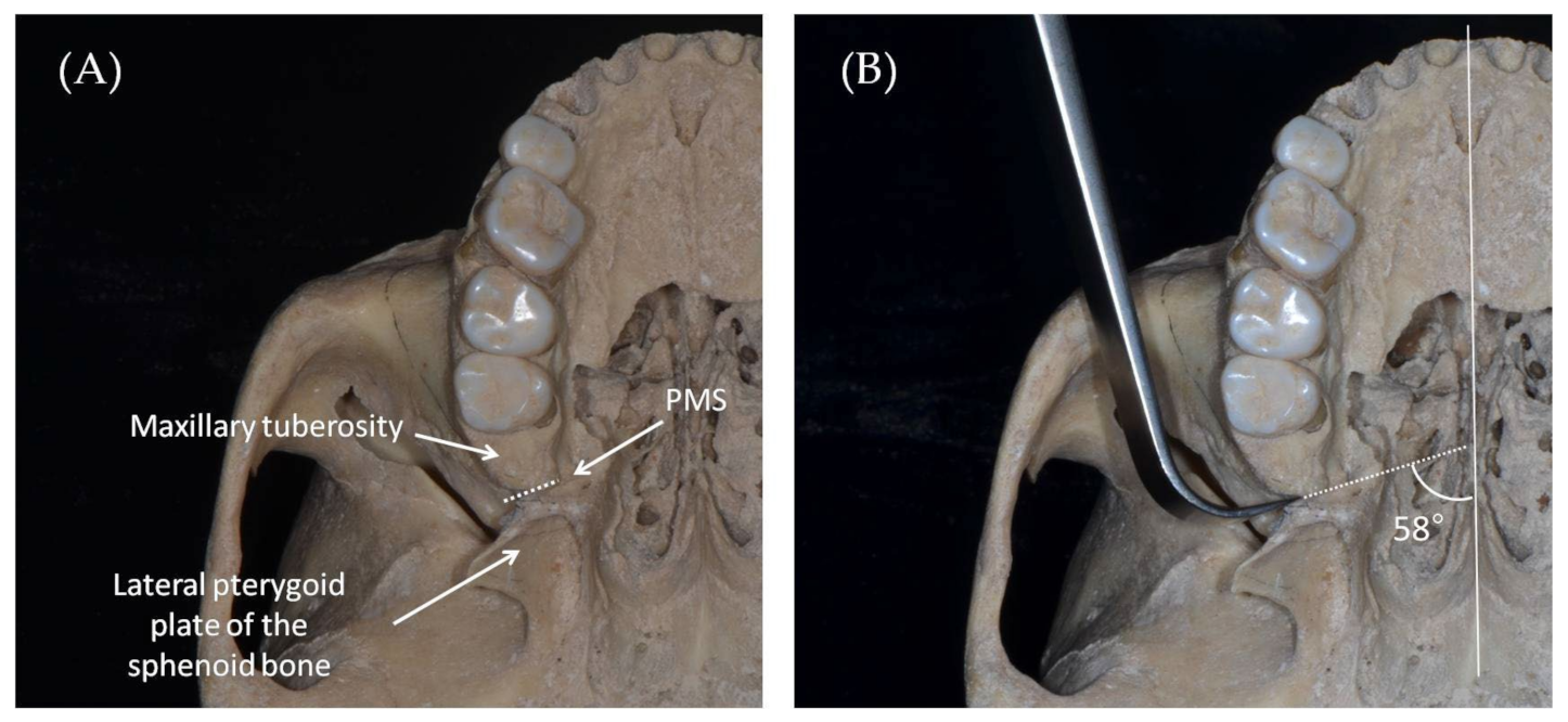
  • PMS angulation (°): The angle created between the PMS thickness line and the midsagittal plane (Figure 3).
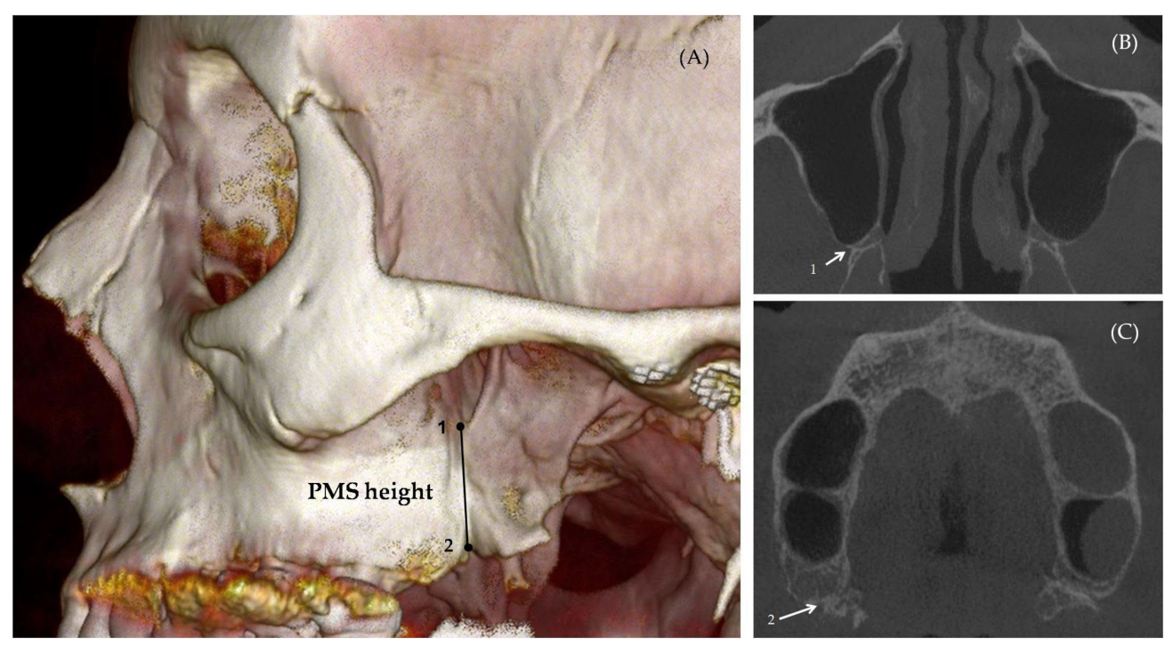
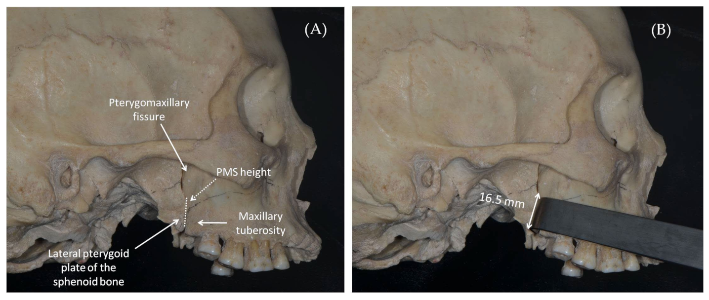
  • PMS height (mm): The suture height was calculated based on the number of axial sections counted from the suture initiation (the section where the maxillary lump connects to the pterygoid hamulus) and the suture termination (the last section before the suture is opened into a pterygomaxillary fissure) multiplied by the slice thickness (Figure 4).

2.3. Statistical Analysis

The data were summarized and analyzed using IBM SPSS statistics software (version 20, IBM Corp., Armonk, NY, USA). A normal distribution of the variables was examined using the one-sample Kolmogorov–Smirnov test. Descriptive statistics (the mean, standard deviation, and range) were carried out for all variables. An independent samples t-test was performed to compare the age and suture parameters between males and females. A related samples t-test was performed to compare the PMS measurements between the right and left sides. A Pearson correlation analysis was carried out to determine whether an association exists between the PMS parameters and age. The level of statistical significance was set at p < 0.05.

2.4. Reliability

In order to determine the intra-tester’s ability to replicate the studied measurements, a researcher (S.D.) evaluated twice, using a blind method, the CBCT of 10 cases (totaling 20 sets of measurements, including the right and left sides). For determining the inter-tester reliability, the same cases were evaluated by another independent researcher (L.K.) in a one-month period. The reliability analysis was performed using an ICC test.

3. Results

3.1. Reliability Test

The intra- and inter-tester reliability analysis revealed a high reproducibility level for all PMS measurements (0.789 ≤ ICC ≤ 0.987) (p < 0.001).

3.2. Study Sample

The sample included 50 CBCT images: 27 male individuals (54%) and 23 female individuals (46%). The mean age of the sample was 46.46 ± 16.411 years; it ranged between 19 and 79 years. Females were significantly younger when compared with males (41.8 years vs. 51.04 years, respectively) (p = 0.048) (Table 1).

3.3. PMS Morphology by the Right and Left Sides in Males and Females

No significant differences were found in any of the measured PMS parameters between the right and left sides of the patient, in both males and females (p > 0.225) (Table 2 and Table 3). Descriptive statistics and p-values are presented in Table 2 and Table 3.

3.4. Comparison of the PMS Morphology between the Sexes

Since no significant differences were noted between the right and left sides of the patient for all the PMS measurements, the mean of both sides was calculated and compared between males and females. For a comparison between the sexes, the descriptive statistics and p-values are presented in Table 4. No statistically significant difference was found regarding PMS height and angulation between males and females (p > 0.486). Regarding PMS thickness, a statistically significant difference between the sexes was found (p = 0.029) when males were presented with a wider suture than were females (1.00 mm vs. 0.90 mm, respectively).
Although the difference in PMS thickness was statistically significant, the biological significance of such a difference (0.1 mm) seems to have only a minor effect.

3.5. PMS Morphology and Age by Sexes

No statistically significant difference was found between the PMS parameters and age in both males and females (p > 0.400) (Table 5).

4. Discussion

To date, controversy exists in the literature regarding the preferred method for pterygomaxillary separation [18,19,20]. Turvey and Fonseca [9] suggested that the position and orientation of the curved osteotome are critical to avoid complications during PMS separation, in particular, to avoid damage to the maxillary artery. In contrast, Robinson and Hendy [15] reported that use of a curved osteotome created unfavorable fractures in the pterygoid plate when used to separate the PMS. Precious et al. [19] compared the outcomes of PMS separations using CT when it was performed with and without an osteotome. They found that an ideal separation was successful in 27% of cases in which it was performed without an osteotome, and in 29% of cases in which it was performed with an osteotome. Others proposed a technique for pterygomaxillary separation performed anterior to the PMS in order to avoid vascular damage during the osteotomy [20,21]. A survey of 205 oral and maxillofacial surgeons investigated surgeons’ preferences for pterygomaxillary separation [22]. Importantly, 78% of the participants reported that they use an osteotome or a micro-oscillating saw for pterygomaxillary separation: 8% reported serious vascular complication outcomes. Others, who used leverage alone or tuberosity osteotomy, did not report any vascular complications. Although the literature presents diverse approaches for pterygomaxillary separation [12,23], the most popular approach is to use a curved osteotome through PMS separation. Therefore, knowing the PMS size and position relative to the midsagittal plane is essential for surgeons performing this procedure. Our study investigated PMS morphology in an adult Israeli population with respect to patients’ age, sex, and the measured side. The vertical height of the PMS, its maximal thickness, and its angulation relative to the midsagittal plane were measured based on the CBCT scans. We found that in an adult Israeli population, the PMS mean height was 16.9 ± 2.3 mm and 16.4 ± 3.0 mm, in males and females, respectively, with no significant difference between the sexes (Figure 5).
However, this finding differs from what was previously reported in the Thai (15.14 ± 2.5 mm), American (14.6 ± 3.1 mm), and Chinese (12.1 ± 2.0 mm) populations [7,9,24]. These differences could result from a different methodology used in these studies; however, they also may reflect ethnical anatomical differences [5]. The blade length of the osteotome used to separate the PMS should resemble the PMS height. Clinically, this measurement, based on the Israeli population, implies that when a curved osteotome with a cutting edge of about 15 mm is used to separate the PMS in a Le Fort I osteotomy, the mean safety margin from the pterygomaxillary fissure is 1.5 mm. However, it should be remembered that anatomical variations exist regarding the PMS height and that an individual CBCT evaluation is critical prior to the surgical procedure.
Regarding the PMS thickness, we found that in our sample the mean thickness was 1.0 ± 0.13 mm and 0.9 ± 0.17 mm in males and females, respectively. Although the biological difference between the sexes seems to be minimal, this difference was found to be statistically significant. Previous studies evaluated the thickness using a different method [8,17]; however, some sex-related differences were also found. Kanawaza et al. [25] found an association between PMS thickness and unwanted fractures in the pterygoid process. It was suggested that the force applied from the osteotome is easily dissipated when a thin pterygomaxillary junction is present, which may lead to an unwanted pterygoid plate fracture. Thus, the PMS thickness should serve as an approximate estimate for the surgeon regarding the amount of force that should be applied to the osteotome during PMS separation.
The ideal fracture line begins laterally to the PMS and progresses medially along the PMS between the maxilla and the pterygoid process. Knowing the angle between the sagittal plane and the PMS can help the oral and maxillofacial surgeon to determine at which angulation to place an osteotome for PMS separation and thus possibly minimize the incidence of unwanted fracture lines. In our study, we found that the PMS angulation, measured at the greatest thickness of the suture, was about 58 ± 4.6° for both males and females, regardless of the measured side (right and left) and the patient’s age. Therefore, it is reasonable to assume that the sharp edge of the osteotome used to separate the suture should be placed at this angle in order to create ideal separation (Figure 6).
Using 12 cadavers, Stajčić [26] compared frequency that a pterygoid plate fracture occurred when a regular osteotome (50 degrees) and a large-angle osteotome (80 degrees) were used during Le Fort I osteotomy. His findings suggest that although the incidence of pterygoid plate fracture was lower using a large angle osteotome, there was a greater risk of unwanted palatinal bone separation. The mean PMS angulation in an Israeli population was found to be 58 degrees; however, this angle may differ between patients due to anatomical variations. Owing to the “blind” approach of PMS separation, it is difficult to identify the instrument’s angle relative to the sagittal plane in the operating room. Therefore, we suggest estimating the PMS size and the position measurements using the patients’ CBCT as well as practicing the PMS angle on a printed 3D model prior to performing the surgical procedure.

5. Limitations of the Study

Although the study sample was randomly selected based on the inclusion criteria and showed a similar age range in male and female populations, the mean age difference was noted between the sexes. Nevertheless, no significant association was found between the age and the PMS morphometrics in both males and females; moreover, the results were presented separately for males and females. However, the association between the age and suture parameters may need further confirmation and validation from other populations of a similar mean age. Deducing data on the PMS morphology from the current study sample was limited to average facial type patients, which justifies a further investigation of different facial type populations. Additionally, the findings of the current study should be validated by a future clinical study providing CBCTs prior to and following the Le Fort I osteotomy.

6. Conclusions

Precise disjunction of the maxilla from the pterygoid process of the sphenoid bone requires recognizing of the pterygomaxillary suture morphology and position with regard to the patient’s sex and age. Our findings show that PMS morphology is similar on the right and left sides in both males and females. Only PMS thickness is statistically significantly greater in males than in females; however, such a difference is of little clinical relevance. No significant age-related changes are expected regarding PMS morphology in individuals above the age of 18 years in both sexes. The mean angle of the PMS was 58 degrees in an Israeli population. Our findings can help in developing the best fitted osteotome in order to provide a safe pterygomaxillary separation during Le Fort I osteotomy.

Author Contributions

Conceptualization, D.M.A. and T.S.T.; methodology, L.K.; software, L.K.; validation, L.K., S.D. and D.M.A.; formal analysis, T.S.T.; investigation, S.D.; resources, D.M.A.; data curation, T.S.T.; writing—original draft preparation, S.D. and T.S.T.; writing—review and editing, T.S.T. and D.M.A.; visualization, L.K.; supervision, D.M.A. and T.S.T.; and project administration, S.D. All authors have read and agreed to the published version of the manuscript.

Funding

This research received no external funding.

Institutional Review Board Statement

The study was conducted in accordance with the Declaration of Helsinki and approved by the Institutional Ethics Committee of Tel Aviv University (date of approval: 5 September 2021).

Informed Consent Statement

Informed consent was obtained from all subjects involved in the study.

Data Availability Statement

The data sets analyzed during the current study are available from the author (D.M.A., [email protected]) on request.

Acknowledgments

The authors would like to thank Steve Manch, who edited the English.

Conflicts of Interest

The authors declare no conflict of interest.

References

  1. Moore, U.J. Surgical Principles and Technique. In Principles of Oral and Maxillofacial Surgery, 6th ed.; John Wiley & Sons Ltd.: Hoboken, NJ, USA; Wiley-Blackwell: New York, NY, USA, 2011; pp. 87–105. [Google Scholar]
  2. Susarla, S.M.; Ettinger, R.E.; Egbert, M.A. Transmucosal pterygomaxillary separation in the Le Fort I osteotomy. Plast. Reconstr. Surg. 2020, 145, 1262–1265. [Google Scholar] [CrossRef] [PubMed]
  3. Naini, F.B.; Gill, D.S. Orthognathic Surgery Principles, Planning and Practice, 1st ed.; John Wiley & Sons Ltd.: West Sussex, UK, 2017. [Google Scholar]
  4. Fehrenbach, M.J.; Herring, S.W. Illustrated Anatomy of the Head and Neck-E-Book, 5th ed.; Elsevier Health Sciences: Amsterdam, The Netherlands, 2015. [Google Scholar]
  5. Osborn, A.G. Radiology of the pterygoid plates and pterygopalatine fossa. AJR Am. J. Roentgenol. 1979, 132, 389–394. [Google Scholar] [CrossRef] [PubMed]
  6. Tashi, S.; Purohit, B.S.; Becker, M.; Mundada, P. The pterygopalatine fossa: Imaging anatomy, communications, and pathology revisited. Insights Imaging 2016, 7, 589–599. [Google Scholar] [CrossRef]
  7. Cheung, L.K.; Fung, S.C.; Li, T.; Samman, N. Posterior maxillary anatomy: Implications for Le Fort I osteotomy. Int. J. Oral Maxillofac. Surg. 1998, 27, 346–351. [Google Scholar] [CrossRef] [PubMed]
  8. Odabaşı, O.; Erkmen, E.; Üçok, C.Ö.; Bakir, M.A.; Keriş, E.Y.; Şahin, O. Morphometric analysis of pterygomaxillary region by using cone beam computed tomography. J. Stomatol. Oral Maxillofac. Surg. 2021, 122, 273–277. [Google Scholar] [CrossRef]
  9. Turvey, T.A.; Fonseca, R.J. The anatomy of the internal maxillary artery in the pterygopalatine fossa: Its relationship to maxillary surgery. J. Oral Surg. 1980, 38, 92–95. [Google Scholar]
  10. Lanigan, D.T.; West, R.A. Management of postoperative hemorrhage following the Le Fort I maxillary osteotomy. J. Oral Maxillofac. Surg. 1984, 42, 367–375. [Google Scholar] [CrossRef]
  11. Lanigan, D.T.; Hey, J.H.; West, R.A. Major vascular complications of orthognathic surgery: Hemorrhage associated with Le Fort I osteotomies. J. Oral Maxillofac. Surg. 1990, 48, 561–573. [Google Scholar] [CrossRef]
  12. Wikkeling, O.M.; Tacoma, J. Osteotomy of the pterygomaxillary junction. Int. J. Oral Surg. 1975, 4, 99–103. [Google Scholar] [CrossRef]
  13. Politis, C. Life-threatening haemorrhage after 750 Le Fort I osteotomies and 376 SARPE procedures. Int. J. Oral Maxillofac. Surg. 2012, 41, 702–708. [Google Scholar] [CrossRef]
  14. Zaroni, F.M.; Cavalcante, R.C.; da Costa, D.J.; Kluppel, L.E.; Scariot, R.; Rebellato, N.L.B. Complications associated with orthognathic surgery: A retrospective study of 485 cases. J. Maxillofac. Surg. 2019, 47, 1855–1860. [Google Scholar] [CrossRef] [PubMed]
  15. Robinson, P.P.; Hendy, C.W. Pterygoid plate fractures caused by the Le Fort I osteotomy. Br. J. Oral Maxillofac. Surg. 1986, 24, 198–202. [Google Scholar] [CrossRef] [PubMed]
  16. Tung, T.C.; Chen, Y.R.; Bendor-Samuel, R. Surgical complications of the Le Fort I osteotomy-a retrospective review of 146 cases. Changgeng Yi Xue Za Zhi. 1995, 18, 102–107. [Google Scholar] [PubMed]
  17. Chin, Y.P.; Leno, M.B.; Dumrongwongsiri, S.; Chung, K.H.; Lin, H.H.; Lo, L.J. The pterygomaxillary junction: An imaging study for surgical information of Le Fort I osteotomy. Sci. Rep. 2017, 7, 9953. [Google Scholar] [CrossRef] [Green Version]
  18. Bilge, S.; Kaba, Y.N.; Demirbas, A.E.; Kütük, N.; Kiliç, E.; Alkan, A. Evaluation of the pterygomaxillary separation pattern in Le Fort I osteotomy using different cutting instruments. J. Oral Maxillofac. Surg. 2020, 78, 1820–1831. [Google Scholar] [CrossRef]
  19. Precious, D.S.; Morrison, A.; Ricard, D. Pterygomaxillary separation without the use of an osteotome. J. Oral Maxillofac. Surg. 1991, 49, 98–99. [Google Scholar] [CrossRef]
  20. Dupont, C.; Ciaburro, T.H.; Prévost, Y. Simplifying the Le Fort I type of maxillary osteotomy. Plast. Reconstr. Surg. 1974, 54, 142–147. [Google Scholar] [CrossRef] [PubMed]
  21. Trimble, L.D.; Tideman, H.; Stoelinga, P.J. A modification of the pterygoid plate separation in low-level maxillary osteotomies. J. Oral Maxillofac. Surg. 1983, 41, 544–546. [Google Scholar] [CrossRef]
  22. O’Regan, B.; Bharadwaj, G. Pterygomaxillary separation in Le Fort I osteotomy UK OMFS consultant questionnaire survey. Br. J. Oral Maxillofac. Surg. 2006, 44, 20–23. [Google Scholar] [CrossRef]
  23. Breeze, J.; Verea Linares, C.; Stockton, P. Is an osteotome necessary for pterygomaxillary disjunction or disjunction through the tuberosity during Le Fort I osteotomy? A systematic review. Br. J. Oral Maxillofac. Surg. 2016, 54, 248–252. [Google Scholar] [CrossRef]
  24. Apinhasmit, W.; Chompoopong, S.; Methathrathip, D.; Sangvichien, S.; Karuwanarint, S. Clinical anatomy of the posterior maxilla pertaining to Le Fort I osteotomy in Thais. Clin. Anat. 2005, 18, 323–329. [Google Scholar] [CrossRef] [PubMed]
  25. Kanazawa, T.; Kuroyanagi, N.; Miyachi, H.; Ochiai, S.; Kamiya, N.; Nagao, T.; Shimozato, K. Factors predictive of pterygoid process fractures after pterygomaxillary separation without using an osteotome in Le Fort I osteotomy. Oral Surg. Oral Med. Oral Pathol. Oral Radiol. 2013, 115, 310–318. [Google Scholar] [CrossRef] [PubMed]
  26. Stajcić, Z. Altering the angulation of a curved osteotome-does it have effects on the type of pterygomaxillary disjunction in Le Fort I osteotomy? An experimental study. Int. J. Oral Maxillofac. Surg. 1991, 20, 301–303. [Google Scholar] [CrossRef] [PubMed]
Figure 1. Anatomical skeletal structures surrounding the pterygomaxillary suture from the lateral view of the skull (the mandibular ramus is removed).
Applsci 13 03825 g001
Figure 2. Schematic presentation of the Le Fort I maxillary osteotomy surgical procedure using a volume rendering 3D CT scan of the skull. (A) Lateral view of the skull demonstrating the Le Fort I osteotomy line (white line). (B) Enlarged view of the maxilla “sectioned” through the Le Fort I osteotomy line. The final stage of maxillary release from the base of the skull is a PMS separation. The PMS separation line is denoted by a blue dotted line and a white arrow. (C) Axial cranial view of the maxilla “sectioned” through the Le Fort I osteotomy line. The right and left PMS separation lines are denoted by blue dotted lines and white arrows. (D) Axial caudal view of the maxilla “sectioned” through the Le Fort I osteotomy line. The right and left PMS separation lines are denoted by blue dotted lines and white arrows.
Applsci 13 03825 g002
Figure 3. The method for measuring PMS thickness and angulation. The blue line denotes the midsagittal plane, the white line denotes PMS thickness measured from the most convex point on the lateral surface of the suture and the most medial point of the suture, and the α angle represents the PMS angulation, measured between the midsagittal plane and the continuation of the PMS thickness line (the yellow dotted line).
Applsci 13 03825 g003
Figure 4. The method for measuring the PMS height. (A) A volume rendering of the lateral view of the skull representing the PMS height defined by the most superior point (landmark number 1) and the most inferior point (landmark number 2). (B) A CBCT section that represents the superior most point (landmark number 1) on the PMS height measurement, where the PMS is seen just before it opens into a pterygomaxillary fissure (the white arrow). (C) A CBCT section that represents the inferior most point (landmark number 2) on the PMS height measurement, where the maxillary tuberosity and the pterygoid hamulus are seen (the white arrow).
Applsci 13 03825 g004
Figure 5. Simulation of the osteotome position during PMS separation on the dry skull, lateral view. (A) Dry skull from the lateral view. Dotted line represents PMS height. (B) Osteotome placed on the PMS height of the dry skull. The cutting edge of the osteotome is about 1.5 mm less than the mean PMS height (16.5 mm, white arrow).
Applsci 13 03825 g005
Figure 6. Simulation of the osteotome position during PMS separation on the dry skull, caudal view. (A) Dry skull from the caudal view. Dotted line represents PMS direction. (B) Osteotome oriented at 58 degrees to the midsagittal plane through the PMS of the dry skull.
Applsci 13 03825 g006
Table 1. Descriptive statistics of the study sample.
Sexn (%)Mean Age (Years)± SDMinMaxp-Value *
Males27 (54)51.0416.21120790.048
Females23 (46)41.8815.7421974
Total50 (100)46.4616.4111979
* p-value for the age difference between the sexes. The significant value is denoted in bold. SD: standard deviation; min: minimum; and max: maximum.
Table 2. Comparison of PMS morphology between the sides, males only.
PMS MeasurementSideMean± SDMinMaxp-Value *
Thickness (mm)Right1.020.1390.751.250.225
Left0.980.1440.701.25
Height (mm)Right16.672.56911.4022.500.392
Left17.102.74412.8022.90
Angulation (°)Right57.865.21267.2049.100.380
Left58.745.25966.7049.20
* p-values for the differences between the sides. PMS: pterygomaxillary Suture; SD: standard deviation; min: minimum; and max: maximum.
Table 3. Comparison of PMS morphology between the sides, females only.
PMS MeasurementSideMean± SDMinMaxp-Value *
Thickness (mm)Right0.910.1780.511.180.738
Left0.900.1790.571.36
Height (mm)Right16.352.74811.4022.500.973
Left16.373.46712.5024.20
Angulation (°)Right58.426.21067.6047.900.975
Left58.465.30569.9049.20
* p-values for the differences between the sides. PMS: pterygomaxillary Suture; SD: standard deviation; min: minimum; and max: maximum.
Table 4. Comparison of the PMS morphology between the sexes.
PMS MeasurementSexMean± SDMinMaxp-Value *
Thickness (mm)Male1.000.1300.761.220.029
Female0.900.1700.541.27
Height (mm)Male16.892.32212.8522.450.486
Female16.362.99412.0523.35
Angulation (°)Male58.304.55766.8550.350.915
Female58.444.75567.3549.95
* p-values for the differences between the sexes. Significant values are denoted in bold. PMS: pterygomaxillary suture; SD: standard deviation; min: minimum; and max: maximum.
Table 5. Association between PMS morphology and age (Pearson correlation analysis) by sex.
PMS MeasurementSexrp-Value *
Thickness (mm)Male0.170.400
Female0.040.858
Height (mm)Male−0.160.411
Female0.130.558
Angulation (°)Male0.160.439
Female0.140.516
* p-values for the correlation analysis between the PMS parameters and age.
Disclaimer/Publisher’s Note: The statements, opinions and data contained in all publications are solely those of the individual author(s) and contributor(s) and not of MDPI and/or the editor(s). MDPI and/or the editor(s) disclaim responsibility for any injury to people or property resulting from any ideas, methods, instructions or products referred to in the content.

