Article

Synergistic Antimicrobial Effect of Photodynamic Inactivation and SWEEPS in Combined Treatment against Enterococcus faecalis in a Root Canal Biofilm Model: An In Vitro Study

1
Laser Research Center of Dentistry, Dentistry Research Institute, Tehran University of Medical Sciences, Tehran 1441987566, Iran
2
Faculty of Health Sciences, De Montfort University, Leicester LE1 9BH, UK
*
Author to whom correspondence should be addressed.
Appl. Sci. 2023, 13(9), 5668; https://doi.org/10.3390/app13095668
Submission received: 7 February 2023 / Revised: 11 April 2023 / Accepted: 2 May 2023 / Published: 4 May 2023
(This article belongs to the Special Issue Photodynamic Therapy for Oral Health)

Abstract

:
Background: Persistent bacterial infections caused by biofilm-associated Enterococcus faecalis are a global public health concern. This study aims to compare the biofilm removal effects of methylene blue (MB) or hydrogen peroxide (HP) mediated photodynamic inactivation (PDI) with a shock wave enhanced emission photoacoustic streaming (SWEEPS) method laser irradiation enhanced bactericidal activity. Methods: A total of fifty extracted single-rooted human teeth were used. Each canal was then inoculated with a bacterial suspension of E. faecalis and left for ten days to induce biofilm formation. The antibacterial effects within treated root canals using MB or HP + diode laser or SWEEPS alone or in combination were assessed and compared with 5.25% sodium hypochlorite (NaOCl) as the positive control group. Data were analyzed using one-way ANOVA and Tukey’s post hoc test. Results: The MB + diode laser + SWEEPS and HP + diode laser + SWEEPS groups displayed significantly lower amounts of bacteria than either the MB + diode laser or SWEEPS and HP + diode laser or SWEEPS groups (p < 0.001). There was no statistically significant difference detected between the diode laser and SWEEPS groups (p = 0.982). Conclusions: SWEEPS can enhance the photosensitizer distribution in the root canal, leading to effective biofilm removal. This technique could thus help promote the future development of endodontic treatments.

1. Introduction

Currently, endodontic treatment aims to reduce the bacterial load well below a certain threshold level, followed by the provision of adequate seals to promote apical tissue healing or to maintain apical tissue health [1,2]. Chemomechanical treatment has often been used as a standard approach. However, bacterial ingrowth in complicated intracanal anatomy, such as isthmus and subsequent biofilm formation, has reduced the successful chemomechanical intervention in the root canal [3]. With the persistence of infection in the pulp space, microorganisms also occupy the canal space, further complicating the disinfection process [4]. Enterococcus faecalis is often found in failed endodontic treatment. The ability of this species to form bacterial biofilm has been well established [5]. Biofilm formation is a complex adaptive manner that enables bacterial survival in harsh growth conditions as well as increased tolerance to antimicrobial agents [5,6]. The removal of bacterial biofilm in the root canal has remained a challenge because chemical and/or mechanical actions alone are insufficient to cause biofilm removal. Hence, disruptive physical cleaning procedures are required that can eliminate biofilm using physical force at an adjacent site that is inaccessible with existing techniques [3].
There are several methods for disinfecting the root canal. The most basic method is the use of an ultrasonic device or endodontic irrigant. These irrigants include normal saline, sodium hypochlorite (NaOCl), ethylenediaminetetraacetic acid (EDTA), chlorhexidine gluconate, MTAD (a mixture of a tetracycline isomer, an acid, and a detergent), calcium hydroxide (Ca(OH)2), etc. The repeated irrigations are necessary to reach all areas of the root canal and remove debris. The most common cleaning agent is NaOCl. However, the pungent odor and strong cytotoxicity are among the disadvantages of this corrosive detergent [7,8,9].
Among the irrigation techniques, photodynamic inactivation (PDI) using hydrodynamic force generates a physical effect during root canal irrigation [3]. PDI is based on the interactions of a photosensitizer and a harmless light source. The excited photosensitizer in the presence of oxygen generates reactive oxygen species, which induces the death of microorganisms. This technique is promising to affect oral bacterial infections [10]. There have been several studies of PDI in endodontics using methylene blue (MB), with a peak wavelength absorption of 664 nm [11,12,13]. MB is a hydrophilic and positively charged photosensitizer with a low molecular weight [13]. Several investigators have reported that E. faecalis is effectively reduced by MB-PDI [14,15]. However, MB has some disadvantages for root canal treatment, including a tooth staining effect [16,17]. Additionally, excessive concentration of MB can reduce the PDI effect due to light-induced quenching [12]. Furthermore, E. faecalis can infect the deeper dentin tubules, where the absence of adequate oxygen can suppress the PDI- mediated bacterial killing. Therefore, the traditional PDI effect at a low oxygen concentration condition inside the root canals may be significantly reduced [18]. Hence, the development of novel efficient photosensitizers or new PDI protocols may be a solution to this issue [19].
High-level laser therapy is based on the combination of a penetrating laser with a stabilized HP [20]. Multiple studies have concluded improved antimicrobial efficacy when hydrogen peroxide (HP) was employed in PDI [19,21,22,23]. Sales, et al., stated that HP has provided similar characteristics as a photosensitizer [23]. The combination of a high-frequency diode laser (980 nm) and HP, if compared with classic PDI accomplished with photosensitizers, allows deeper penetration into the biofilm [24]. This technique is based on hydroxyl radical formation that causes lethal oxidative stress to bacterial cells [25].
PDI has been found to positively agitate root canal irrigants [11,15,26], but the efficiency of this technique can be improved [27]. The laser-activated irrigation (LAI) cleaning mechanism depends on high-speed liquid streaming in the root canal, which generates progressive pressure waves through the rapid expansion and collapse of laser-induced bubbles [28]. Moreover, advanced irrigation activation methods are developed, such as shock wave enhanced emission photoacoustic streaming (SWEEPS), which enables the generation of strong shock waves in narrow root canals [29]. The newly introduced SWEEPS employs a pulse train of a shorter pulse time compared to that of conventional LAI. In addition, SWEEPS provides an exact dose of shock waves into irrigants, penetrating dentinal tubules to remove tissue, dentine debris, and bacterial biofilm [27]. This study aimed to analyze the synergistic antimicrobial effect of photodynamic inactivation and SWEEPS in combined treatment against Enterococcus faecalis in terms of biofilm removal in a root canal biofilm model.

2. Materials and Methods

2.1. Photosensitizer and Light Source

Stock solutions of MB (Blue+M, Novateb Pars Co, Teheran, Iran) at 0.1 mg/mL were prepared and kept under dark conditions before use. In addition, 3% HP in stabilized water solution glycerophosphate (SIOXYL+ Solution, Doctor Smile Wiser, Vicenza, Italy) was used as a photosensitizer [30]. The absorption spectrum of HP is given in Figure 1. The irradiation source was a diode laser (Konftec, Sijhih City, Taiwan) with output power of 150 mW and a wavelength of 660 nm in the presence of MB. A diode laser (Doctor Smile, Wiser, Vicenza, Italy) with a 320-μm optic fiber at a wavelength of 980 nm, set to 2.5 W average power, and with pulse duration of 30 μs was used to activate the HP. The SWEEPS action was achieved using an erbium yttrium aluminum garnet (Er:YAG) laser at 2940 nm wavelength (LightWalker AT, Fotona, Ljubljana, Slovenia) and operating parameters of 0.3 W average power, 15 Hz, and 20 mJ per pulse, was employed in ultra-short, free-running pulse mode (25 μs). In each case, the true emission parameters were confirmed using a power meter.

2.2. Preparation of Tooth Specimens

The study protocol was approved by the Ethics Committee of the Tehran University of Medical Sciences. Consent was obtained from each patient for the use of their teeth in this study. Fifty human single-rooted teeth extracted for orthodontic and/or periodontal reasons with intact and mature apices and similar lengths (13–15 mm of root length) were selected. According to Korkut’s study [31], the sample size was determined using One-Way Anova Power Analysis, PASS 11, considering the effect size = 0.66, α  =  0.05, and β  =  0.2. Teeth with caries, multiple roots and structural defects were excluded from the sample, and selected teeth were stored in normal saline until use. The crowns were sectioned, and the root canal area was instrumented using the ProTaper rotary system (Dentsply Maillefer, Ballaigues, Switzerland) to size F4 [32,33]. The apex of each specimen was sealed using glass ionomer cement (XtraCemS, Medicept, Harrow, UK). To remove the smear layer, root canals were rinsed with 17% EDTA, each for 60 s [33,34]. Final irrigation of all samples was performed with normal saline, after which the canals were dried using paper cone, size #35. The teeth were put in 1.5 mL microtubes and autoclave-sterilized at 121 °C for 20 min~under 15 psi steam [35]. The method used to ensure standardization of samples is reported here [36]. All specimens are selected from human single-rooted and single-canal anterior teeth. The preparation steps are performed by the operator of the unit. All the specimens are prepared with the rotary system and the same files. The root canals were instrumented 1 mm beyond the apical foramen. All are prepared up to size F4. A laser device with the same wavelength was used in all groups. In all specimens, the root length after cutting the crown is 13–15 mm. The homogeneous root canal samples were randomly divided into ten groups.

2.3. Root Canal Infection with E. faecalis

E. faecalis IBRC-M 11,130 strain, grown aerobically in brain heart infusion (BHI) broth (Merck, Darmstadt, Germany) at 37 °C. E. faecalis suspension of approximately 1.5 × 10⁸ colony-forming unit (CFU)/mL was prepared using spectrophotometry. All the 50 prepared specimens were transferred into 1.5 mL sterile microtubes under sterile conditions. Five microliters of bacterial suspension containing 1.5 × 10⁸ CFU/mL of E. faecalis were syringed into the prepared root canal system. After injection, the microtubes were kept in an aerobic condition at 37 °C for 10 days to allow infection to occur. During this period, each canal was inoculated every 48 h with a fresh bacterial suspension at a concentration of 1.5 × 10⁸ CFU/mL.

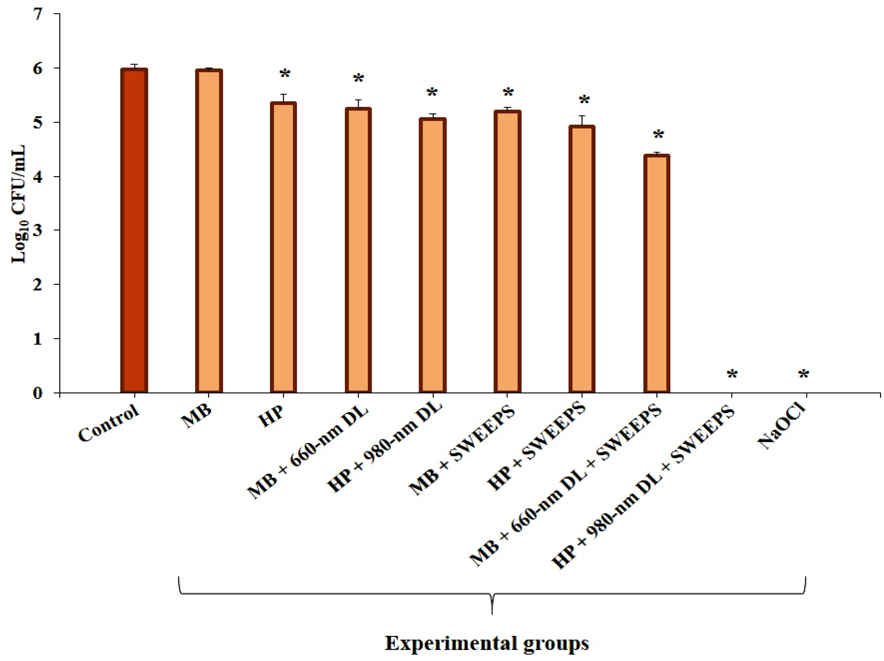
2.4. Experimental Design

The operation steps of each procedure were as follows (Figure 2):
A.
MB (0.1 mg/mL): The root canals were filled with 5 μL MB, followed by dark incubation for 5 min at 25 °C.
B.
3% HP: The root canals were filled with 5 μL HP, followed by dark incubation for 2 min at 25 °C [30].
C.
MB (0.1 μg/mL) + 660 nm diode laser: The root canals were treated with MB similar to group A, and the test samples were then exposed to a 660 nm diode laser for 60 s.
D.
3% HP + 980 nm diode laser: The root canals were treated by HP similar to group B and the test samples were then exposed to a 980 nm diode laser for 30 s.
E.
MB (100 μg/mL) + SWEEPS: The root canals were treated by MB similar to group A, and the test samples were then irradiated with Er:YAG laser with 2940 nm wavelength. The device tip (SWEEPS600, Fotona, Slovenia) was placed in the pulp chamber and activated for 90 s (30 s of activation, 30 s of rest, and 30 s of activation again).
F.
3% HP + SWEEPS: The root canals were treated by HP similar to group B and the test samples were then subjected to SWEEPS technique similar to group E.
G.
MB (100 μg/mL) + 660 nm diode laser + SWEEPS: The root canals were treated by MB-mediated SWEEPS similar to group E, and then after 5 min, 660 nm diode laser irradiation was performed similar to group C.
H.
3% HP + 980 nm diode laser + SWEEPS: The root canals were treated by HP-mediated SWEEPS similar to group F, and then after 5 min, 980 nm diode laser irradiation was performed similar to group D.
I.
5.25% NaOCl (positive control): The root canals were filled with 5 μL NaOCl, followed by incubation for 5 min at 25 °C.
J.
Normal saline (negative control): The root canals were filled with 5 μL normal saline, followed by incubation for 5 min at 25 °C.
Figure 2. Schematic representation of study groups. MB: methylene blue, HP: hydrogen peroxide, DL: diode laser, nm: nanometer, SWEEPS: shock wave enhanced emission photoacoustic streaming, NaOCl: 5.25% sodium hypochlorite.
Applsci 13 05668 g002

3. Evaluation of Antibacterial Activity

Quantifications of the bacteria present in infected root canals were conducted following a previously reported procedure [26]. Briefly, after all treatments, the contents of root canals were sampled by flushing the root canals with an application of 1 mL of BHI broth with a fine needle. The bacterial suspension was collected in a 1.5 mL microtube. Serial dilutions were performed after 60 s processing in agitating mode, and 10 μL of each dilution series were spread onto BHI agar (Merck). After 24 h, bacterial colonies were counted for each root specimen.

Statistical Analysis

Data were calculated by log10 CFU/mL and evaluated by one-way analysis of variance using the statistical software package IBM SPSS Statistics version 26.0 (IBM, Chicago, IL, USA). In addition, specific multiple comparisons among treatments were performed by Tukey’s multiple range test.

4. Results

The results in Figure 3 illustrated that except for MB alone, all experimental groups could decrease the viability of E. faecalis in the root canal biofilm model, compared with the control group (p < 0.001). As shown in Table 1, a highly significant difference was found between the MB alone and the groups treated with MB in combination with 660 nm diode laser, SWEEPS, or both (p < 0.001). Additionally, there was no significant difference found between the MB plus 660 nm diode laser and MB plus SWEEPS (p = 0.982). Compared to the HP alone, the bacterial counts in the HP + 980 nm diode laser group (p = 0.014), HP + SWEEPS group (p = 0.002), and HP + 980 nm diode laser + SWEEPS group (p < 0.001) were significantly lower, while those in the HP + 980 nm diode laser group and HP + SWEEPS group did not differ significantly (p = 0.982). In addition, no bacteria were detected in the presence of 5.25% NaOCl (p < 0.001).

5. Discussion

It is widely accepted that the main reason for root canal treatment failure is the presence of residual microorganisms in the root canal. Therefore, one of the most important challenges for dentists is the proper disinfection of the dental canals and the removal of pathogens [2,37]. This in vitro study compared the efficacy of three laser modalities, including diode laser, high-frequency diode laser, and SWEEPS, on E. faecalis infected root canal. Due to its resilience in comparison to other species and resistance to alkaline environments, E. faecalis might be a problem germ in persistent root canal infections. According to a study, the susceptibility to disinfection of E. faecalis in the dual-species biofilms did not change, suggesting that the shift in biofilm composition toward more E. faecalis indicates a shift toward more resilient species that may ultimately become more difficult to treat [38]. We have employed a root canal biofilm model, which can closely simulate in vivo situations in terms of morphology, to compare the effectiveness of biofilm removal procedures. Removing the root canal biofilm is still challenging [39]. The biofilm removal techniques still require a more reliable treatment protocol [3]. In an earlier study, the authors found that SWEEPS is an effective intervention in this regard. Moreover, the SWEEPS technique associated with a light-emitting diode (LED) can cause a more significant response on the root canal biofilm model [40]. In this study, SWEEPS exhibited the capacity to remove biofilm during root canal irrigation, which indicated that SWEEPS has the potential to affect biofilm removal from the root canal. The results of our bacterial quantification analysis demonstrated that laser irradiation is effective in reducing bacterial content.
In this study, MB and HP, along with a diode laser or SWEEPS, were employed for targeting E. faecalis in the root canals. Our findings are consistent with Müller, et al., [41], whose study reported less than 1 log10 CFU/mL reduction of six-species oral biofilm on a bovine-enamel disc model after their treatment with MB plus 665 nm diode laser. Similarly, 5% NaOCl eliminated all strains. Therefore, NaOCl is highly suggested as the main irrigant due to its broad antibacterial range, its special ability to dissolve necrotic tissue debris and the organic components of the smear layer, and its capacity to eliminate deep-rooted pathogens organized in biofilms and dentinal tubules [42].
Subsequently, pre-treatment with HP affected PDI disinfection capacity [12,43]. HP is identified by a moderate antibacterial activity, and the PDI process improves its efficiency due to peroxide activation [44]. Up until now, most studies have investigated the combined use of HP and lasers for managing periodontitis [45,46]. A previous study from our group, which was performed using a 96-well plate, showed that HP significantly reduces biofilm development and metabolic activity of E. faecalis [22].
During the SWEEPS procedure, a laser pulse is delivered to enhance the dynamic movement of irradiated liquid. The primary laser pulse is followed by a subsequent pulse after secondary bubbles have occurred at the fiber tip. This leads to accelerated violent collapse and advances the secondary bubbles deeper into the root canals [29]. These bubbles gradually grow to approach the maximum size. With irradiation cessation, the bubble volume would begin to shrink. With consecutive pulse delivery, secondary bubbles are formed. Due to the collapse of bubbles, cavitation shock waves give rise to strong shear stress acting on the root canal wall, aiding debridement and improving the root canal disinfection efficiency [28]. Therefore, the bacterial biofilm attached to the root canal walls can be simply fragmented and removed [3]. The pulse duration of the SWEEPS technique used in this study was 25 μ, which results in a pulse peak power as high as 800 W. This technique generates a physical force in the irrigating liquid and enhances its flow. The auto-SWEEPS cycle was designated in this study as the temporal separation between the SWEEPS laser pulse pair with 200 and 650 μs. In this regard, the auto-SWEEPS cycle has a further benefit in order to reach an optimal pulse separation with respect to the different activating mechanisms at least twice during each SWEEPS cycle [47].
The results of this study indicate that the dual-stage light strategy (660 or 980 nm diode laser + SWEEPS) is significantly more effective in biofilm removal in comparison with the already highly effective 660 or 980 nm diode laser mediated PDI. As can be seen from Figure 3, the MB + 660 nm diode laser + SWEEPS outperforms the MB + 660 nm diode laser or MB + SWEEPS (1.58 vs. 0.73 or o.77 log10 CFU/mL reduction, respectively). Additionally, the HP + 980 nm diode laser + SWEEPS was more than ~ 5 log10 CFU/mL more effective than HP + 980 nm diode laser or HP + SWEEPS. This is in agreement with another study where the most effective biofilm removal from the root canal was detected when the curcumin-LED was coupled with SWEEPS [32]. Our findings also show that for biofilm removal from the root canal, although the MB or HP-mediated SWEEPS outperforms the MB or HP-mediated PDI, there are no significant differences between these two groups (p = 0.982).
Wang, et al. [48] observed improved bacterial clearance of E. faecalis in root canals using auto-SWEEPS when compared to 3% NaOCl alone and photon-induced photoacoustic streaming (PIPS). In addition, this efficacy was confirmed by SEM images, which showed that the SWEEPS group resulted in complete biofilm removal from the canals. Notably, shock waves cannot be emitted in narrow root canals simply during LAI [27]. The higher single pulse energy will rapidly increase cavitation bubbles and slow bubble dynamics at higher laser energies. In addition, the laser pulse-shaping optimization by using the SWEEPS technique enhances the effects of photoacoustic flow [27,49]. Developing more effective root canal cleaning techniques will improve the disinfecting capability of root canals. Although more research is needed using optimized irrigation parameters and confirming the safety of these procedures, our current evidence suggests that SWEEPS can improve irrigant activation for better biofilm removal from infected root canals. Given the polymicrobial nature of root canal infections, additional research using multi-species biofilm is needed to support the application of this technique. Within the limitations of this study, the use of the confocal laser scanning microscopy technique may better show the structural characteristics of the biofilms in the root canal. The use of single-rooted teeth, which do not have the common complex anatomy of posterior teeth, is another limitation of this study.

6. Conclusions

Overall, the results of this study confirmed that SWEEPS is superior to diode laser regarding biofilm removal from infected root canals; however, there was no significant difference between SWEEPS and diode laser. Remarkably, all methods were able to eliminate biofilm; however, the combined use of the diode laser and SWEEPS revealed a significantly higher reduction of biofilm compared to either technique alone.

Author Contributions

Conceptualization, S.A., S.P. and N.C.; methodology, S.A. and N.C.; software, S.A.; writing—original draft preparation, S.A.; writing—review and editing, S.A., S.P. and N.C. All authors have read and agreed to the published version of the manuscript.

Funding

This research received no external funding.

Institutional Review Board Statement

Not applicable.

Informed Consent Statement

Not applicable.

Data Availability Statement

Not applicable.

Conflicts of Interest

The authors declare no conflict of interest.

References

  1. Siqueira, J.F., Jr.; Rôças, I.N. Clinical implications and microbiology of bacterial persistence after treatment procedures. J. Endod. 2008, 34, 1291–1301.e3. [Google Scholar] [CrossRef] [PubMed]
  2. Al Yahya, R.S.; Al Attas, M.H.; Javed, M.Q.; Khan, K.I.; Atique, S.; Abulhamael, A.M.; Bahammam, H.A. Root Canal Configuration and Its Relationship with Endodontic Technical Errors and Periapical Status in Premolar Teeth of a Saudi Sub-Population: A Cross-Sectional Observational CBCT Study. Int. J. Environ Res. Public Health 2023, 20, 1142. [Google Scholar] [CrossRef] [PubMed]
  3. Nagahashi, T.; Yahata, Y.; Handa, K.; Nakano, M.; Suzuki, S.; Kakiuchi, Y.; Tanaka, T.; Kanehira, M.; Venkataiah, V.S.; Saito, M. Er: YAG laser-induced cavitation can activate irrigation for the removal of intraradicular biofilm. Sci. Rep. 2022, 12, 4897. [Google Scholar] [CrossRef] [PubMed]
  4. Ørstavik, D. Endodontic Treatment of Apical Periodontitis. In Essential Endodontology, 3rd ed.; Ørstavik, D., Ed.; John Wiley & Sons Ltd.: Hoboken, NJ, USA, 2020; pp. 313–343. [Google Scholar] [CrossRef]
  5. Duggan, J.M.; Sedgley, C.M. Biofilm formation of oral and endodontic Enterococcus faecalis. J. Endod. 2007, 33, 815–818. [Google Scholar] [CrossRef]
  6. Kishen, A.; George, S.; Kumar, R. Enterococcus faecalis-mediated biomineralized biofilm formation on root canal dentine in vitro. J. Biomed. Mater. Res. A 2006, 77, 406–415. [Google Scholar] [CrossRef]
  7. Mozo, S.; Llena, C.; Forner, L. Review of ultrasonic irrigation in endodontics: Increasing action of irrigating solutions. Med. Oral Patol. Oral Cir. Bucal 2012, 17, e512. [Google Scholar] [CrossRef]
  8. Haapasalo, M.; Shen, Y.; Wang, Z.; Gao, Y. Irrigation in endodontics. Br. Dent. J. 2014, 216, 299–303. [Google Scholar] [CrossRef]
  9. Khan, A.M.; Gangoo, I.K.A.; Ali, N.A.; Khan, M.; Javed, M.Q.; AlAttas, M.H.; Abulhamael, A.M.; Bahammam, H.A.; Alsofi, L.; Yahya, R.S.A. The Effect of Calcium Hydroxide, Triple Antibiotic Paste and Chlorhexidine on Pain in Teeth with Symptomatic Apical Periodontitis: A Randomised Controlled Trial. Int. J. Environ. Res. Public Health 2023, 20, 3091. [Google Scholar] [CrossRef]
  10. Garcez, A.S.; Ribeiro, M.S.; Tegos, G.P.; Nunez, S.C.; Jorge, A.O.; Hamblin, M.R. Antimicrobial photodynamic therapy combined with conventional endodontic treatment to eliminate root canal biofilm infection. Lasers Surg. Med. 2007, 39, 59–66. [Google Scholar] [CrossRef]
  11. George, S.; Kishen, A. Photophysical, photochemical, and photobiological characterization of methylene blue formulations for light-activated root canal disinfection. J. Biomed. Opt. 2007, 12, 034029. [Google Scholar] [CrossRef]
  12. Garcez, A.S.; Hamblin, M.R. Methylene blue and hydrogen peroxide for photodynamic inactivation in root canal-a new protocol for use in endodontics. Eur. Endod. J. 2017, 2, 29. [Google Scholar] [CrossRef]
  13. George, S.; Hamblin, M.R.; Kishen, A. Uptake pathways of anionic and cationic photosensitizers into bacteria. Photochem. Photobiol. Sci. 2009, 8, 788–795. [Google Scholar] [CrossRef]
  14. Komine, C.; Tsujimoto, Y. A small amount of singlet oxygen generated via excited methylene blue by photodynamic therapy induces the sterilization of Enterococcus faecalis. J. Endod. 2013, 39, 411–414. [Google Scholar] [CrossRef]
  15. Karaoğlu, G.E.; Erdönmez, D.; Göl, C.; Durmuş, M. Efficacy of antimicrobial photodynamic therapy administered using methylene blue, toluidine blue and tetra 2-mercaptopyridine substituted zinc phthalocyanine in root canals contaminated with Enterococcusaecalis. Photodiagn. Photodyn. Ther. 2020, 32, 102038. [Google Scholar] [CrossRef]
  16. Costa, L.M.; De Souza Matos, F.; De Oliveira Correia, A.M.; Carvalho, N.C.; Faria-e-Silva, A.L.; Paranhos, L.R.; Ribeiro, M.A.G. Tooth color change caused by photosensitizers after photodynamic therapy: An in vitro study. J. Photochem. Photobiol. B 2016, 160, 225–228. [Google Scholar] [CrossRef]
  17. Ramalho, K.M.; Cunha, S.R.; Mayer-Santos, E.; De Paula Eduardo, C.; De Freitas, P.M.; Aranha, A.C.C.; Moura-Netto, C. In vitro evaluation of methylene blue removal from root canal after Photodynamic Therapy. Photodiagn. Photodyn. Ther. 2017, 20, 248–252. [Google Scholar] [CrossRef]
  18. Uekubo, A.; Hiratsuka, K.; Aoki, A.; Takeuchi, Y.; Abiko, Y.; Izumi, Y. Effect of antimicrobial photodynamic therapy using rose bengal and blue light-emitting diode on Porphyromonas gingivalis in vitro: Influence of oxygen during treatment. Laser Ther. 2016, 25, 299–308. [Google Scholar] [CrossRef]
  19. Nie, M.; Silva, R.C.e.; De Oliveira, K.T.; Bagnato, V.S.; De Souza Rastelli, A.N.; Crielaard, W.; Yang, J.; Deng, D. Synergetic antimicrobial effect of chlorin e6 and hydrogen peroxide on multi-species biofilms. Biofouling 2021, 37, 656–665. [Google Scholar] [CrossRef]
  20. Caccianiga, G.; Rey, G.; Caccianiga, P.; Leonida, A.; Baldoni, M.; Baldoni, A.; Ceraulo, S. Rough Dental Implant Surfaces and Peri-Implantitis: Role of Phase-Contrast Microscopy, Laser Protocols, and Modified Home Oral Hygiene in Maintenance. A 10-Year Retrospective Study. Appl. Sci. 2021, 11, 4985. [Google Scholar] [CrossRef]
  21. Cai, Z.; Li, Y.; Wang, Y.; Chen, S.; Jiang, S.; Ge, H.; Lei, L.; Huang, X. Antimicrobial effects of photodynamic therapy with antiseptics on Staphylococcus aureus biofilm on titanium surface. Photodiagn. Photodyn. Ther. 2019, 25, 382–388. [Google Scholar] [CrossRef]
  22. Afrasiabi, S.; Chiniforush, N. An in vitro study on the efficacy of hydrogen peroxide mediated high-power photodynamic therapy affecting Enterococcus faecalis biofilm formation and dispersal. Photodiagn. Photodyn. Ther. 2023, 41, 103310. [Google Scholar] [CrossRef] [PubMed]
  23. Sales, L.S.; Guimaraes, G.N.; Wijesinghe, G.K.; Moreira, K.M.S.; Joia, F.; Stipp, R.N.; Rodrigues, L.; Nobre-Dos-Santos, M.; Steiner-Oliveira, C. Addition of hydrogen peroxide to methylene blue conjugated to β-cyclodextrin in photodynamic antimicrobial chemotherapy in S. mutans biofilm. Photodiagn. Photodyn. Ther. 2019, 28, 226–233. [Google Scholar] [CrossRef] [PubMed]
  24. Caccianiga, G.; Rey, G.; Caccianiga, P.; Leonida, A.; Baldoni, M.; Baldoni, A.; Ceraulo, S. Laser management of peri-implantitis: A comparison between photodynamic therapy combined with hydrogen peroxide (OHLLT) and OHLLT+ Er: YAG laser. A retrospective controlled study. Appl. Sci. 2021, 11, 6771. [Google Scholar] [CrossRef]
  25. Nakamura, K.; Shirato, M.; Tenkumo, T.; Kanno, T.; Westerlund, A.; Örtengren, U.; Sasaki, K.; Niwano, Y. Hydroxyl radicals generated by hydrogen peroxide photolysis recondition biofilm-contaminated titanium surfaces for subsequent osteoblastic cell proliferation. Sci. Rep. 2019, 9, 4688. [Google Scholar] [CrossRef]
  26. Foschi, F.; Fontana, C.R.; Ruggiero, K.; Riahi, R.; Vera, A.; Doukas, A.G.; Pagonis, T.C.; Kent, R.; Stashenko, P.P.; Soukos, N.S. Photodynamic inactivation of Enterococcus faecalis in dental root canals in vitro. Lasers Surg. Med. 2007, 39, 782–787. [Google Scholar] [CrossRef]
  27. Lukac, N.; Muc, B.T.; Jezersek, M.; Lukac, M. Photoacoustic endodontics using the novel SWEEPS Er: YAG laser modality. J. Laser Health Acad. 2017, 1, 1–7. [Google Scholar]
  28. Matsumoto, H.; Yoshimine, Y.; Akamine, A. Visualization of irrigant flow and cavitation induced by Er: YAG laser within a root canal model. J. Endod. 2011, 37, 839–843. [Google Scholar] [CrossRef]
  29. Arıcıoğlu, B.; Çıkman, A.Ş.; Babacan, M. The comparison of cleaning efficacy and apical extrusion of advanced irrigation activation methods with a novel Er: YAG laser modality: Sweeps. Lasers Dent. Sci. 2021, 5, 43–52. [Google Scholar] [CrossRef]
  30. Caccianiga, G.; Rey, G.; Baldoni, M.; Caccianiga, P.; Baldoni, A.; Ceraulo, S. Periodontal decontamination induced by light and not by heat: Comparison between oxygen high level laser therapy (OHLLT) and LANAP. Appl. Sci. 2021, 11, 4629. [Google Scholar] [CrossRef]
  31. Korkut, E.; Torlak, E.; Gezgin, O.; Özer, H.; Şener, Y. Antibacterial and smear layer removal efficacy of Er: YAG laser irradiation by photon-induced photoacoustic streaming in primary molar root canals: A preliminary study. Photomed. Laser Surg. 2018, 36, 480–486. [Google Scholar] [CrossRef]
  32. Zhu, X.; Yin, X.; Chang, J.W.; Wang, Y.; Cheung, G.S.; Zhang, C. Comparison of the antibacterial effect and smear layer removal using photon-initiated photoacoustic streaming aided irrigation versus a conventional irrigation in single-rooted canals: An in vitro study. Photomed. Laser Surg. 2013, 31, 371–377. [Google Scholar] [CrossRef]
  33. Dewi, A.; Upara, C.; Chaiariyakul, D.; Louwakul, P. Smear Layer Removal from Root Canal Dentin and Antimicrobial Effect of Citric Acid-modified Chlorhexidine. Eur. Endod. J. 2020, 5, 257–263. [Google Scholar] [CrossRef]
  34. Sebbane, N.; Steinberg, D.; Keinan, D.; Sionov, R.V.; Farber, A.; Sahar-Helft, S. Antibacterial Effect of Er:YAG Laser Irradiation Applied by a New Side-Firing Spiral Tip on Enterococcus faecalis Biofilm in the Tooth Root Canal—An Ex Vivo Study. Appl. Sci. 2022, 12, 12656. [Google Scholar] [CrossRef]
  35. Kasić, S.; Knezović, M.; Beader, N.; Gabrić, D.; Malčić, A.I.; Baraba, A. Efficacy of three different lasers on eradication of Enterococcus faecalis and Candida albicans biofilms in root canal system. Photomed. Laser Surg. 2017, 35, 372–377. [Google Scholar] [CrossRef]
  36. Nagendrababu, V.; Murray, P.E.; Ordinola-Zapata, R.; Peters, O.A.; Rôças, I.N.; Siqueira, J.F., Jr.; Priya, E.; Jayaraman, J.; Pulikkotil, S.J.; Camilleri, J.; et al. PRILE 2021 guidelines for reporting laboratory studies in Endodontology: A consensus-based development. Int. Endod. J. 2021, 54, 1482–1490. [Google Scholar] [CrossRef]
  37. Siqueira, J.F., Jr.; Rôças, I.N.; Favieri, A.; Lima, K.C. Chemomechanical reduction of the bacterial population in the root canal after instrumentation and irrigation with 1%, 2.5%, and 5.25% sodium hypochlorite. J. Endod. 2000, 26, 331–334. [Google Scholar] [CrossRef]
  38. Van Der Waal, S.; Connert, T.; Crielaard, W.; De Soet, J. In mixed biofilms Enterococcus faecalis benefits from a calcium hydroxide challenge and culturing. Int. Endod. J. 2016, 49, 865–873. [Google Scholar] [CrossRef]
  39. Ricucci, D.; Siqueira, J.F., Jr. Biofilms and apical periodontitis: Study of prevalence and association with clinical and histopathologic findings. J. Endod. 2010, 36, 1277–1288. [Google Scholar] [CrossRef]
  40. Ensafi, F.; Fazlyab, M.; Chiniforush, N.; Akhavan, H. Comparative effects of SWEEPS technique and antimicrobial photodynamic therapy by using curcumin and nano-curcumin on Enterococcus faecalis biofilm in root canal treatment. Photodiagn. Photodyn. Ther. 2022, 40, 103130. [Google Scholar] [CrossRef]
  41. Müller, P.; Guggenheim, B.; Schmidlin, P.R. Efficacy of gasiform ozone and photodynamic therapy on a multispecies oral biofilm in vitro. Eur. J. Oral Sci. 2007, 115, 77–80. [Google Scholar] [CrossRef]
  42. Zehnder, M. Root canal irrigants. J. Endod. 2006, 32, 389–398. [Google Scholar] [CrossRef] [PubMed]
  43. Li, Y.; Du, J.; Huang, S.; Wang, S.; Wang, Y.; Cai, Z.; Lei, L.; Huang, X. Hydrogen peroxide potentiates antimicrobial photodynamic therapy in eliminating Candida albicans and Streptococcus mutans dual-species biofilm from denture base. Photodiagn. Photodyn. Ther. 2022, 37, 102691. [Google Scholar] [CrossRef] [PubMed]
  44. Caccianiga, G.; Baldoni, M.; Ghisalberti, C.A.; Paiusco, A. A preliminary in vitro study on the efficacy of high-power photodynamic therapy (HLLT): Comparison between pulsed diode lasers and superpulsed diode lasers and impact of hydrogen peroxide with controlled stabilization. Biomed. Res. Int. 2016, 2016, 1386158. [Google Scholar] [CrossRef] [PubMed]
  45. Caccianiga, G.; Rey, G.; Paiusco, A.; Lauritano, D.; Cura, F.; Ormianer, Z.; Carinci, F. Oxygen high level laser therapy is efficient in treatment of chronic periodontitis: A clinical and microbiological study using PCR analysis. J. Biol. Regul. Homeost. Agents 2016, 30, 87–97. [Google Scholar]
  46. Caccianiga, G.; Rey, G.; Fumagalli, T.; Cambini, A.; Denotti, G.; Giacomello, M. Photodynamic therapy (association diode laser/hydrogen peroxide): Evaluation of bactericidal effects on periodontopathy bacteria: An in vitro study. Eur. J. Inflamm. 2012, 10, 101–106. [Google Scholar] [CrossRef]
  47. Jezeršek, M.; Lukač, N.; Lukač, M. Measurement of simulated debris removal rates in an artificial root canal to optimize laser-activated irrigation parameters. Lasers Surg. Med. 2021, 53, 411–417. [Google Scholar] [CrossRef]
  48. Wang, X.-N.; Shi, J. Shock wave-enhanced emission photoacoustic streaming versus photon-induced photoacoustic streaming modes for clearing root canal bacteria using erbium-doped yttrium aluminum garnet lasers: An in vitro study. Res. Sq. 2020. [Google Scholar] [CrossRef]
  49. Chen, X.; Xu, R.-Q.; Chen, J.-P.; Shen, Z.-H.; Jian, L.; Ni, X.-W. Shock-wave propagation and cavitation bubble oscillation by Nd: YAG laser ablation of a metal in water. Appl. Opt. 2004, 43, 3251–3257. [Google Scholar] [CrossRef]
Figure 1. Absorption spectrum of hydrogen peroxide.
Applsci 13 05668 g001
Figure 3. Effect of different treatment groups on cell viability of Enterococcus faecalis biofilm. * Significantly different from the control group, p < 0.001. MB: methylene blue, HP: hydrogen peroxide, DL: diode laser, nm: nanometer, SWEEPS: shock wave enhanced emission photoacoustic streaming, NaOCl: 5.25% sodium hypochlorite.
Applsci 13 05668 g003
Table 1. Comparative data (Log10 CFU/mL reduction) of root canal biofilm model following each treatment.
Experiments Log10 CFU/mL
Reduction
p Value
MBvs. Control−0.021.00
HP −0.61<0.001
MB + 660 nm DL −0.73<0.001
HP + 980 nm DL −0.91<0.001
MB + SWEEPS −0.77<0.001
HP + SWEEPS −1.05<0.001
MB + 660 nm DL + SWEEPS −1.58<0.001
HP + 980 nm DL + SWEEPS −5.97<0.001
NaOCl −5.97<0.001
MB + 660 nm DLvs. MB−0.71<0.001
MB + SWEEPS −0.75<0.001
MB + 660 nm DL + SWEEPS −1.56<0.001
NaOCl −5.95<0.001
MB + SWEEPSvs. MB + 660 nm DL−0.040.982
MB + 660 nm DL + SWEEPS −0.85<0.001
NaOCl −5.24<0.001
MB + 660 nm DL + SWEEPSvs. MB + SWEEPS−0.81<0.001
NaOCl −5.20<0.001
HP + 980 nm DLvs. HP−0.300.014
HP + SWEEPS −0.440.002
HP + 980 nm DL + SWEEPS −5.36<0.001
NaOCl −5.36<0.001
HP + SWEEPSvs. HP + 980 nm DL−0.140.982
HP + 980 nm DL + SWEEPS −5.06<0.001
NaOCl −5.06<0.001
HP + 980 nm DL + SWEEPSvs. HP + SWEEPS−4.92<0.001
NaOCl−4.92<0.001
Abbreviations: MB: methylene blue, HP: hydrogen peroxide, nm: nanometer, DL: diode laser, SWEEPS: shock wave enhanced emission photoacoustic streaming, NaOCl: 5.25% sodium hypochlorite.
Disclaimer/Publisher’s Note: The statements, opinions and data contained in all publications are solely those of the individual author(s) and contributor(s) and not of MDPI and/or the editor(s). MDPI and/or the editor(s) disclaim responsibility for any injury to people or property resulting from any ideas, methods, instructions or products referred to in the content.

