Article

Effects of Functional Fatigue Protocol and Visual Information on Postural Control in Patients with Chronic Ankle Instability

1
Department of Physical Education, Yonsei University, Seoul 03722, Republic of Korea
2
Department of Kinesiology, West Chester University, West Chester, PA 19383, USA
3
International Olympic Committee Research Centre Korea, Yonsei University, Seoul 03722, Republic of Korea
4
Institute of Convergence Science, Yonsei University, Seoul 03722, Republic of Korea
*
Author to whom correspondence should be addressed.
Appl. Sci. 2024, 14(11), 4445; https://doi.org/10.3390/app14114445
Submission received: 29 April 2024 / Revised: 17 May 2024 / Accepted: 20 May 2024 / Published: 23 May 2024
(This article belongs to the Special Issue Advances in Foot Biomechanics and Gait Analysis)

Abstract

:
Chronic ankle instability (CAI) patients often exhibit postural control deficits and rely on visual information to maintain static balance to compensate for decreased proprioception. Fatigue impairs neuromuscular control, in addition to postural control, in CAI patients. However, whether functional fatiguing exercises alter postural control and sensory organization strategies during single-leg balance tests in CAI patients remains unclear. This study involved a controlled trial on 28 CAI patients in a laboratory setting. Each participant performed a single-leg balance test with eyes open (EO) and eyes closed (EC) before and after a functional fatigue protocol. Two-way repeated-measures ANOVA evaluated fatigue (pre- vs. post-fatigue) × vision (EO vs. EC) interactions for outcome variables. Additionally, paired-sample t-tests examined differences between two conditions (pre- vs. post-fatigue) for time-to-boundary (TTB) minima (%modulation). We found significant interactions between fatigue and vision conditions in ML and AP TTBmeans and AP TTBsds. %Modulations were significantly decreased after fatigue in AP TTBmean, ML TTBsd, and AP TTBsd. In conclusion, static postural control ability decreased after the functional fatigue protocol with EO, but was unchanged with EC. This suggests that decreased balance ability is more pronounced with EO under fatigue due to less visual dependence. This may increase ankle sprain incidence under fatigue.

1. Introduction

Injuries to the lateral ankle complex upon performing physical activity are notably frequent and pervasive [1]. In epidemiological studies, it has been indicated that up to 76% of individuals suffering an acute lateral ankle sprain (LAS) are at risk of developing chronic ankle instability (CAI) [2,3]. Such instability is characterized by ankle joint instability, repetitive giving-way episodes, and functional disability [4,5]. In CAI patients, persistent impairments in proprioception [6], muscle strength [7], neural reflexes [8], and neuroplasticity [9] are common, which can take a long time to recover from and involve a substantial decrease in quality of life [10].
Postural control involves complex neural processes, regulating sensory information from the visual, vestibular, and somatosensory systems to produce adequate motor output to maintain a controlled, upright posture [11]. Studies have revealed associations between CAI and deficits in postural control, which have been attributed to there being less time available for postural corrections when adopting a single-limb stance [12,13,14,15]. Sensorimotor impairments in CAI patients can be attributed to the structural damage to the ligaments and alterations in the mechanoreceptors within the ankle joint, which disrupt the transmission of the proprioceptive signals responsible for the provision of accurate information about joint position and movement [16]. CAI patients often adapt their postural control strategies to compensate for changes in the sensory information being received from the injured joint. In previous studies, such compensatory mechanisms employed by CAI patients were investigated by examining the interaction between visual reliance and balance control under two conditions: with and without vision [17,18]. CAI patients often exhibit reduced reliance on the somatosensory system and increased dependence on visual information when adopting a single-limb stance, compared with those without an ankle sprain [19]. This increased visual reliance is considered to be an adaptive mechanism for coping with the sensorimotor deficits that such patients commonly suffer [15,20].
Neuromuscular fatigue is considered to negatively affect performance and impair neuromuscular control of the ankle [21]. This may occur via raising of the threshold for muscle spindle discharge, which can disrupt afferent feedback and subsequently impact conscious sensing of the joint’s position [22]. Fatigue-induced changes in somatosensory input can thus potentially lead to deficits in neuromuscular control, which could manifest as deficiencies in postural control [23]. The impact of fatigue in specific muscles of the hip, knee, and ankle on lower-extremity function and postural control deficits was previously investigated [22,24,25] and it was postulated that injuries affecting joint integrity, such as CAI, may disrupt afferent–efferent pathways that are essential for maintaining proprioception, kinesthesia, and ultimately neuromuscular control [26].
Fatigue in sports-specific conditions bears certain resemblances to general muscle fatigue. However, the majority of studies on muscle fatigue have primarily involved open kinetic chain movements, emphasizing isolated joint motions and specific muscle groups [27]. This raises concerns about whether the obtained findings can be extrapolated to broader contexts such as overall physical conditioning and actual athletic competitions. It is thus possible that recommendations for prophylactic measures and rehabilitation based on these outcomes are not universally applicable or appropriate. In studies on sports-specific fatigue, closed kinetic chain exercises and their impact are specifically considered. However, it remains unclear whether sports-specific functional fatigue influences the capacity to perform postural control and sensory organization strategies in CAI patients. Moreover, few studies have investigated the cumulative effects of neuromuscular fatigue on reliance on visual input in such patients.
According to recent research, athletes are particularly susceptible to injury towards the end of training or competition, potentially due to accumulating fatigue [28]. This suggests that valuable insights into the complexities of balance control in this population could be obtained via further investigation of the interaction between sports-specific fatigue and visual input reliance on postural control in CAI patients. Against this background, this study was implemented to examine the interaction of fatigue and visual information on time-to-boundary (TTB) measures of postural control in such patients. We also aimed to determine whether a functional fatigue protocol alters the reliance on visual information when CAI patients adopt a single-leg stance. We hypothesized that such patients lacking visual information (eyes closed) would show greater reductions in TTB measures of postural control than those for whom visual information was available (eyes open), after fatigue. We also hypothesized that fatigue would increase visual reliance during postural control in CAI patients.

2. Methods

2.1. Design

This research was a cross-sectional and repeated measure design with controlled laboratory setting with two independent factors: fatigue (pre- vs. post-fatigue) and condition (eyes open vs. closed). The dependent variables included time-to-boundary (TTB) measures, which encompassed TTB minima means and standard deviations (SD) in both mediolateral (ML) and anteroposterior (AP) directions, as well as %modulation.

2.2. Participants

Twenty-eight physically active subjects with CAI (age: 24.07 ± 2.62 years, height: 166.84 ± 7.45 cm, body mass: 61.81 ± 17.09 kg) who exercise more than twice a week were recruited to participate in this study (Table 1). We estimated a priori sample size based on previous literature with 80% statistical power, an α level of 0.05, and an effect size of 0.64 [29]. Inclusion criteria for CAI patients were based on a position statement of the International Ankle Consortium [4]. CAI patients were identified using the Foot and Ankle Ability Measure (FAAM) [30] and identification of functional ankle instability (IdFAI) [31] questionnaires. Specific inclusion criteria for the CAI patients were as follows: (1) a history of an ankle sprain that occurred at least 12 months prior to data collection, (2) at least two episodes of the ankle “giving way” in the last 6 months, (3) a history of a unilateral recurrent ankle sprain within the last 6 months prior to testing, and (4) scoring greater than 11 on IdFAI, less than 90% on the FAAM Activities of Daily Living (ADL), and less than 80% on FAAM Sports. Exclusion criteria included: (1) a history of ankle sprain within 3 months prior to data collection and (2) a history of lower-extremity fracture, surgery, other musculoskeletal injuries, or any other conditions that alter movement patterns and activity levels. All procedures, which were approved by the Institutional Review Board of Yonsei University (approval No.: 7001988-202210-HR-1626-04), included the provision of an informed consent form by each participant prior to data collection.

2.3. Experimental Procedure

Upon arrival for laboratory testing, the participants read and signed the informed consent form and completed self-reported questionnaires, including IdFAI and FAAM. The researchers provided all participants with standardized spandex clothing. Anthropometric data, such as height, mass, foot length, foot width, age, and resting heart rate (HR), were recorded. Before the warm-up, resting HR was measured using an Apple Watch for after one minute of break. Afterward, the Brannock Device was used to measure accurate foot length and width. After a 5 min warm-up on a treadmill at a walking speed of 3 mph, participants underwent two practice trials of the single-leg balance test (Figure 1). They subsequently completed three successful trials of the single-leg balance test both before and after fatiguing exercises, maintaining balance on the leg affected by CAI. The post-fatigue tests were performed immediately after the fatigue protocol upon relocation to the laboratory.

2.4. Static Postural Control

Static postural sway was assessed using the single-leg balance test. Participants were instructed to stand barefoot on the affected leg for 10 s while keeping their foot in the center of the force plate and their hands on their hips. The non-weight-bearing, unaffected leg was positioned with hip and knee flexion of approximately 90°. Each participant completed three successful trials for both eyes open (EO) and eyes closed (EC) conditions, with a 10 s resting period between trials. The results of a trial were discarded and the trial itself was repeated if any of the following conditions occurred: (1) inability to maintain balance on the force plate, including lifting the forefoot or heel, stumbling, stepping, or falling, (2) touching down or leaning on the unaffected leg, or (3) trunk lateral flexion exceeding 30°.

2.5. Functional Fatigue Protocol

Each participant underwent a functional fatigue protocol, which was confirmed to induce muscle fatigue in our previous study [32]. The fatigue protocol consisted of a series of agility exercises, including the following: 5 m forward dashes, side dashes, backward dashes, and L-shaped running, as well as repetitive side jumps, forward countermovement jumps, and maximum jumps while bringing both knees up to the chest (Figure 2). To be considered to have reached a condition of fatigue, participants had to meet the following criteria: (1) a rating of perceived exertion (RPE) score of at least 17 (indicating “very hard”) [33], (2) a heart rate (HR) exceeding 90% of their maximum, and (3) a maximum vertical jump height (MVJH) of less than 80%. The fatigue protocol continued until the fatigue criteria were reached (Table 2). In this study, fatigue was determined based on both subjective assessment using Borg’s RPE scale and objective assessment, which included a reduction in MVJH and an increase in heart rate to measure neuromuscular fatigue [34].

2.6. Data Analysis

We extracted the data using Balance Clinic software (ver. 2.02.01; AMTI, Watertown, MA, USA), while a forceplate (Accusway Plus; AMTI, Watertown, MA, USA) was used to record center of pressure (COP) data at a sampling rate of 50 Hz. The data were filtered with a fourth-order zero lag, low-pass filter with a cut-off frequency of 5 Hz [35]. COP data files were processed with a custom MATLAB program (MathWorks Inc., Natic, MA, USA) to calculate the mean and standard deviations of TTB minima in AP and ML directions [36,37,38]. For each ML COP data point, TTB in the TTBML was computed using COP ML and velocity (Formula (1)). When the COP MLi indicated a medial movement, we calculated the distance between COP MLi and the medial border of the foot, and then divided it by the COP MLi velocity to determine the time required for COP MLi to reach the medial border, assuming constant movement without acceleration or deceleration. Conversely, if COP MLi showed lateral movement, we computed the distance from COP MLi to the lateral border of the foot and divided it by the corresponding COP MLi velocity. Additionally, we generated a time series of TTB measures in the AP direction in a similar manner, determining the time it would take for COP APi to reach either the anterior or the posterior foot boundary.
V C O P M L i = d C O P M L i / 0.02 s T T B M L i = d M L b o u n d i / / V C O P M L i
Formula (1) shows how TTB was calculated based on center of pressure (COP) excursions in the mediolateral direction.
TTB, which incorporates both spatial and temporal aspects within the context of the base of support size, has been demonstrated to be particularly sensitive in CAI patients. It represents a distinct pattern characterized by peaks and valleys, with each valley corresponding to a change in the COP direction. These valley points within the data can be interpreted as potential instances of postural instability, whereas the peaks signify moments of postural stability [39]. Essentially, these valleys mark the transition points when the COP is closest, in terms of time, to one edge of the base of support just before changing its course to move towards the opposite edge, which is further away. Lower TTB values indicate less stability due to a more rapid rate of change in the COP or proximity to the boundary. In our study, TTB measures served as dependent variables, representing the mean of minimum samples and the standard deviation (SD) of minimum samples in both ML and AP directions. The mean of minimum samples signifies the temporal margin to the boundary of support, indicating how much time is available before the boundary is reached. Meanwhile, the SD of minimum samples represents the variability among all of the identified valley minima observed throughout the entire trial. These values were then utilized to generate “%modulation” scores for each outcome, representing the extent of impairment of postural control when visual input is eliminated. The %modulation scores were calculated using the following formula: [(eyes open − eyes closed)/eyes open] × 100 [40]. A lower %modulation score suggests decreased reliance on visual information and a greater emphasis on somatosensory information to maintain postural control.

2.7. Statistical Analysis

Two-way repeated-measures analysis of variance (ANOVA) was conducted to examine the interaction effects of fatigue (pre- vs. post-fatigue) and vision (eyes open vs. closed). Bonferroni post hoc tests were performed when the omnibus F p-value was <0.05. Paired-sample t-tests were also conducted with a significance level of α = 0.05 to determine the differences in visual reliance after fatigue. In addition, Cohen’s d effect sizes and 95% confidence intervals were calculated to estimate the magnitude of time and condition effects. According to Cohen [41], d < 0.50 indicates small effects, 0.50 ≤ d < 0.80 indicates medium effects, and d ≥ 0.80 indicates large effects.

3. Results

3.1. Static Postural Control

In the analysis of static postural control, significant fatigue × vision interactions were revealed for TTB ML mean of minima (p = 0.009), TTB AP mean of minima (p < 0.001), and TTB AP SD of minima (p < 0.001) outcomes (Table 3). Significant fatigue main effects were also observed, indicating that, on average, the CAI patients demonstrated significantly lower TTB ML mean of minima (p = 0.009), TTB AP mean of minima (p < 0.001), and TTB AP SD of minima (p < 0.001) after fatiguing exercises. In addition, significant vision main effects were observed across all variables (p < 0.001), indicating that postural control was compromised when the eyes were closed (Figure 3).

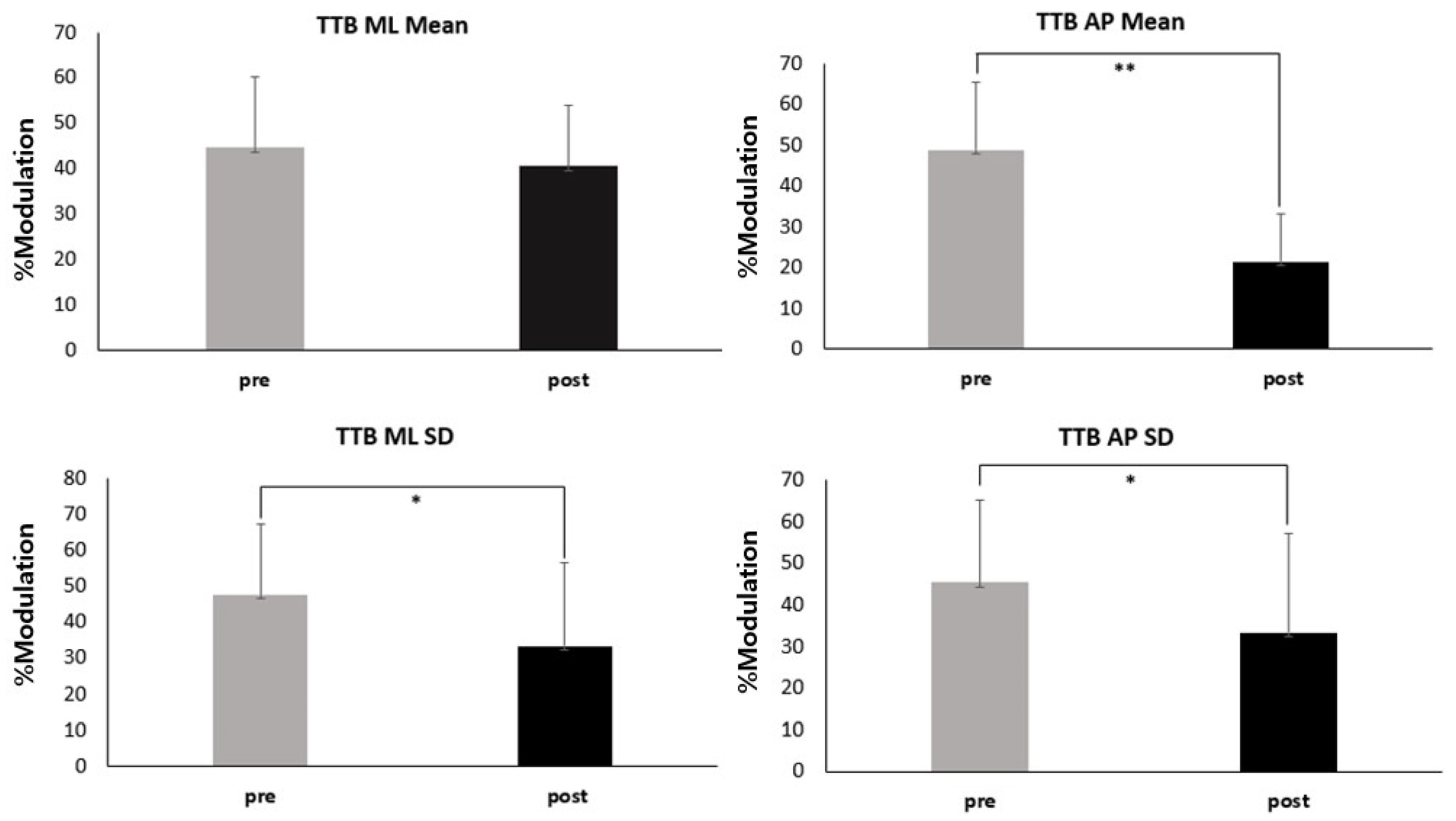
3.2. Visual Reliance

Figure 4 and Table 4 presents the %modulation results. We found significant decreases in %modulation after the fatigue protocol for TTB AP means (PRE: 48.7 ± 16.54; POST: 21.37 ± 11.65, p < 0.001), TTB ML SD (PRE: 49.17 ± 18.23; POST: 34.73 ± 24.66, p = 0.05), and TTB AP SD (PRE: 44.95 ± 20.02; POST: 22.67 ± 12.83, p < 0.001). However, TTB ML means did not differ significantly between pre- and post-fatigue (PRE: 44.59 ± 15.64; POST: 40.52 ± 13.57, p = 0.119).

4. Discussion

This study examines the effects of a functional fatigue protocol and visual information on postural control in patients with CAI. Specifically, we investigate whether open versus closed eyes influence postural control differently in CAI patients following the induction of functional fatigue. To the best of our knowledge, this study is the first to investigate postural control and visual reliance in CAI patients under conditions of functional fatigue. The findings revealed that the blocking of visual input and the implementation of sports-specific fatigue protocols reduced the control of static posture among those with CAI. Specifically, this study demonstrated a negative impact of functional fatigue on the capacity for postural control among CAI patients when performing tasks with their eyes open, while no significant changes were observed after the fatigue protocol under conditions with the eyes closed. Moreover, the research highlighted a significant decrease in reliance on visual input, indicating that functional fatigue alters the sensory strategies used by individuals with CAI, causing them to rely less on visual information.
Regardless of whether individuals were fatigued, a significant main effect of vision was observed. Individuals with CAI exhibited overall decreased TTB minima in eyes closed conditions compared with the case with their eyes open. These results align with previous findings [15,42,43,44,45,46] indicating that, in CAI patients, postural deficits worsen in the absence of visual information. Chronic injuries of the ankle joints can lead to damaged sensory afferents, reducing available somatosensory feedback and diminishing the ability of the postural control system to generate effective corrections to maintain equilibrium during unilateral weight-bearing [47]. This observation could potentially highlight a factor contributing to the recurrent episodes of the ankle “giving way” during functional activities, as the CAI patients exhibited a diminished ability to regulate COP excursions concerning the ML and AP limits of stability.
The results of this study regarding fatigue’s main effect provide compelling evidence that fatigue significantly impairs static postural control in CAI patients, particularly when assessed with the visual assistance of open eyes. These findings are consistent with previous research indicating that CAI patients often experience a prolonged decrease in their ability to interpret sensory information from the somatosensory function, which includes the foot and ankle complex [15]. When CAI is compounded with functional fatigue, the reduction in somatosensory function may become more pronounced, exacerbating the challenges faced by affected individuals to maintain precise control over their movements.
Ankle instability and fatigue are two factors confirmed to exert adverse effects on somatosensory function, specifically impacting the transmission and processing of sensory signals originating from the ankle joint and relaying them to the central nervous system. However, a key finding of this study was that CAI patients were unable to compensate for somatosensory deficits through visual feedback, even when they kept their eyes open under fatigued conditions. This is supported by the %modulation data obtained in this study, and a decrease in visual reliance was observed after fatigue. This can be explained by the SD of TTB minima, which indicates the consistency of TTB measurements and reflects the level of constraints on the sensorimotor system [48]. In essence, it quantifies the effectiveness with which the body and central nervous system employ various sensory organs to maintain balance. A decrease in TTB SD indicates a diminished ability to use non-visual sensory organs for positioning, signifying a reduced ability upon an increase in sensory complexity, such as in fatigued conditions [49]. The diminished ability of CAI patients to find effective movement strategies when fatigued implies that their reliance on visual information, their chosen strategy for maintaining balance, failed under such conditions. Consequently, the average TTB minima decreased and their dependence on visual information diminished. While this might be associated with long-term issues, in the short term, it is crucial to enhance the ability to utilize alternative sensory organs. These findings emphasize the need for training under fatigued conditions, rather than focusing solely on balance training with the eyes open in CAI patients.

5. Clinical Implications

Fatigue-induced impairments significantly contribute to the elevated rates of injury observed in the later stages of sporting competitions, with a substantial proportion of injuries in sports such as soccer (48%) [50] and rugby (71%) [51] occurring during the second half. It was also reported that 47% of ice hockey injuries [52] occur within the final 5 min of a period. Additionally, fatigue-related impairments highlighted in this study are not exclusive to athletes but can also adversely affect individuals with CAI in the physically active individuals. Fatigue has the potential to compromise postural stability, motor control, and movement strategies, ultimately increasing the risk of injury for those without competitive athletic backgrounds. Thus, understanding fatigue’s influence on this vulnerable group is crucial for developing targeted rehabilitation and injury prevention strategies that can benefit a wider spectrum of individuals. Understanding the intricate relationship between fatigue and sensory processing is crucial for unraveling the multiple factors contributing to deficits in postural control in CAI patients. In particular, it is crucial to understand that the fatigue experienced in isolated muscle groups significantly differs from the fatigue encountered during closed kinetic chain exercises, typical sports activities, or athletic performance. Our study presents substantial evidence emphasizing the heightened vulnerability of individuals with CAI to re-injury under sports-specific fatigue conditions. It is thus crucial to understand how fatigue conditions resembling real sports scenarios impact the abilities of CAI patients to achieve postural control, in order to develop future injury prevention strategies and training programs.
Previous research has consistently shown that implementing a balance training program, progressively introducing unique and challenging tasks to encourage CAI patients to explore the limits of their stability, results in an increase in the degree of freedom of the ankle [12]. However, a recent meta-analysis indicated that traditional balance training fails to induce a shift in visual reliance in CAI patients adopting a single-limb stance [19]. Therefore, this study emphasizes the importance of investigating the effects of balance training under fatigue conditions as a potential approach to mitigate the risk of ankle re-injury and its potential impact on reliance on visual input in CAI patients in future studies. The obtained findings not only have practical implications for determining the appropriate timing for returning to sports after a lateral ankle sprain, but also offer valuable insights for assessing proprioceptive function as part of preventive and rehabilitation programs for CAI patients.

6. Limitations

The study has some limitations. First, we did not collect information regarding participants’ physical activity levels through a survey or any other means. Although we did not gather detailed activity levels, we recruited participants who exercise twice a week. Second, our study did not take into account the structural characteristics of the foot, such as the Foot Posture Index, of our individuals. However, we screened our subjects for CAI characteristics using a questionnaire. Lastly, the participants in our study were selected based on their CAI rather than their athletic status. This distinction is important as it focuses on the specific vulnerabilities and biomechanical behaviors associated with CAI, which might differ from those of elite athletes or those actively engaged in sports competitions.

7. Conclusions

Our findings reveal that functional fatigue significantly impairs postural control in CAI individuals when performing tasks with their eyes open. However, no significant changes were observed in postural control when the eyes were closed following functional fatigue. These results suggest that fatigue may negatively impact somatosensory function and decrease reliance on visual cues, contributing to difficulties in maintaining balance among individuals with CAI. Such effects may elucidate the heightened risk of ankle sprains under fatigue conditions. By comprehensively understanding these effects, we can develop more effective interventions and strategies to mitigate the impact of fatigue on postural control in individuals with CAI.

Author Contributions

Conceptualization, S.Y.L., H.K. and K.S.; formal analysis, K.K. and K.S.; investigation, S.Y., E.J.H., H.G.J. and K.U.O.; data curation, K.K., K.S., S.Y., E.J.H., H.G.J. and K.U.O.; writing—original draft preparation, K.K.; writing—review and editing, H.K.; visualization, K.S.; supervision, S.Y.L.; project administration, S.Y.L. and K.K.; funding acquisition, K.K. All authors have read and agreed to the published version of the manuscript.

Funding

This research was funded by Yonsei University Research Grant, grant number No. R202206068.

Institutional Review Board Statement

The study was conducted from 30 October 2022 to 30 April 2023, and all procedures were approved by the Institutional Review Board of Yonsei University (7001988-202210-HR-1626-04).

Informed Consent Statement

Informed consent was obtained from all subjects involved in the study. Written informed consent has been obtained from the patients to publish this paper.

Data Availability Statement

Our data are unavailable due to privacy or ethical restrictions.

Conflicts of Interest

The authors declare no conflicts of interest.

References

  1. Doherty, C.; Delahunt, E.; Caulfield, B.; Hertel, J.; Ryan, J.; Bleakley, C. The incidence and prevalence of ankle sprain injury: A systematic review and meta-analysis of prospective epidemiological studies. Sports Med. 2014, 44, 123–140. [Google Scholar] [CrossRef] [PubMed]
  2. Herzog, M.M.; Kerr, Z.Y.; Marshall, S.W.; Wikstrom, E.A. Epidemiology of ankle sprains and chronic ankle instability. J. Athl. Train. 2019, 54, 603–610. [Google Scholar] [CrossRef] [PubMed]
  3. Lin, C.-I.; Houtenbos, S.; Lu, Y.-H.; Mayer, F.; Wippert, P.-M. The epidemiology of chronic ankle instability with perceived ankle instability—A systematic review. J. Foot Ankle Res. 2021, 14, 41. [Google Scholar] [CrossRef] [PubMed]
  4. Gribble, P.A.; Delahunt, E.; Bleakley, C.; Caulfield, B.; Docherty, C.L.; Fourchet, F.; Fong, D.; Hertel, J.; Hiller, C.; Kaminski, T.W.; et al. Selection criteria for patients with chronic ankle instability in controlled research: A position statement of the international ankle consortium. J. Orthop. Sports Phys. Ther. 2013, 43, 585–591. [Google Scholar] [CrossRef] [PubMed]
  5. Lee, I.; Ha, S.; Chae, S.; Jeong, H.S.; Lee, S.Y. Altered biomechanics in individuals with chronic ankle instability compared with copers and controls during gait. J. Athl. Train. 2022, 57, 760–770. [Google Scholar] [CrossRef] [PubMed]
  6. Webster, K.A.; Pietrosimone, B.G.; Gribble, P.A. Muscle activation during landing before and after fatigue in individuals with or without chronic ankle instability. J. Athl. Train. 2016, 51, 629–636. [Google Scholar] [CrossRef] [PubMed]
  7. Hertel, J. Functional anatomy, pathomechanics, and pathophysiology of lateral ankle instability. J. Athl. Train. 2002, 37, 364–375. [Google Scholar] [PubMed]
  8. Kim, K.-M.; Hart, J.M.; Saliba, S.A.; Hertel, J. Modulation of the fibularis longus Hoffmann reflex and postural instability associated with chronic ankle instability. J. Athl. Train. 2016, 51, 637–643. [Google Scholar] [CrossRef] [PubMed]
  9. Grooms, D.; Appelbaum, G.; Onate, J. Neuroplasticity following anterior cruciate ligament injury: A framework for visual-motor training approaches in rehabilitation. J. Orthop. Sports Phys. Ther. 2015, 45, 381–393. [Google Scholar] [CrossRef] [PubMed]
  10. Gribble, P.A.; Bleakley, C.M.; Caulfield, B.M.; Docherty, C.L.; Fourchet, F.; Fong, D.T.-P.; Hertel, J.; Hiller, C.E.; Kaminski, T.W.; McKeon, P.O.; et al. Evidence review for the 2016 International Ankle Consortium consensus statement on the prevalence, impact and long-term consequences of lateral ankle sprains. Br. J. Sports Med 2016, 50, 1496–1505. [Google Scholar] [CrossRef]
  11. Winter, D.A.; Patla, A.E.; Prince, F.; Ishac, M.; Gielo-Perczak, K. Stiffness control of balance in quiet standing. J. Neurophysiol. 1998, 80, 1211–1221. [Google Scholar] [CrossRef]
  12. Wikstrom, E.A.; Naik, S.; Lodha, N.; Cauraugh, J.H. Balance capabilities after lateral ankle trauma and intervention: A meta-analysis. In Database of Abstracts of Reviews of Effects (DARE): Quality-Assessed Reviews; Centre for Reviews and Dissemination (UK): York, UK, 2009. [Google Scholar]
  13. Munn, J.; Sullivan, S.J.; Schneiders, A.G. Evidence of sensorimotor deficits in functional ankle instability: A systematic review with meta-analysis. J. Sci. Med. Sport 2010, 13, 2–12. [Google Scholar] [CrossRef]
  14. Hiller, C.E.; Nightingale, E.J.; Lin, C.-W.C.; Coughlan, G.F.; Caulfield, B.; Delahunt, E. Characteristics of people with recurrent ankle sprains: A systematic review with meta-analysis. Br. J. Sports Med. 2011, 45, 660–672. [Google Scholar] [CrossRef]
  15. Song, K.; Burcal, C.J.; Hertel, J.; Wikstrom, E.A. Increased visual use in chronic ankle instability: A meta-analysis. Med. Sci. Sports Exerc. 2016, 48, 2046–2056. [Google Scholar] [CrossRef]
  16. Holmes, A.; Delahunt, E. Treatment of common deficits associated with chronic ankle instability. Sports Med. 2009, 39, 207–224. [Google Scholar] [CrossRef]
  17. McKeon, P.O.; Booi, M.J.; Branam, B.; Johnson, D.L.; Mattacola, C.G. Lateral ankle ligament anesthesia significantly alters single limb postural control. Gait Posture 2010, 32, 374–377. [Google Scholar] [CrossRef]
  18. Knapp, D.; Lee, S.Y.; Chinn, L.; Saliba, S.A.; Hertel, J. Differential ability of selected postural-control measures in the prediction of chronic ankle instability status. J. Athl. Train. 2011, 46, 257–262. [Google Scholar] [CrossRef]
  19. Song, K.; Rhodes, E.; Wikstrom, E.A. Balance training does not alter reliance on visual information during static stance in those with chronic ankle instability: A systematic review with meta-analysis. Sports Med. 2018, 48, 893–905. [Google Scholar] [CrossRef]
  20. Kim, K.M. Higher visual reliance during single-leg balance bilaterally occurring following acute lateral ankle sprain: A potential central mechanism of bilateral sensorimotor deficits. Gait Posture 2020, 78, 26–29. [Google Scholar] [CrossRef]
  21. Gribble, P.A.; Hertel, J.; Denegar, C.R.; Buckley, W.E. The effects of fatigue and chronic ankle instability on dynamic postural control. J. Athl. Train. 2004, 39, 321–329. [Google Scholar]
  22. Gribble, P.A.; Hertel, J. Effect of lower-extremity muscle fatigue on postural control. Arch. Phys. Med. Rehabil. 2004, 85, 589–592. [Google Scholar] [CrossRef]
  23. Lane, J.; Bansbach, H.; Connaboy, C.; Darnell, M.; Keenan, K.A.; Lovalekar, M.T.; Nagai, T.; Allison, K.F. The Effect of Loaded Fatigue on Loaded Postural Stability; University of Pittsburgh: Bradford, PA, USA, 2018. [Google Scholar]
  24. Bizid, R.; Margnes, E.; François, Y.; Jully, J.L.; Gonzalez, G.; Dupui, P.; Paillard, T. Effects of knee and ankle muscle fatigue on postural control in the unipedal stance. Eur. J. Appl. Physiol. 2009, 106, 375–380. [Google Scholar] [CrossRef]
  25. Gimmon, Y.; Riemer, R.; Oddsson, L.; Melzer, I. The effect of plantar flexor muscle fatigue on postural control. J. Electromyogr. Kinesiol. 2011, 21, 922–928. [Google Scholar] [CrossRef]
  26. Steib, S.; Zech, A.; Hentschke, C.; Pfeifer, K. Fatigue-induced alterations of static and dynamic postural control in athletes with a history of ankle sprain. J. Athl. Train. 2013, 48, 203–208. [Google Scholar] [CrossRef]
  27. Reimer, R.C., III; Wikstrom, E.A. Functional fatigue of the hip and ankle musculature cause similar alterations in single leg stance postural control. J. Sci. Med. Sport 2010, 13, 161–166. [Google Scholar] [CrossRef]
  28. Jones, C.M.; Griffiths, P.C.; Mellalieu, S.D. Training load and fatigue marker associations with injury and illness: A systematic review of longitudinal studies. Sports Med. 2017, 47, 943–974. [Google Scholar] [CrossRef]
  29. Cortes, N.; Greska, E.; Kollock, R.; Ambegaonkar, J.; Onate, J.A. Changes in lower extremity biomechanics due to a short-term fatigue protocol. J. Athl. Train. 2013, 48, 306–313. [Google Scholar] [CrossRef]
  30. Martin, R.L.; Irrgang, J.J.; Burdett, R.G.; Conti, S.F.; Van Swearingen, J.M. Evidence of validity for the Foot and Ankle Ability Measure (FAAM). Foot Ankle Int. 2005, 26, 968–983. [Google Scholar] [CrossRef]
  31. Ross, S.E.; Guskiewicz, K.M.; Gross, M.T.; Yu, B. Assessment tools for identifying functional limitations associated with functional ankle instability. J. Athl. Train. 2008, 43, 44–50. [Google Scholar] [CrossRef]
  32. Lee, S.C.; Kim, C.Y.; Jeong, H.S.; Lee, S.Y.; Kim, S. Effect of functional fatigue protocol on static and dynamic postural control of lower extremities. Int. J. Appl. Sports Sci. 2018, 29, 603–612. [Google Scholar]
  33. Borg, G. Borg’s Perceived Exertion and Pain Scales; Human Kinetics: Champaign, IL, USA, 1998. [Google Scholar]
  34. Kim, H. Lower Extremity Neuromechanics During a Forward-Side Jump Following Functional Fatigue in Patients with Ankle Instability; Brigham Young University: Provo, UT, USA, 2015. [Google Scholar]
  35. Hertel, J.; Buckley, W.E.; Denegar, C.R. Serial testing of postural control after acute lateral ankle sprain. J. Athl. Train. 2001, 36, 363–368. [Google Scholar]
  36. van Wegen, E.E.; van Emmerik, R.E.; Wagenaar, R.C.; Ellis, T. Stability boundaries and lateral postural control in Parkinson’s disease. Mot. Control 2001, 5, 254–269. [Google Scholar] [CrossRef]
  37. Song, K.; Kang, T.K.; Wikstrom, E.A.; Jun, H.-P.; Lee, S.Y. Effects of reduced plantar cutaneous sensation on static postural control in individuals with and without chronic ankle instability. J. Sci. Med. Sport 2017, 20, 910–914. [Google Scholar] [CrossRef]
  38. Hertel, J.; Olmsted-Kramer, L.C. Deficits in time-to-boundary measures of postural control with chronic ankle instability. Gait Posture 2007, 25, 33–39. [Google Scholar] [CrossRef]
  39. Hertel, J.; Olmsted-Kramer, L.C.; Challis, J.H. Time-to-boundary measures of postural control during single leg quiet standing. J. Appl. Biomech. 2006, 22, 67–73. [Google Scholar] [CrossRef]
  40. Wikstrom, E.A.; Song, K.; Pietrosimone, B.G.; Blackburn, J.T.; Padua, D.A. Visual utilization during postural control in anterior cruciate ligament-deficient and–reconstructed patients: Systematic Reviews and Meta-Analyses. Arch. Phys. Med. Rehabil. 2017, 98, 2052–2065. [Google Scholar] [CrossRef]
  41. Cohen, J. Statistical power analysis. Curr. Dir. Psychol. Sci. 1992, 1, 98–101. [Google Scholar] [CrossRef]
  42. Thompson, C.; Schabrun, S.; Romero, R.; Bialocerkowski, A.; van Dieen, J.; Marshall, P. Factors contributing to chronic ankle instability: A systematic review and meta-analysis of systematic reviews. Sports Med. 2018, 48, 189–205. [Google Scholar] [CrossRef]
  43. Gribble, P.A.; Tucker, W.S.; White, P.A. Time-of-day influences on static and dynamic postural control. J. Athl. Train. 2007, 42, 35–41. [Google Scholar]
  44. Corbin, D.M.; Hart, J.M.; McKeon, P.O.; Ingersoll, C.D.; Hertel, J. The effect of textured insoles on postural control in double and single limb stance. J. Sport. Rehabil. 2007, 16, 363–372. [Google Scholar] [CrossRef]
  45. McKeon, P.O.; Hertel, J. Diminished plantar cutaneous sensation and postural control. Percept. Mot. Skills 2007, 104, 56–66. [Google Scholar] [CrossRef]
  46. McKeon, P.O.; Hertel, J. Spatiotemporal postural control deficits are present in those with chronic ankle instability. BMC Musculoskelet. Disord. 2008, 9, 76. [Google Scholar] [CrossRef]
  47. Simpson, J.D.; Rendos, N.K.; Stewart, E.M.; Turner, A.J.; Wilson, S.J.; Macias, D.M.; Chander, H.; Knight, A.C. Bilateral spatiotemporal postural control impairments are present in participants with chronic ankle instability. Phys. Ther. Sport 2019, 39, 1–7. [Google Scholar] [CrossRef]
  48. Doherty, C.; Bleakley, C.; Hertel, J.; Caulfield, B.; Ryan, J.; Delahunt, E. Postural control strategies during single limb stance following acute lateral ankle sprain. Clin. Biomech. 2014, 29, 643–649. [Google Scholar] [CrossRef]
  49. Koldenhoven, R.M.; Feger, M.A.; Fraser, J.J.; Hertel, J. Variability in center of pressure position and muscle activation during walking with chronic ankle instability. J. Electromyogr. Kinesiol. 2018, 38, 155–161. [Google Scholar] [CrossRef]
  50. Woods, C.; Hawkins, R.D.; Maltby, S.; Hulse, M.; Thomas, A.; Hodson, A.; Football Association Medical Research Programme. The Football Association Medical Research Programme: An audit of injuries in professional football—Analysis of hamstring injuries. Br. J. Sports Med. 2004, 38, 36–41. [Google Scholar] [CrossRef]
  51. Gabbett, T.J. Incidence, site, and nature of injuries in amateur rugby league over three consecutive seasons. Br. J. Sports Med. 2000, 34, 98–103. [Google Scholar] [CrossRef]
  52. Pinto, M.; Kuhn, J.E.; Greenfield, M.L.V.; Hawkins, R.J. Prospective analysis of ice hockey injuries at the Junior A level over the course of one season. Clin. J. Sport Med. 1999, 9, 70–74. [Google Scholar] [CrossRef]
Figure 1. Single-leg balance test.
Applsci 14 04445 g001
Figure 2. Functional fatigue protocol.
Applsci 14 04445 g002
Figure 3. Effects of fatigue and vision on postural control by two-way ANOVA. Abbreviations: AP, anteroposterior; ML, mediolateral; SD, standard deviation; TTB, time to boundary. * Indicates a statistically significant time by visual condition interaction (p < 0.01). ** Indicates a statistically significant time by visual condition interaction (p < 0.001).
Applsci 14 04445 g003
Figure 4. %Modulation changes between pre- and post-fatigue for time-to-boundary (TTB) minima mean and TTB minima standard deviation (SD) in the mediolateral (ML) and anteroposterior (AP) directions. Abbreviations: AP, anteroposterior; ML, mediolateral; SD, standard deviation; TTB time to boundary. * Indicates a statistically significantly difference between PRE and POST (p < 0.05). ** Indicates a statistically significantly difference between PRE and POST (p < 0.001).
Applsci 14 04445 g004
Table 1. Demographic characteristics (mean ± SD).
VariableCAI (n = 28)
Age (years)24.07 ± 2.62
Height (cm)166.87 ± 7.45
Body mass (kg)61.81 ± 17.09
Foot length (cm)22.96 ± 1.66
Foot width (cm)8.86 ± 1.21
Resting heart rate95 ± 7.53
IdFAI18.26 ± 3.82
FAAM ADL79.09 ± 9.34
FAAM Sports67.83 ± 11.83
Abbreviations: CAI, chronic ankle instability; FAAM, foot and ankle ability measure; ADL, activities of daily living; IdFAI, Identification of Functional Ankle Instability.
Table 2. The average completion time, heart rate, perceived exertion, and vertical jump height at the endpoint of the functional fatigue protocol (mean ± SD).
VariablesValue
Rep. time (s)44.30 ± 7.05
HR184.80 ± 6.34
RPE17.73 ± 1.35
MVJH (cm)39.00 ± 16.46
Abbreviations: HR, heart rate; RPE, rated perceived exertion; MVJH, maximum vertical jump height; Rep. time, repetition time.
Table 3. Mean (±SD) for the time-to-boundary (TTB) measures of postural control with eyes open and closed before and after the fatigue protocol.
Eyes OpenEyes ClosedANOVAFEffect Size (95% CI)p
Measures (s)Pre-FatiguePost-FatiguePre-FatiguePost-Fatigue
TTB ML
Mean of minima
3.4 ± 0.763 ± 0.341.78 ± 0.461.74 ± 0.43Fatigue Effect7.8267410.6 (0.055 to 0.360)0.009
Condition Effect201.09510.98 (1.221 to 1.634)<0.001
Fatigue × Condition Interaction7.81124 0.009
TTB AP
Mean of minima
9.99 ± 1.434.53 ± 0.914.92 ± 1.14.79 ± 0.94Fatigue Effect426.79280.45 (2.518 to 3.074)<0.001
Condition Effect81.724110.52 (1.860 to 2.953)
Fatigue × Condition Interaction240.4783
TTB ML
SD on minima
2.9 ± 1.172.45 ± 1.161.59 ± 0.831.56 ± 0.63Fatigue Effect63.983710.57 (−0.040 to 0.515)0.091
Condition Effect2.5639060.96 (0.696 to 1.499)<0.001
Fatigue × Condition Interaction12.21461 0.176
TTB AP
SD on minima
6.11 ± 1.232.69 ± 0.533.33 ± 0.943.19 ± 0.83Fatigue Effect46.880570.54 (1.531 to 1.963)<0.001
Condition Effect106.86570.33 (0.717 to 1.629)
Fatigue × Condition Interaction364.4761
Abbreviations: AP, anteroposterior; ML, mediolateral; SD, standard deviation; TTB time to boundary.
Table 4. Mean (±SD) for %modulation changes between pre- and post-fatigue for the time-to-boundary (TTB) measures.
%ModulationPre-FatiguePost-Fatiguet95% CIdp
TTB ML
Mean of minima
44.59 ± 15.6440.52 ± 13.571.61(−0.078 to 0.681)0.280.119
TTB AP
Mean of minima
48.7 ± 16.5421.37 ± 11.657.604(0.899 to 1.962)1.91<0.001
TTB ML
SD on minima
49.17 ± 18.2334.73 ± 24.663.033(0.168 to 0.969)0.660.05
TTB AP
SD on minima
44.95 ± 20.0222.67 ± 12.834.441(0.401 to 1.266)1.33<0.001
Abbreviations: AP, anteroposterior; ML, mediolateral; SD, standard deviation; TTB time to boundary.
Disclaimer/Publisher’s Note: The statements, opinions and data contained in all publications are solely those of the individual author(s) and contributor(s) and not of MDPI and/or the editor(s). MDPI and/or the editor(s) disclaim responsibility for any injury to people or property resulting from any ideas, methods, instructions or products referred to in the content.

