Article

Eco-Friendly Preservation of Pharaonic Wooden Artifacts using Natural Green Products

by
Neveen S. Geweely
1,
Amira M. Abu Taleb
1,
Paola Grenni
2,*,
Giulia Caneva
3,
Dina M. Atwa
4,
Jasper R. Plaisier
5 and
Shimaa Ibrahim
1,6
1
Botany and Microbiology Department, Faculty of Science, Cairo University, Giza 12613, Egypt
2
Water Research Institute, National Research Council (CNR-IRSA), Via Salaria km 29.300, Monterotondo, 00010 Rome, Italy
3
Department of Science, University Roma Tre, Viale Marconi 446, 00146 Rome, Italy
4
Department of Laser Interaction with Matters, Laser Institute for Research and Applications, Beni-Suef University, Beni-Suef P.O. Box 62517, Egypt
5
Elettra Sincrotrone Trieste SCpA, Area Science Park, Basovizza, 34149 Trieste, Italy
6
Grand Egyptian Museum, Conservation Center, Ministry of Antiquities, Museums Sector, El-Remaya Square, Giza 12561, Egypt
*
Author to whom correspondence should be addressed.
Appl. Sci. 2024, 14(12), 5023; https://doi.org/10.3390/app14125023
Submission received: 23 April 2024 / Revised: 31 May 2024 / Accepted: 6 June 2024 / Published: 9 June 2024
(This article belongs to the Section Applied Microbiology)

Abstract

:

Featured Application

The cross-disciplinary investigation conducted here utilized natural green products (essential oils and plant extracts) as effective eco-friendly antifungal agents for the preservation of archaeological wooden artifacts.

Abstract

The biodeterioration of wooden cultural heritage is a severe problem worldwide and fungi are the main deteriorating agents. The identification of effective natural products, safer for humans and the environment, is a current challenge. Ten deteriorated archaeological objects (a wooden statue of a seated man, an anthropoid wooden coffin with a cartonnage mummy of Nespathettawi, and a wooden box of Padimen’s son), stored at the Egyptian museum in Cairo, were considered here. The wood species of the three most deteriorated objects were previously identified as Acacia nilotica, Ficus sycomorus, and Tamarix gennessarensis. Twenty-six fungal species were isolated and identified from the wooden objects and the four most frequent species belonged to the genus Aspergillus. Fourteen fungal species among those isolated showed the greatest biodeterioration activity on the experimental wood blocks of the archaeological objects. The antifungal activities of several eco-friendly plant essential oils (from cinnamon, eucalyptus, frankincense, geranium, lavender, lemongrass, menthe, rosemary, tea tree, and thyme) and plant extracts (from basil, eucalyptus, henna, melia, and teak) were tested against the fungal species with the greatest biodeterioration activity. The essential oils (Eos) were more effective than the plant extracts. Thyme EO, followed by geranium and cinnamon ones, was the most active (minimum inhibitory concentrations: 0.25–1 µL/mL). These EO; also showed inhibitory effects on the enzymatic activities (cellulase, amylase, and protease) of the four most dominant fungal species. Thymol and p-cymene were the two main components of thyme oil, while geraniol and beta-citronellol were those of geranium oil; eugenol and caryophyllene were those of the cinnamon EO. Thyme oil applied to the most deteriorated experimental aged A. nilotica wooden cubes inoculated with the four highly frequent fungal species was effective in wood preservation. Moreover, no significant interference was observed in the wood before and after thyme treatment. Thyme oil seems to be a promising eco-friendly antifungal agent for the preservation of archaeological wooden artefacts.

1. Introduction

Egyptian wooden artifacts constitute an important part of global cultural heritage. The impact of microbial deterioration on cultural heritage is a well-recognized global problem and the preservation thereof is a challenging task. Archaeological pharaonic wood provides valuable information about ancient cultures; therefore, significant effort should be invested to preserve these precious objects against microbial damage to preserve them for future generations in good condition [1].
The biodeterioration of wooden cultural heritage is a severe problem, and fungi have the most significant potential effect on this type of historical artwork [2,3]. Fungi have a detrimental effect on the aesthetic value of the archaeological wood due to colonization by their pigmented mycelium with the production and deposition of melanin pigment [4]. The decay caused by wood-destroying fungi belonging to Ascomycota, Basidiomycota, and Deuteromycetes, and their responsibility for the irreversible heritage loss of archaeological wooden artifacts, has been highlighted by several authors [5,6]. Their sensitivity to fungal attack and their durability can vary among wood species, and we can also note that the ancient Egyptians selected the most resistant woods to preserve their artefacts [1,7]. In the case of archaeological wood, the large contact surface area of artefacts with the funerary burial soil causes wood corrosion, as recently observed for a statue attacked by fungi that caused the cracking and wrinkling of the wooden structures [7].
Wood protection, using biocides, is an important issue since physical and mechanical conservation practices are not efficient in removing deterioration-causing microorganisms. However, many commercial biocides are toxic, and unsafe to apply on archaeological pieces, since they can be hazardous for humans and ecosystems [8,9]. Indeed, some artificial biocides used against fungal corrosion can be degraded by fungal enzymes [6]. The preservation of archaeological wood against microbial deterioration, using products safe for the health of restorers and museum personnel, is urgent to save them from microbial damage.
A growing interest in natural substances is observed in the field of restoration, because they are, in their usual doses of application, safer compared to many traditional biocides, both due to changing regulations and the phenomenon of increasing resistance of many microorganisms to biocides [9,10]. Essential oils (EOs), which are a complex mixture of odorous and volatile compounds, and other plant extracts, and which were used for a long time as natural antiseptics, are natural products that can exhibit a broad spectrum of biocide activities [9,10]. EOs are known for their natural components (e.g., monoterpenes, diterpenes, and hydrocarbons) with various functional groups, and they have been studied for their antifungal activities [9,10,11,12]. Some compounds, such as thyme, eugenol, and cinnamon were widely tested, and much data appear to suggest the antifungal properties of most EOs against wood-decaying fungi, so they are applied as effective wood-protecting agents [11,12]. In fact, many essential oils can be applied for fungal infection prevention, with an antifungal activity greater than some commercial fungicides [13]. The use of phytochemicals to control microbes has been a practical and safe substitute for both human and ecological health [14,15].
In general, plants are a rich source of various chemical compounds; secondary metabolites comprise up to 30% of the dry mass of plants and play an essential role in their protection against microbial pathogens [16]. Plant extracts are obtained from various plant parts including bark, leaves, fruits, flowers, buds, seeds, and wood [17]. The antifungal properties of various oils and plant extracts make them interesting as possible natural substances that can be utilized as alternative wood preservatives to prevent wood decay [11].
Several review papers [9,12] have collected data on such natural compounds but, considering the wideness and chemical variability of such natural compounds, adequate information is still missing for many substances, such as specific efficacy at low doses and performance over time. As a consequence, further tests are needed to check the effectiveness of most of them, and the main objective of this study was to test the effectiveness of the most promising natural products for the protection of archaeological wooden objects from fungal deterioration. The selection of essential oils and plant extracts arose from the compounds tested for antifungal efficiency in field restoration, and from the compounds listed as environmentally friendly, safe, and economical in health regulations and in the wider literature [9,17]. Then, the ultimate aim is to avoid the use of products with harmful effects on public health and the environment such as most of the synthetic fungicides normally used to preserve artefacts.

2. Materials and Methods

2.1. Archaeological Objects

Ten archaeological pharaonic wooden objects (Table 1 and Supplementary Materials, Figure S1) located at the Egyptian Museum (Cairo, Egypt) were selected for their biodegradation status. The objects belong to different ages (Old Kingdom: 2686–2181 BC; New Kingdom: 1550–1069 BC; Third Intermediate Period, Dynasty 21) and are currently stored at 24–26 °C and 50–55% humidity.

2.2. Isolation and Identification of Deteriorating Fungi

The fungal species were sampled (in three replicates) by swabbing from the deteriorated surface of the wooden objects, and isolated using two types of media (potato dextrose agar and Czapek’s Dox agar media). All plates were incubated at 28 °C for 7 days, as reported by Skóra et al. [18]. The fungal species were examined under the microscope and first identified using several fungal identification keys [19,20,21,22,23,24,25,26,27,28]. Molecular identification of the highly frequent deterioration-causing fungal species was performed by polymerase chain reaction (PCR).
For DNA extraction, the fungal species were grown on potato dextrose broth at 27 °C then the mycelium was filtered and converted to a fine powder in liquid nitrogen. The DNA was extracted using the Quick DNA Fungal Microprep Kit (Sigma-Aldrich) [29]. Genomic DNA Amplification (PCR and Primers): PCR was performed using the Maxima Hot Start PCR Master Mix (Thermo K1051, Sigma Aldrich, Merck KGaA, Darmstadt, Germany). First, the Maxima® Hot Start PCR Master Mix (2×) (Thermo K1051, Sigma Aldrich, Merck KGaA, Darmstadt, Germany) was gently vortex and centrifuged briefly after thawing. The PCR amplifications were performed using PCR reactions in 50 μL of sample containing 25 μL of the Maxima® Hot Start PCR Master Mix (2×), 5 μL of the template DNA, 18 μL of water (nuclease-free), 1 μL (20 μM) of ITS1 forward primer, and 1 μL (20 μM) of ITS4 primer. The ITS1 primer sequence was 5’ -TCC GTA GGT GAA CCT GCG G- 3’. The samples were gently vortex and spun down. Amplification of the PCR products was performed by using the thermal cycling conditions: initial denaturing step of 10 min at 95 °C (1 cycle), 35 cycles of denaturation for 30 s at 95 °C, annealing for 1 min at 57 °C, and elongation for 90 s at 72 °C with a final extension cycle for 10 min at 72 °C. PCR-amplified product electrophoresis was carried out with low-melting-point agarose gels (1.5%) at 7 V/cm2 for 1.5 h [30]. Ethidium bromide (0.5 g/mL) was used to stain the PCR products, which were then observed under a 305 nm UV light. The PCR products were cleaned up using the Gene JET PCR Purification Kit (Thermo Scientific, No. K0701). The PCR products were analyzed by the GATC Biotech Company (Konstanz, Germany) by an ABI 3730XL automated DNA sequencer (ThermoFisher, Applied Biosystems, Germany) using forward and reverse primers with the new 454 technology. Isolates were identified using the BLAST program (NCBI software package, NCBI Gen Bank databases, National Center for Biotechnology Information, www.ncbi.nlm.gov/blast) (accessed on 10 February 2023).

2.3. Ageing and Biodeterioration of Experimental Aged Wooden Blocks

The most deteriorated archaeological wooden samples (the wooden statue of a seated man, the anthropoid wooden coffin with a cartonnage mummy of Nespahettawi son of Neskhonsupahred, and the wooden box of Padimen’s son, object Nos. 1, 2, and 3, respectively, Table 1) were previously identified as Acacia nilotica, Ficus sycomorus, and Tamarix gennessarensis, as reported by Geweely et al. [31]. Three commercial experimental wood samples of the same identified species were used as experimental wooden blocks. They were obtained from El-Orman Botanical Garden, Giza, Egypt, and a woodworking shop in Alexandria, Egypt. The experimental wooden blocks were inoculated with the highly frequent deterioration-causing fungal isolates on a potato dextrose agar plate and left for 8 weeks. Uninoculated wood blocks were used as controls.

2.4. Green Eco-Friendly Natural Products

Ten essential oils extracted from plants, which had previously been demonstrated to have antifungal inhibitory activity [13,32,33,34], were obtained from the Natural Oil Department at the National Research Center (NRC) in Dokki, Egypt (Table 2).
Five medicinal plant species (Table 3) previously proven to have antifungal activity [35,36,37] were obtained from El-Orman Botanical Garden, Giza, Egypt.

2.5. Antifungal Effects of the Essential Oils and Plant Extracts on the Highly Deterioration-Causing Fungal Isolates

The effect of the tested essential oils (EOs) and the plant extracts (Table 2 and Table 3) were determined by the agar dilution method on the isolated fungi. Specifically, stock solutions of the oils and extracts were prepared in Dimethyl Sulfoxide (DMSO) and added to melted Czapek´s dox agar medium to obtain various concentrations (0.125, 0.25, 0.5, 0.75, 1, 2, 4, and 8 μL/mL). They were poured into Petri dishes and allowed to solidify. Each plate was inoculated by a fungal disc (6 mm) in the center. The plates (in triplicates) were incubated at 27 °C for 7 days. The minimum inhibitory concentrations (MICs) of the ten tested EOs and five plant extracts were determined as the lowest concentration (µL/mL) at which no visible growth occurred [38]. In particular, MICs were obtained as the minimum concentration that had an effect on mycelial growth diameter (mm), compared to the control (treated with itraconazole) [39]. Fungal growth inhibition was also calculated in percentage of mycelial growth inhibition according to the following equation:
M y c e l i a l   g r o w t h   i n h i b i t i o n   % = 100 × D C D T D C
where DC is the average diameter of the fungal growth of the control and DT is the average diameter of the fungal growth when treated with an EO. The tested efficient essential oils which had the highest significant antifungal activities against the isolated deteriorating fungal species were selected for the subsequent experiments.

2.6. Active Constituents of the Most Efficient Essential Oils

The active constituents of the selected most efficient EOs were analyzed at the Laboratory of Medicinal and Aromatic Plants Research, National Research Centre, Dokki, Egypt, by a gas chromatography–mass spectrometry apparatus (TRACE GC Ultra Gas Chromatograph, THERMO Scientific Corp.,Waltham, MA, USA) coupled with a THERMO mass spectrometer detector (ISQ Single Quadrupole Mass Spectrometer). The GC-MS system was equipped with a TR5 MS column (30 m × 0.32 mm i.d., 0.25 µm film thickness). The column temperature was kept at 60 °C for 5 min, then ramped at 5 °C/minute up to 200 °C, and finally kept for 10 min at that same temperature. A flame injector detector (FID) with an operating temperature of 220 °C and an injector with a temperature of 180 °C was used for manual injection and with nitrogen as the gas carrier [40].

2.7. Effect of the Most Efficient Essential Oils on the Enzymatic Activities of the Highly Frequent Deterioration-Causing Fungal Species

Activities of the different EOs on the amylase, cellulase, and protease of the most dominant deterioration-causing fungal species were determined. In particular, 100 mL of modified Czapek´s dox broth with 2% starch and 2% cellulose instead of sucrose and 1% casein instead of NaNO3 were used. The reaction mixture was performed by adding 1 mL of 1% soluble starch in acetate buffer (0.02 M, pH 5.0) with 1 mL of the fungal filtrate and incubating for 15 min at 50 °C. The ninhydrin test was used to detect the hydrolysis of casein by protease, while the culture filtrate (1 mL) was mixed with 1 mL of 1% casein in acetate buffer (0.05 M, pH 5.6) as a substrate for 15 min at 37 °C; the reaction was terminated by adding 5% trichloroacetic acid (TCA) followed by a centrifugation at 3000 rpm for 10 min. Stock solutions of the EOs and plant extracts were prepared in DMSO and diluted with the broth to obtain different concentrations (0.125, 0.25, 0.5, 0.75, and 1 μL/mL) and added to a 250 mL conical flask followed by inoculation with a mycelia disc (10 mm) cut from the margin of the 7-day-old culture of the tested fungal species. The controls were prepared without the addition of EOs [41,42,43,44]. The results are expressed as units per mL (U/mL). One unit is defined as the amount of enzyme which releases reducing sugars equivalent to 1 µg of glucose.

2.8. Essential Oils Efficiency on the Preservation of the Experimental Aged Wood Samples against the Deterioration-causing Fungal Species

The highest deteriorated experimental aged A. nilotica wooden blocks (10 × 10 × 10 mm) were autoclaved at 121 °C for 20 min and left to cool. Twenty-four blocks from the selected wood type were immersed in different concentrations (0.125, 0.25, 0.5, 0.75, 1 µL/mL) of EOs for 8 h and then the samples were air-dried for 24 h [39]. The untreated and treated experimental woods with the most efficient essential oils were evaluated against the most deterioration-causing fungal species. The plates were inoculated at the center with a disc (5 mm diameter) of each of the fungal species. The treated wood samples with and without essential oils were put over the agar surface. The extent of fungal growth was visually evaluated every week of inoculation for two months. Three replicates were used for each concentration. DMSO (10%) was used as the control [33,37]. The antifungal activity was estimated by fungal growth reduction [45].

2.9. Effect of the Most Efficient Essential Oil on the Characterization of the Experimental Aged Wood Samples

Different analytical techniques were used to study the effect of the most efficient essential oil on both fungi and wood. Scanning electron microscopy (QUANTA FEG 250, FEI Company, Hillsboro, OR, USA) equipped with an X-ray energy dispersive system (EDX), operating at 15 Kv) was used to identify the elemental composition of the wood. XRD (high-energy X-ray diffraction), performed at the MCX beamline of Elettra Synchrotron (Trieste, Italy), was used to measure the wood’s crystallinity. Finally, Fourier-transform infrared spectroscopy (Vertex 70 FTIR spectrometer equipped with an attenuated total reflectance ATR unit, Bruker, Germany) was used to study the effect of the most efficient essential oil on the characterization of the experimental wooden cubes [46].

2.10. Statistical Analysis

Statistical analysis was carried out by one-way analysis of variance (ANOVA) followed by homogenous subsets (Duncuna) using the Statistical Package for the Social Sciences (SPSS) version 21. Duncan’s multiple range tests were used at significance (p < 0.05) according to Colao et al. [47]. The values of antimicrobial activity of the EOs and plant extracts were expressed as the mean ± standard deviation of triplicate experiments for each sample.

3. Results

3.1. Identification of Fungi Isolated from the Ten Archaeological Wooden Objects

Twenty-six fungal species (Supplementary Materials, Table S1) were isolated from the deteriorated pharaonic wooden objects and identified morphologically by light microscopy. A total of 596 fungal colonies were obtained from the wooden objects studied here (Table 1). The wooden objects that resulted with the higher numbers of colonies were the wooden statue of a seated man, the anthropoid wooden coffin with a cartonnage mummy of Nespahettawi son of Neskhonsupahred, and the wooden box of Padimen’s son, with 113, 90, and 77 fungal colonies, respectively.

3.2. Identification of the Highly Frequent Fungal Species

Aspergillus (an Ascomycota in the brown rot fungi group) was the most highly abundant fungal genus isolated from the ten archaeological wooden objects. Aspergillus flavus (14.6%) was the most frequent species, followed by A. niger (13.8%), A. fumigatus (11.7%), and A. terreus (10.2%). The accession numbers found at https://www.ncbi.nlm.nih.gov/genbank/samplerecord (accessed on 15 March 2023), according to the alignment of the sequences of the tested A. flavus, A. niger, A. fumigatus, and A. terreus were OP131954, OP132225, OP132223, OP131931, respectively. The phylogenetic trees obtained from NCBI blast are shown in Supplementary Materials, Figures S2–S5.

3.3. Experimental Aged Wooden Blocks Biodeterioration

The twenty-six isolated fungal species (Supplementary Materials, Table S2) were inoculated onto three experimental wooden blocks (Ficus sycomorus, Tamarix gennessarensis, and Acacia nilotica). Among them, fourteen species were completely ramified over the experimental wooden blocks (such as: Alternaria alternata, Aspergillus flavus, Aspergillus fumigatus, Aspergillus niger, Aspergillus terreus, Cladosporium cladosporioides, Emericella nidulans, Fusarium oxysporum, Penicillium chrysogenum, Penicillium citrinum, Penicillium glabrum, Penicillium multicolor, Rhizopus oryzae, and Trichoderma viridae). On the other hand, four Aspergillus species (A. clavatus, A. ochraceus, A. parasiticus, and A. versicolor) and Penicillium oxalicum showed moderate growth. No growth was observed for Chaetomium globosum, Curvularia clavata, Epicoccum nigrum, Eurotium amstelodami, Mucor fuscus, Paecilomyces variotii, and Ulocladium botrytis. The fourteen most deterioration-causing fungal species were then selected for measuring the effect of various concentrations of the selected EOs and plant extracts.

3.4. Effect of the Plant Extracts on the Highly Frequent Fungal Species

The effects of the plant extracts (Table 3) on the growth of the fourteen deterioration-causing fungal species are presented in Supplementary Materials, Tables S3–S7. The effects are reported as minimum inhibitory concentrations, MICs, expressed as the diameter of mycelia growth and the percentage of mycelia growth inhibition compared to the control (no-treated mycelia). The teak extract was the most effective, with MICs ranging from 0.75 to 2 µL/mL for all isolated fungal species. Emericella nidulans, Fusarium oxysporum, Penicillium glabrum, P. oxalicum, and Rhizopus oryzae were the most sensitive species (MIC: 0.75 µL/mL). On the other hand, Aspergillus flavus, A. terreus, and Penicillium citrinum were completely inhibited at 1.0 µL/mL and the rest of the isolates were completely suppressed by the teak extract at 2.0 µL/mL. The eucalyptus extract was the second extract in terms of inhibition (MIC: 1.0–4.0 µL/mL). A. flavus and P. glabrum were the most susceptible species to it (MIC: 1.0 µL/mL), while the other species were completely inhibited at 2.0 µL/mL, excluding A. fumigatus, which was totally inhibited only at 4.0 µL/mL.

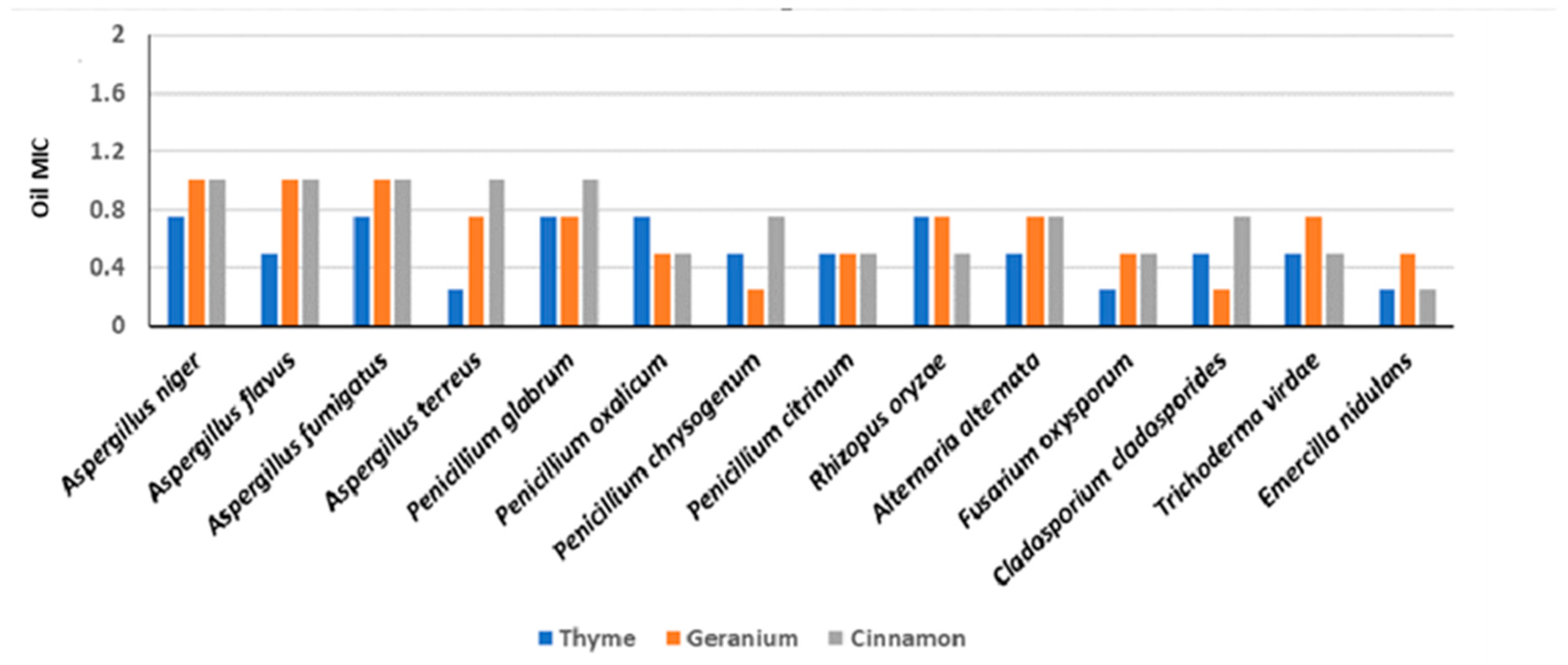
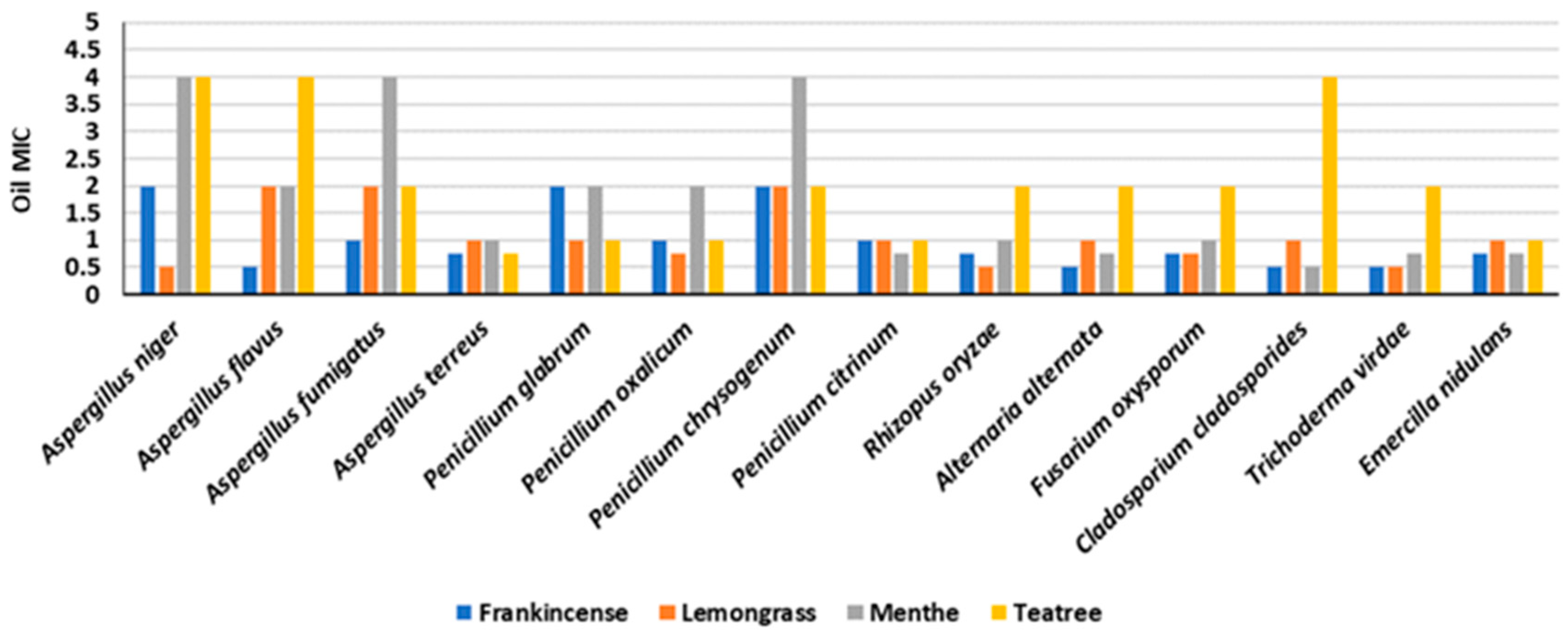
3.5. Effect of Essential Oils (MICs) on Fungal Isolates and Chemical Composition

The thyme, geranium, and cinnamon essential oils were the most effective against the fourteen deteriorating fungal species (Figure 1, Figure 2 and Figure 3).
The chemical composition of these essential oils was investigated by GC-MS (Supplementary Materials, Table S8), and the major constituents were identified. The thyme and geranium oils showed an MIC in the 0.25–1 µL/mL range. The antifungal activity of the thyme oil may be due to thymol (28.2%) and p-cymene (14.5%), being the main constituents. In the case of the geranium oil, the main components with antifungal activity were citronellol (19.4%) and geraniol (36.8%). The cinnamon oil (MIC: 0.5–1 µL/mL) was third in the antifungal effectiveness ranking. The main constituents were eugenol (28.5%), caryophyllene (16.3%), cinnamaldehyde (14.3%), and the acetic acid cinnamyl ester (12.8%). The five moderate EOs in the antimicrobial activity rankings were frankincense oil (MIC: 0.5–2 µL/mL), lemongrass oil, menthe oil, tea tree oil, and eucalyptus oil. Lemongrass oil inhibited three fungal species (Aspergillus niger, Rhizopus oryzae, and T. viridae) at 0.5 µL/mL, whereas A. flavus, A. fumigatus, and P. chrysogenum were the most resistant species, which were completely inhibited at 2 µL/mL. Menthe oil showed an MIC against fourteen deteriorating fungal species in the 0.5–4 µL/mL range. Tea tree oil completely inhibited A. terreus at 0.75 μL/mL. Eucalyptus (MIC: 1–8 μL/mL), rosemary (MIC: 1–8 μL/mL), and lavender (MIC: 2–8 μL/mL) were the oils with the lowest efficiency against the tested fungi. More details on MICs are reported in Supplementary Materials, Tables S3–S8.
In the present study, thyme, geranium, and cinnamon proved to be the most effective EOs. They were selected to further investigate their effect on the cellulase, amylase, and protease activities of the four most frequent fungal species selected, which belonged to the Aspergillus genus (A. flavus, A. fumigatus, A. niger, and A. terreus). The results are presented in Supplementary Materials, Tables S9–S11. A different inhibition was observed on the enzyme production of the four selected dominant fungal species with increasing concentrations (0.125, 0.25, 0.5, 0.75, 1 μL/mL) of the three tested EOs. In particular, thyme oil caused a complete inhibition of the cellulase productivity of Aspergillus fumigatus and A. terreus at 0.5 µL/mL, while productivity was fully inhibited for A. flavus and A. niger at 0.75 µL/mL.
In the same way, fungal cellulase productivities were totally inhibited by geranium and cinnamon oil at 1 µL/mL, except for A. terreus, which was completely inhibited at 0.75 µL/mL, and A. flavus and A. terreus cellulase were reduced at 0.75 and 0.5 µL/mL, respectively.
The amylase activity of A. fumigatus and A. terreus was completely inhibited by thyme oil at 0.5 µL/mL, while A. flavus and A. niger were fully suppressed at 0.75 µL/mL. Only the highest concentration (1 μL/mL) of geranium and cinnamon was necessary to completely inhibit the amylase activity of most species. Geranium oil fully inhibited the protease activity of A. terreus at 0.75 µL/mL, while a concentration of 1 µL/mL totally inhibited the productivity of the other species. The protease activity of A. flavus and A. terreus was fully inhibited at 0.75 µL/mL of cinnamon oil, while A. niger and A. fumigatus enzyme productivities were completely inhibited at the highest concentration (1 µL/mL).
Overall, in the current study, in some cases, the lowest concentrations of the three tested essential oils (thyme, geranium, and cinnamon) induced amylase, cellulase and protease enzymatic activities for the four selected fungal isolates (A. flavus, A. fumigatus, A. niger, and A. terreus, Supplementary Materials, Table S9–S12).
According to the previous results, the antifungal effects of the tested essential oils were higher than the medicinal plant effects against the isolated deteriorating fungal species. Consequently, the subsequent experiments were performed by using the efficient antimicrobial essential oils selected.

3.6. Preservation Effects of Essential Oils on Experimental Wooden Blocks

The antifungal effects of the most efficient essential oils (thyme, geranium, and cinnamon) were evaluated on the most deteriorated experimental aged Acacia nilotica wooden cubes inoculated with the four highly frequent fungal species (Aspergillus niger, A. fumigatus, A. flavus, and A. terreus, Supplementary Materials, Table S12). Thyme oil showed remarkable antifungal effects, with complete inhibition of the growth of all fungal species at 0.5 µL/mL. A. flavus showed some resistance against thyme oil at 0.125 µL/mL with a mark 2 growth retardation. Geranium oil showed complete fungal growth inhibition against the tested fungal species at the higher concentration (0.75 µL/mL). At low concentrations (0.125, 0.25 µL/mL), A. niger and A. fumigatus showed resistance to the geranium oil with retardation marks 2 and 1, respectively. Conversely, cinnamon oil impeded fungal growth at 0.75 µL/mL. The present study revealed that thyme oil was the most efficient essential oil against the tested fungal species, so it was selected for the subsequent experiments.

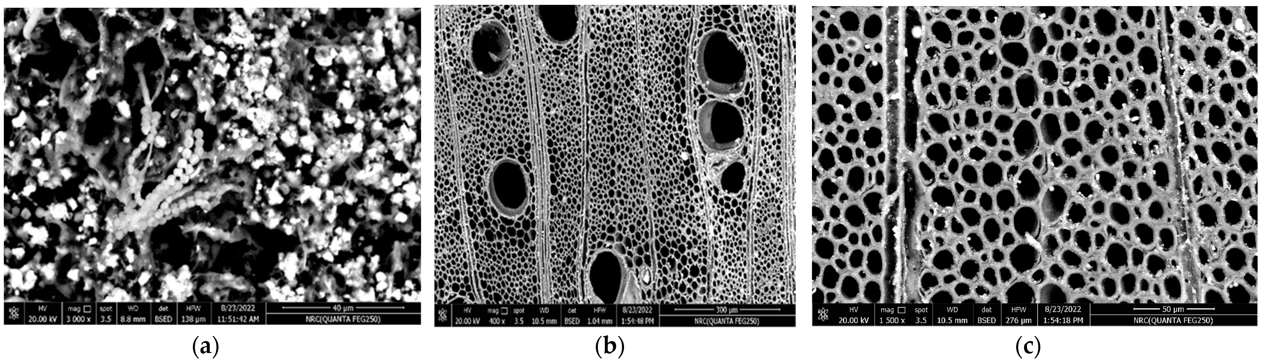
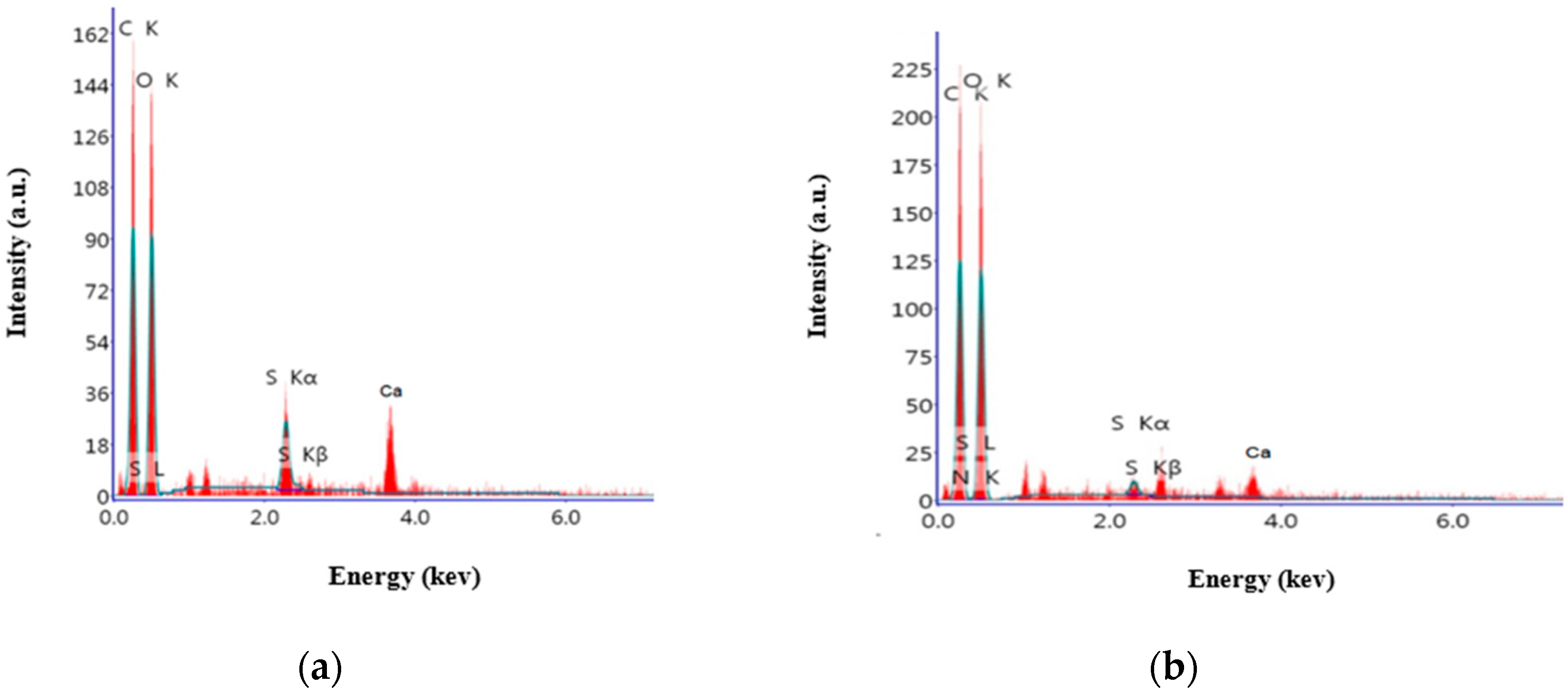
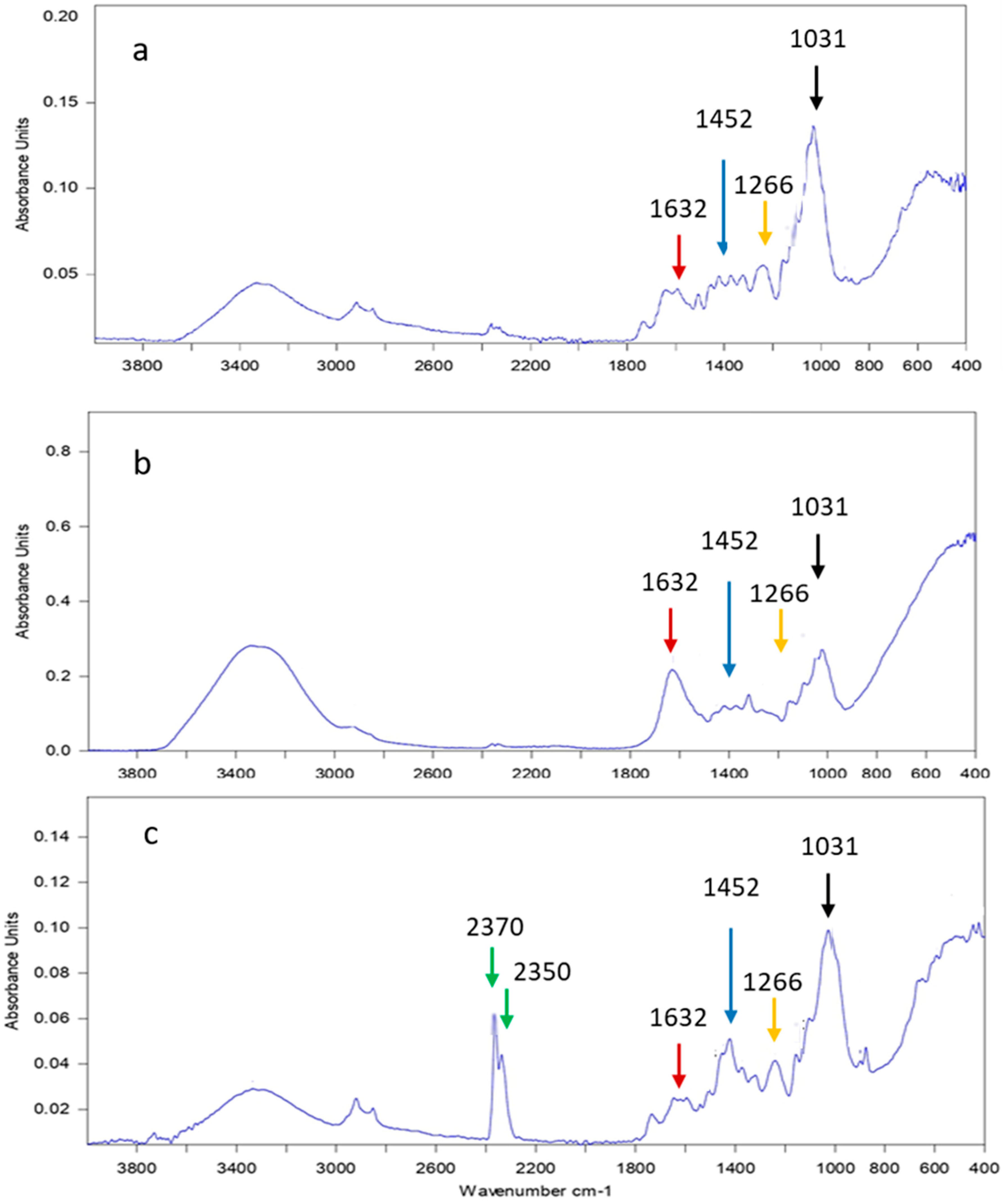
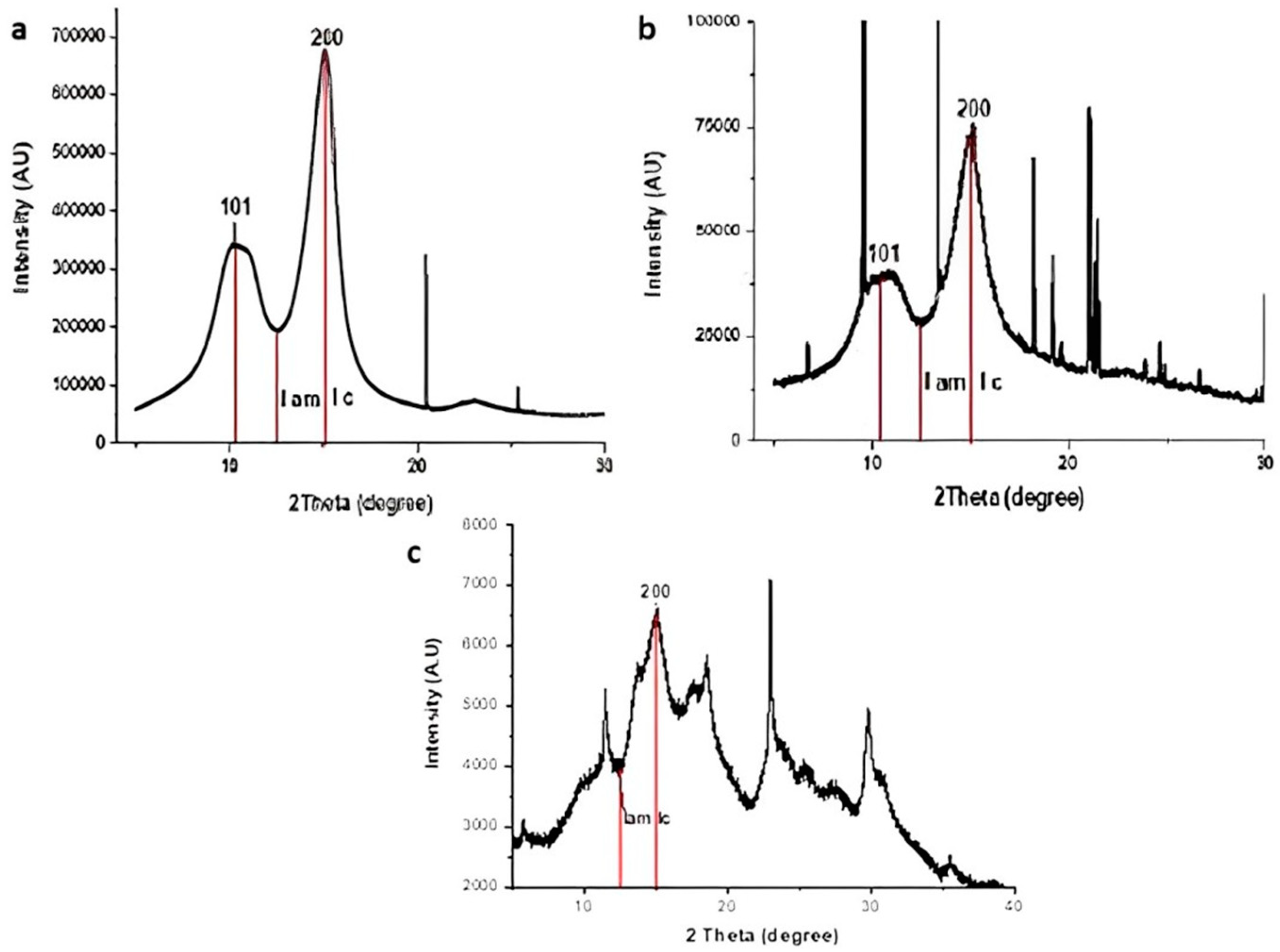
3.7. Analyses of A. nilotica Experimental Wooden Cubes Treated with Thyme Oil as the Most Effective EO

The experimental aged wooden cubes treated with thyme essential oil as the most efficient antimicrobial were analyzed using a scanning electron microscope, energy dispersive X-ray analysis (SEM and EDX), and Fourier-transform infrared spectroscopy (FTIR).
Scanning electron microscope analysis was performed on the experimental wooden cubes of A. nilotica to examine the changes in the morphology of the wooden cells before and after treatment with the most efficient essential oil (thyme oil). The untreated control samples showed fungal hyphae penetrated the cell lumens of the wood via the pits and began to change the structure of the wood by spreading through the lumens, also dense conidial heads and hyphae were mostly located in the untreated wooden cells, where the walls were almost completely degraded (Figure 4a). On the other hand, there were no remarkable morphological variations in the structure of the thyme oil-treated wooden cells. It was also interesting to observe that SEM analyses of the experimental wooden species impregnated with thyme oil were much more resistant to fungal attack than the untreated samples (Figure 4b).
The elemental composition of A. nilotica wood was analyzed by EDX, where the two main elements that appeared before and after treatment by thyme oil were carbon and oxygen. They constitute the main components of the native wood that indicates no significant changes in the major chemical composition of the wood before and after treatment with thyme essential oil (Figure 5a,b).
The FTIR spectra of the experimental aged Acacia nilotica wood samples (standard, untreated, or treated with thyme oil) were investigated (Figure 6).
The peaks at 1632, 1452, 1266, and 1031 cm−1 represent the carbohydrate components. These peaks were different in the wood samples exposed to fungal deterioration if compared to the standard sample and the sample treated with thyme oil. In particular, the absorption peak at 1632 cm−1 (attributed to lignin components) indicates most of the degradation of the cell wall components occurred with the carbohydrates (hemicelluloses and cellulose). The 1452 cm−1 band is attributed to C-H deformation in lignin and carbohydrates, while the C-O stretch in lignin appeared at 1266 cm−1. The band at 1031 cm−1 is attributed to C-O vibration in cellulose and hemicellulose. Moreover, the characteristic peak of thyme oil appeared in the oil-treated wood at the 2200–2400 cm−1 absorption band. The lengths of the peaks at 1632, 1452, 1266, and 1031 cm−1 representing carbohydrate components of the untreated thyme oil wood samples exposed to fungal deterioration were different (Figure 6b) if compared to the standard sample and the sample with treated thyme oil (Figure 6a,c).
The synchrotron radiation XRD results on the crystallinity (as the mass fraction of crystalline cellulose) of the standard, thyme oil-untreated, and thyme oil-treated wood samples are reported in Figure 7. A decrease in the crystalline index was observed in the untreated sample. The crystallinity index (Cr. I%) of cellulose was at 70.5% in the standard wood, 57.1% in the untreated experimental wood, and 62.5% in the wood treated with thyme oil.

4. Discussion

Treating and preserving archaeological wood against microbial deterioration with natural and low-cost compounds is a challenge to prevent damage from microbial attack and, at the same time, there is an urgent need to find products that are safe for the health of conservators and museum staff [9].
The appearance of isolated fungal species may be attributed to their cellulase activities on the tested wooden objects, as suggested by Fazio et al. [48]. The results obtained in this study are in accordance with Ahmad et al. [49] who isolated Aspergillus flavus, A. fumigatus, A. niger, A. terreus, and Penicillium chrysogenum from a Nabataean wooden coffin in Jordon. Most of these fungi produce cellulases, which degrade the cellulose content of the wood; in addition, fungal decay causes a significant reduction in the strength of the wood and changes its morphological aspect. The most frequently and deterioration-causing fungi found in the current study (Aspergillus flavus, A. fumigatus, A. niger, and A. terreus) are similar to those found by other authors. Moreover, Abdallah et al. [50] isolated A. flavus, A. versicolor, and Cladosporium sp. from a wooden offering table dating back to the Middle Kingdom period in Dahshur necropolis located in the desert on the west bank of the Nile, Giza, Egypt. In addition, Aspergillus flavus and A. niger were isolated from ancient funeral masks in Saqqara, Egypt, which revealed various aspects of deterioration (discoloration, cracks, and stains) [51]. Cladosporium, Phoma, and Ulocladium have a black pigment (melanin) that causes distinct dark grey discoloration on wood joints [27].
In the present work, Aspergillus was the most dominant genera isolated from the ten tested archaeological objects, with 322 colonies (8 species) that represent 54% of the total fungal colonies, followed by Penicilli with 98 colonies (5 species) representing 16.4% of the total fungal colonies. The obtained data are in agreement with Osman et al. [52], who found that the most dominant genera isolated from the storage area of Cheops’s solar boat and wooden frames of the Stucco window in the Islamic Art Museum were Aspergillus spp.b and Penicillium spp.
In particular, species of the genus Aspergillus (an Ascomycota in the brown rot fungi group) were as also found by Ortiz et al. [52] who isolated and identified different species of white, brown, and soft rot fungi in the structural woods of eight historical churches in Chile, causing decay in these historical buildings. Aspergillus flavus (14.6%) was the most frequently isolated fungal species recovered from all the archaeological wooden objects studied here, accounting for 87 colonies. This is in line with the fact that it can produce a wide range of hydrolytic enzymes, facilitating the penetration and breaking down of different organic substrates [53]. A. flavus can also develop on protein substrates (mucin and elastin) on complex carbohydrate substrates such as wood components (cellulose, pectin, chitin, and xylan) [54].
The second dominant fungal species was Aspergillus niger (13.8%) with 82 colonies. The high biodeterioration power of A. niger in terms of the cultural heritage is attributed to its great enzymatic activities as found by Romero et al. [55] and Cappitelli et al. [56]; in fact, it can be adapted to different environmental conditions by different metabolic mechanisms [57]. The degradation action of A. niger is performed by the production of pectinases, hemicellulases, and xylanases [58].
A mixture of Eucalyptus camaldulensis and Eucalyptus tereticornis methanolic leaf extracts was found to be effective against wood decay fungi [36]. The antifungal activities of Eucalyptus species (e.g., E. urophylla, E. grandis, E. camaldulensis, and E. citriodora) are due to citronellal and citronellol [59]. Moreover, El Bergadi et al. [60] recorded the antifungal activity of Lawsonia inermis extracts against four cellulolytic wood fungi.
Thyme, geranium, and cinnamon were the most effective essential oils against the fourteen highly deterioration-causing fungal species. Radaelli et al. [61] also revealed that the antimicrobial activity of EOs was a result of the synergistic action of all chemical constituents, not just one constituent. The fungal inhibition by EOs may indicate that the effective ingredients of oil-initiated sugar polymerization may lead to microbial death. Both the affinity for sugar to polymerize into polysaccharides and the decreased degrees of fungal respiration might be causal reasons for the reduction in microbial progress [62]. The disruption of the microbial cell membrane and the precipitation of cellular proteins are connected to the antimicrobial activity of the phenolic compounds in thyme oil [63]. Antonelli et al. [13] found that thymol, which is a phenolic monoterpenoid structure in thyme oil, caused a significant decrease in the vitality of fungal mycelia grown on wood treated with Eos, and could be considered as a possible alternative to the synthetic biocides used currently. The antifungal activity of thyme oil as an alternative preservative for an archaeological Egyptian Coptic cellulosic object was also found by Noshyutta et al. [64].
The main components of geranium oil were citronellol (19.4%) and geraniol (36.8%). They could damage the membrane integrity, inhibited germ tube induction, and the microbial cell cycle [65]. Various low molecular weight proteins or peptides with fungicidal activity have been found in different plants and are involved as germicides [66]. The basis of the essential oils’ antimicrobic efficiency was the particularly lipophilic manner and low molecular weight of terpenes/terpenoids, which block fungal propagation and sporulation causing cell mortality [8].
Cinnamon oil (Cinnamomum cassia, MIC: 0.5–1 µL/mL) was third in the antifungal effectiveness ranking. Its major chemical constituents were eugenol (28.5%), caryophyllene (16.3%), cinnamaldehyde (14.2%), and the acetic acid cinnamyl ester (12.8%). Tahlan [67] concluded that the antimicrobial activity of cinnamon oil may be related to its main antimicrobial components (cinnamaldehyde, eugenol, and linalool), which inhibited amino acid decarboxylase, and which interfere with the production of amylases and proteases, causing the lysis of microbial cells and the deterioration of their cell walls. Wang et al. [68] found that the main active ingredients in Cinnamomum cassia were trans-cinnamaldehyde, citral, trans-geraniol and carvacrol. Cinnamaldehyde, the main component of cinnamon oil, suppressed the growth of the brown rot fungus that was isolated from wood and stone artefacts [69]. Eugenol and cinnamaldehyde were found to be potential wood preservatives for the treatment of timber [70] with a greater effect on the growth of both white and brown rot fungi (wood decay fungi) compared with the other components of cinnamon oil [71].
The main components of lemongrass oil with antimicrobial effects such as fungal sporulation prevention are cineole, citral, geraniol, linalool, and menthol [72]. Ishkeh et al. [73] found that D-limonene (25.3%), spathulenol (14.7%), and α-curcumene (8%) were the main components of Lemon verbena oil, which had antimicrobial activities. Lemongrass oil has been shown to be effective in preventing the colonization of fungi (Aspergillus niger, Chaetomium globosum, Coniophora puteana, Penicillium commune, and Trametes versicolor) on samples of hardwood and softwood by Bahmani and Schmidt [34].
Menthe oil showed an MIC against fourteen deterioration-causing fungal species in the 0.5–4 µL/mL range. Similar effects were recently found by Felšöciová et al. [74] with Mentha piperita extracts against several fungi (Penicillium brevicompactum, P. citrinum, P. crustosum, P. expansum, P. funiculosum, P. glabrum, P. chrysogenum, P. oxalicum, P. polonicum, and Talaromyces purpurogenus).
Tea tree oil (M. alternifolia) caused the complete inhibition of A. terreus at 0.75 μL/mL. Pánek et al. [75], in accordance with our results, indicated that tea tree oil had a lower efficacy against decay fungi than thyme oil, which contains phenolic antifungal compounds.
Eucalyptus oil (E. globulus; MIC: 1–8 μL/mL) was found with a low efficiency if compared with the previous one. Similar results were found by other authors [76,77,78,79,80], who found that it was effective against Ulocladium spp., while Penicillium spp. was resistant to the oil.
Rosemary oil (R. officinalis, MIC: 1–8 μL/mL) and lavender oil (L. latifolia, MIC: 2–8 μL/mL) had the lowest antifungal effects. Felšöciová et al. [74] studied the effect of Lavandula angustifolia volatile essential oils against Penicillium sp., giving an inhibition zone 15.50 ± 1.38 mm at a 0.75 μL/mL concentration. On the other hand, no activity was observed againast Rosmarinus officinalis at 0.125 μL/mL.
In the current study, the isolated Aspergillus spp. were the most resistant species against all the ten tested EOs. This fact can be due to the melanin pigment found in the thick cell walls of Aspergilli that plays a protective role by allowing them to survive in unfavorable environmental conditions [81]. Additionally, many enzymes produced by Aspergillus niger can also detoxify components of EOs into inactive forms, as previously reported [82,83].
In the present study, thyme oil completely inhibited the amylase activity of A. fumigatus and A. terreus at 0.5 µL/mL, while the amylolytic activities of the A. flavus and A. niger were fully suppressed at 0.75 µL/mL. Geweely and Nawar [84] stated that the delay of enzyme production by essential oils may be caused by the efficient components of thyme. Essential oils may have some components that cause the amylase inhibitory effect, according to Yang et al. [85], who found some EOs with strong activity against a-amylase and tyrosinase.
The three tested essential oils (thyme, geranium, and cinnamon), still at the lowest concentrations, can reduce the enzymatic activities tested in this study (amylase, cellulase, and protease) for the four selected fungal isolates (Aspergillus flavus, A. fumigatus, A. niger, and A. terreus). The obtained results are in accordance with Hamedo and El Shamy [44], who showed that EOs affected the enzyme production of two pathogenic fungi (Fusarium solani and Trichoderma virens) with weak enzyme production during the first growth stage, and subsequently the level of enzyme production decreased along with the inhibition of fungal growth. Ekinci et al. [86] used thyme oil at a concentration of 0.01 µL/mL against Neocallimastix spp. to reduce the activity of some enzymes. The results here reported are in agreement with Grande-Tovar et al. [87], who revealed that the activities of extracellular hydrolytic enzymes (amylase, protease, pectinase, and endocellulase) were stimulated by low concentrations of EOs.
The antifungal activities of the tested essential oils in the current study agree with Wilson et al., Chao et al., and Voda et al. [88,89,90].
The experimental aged wooden cubes treated with the most efficient thyme essential oil were analyzed using a scanning electron microscope, energy dispersive X-ray analysis (SEM & EDX), and Fourier-transform infrared spectroscopy (FTIR). The observed drop in the crystallinity index in the untreated wood may be due to fungal deterioration, as recorded by Geweely et al. [31] who stated that the XRD analysis of the archaeological materials revealed a drop in their crystalline index, which was caused by microbial degradation. Moreover, Sparacello et al. [91] evaluated the potential changes to the wooden artwork surfaces pre- and post-treatment with Thymus vulgaris and a hydro-alcoholic solution using SEM, EDX, and FTIR. SEM images and EDX analyses showed no increase in fractures or changes in the elemental composition of the wood surface after treatment. This indicated that the Thymus vulgaris EO was successful when applied as an antifungal agent to control microbial colonization [92,93]. Also, El-Shahawy et al. [94] stated that the natural substance does not produce any environmental hazards.
Yan et al. [95] described the effect of cinnamon essential oil on cell integrity using SEM in which remarkable morphological variations were observed in the control sample infected with Aspergillus niger cells compared with the sample treated with cinnamon oil. Many distinct types of biological degradation, including brown and soft rot, as well as the chemical corrosion of wood by salts that have impacted the various types of wood, were frequently discovered during SEM analyses of wooden artefacts from ancient Egypt kept in museums [96]. Salem et al. [37] stated that FTIR spectra of extractives differ according to the wood species and the type of plant extract. Many groups of extractives have functional groups such as terpenoids, phenolic compounds, hydroxyls, carbonyls, and carboxylic groups [97,98,99,100].
Ali et al. [101] used SEM examination to show the suppression of fungal growth on wood samples after treatment with the Mentha longifolia L. and Citrus reticulata L. essential oils and compared them with untreated samples. Martinez-Pacheco et al. [102] tested the antifungal activities of citrus essential oils on the fungal growth of Alternaria sp. and Trichoderma sp. using SEM. Major alterations in the chemical configuration of the wood due to microbial corrosion and a reduction in the crystallinity index of cellulose were also found by Geweely et al. [31].

5. Conclusions

Archaeological wooden artefacts constitute a crucial part of global cultural heritage. The impact of fungal activity on the degradation of artefacts poses a significant global problem. The preservation of these artefacts over time represents a challenging task. The essential oils were more effective than the plant extracts against the isolated fungi. Thyme oil exhibited the complete inhibition of the growth of all tested deterioration-causing fungal species at 0.5 μL/mL without any harmful effect on the structure of the archaeological objects and was esthetically suitable for the preservation of artefacts. The application of thyme oil on the most deteriorated experimental aged Acacia nilotica wooden cubes inoculated with the four most common fungal species was successful in terms of wood preservation. Analyses (SEM, XRD, and FTIR) of the experimental wood A. nilotica, preserved with thyme oil, highlighted that no significant variations were detected in the characterization of wood before and after treatment. This work shows that thyme oil is highly recommended in the conservation of wooden artefacts, being a promising green and sustainable professional antifungal agent. Moreover, being safe for individuals and the ecosystem and aesthetically acceptable, it is an effective agent for the decontamination and preservation of archaeological wood artefacts that avoids the harmful effects of the frequent use of synthetic fungicides used to protect artefacts.

Supplementary Materials

The following supporting information can be downloaded at: https://www.mdpi.com/article/10.3390/app14125023/s1, Figure S1. The three most deteriorated wooden objects. (a) Wooden statue (L 127 cm, W 47 cm); (b) coffin with cartonnage mummy of Nespahettawi, son of Neskhonsupahred (L 179 cm, W 55 cm); (c) and (d) front and backside of Padimen’s son’s box (L 43.5 cm, W 39 cm). Figure S2. Phylogenetic tree Aspergillus flavus. Figure S3. Phylogenetic tree Aspergillus niger. Figure S4. Phylogenetic tree Aspergillus fumigatus. Figure S5. Phylogenetic tree Aspergillus terreus. Table S1. Total counts, relative density (%), and frequency of occurrence of fungal species isolated from the ten tested deteriorated archaeological pharaonic objects in the Egyptian Museum, El-Tahrir, Egypt. Table S2. Fungal deterioration of the three experimental aged wooden blocks. Symbols: (−) no growth, (+) weak growth, (++) moderate growth, (+++) strong growth. Table S3. Effect of different concentrations (from 0.125 to 8 µL/mL) of teak extract on the highly deteriorating fungal species isolated from ten archaeological pharaonic wooden objects. The minimum inhibitory concentrations, MICs (µL/mL), resulted from the effect on the diameter of mycelia growth and percentage of mycelia growth inhibition, compared to the control (treated with Itraconazole). The numbers are expressed as mean ± standard deviation (n = 3) for each sample. D: diameter (mm) of mycelia growth; P: percentage (%) of mycelia growth inhibition; LSD: least significant difference (p < 0.05). Table S4. Effect of different concentrations (from 0.125 to 8 µL/mL) of eucalyptus extract on the highly deteriorating fungal species isolated from ten archaeological pharaonic wooden objects. The minimum inhibitory concentrations, MICs (µL/mL), resulted from the effect on the diameter of mycelia growth and the percentage of mycelia growth inhibition, compared to the control (treated with Itraconazole). The numbers are expressed as mean ± standard deviation (n = 3) for each sample. D: diameter (mm) of mycelia growth; P: percentage (%) of mycelia growth inhibition; LSD: least significant difference (p < 0.05). Table S5. Effect of different concentrations (from 0.125 to 8 µL/mL) of Melia extract on the highly deteriorating fungal species isolated from ten archaeological pharaonic wooden objects. The minimum inhibitory concentrations, MICs (µL/mL), resulted from the effect on the diameter of mycelia growth and the percentage of mycelia growth inhibition, compared to the control (treated with Itraconazole). The numbers are expressed as mean ± standard deviation (n = 3) for each sample. D: diameter (mm) of mycelia growth; P: percentage (%) of mycelia growth inhibition; LSD: least significant difference (p < 0.05). Table S6. Effect of different concentrations (from 0.125 to 8 µL/mL) of henna (Lawsonia inermis) extract on the highly deterioration-causing fungal species isolated from ten archaeological pharaonic wooden objects. The minimum inhibitory concentrations, MICs (µL/mL), resulted from the effect on the diameter of mycelia growth and the percentage of mycelia growth inhibition, compared to the control (treated with Itraconazole). The numbers are expressed as mean ± standard deviation (n = 3) for each sample. D: diameter (mm) of mycelia growth; P: percentage (%) of mycelia growth inhibition; LSD: least significant difference (p < 0.05). Table S7. Effect of different concentrations (from 0.125 to 8 µL/mL) of basil extract on the highly deterioration-causing fungal species isolated from ten archaeological pharaonic wooden objects. The minimum inhibitory concentrations, MICs (µL/mL), resulted from the effect on the diameter of mycelia growth and the percentage of mycelia growth inhibition, compared to the control (treated with Itraconazole). The numbers are expressed as mean ± standard deviation (n = 3) for each sample. D: diameter (mm) of mycelia growth; P: percentage (%) of mycelia growth inhibition; LSD: least significant difference (p < 0.05). Table S8. Main chemical constituents (%) of the efficient essential oils (thyme, geranium, cinnamon). The symbol (*) indicates the highest percentages for each oil. Table S9. Effect of different concentrations (0.125, 0.25, 0.5, 0.75, and 1 µL/mL) of the most efficient essential oils (thyme, geranium, and cinnamon) on the cellulase activities (expressed as U/mL) of the four most deterioration-causing Aspergillus species (A. flavus, A. fumigatus, A. niger, and A. terreus). The numbers are mean ± standard deviation (n = 3) for each sample. LSD: least significant difference (p < 0.05). Table S10. Effect of different concentrations (0.125, 0.25, 0.5, 0.75, and 1 µL/mL) of the most efficient essential oils (thyme, geranium, and cinnamon) on the amylase activity (U/mL) of the four most deterioration-causing fungal species (Aspergillus flavus, A. fumigatus, A. niger, A. terreus). The numbers are expressed as mean ± standard deviation (n = 3) for each sample. LSD: least significant difference (p < 0.05). Table S11. Effect of different concentrations (0.125, 0.25, 0.5, 0.75, and 1 µL/mL) of the most efficient essential oils (thyme, geranium, and cinnamon) on the protease activity (U/mL) of the four most deterioration-causing Aspergillus species (A. flavus, A. fumigatus, A. niger, and A. terreus). The numbers are expressed as mean ± standard deviation (n = 3) for each sample. LSD: least significant difference (p < 0.05). Table S12. Antifungal activities of the most efficient essential oils (thyme, geranium, and cinnamon) on Acacia nilotica experimental wood cubes inoculated with the four dominant deterioration-causing Aspergillus species (A. niger, A. flavus, A. fumigatus, and A. terreus); 1 = area of colony > 90% of the area of the controls (treated with Itraconazole), 2 = visible signs of retardation (colony < 90% and >60% of control), 3 = pronounced retardation (colony < 60% and >25% of control), 4 = very marked retardation (colony < 25% of control), 5 = no growth.

Author Contributions

Conceptualization and methodology: N.S.G. and A.M.A.T.; formal analysis and investigation S.I., D.M.A. and J.R.P.; data curation, S.I.; writing—original draft preparation, N.S.G. and S.I.; writing—review and editing, P.G. and G.C.; supervision, N.S.G. All authors have read and agreed to the published version of the manuscript.

Funding

This work does not received a financial support.

Institutional Review Board Statement

Not Applicable.

Informed Consent Statement

Not applicable.

Data Availability Statement

The raw data supporting the conclusions of this article will be made available by the authors on re-quest.

Conflicts of Interest

The authors declare no conflicts of interest.

References

  1. Blondel, F.; Huebner, S.R.; Pearson, C.; Stoffel, M. Mummy labels: A witness to the use and processing of wood in Roman Egypt. Int. J. Wood Cult. 2023, 3, 192–223. [Google Scholar] [CrossRef]
  2. Savković, Ž.; Stupar, M.; Unković, N.; Knežević, A.; Vukojević, J.; Ljaljević Grbić, M. Fungal deterioration of cultural heritage objects. In Biodegradation Technology of Organic and Inorganic Pollutants; Mendes, K.F., de Sousa, R.N., Mielke, K.C., Eds.; IntechOpen: London, UK, 2021; pp. 267–288. [Google Scholar] [CrossRef]
  3. Flyen, A.C.; Thuestad, A.E. A review of fungal decay in historic wooden structures in polar regions. Conserv. Manag. Archaeol. Sites 2023, 24, 3–35. [Google Scholar] [CrossRef]
  4. Li, T.; Cui, L.; Song, X.; Cui, X.; Wei, Y.; Tang, L.; Mu, Y.; Xu, Z. Wood decay fungi: An analysis of worldwide research. J. Soils Sediments 2022, 22, 1688–1702. [Google Scholar] [CrossRef]
  5. Blanchette, R.A. A review of microbial deterioration found in archaeological wood from different environments. Int. Biodeterior. Biodegrad. 2000, 46, 189–204. [Google Scholar] [CrossRef]
  6. Gadd, G.; Fomina, M.; Pinzari, F. Fungal biodeterioration and preservation of cultural heritage, artwork, and historical artifacts: Extremophily and adaptation. Microbiol. Mol. Biol. Rev. 2024, 88, e00200–e00222. [Google Scholar] [CrossRef] [PubMed]
  7. Atwa, D.M.; Ibrahim, S.; Stani, C.; Birarda, G.; Ali, N.; Abdullah, E.; Vaccari, L.; Grenni, P.; Visca, A.; Badr, Y.; et al. Biodeterioration assessment of a unique old pharaonic kingdom wooden statue using advanced diagnostic techniques. Appl. Sci. 2022, 12, 7020. [Google Scholar] [CrossRef]
  8. Geweely, N.S. A novel comparative review between chemical, natural essential oils and physical (ozone) conservation of archaeological objects against microbial deterioration. Geomicrobiol. J. 2022, 39, 531–540. [Google Scholar] [CrossRef]
  9. Fidanza, M.R.; Caneva, G. Natural biocides for the conservation of stone cultural heritage: A review. J. Cult. Herit. 2019, 38, 271–286. [Google Scholar] [CrossRef]
  10. Vettraino, A.M.; Zikeli, F.; Humar, M.; Biscontri, M.; Bergamasco, S.; Romagnoli, M. Essential oils from Thymus spp. as natural biocide against common brown-and white-rot fungi in degradation of wood products: Antifungal activity evaluation by in vitro and FTIR analysis. Eur. J. Wood Wood Prod. 2023, 81, 747–763. [Google Scholar] [CrossRef]
  11. Geweely, N.S. Purification and characterization of acido-thermophilic xylanase from Aspergillus terreus. Aust. J. Basic Appl. Sci. 2011, 5, 214–219. [Google Scholar]
  12. Broda, M. Natural compounds for wood protection against fungi—A review. Molecules 2020, 25, 3538. [Google Scholar] [CrossRef] [PubMed]
  13. Zhang, J.; Zhang, M.; Bhandari, B.; Wang, M. Basic sensory properties of essential oils from aromatic plants and their applications: A critical review. Crit. Rev. Food Sci. Nutr. 2023, 19, 1–14. [Google Scholar] [CrossRef] [PubMed]
  14. Antonelli, F.; Bartolini, M.; Plissonnier, M.L.; Esposito, A.; Galotta, G.; Ricci, S.; Davidde Petriaggi, B.; Pedone, C.; Di Giovanni, A.; Piazza, S.; et al. Essential oils as alternative biocides for the preservation of waterlogged archaeological wood. Microorganisms 2020, 8, 2015. [Google Scholar] [CrossRef] [PubMed]
  15. Lillehoj, H.; Liu, Y.; Calsamiglia, S.; Fernandez-Miyakawa, M.E.; Chi, F.; Cravens, R.L.; Oh, S.; Gay, C.G. Phytochemicals as antibiotic alternatives to promote growth and enhance host health. Vet. Res. 2018, 49, 76. [Google Scholar] [CrossRef] [PubMed]
  16. Oladipo, A.; Enwemiwe, V.; Ejeromedoghene, O.; Adebayo, A.; Ogunyemi, O.; Fu, F. Production and functionalities of specialized metabolites from different organic sources. Metabolites 2022, 12, 534. [Google Scholar] [CrossRef] [PubMed]
  17. Elshafie, H.S.; Camele, I.; Mohamed, A.A. A Comprehensive Review on the Biological, Agricultural and Pharmaceutical Properties of Secondary Metabolites Based-Plant Origin. Int. J. Mol. Sci. 2023, 24, 3266. [Google Scholar] [CrossRef] [PubMed]
  18. Skóra, J.; Gutarowska, B.; Pielech-Przybylska, K.; Stępień, Ł.; Pietrzak, K.; Piotrowska, M.; Pietrowski, P. Assessment of microbiological contamination in the work environments of museums, archives and libraries. Aerobiologia 2015, 31, 389–401. [Google Scholar] [CrossRef] [PubMed]
  19. Wozniak, M. Antifungal agents in wood protection—A review. Molecules 2020, 27, 6392. [Google Scholar] [CrossRef] [PubMed]
  20. Booth, C. The Genus Fusarium; Commonwealth Mycological Institute: Kew, UK, 1971; 237p. [Google Scholar]
  21. Gilman, J.C. A Manual of Soil Fungi, Indian Edition Published by Arrangement with the Original American Publishers; The Iowa State University Press: Ames, IA, USA, 1974; pp. 217–251. [Google Scholar]
  22. Raper, K.B.; Funnel, D.I. The Genus Aspergillus; Robert E. Ktieger Publ. Co.:: New York, NY, USA, 1977; 686p. [Google Scholar]
  23. Pitt, J.I. The Genus Pencillium and Its Teleomorph States Eupencillim and Talaromyces; Academic Press: New York, NY, USA, 1979; pp. 50–55. [Google Scholar]
  24. Samson, R.A.; Reenen-Koekstra, E.S. Introduction to Food–Borne Fungi, 3rd ed.; Centraalbureau voor Schimmelcultures: Utrecht, The Netherlands, 1988; 299p. [Google Scholar]
  25. Moubasher, A.H. Soil fungi in Qatar and other Arab countries. Econ. Bot. 1996, 50, 242. [Google Scholar] [CrossRef]
  26. Kern, M.E.; Blevins, K.S. Laboratory procedures for fungal culture and isolation. In Medical Mycology: A Self-Instructional Text, 2nd ed.; FA Davis Company: Philadelphia, PA, USA, 1997; pp. 27–72. [Google Scholar]
  27. Klich, M.A. Identification of Common Aspergillus Species; Centraal Bureau Voor Schim: Utrecht, The Netherlands, 2002; 316p. [Google Scholar]
  28. Domsch, K.H.; Gams, W.; Anderson, T.H. Compendium of Soil Fungi, 2nd ed.; Academic Press: London, UK, 2007; Volume 1, 672p. [Google Scholar]
  29. Siddiquee, S. Practical Handbook of the Biology and Molecular Diversity of Trichoderma Species from Tropical Regions; Springer International Publishing: Cham, Swizerland, 2017; Volume 431. [Google Scholar]
  30. Kwiatkowska, M.; Ważny, R.; Turnau, K.; Wójcik, A. Fungi as deterioration agents of historic glass plate negatives of Brandys family collection. Int. Biodeterior. Biodegrad. 2016, 115, 133–140. [Google Scholar] [CrossRef]
  31. Geweely, N.S.; Abu Taleb, A.; Ibrahim, S.; Grenni, P.; Caneva, G.; Galotta, G.; Abdalla, M.; Atwa, D.M.; Plaisier, J.R.; Antonelli, F. New data on relevant ancient Egyptian wooden artifacts: Identification of wooden species and study of the state of conservation with multidisciplinary analyses. Archaeometry 2023, 65, 165–183. [Google Scholar] [CrossRef]
  32. Sambrook, J.F.; Russell, D.W. Molecular Cloning: A Laboratory Manual; Cold Spring Harbor Laboratory Press: New York, NY, USA, 2001. [Google Scholar]
  33. Veneranda, M.; Blanco-Zubiaguirre, L.; Roselli, G.; Di Girolami, G.; Castro, K.; Madariaga, J.M. Evaluating the exploitability of several essential oils constituents as a novel biological treatment against cultural heritage biocolonization. Microchem. J. 2018, 138, 1–6. [Google Scholar] [CrossRef]
  34. Bahmani, M.; Schmidt, O. Plant essential oils for environment-friendly protection of wood objects against fungi. Maderas. Ciencia Tecnología 2018, 20, 325–332. [Google Scholar] [CrossRef]
  35. Mohareb, A.S.; Badawy, M.E.; Abdelgaleil, S.A. Antifungal activity of essential oils isolated from Egyptian plants against wood decay fungi. J. Wood Sci. 2013, 59, 499–505. [Google Scholar] [CrossRef]
  36. Anda, R.R.; Koch, G.; Richter, H.-G.; Talavera, F.J.F.; Guzmán, J.A.S.; Satyanarayana, K.G. Formation of heartwood, chemical composition of extractives and natural durability of plantation-grown teak wood from Mexico. Holzforschung 2019, 73, 547–557. [Google Scholar] [CrossRef]
  37. Varshney, V.K.; Pandey, A.; Onial, P.K.; Dayal, R. Antifungal activity of phytochemicals from Eucalyptus hybrid leaves against some plant pathogenic and wood decay fungi. Arch. Phytopathol. Plant Prot. 2012, 45, 2347–2354. [Google Scholar] [CrossRef]
  38. Salem, M.Z.; Zidan, Y.E.; El Hadidi, N.M.; Mansour, M.M.; Elgat, W.A.A. Evaluation of usage three natural extracts applied to three commercial wood species against five common molds. Int. Biodeterior. Biodegrad. 2016, 110, 206–226. [Google Scholar] [CrossRef]
  39. Pellegrini, M.; Ricci, A.; Serio, A.; Chaves-López, C.; Mazzarrino, G.; D’Amato, S.; Lo Sterzo, C.; Paparella, A. Characterization of essential oils obtained from Abruzzo autochthonous plants: Antioxidant and antimicrobial activities assessment for food application. Foods 2018, 7, 19. [Google Scholar] [CrossRef] [PubMed]
  40. Mansour, M.M.; Salem, M.Z. Evaluation of wood treated with some natural extracts and Paraloid B-72 against the fungus Trichoderma harzianum: Wood elemental composition, in-vitro and application evidence. Int. Biodeterior. Biodegrad. 2015, 100, 62–69. [Google Scholar] [CrossRef]
  41. Reidel, R.V.B.; Cioni, P.L.; Pistelli, L. Volatiles from different plant parts of Punica granatum grown in Tuscany (Italy). Sci. Hortic. 2018, 231, 49–55. [Google Scholar] [CrossRef]
  42. Ominyi, M.C.; Ogbonna, J.C.; Nwoba, E.G.; Nwagu, K.E.; Ukachi, R. Isolation and screening of α-amylase and glucoamylase producing fungi and their application in bioethanol production. Int. J. Sci. Nat. 2013, 4, 44–50. [Google Scholar]
  43. Ram, L.; Kaur, K.; Sharma, S. Screening isolation and characterization of cellulase producing microorganisms from soil. Int. J. Pharm. Sci. Invent. 2014, 3, 12–18. [Google Scholar]
  44. Marathe, S.K.; Vashistht, M.A.; Prashanth, A.; Parveen, N.; Chakraborty, S.; Nair, S.S. Isolation, partial purification, biochemical characterization and detergent compatibility of alkaline protease produced by Bacillus subtilis, Alcaligenes faecalis and Pseudomonas aeruginosa obtained from sea water samples. J. Genet. Eng. Biotechnol. 2018, 16, 39–46. [Google Scholar] [CrossRef] [PubMed]
  45. Humar, H.; Pohleven, F. Influence of a nitrogen supplement on the growth of wood decay fungi and decay of wood. Int. Biodeterior. Biodegradation 2005, 56, 34–39. [Google Scholar] [CrossRef]
  46. Balzano, V.; Cavaliere, E.; Fanetti, M.; Gardonio, S.; Gavioli, L. The role of substrate on thermal evolution of Ag/TiO2 nanogranular thin films. Nanomaterials 2021, 11, 2253. [Google Scholar] [CrossRef] [PubMed]
  47. Colao, F.; Fantoni, R.; Fiorani, L.; Palucci, A.; Gomoiu, I. Compact scanning lidar fluorosensor for investigations of biodegradation on ancient painted surfaces. J. Optoelectron. Adv. Mater. 2005, 7, 3197–3208. [Google Scholar]
  48. Fazio, A.T.; Papinutti, L.; Gómez, B.A.; Parera, S.D.; Romero, A.R.; Siracusano, G.; Maier, M.S. Fungal deterioration of a Jesuit South American polychrome wood sculpture. Int. Biodeterior. Biodegrad. 2010, 64, 694–701. [Google Scholar] [CrossRef]
  49. Ahmad, A.; Elserogy, A.; Al-Muheisen, Z.; Villeneuve, F.; El-Oqlah, A. The conservation of a wooden nabataean coffin box from Jordan-application of non-destructive ultrasonic technique. Wood Res. 2018, 63, 1–14. [Google Scholar]
  50. Abdallah, M.; Kamal, H.M.; Abdrabou, A. Investigation, preservation and restoration processes of an ancient Egyptian wooden offering table. Int. J. Conserv. Sci. 2016, 7, 1047–1064. [Google Scholar]
  51. Helmi, F.M.; Ali, N.M.; Ismael, S.M. Nanomaterials for the inhibition of microbial growth on ancient Egyptian funeral masks. Mediterr. Archaeol. Archaeom. 2015, 15, 87–95. [Google Scholar] [CrossRef]
  52. Osman, M.E.; El-Shaphy, A.A.; Meligy, D.A.; Ayid, M.M. Survey for fungal decaying archaeological wood and their enzymatic activity. Int. J. Conserv. Sci. 2014, 5, 295–308. [Google Scholar]
  53. Hedayati, M.T.; Pasqualotto, A.C.; Warn, P.A.; Bowyer, P.; Denning, D.W. Aspergillus flavus: Human pathogen, allergen and mycotoxin producer. Microbiology 2007, 153, 1677–1692. [Google Scholar] [CrossRef] [PubMed]
  54. Mellon, J.E.; Cotty, P.J.; Dowd, M.K. Aspergillus flavus hydrolases: Their roles in pathogenesis and substrate utilization. Appl. Microbiol. Biotechnol. 2007, 77, 497–504. [Google Scholar] [CrossRef] [PubMed]
  55. Romero, S.M.; Giudicessi, S.L.; Vitale, R.G. Is the fungus Aspergillus a threat to cultural heritage? J. Cult. Herit. 2021, 51, 107–124. [Google Scholar] [CrossRef]
  56. Cappitelli, F.; Vicini, S.; Piaggio, P.; Abbruscato, P.; Princi, E.; Casadevall, A.; Nosanchuk, J.D.; Zanardini, E. Investigation of fungal deterioration of synthetic paint binders using vibrational spectroscopic techniques. Macromol. Biosci. 2005, 5, 49–57. [Google Scholar] [CrossRef] [PubMed]
  57. Sumthong, P.; Damveld, R.A.; Choi, Y.H.; Arentshorst, M.; Ram, A.F.; Van Den Hondel, C.A.; Verpoorte, R. Activity of quinones from teak (Tectona grandis) on fungal cell wall stress. Planta Medica 2006, 72, 943–944. [Google Scholar] [CrossRef] [PubMed]
  58. Haupt, M.; Leithoff, H.; Meier, D.; Puls, J.; Richter, H.G.; Faix, O. Heartwood extractives and natural durability of plantation-grown teakwood (Tectona grandis L.). A case study. Holz RohWerkst 2003, 61, 473–474. [Google Scholar] [CrossRef]
  59. Chang Su, Y.; Ho, C.L.; Wang, I.C.; Chang, S.T. Antifungal activities and chemical compositions of essential oils from leaves of four eucalypts. Taiwan J. Sci. 2006, 21, 49–61. [Google Scholar]
  60. El Bergadi, F.; Laachari, F.; Sadiki, M.; Megzari, A.; El-Abed, S.; Iraqui, H.M.; Ibnsouda, K.S. Antifungal effect of Moroccan Lawsonia inermis leaf extracts on the growth of filamentous fungi isolated from historical wood. Int. J. Curr. Res. 2014, 7, 14237–14240. [Google Scholar]
  61. Radaelli, M.; Silva, B.P.D.; Weidlich, L.; Hoehne, L.; Flach, A.; Costa, L.A.M.A.D.; Ethur, E.M. Antimicrobial activities of six essential oils commonly used as condiments in Brazil against Clostridium perfringens. Braz. J. Microbiol. 2016, 47, 424–430. [Google Scholar] [CrossRef]
  62. Amany, A.E.; Geweely, N.S. Comparative studies on the biochemistry of Penicillium albicans (alkalosensitive) and Verticillium lateritium, (facultative alkalophile) with special reference to the role of sodium ion in alkalophily. Eur. J. Soil Biol. 1999, 35, 1–7. [Google Scholar] [CrossRef]
  63. Baghdady, K.Z.; Tolba, S.T.; Houssien, S.S. Biogenic deterioration of Egyptian limestone monuments: Treatment and conservation. J. Cult. Herit. 2019, 38, 118–125. [Google Scholar] [CrossRef]
  64. Noshyutta, W.; Osman, E.; Mansour, M. An investigation of the biological fungicidal activity of some essential used as preservatives for a 19th century Egyptian Coptic cellulosic manuscript. Int. J. Conserv. Sci. 2016, 7, 41–56. [Google Scholar]
  65. Zore, G.B.; Thakre, A.D.; Jadhav, S.; Karuppayil, S.M. Terpenoids inhibit Candida albicans growth by affecting membrane integrity and arrest of cell cycle. Phytomedicine 2011, 18, 1181–1190. [Google Scholar] [CrossRef] [PubMed]
  66. Chiu, T.; Poucet, T.; Li, Y. The potential of plant proteins as antifungal agents for agricultural applications. Synth. Syst. Biotechnol. 2022, 7, 1075–1083. [Google Scholar] [CrossRef] [PubMed]
  67. Tahlan, V. Antimicrobial Activity of Essential Oil Emulsions and Possible Synergistic Effect on Food Borne Pathogens. Master’s Thesis, Wayne State University, Detroit, MI, USA, 2014. Available online: http://digitalcommons.wayne.edu/oa_theses (accessed on 10 February 2023).
  68. Wang, H.; Yang, Z.; Ying, G.; Yang, M.; Nian, Y.; Wei, F.; Kong, W. Antifungal evaluation of plant essential oils and their major components against toxigenic fungi. Ind. Crops Prod. 2018, 120, 180–186. [Google Scholar] [CrossRef]
  69. Axinte, L.; Cuzman, O.A.; Feci, E.; Palanti, S.; Tiano, P. Cinnamaldehyde, a potential active agent for the conservation of wood and stone religious artefacts. Eur. J. Sci. Theol. 2011, 7, 25–34. [Google Scholar]
  70. Chittenden, C.; Singh, T. Antifungal activity of essential oils against wood degrading fungi and their applications as wood preservatives. Int. Wood Prod. J. 2011, 2, 44–48. [Google Scholar] [CrossRef]
  71. Wang, S.Y.; Chen, P.F.; Chang, S.T. Antifungal activities of essential oils and their constituents from indigenous cinnamon (Cinnamomum osmophloeum) leaves against wood decay fungi. Bioresour. Technol. 2005, 96, 813–818. [Google Scholar] [CrossRef]
  72. Li, C.C.; Yu, H.F.; Chang, C.H.; Liu, Y.T.; Yao, H.T. Effects of lemongrass oil and citral on hepatic drug-metabolizing enzymes, oxidative stress, and acetaminophen toxicity in rats. J. Food Drug Anal. 2018, 26, 432–438. [Google Scholar] [CrossRef]
  73. Ishkeh, S.R.; Asghari, M.; Shirzad, H.; Alirezalu, A.; Ghasemi, G. Lemon verbena (Lippia citrodora) essential oil effects on antioxidant capacity and phytochemical content of raspberry (Rubus ulmifolius subsp. sanctus). Sci. Hortic. 2019, 248, 297–304. [Google Scholar] [CrossRef]
  74. Felšöciová, S.; Vukovic, N.; Jeżowski, P.; Kačániová, M. Antifungal activity of selected volatile essential oils against Penicillium sp. Open Life Sci. 2020, 15, 511–521. [Google Scholar] [CrossRef] [PubMed]
  75. Pánek, M.; Reinprecht, L.; Hulla, M. Ten essential oils for beech wood protection—Efficacy against wood-destroying fungi and moulds, and effect on wood discoloration. BioResources 2014, 9, 5588–5603. [Google Scholar] [CrossRef]
  76. Schroder, T.; Gaskin, S.; Ross, K.; Whiley, H. Antifungal activity of essential oils against fungi isolated from air. Int. J. Occup. Environ. Health 2017, 23, 181–186. [Google Scholar] [CrossRef] [PubMed]
  77. Stupar, M.; Grbić, M.L.; Džamić, A.; Unković, N.; Ristić, M.; Jelikić, A.; Vukojević, J. Antifungal activity of selected essential oils and biocide benzalkonium chloride against the fungi isolated from cultural heritage objects. S. Afr. J. Bot. 2014, 93, 118–124. [Google Scholar] [CrossRef]
  78. Farooq, A.; Choudhary, I.; Tahara, S.; Başer, K.H.C.; Demirci, F. Detoxification of terpinolene by plant pathogenic fungus Botrytis cinerea. Z. Für Naturforschung C 2002, 57, 863–866. [Google Scholar] [CrossRef] [PubMed]
  79. Salama, M.A.; Ismail, K.M.I.; Amany, H.A.; El-Lill, A.; Geweely, N.S. Biochemical studies of purified extracellular xylanases from Aspergillus versicolor. Int. J. Bot. 2008, 4, 41–48. [Google Scholar] [CrossRef]
  80. Geweely, N.S. Purification and characterization of intracellular urease enzyme isolated from Rhizopus oryzae. Biotechnology 2006, 5, 358–364. [Google Scholar] [CrossRef]
  81. Sarikurkcu, C.; Ceylan, O.; Targan, S.; Zeljković, S.Ć. Chemical composition and biological activities of the essential oils of two endemic Nepeta species. Ind. Crops Prod. 2018, 125, 5–8. [Google Scholar] [CrossRef]
  82. Xu, F.; Gu, D.; Wang, M.; Zhu, L.; Chu, T.; Cui, Y.; Tian, J.; Wang, Y.; Yang, Y. Screening of the potential α-amylase inhibitor in essential oil from Cedrus deodara cones. Ind. Crops Prod. 2017, 103, 251–256. [Google Scholar] [CrossRef]
  83. Abolellil, A.; Geweely, N.S. An enrichment of xylanolytic organism with high pH optima. Biotechnology 2005, 4, 49–55. [Google Scholar] [CrossRef]
  84. Geweely, N.S.; Nawar, L.S. Production, optimization, purification and properties of uricase isolated from some fungal flora in Saudi Arabian soil. Aust. J. Basic Appl. Sci. 2011, 5, 220–230. [Google Scholar]
  85. Yang, J.; Gu, D.; Wang, M.; Kou, D.; Guo, H.; Tian, J.; Yang, Y. In silico-assisted identification of α-amylase inhibitor from the needle oil of Pinus tabulaeformis Carr. Ind. Crops Prod. 2019, 111, 360–363. [Google Scholar] [CrossRef]
  86. Ekinci, M.S.; Dalfesoğlu, İ.A.K.; Ozkose, E. Effects of essential oils supplementation on survival rate and lignocellulolytic enzyme activities of rumen fungi isolated from cattle. KSU J. Nat. Sci. 2017, 20, 235–241. [Google Scholar] [CrossRef]
  87. Grande-Tovar, C.D.; Chavez-Lopez, C.; Viuda-Martos, M.; Serio, A.; Delgado-Ospina, A.; Perez-Alvarez, J.A.; Ospina, N.; la Tora, S.; Palmieri, S.; Paparella, A. Sub-lethal concentrations of Colombian Austroeupatorium inulifolium (HBK) essential oil and its effect on fungal growth and the production of enzymes. Ind. Crops Prod. 2016, 87, 315–323. [Google Scholar] [CrossRef]
  88. Wilson, C.L.; Solar, J.M.; El Ghaouth, A.; Wisniewski, M.E. Rapid evaluation of plant extracts and essential oils for antifungal activity against Botrytis cinerea. Plant Dis. 1997, 81, 204–210. [Google Scholar] [CrossRef] [PubMed]
  89. Chao, S.C.; Young, G.D.; Oberg, C.J. Screening for inhibitory activity of essential oils on selected bacteria, fungi and viruses. J. Essent. Oil Res. 2000, 12, 639–649. [Google Scholar] [CrossRef]
  90. Voda, K.; Boh, B.; Vrtačnik, M.; Pohleven, F. Effect of the antifungal activity of oxygenated aromatic essential oil compounds on the white-rot Trametes versicolor and the brown-rot Coniophora puteana. Int. Biodeterior. Biodegrad. 2003, 51, 51–59. [Google Scholar] [CrossRef]
  91. Sparacello, S.; Gallo, G.; Faddetta, T.; Megna, B.; Nicotra, G.; Bruno, B.; Palla, F. Thymus vulgaris essential oil and hydro-alcoholic solutions to counteract wooden artwork microbial colonization. Appl. Sci. 2021, 11, 8704. [Google Scholar] [CrossRef]
  92. Palla, F.; Bruno, M.; Mercurio, F.; Tantillo, A.; Rotolo, V. Essential oils as natural biocides in conservation of cultural heritage. Molecules 2020, 25, 730. [Google Scholar] [CrossRef]
  93. Palla, F.; Caruana, E.; Di Carlo, E.; Rotolo, V. Plant essential oils in controlling fungal colonization on wooden substrate. Borziana 2021, 2, 5–14. [Google Scholar] [CrossRef]
  94. El-Shahawy, A.; Elsawi, N.M.; Baker, W.; Khorshid, F.; Geweely, N.S. Spectral analysis, molecular orbital calculations and antimicrobial activity of PMF-G fraction extracted from PM-701. Int. J. Pharma Bio Sci. 2010, 1, 1–19. [Google Scholar]
  95. Yan, J.; Niu, Y.; Wu, C.; Shi, Z.; Zhao, P.; Naik, N.; Yuan, B. Antifungal effect of seven essential oils on bamboo. Adv. Compos. Hybrid Mater. 2021, 4, 552–561. [Google Scholar] [CrossRef]
  96. Geweely, N.S. Non-toxic fumigation and alternative control techniques against fungal colonization for preserving archaeological oil painting. Int. J. Bot. 2006, 2, 353–362. [Google Scholar] [CrossRef]
  97. Nuopponen, M.; Vuorinen, T.; Viitaniemi, P.; Jamsa, S. Effects of heat treatment on the behaviour of extractives in softwood studied by FTIR spectroscopic methods. Wood Sci. Technol. 2003, 37, 109–115. [Google Scholar] [CrossRef]
  98. Aluong, E.M.A.; Redington, M. Fourier transform infrared analyses of Bog and Modern Oak wood (Quercus petraea) extractives. Wood Sci. Technol. 2004, 38, 181–190. [Google Scholar] [CrossRef]
  99. Nuopponen, M. FTIR and UV Raman Spectroscopic Studies on Thermal Modification of Scots Pine Wood and Its Extractable Compounds. Ph.D. Thesis, Helsinki University of Technology, Helsinki, Finland, 2005; pp. 1–29. [Google Scholar]
  100. Geweely, N.S. New frontiers review on some recent conservation techniques of organic and inorganic archaeological artefacts against microbial deterioration. Front. Microbiol. 2023, 14, 1146582. [Google Scholar] [CrossRef] [PubMed]
  101. Ali, H.M.; Elgat, W.A.; El-Hefny, M.; Salem, M.Z.; Taha, A.S.; Al Farraj, D.A.; Abdel-Salam, E.M. New approach for using of Mentha longifolia L. and Citrus reticulata L. essential oils as wood-biofungicides: GC-MS, SEM, and MNDO quantum chemical studies. Materials 2021, 14, 1361. [Google Scholar] [CrossRef]
  102. Martinez-Pacheco, M.M.; Reynoso, W.E.; León, J.C.; González, D.R.; García, A.F.; Rojas, A.M.; Becerra, C.V. Pinewood protection against sapstain using citrus essential oils. Revista Árvore 2022, 46, 1–12. [Google Scholar] [CrossRef]
Figure 1. The minimum inhibitory concentrations (MICs, µL/mL) of thyme, geranium, and cinnamon oils against the most deterioration-causing fungal species. The least significant differences (p < 0.05) were 2.51, 3.34, and 3.86 for the thyme, geranium, and cinnamon oils, respectively.
Applsci 14 05023 g001
Figure 2. The minimum inhibitory concentrations (MICs, µL/mL) of the frankincense, lemongrass, menthe, and tea tree oils against the most deterioration-causing fungal species. The least significant differences (p < 0.05) were 4.12, 5.42, and 5.84 for the frankincense, lemongrass, menthe, and tea tree oils, respectively.
Applsci 14 05023 g002
Figure 3. The minimum inhibitory concentrations (MICs, µL/mL) of the eucalyptus, rosemary, and lavender oils against the most deterioration-causing fungal species. The least significant differences (p < 0.05) were 0.84, 7.17, and 8.12 for the eucalyptus, rosemary, and lavender oils, respectively.
Applsci 14 05023 g003
Figure 4. Scanning electron microscope (QUANTA FEG250) images of Acacia nilotica treated and not treated with thyme oil. (a) A. nilotica not treated with thyme oil revealed changes in the morphology of the wooden cells and penetration of hyphae between cells; the scale bar image (on the bottom-right of the figure) is 40 µm; other indications: HV (High Voltage) 20.00 kV; magnification: 3000×; spot: 3.5; WD: 8.8 mm; det: BSED (Backscattered electron detectors); HPW: 138 µm; date: 8/23/2022. (b) A. nilotica treated with thyme oil observed no remarkable changes in the structure of the wooden cells of standard A. nilotica wood; the scale bar image (on the bottom-right of the figure) is 300 µm; other indications: HV 20.00 kV; magnification: 400×; spot: 3.5; WD: 10.5 mm; det: BSED; HPW: 1.01 mm; date: 8/23/2022. (c) SEM image of the standard wood sample; the scale bar image (on the bottom, on the right) is 50 µm; other indications: HV 20.00 kV; magnification: 1500×; spot: 3.5; WD: 10.5 mm; det: BSED; HPW: 276 µm; date: 8/23/2022.
Applsci 14 05023 g004
Figure 5. EDX spectra of the Acacia nilotica before and after thyme oil treatment. (a) Untreated wood. (b) Wood sample treated with thyme oil.
Applsci 14 05023 g005
Figure 6. FTIR spectra of the experimental aged Acacia nilotica wood samples. (a) A. nilotica standard wood; (b) untreated thyme oil wood. (c) Thyme oil-treated wood sample, where thyme oil characteristic peaks 2350 cm−1 and 2370 cm−1 appeared in the oil-treated wood. Bands: lignin components at 1632 cm−1; C-H deformation in lignin and carbohydrates at 1452 cm−1 and 1266 cm−1, respectively; cellulose and hemicellulose at 1031 cm−1.
Applsci 14 05023 g006
Figure 7. XRD patterns of the experimental wood samples. The XRD patterns were recorded in a 2θ diffraction angle range from 2° to 40°. (a) Acacia nilotica standard wood; (b) thyme oil-untreated wood. (c) Wood sample treated with thyme oil. The 101 and 200 (red lines) are the diffraction peak intensities pertaining to crystalline cellulose.
Applsci 14 05023 g007
Table 1. The main characteristics of the ten wooden objects considered in this study. All the objects are actually stored at the Egyptian Museum of EL-Tahrir, Cairo, Egypt.
Object No.Object NamePeriod/DynastyDimensionsProvenanceID
1Wooden statue of a seated manOld Kingdom
(2686–2181 BC)
Length 127 cm
Width (shoulders) 47 cm
Base (length) 58 cm
Saqqara excavations site (Memphite region)SR2/15969
2Anthropoid wooden coffin with cartonnage mummy of Nespahettawi, son of Neskhonsupahred (male)New Kingdom
(1550–1069 BC)
Length 179 cm
Width (shoulders) 55 cm
Width (foot) 32 cm
Deir el-Bahari, (Upper Egypt, Luxor)SR3/643
3Wooden box of Padimen’s sonNew Kingdom
(1550–1069 BC)
Length 43.5 cm
Width 39 cm
Depth 37.5 cm
Deir el-Bahari (Upper Egypt, Luxor)CG5028
4Inner coffin of Khonsuemrenep (lid)3rd intermediate period
Dynasty 21
Length 195 cm
Width 42 cm
Depth 32 cm
Deir el-Bahari (Upper Egypt, Luxor)SR7/23476(b).2
5Upper part of a wooden statue of a woman (Wife of Kaaper)Old Kingdom
(2686–2181 BC)
Length 61 cm
Width 25 cm
Depth cm
Saqqara excavations site (Memphite region)SR 2/14958
6Wooden block statue of Kanefer with figure of PtahNew Kingdom
(1550–1069 BC)
Length 18.7 cm
Width 13 cm
Depth 8.5 cm
Saqqara excavations site (Memphite region)SR 5/13750
7Wooden Shabti boxNew Kingdom
(1550–1069 BC)
Height 27 cm
Depth 40 cm
Deir el-Bahari (Upper Egypt, Luxor)SR 4/9099
8Wooden statue of an Ibis perchingLate period
Dynasty 26
Length 32.5 cm
Width 12 cm
Depth 6.5 cm
Tuna El-Gabal
Al Minya Governorate Middle Egypt
SR 3/6135
9Wooden box engraved with lions, gazelles, and calvesNew Kingdom
(1550–1069 BC)
Height 16 cm
Diameter 8 cm
Fayoum Region
Sidmant al Jabal
SR 3/1885.1
10Painted wooden anthropoid coffin of Taa (lid)Late period
Dynasty 26
Length 170 cm
Width 48 cm
Depth 30 cm
Deir el-Bahari (Upper Egypt, Luxor)SR 4/11307
Table 2. Tested essential oils, plants and the parts from which they were obtained.
No.Common NameLatin NameFamilyUsed Part
1CinnamonCinnamomum cassiaLauraceaeBark
2EucalyptusEucalyptus globulusMyrtaceaeLeaves
3FrankincenseBoswellia carteriiBurseraceaeSeeds
4GeraniumPelargonium graveolensGeraniaceaeLeaves
5LavenderLavandula latifoliaLamiaceaeLeaves
6LemongrassCymbopogon citratusPoaceaeLeaves
7MentheMentha piperitaLamiaceaeLeaves
8RosemaryRosmarinus officinalisLamiaceaeLeaves
9Tea treeMelaleuca alternifoliaMyrtaceaeLeaves
10ThymeThymus vulgarisLamiaceaeLeaves
Table 3. Plant extracts tested in this work and the parts from which they were obtained.
No.Common NameLatin NameFamilyUsed Part
1BasilOcimum basilicumLamiaceaeLeaves
2EucalyptusEucalyptus camaldulensisMyrtaceaeLeaves
3HennaLawsonia inermisLythraceaeLeaves
4MeliaMelia azedarachMeliaceaeLeaves
5TeakTectona grandisVerbenaceaeLeaves
Disclaimer/Publisher’s Note: The statements, opinions and data contained in all publications are solely those of the individual author(s) and contributor(s) and not of MDPI and/or the editor(s). MDPI and/or the editor(s) disclaim responsibility for any injury to people or property resulting from any ideas, methods, instructions or products referred to in the content.

