Review

Food and Drinking Water as Sources of Pathogenic Protozoans: An Update

1
Istituto Zooprofilattico Sperimentale dell’Abruzzo e Molise (IZSAM), 86100 Campobasso, Italy
2
Dipartimento di Medicina e Scienze della Salute “V. Tiberio”, Università degli Studi del Molise, 86100 Campobasso, Italy
*
Author to whom correspondence should be addressed.
Appl. Sci. 2024, 14(12), 5339; https://doi.org/10.3390/app14125339
Submission received: 15 May 2024 / Revised: 17 June 2024 / Accepted: 18 June 2024 / Published: 20 June 2024
(This article belongs to the Section Applied Microbiology)

Abstract

:
This narrative review was aimed at collecting updated knowledge on the risk factors, illnesses caused, and measures for the prevention of protozoan infections transmitted by food and drinking water. Reports screened dated from 2019 to the present and regarded global prevalence in food handlers, occurrence in food and drinking water, impact on human health, and recently reported outbreaks and cases of severe infections attributable to the dietary route. Cryptosporidium spp., Cyclospora cayetanensis, Entamoeba histolytica, and Cystoisospora belli were the protozoans most frequently involved in recently reported waterborne and foodborne outbreaks and cases. Blastocystis hominis was reported to be the most widespread intestinal protozoan in humans, and two case reports indicated its pathogenic potential. Dientamoeba fragilis, Endolimax nana, and Pentatrichomonas hominis are also frequent but still require further investigation on their ability to cause illness. A progressive improvement in surveillance of protozoan infections and infection sources took place in developed countries where the implementation of reporting systems and the application of molecular diagnostic methods led to an enhanced capacity to identify epidemiological links and improve the prevention of foodborne and waterborne protozoan infections.

1. Introduction

Protozoans are unicellular eukaryotes once classified in the subkingdom Protozoa [1] and currently reclassified in different clades of the lineage Eukaryota [2]. Among protozoans, some are waterborne and foodborne parasites of humans and animals that can invade host intestinal cells through mechanical intrusion, attachment, and enzymatic digestion [3]. The oocysts and cysts of protozoans are the infectious stages of these organisms, resistant to disinfectants and other external agents, released into the environment by the feces of infected hosts. Consequently, they are widely distributed in the environment, in water, and in food and persist for a long time in different conditions [4,5,6,7,8,9,10,11,12,13,14].
In 1987, the World Health Organization (WHO) designated some of the protozoan parasites commonly transmitted through contaminated food and water—namely Cryptosporidium spp., Entamoeba histolytica, and Giardia intestinalis (basonyms G. lamblia and G. duodenalis [15])—as a heavy public health concern since they cause malabsorption in children mainly in developing countries [16]. These microorganisms cause intestinal parasitic infections (IPIs) with prolonged diarrhea that impairs children’s growth [17]. Based on data from 7800 children infected by intestinal protozoan parasites enrolled in the Global Enteric Multicenter Study (GEMS) in South Asia and Sub-Saharan Africa, the growth parameters length/height-for-age (HAZ), weight-for-age (WAZ), and weight-for-length/height (WHZ) determined after 60 days from infection significantly decreased in children 0–11 months of age infected with G. intestinalis, Cryptosporidium spp. or E. histolytica [17].
The Food and Agricultural Organization of the United Nations (FAO) and World Health Organization (WHO) published a multi-criteria-based ranking of foodborne parasites that included, in descending order of pathogenicity, protozoans transmissible by the oral route, namely Toxoplasma gondii, Cryptosporidium spp., E. histolytica, Trypanosoma cruzi, G. intestinalis, Cyclospora cayetanensis, Balantioides coli (basonym Balantidium coli [2]), and Sarcocystis species [18]. Other species of protozoans causing foodborne infections with lower prevalence in humans or less defined pathogenic roles are Cystoisospora belli (basonym Isospora belli [19]), Blastocystis hominis, Dientamoeba fragilis, Endolimax nana, and Pentatrichomonas hominis [8,20,21,22,23,24,25].
These organisms can cause acute, chronic infections and sequelae [17,26,27,28,29,30,31,32,33]. Moreover, some studies reported a significantly higher prevalence of B. hominis and Cryptosporidium spp. in colorectal cancer (CRC) patients and of C. parvum also in patients with other gastrointestinal cancer types [34,35]. The latter favored the development of CRC in a murine model [36].
This review aimed to collect the latest information on global prevalence, diseases caused, risk factors, infection sources, and prevention measures for protozoan infections acquired through the dietary route. Only scientific articles published since 2019 were included, except for references to older taxonomy, definitions, norms, and official public health acts. Protozoan pathogens considered in this literature survey were those officially indicated as major public health concerns [16,18] and those of wide distribution with evidence of detrimental effects on human health [8,20,21,22,23,24,25]. The criteria of choice of the literature sources are the clear or most probable connection of infections and severe manifestations with contaminated food and drinking water, the occurrence in these sources, their origin, and measures for prevention.

2. Methods

The scientific articles were first searched in the databases Google Scholar (https://scholar.google.com/schhp?hl=it, accessed on 28 December 2023) and Scopus (https://www-scopus-com.bibliosan.idm.oclc.org/search/form.uri?display=basic#basic, accessed on 27 December 2023) with the keyword combinations “protozoan food (or drinking water) human infection case (or outbreak)”. Then, new database consultations were carried out by substituting the names of individual organisms with the term “protozoan” in the search string. In the second search round articles not focused on specific protozoan parasites were excluded. The type of articles included were narrative and systematic reviews with meta-analyses and epidemiological studies evaluating food, food hygiene, or drinking water as risk factors or infection sources; studies on occurrence in food and food handlers; and recent case and outbreak reports caused by contaminated food or drinking water.
The search results were ordered by pertinence in Google Scholar and relevance in Scopus. The database Google Scholar was browsed until no further relevant articles were retrieved. The database Scopus was consulted by searching the complete strings in “all fields”, for the years 2019–2024 and limitedly to research articles and reviews in the subject areas “Immunology and Microbiology” and “Agricultural and Biological Sciences” and to the keyword “human”. The different bibliographic searches with the number of bibliographic sources identified and screened are reported in Figure 1.
After eliminating duplicate items, the articles were screened for relevant and original information in the full text and those finally cited were selected based on pertinence and amount of information.

3. Waterborne and Foodborne Protozoan Infections

The first database search resulted in a collection of studies on prevalence in the population with associated risk factors linked to food, drinking water and food handling hygiene, and occurrence in food and drinking water. Experimental studies on protozoan detection used optical microscopy, molecular methods, or both, applied to stool, water, and food samples. Microscopy with wet mount observation or with Ziehl–Neelsen (ZN) acid-fast staining, its modifications, and other staining methods was applied for identification based on morphological features and cell dimensions [37,38,39,40]. Microscopic examination is labor-intensive, requires experienced personnel, and is poorly sensitive. Moreover, it cannot differentiate species and pathogens from non-pathogenic members of some genera, such as Entamoeba [41,42,43,44]. To increase sensitivity, the microscopical examination must be executed for at least three stool samples collected over 10 days. Moreover, polymerase chain reaction (PCR) should also be performed [45]. A higher sensitivity of molecular methods for protozoan detection was indicated by some studies [39,46,47]. Other techniques that were used for the diagnosis of protozoan infections are immunoassays, such as enzyme-linked immunosorbent assay (ELISA), enzymatic immunoassays (EIA), rapid immunochromatographic tests, and immunofluorescence assays (IFA) [17,48,49].

3.1. Parasitic Protozoans in the General Population in Different World Regions

An indirect overview of the distribution of four intestinal protozoan parasites in tropical regions was recently delineated by the GeoSentinel network, a global, clinical-care-based surveillance and research network created by the collaboration between the US Centers for Disease Control and Prevention (CDC) and the International Society of Travel Medicine [49]. In 2517 international travelers returning with illnesses giardiasis accounted for 82.3% cases and was most frequently acquired in south Central Asia (45.8%) and Subsaharan Africa (22.6%), cryptosporidiosis was acquired for 24.7% in Subsaharan Africa and 19.5% in south Central Asia, cyclosporiasis was acquired for 31.3% in Southeast Asia and 27.3% in Central America, and cystoisosporiasis was acquired for 62.5% in Subsaharan Africa [50].
In European countries, according to a review of approximately 1000 studies published in the five years preceding 2021, IPI prevalence was 5.9% in children aged 0–19. B. hominis was most commonly detected, with a prevalence of 10.7% of positive samples, followed by Cryptosporidium spp., G. intestinalis, E. nana, and D. fragilis. Prevalence in single studies ranged between 1.3% for Cryptosporidium and 68.3% for D. fragilis [51].
E. hystolitica/dispar was found to be predominant in Ethiopia, Jordan, Brazil, Iraq, and Pakistan [44,52,53,54,55,56,57] and to be present in Argentina [58]. G. intestinalis was the second-most prevalent protozoan in Spain, Argentina, Ethiopia [46,52,53,57], and Pakistan [56] and the third most prevalent in Brazil [44]. Moreover, Cryptosporidium spp. were detected in Ethiopia, and the species C. parvum in Jordan and Iraq [53,54,55]. B. hominis was the most prevalent intestinal protozoan in Spain and Argentina and the second most prevalent in Jordan [46,54,58]. E. nana and P. hominis were detected in Brazil and Pakistan, respectively [44,56]. In Brazil, G. intestinalis showed a higher prevalence in children aged 0–10 years and teenagers. The decreased infection rate with increasing age was attributed to acquired immunity and improvement of personal hygiene [44]. Additionally, in Pakistan, parasitic infections were more prevalent in children, possibly as a result of poorer hygiene [56]. Multiple infections with two (3.5%) or three parasites (0.1%) comprising B. hominis, G. intestinalis, and Cryptosporidium spp. were observed among schoolchildren in Spain by using PCR tests [46].
Source of drinking water, eating raw vegetables, and hygiene-related factors such as unavailability of toilets, handwashing habits, and fingernail trimming were positively associated with IPIs [52,53,55,56]. Washing vegetables was a protective factor for B. hominis infection [46].
The concern of under-reporting protozoan infections was demonstrated by a prospective study on the prevalence of cryptosporidiosis and giardiasis in children from January to February 2021 in Latvia. Notably, it was found that prevalence in one month equalized the one officially reported annually in the years 2000–2020 by the National Centre for Disease Prevention and Control [59].

3.2. Parasitic Protozoans in Immunocompromised Patients

Intestinal protozoan parasites can cause opportunistic infections [60,61], defined as “serious, usually progressive infections by a micro-organism that has limited (or no) pathogenic capacity under ordinary circumstances, and that has been able to cause serious disease as a result of the predisposing effect of another disease or of its treatment” [62]. Therefore, exposure of immunocompromised individuals to these pathogenic agents should be carefully prevented. However, intestinal protozoans, namely Cystoisospora and Cryptosporidium spp., in patients with diarrhea infected with the human immunodeficiency virus (HIV) showed a prevalence higher than in not-infected controls in South Africa [63], and prevalence of intestinal protozoans—including Cryptosporidium spp., E. histolytica, G. intestinalis, and P. hominis—among HIV-infected patients in central Ethiopia was high (12.9%). Regularly eating uncooked food was a risk factor significantly associated with protozoan infections in these patients [64].
Case–control studies carried out in Lybia and Ghana indicated a higher prevalence of Cryptosporidium spp., C. belli, and D. fragilis among diabetic patients compared to not-diabetic controls, most probably for the reduced activity of phagocytes and cytokine production resulting from chronic inflammation in diabetes [65,66]. Coinfection with different parasites was more frequent in diabetic patients [65]. A global systematic review and meta-analysis indicated a higher prevalence of Cryptosporidium spp. and B. hominis among patients with diabetes compared to healthy individuals [67].
In children with chronic liver diseases (CLD) of different etiology, protozoan parasites detectable by unstained microscopy were significantly higher compared to children with no CLD, most probably due to immunity impairment in CLD [68].
In a case–control study carried out in Poland, the prevalence of pathogenic protozoans G. intestinalis, Cryptosporidium spp., and B. hominis was significantly higher in patients with hematological malignancies, especially those with large B-cell lymphoma and plasma cell myeloma, than in healthy controls [69].
G. intestinalis and Cryptosporidium were the most common causative pathogens of diarrhea in kidney transplant recipients [70].

3.3. Parasitic Protozoans in Food Handlers

Different studies analyzed the presence of intestinal parasites in food handlers since these individuals can transmit infections to numerous subjects. According to a recent systematic review and meta-analysis, protozoan intestinal parasite occurrence in food handlers was documented in 28 countries, with more studies from Ethiopia (39) and Iran (24). The estimated pooled prevalence of all intestinal protozoans was 0.143% globally. B. hominis had the highest pooled prevalence (0.077%), followed in decreasing in order by E. histolytica/dispar, Cryptosporidium spp., G. intestinalis, E. nana, and D. fragilis. Protozoan pooled prevalence decreased from 1990 to 2020, and in different World Health Organization (WHO) regions (WHO region Codelist https://apps.who.int/gho/data/node.metadata.REGION?lang=en, accessed on 23 January 2024) it ranged between 0.010% and 0.318%, decreasing in the following order: Western Pacific Region, Americas, Eastern Mediterranean Region, African region, Southeast Asia Region, and European Region. Gambia had the highest pooled prevalence (0.501%) among individual countries [71].
A systematic review and meta-analysis on the prevalence of intestinal parasites among Ethiopian food handlers indicated E. histolytica/dispar as the most frequent protozoan (prevalence 11.95%) [72]. This was confirmed by more recent studies in the country [73,74,75]. Moreover, E. histolytica was the most prevalent parasite in food handlers in Kano, Nigeria [76] and Erbil City, Iraq, accounting for 80.1% of samples positive for parasites [77].
Among food handlers in Belgarn province, Saudi Arabia, 52.7% were infected with parasites, among which B. hominis was the most frequent (78.4%), followed by G. intestinalis (8.1%), C. parvum, and E. histolytica (2.7%). Double and triple infections were frequent [78].
Among food handlers preparing and distributing foods in hospitals in Turkey, 59% had intestinal parasites, of whom 27% were positive for B. hominis, 25% for T. gondii, 10% for E. histolytica, and 5% for G. intestinalis. The results underline the risk for patients in hospitals to get infected [79].
The studies on food handlers reported links between intestinal infections and washing hands habits after toilet usage and before eating or cooking, trimming nails, and access to tap water [73,74,75,76].

3.4. Pathogenic Protozoans in Drinking Water

According to a systematic review of waterborne parasitic infections occurring worldwide from 2017 to 2020, drinking water was the source of infection in about 18 outbreaks and protozoans isolated from patients were Cryptosporidium spp., Giardia spp., B. hominis, C. cayetanensis, E. histolytica, D. fragilis, and T. gondii [80].
A comprehensive review found that drinking water was involved in 17.06% of 416 outbreaks occurring from 2017 to 2022, while the remainder was associated with recreational water and swimming pools [81]. Reports regarded mainly developed countries with efficient national surveillance systems and adoption of sensitive detection methods, while outbreaks in developing countries with poor hygiene levels and inadequate water treatment standards were likely underestimated. Not publishing reports in peer-reviewed journals contributes to underestimation [80,81].
In a study on the occurrence of pathogenic protozoans in drinking water in African countries, Cryptosporidium spp. was detected in 4–100% samples of tap water and sachet water in Egypt, Ethiopia, Ghana, Uganda, Zambia, and Zimbabwe. G. intestinalis was detected in tap water in Egypt, Ethiopia, South Africa, and Sudan, with prevalence ranging between 1% and 38%. C. cayetanensis was detected with a prevalence of 5% in tap water in Ghana [14].

3.5. Pathogenic Protozoans in Raw Vegetables

Contamination of leafy greens caused many outbreaks of protozoan infections worldwide [37]. According to a systematic review and meta-analysis, protozoan contamination in vegetables and fruits was documented in 44 countries with a pooled global prevalence of 20% for vegetables and 13% for fruits. The pooled prevalence in vegetables was 11% for Cryptosporidium spp.; 9% for G. intestinalis; 8% for B. coli and E. histolytica; and 6% for T. gondii, C. cayetanensis, and B. hominis. In fruits, the pooled prevalence was 9% for E. histolytica, 7% for G. intestinalis, and 5% for Cryptosporidium species [82]. The estimated pooled prevalence of parasitic protozoans in vegetables and fruits for different WHO regions was 37% in the Southeast Asian region, 26% in the Americas, 22% in the Eastern Mediterranean region, 16% in the African region, 14% in the Western Pacific region, and 14% in the European region. At the country level, Nepal had the highest pooled prevalence of 92% [82].
Studies from Egypt, Ethiopia, Ghana, Libya, and Nigeria reported prevalences of Cryptosporidium spp. and G. intestinalis in fresh produce of 0.8–81% and 2–99%, respectively, while C. cayetanensis had an overall prevalence of 12–21% in Egypt, Ethiopia, and Ghana. E. histolytica showed an overall prevalence of 5–99% in Burkina Faso, Egypt, Ethiopia, Nigeria, and Sudan [14].
Other studies reported the detection of G. intestinalis in vegetables in Morocco, the United Arab Emirates (UAE), Mozambique, Ethiopia, Italy, and Spain; Cryptosporidium spp. in Morocco, Ethiopia, Italy, and Spain; E. histolytica/dispar/moskowsky in the UAE, Mozambique, and Italy; T. gondii in Morocco and Spain; C. cayetanensis in Spain; B. hominis in Mozambique and Spain; and B. coli and E. nana in Mozambique [11,37,38,43,47,48,83,84]. In one study, Cryptosporidium spp. was detected only by quantitative PCR (qPCR) and not confirmed by microscope observation [47]. B. hominis was the most frequently detected pathogenic protozoan in vegetable samples in Lebanon markets, with some samples also contaminated by Entamoeba spp., B. coli, and G. intestinalis [85].
Eating crops irrigated with reused water represents a risk for protozoan infection. A study carried out on wastewater treatment plants in Japur, India, reported a risk of infection decreasing in the order moving-bed bioreactor, activated sludge process, and sequencing batch reactor [86].
Data on pathogenicity, epidemiology, and occurrence in food and drinking water, with risk factors and prevention measures are overviewed in the following sections dedicated to individual pathogenic protozoans. The pathogenic agents are ordered according to the number of literature sources screened in the second search round with priority according to the FAO/WHO ranking [18] for T. gondii and E. histolytica with an equal number of screened articles (Figure 1).

4. Cryptosporidium spp.

The genus Cryptosporidium, clade Sar Alveolata, phylum Apicomplexa, class Conoidasida, subclass Coccidia, family Cryptosporidiidae [2] comprises 44 validated species and more than 120 genotypes [87,88]. The species C. hominis and C. parvum are responsible for nearly 95% of human infections, and one or the other was reported to be dominant in different countries [89,90,91,92,93,94,95,96]. C. meleagridis can infect both birds and mammals, and it is the third-most common species involved in human cryptosporidiosis [97]. Cryptosporidium chipmunk genotype I, associated with rodents, was the third cause of human cryptosporidiosis in Sweden [98]. Other species and genotypes reported in humans are C. andersoni, C. bovis, C. canis, C. cuniculus, C. ditrichi, C. erinacei, C. fayeri, C. felis, C. meleagridis, C. mortiferum, C. muris, C. scrofarum, C. suis, C. tyzzeri, C. ubiquitum, C. viatorum, C. xiaoi, C. occultus, Cryptosporidium chipmunk genotype I, skunk genotype, horse genotype and mink genotype [10,88,91,93,95,99,100].
Cryptosporidium subtype families are distinguished based on glycoprotein (gp60) gene sequences. Five subtype families of C. hominis with very divergent sequences, Ia, Ib, Id, Ie, and If and C. parvum subtype family IIc—followed by IIa, and IId—are common in different countries [101].
C. hominis subtype IfA12G1R5, also common in Australia and New Zealand, was associated with increased incidence of cryptosporidiosis in the USA and became dominant over the C. hominis subtype IbA10G2, which was involved in a massive outbreak with more than 400,000 infections and 69 deaths in Milwaukee, Wisconsin, in 1993 caused by insufficiently sanitized drinking water caught from Lake Michigan [4,10,102,103]. The latter was found to be dominant in the American continent [104], together with C. hominis subtype IeA11G3T3, and in other countries [90,91].
C. parvum subtype family IIa was frequently reported in dairy calves, and subtype family IId in sheep and goats [94,97]. The C. parvum subtype IIaA15G2R1, associated with calves, predominated in the Americas, UK, and Jordan [89,93,95,104] and became increasingly prevalent in China with the diffusion of intensive farming [94]. The C. parvum subtype IIaA18G3R1 predominated in New Zealand [91], IIaA19G1R1 predominated in Norway [10], and the C. parvum subtype IIdA20G1b predominated among asymptomatic food handlers in Qatar [97]. In Sweden, the new subtype families IIy and IIz were identified for C. parvum, and the new subtype families IIIk and VId were identified for C. mortiferum, which was involved in foodborne outbreaks [100].
Cryptosporidium life cycle comprises an asexual and a sexual phase. The thick-walled oocyst represents the infectious stage of this organism shed with the feces of infected hosts. The oocyst contains four sporozoites that, once ingested, are released in the host gastrointestinal tract. These attach to the enterocyte surface and are incorporated in parasitophorous vacuoles derived from the host-cell membrane. Here the sporozoites divide asexually, giving rise to trophozoites, and then to type-1 meronts, which contain eight merozoites that either reproduce asexually in the epithelial cells or initiate the sexual cycle by differentiating into type-II meronts. These give origin to four merozoites via asexual division, which infect other enterocytes and differentiate into micro- and macro-gametes. The mature micro-gametes leave their host cell and fertilize the macrogametes forming a zygote that goes through meiosis and gives rise to the oocysts. The oocysts are either thin- or thick-walled; the thin-walled oocysts cause autoinfection, while the thick-walled oocysts are excreted and can infect new hosts. One infected host can shed up to one thousand oocysts [10]. The infective dose calculated in healthy volunteers was 132 oocysts for C. parvum and 10 to 83 oocysts for C. hominins [90].
The thick-walled oocysts are resistant to all the available anti-coccidial drugs. These are inactivated by exposure to 64.2 °C for more than 5 min or 72.4 °C for 1 min and by ultraviolet (UV) light. C. parvum oocysts can survive at −20 °C for long, but not at −70 °C. The concentrations and exposure times for chlorine dioxide, hydrogen peroxide, and ammonia needed to kill Cryptosporidium oocysts cannot be applied in practice, and only ozone is effective in killing these microorganisms in water [10].

4.1. Diseases Caused by Cryptosporidium spp.

Cryptosporidia are carried asymptomatically or cause acute diarrhea that, in immunocompetent hosts, is self-limiting and lasts for 3–12 days [10]. However, the infection can be life-threatening in immunocompromised patients and is the primary cause of chronic diarrhea that increases the mortality rates in HIV-infected patients with acquired immunodeficiency syndrome (AIDS) [105]. In these patients, Cryptosporidium spp. can cause biliary tree inflammation and biliary tract blockage, sclerosing cholangitis, papillary stenosis, and pancreatitis [106,107]. In southern Africa, cryptosporidiosis showed a higher pooled prevalence in HIV/AIDS patients (25.2%) compared to children (20.5%) and diarrhoeic individuals (17.9%) [92].
In children with primary immunodeficiency Cryptosporidium spp. caused sclerosing cholangitis and pulmonary infections [108]. In AIDS patients, Cryptosporidium spp. were isolated also from the respiratory tract, including bronchioles [10]. The prevalence of cryptosporidiosis in HIV-infected individuals ranged from 5.6% to 25.7% in Africa, 3.7% to 45.0% in Asia, 5.6% to 41.6% in South America, and 2.6% to 15.1% in Europe [101]. Higher infection rates and more severe clinical outcomes were seen in HIV-positive individuals with CD4+ cell counts lower than 200 cells/μL [105]. In AIDS patients and children, C. parvum is less virulent than C. hominis and has been mostly associated with vomiting and chronic diarrhea [10]. Cryptosporidium infections in immunocompromised subjects should not be treated with the only approved drug, nitazoxanide, that can be safely administered to immunocompetent patients only. Moreover, there are presently no vaccines available [108,109,110].
Some studies reported a higher prevalence of cryptosporidiosis in immunocompromised patients. In a case–control study performed in western Iran, this infection was significantly more prevalent in CRC patients [111]. In low- and middle-income countries, cryptosporidiosis was reported to be more frequent in hemodialysis and renal transplant patients. Instead, in industrialized nations, it occurs in individuals of various ages and immune statuses [101]. In particular, in France, cryptosporidiosis cases—previously more frequent in immune-deficient patients—became dominant in immunocompetent individuals after 2019, possibly as an effect of the improvement in monitoring [90].
The most common sequelae of cryptosporidiosis are long-lasting diarrhea (25%), abdominal pain (25%), nausea (24%), fatigue (24%), and headache (21%). Symptoms meeting the definition for IBS were described in 10% of cases for up to 36 months, more frequently in children than in adults [28]. Massive C. hominis infection with chronic diarrhea, large intestine–cryptitis and damage of lamina propria, joint-related symptoms, and chronic urticaria in immunocompetent individuals were recently reported [112,113,114,115].
Diarrhea is a common problem in solid organ transplanted patients, with a prevalence of 20–50%. A large proportion of diarrhea cases in liver transplanted patients was caused by Cryptosporidium spp. but the lack of vigilance and awareness led to delays in diagnosis and treatment, resulting in immune rejection and shock. Inappropriate treatments with antibiotics and antifungals led to septic shock and multiorgan functional impairment in a 55-year-old Chinese man with long-lasting diarrhea of food origin. After ten days of hospitalization, Cryptosporidium oocysts were detected in stool, and the organism was detected in blood by new generation sequencing (NGS), so the infection was successfully treated with nitazoxanide [108].
In a case–control study carried out in China, a correlation was found between C. parvum infection and CRC, liver cancer, and esophageal and small intestine cancers, with infection rates of 40%, 6.25%, 17.24%, and 14.29%, respectively. The isolates showed 18S rRNA sequences identical to those of cattle isolates, suggesting zoonotic transmission. C. parvum subtypes IIaA15G2R1 and IIaA15G2R2 were predominant in CRC, while IIaA13G2R2 was identified for the first time in CRC and liver cancer [35].

4.2. Epidemiology of Cryptosporidiosis

According to the GEMS, Cryptosporidium spp. was among the enteric pathogens associated with an increased risk of death in children aged 12–23 months with moderate to severe diarrhea (MSD) and was among the four major pathogens associated with MSD and faltering growth in developing countries. Most cases of cryptosporidiosis occurred before the age of two in low-income regions such as rural Western Gambia, Kenya, Mali, and Mozambique [116,117] confirming the results of a multisite birth cohort community-based study the MAL-ED (Etiology, Risk Factors and Interactions of Enteric Infections and Malnutrition and the Consequences for Child Health and Development Project, https://www.fic.nih.gov/About/Staff/Pages/mal-ed.aspx#:~:text=Etiology%2C%20Risk%20Factors%20and%20Interactions,infections%20and%20their%20effects%20, accessed on 4 April 2024) for eight countries of Africa, Asia, and South America [114].
The Global Burden of Disease Study (GBD) in 2016 listed Cryptosporidium as the fifth leading cause of diarrhea in children younger than five years, accounting for more than 57,000 deaths and more than 12.9 million disability-adjusted life years (DALYs) considering growth impairment and increased risk of subsequent infectious diseases [113]. In southwest Ethiopia, diarrhea caused by Cryptosporidium in children younger than two years was strongly associated with malnutrition [118].
In studies carried out in Cameroon [101], Egypt, and Iraq [119], it was found that breastfeeding was protective against Cryptosporidium infection in children within six months, and for a cryptosporidiosis outbreak in Botswana, hospitalization and mortality in children were associated primarily with non-breastfeeding. However, breastfeeding for more than two years was found to be a risk factor for pediatric cryptosporidiosis in Malaysia [101].
Cryptosporidiosis was more frequent over the age of two in industrialized nations, probably because exposure occurs later as a consequence of better hygienic conditions [28]. In many of these countries, Cryptosporidium was recognized as a relevant human pathogen with consequent improvement in surveillance. In 2016, Cryptosporidium spp. were ranked as the foodborne parasite of second-highest priority in northern and western Europe and the eighth-highest-priority foodborne parasite in eastern and southwestern Europe by the European Network for Foodborne Parasites (Euro-FBP) [120]. Cryptosporidiosis became a notifiable disease in the year 2000 in Canada [89], in Ireland and Sweden in 2004, and in the UK in 2010 [90,100]. Foodborne Cryptosporidium infections are mandatorily notifiable in the UK [90]. In Sweden, the incidence of human cryptosporidiosis increased from 0.8 cases/100,000 inhabitants in 2005 to 6.8 cases/100,000 inhabitants in 2022, possibly as a consequence of using molecular diagnostic tools and increased awareness after two large waterborne outbreaks that occurred in 2010 and 2011 [100].
In France, reporting is not mandatory, but the National Reference Center-Expert Laboratory (CNR-LE) monitored the national epidemiology of cryptosporidiosis since 2017 on the basis of private laboratory reporting from the entire French territory [90]. Proactive monitoring led to an increase in outbreak reports, with 11 outbreaks caused by drinking water and food identified in France and French Guiana from 2017 to 2020 [9]. In the Netherlands, cryptosporidiosis is not a notifiable disease and no surveillance program exists, but after a large increase in cryptosporidiosis cases in 2012, Cryptosporidium infections were better monitored in the country with the participation of private laboratories [121].
On the other hand, just 28 cryptosporidiosis outbreaks were reported from the Gulf countries of Kuwait, Saudi Arabia, the United Arabian Emirates (UAE), Qatar, and Oman since 1988, with no records for Bahrain, thus indicating underestimation and under-reporting of cryptosporidiosis. Indeed, Cryptosporidium spp. was detected in all drinking water resources—namely desalinated water, underground water, and bottled water—in those countries [115].

4.3. Cryptosporidium spp. in Drinking Water

Control of Cryptosporidium is a major challenge for drinking water sanitization since oocysts can pass through different types of filters. Though the conventional filtration methods using coagulation, flocculation, and sedimentation could remove 99% of Cryptosporidium cells [10], ubiquitous presence, resistance to disinfectants, and lack of retention of the filtration stage [4] have led to large waterborne outbreaks [4,9,10,122,123]. Indeed, Cryptosporidium oocysts can survive for more than seven days at concentrations higher than 1 ppm of free available chlorine, the recommended concentration for sanitization of drinking water [10]. Moreover, Cryptosporidium spp. was found to be able, under defined conditions, to aggregate and reproduce in biofilms formed in domestic water networks so that oocyst could be retained and gradually released [5].
The prevalence of Cryptosporidium spp. was higher among people with no access to household water or dependent on river water in Qatar [97] and in people using desalinated potable water transported with tanker trucks and stored in overhead tanks in Kuwaiti [115]. In Gambia consumption of stored drinking water was associated with an increased risk of cryptosporidiosis in children below five years of age [114] and diarrhea caused by Cryptosporidium in children younger than two years was associated with public tap water use in southwest Ethiopia [117].
In the years 2010–2020, drinking water was the origin of 7% of waterborne outbreaks caused worldwide by C. hominis, with genotype IbA10G2 most frequently involved [113]. In the USA, drinking-water-related cryptosporidiosis outbreaks accounted for 28% of cases between 2013 and 2014, and it was estimated that 748,000 cases occur annually, of which only 2% are officially reported [89].
In France, from 2017 until 2019, 60% of notified cases of cryptosporidiosis were associated with unbottled water consumption [90], and in an outbreak involving military trainees in southwest France, C. hominis IbA10G2 was detected in the water network. The contamination was eliminated by restricting the polluting activities and installing an additional ultrafiltration module in the water treatment plant [9,123].
The largest waterborne outbreak described in France, with several thousand cases, occurred in the Provence-Alpes-Côte d’Azur region from October to December 2019 and was caused by leaching after intense precipitation. Water sources were contaminated by multiple C. parvum subtypes (IIdA22G1, IIaA15G2R1, IIdA17G2, IIdA18G1, IIaA17G1R1). The high number of waterborne cryptosporidiosis outbreaks in France can be explained by leaching from livestock farms; the ineffectiveness of drinking water treatment plants, mainly based on sedimentation and chlorination; and the prevalent use of tap water for human consumption. Nevertheless, Cryptosporidium spp. are not yet included among the microbiological criteria for drinking water in the country [9].
In Italy, drinking water contaminated by C. parvum subtype IIdA25G1 caused an outbreak of cryptosporidiosis among tourists staying in a small town in the Apennines. Since water disinfection measures adopted in the area may be insufficient to inactivate these parasites, the authors warned that testing drinking water for protozoan pathogens, which is not commonly included in routine water analysis in Italy, should be implemented [122].
In contrast, in China, Cryptosporidium was listed as one of the microbial contaminant indicators in Chinese Standards for Drinking Water Quality since 2006, though no waterborne outbreaks were reported in the country. A retrospective epidemiological analysis showed that Cryptosporidium prevalence was significantly associated with the use of well water and with drinking unboiled water [124].
The first global comprehensive scoping review on the presence of Cryptosporidium spp. in drinking water regarded groundwater sources and included literature records from the year 1992 to July 2019 [5]. Pooled detection rates of Cryptosporidium were 19.6% and 13.3% in supply sources and samples, respectively, and a baseline prevalence of Cryptosporidium spp. of 10–20% was estimated in global groundwater supplies. The Greater Middle East presented the highest detection rates for samples and Europe the highest detection rates for supply sources. Private supplies, which are largely unregulated, had higher sample and source contamination levels than public supplies. The highest average oocyst counts were recorded in Saudi Arabia (210 oocyst/L), followed by the USA (25 oocyst/L) and Haiti (12.7 oocyst/L). Pollution sources were of animal origin in 71.4% of cases, of which 53.3% were cattle, and C. parvum was the most prevalent species [5].
The mechanism hypothesized for Cryptosporidium entry was groundwater recharge. Indeed, in situ and ex situ investigations demonstrated the ability of oocysts to migrate through soil columns, despite soil attenuation. Protected groundwater sources exhibited high levels of Cryptosporidium spp. prevalence (21.4%) as a consequence of bedrock percolation, casing/liner cracking, well deterioration, and insufficient or absent wellhead protection, representing a significant public health concern [5].
In a study carried out in northern Italy on a small drinking water treatment plant (DWTP) distributing surface water after filtration, chlorination, and UV treatment, Cryptosporidium oocysts were found in 100% influent and 77.8% effluent samples. Oocysts in the effluent presented intact nuclei, suggesting no inactivation by the high-power UV treatment of the system. The sequence of the 18S ribosomal DNA of the microorganism in the influent was closely related to a genotype found in bank voles, suggesting water contamination by the feces of wild rodents [4].
The global prevalence of Cryptosporidium spp. was estimated to be 25.5% in drinking water, 24.5% in reservoir water, and 18.8% in groundwater [125]. In Minnesota, 40% of the drinking water wells examined were contaminated with Cryptosporidium species [126].
It was reported that Cryptosporidium spp. can go through a whole life cycle and multiply in biofilms in water networks, a finding that must be confirmed [5]. Moreover, biofilms protect oocysts from inactivation by UV light [102].

4.4. Cryptosporidium spp. in Food

Until 2020, more than 40 foodborne cryptosporidiosis outbreaks were documented worldwide. Unpasteurized milk, unpasteurized apple cider, and salads were among the foods most commonly implicated [113,127]. Until 2000, most foodborne outbreaks of cryptosporidiosis were reported in the USA and UK, while later, most cryptosporidiosis outbreaks were related to fresh produce and were reported in Sweden, Denmark, and Norway [127].
A systematic review and a meta-analysis revealed significant associations of cryptosporidiosis with meat and dairy foods for the mixed population and with composite foods for children. The association of cryptosporidiosis with raw milk was significant for both the mixed population and children. Moreover, associations with cryptosporidiosis were observed for barbecued foods, meat of non-specified origin, except beef, and dishes prepared outside the home. The association with dishes prepared outside the home and barbecued foods could be linked to contamination by an infected handler. The association with meat could reflect fecal contamination of carcasses during the slaughter process though Cryptosporidium spp. was isolated from animal feces but not from carcasses [128].
In a retrospective study carried out in China, eating raw foods was found to increase the prevalence of cryptosporidiosis, despite the absence of reports of foods contaminated with Cryptosporidium oocysts in the country [124].
A 3-year case–control study involving 17 regional laboratories in the Netherlands led to the identification of hundreds of cases each year with a predominance of C. parvum infections. In the first and third years, foodborne transmission cases were more likely to have had a picnic or barbeque. In the second year, no association with dietary routes was found, while in the third year, cases were associated with drinking water sources other than tap water [121].
In Quebec, from 2016 to 2017, confirmed cases of cryptosporidiosis were linked to raw vegetables (nine cases), raw fruits (eight cases), unpasteurized drinks (six cases), raw herbs (one case), and raw shellfish (one case), with the majority of cases attributed to C. parvum subtypes IIaA15G2R1, IIaA16G3R1, and IIaA16G2R1 [89]. In France from 2017 until 2019, 36 cases were caused by shellfish and 6 by farm cider [90].
Outbreaks of cryptosporidiosis from unpasteurized milk occurred in the USA, Australia, and the UK before 2020 [90,113], and C. parvum was listed among the microbiological hazards potentially transmissible through milk in the EU [129,130]. In France, 180 cases were attributed to unpasteurized milk in 2017–2019 [90]. In the same country, C. parvum IIaA15G2R1 caused an outbreak linked to unpasteurized curd cheese and an outbreak from contaminated milk. The contaminated dairy foods could not be analyzed, but C. parvum subtype IIaA15G2R1 was isolated from calves in the dairy farms of origin [9]. Pasteurized milk caused an outbreak involving C. parvum subtype IIaA19G1R1 in England because of post-pasteurization contamination of the vending machine placed in the producing farm. Multiple locus variable number of tandem repeats (MLVA) profiling showed that isolates from two patients and one calf in the farm were identical. Indeed, C. parvum is a common cause of enteritis in calves and its oocysts can persist in the farm environment even after cleaning and disinfection. Since bacteriological indicators of post-pasteurization contamination were not eliminated by sanitization of the vending machine, the authors warned that vending machines on-farm represent a public health concern [131].
Apple juice was the cause of cryptosporidiosis outbreaks in the USA, France, and Norway [90,132]. In the latter country, C. parvum subtype IIaA14G1R1 caused illnesses among employees who consumed unpasteurized apple juice from the same container. After inspecting the producing farm, it was inferred that contamination came from a few apples accidentally dropped during collection. In spiking experiments, it was observed that even vigorous washing with a detergent did not completely remove Cryptosporidium oocysts from the apple surface and these retained their infectivity for at least 4 weeks, so drinking unpasteurized apple juice can expose to the risk of cryptosporidiosis [132].
Fresh produce, washed or unwashed, was not identified as a risk factor [121,128] despite being the principal source of foodborne cryptosporidiosis outbreaks [89,100,113,127,133]. Indeed, several case–control studies suggested that vegetable consumption might lead to the acquisition of immunity following repeated exposure to low doses of oocysts [121,128]. Moreover, eating chicken and hard cheese was suggested to protect from Cryptosporidium infection [121]. Nevertheless, in a study in Ghana, not regularly washing vegetables was found to be significantly associated with the prevalence of cryptosporidiosis in HIV-infected patients [134]. In addition, in Sweden, cases of cryptosporidiosis peaked in 2019 and 2022, due mostly to outbreaks caused by leafy greens [100].
Cryptosporidium spp. contamination was detected in 6% vegetables and fruits sampled in Norway, most often in mung bean sprouts, in 5% vegetable samples from an area of high livestock production in Poland, in 5% of cilantro leaves, 8.7% cilantro roots, 2.5% lettuce, 1.2% in radish, tomato, cucumbers, and carrot samples in Costa Rica, and 5.9% ready-to-eat packaged leafy greens in Canada. Contamination rates in Peru were 8.5%, 14.5%, and 19.35% for pooled vegetable samples collected in three sampling periods in markets. In African countries, 35% of vegetable samples were found to be contaminatedin Kaduna State, Nigeria; between 11.7% (green onion) and 46.7% (rocket) in Egypt; and 16% in the Accra Metropolis, Ghana, with vegetables sold in open-air markets showing a 10 times higher contamination level than those from supermarkets for possible unhygienic handling. C. parvum was detected in all the studies performing species identification [127].
Cryptosporidium spp. occurrence was also observed in fish and seafood, which could thus represent an infection route. The first epidemiological study on Cryptosporidium spp. in edible marine fish was carried out in seas surrounding France and found an overall prevalence of 2.3% and 3.2% in two sampling periods, respectively. C. parvum subtypes IIaA13G1R1, IIaA15G2R1, IIaA17G2R1, and IIaA18G3R1, and eight genotypes closely related to C. molnari were detected. Saithe fish showed the highest prevalence (19%), but positive samples were also found for cod, ling, mackerel, sardine, anchovy, hake, and herring. A higher prevalence of Cryptosporidium spp. was observed in larger fishes during the spring–summer period in the northeast Atlantic [135].
In the western Mediterranean region, samples positive for Cryptosporidium spp. were found among synanthropic fish from all four farms examined, and in cultivated sardinellas, European seabass, Mediterranean horse mackerel, blotched picarel, and pompano from three of the farms. The only positive fish from the extractive fisheries was a bogue. The identified species were C. molnari, the zoonotic C. ubiquitum, able to affect a wider range of hosts compared with other species, C. scophthalmi, and one isolate highly divergent from known species/genotypes. Cryptosporidium spp. was also detected in fish fillets possibly for cross-contamination during evisceration [136]. In shellfish aquaculture sites on Thailand’s Gulf Coast, 13.8% of oyster samples were contaminated by Cryptosporidium species [137]. Therefore, fish and seafood consumed raw or undercooked can represent a risk for cryptosporidiosis.

5. Giardia intestinalis

Giardia genus of flagellated protozoans of the family Hexamitidae, phylum Fornicata, clade Metamonada [2] comprises the eight species—G. agilis, G. ardeae, G. cricetidarum, G. microti, G. muris, G. peramelis, and G. psittaci—associated with amphibians, herons, hamsters, voles and muskrats, rodents, in bandicoots, and budgerigars, respectively, and G. intestinalis, responsible for human infections. These organisms colonize the small intestine of mammals [138]. G. intestinalis is divided into assemblages, or subtypes, A–H, and sub-assemblages are distinguished based on different conventional PCR or qPCR assays on genes encoding the small subunit ribosomal RNA (SSU rRNA), glutamate dehydrogenase (gdh), triosephosphate isomerase (tpi) or β-giardin (bg) [15,139]. Assemblages A and B have a wide host range and cause most infections in humans with the latter responsible for most cases. Co-infections with Assemblages A and B were reported by 41 studies in the MENA region. Assemblages C and D are associated with dogs and other canids, E with livestock, F with cats, G with rodents, and H with seals [26,95,138]. Human infections with Assemblages C, E, and F were reported in Egypt and Iraq [95]. The possibility that Giardia assemblages could represent distinct species is under consideration [15].
G. intestinalis Assemblage A can be subtyped into sub-assemblages AI, AII, and AIII by multilocus sequence typing (MLST), with sub-assemblages AI and AII mainly involved human and animal giardiasis worldwide, and sub-assemblage AIII more common in hoofed animals. An Assemblage-A-specific multilocus sequence typing (MLST) method scheme can facilitate source tracking in foodborne outbreaks [26]. Assemblage B was previously subtyped into BIII and BIV, but later, a new typing system for Assemblage B was developed [95].
G. intestinalis has a simple life cycle comprising the trophozoite, which causes the symptoms, and the cyst, which represents the infective form shed with the stools of the host. Its ability to cause infection is favored by the low infective dose of 10 to 100 cysts in humans and by the large number of cysts shed by a single individual, reported to be 2.5 × 10⁷ G. intestinalis cysts annually in a study in the Netherlands. High-level shedding by different animal hosts, including insects, determines high environmental contamination. Moreover, a high survival ability of cysts from weeks to months was reported in drinking water and food. The small size of G. intestinalis cysts, 8–12 μm in length, allows their penetration and survival in water filters such as sand filters. In addition, their ability to survive at low temperatures determines their persistence in refrigerated conditions [26].

5.1. Diseases Caused by G. intestinalis

The pathogenic mechanism of Giardia spp. includes disruption of the mucus layer and the gastrointestinal epithelium [138,140]. Symptoms include diarrhea, abdominal bloating, and cramps. If left untreated, the infection can become chronic and asymptomatic over time. Chronicization is associated with malabsorption, which causes growth retardation, weight loss, iron deficiency, anemia, and sometimes cognitive deficit in children [26,29,140]. The GEMS reported that G. intestinalis was not associated with severe diarrhea in children and protected from acute diarrhea [141] but increased the risk of persistent diarrhea [26]. In the GEMS study, G. intestinalis spp. were more frequently detected among children older than two years and were associated with growth deficit [17]. G. intestinalis was implicated in stunting at 2 years of age and presented two peaks of morbidity at ages 1–9 years and 45–49 years [29].
Chronic G. intestinalis infections are associated with food allergies, irritable bowel syndrome (IBS), chronic fatigue, and arthritis [26,29,30,31]. Two cohort studies showed that beyond Clostridium difficile, Helicobacter pylori, Norovirus, and Candida being implicated in the initiation and exacerbation of the IBS syndrome, G. intestinalis is significantly associated with IBS incidence and increased, though not significantly, risk of IBS [31]. Recently reported cases of giardiasis regarded a 59-year-old man with severe liver diseases, prolonged watery diarrhea, and rectum bleeding that resolved spontaneously [142] and a 67-year-old patient with pancreatic cancer. The association of G. intestinalis infection with pancreatic or gallbladder cancer was also reported previously [143].
Giardiasis is treated with nitroheterocyclic antiparasitic drugs including the 5-nitroimidazoles secnidazole and tinidazole whose activity depends on the reduction by ferredoxin or flavodoxin in organisms lacking mitochondria like G. duodenalis [140,144]. The emergence of resistance mediated by epigenetic or posttranslational modifications led to the evaluation of alternative substances for the treatment of giardiasis, including other nitroheterocyclic substances, benzimidazoles, nitazoxanide, quinacrine anti-malarian drug, aminoglycosides hygromycin and paromomycin, ciprofloxacin, bacitracin, anti-viral protease inhibitors, anti-rheumatic and anti-tumoral substances, molecule combinations, and hybrid molecules. Omeprazole, which inhibits the glycolytic enzyme triosephosphate isomerase of G. intestinalis leading the organism to death, had effects in vitro [144].

5.2. Epidemiology of Giardiasis

G. intestinalis was responsible for approximately 180 million symptomatic infections annually, and its prevalence in the stool of asymptomatic children was between 17% and 18% in Spain and between 18% and 64% in subtropical countries, such as Brazil, Ethiopia, Argentina, and Mozambique [15]. Giardiasis was the most frequently reported food- and waterborne parasitic disease in the EU according to the report on giardiasis of the European Centre of Disease and Prevention (ECDC) for 2019, with 5.2 cases per 100,000 inhabitants, highest for Belgium and Bulgaria in the age group 0–4 years [145].
Between 2011 and 2017 over 140 waterborne outbreaks of giardiasis occurred globally, while foodborne transmission occurred in approximately 23.2 million cases annually, but these could be underestimated since the detection of G. intestinalis in food is inefficient, and reporting and surveillance are carried out mainly in countries in which giardiasis is notifiable, namely the USA, European Union (EU), some Australian states, and New Zealand [26,138,146].
G. intestinalis showed a prevalence of 53.3% in 2020 and 56% in 2021 among 5–8 year-old children in Sheno, Ethiopia 2020 [147]; of 52% in Zambézia province, Mozambique [148], where one of the associated risk factors is drinking untreated river/spring water [149]; of 37% in marginalized rural areas in the north of the Palestinian West Bank Region, where it was the predominant parasite [150]; an estimated prevalence in Colombia between 0.9 and 48.1% when analyzed with classical microscopy and between 4.2 and 87% when analyzed by PCR [151]; and a prevalence of 10% in asymptomatic school children in Lusaka, Zambia [152]. G. intestinalis accounted for 13.8% of intestinal infections in immunocompromised patients with intestinal illnesses in Yemen, a country with limited water resources [153].
According to a meta-analysis of cases reported globally between 1977 and 2016, impaired immunity was associated with giardiasis, while breastfeeding was a protective factor in African countries. This was explained by the inhibitory activity of unsaturated fatty acids and specific secretory IgA antibodies present in human milk in areas of endemicity and with reduced exposure to contaminated milk. Not washing hands after the toilet and before eating was significantly associated with giardiasis in children, while not using soap was associated with giardiasis in the general population in Asia, Africa, and South America. Consumption of fresh produce was identified as a risk factor in children and consumption of composite ready-to-eat foods represented a highly significant risk factor in Canada. The consumption of unwashed vegetables increased the risk of giardiasis in Cuba and Malaysia [29]. A recent global meta-analysis showed significant positive associations between G. intestinalis infection and lack of safe drinking water and no access to the toilet. Meta-regression showed that G. intestinalis infection risk decreased significantly over time [140].

5.3. G. intestinalis Infections from Food and Drinking Water

Foodborne transmission of giardiasis achieved increased relevance in recent years, and in the USA, 7–15% of G. intestinalis infections were probably foodborne [26], with 38 outbreaks reported from 1971 to 2011. Foods involved were fresh produce, fresh fruits, canned salmon, raw oysters, ice cream, noodle salad, chicken salad, dairy products, sandwiches, and tripe soup [147]. In the USA, some outbreaks until 2018 were also caused by unpasteurized milk, shellfish, and non-identified foods served in restaurants or communities such as offices, schools, and camps. In twelve cases, infected food handlers were the cause of food contamination and G. intestinalis was frequently identified in fecal samples and under their nails. Sub-assemblages AII and BIV were detected both in food handlers and in students in public schools in Angulo, Brazil, and sub-assemblage BIII transmitted by infected food handlers in a grocery store chain caused an outbreak in New York, USA [26].
A study in an urban area of southern Brazil identified the same assemblage B genotype, with 100% gdh gene sequence similarity in humans, one dog, and two lettuce samples, suggesting a linkage with irrigation water possibly contaminated from septic tanks. In another study, whole-genome sequencing (WGS) showed that G. intestinalis from beavers was the cause of two waterborne outbreaks in a small community [26].
Waterborne outbreaks of giardiasis in Europe were mainly reported in Nordic countries, including a large outbreak in Bergen, Norway, which involved around 6000 cases in 2004 [154]. In Africa, the pooled prevalence estimate of G. intestinalis contamination from waterbodies detected by microscopy was 11.9% from a total of 7950 samples, with the highest rate of 37.3% in Tunisia [155]. In January 2019, a giardiasis outbreak caused by tap water, according to patients’ interviews, involved more than 200 individuals in Bologna, Italy. G. intestinalis cysts were not detected in water samples from the distribution network during the outbreak and the additional monitoring period, so it was hypothesized that water contamination occurred during working operations carried out on the water supply network before the outbreak and was no longer detectable [154].
G. intestinalis cysts have been detected on fresh produce, including leafy greens, herbs, berries, fruits, green onions, carrots and tomatoes, dairy products, meat, shellfish, and processed foods [26,147]. For ready-to-eat vegetables and salads in supermarkets, the contamination percentage ranged between 0.5% in Palermo, Italy and 1.8% in Canada. On unwashed vegetables, the highest contamination percentage was 55% in Ilam City, Iran, followed by field-collected vegetables in Valencia, Spain (52.6%). G. intestinalis assemblages A, B, C, and D were reported in the hemolymph of shellfish [26].
Generally, the numbers of cysts recovered from fruits and vegetables were low but real contamination rates were probably higher due to the poor recovery of G. intestinalis cysts from food. Washing fruits and vegetables can reduce the risk of contamination. However, in a study in Nepal, it was reported that even when treated water was used for washing vegetables, the contamination levels were higher than the infectious dose of 10 cysts [26]. Contamination of fresh produce may originate either from contaminated fertilizers, livestock, and other animals with access to the fields or directly from the hands of infected workers or equipment [147].
In Côte d’Ivoire, quantitative microbial risk assessment (QMRA), which estimates the risk of infection from exposure to a microorganism, assessed an annual risk of infection with G. intestinalis of 0.36 and a probability of becoming ill from eating vegetables grown locally of 1.0%. In many countries, the risk of giardiasis from eating vegetables irrigated with wastewater was higher than acceptable, with a maximum of 100% in Thailand. An “adjusted likelihood ratio” statistical tool that examines the association between outbreak cases and food distribution was developed to improve the identification of food products that should be analyzed for G. intestinalis cysts [26].
The detection of G. intestinalis in food was improved by the use of immunomagnetic separation (IMS) to isolate and elute cysts from the food sample before DNA extraction, followed by PCR targeted on gdh, tpi, bg, and the 18S rRNA encoding gene. A standardized method for the detection and enumeration of G. intestinalis cysts on berry fruits and fresh leafy green vegetables based on IMS became available in 2016. However, the cost of IMS beads limits the implementation of this method in developing countries [26].

6. Toxoplasma gondii

Toxoplasmosis is a zoonotic opportunistic parasitic infection that affects humans and animals worldwide [156,157] and is caused by the protozoan Toxoplasma gondii, clade Sar Alveolata, phylum Apicomplexa, class Conoidasida, subclass Coccidia, family Sarcocystidae [2]. Genotyping of this parasite is carried out by multiplex PCR tests targeted on microsatellite markers located in different genes. These markers are distinguished in typing markers (TUB2, W35, TgM-A, B18, B17, M33, IV.1, and XI.1) that differentiate T. gondii genotypes I, II, and III and fingerprinting markers (M48, M102, N60, N82, AA, N61, and N83) that differentiate between strains [156].
It was estimated that about one-third of the global population is infected with T. gondii, which is ranked fourth globally and second in Europe among foodborne parasites [157]. Felids are the definitive hosts, in which sexual reproduction occurs, and shed the infective oocysts, while other animals and humans are intermediate hosts. Up to 70% of the cat population is infected with T. gondii, and cats can shed millions of oocysts in their feces. Oocysts may survive for several years in the environment and infect intermediate hosts through water and food [11,157,158,159,160,161,162]. Upon infection, the parasites differentiate into tachyzoites that rapidly divide asexually and spread in the body, causing toxoplasmosis [160,161].
Humans and animals can become infected with T. gondii also through the ingestion of the parasite in the form of tissue cysts containing slowly replicating bradyzoites [161,162] that originate from tachyzoites and can differentiate back into tachyzoites [160].

6.1. Diseases Caused by T. gondii

In immunocompetent individuals, T. gondii infection is mostly asymptomatic or mild and self-limiting, but it can be life-threatening in fetuses and immunocompromised individuals [158,160,163]. Severe toxoplasmosis in immunocompromised individuals mainly manifests as central nervous system (CNS) disease, myocarditis, or pneumonitis [157,159,164]. Latent infection may be associated with specific neuropsychiatric conditions [165]. Infection during pregnancy can cause congenital infection and lead to miscarriage, stillbirth, prematurity, neonatal death, and clinical manifestations in the newborn in both humans and animals. In Europe, 75% of children with congenital T. gondii infection were asymptomatic at birth, but—if left untreated—they could develop symptoms later, including chorioretinitis, intracranial calcifications, or hydrocephalus [159].
When the organism is disseminated throughout the body via macrophages, toxoplasmosis can manifest with fever, pneumonia, or brain cysts. Clinical manifestations include myelopathy, encephalitis, brain abscesses, hydrocephalus, short-term memory loss, cognitive impairment, altered mental status, cachexia, hypercalcemia, and stroke. Rare voluminous lesions mimic primary and metastatic tumors. Central nervous system (CNS) involvement, most commonly acute or chronic meningitis, has been reported in 5–10% of cases and usually occurs both in disseminated or focal CNS infection [163]. In patients with AIDS, toxoplasmosis can manifest as encephalitis [166].
The bradyzoites form cysts in the heart, liver, kidney, skeletal muscle, and—due to the high affinity for nerve cells—also in the brain. This chronic form of the pathogen may persist even for the whole life of the host and reactivate, leading to a severe pathology in case of weakened immune response, as occurs in patients with dormant toxoplasmosis who receive allogeneic hematopoietic stem cell transplants or grafts [163]. Patients treated with immunosuppressants can develop symptomatic toxoplasmosis as recently reported for a 34-year-old woman treated with ixekizumab for chronic psoriasis who regularly consumed rare meat [157]. Moreover, a case–control study based on serological diagnosis showed that rheumatic patients had a significantly higher T. gondii seroprevalence than control subjects. Consumption of raw shellfish was identified through multivariate analysis as being among the risk factors that affected the T. gondii seroprevalence in these patients [167].
The diagnosis of T. gondii infection is carried out by serological tests, with detection in serum of anti-toxoplasma-specific antibodies and the presence of IgM specific for T. gondii being indicative of acute infection [159]. Laboratory tests are less reliable in immunosuppressed patients, so clinical symptoms are interpreted by investigating possible foodborne transmission routes, such as rare and cured meat, raw shellfish, and unpasteurized milk. In addition, PCR, histological examination, isolation of the organisms, and imaging are carried out. Acute infection can be diagnosed by visualization of tachyzoites and infiltration of inflammatory cells in tissues or body fluids with immunohistochemical or Giemsa staining [157].

6.2. Involvement of Food and Drinking Water in T. gondii Infections

Brazil has one of the highest rates of T. gondii infection, and the severity of toxoplasmosis in congenitally infected children is considered to be the highest in the world. Outbreak notification and epidemiological investigations are compulsory in the country and, among 35 outbreak reports published worldwide since 1967, most were from Brazil. The main transmission routes were water, vegetables, fruits, raw or undercooked meats, and unpasteurized goat’s milk. The largest outbreak ever registered occurred in 2018 and affected more than 900 persons in Santa Maria, Rio Grande do Sul Municipality. The outbreak investigation began when physicians reported increased cases of a syndrome characterized by fever, myalgia, headache, rash, and mild gastrointestinal and respiratory symptoms. Pregnant women represented 15% of cases, and 3 fetal deaths, 9 abortions and 28 cases of congenital toxoplasmosis occurred. Drinking water was the source of infection, as ascertained by bioassay in piglets and mice carried out with the contaminated water. The necessity of continuous monitoring of public water supplies for T. gondii contamination emerged [168].
A recent outbreak of acute toxoplasmosis involved 73 employees of an institution over 4 months and a case–control investigation revealed a significant association of cases with eating raw salad at the institution’s restaurant [169]. In June 2016, acute toxoplasmosis cases were reported in Montes Claros de Goiás, Brazil, with symptoms including fever, lymphadenomegaly, ophthalmic alteration, seizure, and myalgia. Since vegetables and artisan fresh cheese from raw cow’s milk were consumed by 100.0% and 78.6% of the involved patients, respectively, these sources were evaluated in a case–control study. The consumption of artisan fresh cheese from raw cow’s milk was found to be the only significant variable, and two samples of artisan fresh cheese and one irrigation water sample tested positive for T. gondii by PCR. One of the inspected cheese manufacturers presented an inadequate factory structure, allowing access to cats, which were probably the source of the parasite [170].
In a systematic review and meta-analysis on seroprevalence of T. gondii in pregnant women in eighteen countries of the WHO Eastern Mediterranean region, a pooled prevalence of 36.5% was found. From the studies that examined foodborne transmission, it emerged that 32.9% of positive women drank unprocessed milk, 43.7% ate raw or undercooked meat, and 40.8% ate unwashed raw vegetables [164]. Based on the determination of antibodies against T. gondii IgM, unpasteurized milk consumption was found to be a major risk factor for infection in a case–control study comparing women of childbearing age with a previous history of recurrent pregnancy loss and controls in the province of Khyber Pakhtunkhwa, Pakistan [171].
Multiple outbreaks and cases of toxoplasmosis have been reported recently and also regarded immunocompetent individuals, including hunters who consumed undercooked venison in Wisconsin, USA. An atypical genotype (haplogroup 12, polymerase chain reaction restriction fragment length polymorphism genotype 5) common in North America was isolated and characterized for the first time for human clinical manifestations [172].
In a case–control study in the Netherlands, it was found that in the cases ascertained by fourteen regional medical laboratories consumed beef, veal, and raw/undercooked beef such as steak, steak tartare, and roast beef more frequently than the controls in the nine months preceding the analysis. Consumption of lamb, duck/goose, big game animals, undercooked pork prepared as raw bacon, spreadable sausages, toppings, raw or undercooked crustaceans, and shellfish was more often reported by cases. After adjustment for age, gender, and pregnancy, two factors remained as risk factors, i.e., consumption of meat from large game animals and washing hands occasionally or never before preparing food. These results confirmed those obtained in a study in England and Wales, in which beef consumption, hand-washing habits before food preparation, and cross-contamination of food via sources other than food were identified as causes of infection [159]. In a meta-analysis it was observed that individuals who eat raw or undercooked meat have, respectively, 1.2–1.3 times the risk of T. gondii infection compared to those who thoroughly cook meat [161].
Marine mammals such as whales, dolphins, and seals are parasitized by T. gondii. In June 2020, in a suspected food poisoning case reported in Tokyo, Japan, five of nine people who ingested the raw meat of a common minke whale (Balaenoptera acutorostrata) not previously frozen before consumption showed symptoms such as diarrhea and fever up to 39 °C from 12 h to 5 days post-ingestion. The anti-Toxoplasma antibodies could not be investigated in the patients due to a lack of cooperation. However, molecular and histopathological examinations of whale meat indicated Sarcocystis spp. and T. gondii of the atypical type II genotype. The latter belonged to the ToxoDB genotype #39 isolated from sea otters, sheep, and goats in the USA. Occurrence of T. gondii has already been reported in marine mammalians, namely, genotype ToxoDB-RFLP genotype #300 in Bryde’s whale (Balaenoptera edeni) in Brazil, and type II strain in fin whales (B. physalus) in Italy. Minke whales in Scotland were positive for T. gondii serum antibodies, and 60% seroprevalence of T. gondii was found in Inuits in Canada and linked to the consumption of seal meat [173].
According to a systematic review, the published up to March 2018 showed that 44.1% of the outbreaks documented worldwide were oocyst-related. Waterborne infections gave rise to large-scale outbreaks since oocysts can survive exposure to sodium hypochlorite and chlorine. T. gondii oocysts were found in soil in 28 out of 34 studies; in water in 25 out of 40 studies; in fresh produce comprising leafy greens and non-leafy vegetables, including roots, herbs, and fruits in 13 out of 23 studies; and bivalve mollusks in 19 out of 22 studies. For soil and water, some studies reported a 100% detection rate, while for fresh produce and bivalve mollusks, the maximum detection rates were 46% and 50%, respectively. In 13 of the selected articles, the One Health concept was applied to establish links between soil, water, and food contamination with infection cases. These mostly regarded North and South America [11].
Recent monitoring in the Emilia-Romagna region, Italy, highlighted a prevalence of human toxoplasmosis between 20.0% and 20.8%, higher than reported for other European countries. Over three years, 22.3% of women tested positive for toxoplasmosis in early pregnancy, with a frequency of active toxoplasmosis of 0.39% and a higher likelihood of infection for women of African, Asian, Eastern European, or South American origin compared to Italian women. Acute infection was diagnosed in 161 patients, of whom 113 were pregnant women [165]. In a cross-sectional study on pregnant women the overall seroprevalence of anti-T. gondii antibodies (IgG and IgM) was 21.2%, and a significant association of the infection with raw vegetable consumption was found [174].

6.3. Distribution of T. gondii in Food Producing Animals and Derived Products

In a molecular epidemiological study carried out in Belgium, T. gondii could be isolated and detected by magnetic capture-qPCR in 14 out of 92 pig hearts collected in the years 2016 and 2017 from pigs raised on organic farms [156]. Mouse bioassay demonstrated the viability of T. gondii in nine of the positive animals. Isolate genotyping by multiplex PCR on microsatellite markers showed that pig isolates belonged to type II, which is predominant in Europe [159]. Genotypes were highly related to those of human isolates, leading to hypothesize either transmission of T. gondii from pigs to humans or infection of both species via a common source. In Northwestern Italy, genotype I was the most prevalent in different animal species; in Denmark, more than one-third of the isolates were attributed to genotypes Africa 1, HG12-like, and other atypical genotypes; in Portugal, 12.5% and 20.8% of isolates were identified as type I and mix/recombinant, respectively; and in Poland, the circulation in humans of type III strains associated with wildlife was was reported [156].
Pork and lamb are major sources of T. gondii infection, but tissue cysts can also be present in poultry, rabbits, cattle, and horses. Regarding swine farming, conventional finisher herds may be classified as low-risk, organic finisher herds as medium-risk, and sow herds, both conventional and organic, as high-risk. Recently, the global T. gondii seroprevalence in pigs was reported to be 19%, with the lowest in Europe (13%) and the highest in Africa and North America (25%). The seroprevalences in Asian and South American regions were 21% and 23%, respectively. Processed pork products pose a lower infection risk than fresh raw pork because salting, freezing at −12 °C, hot smoking, long fermentation, heating above 67 °C, irradiation, and high hydrostatic pressure can reduce or inactivate T. gondii cysts in meat. Several studies confirmed the safety of ready-to-eat pork prepared using typical NaCl concentrations of 13% or higher, and industry fermentation and drying standard procedures [158].
Data on prevalence in sheep flocks are available for the Emilia Romagna region, Italy, where a high prevalence of toxoplasmosis of 41.9% was reported. A seroprevalence of 18.7% was reported in goats for which T. gondii represents a primary cause of abortion [165]. In farmed and free-ranging wild boars, seroprevalences were reported at 32% in North America; 26% in Europe; 13% in Asia; 5% in South America; 24.6% on Sardinia, Italy; 18.9% in southern Italy; and 15.5% in northern Italy [158,162,165,175].
In the Emilia-Romagna region, Italy, a prevalence of 14% among wild water birds and a seroprevalence of 25% in roe deer were reported. The significance of toxoplasmosis in these animal species is of direct concern for public health since some are used to produce raw gastronomic preparations [165]. In southern Italy, the prevalence of T. gondii determined by qPCR was 28.6% in wolves, 27.3% in badgers, 23.9% in foxes, and 14.3% in roe deer [162]. Moreover, the parasite was also detected in 6 out of 11 blue crabs from the Lesina Lagoon. Among 55 tissue samples, 12.7% were positive for T. gondii, with hemolymph and gills showing higher infection levels [176].
Raw milk can pose a risk of infection due to tachyzoites of T. gondii that can be shed in the milk of naturally infected animals. In Turkey, T. gondii DNA was detected in 8% ewes and 4% goat raw milk samples but not in cheese samples [177]. In addition, according to a study from Iran, chicken, duck, and quail eggs can be contaminated by T. gondii and pose an infection risk if consumed undercooked [178].

7. Entamoeba spp.

The protozoan species Entamoeba histolytica, E. dispar, E. moshkovskii, and E. bangladeshi are referred to as the Entamoeba complex, are morphologically identical, and can all cause disease, with E. histolytica showing the highest pathogenic potential [179]. E. gingivalis is the only Entamoeba species known to colonize the human oral cavity, and evidence suggests its involvement in periodontitis [180]. Entamoeba coli and E. hartmanii are considered non-pathogenic species, also transmitted through the fecal–oral route [181]. These protozoans belong to the Entamoebidae family of the Eukaryota clade Amoebozoa, phylum Evosea [2].
E. histolytica can cause invasive intestinal and extraintestinal infections and has a simple life cycle involving a quadrinucleated cyst, which is shed with feces by infected hosts, and, when ingested with contaminated water or food, undergoes excystation in the large intestine, releasing trophozoites that reproduce via binary fission. These penetrate the intestinal mucosa, where they form typical flask-shaped ulcers. From the intestine trophozoites can have access to the portal circulation reaching the liver where they can cause an inflammatory reaction that leads to hepatocyte necrosis and abscess formation (amoebic liver abscess, ALA). The progression of the disease depends on factors such as intestinal microbiome composition, dysbiosis, and reduced cell-mediated immunity. The intestinal infection is more frequent, and most infected individuals are asymptomatic [7].
The first step of intestinal invasion by E. histolytica is breaching the mucus layer by modulating the transcription of the MUCIN2 gene in host epithelial cells. Once in contact with epithelial cells, the amoeba induces apoptosis, causing epithelial damage, tissue invasion, and strong upregulation of the inflammatory cytokine interleukin 8 (IL-8), with neutrophil infiltration. The amoeba also actively increases the production of host matrix metalloproteinases (MMPs) that break down the extracellular matrix favoring the translocation of intestinal bacteria into the tissue and their dissemination in organs with risk of systemic disease [182]. Cysteine proteases, prostaglandin E2, and amoebaepores play important roles in trophozoite colonization and intestinal mucosa invasion by E. histolytica. Gal-lectin and a 21 KDa surface protein are involved in extraintestinal dissemination [183].
E. histolytica genotypes are distinguished on the basis of polymorphic repetitive markers, such as tRNA-linked short tandem repeat (STR), serine-rich E. histolytica protein (SREHP) gene and the chitinase (CHI) gene. These markers can discriminate between strains of different geographical origins with high resolution [184]. Multilocus sequence typing (MLST) of non-repetitive markers—such as kerp1 and kerp2, encoding plasma membrane proteins involved in the interaction of the parasite with the brush border of enterocytes, and amoebapore C (apc)—can be adopted to distinguish between genotypes associated with different clinical outcomes, namely asymptomatic infection, diarrhea, and ALA [184]. The tRNA-linked STR at different loci were also recently exploited to differentiate E. histolytica haplotypes associated to different clinical manifestations [185]. Moreover, single nucleotide polymorphisms (SNPs) in KERP1 and amoebapore C can distinguish among E. moshkovskii genotypes able to cause diarrhea [186].

7.1. Diseases Caused by Entamoeba spp.

A few days after exposure to E. histolytica, infected individuals can show symptoms such as diarrhea and fever lasting 4–6 weeks. Some reports described infection manifestation years after exposure [187,188]. Symptoms similar to ulcerative colitis lasting for several months and IBS-like symptoms with abdominal pain and episodes of diarrhea alternated with constipation were also reported [187,189]. Severe forms of intestinal amoebiasis manifest with severe dysentery, fever above 38 °C, tachycardia, hypertension, nausea, and anorexia [190].
The mortality rate of dysenteric amoebiasis is less than 1%, but mortality due to complications increases up to 75%. Fulminant necrotizing amoebic colitis, associated with large bowel gangrenous necrosis, perforation, and peritonitis with mucosal and submucosal ulcers, occurs in more than 50% of cases, with severe colitis representing 0.5–3% of E. hystolitica infections. Other severe forms of E. histolytica intestinal infections are colon amoeboma and toxic megacolon. Intestinal amoebiasis is easily misdiagnosed as IBD and treated with steroids, thus increasing the possibility of severe complications [191].
Extra-intestinal infections may develop in some patients with severe outcomes that can be fatal. These include amoebic liver abscesses (ALA), which is the most common, peritonitis, venous thrombosis, lung abscesses/pneumonitis, hepatic encephalopathy, and pyopericardium/tamponade mediastinitis which are direct or indirect complications of ALA rupture [183,189,192]. These severe forms can be observed in young children, pregnant women, the elderly, and immunocompromised subjects. Indeed, the destroyed host immunity, combined with tissue damage facilitates invasive amoebic infection, leading to a rapid progression of the disease [193]. The risk of ALA development is strongly associated with alcohol consumption [192].
ALA is a major parasitological health concern in northern Sri Lanka, where it is a common cause of emergency hospitalization. It was found to be associated with the consumption of the alcoholic fermented sap of the Palmyra toddy (Borassus falbellifer). This might be contaminated with E. histolytica through water or unhygienic practices in preparation since cysts are resistant to low doses of chlorination [7]. Prevention could be accomplished by supplying safe drinking water, according to the WHO Guidelines for Drinking-Water Quality [194], and through safe consumption of alcoholic beverages [7].
Recent case reports of ALA regarded a 35-year-old man who also developed a right atrial thrombus that possibly originated from a hepatic vein thrombus [195] and a woman in early pregnancy seven years after a prolonged episode of intermittently bloody diarrhea that she developed during a journey to Indonesia. In this case, infection with E. histolytica was diagnosed by serological testing, while abscess fluid and blood were negative for the parasite [196].
ALA generally responds well to metronidazole treatment, and percutaneous catheter drainage is required in only 15% of ALA cases [197]. Other amoebicidal agents used to treat ALA include ornidazole, tinidazole, nitazoxanide, and chloroquine. At present, no vaccine exists to prevent E. histolytica-induced ALA [7]. Metronidazole treatment in invasive amoebiasis should always be followed by treatment with an agent able to eliminate cysts, such as paromomycin, to prevent relapse [195].
Three cases of infiltrative appendicitis caused by E. histolytica were reported recently in a 59-year-old Indonesian man who necessitated hemicolectomy and recovered after treatment with metronidazole, in a 29-year-old woman, and in a 46-year-old man who was diagnosed post-operatively [191,198,199]. Amoebic appendicitis, if identified late, is associated with postoperative morbidity and mortality due to complications such as sepsis, ALA, postoperative colonic fistula, necrotic surgical site infection, necrotizing fasciitis, fulminant colitis, and brain abscess [199].
Misdiagnosis as Crohn’s disease and administration of corticosteroids has led to life-threatening worsening of E. histolytica infections [193,200]. One case occurred in a 49-year-old man who developed multiple ulcerations in the entire colon and died of sepsis and multiple organ failure after total colectomy, small bowel resections, and other interventions. E. histolytica with phagocytosed red cells was identified late in ulcers of the colon mucosa [192]. Another case involved a 40-year-old man with persistent diarrhea and patchy colitis who was treated with steroids, and symptoms worsened. E. histolytica trophozoites were seen during endoscopy. The infection probably originated from a coffee consumed in a street store in India [200]. In a 30-year-old male patient who developed a large liver abscess two months after misdiagnosis and treatment of Crohn’s disease, specific tests for E. histolytica were carried out, obtaining positive serum ELISA and PCR tests [200]. For a 35-year-old German patient who had recently traveled to Indonesia, E. histolytica infection was initially misidentified as colitis associated with non-steroidal anti-inflammatory drug use because initial stool examination by microscopy and Entamoeba fecal antigen ELISA did not reveal the infection. The patient did not improve with the therapy, so reinvestigation was carried out on colon biopsies using real-time PCR and fluorescence in situ hybridization (FISH) that revealed E. histolytica infection [201]. These cases indicated the need to suspect E. histolytica infection if a patient does not respond to immunosuppression. Moreover, a greater prevalence of amoebiasis was observed in patients with IBD (16%), compared to the normal population (1.7%), so the amoebic infection should be ruled out in IBD patients before the administration of corticosteroids [193]. The mentioned cases of Crohn’s disease misdiagnosis, except the fatal one, were successfully treated with metronidazole with or without paromomycin [200,201].
E. dispar was also isolated from patients with ALA, thus indicating its pathogenic potential [192,197,202]. Moreover, this species was significantly associated with abdominal pain [190]. E. moshkovskii was found to infect humans in Yemen, India, Indonesia, Colombia, Malaysia, Tunisia, Tanzania, Australia, and Kenya and it was reported to cause mild abdominal discomfort or diarrhea [203]. E. gingivalis was significantly higher in inflamed periodontal pockets in a metagenomics analysis and a case–control study, it was detected in the oral cavity of 15% of controls and 77% of patients with inflamed gingival pockets. This amoeba was able to penetrate the cytoplasm of gingival epithelial cells in wounded live gingival biopsies and damaged host cells, thus indicating its active role in the pathogenesis of periodontitis in turn associated with increased risk for cardiovascular disease, rheumatoid arthritis, and oral cancer [183].

7.2. Epidemiology of Entamoeba spp.

E. histolytica has been listed as one of the top 15 causes of diarrhea in the first 2 years of life. Breastfeeding is also protective against this parasite, which affects about 50 million people, causing 100,000 deaths annually worldwide, although it is more common in tropical and subtropical countries. Entamoeba spp. prevalence ranged between 0.43% in Belgium and 82.64% in Malaysia [7]. Infections most often occur in people living in communities, and contaminated food is the most common method of transmission, especially in cases of inadequate hygiene of food handlers [187].
In Iraq, E. histolytica infection is endemic and prevalence does not vary seasonally. Real-time PCR showed prevalences of 35.0% for E. histolytica and 18.8% for E. dispar [204]. The prevalence of E. histolytica in 1-to-10-year-old children from Sulaimani Province, Iraq, was 19.3% and significantly associated with raw vegetable consumption [205].
In Abuja, Nigeria, the prevalence of E. histolytica in children was 12% [181]. In northern South Africa, E. histolytica was significantly associated with diarrhea and more prevalent among HIV patients [190]. A prevalence of 19.8%, was found among school children in Arsi Town, Ethiopia [206] and Perak, Malaysia among children aged between 7 and 12 years old; the overall prevalence of Entamoeba complex infections was 21.3%, of which E. moshkovskii, E. dispar, and E. histolytica represented 10.7%, 9.0%, and 5.0%, respectively [207]. Hand-washing habits were significantly associated with E. histolytica infection [206,207].
A systematic review and meta-analysis based on 36 studies from Brazil, India, Ethiopia, Cote D’lvoire, Kenya, Lesotho, Mexico, Vietnam, Colombia, Ecuador, Cuba, Chile, Cambodia, Iraq, Nigeria, South Africa, Uganda, and Yemen found that the prevalence of intestinal Entamoeba spp. infection was positively, though not significantly, correlated with the lack of safe drinking water and significantly correlated with the lack of toilets [208].
In an outbreak involving 250 cases in Idahluye Bozorg village, Iran, the source of infection was network drinking water contaminated by sewage pipe erosion [209]. E. dispar was reported to be a pioneering agent in biofilm formation, as observed in an industrial wastewater deep injection disposal well in Florida, where it was the most abundant microorganism. This aspect is probably implied in the occurrence and persistence of Entamoeba spp. in water [210].

8. Blastocystis hominis

Blastocystis hominis is an anaerobic intestinal protozoan of the Sar Stramenophiles clade, phylum Bigyra, family Blastocystidae [2]. It has a broad host range and is one of the most prevalent microeukaryotes worldwide [22,24,33,211]. It is highly polymorphic and has four development stages—i.e., cyst, granular, vacuolar, and amoeboid forms—with a complex life cycle not yet described [211,212]. All the forms were observed in stool or cultures, and the cyst is the dominant form in the environment responsible for transmission to the host. The division processes include binary fission with rosette formation, plasmotomy, schizogony, endodyogeny, and budding. Culture in different media can be used for B. hominis detection in stool after incubation at 37 °C for 2/3 days, but it is time-consuming. Moreover, it does not allow the growth of all genotypes in the sample. Other detection methods are light and scanning or transmission electron microscopy [213,214]. Sequence analysis of the small subunit ribosomal RNA (SSU-rRNA) allowed the distinction of more than 30 subtypes (STs) of B. hominis of which STs ST1 to ST8 were found both in humans and animals, ST9 was exclusively identified in humans, and ST10 to ST17 were identified only in animals [215]. A study regarding Southeast Asian populations was the first to report ST8, ST10, and ST14 in humans [216].
The most frequently isolated STs in humans were ST1 to ST4, with ST3 being the most common worldwide and the most pathogenic [22,34,212,216,217,218,219,220,221]. This invades the intestine by releasing a cysteine protease, which induces an increase of interleukin-8 (IL-8) in human intestinal epithelial cells. It was associated with 53.3% of the abnormal findings from colonoscopies—including colitis, CRC, inflammatory ulcer, and ileitis—and with 57.7% of histopathological abnormal findings. ST1 was associated with 4.4% of abnormal observations from colonoscopies and 2.7% of abnormal observations from histopathology [34]. Subtype ST3 was significantly associated with chronic spontaneous urticaria, and a statistically significant association was found between total IgE value and subtypes ST2 and ST3 [32]. Subtypes ST2 and ST1 showed higher intra-subtype variability than other subtypes [220,222].
ST1 was widespread in various water resources in Asiatic countries, so it is probably transmitted through water contaminated by feces from different hosts [216]. A large number of individuals were colonized with ST7 genotypes in Asian countries, and the majority of them did not report contact with animals so they were likely infected after contact with bird droppings in water or on vegetables. A similar hypothesis could explain the high frequency of bovine ST10 and ST14 genotypes in the Vietnamese population [216].
It is estimated that about 1 billion people worldwide are infected with B. hominis and that 50% of them remain asymptomatic carriers for months or years. B. hominis is the most commonly detected eukaryote in human fecal samples, especially in hot climates, with reported prevalence values of 58% in Alexandria, Egypt; 67% in Morocco [34,218,220,223]; and 100% in 25 children below five years of age examined in Medellin, Colombia [224]. The infection occurs via the ingestion of cysts present in water or food or by direct contact with animal reservoirs and symptoms manifest as lack of appetite, nausea, loose stools, abdominal pain, flatulence, bloating, constipation, and—less frequently—skin lesions or pruritus [225] and are self-limiting [24]. The pathogenicity of B. hominis could derive from oxidative damage and invasive activity, as demonstrated in a rat model [34].
In a large case–control study carried out in Zaragoza, Spain, B. hominis was detected in 9.18% of patients and anorexia, aerophagia, halitosis, urticaria, anal itching, and dyspepsia were significantly more common in those patients than in controls. Significant associations of B. hominis infection were observed with type 2 diabetes, bacterial co-infection, and immunosuppressive treatments, except corticosteroid treatment [23]. B. hominis infection was associated with cutaneous manifestations such as urticaria, rash, and itching and these symptoms ceased when the infection was treated [27] as in a recent case of urticaria complicated by arthritis and tenosynovitis that did not improve with antihistamine treatment in an 18-year-old woman. The patient was found to be infected with B. hominis and completely recovered after administration of metronidazole, naproxen, and prednisolone [226].
B. hominis infection was found to be associated with the development of irritable bowel syndrome (IBS), inflammatory bowel disease (IBD) [27,30,225,227], and autoimmune disorders [33]. In a case–control study that included patients with different autoimmune diseases divided into a group with gastrointestinal problems (cases) and a group without gastrointestinal symptoms (controls), B. hominis was significantly more frequent in cases (16.6%) [33]. B. hominis infection can exacerbate symptoms of the autoimmune disease Hashimoto’s thyroiditis (HT) by increasing serum levels of interleukin (IL-17) which has a relevant role in pathogenesis. HT patients improved after treatment of B. hominis infection which led to lower levels of IL-17, thyroid-stimulating hormone (TSH), and thyroid peroxidase antibodies (anti-TPO) [228].
Some studies found an association between B. hominis and CRC, suggesting that its presence was favored by immunosuppression. It was also suggested that the B. hominis antigen induces the proliferation of HCT116 colon carcinoma cells through the downregulation of p53 and proapoptotic genes and upregulation of IL-6 [34]. However, a case–control study in Egypt showed that patients with CRC did not have a higher prevalence of B. hominis infection but a significantly higher frequency of urticaria. Moreover, B. hominis infection rates increased with CRC stage advancement [32].
A recent study demonstrated the alteration of the gut microbiota in the presence of B. hominis. In particular, individuals positive for B. hominis showed higher abundances of Prevotellaceae and Ruminococcaceae, while B. hominis-negative subjects showed a higher abundance of Akkermansia spp., Bacteroides spp., Verrucomicrobiota, and Firmicutes. In the presence of bacterial suspension from symptomatic individuals, a B. hominis isolate obtained from asymptomatic individual showed an increased cysteine protease activity. In addition, antigens of B. hominis isolated from asymptomatic individuals which were co-cultured with bacteria from symptomatic individuals showed an increased capacity to induce colon cell proliferation. Protease expression in B. hominis is considered a virulence factor for causing the degradation of secretory immunoglobulin A and activation of IL-8 expression. Therefore, it was proposed that the gut microbiota influences the pathogenic potential of B. hominis [229].
A study on discovery and validation cohorts highlighted that B. hominis STs 1, 2, 3 and—to a lesser extent—ST4 negatively affected executive functions. Significant differences in the gut bacterial microbiota composition were revealed for all STs with negative associations of these with the actinomycetes Bifidobacterium; Actinomyces and Collinsella; Lachnospiraceae Blautia; Roseburia and Faecalibacterium, which are short-chain fatty acid (SCFA)-producers; and Lactobacillus species. Microbiome functionality analysis showed that B. hominis STs 1–3 were positively correlated with aromatic amino acid, folate-mediated pyrimidine, and one-carbon microbial metabolism. Analysis of the circulating metabolome in the presence of B. hominis STs 1–3 revealed the activation of pathways with important roles in central nervous system (CNS) function, including neurotransmitter metabolism, synaptic vesicle cycle, and transmission across chemical synapses. A fecal microbiota transplantation experiment in mice showed that B. hominis STs 1–3 alter cognitive function and prefrontal cortex gene expression, thus suggesting further detrimental effects of B. hominis on well-being [230].
The global estimated pooled prevalence of B. hominis infection in immunocompromised people was 10%—specifically, 21% in Australia, 12% in America, 11% in Europe, 10% in Asia, and 7% in Africa [211]. A cross-sectional study involved patients immunocompromised for different reasons and found that B. hominis infection was significantly higher in immunocompromised patients and not statistically different for immunodeficiency of distinct origin [231]. Contrasting results were obtained in Turkey where B. hominis infection was more frequent in immunocompetent patients (21.8%) compared to immunocompromised patients (12.7%) [39].
Recent symptomatic cases that could be of food origin involved a previously healthy 13-year-old boy who had fever, headache gastrointestinal symptoms for two days after consumption of food in a restaurant [232] and a 3-year-old boy with HIV at stage three and under treatment for pulmonary tuberculosis, who suffered from gastrointestinal symptoms for about seven days and presented sunken eyes, decreased skin turgor, and a rash on the extremities [233]. After the detection of B. hominis in feces, patients were treated with TMP-STX or metronidazole and recovered [232,233].
B. hominis was detected in fresh vegetables [85], and contaminated handmade food eaten on the way home was possibly a source of B. hominis infection in schoolchildren aged 3 to 15 years old from a rural community in Mexico who showed 44% prevalence of the parasite, represented by subtypes ST1, ST2, and ST3 [234].
B. hominis was detected in tap water and drinking water treatment facilities worldwide, and it is not destroyed by chlorine treatment at the adopted concentrations of 0.2–5.0 ppm. Indeed, isolates exposed to chlorine recovered growth capacity after 24 h of ceased exposure, suggesting that resistant forms, i.e., cysts, were formed during exposure. Chlorine concentrations ranging between 178 and 2857 ppm were required for complete inhibition and subtypes ST1 and ST7 were the most resistant. All subtypes were also resistant to hydrogen peroxide, with ST8 and ST9 showing the highest resistance. Concentrations ranging from 103 ppm to 3338 ppm were necessary to impede recovery after 24 without exposure. Therefore, hydrogen peroxide at concentrations used for disinfection, i.e., 5000 ppm, is adequate to eliminate the organism from surfaces and objects [13].

9. Cyclospora cayetanensis

Cyclospora is an oocyst-forming protozoan belonging to the clade Sar Alveolata, phylum Apicomplexa, class Conoidasida, subclass Coccidia, family Eimeriidae [2]. Currently, nineteen Cyclospora species have been reported to cause disease in various animals, but only C. cayetanensis was associated with human disease [6,235]. However, a recent study on the genotyping of thousands of Cyclospora isolates from the USA and one from China, based on sequencing of two mitochondrial and six nuclear genetic loci, reported that at least three genetic lineages of C. cayetanensis were responsible for human cyclosporiasis—Lineages A and B, which caused outbreaks in North America, and Lineage C from Henan Province, China. The reclassification of Lineage A as C. cayetanensis, Lineage B as the novel species C. ashfordi, and Lineage C as the novel species C. henanensis was proposed [236].
The C. cayetanensis life cycle starts with the release of non-infective oocysts with the feces of infected individuals. These sporulate in the environment and become infectious after days or weeks from release at 22 °C to 32 °C [237,238]. The sporulated oocysts contain two ovoid sporocysts, each containing two sporozoites, which are released in the gastrointestinal tract when ingested and invade the epithelial cells of the small intestine [235,239]. Asexual and sexual multiplication are analogous to those described for Cryptosporidium species [235]. There is still little information on the infective dose, the environmental conditions that favor oocyst persistence and sporulation, and reservoir existence [235,239].

9.1. Diseases Caused by C. cayetanensis

Enterocyte invasion by C. cayetanensis causes damage to the epithelium of the small intestine with disruption of the brush border, loss of membrane-bound digestive enzymes, and atrophy of villi. Consequently, water, nutrients, and electrolyte uptake in the small intestine are reduced, and symptoms manifest as watery diarrhea, abdominal cramps, bloating, nausea, fatigue, low-grade fever, anorexia, and weight loss [235,239]. Prolonged diarrhea may cause death in rare cases involving infants and individuals immunocompromised or with co-morbidities [235]. Infection is often mild or asymptomatic in residents of countries in which C. cayetanensis is endemic, while a more severe disease occurs in travelers from non-endemic countries [235,239].
Late identification of the etiology of C. cayetanensis diarrhea can lead to complications such as acute kidney injury (AKI), which can turn into chronic kidney disease (CKD). A recent case of AKI involved a kidney-transplanted 35-year-old Mexican male who was most probably infected after eating fresh vegetables in a Dutch restaurant during a trip [240].
This parasite is endemic in the Indian subcontinent. However, the first case of C. cayetanensis infection was reported only recently in India and regarded a 30-year-old woman who was in complete clinical remission from Hodgkin’s lymphoma and was affected by diarrhea lasting two months that was unfruitfully treated with quinolones. C. cayetanensis was isolated from stool and identified by PCR and DNA sequencing, so she was treated with tab nitazoxanide and recovered. The source of infection was suspected to be contaminated water or fruits and fresh vegetables available locally [239]. A 76-year-old Spanish man with diffuse large B cell lymphoma and long-lasting diarrhea was also treated with ciprofloxacin without success. Campylobacter jejuni and C. cayetanensis were detected by multiplex one-step real-time PCR on stool samples. After treatment with azithromycin and an initial improvement, the patient relapsed and C. cayetanensis was still detected in stool. Therefore, he was treated with TMP–STX, and his diarrhea symptoms improved [241].
TMP–STX treatment is effective against cyclosporiasis, and in immunocompromised patients, such as those with AIDS and transplant recipients, it should be administered for longer or at higher doses, and as prophylaxis for relapse prevention [235].
Infection prevention measures that should be followed, especially by immunocompromised patients, include avoiding eating uncooked raw vegetables and unpeeled fruits, eating fully cooked foods since C. cayetanensis oocysts are inactivated by cooking and avoiding consuming untreated well water and surface water [235].

9.2. Epidemiology of C. cayetanensis

Latin America, Central and Southeast Asia, the Middle East, and North Africa are hotspots of endemicity for C. cayetanensis, where it caused several outbreaks linked to the consumption of fresh fruits, berries, leafy greens, and aromatic herbs. Since C. cayetanensis is highly resistant to disinfectants used in the food industry, the contamination of fresh vegetables and fruits persists even after sanitation [6].
In the USA, outbreaks of cyclosporiasis linked to berries, cilantro, basil, and, more recently, ready-to-eat bagged salads have been documented since the 1990s and have affected thousands of individuals annually in the past decade [237]. The first outbreak was reported in Chicago and was possibly linked to a hospital water supply [242]. From 2018 to 2022 annual summertime outbreaks became regular in people with no recent international travel history [235,243]. Between May and August 2021, 1123 laboratory-confirmed cases occurred in 36 states, and 561 isolates were distinguished in 31 temporal genetic clusters (TGC). Ten of these were associated with epidemiological clusters. Two of these were multistate and were linked to lettuce from a single brand, and to lettuce not associated with a brand, respectively, as inferred by isolate typing and patient interviews [242].
In a case–control study of an outbreak in Canada, with 87 cases and 1 hospitalization, blackberries imported from Mexico were identified as the most probable source of infection. In South Korea, the first reported outbreak of cyclosporiasis occurred among travelers returning from Nepal, and it was probably caused by infected vegetables, water, or fruits from the visited country [237].
In Europe and Australia, most of the reported cases of cyclosporiasis were linked to international travel to endemic areas. A foodborne outbreak in Stockholm, Sweden, was linked to sugar snap peas imported from Guatemala, and a foodborne outbreak in Germany was epidemiologically traced to lettuce and herbs from Germany, France, and Italy; the contamination of food crops probably derived from agricultural workers who had no access to sanitary facilities [243].
The prevalence of C. cayetanensis among populations reported in different studies varied greatly. In China, it was 0.2% among patients with diarrheal illnesses in Zhenjiang City, Shanghai Municipality and 10.0% in Yongfu County and in southeast China among workers in a laboratory animal facility. In children younger than 5 years in African countries, the prevalence of C. cayetanensis was 3.3% in Gabon, 2.7%, in Ghana, and 0.9%–1.5% in Tanzania. In Iraq, the prevalence of C. cayetanensis varied between 1.0% in a hospital in Baqdad and 14.5% in rural areas, and a high rate of infection was observed in children of 1–9 years of age (25.8%) and in the age group 10–29 years (21.8%–16.6%), while it comprised between 1.0 and 3.0% in older individuals. In Thailand, C. cayetanensis was not detected by molecular methods in 254 schoolchildren in Ratchaburi Province [237].
A significantly higher prevalence of 8.7% was reported in Ghana in HIV-positive individuals compared to 1.2% in HIV-negative individuals. The prevalence reached 13.6% in patients with CD4+ T cell counts below 200 cells/µL. A pooled prevalence of C. cayetanensis infection of 3.9% was estimated in HIV-infected individuals in northwestern Iran [244]. In patients with gastroenteritis from Sweden, the parasite was only detected in 2 of 803 fecal samples by microscopy and real-time PCR. The incidence and suspected origin of those infections were unknown [245]. In Venezuela, cyclosporiasis was associated with poverty and soil transmission [246].

9.3. Dietary Sources of C. cayetanensis

Contamination of fruits by C. cayetanensis was recently observed in imported blueberries in Italy; in Norway for 8.7%, 5.5%, and 4.8% of imported and locally produced raspberry, blueberry, and strawberry, respectively; and in central Mexico for 16.6% and 27.3% of blueberries and blackberries from commercial farms, respectively; and in one strawberry sample among 120 analyzed in
Bogotá, Colombia. In a dry area of northwest Mexico Cyclospora spp. was detected in 1.0%, 5.0%, and 30.0% of melon, peach, and grape samples, respectively, with no differences in the percentage of positive samples between those collected in open-air markets or closed markets [38,237,247,248].
Contamination of leafy greens and other vegetables by C. cayetanensis was detected in 0.3% of samples in the USA and 0.28% in Canada [243,249]. In Iraq, C. cayetanensis was detected in 3.7% of fresh produce in Baghdad Province and in washing water from at least one sample of garden cress, radish, leek, green onions, and purslane with 6, 7.8, 7.2, 4.4, and 3.2 oocysts per liter, respectively, in Anbar Province. In China, C. cayetanensis was only identified in two vegetable samples, 0.2% of those analyzed, and isolates showed the identity of the 18S ribosomal RNA gene with a C. cayetanensis human isolate from Shanghai. In Mexico, C. cayetanensis was detected in 3% fresh asparagus samples and 3 out of 77 samples of fresh produce collected at local markets in Venezuela. Cyclospora spp. was detected in 3–4% tomato, green pepper, and salad samples and eight types of fruits and vegetables from markets of Dire Dawa City, Ethiopia as well as in 2.9% vegetable samples, including watercress, parsley, radish, and green onion, but not lettuce or coriander, In Egypt, in the El-Kharga Oasis, Upper Egypt, 20% positive samples of arugula and radish were identified in public markets. No Cyclospora-positive samples of fresh-cut fruit products were found at retail in Korea by qPCR [237].
Cyclospora cayetanensis was found to occur in marine crustaceans and mollusks such as bivalve shellfish and wild clams in Tunisia, Egypt, and Turkey; mussels (Mytilus galloprovincialis) near the west coast of Turkey; and blue crabs (Callinectes sapidus) from Lesina lagoon, Italy, thus showing its occurrence in the marine ecosystem [176,243].
Cyclospora spp. in drinking water was reported in a major watershed in the Philippines after rainfalls and in a watershed in Bamenda, Cameroon, among other protozoan parasites, in a concentration of 141.31 ± 143.19 oocysts/L in the rainy season and 471.42 ± 216.32 oocysts/L in the dry season. In Turkey, Cyclospora was found in agricultural irrigation water samples collected in Denizli City Center but not in drinking water [237].
In Italy, 30% (3/10) of train tap water samples were positive for C. cayetanensis, and in the Apulia region, the parasite was identified in treated wastewater (21.3%), well water (6.2%), soil (11.8%), and vegetables (12.2%) [243].
The low number of oocysts present makes it difficult to detect C. cayetanensis in fresh produce and environmental samples. Therefore, molecular techniques, including quantitative PCR (qPCR) tests, were recently developed for its detection in these samples and in water to improve outbreak investigation and prevention [243,250]. The genotyping system based on eight genetic markers applied to human clinical samples was not yet successfully employed for food samples. However, a workflow was recently developed and successfully applied to obtain complete mitochondrial genome sequences from produce samples spiked with low numbers of C. cayetanensis [243].

10. Trypanosoma cruzi

Trypanosoma cruzi is a flagellated protozoan of the clade Discoba [2], phylum Euglenozoa, family Trypanosomatidae, that causes Chagas disease (CD), an infection endemic in Latin America that can be severe and lead to disability and death. Among the transmission mechanisms, the oral route is the most important for the maintenance of the zoonotic cycle of the parasite [251].
Confirmed human cases of transmission through food were reported in Brazil, Bolivia, Ecuador, Venezuela, Colombia, Argentina, and French Guiana and mostly attributed to unpasteurized fruit juices, fruits, and other food and water infected with feces of triatomine (Triatomine subfamily, Hemiptera, Reduviidae) vectors of the parasite or with secretions of animal reservoirs [251]. The genera Triatoma, Rhodnius, and Panstrongylus are the most epidemiologically relevant vectors of T. cruzi [12].
The infection can also be acquired by ingesting the raw meat of infected animals containing metacyclic trypomastigotes. These are the infective forms of the parasite and, in oral infection, enter the bloodstream through the mucosal membranes after binding to the gastric epithelium, a process modulated by glycoproteins gp82 and gp90. The metacyclic trypomastigotes are covered with mucin-like molecules that make them highly resistant to proteolysis in the gastric tract. In the host cell, the trypomastigotes transform into proliferative forms, the amastigotes, that multiply by binary division. These burst the host cells, infecting other cells and entering the bloodstream thus reaching many organs. In oral infection, the intensity of the inflammatory response depends on the parasitic load and determines mild or severe gastrointestinal manifestations [252].
T. cruzi is classified into at least seven discrete typing units (DTUs), TcI to TcVI, and Tcbat. TcI is the most common DTU found in oral outbreaks and is associated with severe cardiopathy, while TcII, TcV, and TcVI—also associated with cardiopathy—give milder or no symptomatology. DTUs related to oral outbreaks in Brazil, Colombia, Venezuela, Bolivia, and French Guiana were TcI, TcV, TcIII, and TcIV [253]. TcI presents an extensive genetic diversity and is divided, according to the transmission route, into domestic (TcIDom) and sylvatic (TcISylv) genotypes. Methods for DTU distinction are specific PCR on genetic markers, including the spliced-leader intergenic region (SL-IR), microsatellites, kinetoplast DNA (kDNA), heat shock proteins (HSP), 18S rRNA gene, cytochrome c oxidase subunit 2 (COII), cytochrome b (Cytb), Glycosylphosphatidylinositol (GPI) and 24Sα rDNA/rDNA subunits (24Sα). Older PCR-RFLP methods are still in use. A map of DTU distribution in different countries of parasites isolated from clinical samples, insects and food is available [254].

Epidemiology and Symptoms of T. cruzi Infection

A meta-analysis of 2,470 orally-transmitted acute CD cases that resulted in 97 deaths in Venezuela, Colombia, Bolivia, French Guiana, and Brazil, showed that the food source was unreported in many studies. However, açaí was the most common, followed by sugar cane juice, palm, mayo fruits, mango, orange, and other juices. Meta-regression showed that the lethality displayed a statistically significant reduction over the years, reflecting increased awareness, more rapid diagnosis, and prompt appropriate therapy [251].
Inadequate hygiene in food preparation at home caused CD in many instances, as in a recent outbreak in southwest Amazonas that involved 27 individuals who consumed açaí juice prepared by a single person in a house with a palm leaf roof, amid palm trees, which are typical habitats for triatomines. Five of the exposed individuals had a confirmed diagnosis of CD and all had prolonged fever after a few days from ingestion of the juice. Symptoms, such as lower limb and face edema, headache, abdominal pain, vomiting, back pain, polyarthralgia, asthenia, exanthema, retro-orbital pain, generalized rash, and itching, were variable. None of the patients had severe manifestations, such as acute myocarditis or digestive hemorrhage. However, the youngest patient, a 10-year-old boy, had an allergic reaction to the drug benznidazole, used to treat CD, and needed a longer treatment along with antiallergic drugs [255].
An acute CD case occurred in the Para State, Brazil, in a 22-month-old infant after ingestion of bacaba palm fruit wine. The infant was admitted in a fair general condition, with bi-palpebral edema and anasarca and she had fever, vomiting, diarrhea, and upper abdominal pain thirteen days before admission to the hospital. She was the first patient detected in an outbreak with more than eight infected individuals. She was treated with benznidazole and discharged asymptomatic with a negative blood smear and a prescribed 60-day treatment with benznidazole. However, she suddenly died three days later for an unascertained reason. Fatality due to stroke and upper gastrointestinal bleeding has been described in CD and delay in treatment, low platelets, and altered liver enzymes with a very high parasitemia might have all contributed to the fatal evolution in this case [256].
In the acute phase, with elevated parasitemia, trypomastigotes can be visualized in peripheral blood by direct microscopy. Instead, in the chronic phase, with low parasitemia, serological tests are carried out to detect IgG antibodies against the antigens of T. cruzi by ELISA (enzyme-linked immunosorbent assay), IFA (indirect immunofluorescence assay) and HAI (hemagglutination inhibition test). Molecular methods such as qPCR are suitable for molecular diagnosis both for the acute and chronic phase. For oral cases of CD, one of the main diagnostic phases is the epidemiological inquiry which must consider travel history, raw food consumption, and geographical location [252].
T. cruzi cannot multiply out of its host but can survive at room temperature for 10 h on melon and tomato, 18 h on papaya and banana, 24 h on apple, and 48 h on potato and carrot. T. cruzi can survive in açai juice at 4 °C for 144 h, at −20 °C for 26 h, and 24 h at room temperature [257]. Prevention technologies to eliminate the parasites include inactivation by blanching fruits at 70/80 ± 1 °C for 10 s or pasteurizing juice at 82.5 °C for 1 min. Triatomine contact with food can be prevented by applying good manufacturing practices (GMP) and hazard analysis critical control points (HACCP) [256,257]. Brazilian regulations recommend these rules, but their observance is not surveilled [256].
T. cruzi detection in food matrices is important to identify potentially infectious food and implement prevention procedures, so culture and microscopic observation—which are difficult, labor- and time-intensive, and not sensitive enough—were replaced by molecular techniques [257].

11. Sarcocystis

Genus Sarcocystis belongs to the clade Sar Alveolata, phylum Apicomplexa, class Conoidasida, subclass Coccidia, family Sarcocistidae [2]. It includes over 200 species of intracellular protozoan parasites, of which some species are zoonotic. The complete lifecycle is known for only 26 species and requires two hosts, generally an herbivore or carnivore intermediate host and a carnivore or omnivore definitive host [258]. The 18S rDNA sequence can differentiate members of the genus, while ribosomal ITS-1 and mitochondrial cytochrome oxidase (COI) sequences better highlight parasite diversity, relationships, and transmission cycles [8].
Oocysts containing two sporocysts, each with four sporozoites, are formed in the small intestine of the intermediate host and are excreted with the stool into the environment either as oocysts or sporocysts when the oocysts rupture. The sporocyst ingested by the intermediate host releases the sporozoites in the gastrointestinal tract. These enter the endothelial cells of blood vessels and undergo schizogony, resulting in first-generation schizonts or merozoites that invade small capillaries and blood vessels. These become second-generation schizonts and second-generation merozoites that invade skeletal and heart myocytes and neurons. Second-generation merozoites develop into metrocytes and undergo internal mitotic divisions until becoming filled with bradyzoites and, finally, a sarcocyst resistant to digestion. Sarcocysts in muscles do not spread to new cells. Thus, there seems to be no risk of recrudescence [258].
Sarcocystis hominis and S. heydorni produce either thick-walled cysts or thin-walled cysts in cattle tissue, preferentially in the heart muscle. Humans can become definitive hosts of these Sarcocystis species after consumption of undercooked beef containing cysts [8].

11.1. Diseases Caused by Sarcocystis spp.

Most individuals with intestinal sarcocystosis remain asymptomatic or infections may manifest with nausea, abdominal discomfort, and self-limiting diarrhea with severity varying with the amount of meat consumed. Diarrhea occurs between 3 and 48 h post-ingestion and resolves within 36 h. Cases of extraintestinal sarcocystosis are most often asymptomatic but can give symptoms when the merozoites invade striated muscles inducing vasculitis and musculoskeletal symptoms. In this case, painful swelling of muscles from 1 to 3 cm initially associated with erythema of the overlying skin that lasts from 2 days to 4 weeks, fever, diffuse myalgia, muscle tenderness, weakness, eosinophilia, and bronchospasm can be present. Treatments are directed toward the replicating stages of Sarcocystis spp. but do not affect sarcocysts in muscles. Therefore, symptomatic Sarcocystis infection is treated by palliative therapies with corticosteroids or other drugs that reduce the allergic and inflammatory reactions. Diagnosis of Sarcocystis infection by biopsy must exclude other muscle cyst-forming organisms, such as T. gondii and T. cruzi. Eosinophilia, compatible symptoms, and epidemiologic exposure can also allow a diagnosis [8].
A clinical report described marked symptoms in a 31-year-old AIDS patient, indicating that Sarcocystis should be considered an opportunistic pathogen in these individuals [8].
S. suihominis can cause a transient infection restricted to the gastrointestinal tract after consumption of undercooked pork. S. nesbitti, associated with macaques and snakes, was also reported as a cause of outbreaks in humans with symptoms including fever, myalgia, headache, and cough. Indeed, Sarcocystis infection can also be transmitted by water or food contaminated with feces of predatory carnivores [8].
S. fayeri, found in horses and deer, can cause acute food poisoning with vomiting and diarrhea after a short post-consumption period by producing a 15 kDa proteinaceous diarrheal toxin [8]. A similar toxin is probably formed by S. sybillensis and S. wapiti, which caused food poisoning from venison, and in S. truncata, identified in raw deer meat, that caused an outbreak in three Japanese persons [259]. Whale meat caused similar symptoms in four patients 12 h post-consumption, so it was suggested that a Sarcocystis species associated with whale meat also produces a toxin [173].

11.2. Epidemiology of Sarcocystis spp.

The epidemiology of human sarcocystosis relies primarily on case reports and outbreaks from Southeast Asia. In Malaysia and Thailand, one study on routine autopsy specimens showed a prevalence of more than 20% and seroprevalence of 10–20% was observed in adults in rural areas of Laos and Tibet. In Malaysia, an unidentified Sarcocystis species detected in a muscle biopsy caused an outbreak of eosinophilic myositis in a USA military unit, and one patient had severe, chronic sequelae. A recent outbreak in Malaysia, involving 93 suspected cases, was caused by S. nesbitti [8].

11.3. Prevalence of Sarcocystis spp. in Food-Producing Animals

Cattle are the intermediate host of several Sarcocystis species in which the infection manifests as bovine eosinophilic myositis (BEM), an inflammatory myopathy most often caused by S. cruzi and S. hominis, S. bovifelis, and S. hirsuta [258,260]. It appears as diffuse grey–green patches on masticatory muscles, tongue, heart, and diaphragm, and in severe cases, all striated muscles can be involved [261], causing carcass depreciation and economic losses [258].
In a Belgian slaughterhouse, it was recently found that female dairy cattle had the highest Sarcocystis occurrence rate (91%), and this reached 100% in animals older than 7 years. Beef cattle males had the lowest Sarcocystis occurrence rate (22%), partially explained by the lower age of the animals. S. cruzi was the most prevalent species, followed by S. hominis and S. bovifelis. Moreover, S. heydorni, S. bovini, and S. hirsuta were found in female dairy cattle. Other species occurring in cattle are S. rommeli, S. bovini, S. sinensis, S. gigantea, S. fusiformis, S. hjorti, and S. tenella [261]. S. cruzi is the most pathogenic species in cattle, and acute infection can lead to weakness and reduced milk yield [262]. Though the prevalence of Sarcocystis in cattle is high, the prevalence of BEM is quite low, ranging from 0.002% to 5% worldwide. However, the association of Sarcocystis with BEM was confirmed in a study in which Sarcocystis DNA was detected in 91.7% of the lesions [260].
S. miescheriana is associated with wild boars. In those hunted in northern Italy Sarcocystis spp. showed a prevalence of 97% [263,264,265]. Species that are associated with goats and sheep, S. capracanis and S. tenella, which do not have humans as the definitive host, can cause anorexia, weight loss, fever, anemia, hair loss, abortion, premature birth, neurologic signs, myositis, and death in sheep [266,267,268].
Boiling water that could be contaminated and avoiding the consumption of raw or undercooked beef and pork are measures to prevent Sarcocystis spp. infection. Sarcocysts in pork can be destroyed by heat treatment at 60 °C for 20 min, 70 °C for 15 min, 100 °C for 5 min, or freezing at −4 °C for 2 days or −20 °C for 24 h. Sporocysts die when heated to 60 °C for 1 min, 55 °C for 15 min, or 50 °C for one hour but can survive freezing. Commonly used disinfectants—e.g., 1% iodine, 10% formalin, 12% phenol, and 2% chlorhexidine—fail to kill sporocysts, but 5.25% sodium hydroxide is effective [8].

12. Cystoisospora belli

Cystoisospora belli is an obligate intracellular parasite of the phylum Apicomplexa, class Conoidasida, subclass Coccidia, family Sarcocystidae [2] that occurs globally but is more common in tropical and subtropical regions and is most frequently transmitted by contaminated food and water. The life cycle of this parasite is similar to that of Cryptosporidium spp.; its oocysts can survive for months in the environment, where they become infectious [60,269].

12.1. Diseases Caused by C. belli

C. belli infection in immunocompetent hosts can be asymptomatic or cause self-limiting watery diarrhea. However, it causes prolonged diarrhea or chronic diarrhea with relapses in immunocompromised patients, such as those infected with HIV, sometimes associated with headache, fever, malaise, abdominal pain, vomiting, dehydration, and weight loss. Moreover, in these patients, it can cause extra-intestinal complications with monozoic tissue cyst-like stages involving the liver, spleen, gallbladder, biliary tract, trachea–bronchial tract, and lymph nodes. This parasite can also infect epithelial cells of bile ducts and gallbladder, especially in severely immunocompromised patients. Other patients at risk are transplant recipients under immunosuppressive therapy [269,270,271,272].
Often, patients infected with C. belli are asymptomatic carriers before contracting HIV and may become symptomatic once infected with HIV regardless of the CD4 count, suggesting the absence of a protective CD4 count threshold [45]. Cystoisosporiasis recurrence is observed in HIV/AIDS patients and could be related to the reactivation of tissue cysts formed in extra-intestinal organs or the lamina propria of the intestine. Diagnosis is made difficult by intermittent shedding in stools and there is no reported serological test at present [269].
Infections and even fatal cases have been sporadically reported in immunocompromised patients, including those with Hodgkin’s and non-Hodgkin’s lymphoma, and acute lymphoblastic leukemia and in human T-cell leukemia virus type 1 (HTLV-1) infected patients [271]. Prevalence rates in HIV-infected patients range between 0.4% and 28% [269].
Recent cases in immunocompromised patients regarded a 66-year-old male with myeloma in Turkey [271]; six HIV-infected patients, including a 25-year-old kidney-transplanted male patient with deteriorated renal function [269]; a 45-year-old Nigerian woman with disseminated Kaposi sarcoma [19]; a 57-year-old man in Colombia [45]; a 34-year-old woman also with chronic hepatitis B and latent tuberculosis; a 61-year-old male with a very low CD4 cell count who had a dramatic weight loss and presented multiple erosions and ulcers in the colon [273]; and a 27-year-old male patient in Cameroon who contracted the infection in Morocco and required hemodynamic support in an intensive care unit (ICU) [274]. In five of the reported cases, the patients experienced prolonged diarrhea, even for one year, and in all cases, C. belli infection was successfully treated with TMP-SMX [45,269,271,273,274] which should be administered as a long-term maintenance therapy in HIV patients [19].
Misdiagnoses were also reported for this parasite, namely, ovoid perinuclear cytoplasmic structures composed of dense fibrillar aggregates in the gallbladder of pediatric patients [275] and aggregates of degenerated cytoplasmic components in the gallbladder in immunocompetent individuals were reported as C. belli infection in a large series of studies but not confirmed by molecular and ultrastructural analysis [276].

12.2. Epidemiology of C. belli

In a study investigating the prevalence of C. belli in resected human gallbladders in a large cohort, the parasite was identified in 9.7% of specimens in a retrospective analysis and 27.3% of specimens in a prospective study, and this organism appeared more prevalent among immunocompetent humans than previously recognized. Therefore, it was proposed that C. belli might be conceived as a commensal organism with a latent presence in the gallbladder, giving rise to symptoms in case of pronounced immunodeficiency or immunosuppression [20].
In a cross-sectional study involving 156 children suffering from diarrhea or abdominal discomfort in Sulaimaniyah, Iraq, C. belli oocysts, missed by the direct wet mount technique, were detected in 26.92% of cases with the modified ZN staining method. The highest infection rate, 15.38%, was found among male children aged 4–6 years, who lived in an urban area (23.08%) and assumed bottled water (15.38%) [277].
Vegetables are the main food source of C. belli. This was the parasite with the highest contamination rate (16.67%) and mean density (13 oocysts/g) in a study on wet markets and supermarkets of San Jose City, Philippines [278] and the most frequent among parasites contaminating fruit preparations (tamarind water, sliced fruits, and fruit juices) sold in schools in Dhaka City, Bangladesh [279].

13. Balantioides coli

Balantioides coli, clade Sar Alveolata, phylum Ciliophora, family Balantidiidae [2], is the only ciliated protozoan and the largest among those infecting humans [280]. Balantidiasis is a neglected parasitic infection of zoonotic significance and worldwide distribution, most frequent in subtropical and tropical regions [281].
B. coli is considered a commensal of the intestine in several mammalian hosts. The main reservoirs are domestic and wild pigs as well as wild boars, which carry the parasite in the large intestine. Its life cycle comprises the cyst and trophozoite stages [280,281]. A prevalence of 0.02%–1% reported in human hosts might be an underestimation since the infection most often remains asymptomatic [280]. High prevalence values reported for Latin America, the Philippines, Papua New Guinea, and the Middle East regarded pig farmers for 29% of the studies. Transmission may occur by ingesting food and water contaminated with cysts beyond contact with animals. Predisposing factors for illness manifestation are malnutrition, ongoing infections, and debilitating diseases [280]. Symptomatic cases present loose feces or watery diarrhea, loss of appetite and body condition, dehydration, and retarded growth. Severe cases manifest with ulceration of the intestinal mucosa and bloody diarrhea with possible fatal outcomes, liver abscess, vertebral osteomyelitis, and myelopathy [280,281]. Extra-intestinal infections may occur through dissemination into the appendix, liver, peritoneum, lung, and genitourinary tract, especially in immunocompromised individuals. Uterine infection, vaginitis, and cystitis may occur through direct spread from the anal area or rectovaginal fistula [280].
A recent case of urinary tract B. coli infection was casually discovered during routine analysis for preeclampsia and preterm delivery in a 24-year-old pregnant woman in Northwest Ethiopia. The patient might have acquired the infectious agent by contact with goats, sheep, and other domestic animals or by eating raw vegetables. This was probably facilitated by immune suppression in pregnancy [280]. A symptomatic case regarded a 48-year-old Thai woman with a 3-month history of intermittent swelling of both lower limbs who was found to be affected by a urinary tract infection caused by B. coli. The patient had no direct contact with pigs, but most probably ingested cysts through water or food, possibly an undercooked grilled pig intestine dish popular in some parts of Thailand [282].
B. coli is frequently found in pigs, and a recent study carried out in southern Italy showed that of 177 samples from swine reared and slaughtered locally, 46.89% tested positive for B. coli. The infection rate was significantly higher in commercial hybrid races (64.84%) compared to the autochthonous breed (27.91%) and significantly higher in pigs reared in the intensive system, possibly due to the increased difficulty of maintaining hygienic conditions. B. coli was previously found to be highly prevalent in other national and European areas. However, its public health significance has been poorly considered, despite its high risk of zoonotic transmission. Since B. coli can survive on carcass surfaces, good slaughtering hygiene is essential to prevent transmission [281].
The presence of this parasite on leafy greens marketed was recently reported in Brazil and Mozambique and also in older studies [82,84,283].

14. Dientamoeba fragilis

Dientamoeba fragilis belongs to the clade Metamonada, phylum Parabasalia, family Dientamoebidae [2] and has been reported in humans with a worldwide distribution. Two described genotypes of D. fragilis, 1 and 2, can be distinguished by restriction fragment length polymorphisms of the SSU rRNA gene [39].
Most studies on this protozoan parasite were conducted in industrialized countries and prevalence values ranged between 0.3% and 82.9%. The few studies available for developing countries reported prevalence values between 0% and 60.6% [39,284]. Spreading by the fecal–oral route is most likely though zoonotic transmission was also suggested since D. fragilis was detected in pigs, cats, and dogs [21].
The life cycle and clinical significance of this parasite are not well defined. Recently, a cyst stage was discovered and its transmission and association of the protozoan with symptomatic disease was shown in a rat model [285]. Acquisition together with pinworms (Enterobius vermicularis) was also suggested [284]. The advent of PCR methods to replace microscopy methods which do not allow a definitive diagnosis determined an apparent increase in D. fragilis prevalence and the proportion of asymptomatic carriage, ranging between 11% and 39%, raised doubts on its pathogenicity [21].
However, symptoms such as abdominal pain and diarrhea or loose stools and a chronic course lasting up to several years have been reported for 2–32% of patients. In a retrospective study in Finland, 85% of patients were infected with D. fragilis, and 54% were also infected with B. hominis. These patients reported loose stool, abdominal pain, constipation, and fecal urgency for a median duration of 180 days [21]. Of 438 patients with diarrhea, 11.9% were infected with D. fragilis in Turkey [39].

15. Endolimax nana

The genus Endolimax, like Entamoeba, belongs to the clade Amoebozoa, phylum Evosea family Endamoebidae [2]. Endolimax spp. occur in mammalians, birds, reptiles, amphibians, fishes, and insects and have two life cycle stages—an amoeboid trophozoite, and a cyst.
The intestinal colonization with Endolimax occurs after ingestion of mature cysts via the oral-fecal route. The trophozoite originates from the cyst and multiplies by binary fission in the host [25] where it feeds exclusively on bacteria [286]. The cysts are excreted in feces and can survive for two weeks at room temperature and two months at lower temperatures [25].
E. nana is considered a commensal in the colon and appendix but rare cases of abdominal pain, diarrhea, polyarthritis, and urticaria were found to be associated with this protozoan. A systematic review of E. nana prevalence showed that most data were obtained from general studies on intestinal parasites and very few case reports are available [25]. A recent case of chronic urticaria and prolonged occasional abdominal pain and diarrhea with weight loss after a journey to Vietnam involved a 34-year-old Italian woman. A stool examination showed the presence of numerous cysts of E. nana and she completely recovered after treatment with metronidazole with no need for antihistamines. Similar cases were observed previously in patients treated in the same dermatological center who had traveled to tropical and subtropical countries [286]. Therefore, dedicated studies on the involvement of this protozoan in urticaria appear opportune.

16. Pentatrichomonas hominis

The parasite Pentatrichomonas hominis belongs to the clade Metamonada, phylum Parabasalia, family Trichomonadidae [2]. It can be transmitted via the fecal–oral route, it is potentially zoonotic [287], and some studies suggest that it can cause gastrointestinal or pulmonary diseases in children and older people though its pathological role is still unclear [288]. This protozoan was detected in 13.8% of schoolchildren examined in Egypt [288,289].
The fecal–oral transmission might occur by ingestion of trophozoites or a pseudocyst stage that P. hominis forms under unfavorable environmental conditions and allows the survival of the parasite for some days [287].
P. hominis was recently detected in the stool of immunocompromised patients and it was reported that it significantly increased CRC incidence. However, this observation was based on a study with only 25 participants. NGS of the 16S rRNA gene sequences of the V3 and V4 regions showed that in the P. hominis-infected patients, the richness of the gut microbiota was reduced compared to the other patients and that the bacterial genera Flavonifractor spp., Lachnoclostridium spp., and the R. gnavus group, which are associated with the development of CRC, significantly increased in infected patients. Therefore, it was concluded that the infection with P. hominis can aggravate CRC by inducing an increase in intestinal bacteria associated with its development [290].

17. Methods for the Detection/Identification of Protozoans in Food and Drinking Water

From the literature reviewed here, variability in the methods used to detect and identify protozoans in food and drinking water emerged. Since method performance can influence result sensitivity and comparability, standardization is needed.
Detection of protozoan oocysts from vegetable samples includes elution, concentration, and identification. The elution step is crucial for the efficiency of parasite recovery and is affected by changes in elution buffer composition, volume, incubation times, and agitation methods [37,38,47]. Elution optimization allowed the simultaneous detection of C. parvum, G. intestinalis, T. gondii and C. cayetanensis by multiplex polymerase chain reaction (PCR) with 90% certainty of detection for contamination levels of 1–10 oocysts per g of spinach [37]. The washing solution commonly applied and recommended by standardized detection methods consists of 0.1–1% Alconox [38,127,247,248,291], but suspension solutions with a different composition were also reported [47]. Moreover, spontaneous sedimentation of wash suspension instead of centrifugation is still in use [278,283]. After washing, oocyst concentration was carried out via different multi-step centrifugation methods [38,43,47,83,84] that also required optimization for most genera and species. Comparison of the yield for different elution methods would lead to increased repeatability, reproducibility, and sensitivity of protozoan detection in food and drinking water.
Detection by optical microscope examination was preceded by flotation in some experimental procedures [38,237,292]. However, this step is not appropriate to detect Cryptosporidium oocysts that can be present in too-low numbers in fresh produce and are too small. PCR was proven to be 10-fold more sensitive than direct microscope examination with modified Ziehl–Neelsen staining for Cryptosporidium spp. or Lugol’s iodine staining for G. intestinalis. Staining methods are largely adopted for their low cost [37] but these should be replaced with PCR-based detection.
The IFA technique, which is recommended by the ISO 18744:2016 norm for the detection and enumeration of Cryptosporidium spp. and G. intestinalis in vegetables and berry fruits [293], allows oocyst identification based on immunofluorescence, size, and morphology [37,43,83,84,293]. It was applied to apple juice, coupled with IMS [132]. However, this detection method is affected by interference from the auto-fluorescing particles in the food matrices [37], while molecular detection methods based on DNA extraction followed by conventional PCR or TaqMan probe-based qPCR are not [37,230]. These methods can be easily standardized since commercial DNA extraction kits can be used to analyze the pellets derived from vegetable washing, apple juice, and water [4,38,47,122,123,133], and commercial multiplex assays are available for PCR detection [47,294].
For water analysis, in different studies, sampling and analysis were conducted according to the standard ISO 15553:2006 and the NF T90-455 standard method for sampling and enumeration of Cryptosporidium oocysts and Giardia cysts in water samples [41,42,123] based on the same phases of concentration by filtration on site of large volumes of water, e.g., 250 L, release of the microorganisms by washing the filters, and detection by IMS and IFA. This procedure does not accomplish species-level identification and gives no information on viability or infectivity. Moreover, it can be used only by experienced analysts who have passed competency tests [122,154]. Therefore, a simplification and evolution of such methods towards the identification of the parasite is desirable. It must be noted that standardized methods of elution and concentration from water had not yet been developed for some protozoans, such as Entamoeba species [210]. A recent study demonstrated that dead-end ultrafiltration (DEUF) coupled with quantitative PCR allowed consistent detection of C. cayetanensis DNA in surface water samples [237].
Methods were made available to detect pathogenic protozoans from challenging food matrices, such as meat, milk, seafood, and cheese, and are available for validation. Molecular detection in meat was carried out for T. gondii by homogenizing tissue samples and extracting DNA from 25 mg of homogenate using commercial kits [168,173,175,176,295]. Detection of T. gondii from milk was accomplished by centrifugation, separation of fat, washing, and resuspension of the concentrated pellet before DNA extraction. The detection in cheese was carried out by homogenization in stomacher, washing the pellet from 1 mL of the homogenate, and using a commercial kit for DNA extraction from 200 µL of pellet suspension [177]. A method used for serological analysis in meats of T. gondii, applied to wild boar, is based on freezing at −20 °C in a plastic bag and meat juice collection after thawing for subsequent analysis [175].
A method of detection from oysters was described for Cryptosporidium spp. and comprises the homogenization of gills and gastrointestinal tract, lipid extraction with diethyl ether concentration by centrifugation, and detection with fluorescent anti-C. parvum monoclonal antibodies [137]. Cryptosporidium spp. in fish was detected in gastrointestinal tissue scrapings mixed with intestinal contents used for direct DNA extraction with a commercial kit [136]. Detection from mussels was carried out for Cryptosporidium and Giardia on the entire contents of the shells including the intravalvular liquid. Alternatively, water and coarse-sieving, diethyl ether lipid removal, and digestion with pepsin, followed by IFA or IMS+IFA, were carried out. The digestion method was the most effective protocol to detect both parasites [296]. Detection from blue crabs was carried out for C. cayetanensis and T. gondii on the homogenate obtained from gills, hepatopancreas, stomach, and gonads sieved through a double layer of gauze, pelleted via centrifugation, washed with an appropriate buffer, and lysed through freeze–thawing cycles before DNA extraction [176].
Reported methods for the detection of Sarcocystis spp. are the most varied and include the isolation of individual sarcocysts from the heart and diaphragm with tweezers and needles for microscope examination [173,266], homogenization of tissues, and extraction/decantation methods with or without pepsin/HCl digestion, microscope observation with or without staining with different dyes on sediments or directly on tissues not stained, stained, paraffin-embedded, or not [258,266,267,297]. DNA extraction for molecular identification was carried out from individual sarcocysts or infected tissues by using commercial DNA extraction kits [173,265,297].
PCR methods of detection were targeted on the SSU rDNA for Cryptosporidium [4,122,123,132], C. cayetanensis [237], and Sarcocystis species [173,258,260,263,297]. Specific genetic targets were the internal transcribed spacer 2 (ITS 2), the hsp70 gene and the ITS1, the mitochondrial cytochrome oxidase gene for C. cayetanensis [37,176,248,291] and a specific real-time PCR detection method using mitochondrial primers (Mit1C qPCR) [292], the ITS118S-5,8S rRNA, ROP5 and the 529 bp repetitive element for T. gondii, [37,173,176,295], the ITS1 region, and the mtDNA cox1 gene for Sarcocystis species [173,258,260,263,264,265,266,297].
The sensitivity of detection was found to be affected by the target gene chosen for PCR. As an example, the LODs of T. gondii in lettuce were 10 and 100 oocysts per μL, with PCR targeted on the B1 gene and the 529 bp fragment, respectively, since the B1 gene is present in 35 copies while the 529 bp fragment is repeated 200–300 times in the genome [37]. Limits of detection (LODs) of 3 oocysts/g of G. duodenalis and C. parvum and less than 1 oocyst/g for T. gondii, were obtained in the analysis of basil using quantitative PCR (qPCR). T. gondii could be detected with an LOD of 0.5 oocysts per g, using loop-mediated isothermal amplification (LAMP) chromatographic lateral-flow dipstick, a cheaper technique suitable for resource-limited contexts. The US-FDA-validated method enables the detection of as few as five oocysts of C. cayetanensis in 25 g samples of RTE mix salads [291]. Modifications to the official detection method from vegetables enabled the processing of food matrices with multiple ingredients and fat by optimization of washing, detaching, and DNA extraction steps for C. cayetanensis [298]. In addition, an optimized qualitative Mit1C qPCR assay demonstrated increased specificity compared to methods previously published, being as robust and sensitive as the validated FDA method. The study provided several improvements for detection in agricultural water including a new dialysis filter for water filtration [238].
Moreover, droplet digital PCR (ddPCR) has been proven to be more sensitive than qPCR in detecting T. gondii in mussels [295]. Finally, a variety of detection methods based on advanced techniques such as nanotechnology, on-chip biosensors, capacitance, electrochemical, colorimetric, on-site microscopy, optic fibers, Raman spectroscopy, and surface plasmon resonance reduced processing time from days to hours with detection limits as low as a single oocyst for some. These methods could allow a rapid point-of-care application for efficient outbreak identification and infection prevention [299].

18. Discussion

This review summarizes recent records on the transmission of protozoan infections from food and drinking water, with the number of published outbreaks and cases reflecting the relevance of the pathogen for public health, albeit representing a small fraction of the actual number of infections occurring. The number of cases and outbreaks of waterborne and foodborne protozoan infections published for each pathogen since 2019 are shown in Figure 2.
In Figure 2, 271 cases reported since 2019 for Cryptosporidium spp. [90,127,132] are not shown because they were too numerous to be presented in the graph. Moreover, it was not clear if they were from outbreaks. The low number of cases and outbreaks reported for G. intestinalis and B. coli reflects the rarity of severe infections caused by these microorganisms that are, for this reason, poorly investigated. Nevertheless, the long-term detrimental effects on the well-being of G. intestinalis [23,29,30,31] and the possible severe infections caused by B. coli [280,281] justify further studies on their occurrence in the general population, associations with chronic pathologies, and sources of contamination. All cases reported for E. histolytica and C. belli were included despite the lack of information on the causative dietary source since the acquisition from food and water is the most probable for these infectious agents [7,269]. It can be observed that B. hominis, B. coli, E. histolytica, and C. belli were identified in sporadic cases rather than outbreaks.
The lack of reports for D. fragilis and P. hominis reflects the poorly defined pathogenicity of these species so further investigation into the occurrence of symptomatic cases after the consumption of contaminated food or water should be considered [21,284,286].
The identified or most probable sources of infection associated with recent cases and outbreaks of waterborne and foodborne protozoan infections are shown in Table 1.
In most cases, links to infection sources were defined by epidemiological investigations since the organisms were not detected from food or water samples causing the illnesses either because the samples were not anymore available at the time of the outbreaks or due to lack of sensitivity of the analytical methods [9,113]. Indeed, the long incubation period, lasting some weeks before symptom manifestation, and intermittent remissions can confound case recognition and the identification of the infection source in most foodborne outbreaks [9,129,131,154,203]. In addition, methods available for protozoan detection in food matrices are considered inefficient. A guideline from the International Organization for Standardization (ISO), ISO 18744:2016 [293] is available only for the detection and enumeration of Cryptosporidium and G. intestinalis in vegetables and berry fruits. However, it does not evaluate viability or identify the species and the genotype. Therefore, the development of standard analytical methods is still needed for other food matrices from which the recovery of the parasites is difficult, as in the case of Cryptosporidium from milk and meat [9,128,169]. The standardization of reliable detection methods would greatly improve prevention and could represent an essential tool for surveillance.
Figure 3 shows the identified sources of protozoan infections in the most recent cases and outbreaks and the origin of contamination if known.
Figure 3 shows that in most outbreaks the origin of contamination was not identified. Therefore, future research should be aimed at finding links between protozoan pathogens in food or water and the sources of contamination. In particular, advanced molecular detection methods should be applied to soil, irrigation water, or washing water that comes into contact with fresh produce. To this end, new genotyping methods, still unavailable for some species and subtypes, would be essential for pathogen source tracking.
Additionally, metagenomics techniques can be exploited to elucidate the protozoan pathogen transmission chain, as shown by a study that used metataxonomic and metagenomic techniques, coupled with traditional outbreak investigation, to trace a foodborne outbreak caused by C. parvum IIdA24G1 in Sweden. In that study, case interviews, sampling of patients, and back-tracing identified romaine lettuce from a specific farm as the outbreak source. The microorganism was found in other samples of lettuce from the farm and the microbiota of the contaminated lettuce and other crops, identified with both 16S rRNA gene V4 hypervariable region amplicon metagenomics and shotgun sequencing, comprised species associated with human and animal fecal environments and untreated sewage. Since the farm used only chemical fertilizers and irrigation water from a deep well that was not contaminated at the time of the outbreak, it was concluded that the most likely source of fecal pollution was human and derived either from wastewater contamination of the irrigation water or water used for washing the lettuce or else from infected people handling the lettuce [133].
The implementation of surveillance programs in countries that still lack plans for protozoan infection monitoring is another critical aspect to be considered since even protozoans with a high pathogenic potential are still not included among microbial pathogens under control [156].
Nearly all the studies on prevalence considered in this review indicated that improving people’s hygiene habits, primarily hand washing, is essential to reduce parasitic protozoan infection prevalence. In this respect, the application of the WHO WASH collection of integrated prevention and control strategies for infectious diseases, including handwashing among the most effective prevention measures [28,101,110,300], represents a valuable tool for decreasing the public health burden of protozoan infections. The WASH strategies also include making available safe water sources and clean toilets. These were found to decrease the occurrence of diarrhea by 15 to 50%. Washing hands with soap can reduce up to 40% of non-emergency diarrheal cases. WASH application allowed a 27 to 56% reduction in diarrhea occurrence in children younger than 5 years of age in low-income countries, e.g., giardiasis in rural Bangladesh, with handwashing as the most effective long-term intervention. Moreover, a point-of-use drinking water filter reduced the occurrence of Cryptosporidium infection in children in Rwanda.
Finally, wider adoption of effective water sanitization methods, such as UV light and ozone disinfection, would effectively reduce protozoan oocyst contamination of drinking water.
Some ongoing trends in protozoan foodborne and waterborne infection prevalence can be inferred from this review. In particular, C. parvum appeared to become increasingly dominant over C. hominis in waterborne outbreaks according to reports from France and Italy [9,122]. C. parvum was also more frequent in infections originating from fresh produce [89,100,132,133] and was implied in 90% of infection cases in Sweden [100]. This tendency indicates a strong implication of the contamination of animal origin in recent cryptosporidiosis cases. Therefore, the necessity to implement a One Health intervention strategy to reduce the prevalence of C. parvum in farmed animals should be persecuted to prevent pollution deriving from farming activities on the contamination of tap and irrigation water.
According to recent reports, the improved vigilance on cryptosporidiosis cases in some countries, mostly the Netherlands, France, and Sweden [9,90,100,121], will provide novel indications on the primary sources of Cryptosporidium spp. and fill knowledge gaps regarding their transmission. This new direction was made possible by the introduction of molecular typing methods which proved essential to establish epidemiological links to infection sources and reservoirs [9,131,132].
Regarding G. intestinalis, a global meta-regression showed a decreased risk of infections over time [140] probably connected to an improvement of hygiene practices, that could be linked to a reduced transmission rate by infected food handlers. However, recently, waterborne outbreaks reported in developed countries [154] can only be prevented through intensified monitoring of water treatment plant efficiency.
T. gondii showed a still widespread distribution based on recent monitoring activities [165] and an increase in cases was reported in Brazil, with the largest outbreak ever reported and other smaller outbreaks [168,169,170]. Drinking water was the infection source in the largest outbreak, highlighting the opportunity to consider this pathogen among those to be monitored in water and to apply adequate measures to prevent access to water resources of animal shedders.
C. cayetanensis is a pathogen with increased prevalence, and outbreaks were reported to be on the rise, especially in the US, with regular annual summertime outbreaks [235,243]. The largest multistate outbreaks were linked to lettuce, but the source of contamination was not identified. Therefore, further investigations should be aimed at tracing the pathogen with high resolution genotyping methods to identify the critical points at which contamination may occur and improve the production practices accordingly.

19. Conclusions

This literature survey jointly took into account all the protozoan pathogens involved in foodborne and waterborne infections, thus allowing a comparison of prevalence in the population, number of recent cases and outbreaks, and ongoing efforts to establish epidemiological links on which to base prevention measures. In the case of cryptosporidiosis, enhancement of notification activities revealed that many illnesses might have gone undetected in past years because mild symptoms in immunocompetent subjects were not investigated. Therefore, further insights into the actual distribution of all these pathogens will make better transmission prevention and reduction of severe opportunistic infections possible. To this aim, the application and improvement of methods for molecular detection in food, water, and their contamination sources is essential.

Author Contributions

G.C. and F.R.: conceptualization; F.R.: methodology, data curation, and original draft preparation; S.S., C.A. and L.M.: investigation, review, and editing. All authors have read and agreed to the published version of the manuscript.

Funding

This research received no external funding.

Institutional Review Board Statement

Not applicable.

Informed Consent Statement

Not applicable.

Data Availability Statement

Not applicable.

Conflicts of Interest

The authors declare no conflicts of interest.

References

  1. Yaeger, R.G. Chapter 77: Protozoa: Structure, Classification, Growth, and of the Development. In Medical Microbiology, 4th ed.; Baron, S., Ed.; University of Texas Medical Branch at Galveston: Galveston, TX, USA, 1996. Available online: https://www.ncbi.nlm.nih.gov/books/NBK7627/ (accessed on 1 December 2023).
  2. National Center for Biotechnology Information. Taxonomy Browser. Available online: https://www.ncbi.nlm.nih.gov/Taxonomy/Browser/wwwtax.cgi?mode=Info&id=2698737&lvl=3&lin=f&keep=1&srchmode=1&unlock (accessed on 1 December 2023).
  3. Baig, A.M.; Suo, X.; Liu, D. Chapter 148—Pathogenesis of protozoan infections. In Molecular Medical Microbiology, 3rd ed.; Tang, Y.-W., Hindiyeh, M.Y., Liu, D., Sails, A., Spearman, P., Zhang, J.-R., Eds.; Academic Press: Cambridge, MA, USA, 2024; pp. 2921–2940. [Google Scholar]
  4. Pignata, C.; Bonetta, S.; Bonetta, S.; Cacciò, S.M.; Sannella, A.R.; Gilli, G.; Carraro, E. Cryptosporidium Oocyst Contamination in Drinking Water: A Case Study in Italy. Int. J. Environ. Res. Public Health 2019, 16, 2055. [Google Scholar] [CrossRef] [PubMed]
  5. Chique, C.; Hynds, P.D.; Andrade, L.; Burke, L.; Morris, D.; Ryan, M.P.; O’Dwyer, J. Cryptosporidium spp. in Groundwater Supplies Intended for Human Consumption—A Descriptive Review of Global Prevalence, Risk Factors and Knowledge Gaps. Water Res. 2020, 176, 115726. [Google Scholar] [CrossRef] [PubMed]
  6. Hadjilouka, A.; Tsaltas, D. Cyclospora Cayetanensis-Major Outbreaks from Ready to Eat Fresh Fruits and Vegetables. Foods 2020, 9, 1703. [Google Scholar] [CrossRef] [PubMed]
  7. Tharmaratnam, T.; Kumanan, T.; Iskandar, M.A.; D’Urzo, K.; Gopee-Ramanan, P.; Loganathan, M.; Tabobondung, T.; Tabobondung, T.A.; Sivagurunathan, S.; Patel, M.; et al. Entamoeba Histolytica and Amoebic Liver Abscess in Northern Sri Lanka: A Public Health Problem. Trop. Med. Health 2020, 48, 2. [Google Scholar] [CrossRef] [PubMed]
  8. Rosenthal, B.M. Zoonotic Sarcocystis. Res. Vet. Sci. 2021, 136, 151–157. [Google Scholar] [CrossRef] [PubMed]
  9. Costa, D.; Razakandrainibe, R.; Basmaciyan, L.; Raibaut, J.; Delaunay, P.; Morio, F.; Gargala, G.; Villier, V.; Mouhajir, A.; Levy, B.; et al. A Summary of Cryptosporidiosis Outbreaks Reported in France and Overseas Departments, 2017–2020. Food Waterborne Parasitol. 2022, 27, e00160. [Google Scholar] [CrossRef] [PubMed]
  10. Helmy, Y.A.; Hafez, H.M. Cryptosporidiosis: From Prevention to Treatment, a Narrative review. Microorganisms 2022, 10, 2456. [Google Scholar] [CrossRef] [PubMed]
  11. López Ureña, N.M.; Chaudhry, U.; Calero Bernal, R.; Cano Alsua, S.; Messina, D.; Evangelista, F.; Betson, M.; Lalle, M.; Jokelainen, P.; Ortega Mora, L.M.; et al. Contamination of Soil, Water, Fresh Produce, and Bivalve Mollusks with Toxoplasma Gondii Oocysts: A Systematic Review. Microorganisms 2022, 10, 517. [Google Scholar] [CrossRef] [PubMed]
  12. López-García, A.; Gilabert, J.A. Oral Transmission of Chagas Disease from a One Health Approach: A Systematic Review. Trop. Med. Int. Health 2023, 28, 689–698. [Google Scholar] [CrossRef]
  13. Martín-Escolano, R.; Ng, G.C.; Tan, K.S.W.; Stensvold, C.R.; Gentekaki, E.; Tsaousis, A.D. Resistance of Blastocystis to Chlorine and Hydrogen Peroxide. Parasitol. Res. 2023, 122, 167–176. [Google Scholar] [CrossRef]
  14. Siwila, J.; Mwaba, F.; Chidumayo, N.; Mubanga, C. Food and Waterborne Protozoan Parasites: The African Perspective. Food Waterborne Parasitol. 2020, 20, e00088. [Google Scholar] [CrossRef]
  15. Brožová, K.; Jirků, M.; Lhotská, Z.; Květoňová, D.; Kadlecová, O.; Stensvold, C.R.; Samaš, P.; Petrželková, K.J.; Jirků, K. The Opportunistic Protist, Giardia Intestinalis, Occurs in Gut-Healthy Humans in a High-Income Country. Emerg. Microbes Infect. 2023, 12, 2270077. [Google Scholar] [CrossRef]
  16. World Health Organization Team Control of Neglected Tropical Diseases. Prevention and Control of Intestinal Parasitic Infections: WHO Technical Report Series N°749. Guideline. Available online: https://www.who.int/publications/i/item/WHO-TRS-749 (accessed on 16 November 2023).
  17. Das, R.; Palit, P.; Haque, M.A.; Levine, M.M.; Kotloff, K.L.; Nasrin, D.; Hossain, M.J.; Sur, D.; Ahmed, T.; Breiman, R.F.; et al. Symptomatic and Asymptomatic Enteric Protozoan Parasitic Infection and Their Association with Subsequent Growth Parameters in under Five Children in South Asia and Sub-Saharan Africa. PLoS Negl. Trop. Dis. 2023, 17, e0011687. [Google Scholar] [CrossRef]
  18. FAO/WHO. Multicriteria-Based Ranking for Risk Management of Food-Borne Parasites. Rome. Available online: http://www.who.int/iris/handle/10665/112672 (accessed on 16 February 2024).
  19. Iordanov, R.B.; Leining, L.M.; Wu, M.; Chan, G.; DiNardo, A.R.; Mejia, R. Case Report: Molecular Diagnosis of Cystoisospora Belli in a Severely Immunocompromised Patient with HIV and Kaposi Sarcoma. Am. J. Trop. Med. Hyg. 2021, 106, 678–680. [Google Scholar] [CrossRef] [PubMed]
  20. Noor, M.; Katzman, P.J.; Huber, A.R.; Findeis-Hosey, J.J.; Whitney-Miller, C.; Gonzalez, R.S.; Zhou, Z.; N’kodia, H.D.; Skonick, K.; Abell, R.L.; et al. Unexpectedly High Prevalence of Cystoisospora Belli Infection in Acalculous Gallbladders of Immunocompetent Patients. Am. J. Clin. Pathol. 2019, 151, 100–107. [Google Scholar] [CrossRef]
  21. Pietilä, J.-P.; Meri, T.; Siikamäki, H.; Tyyni, E.; Kerttula, A.-M.; Pakarinen, L.; Jokiranta, T.S.; Kantele, A. Dientamoeba Fragilis—The Most Common Intestinal Protozoan in the Helsinki Metropolitan Area, Finland, 2007 to 2017. Euro Surveill. 2019, 24, 1800546. [Google Scholar] [CrossRef] [PubMed]
  22. Popruk, S.; Adao, D.E.V.; Rivera, W.L. Epidemiology and Subtype Distribution of Blastocystis in Humans: A Review. Infect. Genet. Evol. 2021, 95, 105085. [Google Scholar] [CrossRef] [PubMed]
  23. Matovelle, C.; Tejedor, M.T.; Monteagudo, L.V.; Beltrán, A.; Quílez, J. Prevalence and Associated Factors of Blastocystis sp. Infection in Patients with Gastrointestinal Symptoms in Spain: A Case-Control Study. Trop. Med. Infect. Dis. 2022, 7, 226. [Google Scholar] [CrossRef]
  24. Cobuccio, L.G.; Laurent, M.; Gardiol, C.; Wampfler, R.; Poppert, S.; Senn, N.; Eperon, G.; Genton, B.; Locatelli, I.; de Vallière, S. Should We Treat Blastocystis sp.? A Double-Blind Placebo-Controlled Randomized Pilot Trial. J. Travel Med. 2023, 30, taac143. [Google Scholar] [CrossRef]
  25. Hooshyar, H.; Arbabi, M.; Rostamkhani, P. Checklist of Endolimax Species Recognized in Human and Animals, a Review on a Neglected Intestinal Parasitic Amoeba. Iraq Med. J. 2023, 7, 28–31. [Google Scholar] [CrossRef]
  26. Ryan, U.; Hijjawi, N.; Feng, Y.; Xiao, L. Giardia: An under-Reported Foodborne Parasite. Int. J. Parasitol. 2019, 49, 1–11. [Google Scholar] [CrossRef] [PubMed]
  27. Bahrami, F.; Babaei, E.; Badirzadeh, A.; Riabi, T.R.; Abdoli, A. Blastocystis, Urticaria, and Skin Disorders: Review of the Current Evidences. Eur. J. Clin. Microbiol. Infect. Dis. 2020, 39, 1027–1042. [Google Scholar] [CrossRef] [PubMed]
  28. Carter, B.L.; Chalmers, R.M.; Davies, A.P. Health Sequelae of Human Cryptosporidiosis in Industrialised Countries: A Systematic Review. Parasit. Vectors 2020, 13, 443. [Google Scholar] [CrossRef] [PubMed]
  29. Thébault, A.; Favennec, L.; Kooh, P.; Cadavez, V.; Gonzales-Barron, U.; Villena, I. Risk Factors for Sporadic Giardiasis: A Systematic Review and Meta-Analysis. Microb. Risk Anal. 2021, 17, 100158. [Google Scholar] [CrossRef]
  30. Wadi, W.F.; Rathi, M.H.; Molan, A.-L. The Possible Link between Intestinal Parasites and Irritable Bowel Syndrome (IBS) in Diyala Province, Iraq. Ann. Parasitol. 2021, 67, 505–513. [Google Scholar] [PubMed]
  31. Abedi, S.H.; Fazlzadeh, A.; Mollalo, A.; Sartip, B.; Mahjour, S.; Bahadory, S.; Taghipour, A.; Rostami, A. The Neglected Role of Blastocystis sp. and Giardia Lamblia in Development of Irritable Bowel Syndrome: A Systematic Review and Meta-Analysis. Microb. Pathog. 2022, 162, 105215. [Google Scholar] [CrossRef] [PubMed]
  32. Ali, S.H.; Ismail, M.A.M.; El-Badry, A.A.; Abu-Sarea, E.Y.; Dewidar, A.M.; Hamdy, D.A. An Association between Blastocystis Subtypes and Colorectal Cancer Patients: A Significant Different Profile from Non-Cancer Individuals. Acta Parasitol. 2022, 67, 752–763. [Google Scholar] [CrossRef] [PubMed]
  33. Mahmoud, A.M.; Ismail, K.A.; Khalifa, O.M.; Abdel-wahab, M.M.; Hagag, H.M.; Mahmoud, M.K. Molecular Identification of Blastocystis hominis Isolates in Patients with Autoimmune Diseases. Appl. Microbiol. 2023, 3, 417–428. [Google Scholar] [CrossRef]
  34. Issa, Y.A.; Ooda, S.A.; Salem, A.I.; Idris, S.N.; Elderbawy, M.M.; Tolba, M.M. Molecular Diagnosis and Subtyping of Blastocystis sp.: Association with Clinical, Colonoscopic, and Histopathological Findings. Trop. Parasitol. 2023, 13, 46–53. [Google Scholar] [PubMed]
  35. Zhang, N.; Yu, X.; Zhang, H.; Cui, L.; Li, X.; Zhang, X.; Gong, P.; Li, J.; Li, Z.; Wang, X.; et al. Prevalence and Genotyping of Cryptosporidium Parvum in Gastrointestinal Cancer Patients. J. Cancer 2020, 11, 3334–3339. [Google Scholar] [CrossRef]
  36. Certad, G. Is Cryptosporidium a Hijacker Able to Drive Cancer Cell Proliferation? Food Waterborne Parasitol. 2022, 27, e00153. [Google Scholar] [CrossRef] [PubMed]
  37. Berrouch, S.; Escotte-Binet, S.; Harrak, R.; Huguenin, A.; Flori, P.; Favennec, L.; Villena, I.; Hafid, J. Detection Methods and Prevalence of Transmission Stages of Toxoplasma Gondii, Giardia Duodenalis and Cryptosporidium spp. in Fresh Vegetables: A Review. Parasitology 2020, 147, 516–532. [Google Scholar] [CrossRef] [PubMed]
  38. Barlaam, A.; Temesgen, T.T.; Tysnes, K.R.; Rinaldi, L.; Ferrari, N.; Sannella, A.R.; Normanno, G.; Cacciò, S.M.; Robertson, L.J.; Giangaspero, A. Contamination of Fresh Produce Sold on the Italian Market with Cyclospora Cayetanensis and Echinococcus Multilocularis. Food Microbiol. 2021, 98, 103792. [Google Scholar] [CrossRef] [PubMed]
  39. Sarzhanov, F.; Dogruman-Al, F.; Santin, M.; Maloney, J.G.; Gureser, A.S.; Karasartova, D.; Taylan-Ozkan, A. Investigation of Neglected Protists Blastocystis sp. and Dientamoeba fragilis in Immunocompetent and Immunodeficient Diarrheal Patients Using Both Conventional and Molecular Methods. PLoS Negl. Trop. Dis. 2021, 15, e0009779. [Google Scholar] [CrossRef] [PubMed]
  40. Barlaam, A.; Sannella, A.R.; Ferrari, N.; Temesgen, T.T.; Rinaldi, L.; Normanno, G.; Cacciò, S.M.; Robertson, L.J.; Giangaspero, A. Ready-to-Eat Salads and Berry Fruits Purchased in Italy Contaminated by Cryptosporidium spp., Giardia Duodenalis, and Entamoeba Histolytica. Int. J. Food Microbiol. 2022, 370, 109634. [Google Scholar] [CrossRef] [PubMed]
  41. ISO 15553:2006; Water Quality. Isolation and Identification of Cryptosporidium Oocysts and Giardia Cysts from Water. International Organization for Standardization: Geneva, Switzerland, 2006. Available online: https://www.iso.org/standard/39804.html (accessed on 2 April 2024).
  42. Association Française de Normalisation, AFNOR. NF T90-455 Qualité de L’eau—Echantillonage et/ou Dénombrement des Oocystes de Cryptosporidium et des Kystes de Giardia—Méthode de Concentration et de Dénombrement. Available online: https://www.boutique.afnor.org/fr-fr/norme/nf-t90455/qualite-de-leau-echantillonage-et-ou-denombrement-des-oocystes-de-cryptospo/fa060794/46387#AreasStoreProductsSummaryView (accessed on 9 April 2024).
  43. El Bakri, A.; Hussein, N.M.; Ibrahim, Z.A.; Hasan, H.; AbuOdeh, R. Intestinal Parasite Detection in Assorted Vegetables in the United Arab Emirates. Oman Med. J. 2020, 35, e128. [Google Scholar] [CrossRef] [PubMed]
  44. Dos Santos, T.L.; Medeiros de Carvalho Neto, A.; Da Silva Ferreira, J.R.; Azevedo, P.V.M.; Wallas Lins Da Silva, K.; Alencar dos Nascimento, C.M.; Calheiros, C.M.L.; Wanderley, F.S.G.; Dos Santos Cavalcanti, M.; Alencar Do Nascimento Rocha, M.; et al. Frequency of Intestinal Protozoan Infections Diagnosed in Patients from a Clinical Analysis Laboratory. Biosci. J. 2022, 38, e38001. [Google Scholar]
  45. Fortes, B.H.; Dilmaghani, S.; Ryan, S.M. 34-Year-Old Woman with Human Immunodeficiency Virus and Chronic Diarrhea. Mayo Clin. Proc. 2021, 96, 2474–2479. [Google Scholar] [CrossRef] [PubMed]
  46. Reh, L.; Muadica, A.S.; Köster, P.C.; Balasegaram, S.; Verlander, N.Q.; Chércoles, E.R.; Carmena, D. Substantial Prevalence of Enteroparasites Cryptosporidium spp., Giardia Duodenalis and Blastocystis sp. in Asymptomatic Schoolchildren in Madrid, Spain, November 2017 to June 2018. Euro Surveill. 2019, 24, 190024. [Google Scholar] [CrossRef]
  47. Trelis, M.; Sáez-Durán, S.; Puchades, P.; Castro, N.; Miquel, A.; Gozalbo, M.; Fuentes, M.V. Survey of the Occurrence of Giardia Duodenalis Cysts and Cryptosporidium spp. Oocysts in Green Leafy Vegetables Marketed in the City of Valencia (Spain). Int. J. Food Microbiol. 2022, 379, 109847. [Google Scholar] [CrossRef]
  48. Moreno-Mesonero, L.; Soler, L.; Amorós, I.; Moreno, Y.; Ferrús, M.A.; Alonso, J.L. Protozoan Parasites and Free-Living Amoebae Contamination in Organic Leafy Green Vegetables and Strawberries from Spain. Food Waterborne Parasitol. 2023, 32, e00200. [Google Scholar] [CrossRef] [PubMed]
  49. Brown, A.B.; Miller, C.; Hamer, D.H.; Kozarsky, P.; Libman, M.; Huits, R.; Rizwan, A.; Emetulu, H.; Waggoner, J.; Chen, L.H.; et al. Travel-Related Diagnoses among U.s. Nonmigrant Travelers or Migrants Presenting to U.s. Geosentinel Sites—GeoSentinel Network, 2012–2021. MMWR Surveill. Summ. 2023, 72, 1–22. [Google Scholar] [CrossRef] [PubMed]
  50. Weitzel, T.; Brown, A.; Libman, M.; Perret, C.; Huits, R.; Chen, L.; Leung, D.; Leder, K.; Connor, B.A.; Menéndez, M.D.; et al. GeoSentinel Network. Intestinal protozoa in returning travellers: A GeoSentinel analysis from 2007 to 2019. J. Travel Med. 2024, 31, taae010. [Google Scholar] [CrossRef] [PubMed]
  51. Kantzanou, M.; Karalexi, M.A.; Vrioni, G.; Tsakris, A. Prevalence of Intestinal Parasitic Infections among Children in Europe over the Last Five Years. Trop. Med. Infect. Dis. 2021, 6, 160. [Google Scholar] [CrossRef] [PubMed]
  52. Berhe, B.; Mardu, F.; Tesfay, K.; Legese, H.; Adhanom, G.; Haileslasie, H.; Gebremichail, G.; Tesfanchal, B.; Shishay, N.; Negash, H. More than Half Prevalence of Protozoan Parasitic Infections among Diarrheic Outpatients in Eastern Tigrai, Ethiopia, 2019; A Cross-Sectional Study. Infect. Drug Resist. 2020, 13, 27–34. [Google Scholar] [CrossRef]
  53. Tegen, D.; Damtie, D.; Hailegebriel, T. Prevalence and Associated Risk Factors of Human Intestinal Protozoan Parasitic Infections in Ethiopia: A Systematic Review and Meta-Analysis. J. Parasitol. Res. 2020, 2020, 8884064. [Google Scholar] [CrossRef] [PubMed]
  54. Hijjawi, N.; Zahedi, A.; Ryan, U. Molecular Characterization of Entamoeba, Blastocystis and Cryptosporidium Species in Stool Samples Collected from Jordanian Patients Suffering from Gastroenteritis. Trop. Parasitol. 2021, 11, 122–125. [Google Scholar] [PubMed]
  55. Al-Sultany, A.K.; Al-Morshidy, K.A.H. An Epidemiological Study of Intestinal Parasites in Children Attending the Pediatric Teaching Hospital in the Holy City of Karbala, Iraq. Med. J. Babylon 2023, 20, 95–100. [Google Scholar] [CrossRef]
  56. Karim, A.; Zartashia, B.; Khwaja, S.; Akhter, A.; Raza, A.A.; Parveen, S. Prevalence and Risk Factors Associated with Human Intestinal Parasitic Infections (IPIs) in Rural and Urban Areas of Quetta, Pakistan. Braz. J. Biol. 2023, 84, e266898. [Google Scholar] [CrossRef]
  57. Tilahun, Y.; Gebreananiya, T.A. Prevalence of human intestinal parasitic infections and associated risk factors at Lay Armachiho District Tikildingay town health center, Northwest Ethiopia. Res. Sq. 2023. [Google Scholar] [CrossRef]
  58. Candela, E.; Goizueta, C.; Periago, M.V.; Muñoz-Antoli, C. Prevalence of Intestinal Parasites and Molecular Characterization of Giardia Intestinalis, Blastocystis spp. and Entamoeba Histolytica in the Village of Fortín Mbororé (Puerto Iguazú, Misiones, Argentina). Parasit. Vectors 2021, 14, 510. [Google Scholar] [CrossRef] [PubMed]
  59. Deksne, G.; Krūmiņš, A.; Mateusa, M.; Morozovs, V.; Šveisberga, D.P.; Korotinska, R.; Bormane, A.; Vīksna, L.; Krūmiņa, A. Occurrence of Cryptosporidium spp. and Giardia spp. Infection in Humans in Latvia: Evidence of Underdiagnosed and Underreported Cases. Medicina 2022, 58, 471. [Google Scholar] [CrossRef] [PubMed]
  60. Laksemi, D.A.; Suwanti, L.T.; Mufasirin, M.; Suastika, K.; Sudarmaja, M. Opportunistic Parasitic Infections in Patients with Human Immunodeficiency Virus/Acquired Immunodeficiency Syndrome: A Review. Vet. World 2019, 13, 716–725. [Google Scholar] [CrossRef] [PubMed]
  61. Chatterjee, R.; Mondal, T.; Barik, G.; Ghosh, S.; Pramanik, N.; Maji, A.; Ghosh, T. Multiple Protozoal Infections in a Single Immunocompromised Patient: A Case Report. Trop. Parasitol. 2023, 13, 121–125. [Google Scholar] [PubMed]
  62. Symmers, W.S.C. Opportunistic Infections: The Concept of ‘Opportunistic Infections’. Proc. R. Soc. Med. 1965, 58, 341–346. [Google Scholar] [CrossRef] [PubMed]
  63. Johnstone, S.L.; Erasmus, L.; Thomas, J.; Groome, M.J.; du Plessis, N.M.; Avenant, T.; de Villiers, M.; Page, N.A. Epidemiology and Aetiology of Moderate to Severe Diarrhoea in Hospitalised Patients ≥5 Years Old Living with HIV in South Africa, 2018–2021: A Case-Control Analysis. PLoS Glob. Public Health 2023, 3, e0001718. [Google Scholar] [CrossRef] [PubMed]
  64. Mesfun, M.G.; Fuchs, A.; Holtfreter, M.C.; Tufa, T.B.; Orth, H.M.; Luedde, T.; Feldt, T. The Implementation of the Kinyoun Staining Technique in a Resource-Limited Setting Is Feasible and Reveals a High Prevalence of Intestinal Cryptosporidiosis in Patients with HIV. Int. J. Infect. Dis. 2022, 122, 130–135. [Google Scholar] [CrossRef]
  65. Younis, E.Z.; Elamami, A.H.; El-Salem, R.M. Intestinal Parasitic Infections among Patients with Type 2 Diabetes Mellitus in Benghazi, Libya. JCDO 2021, 1, 1–4. [Google Scholar] [CrossRef]
  66. Konadu, E.; Essuman, M.A.; Amponsah, A.; Agroh, W.X.K.; Badu-Boateng, E.; Gbedema, S.Y.; Boakye, Y.D. Enteric Protozoan Parasitosis and Associated Factors among Patients with and without Diabetes Mellitus in a Teaching Hospital in Ghana. Int. J. Microbiol. 2023, 2023, 5569262. [Google Scholar] [CrossRef]
  67. Taghipour, A.; Javanmard, E.; Rahimi, H.M.; Abdoli, A.; Matin, S.; Haghbin, M.; Olfatifar, M.; Mirjalali, H.; Zali, M.R. Prevalence of Intestinal Parasitic Infections in Patients with Diabetes: A Systematic Review and Meta-Analysis. Int. Health 2023, 16, 23–34. [Google Scholar] [CrossRef]
  68. Sror, S.M.; Galal, S.M.; Mohamed, Y.M.; Dyab, A.K. Prevalence of Intestinal Parasitic Infection among Children with Chronic Liver Diseases, Assiut Governorate, Egypt. Aljouf Univ. Med. J. 2019, 6, 1–8. [Google Scholar] [CrossRef]
  69. Łanocha, A.; Łanocha-Arendarczyk, N.; Wilczyńska, D.; Zdziarska, B.; Kosik-Bogacka, D. Protozoan Intestinal Parasitic Infection in Patients with Hematological Malignancies. J. Clin. Med. 2022, 11, 2847. [Google Scholar] [CrossRef] [PubMed]
  70. Patil, A.D.; Saxena, N.G.; Thakare, S.B.; Pajai, A.E.; Bajpai, D.; Jamale, T.E. Diarrhea after Kidney Transplantation: A Study of Risk Factors and Outcomes. J. Postgrad. Med. 2023, 69, 205–214. [Google Scholar] [CrossRef] [PubMed]
  71. Eslahi, A.V.; Olfatifar, M.; Zaki, L.; Karimipour Saryazdi, A.; Barikbin, F.; Maleki, A.; Abdoli, A.; Badri, M.; Karanis, P. Global Prevalence of Intestinal Protozoan Parasites among Food Handlers: A Systematic Review and Meta-Analysis. Food Control 2023, 145, 109466. [Google Scholar] [CrossRef]
  72. Yimam, Y.; Woreta, A.; Mohebali, M. Intestinal Parasites among Food Handlers of Food Service Establishments in Ethiopia: A Systematic Review and Meta-Analysis. BMC Public Health 2020, 20, 73. [Google Scholar] [CrossRef] [PubMed]
  73. Hajare, S.T.; Gobena, R.K.; Chauhan, N.M.; Erniso, F. Prevalence of Intestinal Parasite Infections and Their Associated Factors among Food Handlers Working in Selected Catering Establishments from Bule Hora, Ethiopia. Biomed Res. Int. 2021, 2021, 6669742. [Google Scholar] [CrossRef]
  74. Lette, A.; Negash, G.; Kumbi, M.; Hussen, A.; Kassim, J.; Zenbaba, D.; Gezahgn, H.; Bonsa, M.; Aman, R.; Abdulkadir, A. Predictors of Intestinal Parasites among Food Handlers in, Southeast Ethiopia, 2020. J. Parasitol. Res. 2022, 2022, 3329237. [Google Scholar] [CrossRef] [PubMed]
  75. Wondimu, H.; Mihret, M. Prevalence and Associated Factors of Intestinal Parasites among Food Handlers Working in Food Service Establishments in Northwest Ethiopia, 2022. J. Parasitol. Res. 2023, 2023, 3230139. [Google Scholar] [CrossRef] [PubMed]
  76. Muhammad Bunza, N.; Sale Kumurya, A.; Muhammad, A. Prevalence and Associated Risk Factors of Intestinal Parasitic Infections among Food Handlers in Kano Metropolis, Kano State, Nigeria. Microbes Infect. Dis. 2021, 2, 590–596. [Google Scholar] [CrossRef]
  77. Al-Daoody, A.A.K.A.; Ali, F.; Sadiq, L.B.; Samer Mamand, A.; Salah Ismail, R.; Fatah Muhammed, H. Investigation of Intestinal Protozoan Infections among Food-Handlers in Erbil City, Iraq. Plant Arch. 2021, 21 (Suppl. S1), 1367–1372. [Google Scholar] [CrossRef]
  78. Alqarni, A.S.; Wakid, M.H.; Gattan, H.S. Prevalence, Type of Infections and Comparative Analysis of Detection Techniques of Intestinal Parasites in the Province of Belgarn, Saudi Arabia. PeerJ 2022, 10, e13889. [Google Scholar] [CrossRef] [PubMed]
  79. Şahin, M.; Ödemiş, N.; Yılmaz, H.; Beyhan, Y.E. Investigation of Parasites in Food Handlers in Turkey. Foodborne Pathog. Dis. 2023, 20, 381–387. [Google Scholar] [CrossRef] [PubMed]
  80. Ma, J.-Y.; Li, M.-Y.; Qi, Z.-Z.; Fu, M.; Sun, T.-F.; Elsheikha, H.M.; Cong, W. Waterborne Protozoan Outbreaks: An Update on the Global, Regional, and National Prevalence from 2017 to 2020 and Sources of Contamination. Sci. Total Environ. 2022, 806 Pt 2, 150562. [Google Scholar] [CrossRef] [PubMed]
  81. Bourli, P.; Eslahi, A.V.; Tzoraki, O.; Karanis, P. Waterborne Transmission of Protozoan Parasites: A Review of Worldwide Outbreaks—An Update 2017–2022. J. Water Health 2023, 21, 1421–1447. [Google Scholar] [CrossRef] [PubMed]
  82. Badri, M.; Olfatifar, M.; Karim, M.R.; Modirian, E.; Houshmand, E.; Abdoli, A.; Nikoonejad, A.; Sotoodeh, S.; Zargar, A.; Samimi, R.; et al. Global Prevalence of Intestinal Protozoan Contamination in Vegetables and Fruits: A Systematic Review and Meta-Analysis. Food Control 2022, 133, 108656. [Google Scholar] [CrossRef]
  83. Kifleyohannes, T.; Debenham, J.J.; Robertson, L.J. Is Fresh Produce in Tigray, Ethiopia a Potential Transmission Vehicle for Cryptosporidium and Giardia? Foods 2021, 10, 1979. [Google Scholar] [CrossRef] [PubMed]
  84. Salamandane, C.; Lobo, M.L.; Afonso, S.; Miambo, R.; Matos, O. Occurrence of Intestinal Parasites of Public Health Significance in Fresh Horticultural Products Sold in Maputo Markets and Supermarkets, Mozambique. Microorganisms 2021, 9, 38. [Google Scholar] [CrossRef]
  85. El Safadi, D.; Osman, M.; Hanna, A.; Hajar, I.; Kassem, I.I.; Khalife, S.; Dabboussi, F.; Hamze, M. Parasitic Contamination of Fresh Leafy Green Vegetables Sold in Northern Lebanon. Pathogens 2023, 12, 1014. [Google Scholar] [CrossRef] [PubMed]
  86. Chaudhary, A.; Rana, S.; Singh, R.; Gurian, P.L.; Betancourt, W.; Kumar, A.; Kumar, A. Non-Potable Water Reuse and the Public Health Risks from Protozoa and Helminths: A Case Study from a City with a Semi-Arid Climate. J. Water Health 2023, 21, 981–994. [Google Scholar] [CrossRef]
  87. Ryan, U.M.; Feng, Y.; Fayer, R.; Xiao, L. Taxonomy and Molecular Epidemiology of Cryptosporidium and Giardia—A 50 Year Perspective (1971–2021). Int. J. Parasitol. 2021, 51, 1099–1119. [Google Scholar] [CrossRef]
  88. Ryan, U.; Zahedi, A.; Feng, Y.; Xiao, L. An Update on Zoonotic Cryptosporidium Species and Genotypes in Humans. Animals 2021, 11, 3307. [Google Scholar] [CrossRef] [PubMed]
  89. Ayres Hutter, J.; Dion, R.; Irace-Cima, A.; Fiset, M.; Guy, R.; Dixon, B.; Aguilar, J.L.; Trépanier, J.; Thivierge, K. Cryptosporidium spp.: Human Incidence, Molecular Characterization and Associated Exposures in Québec, Canada (2016–2017). PLoS ONE 2020, 15, e0228986. [Google Scholar] [CrossRef] [PubMed]
  90. Costa, D.; Razakandrainibe, R.; Valot, S.; Vannier, M.; Sautour, M.; Basmaciyan, L.; Gargala, G.; Viller, V.; Lemeteil, D.; Ballet, J.-J.; et al. Epidemiology of Cryptosporidiosis in France from 2017 to 2019. Microorganisms 2020, 8, 1358. [Google Scholar] [CrossRef] [PubMed]
  91. Garcia-R, J.C.; Pita, A.B.; Velathanthiri, N.; French, N.P.; Hayman, D.T.S. Species and Genotypes Causing Human Cryptosporidiosis in New Zealand. Parasitol. Res. 2020, 119, 2317–2326. [Google Scholar] [CrossRef] [PubMed]
  92. Omolabi, K.F.; Odeniran, P.O.; Soliman, M.E. A Meta-Analysis of Cryptosporidium Species in Humans from Southern Africa (2000–2020). J. Parasit. Dis. 2022, 46, 304–316. [Google Scholar] [CrossRef] [PubMed]
  93. Guy, R.A.; Yanta, C.A.; Muchaal, P.K.; Rankin, M.A.; Thivierge, K.; Lau, R.; Boggild, A.K. Molecular Characterization of Cryptosporidium Isolates from Humans in Ontario, Canada. Parasit. Vectors 2021, 14, 69. [Google Scholar] [CrossRef] [PubMed]
  94. Guo, Y.; Ryan, U.; Feng, Y.; Xiao, L. Emergence of Zoonotic Cryptosporidium Parvum in China. Trends Parasitol. 2022, 38, 335–343. [Google Scholar] [CrossRef] [PubMed]
  95. Hijjawi, N.; Zahedi, A.; Al-Falah, M.; Ryan, U. A Review of the Molecular Epidemiology of Cryptosporidium spp. and Giardia Duodenalis in the Middle East and North Africa (MENA) Region. Infect. Genet. Evol. 2022, 98, 105212. [Google Scholar] [CrossRef] [PubMed]
  96. Chen, Y.; Wu, Y.; Qin, H.; Xu, H.; Zhang, L. Prevalence of Cryptosporidium Infection in Children from China: A Systematic Review and Meta-Analysis. Acta Trop. 2023, 244, 106958. [Google Scholar] [CrossRef]
  97. Boughattas, S.; Behnke, J.M.; Al-Sadeq, D.; Ismail, A.; Abu-Madi, M. Cryptosporidium spp., Prevalence, Molecular Characterisation and Socio-Demographic Risk Factors among Immigrants in Qatar. PLoS Negl. Trop. Dis. 2019, 13, e0007750. [Google Scholar] [CrossRef]
  98. Lebbad, M.; Winiecka-Krusnell, J.; Stensvold, C.R.; Beser, J. High Diversity of Cryptosporidium Species and Subtypes Identified in Cryptosporidiosis Acquired in Sweden and Abroad. Pathogens 2021, 10, 523. [Google Scholar] [CrossRef] [PubMed]
  99. Bujila, I.; Troell, K.; Fischerström, K.; Nordahl, M.; Killander, G.; Hansen, A.; Söderlund, R.; Lebbad, M.; Beser, J. Cryptosporidium Chipmunk Genotype I—An Emerging Cause of Human Cryptosporidiosis in Sweden. Infect. Genet. Evol. 2021, 92, 104895. [Google Scholar] [CrossRef] [PubMed]
  100. Bujila, I.; Troell, K.; Ögren, J.; Hansen, A.; Killander, G.; Agudelo, L.; Lebbad, M.; Beser, J. Cryptosporidium species and subtypes identified in human domestic cases through the national microbiological surveillance programme in Sweden from 2018 to 2022. BMC Infect. Dis. 2024, 24, 146. [Google Scholar] [CrossRef] [PubMed]
  101. Yang, X.; Guo, Y.; Xiao, L.; Feng, Y. Molecular Epidemiology of Human Cryptosporidiosis in Low- and Middle-Income Countries. Clin. Microbiol. Rev. 2021, 34, e00087-19. [Google Scholar] [CrossRef] [PubMed]
  102. Lefebvre, M.; Razakandrainibe, R.; Villena, I.; Favennec, L.; Costa, D. Cryptosporidium-Biofilm Interactions: A Review. Appl. Environ. Microbiol. 2021, 87, e02483-20. [Google Scholar] [CrossRef]
  103. Huang, W.; Guo, Y.; Lysen, C.; Wang, Y.; Tang, K.; Seabolt, M.H.; Yang, F.; Cebelinski, E.; Gonzalez-Moreno, O.; Hou, T.; et al. Multiple Introductions and Recombination Events Underlie the Emergence of a Hyper-Transmissible Cryptosporidium Hominis Subtype in the USA. Cell Host Microbe 2023, 31, 112–123.e4. [Google Scholar] [CrossRef] [PubMed]
  104. Jann, H.W.; Cabral-Castro, M.J.; Costa, J.V.B.; Alencar, A.C.M.d.B.; Peralta, J.M.; Peralta, R.H.S. Prevalence of Human Cryptosporidiosis in the Americas: Systematic Review and Meta-Analysis. Rev. Inst. Med. Trop. Sao Paulo 2022, 64, e70. [Google Scholar] [CrossRef] [PubMed]
  105. Ahmadpour, E.; Safarpour, H.; Xiao, L.; Zarean, M.; Hatam-Nahavandi, K.; Barac, A.; Picot, S.; Rahimi, M.T.; Rubino, S.; Mahami-Oskouei, M.; et al. Cryptosporidiosis in HIV-Positive Patients and Related Risk Factors: A Systematic Review and Meta-Analysis. Parasite 2020, 27, 27. [Google Scholar] [CrossRef] [PubMed]
  106. Cohn, I.S.; Henrickson, S.E.; Striepen, B.; Hunter, C.A. Immunity to Cryptosporidium: Lessons from Acquired and Primary Immunodeficiencies. J. Immunol. 2022, 209, 2261–2268. [Google Scholar] [CrossRef]
  107. Abdullah, R.; Azam, M.; Clement, D.; Al-Katib, S. Magnetic Resonance Cholangiopancreatography (MRCP) Findings in a Patient with AIDS Cholangiopathy and Cryptosporidiosis. Cureus 2023, 15, e45869. [Google Scholar] [CrossRef]
  108. Tie, X.; Zhang, Z.; Zhou, R.; Li, Y.; Xu, J.; Yin, W. A Case of Septic Shock Due to Delayed Diagnosis of Cryptosporidium Infection after Liver Transplantation. BMC Infect. Dis. 2023, 23, 260. [Google Scholar] [CrossRef] [PubMed]
  109. Khan, S.M.; Witola, W.H. Past, Current, and Potential Treatments for Cryptosporidiosis in Humans and Farm Animals: A Comprehensive Review. Front. Cell. Infect. Microbiol. 2023, 13, 1115522. [Google Scholar] [CrossRef] [PubMed]
  110. Korpe, P.; Ni, Z.; Kabir, M.; Alam, M.; Ferdous, T.; Ara, R.; Munday, R.M.; Haque, R.; Duggal, P. Prospective Cohort Study of Cryptosporidium Infection and Shedding in Infants and Their Households. Clin. Infect. Dis. 2023, 76, 2178–2186. [Google Scholar] [CrossRef] [PubMed]
  111. Ghanadi, K.; Khalaf, A.K.; Jafrasteh, A.; Anbari, K.; Mahmoudvand, H. High Prevalence of Cryptosporidium Infection in Iranian Patients Suffering from Colorectal Cancer. Parasite Epidemiol. Control 2022, 19, e00271. [Google Scholar] [CrossRef] [PubMed]
  112. Pielok, Ł.; Nowak, S.; Kłudkowska, M.; Frąckowiak, K.; Kuszel, Ł.; Zmora, P.; Stefaniak, J. Massive Cryptosporidium Infections and Chronic Diarrhea in HIV-Negative Patients. Parasitol. Res. 2019, 118, 1937–1942. [Google Scholar] [CrossRef] [PubMed]
  113. Zahedi, A.; Ryan, U. Cryptosporidium—An Update with an Emphasis on Foodborne and Waterborne Transmission. Res. Vet. Sci. 2020, 132, 500–512. [Google Scholar] [CrossRef] [PubMed]
  114. Hossain, M.J.; Saha, D.; Antonio, M.; Nasrin, D.; Blackwelder, W.C.; Ikumapayi, U.N.; Mackenzie, G.A.; Adeyemi, M.; Jasseh, M.; Adegbola, R.A.; et al. Cryptosporidium Infection in Rural Gambian Children: Epidemiology and Risk Factors. PLoS Negl. Trop. Dis. 2019, 13, e0007607. [Google Scholar] [CrossRef] [PubMed]
  115. Azami, M.; Amini Rarani, S.; Kiani, F. Treatment of Urticaria Caused by Severe Cryptosporidiosis in a 17-Month-Old Child—A Case Report. BMC Infect. Dis. 2023, 23, 461. [Google Scholar] [CrossRef] [PubMed]
  116. Ahmed, S.A.; Karanis, P. Cryptosporidium and Cryptosporidiosis: The Perspective from the Gulf Countries. Int. J. Environ. Res. Public Health 2020, 17, 6824. [Google Scholar] [CrossRef]
  117. Levine, M.M.; Nasrin, D.; Acácio, S.; Bassat, Q.; Powell, H.; Tennant, S.M.; Sow, S.O.; Sur, D.; Zaidi, A.K.M.; Faruque, A.S.G.; et al. Diarrhoeal Disease and Subsequent Risk of Death in Infants and Children Residing in Low-Income and Middle-Income Countries: Analysis of the GEMS Case-Control Study and 12-Month GEMS-1A Follow-on Study. Lancet Glob. Health 2020, 8, e204–e214. [Google Scholar] [CrossRef]
  118. Johansen, Ø.H.; Abdissa, A.; Zangenberg, M.; Mekonnen, Z.; Eshetu, B.; Sharew, B.; Moyo, S.; Sommerfelt, H.; Langeland, N.; Robertson, L.J.; et al. A Comparison of Risk Factors for Cryptosporidiosis and Non-Cryptosporidiosis Diarrhoea: A Case-Case-Control Study in Ethiopian Children. PLoS Negl. Trop. Dis. 2022, 16, e0010508. [Google Scholar] [CrossRef] [PubMed]
  119. Alali, F.; Abbas, I.; Jawad, M.; Hijjawi, N. Cryptosporidium Infection in Humans and Animals from Iraq: A Review. Acta Trop. 2021, 220, 105946. [Google Scholar] [CrossRef] [PubMed]
  120. Bouwknegt, M.; Devleesschauwer, B.; Graham, H.; Robertson, L.J.; van der Giessen, J.W.; Euro-FBP workshop participants. Prioritisation of Food-Borne Parasites in Europe, 2016. Euro Surveill. 2018, 23, 17-00161. [Google Scholar] [CrossRef] [PubMed]
  121. Nic Lochlainn, L.M.; Sane, J.; Schimmer, B.; Mooij, S.; Roelfsema, J.; van Pelt, W.; Kortbeek, T. Risk Factors for Sporadic Cryptosporidiosis in the Netherlands: Analysis of a 3-Year Population Based Case-Control Study Coupled with Genotyping, 2013–2016. J. Infect. Dis. 2019, 219, 1121–1129. [Google Scholar] [CrossRef] [PubMed]
  122. Franceschelli, A.; Bonadonna, L.; Cacciò, S.M.; Sannella, A.R.; Cintori, C.; Gargiulo, R.; Coccia, A.M.; Paradiso, R.; Iaconelli, M.; Briancesco, R.; et al. An Outbreak of Cryptosporidiosis Associated with Drinking Water in North-Eastern Italy, August 2019: Microbiological and Environmental Investigations. Euro Surveill. 2022, 27, 2200038. [Google Scholar] [CrossRef] [PubMed]
  123. Watier-Grillot, S.; Costa, D.; Petit, C.; Razakandrainibe, R.; Larréché, S.; Tong, C.; Demont, G.; Billetorte, D.; Mouly, D.; Fontan, D.; et al. Cryptosporidiosis Outbreaks Linked to the Public Water Supply in a Military Camp, France. PLoS Negl. Trop. Dis. 2022, 16, e0010776. [Google Scholar] [CrossRef] [PubMed]
  124. Liu, A.; Gong, B.; Liu, X.; Shen, Y.; Wu, Y.; Zhang, W.; Cao, J. A Retrospective Epidemiological Analysis of Human Cryptosporidium Infection in China during the Past Three Decades (1987–2018). PLoS Negl. Trop. Dis. 2020, 14, e0008146. [Google Scholar] [CrossRef] [PubMed]
  125. Daraei, H.; Oliveri Conti, G.; Sahlabadi, F.; Thai, V.N.; Gholipour, S.; Turki, H.; Fakhri, Y.; Ferrante, M.; Moradi, A.; Mousavi Khaneghah, A. Prevalence of Cryptosporidium spp. in Water: A Global Systematic Review and Meta-Analysis. Environ. Sci. Pollut. Res. Int. 2021, 28, 9498–9507. [Google Scholar]
  126. Stokdyk, J.P.; Spencer, S.K.; Walsh, J.F.; de Lamber, J.R.; Firnstahl, A.D.; Anderson, A.C.; Rezania, L.I.W.; Borchardt, M.A. Cryptosporidium incidence and surface water influence of groundwater supplying public water systems in Minnesota, USA. Environ. Sci. Technol. 2019, 53, 3391–3398. [Google Scholar] [CrossRef]
  127. Nasser, A.M. Transmission of Cryptosporidium by Fresh Vegetables. J. Food Prot. 2022, 85, 1737–1744. [Google Scholar] [CrossRef]
  128. Kooh, P.; Thébault, A.; Cadavez, V.; Gonzales-Barron, U.; Villena, I. Risk Factors for Sporadic Cryptosporidiosis: A Systematic Review and Meta-Analysis. Microb. Risk Anal. 2021, 17, 100116. [Google Scholar] [CrossRef]
  129. Ursini, T.; Moro, L.; Requena-Méndez, A.; Bertoli, G.; Buonfrate, D. A Review of Outbreaks of Cryptosporidiosis Due to Unpasteurized Milk. Infection 2020, 48, 659–663. [Google Scholar] [CrossRef] [PubMed]
  130. European Food Safety Authority. Scientific Opinion. The efficacy and safety of high-pressure processing of food. EFSA J. 2022, 20, e07128. [Google Scholar] [CrossRef] [PubMed]
  131. Gopfert, A.; Chalmers, R.M.; Whittingham, S.; Wilson, L.; van Hove, M.; Ferraro, C.F.; Robinson, G.; Young, N.; Nozad, B. An Outbreak of Cryptosporidium Parvum Linked to Pasteurised Milk from a Vending Machine in England: A Descriptive Study, March 2021. Epidemiol. Infect. 2022, 150, e185. [Google Scholar] [CrossRef] [PubMed]
  132. Robertson, L.J.; Temesgen, T.T.; Tysnes, K.R.; Eikås, J.E. An Apple a Day: An Outbreak of Cryptosporidiosis in Norway Associated with Self-Pressed Apple Juice. Epidemiol. Infect. 2019, 147, e139. [Google Scholar] [CrossRef] [PubMed]
  133. Ahlinder, J.; Svedberg, A.-L.; Nystedt, A.; Dryselius, R.; Jacobsson, K.; Hägglund, M.; Brindefalk, B.; Forsman, M.; Ottoson, J.; Troell, K. Use of Metagenomic Microbial Source Tracking to Investigate the Source of a Foodborne Outbreak of Cryptosporidiosis. Food Waterborne Parasitol. 2022, 26, e00142. [Google Scholar] [CrossRef] [PubMed]
  134. Dankwa, K.; Nuvor, S.V.; Obiri-Yeboah, D.; Feglo, P.K.; Mutocheluh, M. Occurrence of Cryptosporidium Infection and Associated Risk Factors among HIV-Infected Patients Attending ART Clinics in the Central Region of Ghana. Trop. Med. Infect. Dis. 2021, 6, 210. [Google Scholar] [CrossRef] [PubMed]
  135. Certad, G.; Follet, J.; Gantois, N.; Hammouma-Ghelboun, O.; Guyot, K.; Benamrouz-Vanneste, S.; Fréalle, E.; Seesao, Y.; Delaire, B.; Creusy, C.; et al. Prevalence, Molecular Identification, and Risk Factors for Cryptosporidium Infection in Edible Marine Fish: A Survey across Sea Areas Surrounding France. Front. Microbiol. 2019, 10, 1037. [Google Scholar] [CrossRef] [PubMed]
  136. Moratal, S.; Dea-Ayuela, M.A.; Martí-Marco, A.; Puigcercós, S.; Marco-Hirs, N.M.; Doménech, C.; Corcuera, E.; Cardells, J.; Lizana, V.; López-Ramon, J. Molecular Characterization of Cryptosporidium spp. In Cultivated and Wild Marine Fishes from Western Mediterranean with the First Detection of Zoonotic Cryptosporidium Ubiquitum. Animals 2022, 12, 1052. [Google Scholar] [CrossRef]
  137. Srisuphanunt, M.; Wilairatana, P.; Kooltheat, N.; Damrongwatanapokin, T.; Karanis, P. Occurrence of Cryptosporidium Oocysts in Commercial Oysters in Southern Thailand. Food Waterborne Parasitol. 2023, 32, e00205. [Google Scholar] [CrossRef]
  138. Ogbuigwe, P.; Biggs, P.J.; Garcia-Ramirez, J.C.; Knox, M.A.; Pita, A.; Velathanthiri, N.; French, N.P.; Hayman, D.T.S. Uncovering the Genetic Diversity of Giardia Intestinalis in Isolates from Outbreaks in New Zealand. Infect. Dis. Poverty 2022, 11, 49. [Google Scholar] [CrossRef] [PubMed]
  139. Zajaczkowski, P.; Lee, R.; Fletcher-Lartey, S.M.; Alexander, K.; Mahimbo, A.; Stark, D.; Ellis, J.T. The Controversies Surrounding Giardia Intestinalis Assemblages A and B. Curr. Res. Parasitol. Vector Borne Dis. 2021, 1, 100055. [Google Scholar] [CrossRef]
  140. Fakhri, Y.; Daraei, H.; Ghaffari, H.R.; Rezapour-Nasrabad, R.; Soleimani-Ahmadi, M.; Khedher, K.M.; Rostami, A.; Thai, V.N. The Risk Factors for Intestinal Giardia spp. Infection: Global Systematic Review and Meta-Analysis and Meta-Regression. Acta Trop. 2021, 220, 105968. [Google Scholar] [CrossRef] [PubMed]
  141. Shaima, S.N.; Das, S.K.; Ahmed, S.; Jahan, Y.; Khan, S.H.; Mamun, G.M.S.; Shahid, A.S.M.S.B.; Parvin, I.; Ahmed, T.; Faruque, A.S.G.; et al. Anthropometric Indices of Giardia-Infected under-Five Children Presenting with Moderate-to-Severe Diarrhea and Their Healthy Community Controls: Data from the Global Enteric Multicenter Study. Children 2021, 8, 1186. [Google Scholar] [CrossRef] [PubMed]
  142. Groudan, K.; Gupta, K.; Chalhoub, J.; Singhania, R. Giardia Lamblia Diagnosed Incidentally by Duodenal Biopsy. J. Investig. Med. High Impact Case Rep. 2021, 9, 23247096211001649. [Google Scholar] [CrossRef] [PubMed]
  143. Hurník, P.; Žiak, D.; Dluhošová, J.; Židlík, V.; Šustíková, J.; Uvírová, M.; Urban, O.; Dvořáčková, J.; Nohýnková, E. Another Case of Coincidental Giardia Infection and Pancreatic Cancer. Parasitol. Int. 2019, 71, 160–162. [Google Scholar] [CrossRef] [PubMed]
  144. Loderstädt, U.; Frickmann, H. Antimicrobial Resistance of the Enteric Protozoon Giardia Duodenalis—A Narrative Review. Eur. J. Microbiol. Immunol. 2021, 11, 29–43. [Google Scholar] [CrossRef] [PubMed]
  145. European Centre for Disease Prevention and Control (ECDC) Giardiasis (Lambliasis)—Annual Epidemiological Report for 2019. 2022. Available online: https://www.ecdc.europa.eu/sites/default/files/documents/giardiasis-%20annual-epidemiological-report-2019_0.pdf (accessed on 22 February 2024).
  146. European Commission. Commission Implementing Decision (EU) 2018/945 of 22 June 2018 on the Communicable Diseases and Related Special Health Issues to be Covered by Epidemiological Surveillance as well as Relevant Case Definitions (Text with EEA Relevance.). Off. J. Eur. Union 2018, 170, 1–74. Available online: https://eur-lex.europa.eu/legal-content/EN/TXT/?uri=uriserv%3AOJ.L_.2018.170.01.0001.01.ENG (accessed on 16 January 2024).
  147. Dixon, B.R. Giardia Duodenalis in Humans and Animals—Transmission and Disease. Res. Vet. Sci. 2021, 135, 283–289. [Google Scholar] [CrossRef]
  148. Tembo, S.J.; Mutengo, M.M.; Sitali, L.; Changula, K.; Takada, A.; Mweene, A.S.; Simulundu, E.; Chitanga, S. Prevalence and Genotypic Characterization of Giardia Duodenalis Isolates from Asymptomatic School-Going Children in Lusaka, Zambia. Food Waterborne Parasitol. 2020, 19, e00072. [Google Scholar] [CrossRef]
  149. Muadica, A.S.; Balasegaram, S.; Beebeejaun, K.; Köster, P.C.; Bailo, B.; Hernández-de-Mingo, M.; Dashti, A.; Dacal, E.; Saugar, J.M.; Fuentes, I.; et al. Risk Associations for Intestinal Parasites in Symptomatic and Asymptomatic Schoolchildren in Central Mozambique. Clin. Microbiol. Infect. 2021, 27, 624–629. [Google Scholar] [CrossRef]
  150. Al-Jawabreh, A.; Ereqat, S.; Dumaidi, K.; Al-Jawabreh, H.; Abdeen, Z.; Nasereddin, A. Prevalence of Selected Intestinal Protozoan Infections in Marginalized Rural Communities in Palestine. BMC Public Health 2019, 19, 1667. [Google Scholar] [CrossRef] [PubMed]
  151. Fusaro, C.; Chávez-Romero, Y.A.; Prada, S.L.G.; Serrano-Silva, N.; Bernal, J.E.; González-Jiménez, F.E.; Sarria-Guzmán, Y. Burden and Epidemiology of Human Intestinal Giardia Duodenalis Infection in Colombia: A Systematic Review. Trop. Med. Infect. Dis. 2022, 7, 325. [Google Scholar] [CrossRef]
  152. Bekele, D.; Reta, T. Prevalence and distribution of Giardia lamblia infection among patients of children aged 5–11years treated in Sheno town health centers, North Shoa Oromia region, Ethiopia. ClinicSearch 2023. [Google Scholar] [CrossRef]
  153. Al-Yousofi, A.; Yan, Y.; Al Mekhlafi, A.M.; Hezam, K.; Abouelnazar, F.A.; Al-Rateb, B.; Almamary, H.; Abdulwase, R. Prevalence of Intestinal Parasites among Immunocompromised Patients, Children, and Adults in Sana’a, Yemen. J. Trop. Med. 2022, 2022, 5976640. [Google Scholar] [CrossRef]
  154. Resi, D.; Varani, S.; Sannella, A.R.; De Pascali, A.M.; Ortalli, M.; Liguori, G.; Benvenuti, M.; Re, M.C.; Pirani, R.; Prete, L.; et al. A Large Outbreak of Giardiasis in a Municipality of the Bologna Province, North-Eastern Italy, November 2018 to April 2019. Euro Surveill. 2021, 26, 2001331. [Google Scholar] [CrossRef] [PubMed]
  155. Tawana, M.; Onyiche, T.E.; Ramatla, T.; Thekisoe, O. A “One Health” Perspective of Africa-Wide Distribution and Prevalence of Giardia Species in Humans, Animals and Waterbodies: A Systematic Review and Meta-Analysis. Parasitology 2023, 150, 769–780. [Google Scholar] [CrossRef]
  156. Gisbert Algaba, I.; Verhaegen, B.; Murat, J.B.; Coucke, W.; Mercier, A.; Cox, E.; Dorny, P.; Dierick, K.; De Craeye, S. Molecular Study of Toxoplasma Gondii Isolates Originating from Humans and Organic Pigs in Belgium. Foodborne Pathog. Dis. 2020, 17, 316–321. [Google Scholar] [CrossRef]
  157. Lobo, Y.; Holland, T.; Spelman, L. Toxoplasmosis in a Patient Receiving Ixekizumab for Psoriasis. JAAD Case Rep. 2020, 6, 204–206. [Google Scholar] [CrossRef]
  158. Papatsiros, V.G.; Athanasiou, L.V.; Kostoulas, P.; Giannakopoulos, A.; Tzika, E.; Billinis, C. Toxoplasma Gondii Infection in Swine: Implications for Public Health. Foodborne Pathog. Dis. 2021, 18, 823–840. [Google Scholar] [CrossRef] [PubMed]
  159. Friesema, I.H.M.; Hofhuis, A.; Hoek-van Deursen, D.; Jansz, A.R.; Ott, A.; van Hellemond, J.J.; van der Giessen, J.; Kortbeek, L.M.; Opsteegh, M. Risk Factors for Acute Toxoplasmosis in the Netherlands. Epidemiol. Infect. 2023, 151, e95. [Google Scholar] [CrossRef] [PubMed]
  160. Cerutti, A.; Blanchard, N.; Besteiro, S. The Bradyzoite: A Key Developmental Stage for the Persistence and Pathogenesis of Toxoplasmosis. Pathogens 2020, 9, 234. [Google Scholar] [CrossRef] [PubMed]
  161. Ducrocq, J.; Simon, A.; Lemire, M.; De Serres, G.; Lévesque, B. Exposure to Toxoplasma Gondii through Consumption of Raw or Undercooked Meat: A Systematic Review and Meta-Analysis. Vector Borne Zoonotic Dis. 2021, 21, 40–49. [Google Scholar] [CrossRef] [PubMed]
  162. Dakroub, H.; Sgroi, G.; D’Alessio, N.; Russo, D.; Serra, F.; Veneziano, V.; Rea, S.; Pucciarelli, A.; Lucibelli, M.G.; De Carlo, E.; et al. Molecular Survey of Toxoplasma Gondii in Wild Mammals of Southern Italy. Pathogens 2023, 12, 471. [Google Scholar] [CrossRef] [PubMed]
  163. Tawfik, O.; Hassan, A.; Mohamed, W.; Essmyer, C.; Ewing, E.; Lem, V.; Grobelny, B.; Breshears, J.; Akhtar, N. Disseminated Opportunistic Infections Masquerading as Central Nervous System Malignancies. Human Pathology Reports 2022, 28, 300629. [Google Scholar] [CrossRef]
  164. Rabaan, A.A.; Uzairue, L.I.; Alfaraj, A.H.; Halwani, M.A.; Muzaheed; Alawfi, A.; Alshengeti, A.; Al Kaabi, N.A.; Alawad, E.; Alhajri, M.; et al. Seroprevalence, Risk Factors and Maternal-Fetal Outcomes of Toxoplasma Gondii in Pregnant Women from WHO Eastern Mediterranean Region: Systematic Review and Meta-Analysis. Pathogens 2023, 12, 1157. [Google Scholar] [CrossRef] [PubMed]
  165. Dini, F.M.; Morselli, S.; Marangoni, A.; Taddei, R.; Maioli, G.; Roncarati, G.; Balboni, A.; Dondi, F.; Lunetta, F.; Galuppi, R. Spread of Toxoplasma Gondii among Animals and Humans in Northern Italy: A Retrospective Analysis in a One-Health Framework. Food Waterborne Parasitol. 2023, 32, e00197. [Google Scholar] [CrossRef] [PubMed]
  166. Hoshina, T.; Horino, T.; Saiki, E.; Aonuma, H.; Sawaki, K.; Miyajima, M.; Lee, K.; Nakaharai, K.; Shimizu, A.; Hosaka, Y.; et al. Seroprevalence and Associated Factors of Toxoplasma Gondii among HIV-Infected Patients in Tokyo: A Cross Sectional Study. J. Infect. Chemother. 2020, 26, 33–37. [Google Scholar] [CrossRef]
  167. Dong, W.; Zhong, Q.; Gu, Y.-L.; Liang, N.; Zhou, Y.-H.; Cong, X.-M.; Liang, J.-Y.; Wang, X.-M. Is Toxoplasma Gondii Infection a Concern in Individuals with Rheumatic Diseases? Evidence from a Case-Control Study Based on Serological Diagnosis. Microb. Pathog. 2023, 182, 106257. [Google Scholar] [CrossRef]
  168. Minuzzi, C.E.; Fernandes, F.D.; Portella, L.P.; Bräunig, P.; Sturza, D.A.F.; Giacomini, L.; Salvagni, E.; Ribeiro, J.D.S.; Silva, C.R.; Difante, C.M.; et al. Contaminated Water Confirmed as Source of Infection by Bioassay in an Outbreak of Toxoplasmosis in South Brazil. Transbound. Emerg. Dis. 2021, 68, 767–772. [Google Scholar] [CrossRef]
  169. Cabral Monica, T.; Pinto-Ferreira, F.; Martins, F.D.C.; de Matos, R.L.N.; de Matos, A.M.R.N.; Santos, A.C.; Nino, B.d.S.L.; Pereira, L.; Narciso, S.G.; Garcia, J.L.; et al. Epidemiology of a Toxoplasmosis Outbreak in a Research Institution in Northern Paraná, Brazil. Zoonoses Public Health 2020, 67, 760–764. [Google Scholar] [CrossRef] [PubMed]
  170. Da Costa, M.A.; Pinto-Ferreira, F.; de Almeida, R.P.A.; Martins, F.D.C.; Pires, A.L.; Mareze, M.; Mitsuka-Breganó, R.; Freire, R.L.; da Rocha Moreira, R.V.; Borges, J.M.; et al. Artisan Fresh Cheese from Raw Cow’s Milk as a Possible Route of Transmission in a Toxoplasmosis Outbreak, in Brazil. Zoonoses Public Health 2020, 67, 122–129. [Google Scholar] [CrossRef] [PubMed]
  171. Rehman, F.; Shah, M.; Ali, A.; Ahmad, I.; Sarwar, M.T.; Rapisarda, A.M.C.; Cianci, A. Unpasteurised Milk Consumption as a Potential Risk Factor for Toxoplasmosis in Females with Recurrent Pregnancy Loss. J. Obstet. Gynaecol. 2020, 40, 1106–1110. [Google Scholar] [CrossRef] [PubMed]
  172. Schumacher, A.C.; Elbadawi, L.I.; DeSalvo, T.; Straily, A.; Ajzenberg, D.; Letzer, D.; Moldenhauer, E.; Handly, T.L.; Hill, D.; Dardé, M.-L.; et al. Toxoplasmosis Outbreak Associated with Toxoplasma Gondii-Contaminated Venison-High Attack Rate, Unusual Clinical Presentation, and Atypical Genotype. Clin. Infect. Dis. 2021, 72, 1557–1565. [Google Scholar] [CrossRef] [PubMed]
  173. Murata, R.; Kodo, Y.; Maeno, A.; Suzuki, J.; Mori, K.; Sadamasu, K.; Kawahara, F.; Nagamune, K. Detection of Toxoplasma Gondii and Sarcocystis sp. in the Meat of Common Minke Whale (Balaenoptera Acutorostrata): A Case of Suspected Food Poisoning in Japan. Parasitol. Int. 2023, 99, 102832. [Google Scholar] [CrossRef] [PubMed]
  174. Al-Adhroey, A.H.; Mehrass, A.A.-K.O.; Al-Shammakh, A.A.; Ali, A.D.; Akabat, M.Y.M.; Al-Mekhlafi, H.M. Prevalence and Predictors of Toxoplasma Gondii Infection in Pregnant Women from Dhamar, Yemen. BMC Infect. Dis. 2019, 19, 1089. [Google Scholar] [CrossRef]
  175. Sini, M.F.; Manconi, M.; Varcasia, A.; Massei, G.; Sandu, R.; Mehmood, N.; Ahmed, F.; Carta, C.; Cantacessi, C.; Scarano, C.; et al. Seroepidemiological and biomolecular survey on Toxoplasma gondii in Sardinian wild boar (Sus scrofa). Food Waterborne Parasitol. 2024, 34, e00222. [Google Scholar] [CrossRef] [PubMed]
  176. Marangi, M.; Lago, N.; Mancinelli, G.; Antonio, O.L.; Scirocco, T.; Sinigaglia, M.; Specchiulli, A.; Cilenti, L. Occurrence of the protozoan parasites Toxoplasma gondii and Cyclospora cayetanensis in the invasive Atlantic blue crab Callinectes sapidus from the Lesina Lagoon (SE Italy). Mar. Pollut. Bull. 2022, 176, 113428. [Google Scholar] [CrossRef] [PubMed]
  177. Çulbasan, V.; Gungor, C.; Gundog, D.A.; Koskeroglu, K.; Onmaz, N.E. Molecular surveillance of Toxoplasma gondii in raw milk and Artisan cheese of sheep, goat, cow and water buffalo origin. Int. J. Dairy Technol. 2023, 76, 948–954. [Google Scholar] [CrossRef]
  178. Maani, S.; Solhjoo, K.; Bagherzadeh, M.A.; Bazmjoo, A.; Barnaaji, H.G.; Mir, H.; Bahrami, F.; Khademi, S.Z.; Shams, M.; Abdoli, A.; et al. Prevalence of Toxoplasma Gondii and Neospora Caninum Contaminations in Poultry Eggs: Molecular Surveillance in Three Different Geographical Regions of Iran. Food Saf. Risk 2023, 10, 3. [Google Scholar] [CrossRef]
  179. Servián, A.; Lorena Zonta, M.; Navone, G.T. Differential Diagnosis of Human Entamoeba Infections: Morphological and Molecular Characterization of New Isolates in Argentina. Rev. Argent. Microbiol. 2023, 56, 16–24. [Google Scholar] [CrossRef] [PubMed]
  180. Bao, X.; Wiehe, R.; Dommisch, H.; Schaefer, A.S. Entamoeba Gingivalis Causes Oral Inflammation and Tissue Destruction. J. Dent. Res. 2020, 99, 561–567. [Google Scholar] [CrossRef]
  181. Balarabe-Musa, B.; Onyeagba, K.D. Prevalence of Entamoeba Histolytica amongst Infants with Diarrhoea Visiting the University of Abuja Teaching Hospital. J. Vet. Biomed. Sci. 2020, 2, 128–134. [Google Scholar] [CrossRef]
  182. Leon-Coria, A.; Kumar, M.; Chadee, K. The Delicate Balance between Entamoeba Histolytica, Mucus and Microbiota. Gut Microbes 2020, 11, 118–125. [Google Scholar] [CrossRef]
  183. Singh, A.; Banerjee, T. Host-Parasite Interactions in Infections Due to Entamoeba Histolytica: A Tale of Known and Unknown. Trop. Parasitol. 2022, 12, 69–77. [Google Scholar]
  184. Das, K.; Sardar, S.K.; Ghosal, A.; Saito-Nakano, Y.; Dutta, S.; Nozaki, T.; Ganguly, S. Multilocus Sequence Typing (MLST) of Entamoeba Histolytica Identifies Kerp2 as a Genetic Marker Associated with Disease Outcomes. Parasitol. Int. 2021, 83, 102370. [Google Scholar] [CrossRef] [PubMed]
  185. Upadhyay, S.; Das, K.; Ghosal, A.; Manna, S.; Saito-Nakano, Y.; Dutta, S.; Nozaki, T.; Ganguly, S. Multi-Locus Sequence Analysis Reveals Phylogenetically Segregated Entamoeba Histolytica Population. Parasitol. Int. 2024, 100, 102861. [Google Scholar] [CrossRef] [PubMed]
  186. Sardar, S.K.; Ghosal, A.; Haldar, T.; Prasad, A.; Mal, S.; Saito-Nakano, Y.; Kobayashi, S.; Dutta, S.; Nozaki, T.; Ganguly, S. Genetic Characterization of the Entamoeba Moshkovskii Population Based on Different Potential Genetic Markers. Parasitology 2024, 151, 429–439. [Google Scholar] [CrossRef] [PubMed]
  187. Lin, F.-H.; Chen, B.-C.; Chou, Y.-C.; Chien, W.-C.; Chung, C.-H.; Hsieh, C.-J.; Yu, C.-P. The Epidemiology of Entamoeba histolytica Infection and Its Associated Risk Factors among Domestic and Imported Patients in Taiwan during the 2011–2020 Period. Medicina 2022, 58, 820. [Google Scholar] [CrossRef]
  188. Morán, P.; Serrano-Vázquez, A.; Rojas-Velázquez, L.; González, E.; Pérez-Juárez, H.; Hernández, E.G.; Padilla, M.d.L.A.; Zaragoza, M.E.; Portillo-Bobadilla, T.; Ramiro, M.; et al. Amoebiasis: Advances in Diagnosis, Treatment, Immunology Features and the Interaction with the Intestinal Ecosystem. Int. J. Mol. Sci. 2023, 24, 11755. [Google Scholar] [CrossRef]
  189. Tahir, M.; Sendzischew Shane, M.; Deshpande Amar, R.; Milikowski, C. An Unusual Case of Entamoeba Histolytica Infection in Miami Fl: Case Report and Review of the Literature. Res. Rev. Infect. Dis. 2020, 3, 40–43. [Google Scholar] [CrossRef]
  190. Samie, A.; Mahlaule, L.; Mbati, P.; Nozaki, T.; ElBakri, A. Prevalence and Distribution of Entamoeba Species in a Rural Community in Northern South Africa. Food Waterborne Parasitol. 2020, 18, e00076. [Google Scholar] [CrossRef] [PubMed]
  191. Rahadiani, N.; Habiburrahman, M.; Putranto, A.S.; Handjari, D.R.; Stephanie, M.; Krisnuhoni, E. Fulminant Necrotizing Amoebic Colitis Presenting as Acute Appendicitis: A Case Report and Comprehensive Literature Review. J. Infect. Dev. Ctries. 2022, 16, 717–725. [Google Scholar] [CrossRef]
  192. Kumar, R.; Patel, R.; Priyadarshi, R.N.; Narayan, R.; Maji, T.; Anand, U.; Soni, J.R. Amebic liver abscess: An update. World J Hepatol. 2024, 16, 316–330. [Google Scholar] [CrossRef]
  193. Wang, H.; Kanthan, R. Multiple Colonic and Ileal Perforations Due to Unsuspected Intestinal Amoebiasis-Case Report and Review. Pathol. Res. Pract. 2020, 216, 152608. [Google Scholar] [CrossRef] [PubMed]
  194. World Health Organization. Guidelines for Drinking-Water Quality. Fourth Edition Incorporating the First and Second Addenda; WHO: Geneva, Switzerland, 2022; ISBN 978-92-4-004506-4. Available online: https://iris.who.int/bitstream/handle/10665/352532/9789240045064-eng.pdf?sequence=1&isAllowed=y#page=153 (accessed on 15 February 2024).
  195. Fichardt, H.J.; Rocha, J. S2741 Entamoeba Histolytica Dysentery Complicated by Amebic Liver Abscess and Type a Right Heart Thrombus: A Case Report. Am. J. Gastroenterol. 2020, 115, S1432. [Google Scholar] [CrossRef]
  196. Kaiser, R.W.J.; Allgeier, J.; Philipp, A.B.; Mayerle, J.; Rothe, C.; Wallrauch, C.; Op den Winkel, M. Development of Amoebic Liver Abscess in Early Pregnancy Years after Initial Amoebic Exposure: A Case Report. BMC Gastroenterol. 2020, 20, 424. [Google Scholar] [CrossRef] [PubMed]
  197. Usuda, D.; Tsuge, S.; Sakurai, R.; Kawai, K.; Matsubara, S.; Tanaka, R.; Suzuki, M.; Takano, H.; Shimozawa, S.; Hotchi, Y.; et al. Amebic Liver Abscess by Entamoeba Histolytica. World J. Clin. Cases 2022, 10, 13157–13166. [Google Scholar] [CrossRef] [PubMed]
  198. Herrera, G.C.; Herrera, A.C.; Pontón, P.; Molina, G.A.; Constante, J.E.; Delgado, J.A. Amebiasis, a Rare Cause of Acute Appendicitis. J. Surg. Case Rep. 2019, 2019, rjz076. [Google Scholar] [CrossRef]
  199. Patel, S.J.; Pathak, P.; Thejeswi, P.; Bandari, M. Acute Appendicitis Secondary to Entamoeba Histolytica Infestation. Trop. Parasitol. 2023, 13, 114–116. [Google Scholar]
  200. Dominic, C.; Kelly, S.; Melzer, M. Refractory Inflammatory Bowel Disease—Entamoeba Histolytica, the Forgotten Suspect. Clin. Infect. Pract. 2023, 20, 100245. [Google Scholar] [CrossRef]
  201. Müller, A.; Frickmann, H.; Tannich, E.; Poppert, S.; Hagen, R.M. Colitis Caused by Entamoeba Histolytica Identified by Real-Time-PCR and Fluorescence in Situ Hybridization from Formalin-Fixed, Paraffin-Embedded Tissue. Eur. J. Microbiol. Immunol. 2022, 12, 84–91. [Google Scholar] [CrossRef]
  202. Carrero, J.C.; Reyes-López, M.; Serrano-Luna, J.; Shibayama, M.; Unzueta, J.; León-Sicairos, N.; de la Garza, M. Intestinal Amoebiasis: 160 Years of Its First Detection and Still Remains as a Health Problem in Developing Countries. Int. J. Med. Microbiol. 2020, 310, 151358. [Google Scholar] [CrossRef] [PubMed]
  203. Sardar, S.K.; Ghosal, A.; Haldar, T.; Maruf, M.; Das, K.; Saito-Nakano, Y.; Kobayashi, S.; Dutta, S.; Nozaki, T.; Ganguly, S. Prevalence and Molecular Characterization of Entamoeba Moshkovskii in Diarrheal Patients from Eastern India. PLoS Negl. Trop. Dis. 2023, 17, e0011287. [Google Scholar] [CrossRef] [PubMed]
  204. Flaih, M.H.; Khazaal, R.M.; Kadhim, M.K.; Hussein, K.R.; Alhamadani, F.A.B. The Epidemiology of Amoebiasis in Thi-Qar Province, Iraq (2015–2020): Differentiation of Entamoeba Histolytica and Entamoeba Dispar Using Nested and Real-Time Polymerase Chain Reaction. Epidemiol. Health 2021, 43, e2021034. [Google Scholar] [CrossRef] [PubMed]
  205. Aziz, K.T.; Rostam, R.A.; Nasir, K.; Qasem, A.O.; Azize, P.M. Epidemiological Study for Entamoeba Histolytica Infection and Their Risk Factors among Children in Pediatric Hospital in, Sulaimani Province-Iraq. Kufa J. Nurs. Sci. 2022, 12, 199–204. [Google Scholar]
  206. Roro, G.B.; Eriso, F.; Al-Hazimi, A.M.; Kuddus, M.; Singh, S.C.; Upadhye, V.; Hajare, S.T. Prevalence and Associated Risk Factors of Entamoeba Histolytica Infection among School Children from Three Primary Schools in Arsi Town, West Zone, Ethiopia. J. Parasit. Dis. 2022, 46, 776–784. [Google Scholar] [CrossRef]
  207. Tokijoh, N.I.; Bakar, A.A.; Othman, N.; Noordin, R.; Saidin, S. Assessing the Prevalence and Risk Factors Associated with Entamoeba Complex Infection among the Orang Asli School Children in Perak, Malaysia through Molecular Approach. Parasitol. Int. 2022, 91, 102638. [Google Scholar] [CrossRef] [PubMed]
  208. Atabati, H.; Kassiri, H.; Shamloo, E.; Akbari, M.; Atamaleki, A.; Sahlabadi, F.; Linh, N.T.T.; Rostami, A.; Fakhri, Y.; Khaneghah, A.M. The Association between the Lack of Safe Drinking Water and Sanitation Facilities with Intestinal Entamoeba spp. Infection Risk: A Systematic Review and Meta-Analysis. PLoS ONE 2020, 15, e0237102. [Google Scholar] [CrossRef]
  209. Azizi, H.; Davtalab-Esmaeili, E.; Mirzapour, M.; Karimi, G.; Rostampour, M.; Mirzaei, Y. A Case-Control Study of Timely Control and Investigation of an Entamoeba Histolytica Outbreak by Primary Health Care in Idahluy-e Bozorg Village, Iran. Int. J. Epidemiol. Res. 2019, 6, 120–127. [Google Scholar] [CrossRef]
  210. Chowdhury, R.; Esiobu, N.; Meeroff, D.; Bloetscher, F. Different Detection and Treatment Methods for Entamoeba histolytica and Entamoeba dispar in Water/Wastewater: A Review. J. Environ. Prot. 2022, 13, 126–149. [Google Scholar] [CrossRef]
  211. Khorshidvand, Z.; Khazaei, S.; Amiri, M.; Taherkhani, H.; Mirzaei, A. Worldwide Prevalence of Emerging Parasite Blastocystis in Immunocompromised Patients: A Systematic Review and Meta-Analysis. Microb. Pathog. 2021, 152, 104615. [Google Scholar] [CrossRef] [PubMed]
  212. Wakid, M.H.; Aldahhasi, W.T.; Alsulami, M.N.; El-Kady, A.M.; Elshabrawy, H.A. Identification and Genetic Characterization of Blastocystis Species in Patients from Makkah, Saudi Arabia. Infect. Drug Resist. 2022, 15, 491–501. [Google Scholar] [CrossRef]
  213. Clark, C.G.; Diamond, L.S. Methods for Cultivation of Luminal Parasitic Protists of Clinical Importance. Clin. Microbiol. Rev. 2002, 15, 329–341. [Google Scholar] [CrossRef] [PubMed]
  214. Elsayad, M.H.; Tolba, M.M.; Argiah, H.A.; Gaballah, A.; Osman, M.M.; Lewis Mikhael, I. Electron microscopy of Blastocystis hominis and other diagnostic approaches. J. Egypt. Soc. Parasitol. 2019, 49, 373–380. [Google Scholar] [CrossRef]
  215. Maloney, J.G.; Molokin, A.; Seguí, R.; Maravilla, P.; Martínez-Hernández, F.; Villalobos, G.; Tsaousis, A.D.; Gentekaki, E.; Muñoz-Antolí, C.; Klisiowicz, D.R.; et al. Identification and Molecular Characterization of Four New Blastocystis Subtypes Designated ST35-ST38. Microorganisms 2023, 11, 46. [Google Scholar] [CrossRef] [PubMed]
  216. Nguyen, L.D.N.; Gantois, N.; Hoang, T.T.; Do, B.T.; Desramaut, J.; Naguib, D.; Tran, T.N.; Truong, A.D.; Even, G.; Certad, G.; et al. First Epidemiological Survey on the Prevalence and Subtypes Distribution of the Enteric Parasite Blastocystis sp. in Vietnam. Microorganisms 2023, 11, 731. [Google Scholar] [CrossRef] [PubMed]
  217. Piubelli, C.; Soleymanpoor, H.; Giorli, G.; Formenti, F.; Buonfrate, D.; Bisoffi, Z.; Perandin, F. Blastocystis Prevalence and Subtypes in Autochthonous and Immigrant Patients in a Referral Centre for Parasitic Infections in Italy. PLoS ONE 2019, 14, e0210171. [Google Scholar] [CrossRef] [PubMed]
  218. Cinek, O.; Polackova, K.; Odeh, R.; Alassaf, A.; Kramná, L.; Ibekwe, M.U.; Majaliwa, E.S.; Ahmadov, G.; Elmahi, B.M.E.; Mekki, H.; et al. Blastocystis in the Faeces of Children from Six Distant Countries: Prevalence, Quantity, Subtypes and the Relation to the Gut Bacteriome. Parasit. Vectors 2021, 14, 399. [Google Scholar] [CrossRef]
  219. Rauff-Adedotun, A.A.; Meor Termizi, F.H.; Shaari, N.; Lee, I.L. The Coexistence of Blastocystis spp. In Humans, Animals and Environmental Sources from 2010–2021 in Asia. Biology 2021, 10, 990. [Google Scholar] [CrossRef]
  220. Gureser, A.S.; Karasartova, D.; Sarzhanov, F.; Kosar, N.; Taylan-Ozkan, A.; Dogruman-Al, F. Prevalence of Blastocystis and Dientamoeba Fragilis in Diarrheal Patients in Corum, Türkiye. Parasitol. Res. 2023, 122, 2977–2987. [Google Scholar] [CrossRef]
  221. Marangi, M.; Boughattas, S.; De Nittis, R.; Pisanelli, D.; Delli Carri, V.; Lipsi, M.R.; La Bella, G.; Serviddio, G.; Niglio, M.; Lo Caputo, S.; et al. Prevalence and Genetic Diversity of Blastocystis sp. among Autochthonous and Immigrant Patients in Italy. Microb. Pathog. 2023, 185, 106377. [Google Scholar] [CrossRef]
  222. Rojas-Velázquez, L.; Maloney, J.G.; Molokin, A.; Morán, P.; Serrano-Vázquez, A.; González, E.; Pérez-Juárez, H.; Ximénez, C.; Santin, M. Use of Next-Generation Amplicon Sequencing to Study Blastocystis Genetic Diversity in a Rural Human Population from Mexico. Parasit. Vectors 2019, 12, 566. [Google Scholar] [CrossRef]
  223. Boutahar, M.; Belaouni, M.; Ibrahimi, A.; Eljaoudi, R.; Aanniz, T.; Er-Rami, M. Prevalence of Blastocystis sp. in Morocco: Comparative Assessment of Three Diagnostic Methods and Characterization of Parasite Forms in Jones’ Culture Medium. Parasite 2023, 30, 64. [Google Scholar] [CrossRef]
  224. Garcia-Montoya, G.M.; Galvan-Diaz, A.L.; Alzate, J.F. Metataxomics Reveals Blastocystis Subtypes Mixed Infections in Colombian Children. Infect. Genet. Evol. 2023, 113, 105478. [Google Scholar] [CrossRef]
  225. Rudzińska, M.; Sikorska, K. Epidemiology of Blastocystis Infection: A Review of Data from Poland in Relation to Other Reports. Pathogens 2023, 12, 1050. [Google Scholar] [CrossRef]
  226. Alamlih, L.; Abufaied, M.; Al-Allaf, A.-W. An Unusual Cause of Reactive Arthritis with Urticarial: A Case Report. Qatar Med. J. 2020, 2020, 25. [Google Scholar] [CrossRef]
  227. Ascuña-Durand, K.; Salazar-Sánchez, R.S.; Cartillo-Neyra, R.; Ballón-Echegaray, J. Molecular Epidemiology of Blastocystis in Urban and Periurban Human Populations in Arequipa, Peru. bioRxiv 2020. [Google Scholar] [CrossRef]
  228. El-Zawawy, H.T.; Farag, H.F.; Tolba, M.M.; Abdalsamea, H.A. Improving Hashimoto’s Thyroiditis by Eradicating Blastocystis Hominis: Relation to IL-17. Ther. Adv. Endocrinol. Metab. 2020, 11, 2042018820907013. [Google Scholar] [CrossRef] [PubMed]
  229. Rajamanikam, A.; Isa, M.N.M.; Samudi, C.; Devaraj, S.; Govind, S.K. Gut Bacteria Influence Blastocystis sp. Phenotypes and May Trigger Pathogenicity. PLoS Negl. Trop. Dis. 2023, 17, e0011170. [Google Scholar] [CrossRef] [PubMed]
  230. Mayneris-Perxachs, J.; Arnoriaga-Rodríguez, M.; Garre-Olmo, J.; Puig, J.; Ramos, R.; Trelis, M.; Burokas, A.; Coll, C.; Zapata-Tona, C.; Pedraza, S.; et al. Presence of Blastocystis in Gut Microbiota Is Associated with Cognitive Traits and Decreased Executive Function. ISME J. 2022, 16, 2181–2197. [Google Scholar] [CrossRef]
  231. Sayed Ahmed Nasr, M.; Mohammed Abdellatif, M.; Belal, U.; Abdel-Hafeez, E.; Abdalgelil, N. Detection of Blastocystis Species in the Stool of Immuno-Compromised Patients by Using Two Different Diagnostic Techniques. Minia J. Med. Res. 2023, 34, 103–110. [Google Scholar] [CrossRef]
  232. Bhat Yellanthoor, R. Acute Gastroenteritis Due to Blastocystis Hominis in an Adolescent Boy. BMJ Case Rep. 2020, 13, e237810. [Google Scholar] [CrossRef]
  233. Permasutha, M.B.; Pranata, I.W.A.; Diptyanusa, A.; Murhandarwati, E.E.H. Blastocystis Hominis Infection in HIV/AIDS Children with Extraintestinal Symptom: A Case Report. Bali Med. J. 2022, 11, 498–500. [Google Scholar] [CrossRef]
  234. Hidalgo-Gonzalez, L.A.; Salgado-Lopez, J.; Pineda-Rodriguez, S.A.; Martinez, A.; Romero-Valdovinos, M.; Martinez-Hernandez, F.; Rendon-Franco, E.; Olivo-Diaz, A.; Maravilla, P.; Rodriguez-Bataz, E. Identification of Blastocystis sp. in School Children from a Rural Mexican Village: Subtypes and Risk Factors Analysis. Parasitol. Res. 2023, 122, 1701–1707. [Google Scholar] [CrossRef]
  235. Mathison, B.A.; Pritt, B.S. Cyclosporiasis-Updates on Clinical Presentation, Pathology, Clinical Diagnosis, and Treatment. Microorganisms 2021, 9, 1863. [Google Scholar] [CrossRef]
  236. Barratt, J.L.N.; Shen, J.; Houghton, K.; Richins, T.; Sapp, S.G.H.; Cama, V.; Arrowood, M.J.; Straily, A.; Qvarnstrom, Y. Cyclospora Cayetanensis Comprises at Least 3 Species That Cause Human Cyclosporiasis. Parasitology 2023, 150, 269–285. [Google Scholar] [CrossRef]
  237. Almeria, S.; Chacin-Bonilla, L.; Maloney, J.G.; Santin, M. Cyclospora Cayetanensis: A Perspective (2020–2023) with Emphasis on Epidemiology and Detection Methods. Microorganisms 2023, 11, 2171. [Google Scholar] [CrossRef]
  238. Durigan, M.; Ewing-Peeples, L.; Almeria, S.; Balan, K.V.; Grocholl, J.; Irizawa, S.; Mammel, M. Detection of Cyclospora cayetanensis in Food and Water Samples: Optimized Protocols for Specific and Sensitive Molecular Methods from a Regulatory Agency Perspective. J Food Prot. 2024, 1, 100291. [Google Scholar] [CrossRef]
  239. Sardar, S.K.; Goel, G.; Ghosal, A.; Deshmukh, R.; Bhattacharya, S.; Haldar, T.; Maruf, M.M.; Mahto, R.; Kumar, J.; Bhave, S.J.; et al. First Case Report of Cyclosporiasis from Eastern India: Incidence of Cyclospora Cayetanensis in a Patient with Unusual Diarrheal Symptoms. J. Infect. Dev. Ctries. 2023, 17, 1037–1040. [Google Scholar] [CrossRef]
  240. Lugo, R.; Angulo-Várguez, F.; Ávila-Nava, A.; Gutiérrez-Solis, A.L.; Reyes-Sosa, M.; Medina-Escobedo, M. Acute Kidney Injury Associated with Intestinal Infection by Cyclospora Cayetanensis in a Kidney Transplant Patient. A Case Report. Parasitol. Int. 2021, 80, 102212. [Google Scholar] [CrossRef]
  241. Alkorta, M.; Manzanal, A.; Zeberio, I. A Case of Severe Diarrhoea Caused by Cyclospora Cayetanensis in an Immunocompromised Patient in Northern Spain. Access Microbiol. 2022, 4, 000313. [Google Scholar] [CrossRef]
  242. Ahart, L.; Jacobson, D.; Rice, M.; Richins, T.; Peterson, A.; Zheng, Y.; Barratt, J.; Cama, V.; Qvarnstrom, Y.; Montgomery, S.; et al. Retrospective Evaluation of an Integrated Molecular-Epidemiological Approach to Cyclosporiasis Outbreak Investigations—United States, 2021. Epidemiol. Infect. 2023, 151, e131. [Google Scholar] [CrossRef]
  243. Chacin-Bonilla, L.; Santin, M. Cyclospora Cayetanensis Infection in Developed Countries: Potential Endemic Foci? Microorganisms 2023, 11, 540. [Google Scholar] [CrossRef]
  244. Ramezanzadeh, S.; Barzegar, G.; Osquee, H.O.; Pirestani, M.; Mahami-Oskouei, M.; Hajizadeh, M.; Hosseini, S.A.; Rodrigues Oliveira, S.M.; Agholi, M.; de Lourdes Pereira, M.; et al. Microscopic and Molecular Identification of Cyclospora Cayetanensis and Cystoisospora Belli in HIV-Infected People in Tabriz, Northwest of Iran. Trop. Med. Infect. Dis. 2023, 8, 368. [Google Scholar] [CrossRef]
  245. Ögren, J.; Dienus, O.; Beser, J.; Henningsson, A.J.; Matussek, A. Protozoan Infections Are Under-Recognized in Swedish Patients with Gastrointestinal Symptoms. Eur. J. Clin. Microbiol. Infect. Dis. 2020, 39, 2153–2160. [Google Scholar] [CrossRef]
  246. Berrouch, S.; Escotte-Binet, S.; Amraouza, Y.; Flori, P.; Aubert, D.; Villena, I.; Hafid, J. Cryptosporidium spp., Giardia Duodenalis and Toxoplasma Gondii Detection in Fresh Vegetables Consumed in Marrakech, Morocco. Afr. Health Sci. 2020, 20, 1669–1678. [Google Scholar] [CrossRef]
  247. Chacin-Bonilla, L.; Sanchez, Y.; Cardenas, R. Factors Associated with Cyclospora Infection in a Venezuelan Community: Extreme Poverty and Soil Transmission Relate to Cyclosporiasis. Trans. R. Soc. Trop. Med. Hyg. 2023, 117, 83–90. [Google Scholar] [CrossRef]
  248. Temesgen, T.T.; Stigum, V.M.; Robertson, L.J. Surveillance of berries sold on the Norwegian market for parasite contamination using molecular methods. Food Microbiol. 2022, 104, 103980. [Google Scholar] [CrossRef] [PubMed]
  249. Lalonde, L.; Oakley, J.; Fries, P. Verification and Use of the US-FDA BAM 19b Method for Detection of Cyclospora cayetanensis in a Survey of Fresh Produce by CFIA Laboratory. Microorganisms 2022, 10, 559. [Google Scholar] [CrossRef] [PubMed]
  250. Almeria, S.; Shipley, A. Detection of Cyclospora cayetanensis on bagged pre-cut salad mixes within their shelf-life and after sell by date by the U.S. food and drug administration validated method. Food Microbiol. 2021, 98, 103802. [Google Scholar] [CrossRef] [PubMed]
  251. Bruneto, E.G.; Fernandes-Silva, M.M.; Toledo-Cornell, C.; Martins, S.; Ferreira, J.M.B.; Corrêa, V.R.; da Costa, J.M.; Pinto, A.Y.d.N.; de Souza, D.d.S.M.; Pinto, M.C.G.; et al. Case-Fatality from Orally-Transmitted Acute Chagas Disease: A Systematic Review and Meta-Analysis. Clin. Infect. Dis. 2021, 72, 1084–1092. [Google Scholar] [CrossRef] [PubMed]
  252. Franco-Paredes, C.; Villamil-Gómez, W.E.; Schultz, J.; Henao-Martínez, A.F.; Parra-Henao, G.; Rassi, A., Jr.; Rodríguez-Morales, A.J.; Suarez, J.A. A Deadly Feast: Elucidating the Burden of Orally Acquired Acute Chagas Disease in Latin America—Public Health and Travel Medicine Importance. Travel Med. Infect. Dis. 2020, 36, 101565. [Google Scholar] [CrossRef] [PubMed]
  253. Velásquez-Ortiz, N.; Ramírez, J.D. Understanding the Oral Transmission of Trypanosoma Cruzi as a Veterinary and Medical Foodborne Zoonosis. Res. Vet. Sci. 2020, 132, 448–461. [Google Scholar] [CrossRef] [PubMed]
  254. Velásquez-Ortiz, N.; Herrera, G.; Hernández, C.; Muñoz, M.; Ramírez, J.D. Discrete Typing Units of Trypanosoma Cruzi: Geographical and Biological Distribution in the Americas. Sci. Data 2022, 9, 360. [Google Scholar] [CrossRef] [PubMed]
  255. Brito, A.K.S.B.d.; Sousa, D.R.T.d.; Silva Junior, E.F.d.; Ruiz, H.J.d.S.; Arcanjo, A.R.L.; Ortiz, J.V.; Brito, S.S.d.; Jesus, D.V.; Lima, J.R.C.d.; Couceiro, K.d.N.; et al. Acute Micro-Outbreak of Chagas Disease in the Southeastern Amazon: A Report of Five Cases. Rev. Soc. Bras. Med. Trop. 2022, 55, e0687. [Google Scholar] [CrossRef] [PubMed]
  256. Esper, H.R.; Freitas, V.L.T.d.; Assy, J.G.P.L.; Shimoda, E.Y.; Berreta, O.C.P.; Lopes, M.H.; França, F.O.S. Fatal Evolution of Acute Chagas Disease in a Child from Northern Brazil: Factors That Determine Poor Prognosis. Rev. Inst. Med. Trop. Sao Paulo 2019, 61, e27. [Google Scholar] [CrossRef]
  257. De Oliveira, A.C.; Soccol, V.T.; Rogez, H. Prevention Methods of Foodborne Chagas Disease: Disinfection, Heat Treatment and Quality Control by RT-PCR. Int. J. Food Microbiol. 2019, 301, 34–40. [Google Scholar] [CrossRef]
  258. Zeng, H.; Van Damme, I.; Kabi, T.W.; Šoba, B.; Gabriël, S. Sarcocystis Species in Bovine Carcasses from a Belgian Abattoir: A Cross-Sectional Study. Parasit. Vectors 2021, 14, 271. [Google Scholar] [CrossRef] [PubMed]
  259. Ota, T.; Nakano, Y.; Mizuno, T.; Shiozaki, A.; Hori, Y.; Yamanishi, K.; Hayakawa, K.; Hayakawa, T.; Fujimoto, T.; Nakamoto, C.; et al. First Case Report of Possible Sarcocystis Truncata-Induced Food Poisoning in Venison. Intern. Med. 2019, 58, 2727–2730. [Google Scholar] [CrossRef] [PubMed]
  260. Rubiola, S.; Moré, G.; Civera, T.; Hemphill, A.; Frey, C.F.; Basso, W.; Colasanto, I.; Vercellino, D.; Fidelio, M.; Lovisone, M.; et al. Detection of Sarcocystis Hominis, Sarcocystis Bovifelis, Sarcocystis Cruzi, Sarcocystis Hirsuta and Sarcocystis Sigmoideus sp. Nov. in Carcasses Affected by Bovine Eosinophilic Myositis. Food Waterborne Parasitol. 2024, 34, e00220. [Google Scholar] [CrossRef] [PubMed]
  261. Shams, M.; Shamsi, L.; Asghari, A.; Motazedian, M.H.; Mohammadi-Ghalehbin, B.; Omidian, M.; Nazari, N.; Sadrebazzaz, A. Molecular Epidemiology, Species Distribution, and Zoonotic Importance of the Neglected Meat-Borne Pathogen Sarcocystis spp. in Cattle (Bos Taurus): A Global Systematic Review and Meta-Analysis. Acta Parasitol. 2022, 67, 1055–1072. [Google Scholar] [CrossRef]
  262. Zhu, Z.; Ying, Z.; Feng, Z.; Liu, Q.; Liu, J. The Occurrence and Meta-Analysis of Investigations on Sarcocystis Infection among Ruminants (Ruminantia) in Mainland China. Animals 2022, 13, 149. [Google Scholar] [CrossRef] [PubMed]
  263. Gazzonis, A.L.; Gjerde, B.; Villa, L.; Minazzi, S.; Zanzani, S.A.; Riccaboni, P.; Sironi, G.; Manfredi, M.T. Prevalence and Molecular Characterisation of Sarcocystis Miescheriana and Sarcocystis in Wild Boars (Sus Scrofa) in Italy. Parasitol. Res. 2019, 118, 1271–1287. [Google Scholar] [CrossRef] [PubMed]
  264. Prakas, P.; Kirillova, V.; Dzerkale, A.; Kirjušina, M.; Butkauskas, D.; Gavarāne, I.; Rudaitytė-Lukošienė, E.; Šulinskas, G. First Molecular Characterization of Sarcocystis Miescheriana in Wild Boars (Sus Scrofa) from Latvia. Parasitol. Res. 2020, 119, 3777–3783. [Google Scholar] [CrossRef] [PubMed]
  265. Helman, E.; Dellarupe, A.; Cifuentes, S.; Chang Reissig, E.; Moré, G. Identification of Sarcocystis spp. in Wild Boars (Sus Scrofa) from Argentina. Parasitol. Res. 2023, 122, 471–478. [Google Scholar] [CrossRef] [PubMed]
  266. Metwally, D.M.; Al-Damigh, M.A.; Al-Turaiki, I.M.; El-Khadragy, M.F. Molecular Characterization of Sarcocystis Species Isolated from Sheep and Goats in Riyadh, Saudi Arabia. Animals 2019, 9, 256. [Google Scholar] [CrossRef] [PubMed]
  267. Salehi, M.; Spotin, A.; Rostamian, M.; Adami, M. Prevalence and Molecular Assessment of Sarcocystis Infection in Livestock in Northeast Iran. Comp. Immunol. Microbiol. Infect. Dis. 2022, 80, 101738. [Google Scholar] [CrossRef]
  268. Feng, Y.; Guo, R.; Sang, X.; Zhang, X.; Li, M.; Li, X.; Yang, N.; Jiang, T. A Systematic Meta-Analysis of Global Sarcocystis Infection in Sheep and Goats. Pathogens 2023, 12, 902. [Google Scholar] [CrossRef] [PubMed]
  269. Galván-Díaz, A.L.; Alzate, J.C.; Villegas, E.; Giraldo, S.; Botero, J.; García-Montoya, G. Chronic Cystoisospora Belli Infection in a Colombian Patient Living with HIV and Poor Adherence to Highly Active Antiretroviral Therapy. Biomedica 2021, 41 (Suppl. S1), 17–22. [Google Scholar]
  270. Özmen-Çapın, B.B.; Gürsoy, G.; Tortop, S.; Jabrayilov, J.; İnkaya, A.Ç.; Ergüven, S. Cystoisospora Belli Infection in a Renal Transplant Recipient: A Case Report and Review of Literature. J. Infect. Dev. Ctries. 2021, 15, 599–602. [Google Scholar] [CrossRef] [PubMed]
  271. Tiryaki, T.O.; Anıl, K.U.; Büyük, M.; Yıldırım, A.Y.; Atasoy, A.; Çiftçibaşı Örmeci, A.; Kalayoğlu Beşışık, S. Prolonged Severe Watery Diarrhea in a Long-Term Myeloma Survivor: An Unforeseen Infection with Cystoisospora Belli. Turk. J. Hematol. 2021, 38, 171–173. [Google Scholar] [CrossRef] [PubMed]
  272. Kanaujia, R.; Mewara, A. Cystoisospora Belli: A Cause of Chronic Diarrhea in Immunocompromised Patients. Am. J. Trop. Med. Hyg. 2023, 108, 1084–1085. [Google Scholar] [CrossRef] [PubMed]
  273. Yi, F.; Li, F.; Chen, D.; Ma, Y. Cystoisospora Belli Infection in an AIDS Patient in China: Need for Cautious Interpretation of mNGS. Indian J. Med. Microbiol. 2022, 40, 163–165. [Google Scholar] [CrossRef] [PubMed]
  274. Rial-Crestelo, D.; Otero, B.; Pérez-Ayala, A.; Pinto, A.; Pulido, F. Severe Diarrhea by Cystoisospora Belli in a Well Controlled HIV-Infected Patient. AIDS 2022, 36, 485–486. [Google Scholar] [CrossRef] [PubMed]
  275. Rose, G.S.; Arnold, C.A.; Badizadegan, K.; Carter, C.M.; Conces, M.R.; Kahwash, S.B.; Nicol, K.K.; Arnold, M.A. Cytoplasmic Fibrillar Aggregates in Gallbladder Epithelium Are a Frequent Mimic of Cystoisospora in Pediatric Cholecystectomy Specimens. Arch. Pathol. Lab. Med. 2019, 143, 1259–1264. [Google Scholar] [CrossRef] [PubMed]
  276. Gopinath, A.; Alkhasawneh, A.; Mubeen, A.; Makary, R.; Mohammed, I.; Baskovich, B. Pitfalls in Morphologic Diagnosis of Pathogens: Lessons Learned from the Pseudo-Cystoisospora Epidemic. Int. J. Surg. Pathol. 2021, 29, 169–173. [Google Scholar] [CrossRef]
  277. Ali, F.M. Detection of Cystoisospora belli among Children in Sulaimaniyah, Iraq. Res. Sq. 2014. [Google Scholar] [CrossRef]
  278. Vizon, K.C.C.; Battad, Z.G., 2nd; Castillo, D.S.C. Contamination of Food-Borne Parasites from Green-Leafy Vegetables Sold in Public Markets of San Jose City, Nueva Ecija, Philippines. J. Parasit. Dis. 2019, 43, 651–657. [Google Scholar] [CrossRef]
  279. Barua, P.; Banik, K.S.; Saha, S.; Musa, S. Parasitic contamination of street food samples from schoolbased food vendors of Dhaka city, Bangladesh. Bangladesh J. Zool. 2023, 51, 217–229. [Google Scholar] [CrossRef]
  280. Almaw, A.; Berhan, A.; Solomon, Y.; Malkamu, B.; Eyayu, T.; Workineh, L.; Mekete, G.; Yayehrad, A.T. Balantidium coli; Rare and Accidental Finding in the Urine of Pregnant Woman: Case Report. Int. Med. Case Rep. J. 2022, 15, 105–109. [Google Scholar] [CrossRef] [PubMed]
  281. Giarratana, F.; Nalbone, L.; Napoli, E.; Lanzo, V.; Panebianco, A. Prevalence of Balantidium coli (Malmsten, 1857) Infection in Swine Reared in South Italy: A Widespread Neglected Zoonosis. Vet. World 2021, 14, 1044–1049. [Google Scholar] [CrossRef] [PubMed]
  282. Martviset, P.; Sirisabhabhorn, K.; Pumpa, S.; Rhongbutsri, P.; Taylor, A.; Taylor, W.R.J. Urinary Balantidiasis in a Patient with Systemic Lupus Erythematosus and Lupus Nephritis: A Case Report. J. Med. Case Rep. 2020, 14, 63. [Google Scholar] [CrossRef] [PubMed]
  283. Kennedy Souza Araújo, R.; Rios da Silva, A.; Souza, N.R.; Santos de Melo, K. Parasitological Analysis of Vegetables Sold in Supermarkets and Free Markets in the City of Taguatinga, Federal District, Brazil. Rev. Patol. Trop. 2022, 51, 225–234. [Google Scholar] [CrossRef]
  284. Calderaro, A.; Buttrini, M.; Montecchini, S.; Rossi, S.; Farina, B.; Arcangeletti, M.C.; De Conto, F.; Chezzi, C. Prevalence of Intestinal Parasitoses in a Non-Endemic Setting during a 10-Year Period (2011–2020): A Focus on Dientamoeba fragilis. Microorganisms 2022, 10, 426. [Google Scholar] [CrossRef] [PubMed]
  285. Hall, L.M.; Munasinghe, V.S.; Vella, N.G.F.; Ellis, J.T.; Stark, D. Observations on the Transmission of Dientamoeba Fragilis and the Cyst Life Cycle Stage. Parasitology 2024, 151, 337–345. [Google Scholar] [CrossRef] [PubMed]
  286. Veraldi, S.; Angileri, L.; Rossi, L.C.; Nazzaro, G. Endolimax Nana and Urticaria. J. Infect. Dev. Ctries. 2020, 14, 321–322. [Google Scholar] [CrossRef] [PubMed]
  287. Mahittikorn, A.; Udonsom, R.; Koompapong, K.; Chiabchalard, R.; Sutthikornchai, C.; Sreepian, P.M.; Mori, H.; Popruk, S. Molecular Identification of Pentatrichomonas Hominis in Animals in Central and Western Thailand. BMC Vet. Res. 2021, 17, 203. [Google Scholar] [CrossRef] [PubMed]
  288. Al-Hasnawy, M.H.; Rabee, A.H. A Review on Trichomonas Species Infection in Humans and Animals in Iraq. Iraqi J. Vet. Sci. 2023, 37, 305–313. [Google Scholar] [CrossRef]
  289. Abdo, S.M.; Ghallab, M.M.I.; Elhawary, N.M.; Elhadad, H. Pentatrichomonas hominis and other intestinal parasites in school-aged children: Coproscopic survey. J. Parasit. Dis. 2022, 46, 896–900. [Google Scholar] [CrossRef]
  290. Zhang, H.; Yu, Y.; Li, J.; Gong, P.; Wang, X.; Li, X.; Cheng, Y.; Yu, X.; Zhang, N.; Zhang, X. Changes of Gut Microbiota in Colorectal Cancer Patients with Pentatrichomonas Hominis Infection. Front. Cell. Infect. Microbiol. 2022, 12, 961974. [Google Scholar] [CrossRef]
  291. Murphy, H.R.; Almeria, S.; da Silva, A.J. BAM Chapter 19b: Molecular Detection of Cyclospora cayetanensis in Fresh Produce Using Real-Time PCR. Bacteriological Analytical Manual (BAM) Main Page. 2016, updated April 2022. Available online: https://www.fda.gov/food/laboratory-methods-food/bam-chapter-19b-molecular-detection-cyclospora-cayetanensis-fresh-produce-using-real-time-pcr (accessed on 2 April 2024).
  292. Balan, K.V.; Mammel, M.; Lipman, D.; Babu, U.; Harrison, L.M.; Almeria, S.; Durigan, M.; Leonard, S.R.; Jang, H.; Gebru, S.; et al. Development and Single Laboratory Evaluation of a Refined and Specific Real-Time PCR Detection Method, Using Mitochondrial Primers (Mit1C), for the Detection of Cyclospora Cayetanensis in Produce. J. Food Prot. 2023, 86, 100037. [Google Scholar] [CrossRef]
  293. ISO 18744:2016; Microbiology of the Food Chain. Detection and Enumeration of Cryptosporidium and Giardia in Fresh Leafy Green. International Organization for Standardization: Geneva, Switzerland, 2016. Available online: https://www.iso.org/standard/63252.html (accessed on 28 February 2024).
  294. Dashti, A.; Alonso, H.; Escolar-Miñana, C.; Köster, P.C.; Bailo, B.; Carmena, D.; González-Barrio, D. Evaluation of a Novel Commercial Real-Time PCR Assay for the Simultaneous Detection of Cryptosporidium spp., Giardia Duodenalis, and Entamoeba Histolytica. Microbiol. Spectr. 2022, 10, e0053122. [Google Scholar] [CrossRef]
  295. Mancusi, A.; Proroga, Y.T.R.; Giordano, A.; Girardi, S.; D’Orilia, F.; Pinto, R.; Sarnelli, P.; Rinaldi, L.; Capuano, F.; Maurelli, M.P. First Application of a Droplet Digital PCR to Detect Toxoplasma Gondii in Mussels. Front. Microbiol. 2023, 14, 1238689. [Google Scholar] [CrossRef] [PubMed]
  296. Ligda, P.; Claerebout, E.; Robertson, L.J.; Sotiraki, S. Protocol Standardization for the Detection of Giardia Cysts and Cryptosporidium Oocysts in Mediterranean Mussels (Mytilus Galloprovincialis). Int. J. Food Microbiol. 2019, 298, 31–38. [Google Scholar] [CrossRef]
  297. Januskevicius, V.; Januskeviciene, G.; Prakas, P.; Butkauskas, D.; Petkevicius, S. Prevalence and Intensity of Sarcocystis spp. Infection in Animals Slaughtered for Food in Lithuania. Vet. Med. 2019, 64, 149–157. [Google Scholar] [CrossRef]
  298. Almeria, S.; Assurian, A.; Shipley, A. Modifications of the U.S. Food and Drug Administration Validated Method for Detection of Cyclospora Cayetanensis Oocysts in Prepared Dishes: Mexican-Style Salsas and Guacamole. Food Microbiol. 2021, 96, 103719. [Google Scholar] [CrossRef]
  299. Luka, G.; Samiei, E.; Tasnim, N.; Dalili, A.; Najjaran, H.; Hoorfar, M. Comprehensive Review of Conventional and State-of-the-Art Detection Methods of Cryptosporidium. J. Hazard. Mater. 2022, 421, 126714. [Google Scholar] [CrossRef]
  300. World Health Organization. Ending the Neglect to Attain the Sustainable Development Goals: A Global Strategy on Water, Sanitation and Hygiene to Combat Neglected Tropical Diseases, 2021–2030. World Health Organization. 2021. Available online: https://iris.who.int/handle/10665/340240 (accessed on 28 February 2024).
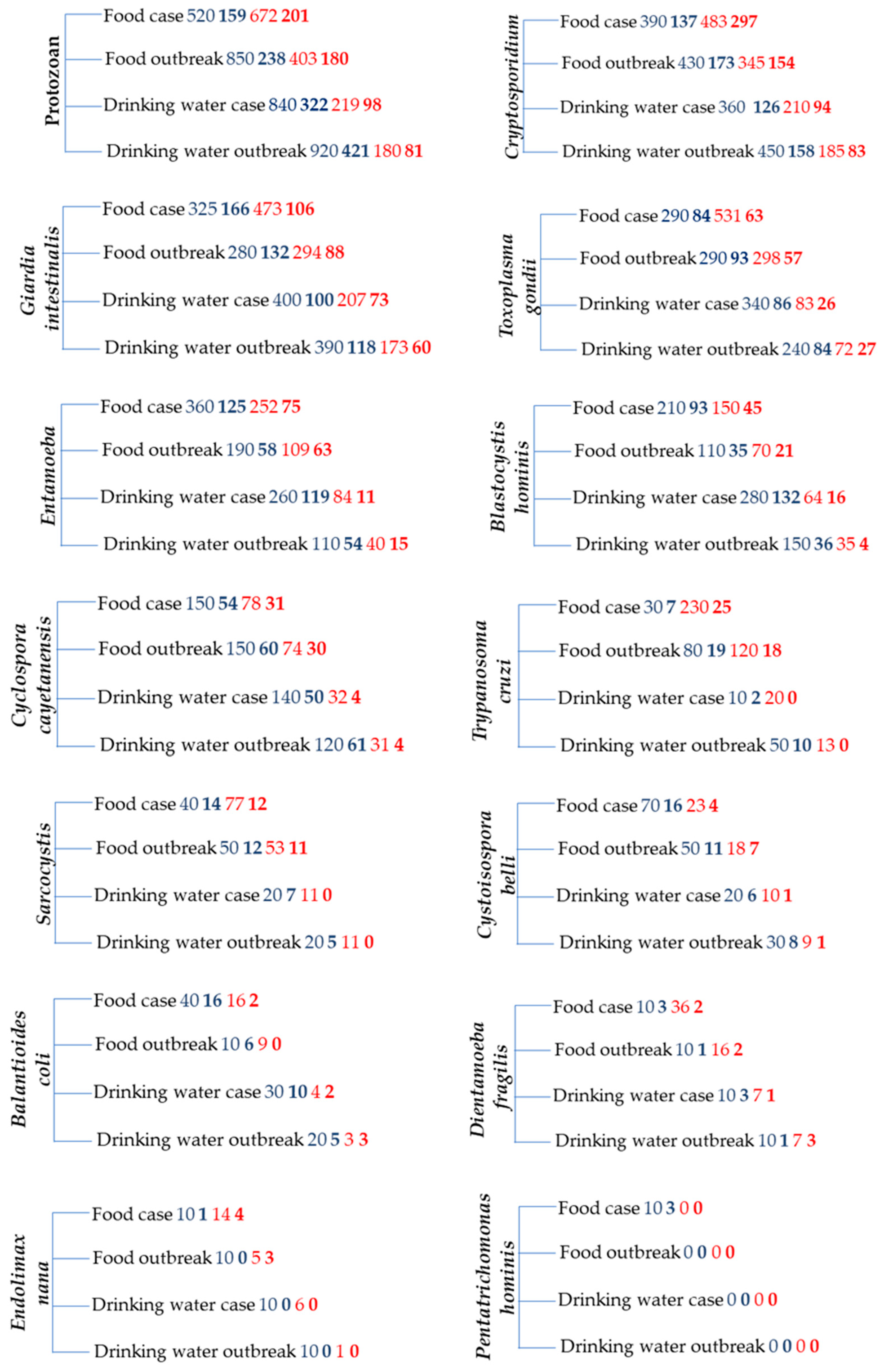
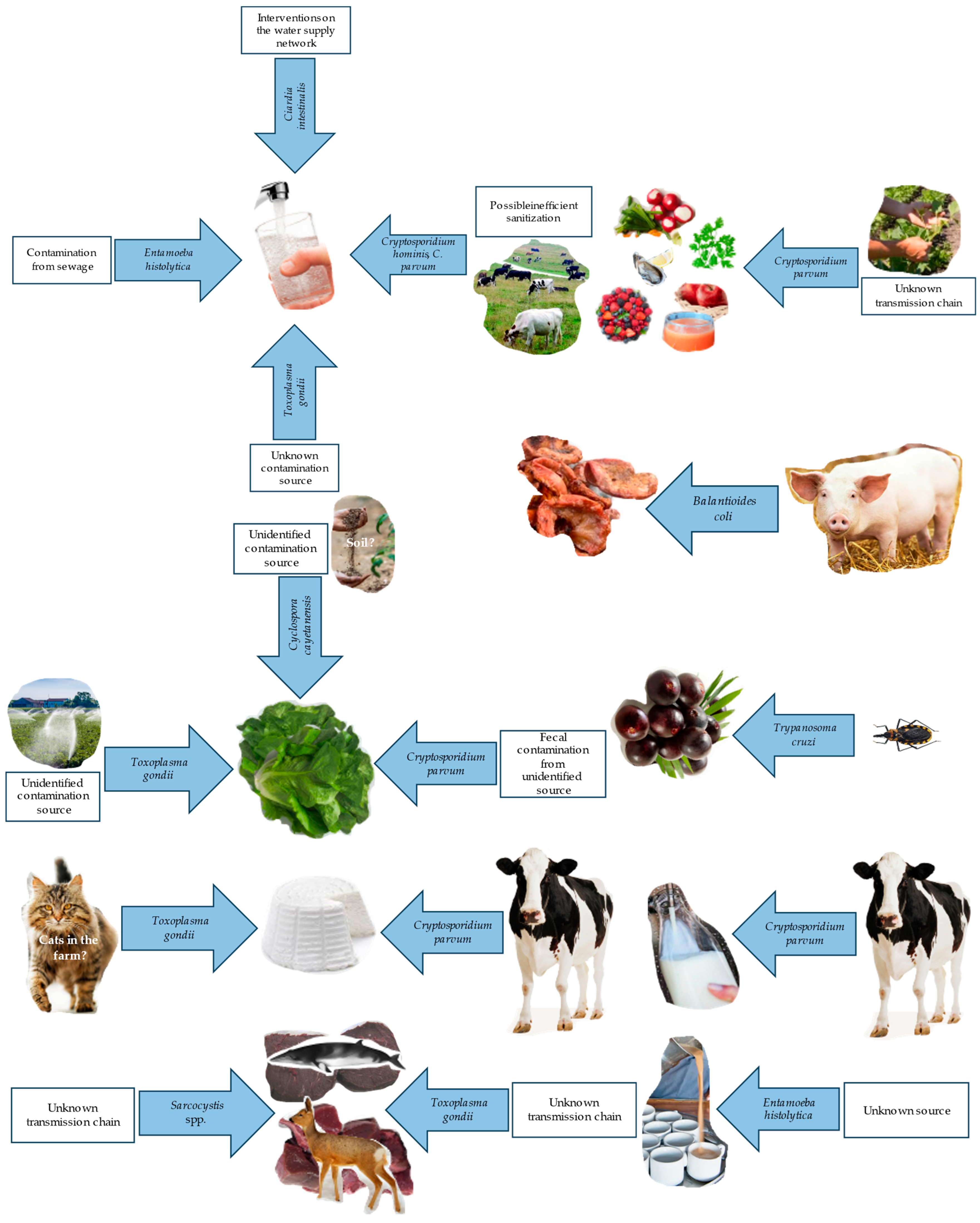
Figure 1. Number of articles retrieved (normal character) and screened (bold character) for the construction of this review from the GoogleScholar (blue numbers) and Scopus (red numbers) databases. The search strings were “protozoan food (or drinking water) human infection case (or outbreak)” or the same strings with individual names of organisms in place of the word “protozoan”. The numbers comprise duplicate items from different searches.
Applsci 14 05339 g001
Figure 2. Number of waterborne or foodborne acute cases/outbreaks reported for pathogenic protozoans since 2019 [8,9,19,35,122,123,131,132,133,142,143,154,157,168,169,170,172,173,191,193,195,196,198,199,200,201,209,226,233,237,239,240,241,242,243,255,256,260,268,269,273,274,280,282,286]. Outbreaks are only shown for Cryptosporidium species.
Applsci 14 05339 g002
Figure 3. Schematic representation of the contamination sources at the origin of cases and outbreaks reported since 2019 for foodborne and waterborne protozoans [90,122,123,131,132,133,154,157,168,169,170,172,173,200,210,237,242,243,255,259,282].
Applsci 14 05339 g003
Table 1. Sources of pathogenic protozoans involved in infection cases and outbreaks according to literature published since 2019.
Infectious AgentDietary Infection Sources in Cases and Outbreaks *
Cryptosporidium spp.Drinking water, unpasteurized or re-contaminated pasteurized milk, meat, dairy foods, dishes prepared outdoors, barbecued foods, raw foods, raw vegetables, raw fruits, unpasteurized drinks, apple juice, raw shellfish [9,89,97,100,113,114,121,122,123,124,127,128,129,130,131,133]
Entamoeba spp.Drinking water, alcoholic fermented sap of the Palmyra toddy [7,209]
Toxoplasma gondiiRaw vegetables, fruits, raw or undercooked meats, unpasteurized milk, raw cow’s milk cheese, raw or undercooked crustaceans or shellfish, whale meat, drinking water [11,157,159,161,164,168,169,170,171,172,173,174]
Giardia intestinalisDrinking water, fresh produce, composite ready-to-eat food, canned salmon, raw oysters, ice cream, noodle salad, chicken salad, dairy products, sandwiches, tripe soup, unpasteurized milk, shellfish, unidentified foods [26,29,140,147,149,154]
Trypanosoma cruziAçaí juice, sugar cane juice, palm, guanabana, guava, milpesillo, majo, mango, mandarin, orange juices, raw meat [12,251]
Balantioides coliNone reported
Cyclospora cayetanensisBerries, cilantro, basil, lettuce, ready-to-eat bagged salads, sugar snap peas [6,237,242,243]
Cystoisospora belliNone reported
Sarcocystis spp.Beef, pork, venison, whale meat [8,173,260]
Blastocystis hominisNone reported
Endolimax nanaNone reported
Dientamoeba fragilisNone reported
Pentatrichomonas hominisNone reported
* Ascertained or most probable, determined by statistical association or by direct evidence in cases and outbreaks.
Disclaimer/Publisher’s Note: The statements, opinions and data contained in all publications are solely those of the individual author(s) and contributor(s) and not of MDPI and/or the editor(s). MDPI and/or the editor(s) disclaim responsibility for any injury to people or property resulting from any ideas, methods, instructions or products referred to in the content.

