Article

Crestal Sinus Lift with the Hydrodynamic Technique: Prospective Clinical Study at 3 Years Follow-Up

1
Department of Dentistry, Dental School, IRCCS San Raffaele Hospital, Vita-Salute San Raffaele University, 20132 Milan, Italy
2
Chair Center for Oral Hygiene and Prevention, Department of Dentistry, Dental School, IRCCS San Raffaele Hospital, Vita-Salute San Raffaele University, 20132 Milan, Italy
3
Dental School, IRCCS San Raffaele Hospital, Vita-Salute University, 20132 Milan, Italy
*
Author to whom correspondence should be addressed.
Appl. Sci. 2024, 14(14), 6204; https://doi.org/10.3390/app14146204
Submission received: 4 June 2024 / Revised: 2 July 2024 / Accepted: 4 July 2024 / Published: 17 July 2024
(This article belongs to the Special Issue Dental Implants: Latest Advances and Prospects)

Abstract

:
Aim: This study aimed to evaluate the implant survival rate, marginal bone loss (MBL), and surgical complications in single dental implants placed in the atrophic posterior maxilla using a transcrestal sinus lift with injectable graft materials. Materials and Methods: A prospective longitudinal study was conducted at IRCCS San Raffaele, Milan, Italy. Fifty-four patients with single edentulous sites and residual bone heights < 5 mm were included. A transcrestal sinus lift was performed using non-cutting drills (Cosci’s technique) and xenogenic bone graft in gel form (Gel40, Tecnoss, Italy). Follow-up visits were at 1 week, 3 and 6 months, and annually for 3 years. Results: The implant survival rate was 98.04%, with one implant lost. MBL values were 0.71 ± 0.94 mm at 6 months, 1.00 ± 0.99 mm at 1 year, 1.03 ± 1.00 mm at 2 years, and 1.02 ± 1.01 mm at 3 years. Our ANOVA showed a significant MBL increase from 6 months to 1 year (p = 0.015), with no significant changes thereafter. Minimal surgical complications were observed, each occurring in 1.85% of cases. Conclusion: Transcrestal sinus lifts with injectable graft materials demonstrate high implant survival, acceptable MBL, and minimal complications, making it a reliable option for posterior maxilla rehabilitation.

1. Introduction

Dental implants are currently recognized as a viable therapeutic alternative for replacing missing teeth [1,2,3]; however, their placement may be hindered by several issues [4,5].
The placement of dental implants in the posterior maxilla is often complicated due to the reduced vertical height of the residual bone ridge, typically resulting from post-extraction bone resorption and subsequent pneumatization of the maxillary sinus [6,7]. Among the various surgical procedures proposed over the years for the implant-prosthetic rehabilitation of the atrophic posterior maxillary sector, sinus elevation with simultaneous graft material placement represents a valid therapeutic solution for restoring correct inter-arch relationships [8,9].
In 1980, Boyne and James introduced the first sinus floor elevation technique, which involves lifting Schneider’s membrane by opening a bone window in the lateral sinus wall and filling the subantral space with graft material [10]. In 1986, Tatum developed a less invasive surgical approach through the crestal route, which was later refined by Summers in 1994 with the introduction of the osteotome technique [11].
The Osteotome Sinus Floor Elevation (OSFE) procedure reduces treatment morbidity and invasiveness by using bone from the osteotomy site preparation to elevate the sinus floor. Conversely, the Bone-Added Osteotome Sinus Floor Elevation (BAOSFE) technique achieves the same result using graft material driven into the surgical site through a series of osteotomes. Both techniques are considered more reliable when the residual bone ridge height is at least 5–6 mm [12,13].
To avoid greenstick fracture of the sinus floor using osteotomes, Cosci and Luccioli proposed a new surgical procedure involving a specific sequence of drills to perforate the sinus floor [14]. Laceration of the Schneiderian membrane during sinus floor perforation with rotary instruments is less frequent than with sinus floor fracture using osteotomes. The main limitation of transcrestal techniques is the uncertainty of a possible perforation of the sinus membrane. However, an endoscopic study has shown that the sinus floor can be raised up to 5 mm without tearing the membrane [15].
Crestal sinus elevation promotes simultaneous implant placement in cases where there is sufficient residual bone crest size to achieve primary implant stability. If this is not possible, crestal sinus elevation can be performed with a delayed approach (Future Site Development) to create adequate bone volume for subsequent implant placement [16].
In 2009, Pommer and Watzek introduced the gel pressure technique, which involves using injectable grafts in gel form to create controlled pressure capable of lifting the sinus membrane [17]. The biomaterials used for this purpose consist of micronized allogeneic, xenogeneic, or alloplastic graft particles embedded in a collagen matrix or water-based gels, characterized by a pasty consistency and a smooth surface that likely prevents accidental perforation of the Schneiderian membrane during elevation [18].
This surgical technique utilizes the hydraulic pressure generated during the injection of the gel graft through the crestal antrostomy to detach the sinus membrane from the bone walls and simultaneously fill the sub-antral space, significantly reducing surgical time. This allows for increased vertical space available for dental implant placement without needing more invasive surgical techniques [19]. Although graftless sinus lift procedures have been described over the years, using bone substitutes in the transcrestal approach allows greater vertical bone gain and can also be applied in cases with residual bone ridge height < 5 mm [20].
The aim of this prospective clinical study was to evaluate the implant survival rate, marginal bone loss, and surgical complications in single dental implants placed in the posterior atrophic maxilla according to the transcrestal sinus lift procedure performed with injectable graft materials in gel.

2. Materials and Methods

This study employed a prospective longitudinal design. All procedures adhered to the ethical standards of the institutional and national research committees, as well as the 1964 Declaration of Helsinki and its subsequent amendments or comparable ethical standards. The ethics committee approval number is CE/INT/10/2015.
Our research was conducted at the Dentistry Department of IRCCS San Raffaele in Milan, Italy. Patients were enrolled from January 2018 to January 2021, with data collection continuing until January 2024.
The first visit included the collection of anamnestic data, clinical objective examination, level I radiographic investigations (intra-oral X-rays and orthopantomography), and level II radiographic investigations (Cone Beam CT).
Sample selection
Inclusion criteria
  • Age > 18 years old;
  • Single edentulous at the posterior maxilla with a residual bone height less than 5 mm;
  • Requiring implant-prosthetic rehabilitation;
  • The absence of any contraindication to dental implant placement or sinus augmentation [21];
  • Compliance with follow-up monitoring and professional oral hygiene maintenance protocol.
Exclusion criteria
  • Absolute contraindications to dental implant placement;
  • Bisphosphonate medication [22];
  • Head and neck radiotherapy within the past year [23];
  • Uncompensated systemic disorders [24];
  • Smoker patients [25];
  • Inability to adhere to protocol checks;
  • Failure to maintain regular oral hygiene sessions;
  • Economic infeasibility in affording the treatment.
Surgical procedure
Pre-surgical protocol
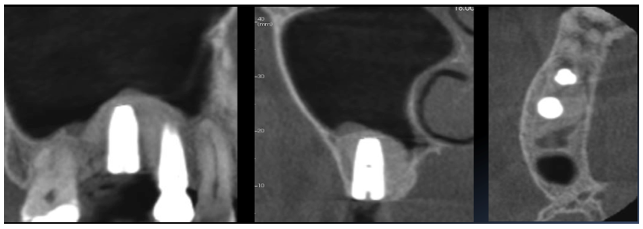
Radiological investigations, performed using Cone Beam Computed Tomography (CBCT), showed the patency of the maxillary sinus and a residual bone ridge height (RBH) of between 3 and 5 mm at the edentulous site, which was insufficient for traditional axial implant placement (Figure 1).
Based on the residual bone measurements, a transcrestal sinus lift using an injectable gel form of xenogenic bone graft (Gel 40, Tecnoss, Giaveno, Italy), along with simultaneous implant placement, was eligible.
In addition to the CBCT evaluation, prior to the surgical procedure, each patient was made to sign the informed consent.
Antibiotic therapy (amoxicillin and clavulanic acid 1 g or clarithromycin 1 g in case of allergy) was prescribed to be started the day before surgery.
Surgical protocol
The patient was prepared for surgery with an oral rinse using a chlorhexidine digluconate mouthwash at 0.12% for one minute. Under local anesthesia with articaine and adrenaline 1:100,000 (Septanest, Saint-Maur-des-Fossés Cedex, France), a crestal marginal incision was made with a full-thickness flap to expose the alveolar crest.
The implant site was created using a sequential set of drills with appropriate cutting angles to gently erode the cortical floor of the sinus (Cosci’s technique). This set of drills comprised 11 pieces of the same diameter (3.1 mm) but of variable and increasing lengths, ranging from 2 to 12 mm. These drills allowed the Schneiderian membrane to be reached without tearing it, enabling subsequent graft insertion and sinus lift.
The implant location was marked using a small-diameter pilot drill, 1 mm in length, working through the cortical bone and using a prefabricated surgical guide. After intermediate, larger-diameter drilling, the implant site was prepared 1 mm below the sinus floor using a lifting drill, 1 mm shorter than the height of the alveolar crest, as indicated by the periapical radiograph. A parallel pin was then inserted into the surgical site to check the exact height of the alveolar ridge below the sinus floor using an intraoperative X-ray. A final lifting drill, 1 mm longer than the crest height, was used until the shoulder stop reached the ridge. The alveolar bone was then checked with a special rounded probe to confirm complete bone erosion prior to lifting the sinus membrane.
After verifying the integrity of the sinus membrane using an appropriate probe and the Valsalva maneuver, a porcine xenogenic bone substitute in the form of a pre-heated (40 °C) gel (Gel 40, Tecnoss, Giaveno, Italy) was injected through the crestal antrostomy to elevate the membrane and fill the sub-antral space (Figure 2A–D).
At this point, an intraoperative intraoral X-ray was performed to confirm the elevation of Schneider’s membrane and measure the new distance from the alveolar crest (Figure 3).
The new bone height, achieved through the sinus lift, allowed for the placement of an implant with an insertion torque higher than 25 Ncm, ensuring primary stability. An intraoperative periapical X-ray was then performed, confirming the correct positioning of the implant and the mechanical engagement between the apex of the fixture and the grafted material. This engagement was considered a crucial factor as it provides additional stability to the implant (Figure 4).
Post-surgical protocol
At the end of the surgery, a control CBCT was performed, which confirmed the adequate prosthetic emergence of the inserted implant and the success of the surgical procedures. Additionally, it clearly showed that the graft material in gel form, by detaching the membrane at points of least resistance, enabled a mini sinus lift even at the apex of the implant fixture (Figure 5).
Antibiotic therapy was continued for the next 5 days (1 g every 12 h—amoxicillin in combination with clavulanic acid or clarithromycin in case of allergy).
In addition, analgesic therapy (non-steroidal anti-inflammatory drugs, as needed) was prescribed for each patient. Mouth rinsing with a chlorhexidine–digluconate-containing solution (0.2%) was recommended twice daily for 10 days [26]. One week after the surgical procedure, the sutures were removed.
Prosthetic rehabilitation
Four months post-surgery, the implant site was reopened. The closure screw was removed and replaced with a healing abutment to facilitate soft tissue healing around the implant. After 10 days, the sutures were removed.
Digital impressions were taken to scan the lower and upper arches, buccal in occlusion, and the scanning body on the implant (PRIMESCAN®, Dentsply Sirona, Bensheim, Germany). This digital workflow ensured precise mapping of the implant position and surrounding structures.
A provisional prosthetic crown was fabricated and placed on the implant one week later. This provisional crown allowed for the assessment of fit, function, and esthetics while the soft tissue matured. Following a satisfactory evaluation period, the provisional crown was replaced with a definitive monolithic zirconia prosthetic restoration, which was screw-retained onto the implant to ensure stability and function.
Clinical outcomes
1.
The Implant Survival Rate
The implant survival rate was assessed based on the number of implants lost during the follow-up period due to mobility associated with progressive marginal bone loss, typically caused by peri-implantitis. Implant loss was classified by the period of occurrence: if it occurred within 6 months of fixture placement, it was termed early failure; if it occurred after 6 months, it was termed late failure. Early failures were usually identified at the reopening stage due to a lack of osseointegration. Late failures were characterized by signs of peri-implantitis, implant mobility, radiolucent areas around fixtures, mucosal suppuration, and/or pain during the follow-up period.
2.
Marginal Bone Loss (MBL)
Marginal bone loss was assessed at 6 months, 1 year, 2 years, and 3 years post-surgery using digital phosphor intraoral radiography. To assess marginal bone trends, measurements were performed only after image calibration. Digora 2.5 software (Soredex, Tuusula, Finland) was utilized for analysis, employing a specific measurement tool. Calibrations (pixels/mm) were performed using the implant diameter as the known unit. Changes in the height of the peri-implant marginal bone relative to the most coronal part of the implant and the point of contact between the implant and the marginal ridge were measured.
A line passing over the shoulder of the implant was used as a reference for measurement, from which a straight line was drawn parallel to the long axis of the implant to the most coronal point where the bone met the fixture both mesially and distally (Figure 6).
The software tool we used automatically provided the distance between these points in millimeters. To minimize human error, three operators performed the measurements, and the average was considered. Mesial and distal measurements were taken, and the averages for each implant site (MBL) were calculated.
3.
Surgical Complications
Surgical complications were categorized based on the procedure performed. Potential complications included sinus membrane perforation, maxillary sinusitis, graft material displacement, excessive bleeding, infection, and insufficient lift of the sinus floor. Additionally, there could be complications such as pain, swelling, and hematoma formation at the surgical site.
Follow-up
Follow-up visits were conducted one-week post-surgery, then at 3 and 6 months, followed by annual check-ups for the next three years. Professional oral hygiene sessions were scheduled every four months after the surgical–prosthetic procedure [26,27,28].
Statistical analysis
The collected data were analyzed using SPSS software (Version 25.0, IBM Corp., Armonk, NY, USA) to evaluate the implant survival rates, MBL, and surgical complications.
Kaplan–Meier survival analysis was performed to estimate the implant survival rate over the follow-up period. A repeated measures ANOVA was used to analyze changes in MBL over time. Pairwise comparisons were conducted to assess differences between specific time points. The incidence of surgical complications was summarized using descriptive statistics. Chi-square tests were employed to compare the complication rates between different time intervals. A p-value of <0.05 was considered statistically significant.
Null Hypothesis (H0): There are no significant differences in implant survival rates, marginal bone loss, or the incidence of surgical complications in patients with single dental implants placed in the atrophic posterior maxilla using the transcrestal sinus lift procedure with injectable graft materials.
Study size: The sample size determination was based on a statistical power analysis using a t-test for two independent samples. We targeted a significance level (α) of 0.05 and a power (1 − β) of 0.80, reflecting a commonly accepted balance between Type I and Type II errors. The required sample size for the two-sample t-test was calculated using the following formula:
n = 2(σ2) (Zα/2 + Zβ)2/δ2
where σ is the estimated standard deviation; Zα/2 and Zβ are critical values for the chosen significance level and power; and delta is the effect size.
We conducted a sensitivity analysis on the effect size used in the power analysis. The required sample size for an effect size of 0.7 was determined to be 50.

3. Results

At each stage of this study, 61 patients were considered potentially eligible and examined for confirmation. According to the exclusion criteria, 54 patients were confirmed as eligible at baseline. Sample details at baseline and implant-prosthetic features are provided in the following table (Table 1).
1.
The Implant Survival Rate
Out of 54 implants placed, only one implant was lost during the follow-up period, resulting in an implant survival rate of 98.04%. The implant was lost during the osseointegration period, leading to early failure.
Kaplan–Meier survival analysis was conducted to estimate the survival probability over time. The survival rate at the end of the three-year period was 98.0% (95% CI: 94.3%—100.0%). The log-rank test indicated no significant differences in survival rates between different implant positions (p = 0.65).
2.
Marginal Bone Loss (MBL)
The mean and standard deviation for each time point were as follows (Figure 7):
A repeated measures ANOVA was performed to determine if there were significant differences in MBL over time. The analysis revealed a significant effect of time on MBL (F(3, 159) = 5.82, p < 0.001). Post-hoc pairwise comparisons using the Bonferroni correction indicated a significant increase in MBL from 6 months to 1 year (p = 0.015). However, the differences in MBL between 1 year and 2 years (p = 0.68) and between 2 years and 3 years (p = 0.82) were not statistically significant.
3.
Surgical Complications
Out of the total cases treated, only one patient (1.85%) experienced a sinus membrane perforation, while an equal number of cases (1.85%) developed maxillary sinusitis. Additionally, we observed a single case (also 1.85%) of bleeding during the procedure. Fortunately, there were no instances of post-operative wound infection (0%) or insufficient lift of the sinus floor (0%). The incidence of surgical complications was recorded as follows (Figure 8):
Given the small number of complications, descriptive statistics were used to summarize the data. Fisher’s exact test was employed to compare the observed frequencies of complications with expected values based on historical data. The test results were not statistically significant for any complication (p > 0.05), suggesting that the incidence of complications in this study was consistent with what might be expected in similar clinical settings.

4. Discussion

The effectiveness and safety of transcrestal sinus lift procedures with simultaneous implant placement have been well documented in the literature. Our study adds to this body of knowledge by demonstrating high implant survival rates and acceptable levels of marginal bone loss over a three-year follow-up period.
The implant survival rate observed in our study was 98.04%, which is consistent with previous reports in the literature. For instance, Corbella et al. (2015) and Chen et al. (2024) reported similar survival rates for implants placed in the atrophic posterior maxilla using various sinus lift techniques [6,9]. These findings are further corroborated by Pjetursson et al. (2008), who highlighted the success of sinus floor elevation and the survival of implants inserted in combination with this procedure [29].
The marginal bone loss (MBL) documented in our study showed a significant increase within the first year post-surgery, followed by stabilization in subsequent years. This trend aligns with findings by Andrés-García et al. (2021) and Elghobashy et al. (2023), who also observed early bone remodeling followed by a plateau phase [12,20].
The average MBL at three years was 1.02 ± 1.01 mm, which is within the acceptable range reported by the International Team for Implantology (ITI) consensus guidelines.
Studies by Chen et al. (2024) and Stacchi et al. (2018) have similarly observed that appropriate techniques and materials can result in stable bone levels over time [9,30]. The use of digital radiography and precise measurement techniques, as described by Chandra et al. (2018), ensures the reliability of these findings [13].
The incidence of surgical complications in our study was minimal and comparable to other studies. Sinus membrane perforation, maxillary sinusitis, and graft material displacement each occurred in 1.85% of cases, consistent with the complication rates reported by Danesh-Sani et al. (2016) and Lie et al. (2022) [8,18]. The careful execution of the surgical technique, including the use of specialized drills and probes to avoid membrane perforation, likely contributed to the low complication rates observed. Studies by Tatum (1986) and Yu et al. (2022) also emphasized the importance of technique in minimizing complications during sinus lift procedures [11,15].
Our study utilized the xenogenic bone graft in gel form, which has shown promising results in terms of ease of use and effective bone regeneration. This approach is supported by findings from Pommer and Watzek (2009), who demonstrated the efficacy of gel pressure techniques in flapless transcrestal sinus lifts [17].
Additionally, the graftless approaches described by Manekar (2020) and Elghobashy et al. (2023) also show comparable outcomes, suggesting that various techniques can be effectively employed depending on the clinical scenario [19,20]. The hydrodynamic properties of the gel form graft, as highlighted by Lombardi et al. (2017), facilitate a controlled and effective lift of the Schneiderian membrane [31].
The osteotome technique, although not very invasive, has the limitation of hammering, which is often poorly tolerated by patients. Conversely, the crestal approach based on the use of sequential drills, as described by Cosci, does not present such a drawback [12]. Adequate elevation of Schneider’s membrane is an essential prerequisite for the creation of a sub-antral space with regenerative potential. Recent studies have shown a direct correlation between adequate membrane elevation, the formation of new bone tissue, and the volumetric stability of the regenerated tissue [32,33,34].
The indirect hydrodynamic detachment of the membrane by the injection of the graft in gel form was simple, quick, and effective [35]. The use of an injectable biomaterial, such as Gel 40 (Tecnoss, Giaveno, Italy), allowed a significant reduction in surgical time, enabling the sinus lift procedure to be performed safely and achieving the expected result. Gel 40 provides adequate mechanical support to the formed blood clot, thus facilitating the healing and bone neoformation processes [36,37]. The simultaneous and immediate insertion of the implant fixture is a key factor in increasing mechanical stability and reducing graft shrinkage [38,39,40]. Bernardello et al. (2011) showed that the percentage shrinkage of the graft is inversely proportional to the length of the implant fixture [41]. The large size of the graft can be explained by the physical and hydrodynamic properties of Gel 40, which exploits Pascal’s law to allow the elevation of Schneider’s membrane. Additionally, in the event of accidental dissemination, the very small particle size of the grafts (0.3 mm) allows easier removal from the sinus cavity [42].

5. Conclusions

Within the limitations of this study, transcrestal elevation of the sinus floor, performed in crests with reduced bone height (<5 mm), using a sequence of non-cutting drills (Cosci’s technique) and a xenogenous bone graft in the form of injectable gel, has successfully allowed implant placement and survival, with a follow-up of 20 months. Therefore, this procedure represents a safe and reliable approach to the rehabilitation of atrophic upper posterior sectors. Our study confirms the effectiveness and safety of the transcrestal sinus lift procedure with simultaneous implant placement. With a high implant survival rate, acceptable marginal bone loss, and minimal complications, this technique represents a reliable option for implant-prosthetic rehabilitation in the posterior maxilla. The findings align with the existing literature and contribute to the growing evidence supporting this approach. Furthermore, further clinical trials may be necessary to confirm the obtained results.

Author Contributions

Conceptualization, S.S. and E.P.; methodology, S.S. and E.P.; software, L.A.; validation, E.F.G., E.P. and S.S.; formal analysis, M.G.; investigation, L.A.; resources, S.S.; data curation, L.C.; writing—original draft preparation, L.C.; writing—review and editing, L.A.; visualization, M.G.; supervision, E.F.G. All authors have read and agreed to the published version of the manuscript.

Funding

This research received no external funding.

Institutional Review Board Statement

This study was conducted in accordance with the Declaration of Helsinki and approved by the Institutional Review Ethics Committee of Vita-Salute San Raffaele University n. CE/INT/10/2015 (15 April 2015), Dental School Department of Dentistry IRCCS San Raffaele Hospital, Milan, 20132, Italy.

Informed Consent Statement

Informed consent was obtained from all subjects involved in this study.

Data Availability Statement

Data is contained within the article.

Conflicts of Interest

The authors declare no conflicts of interest.

References

  1. Capparè, P.; Tetè, G.; D’Orto, B.; Nagni, M.; Gherlone, E.F. Immediate Loaded Full-Arch Mandibular Rehabilitations in Younger vs. Elderly Patients: A Comparative Retrospective Study with 7-Year Follow-Up. J. Clin. Med. 2023, 12, 4524. [Google Scholar] [CrossRef] [PubMed]
  2. Nagni, M.; Pirani, F.; D’Orto, B.; Ferrini, F.; Cappare, P. Clinical and Radiographic Follow-Up of Full-Arch Implant Prosthetic Rehabilitations: Retrospective Clinical Study at 6-Year Follow-Up. Appl. Sci. 2023, 13, 11143. [Google Scholar] [CrossRef]
  3. D’Orto, B.; Chiavenna, C.; Leone, R.; Longoni, M.; Nagni, M.; Capparè, P. Marginal Bone Loss Compared in Internal and External Implant Connections: Retrospective Clinical Study at 6-Years Follow-Up. Biomedicines 2023, 11, 1128. [Google Scholar] [CrossRef] [PubMed]
  4. Al Amri, M.D. Influence of interimplant distance on the crestal bone height around dental implants: A systematic review and meta-analysis. J. Prosthet. Dent. 2016, 115, 278–282.e1. [Google Scholar] [CrossRef] [PubMed]
  5. Tavelli, L.; Barootchi, S.; Avila-Ortiz, G.; Urban, I.A.; Giannobile, W.V.; Wang, H.L. Peri-implant soft tissue phenotype modification and its impact on peri-implant health: A systematic review and network meta-analysis. J. Periodontol. 2021, 92, 21–44. [Google Scholar] [CrossRef] [PubMed]
  6. Corbella, S.; Taschieri, S.; Del Fabbro, M. Long-term outcomes for the treatment of atrophic posterior maxilla: A systematic review of literature. Clin. Implant. Dent. Relat. Res. 2015, 17, 120–132. [Google Scholar] [CrossRef] [PubMed]
  7. Leung, M.; Alghamdi, R.; Guallart, I.F.; Bergamini, M.; Yu, P.Y.; Froum, S.J.; Cho, S.C. Patient-Related Risk Factors for Maxillary Sinus Augmentation Procedures: A Systematic Literature Review. Int. J. Periodontics Restor. Dent. 2021, 41, e121–e128. [Google Scholar] [CrossRef]
  8. Lie, S.A.N.; Claessen, R.M.M.A.; Leung, C.A.W.; Merten, H.A.; Kessler, P.A.W.H. Non-grafted versus grafted sinus lift procedures for implantation in the atrophic maxilla: A systematic review and meta-analysis of randomized controlled trials. Int. J. Oral. Maxillofac. Surg. 2022, 51, 122–132. [Google Scholar] [CrossRef]
  9. Chen, J.; Lu, Y.; Xu, J.; Hua, Z. Clinical evaluation of maxillary sinus floor elevation with or without bone grafts: A systematic review and meta-analysis of randomised controlled trials with trial sequential analysis. Arch. Med. Sci. 2024, 20, 384–401. [Google Scholar] [CrossRef]
  10. Boyne, P.J.; James, R.A. Grafting of the maxillary sinus floor with autogenous marrow and bone. J. Oral. Surg. 1980, 38, 613–616. [Google Scholar]
  11. Tatum, H., Jr. Maxillary and sinus implant reconstructions. Dent. Clin. N. Am. 1986, 30, 207–229. [Google Scholar] [CrossRef] [PubMed]
  12. Andrés-García, R.; Ríos-Santos, J.V.; Herrero-Climent, M.; Bullón, P.; Fernández-Farhall, J.; Gómez-Menchero, A.; Fernández-Palacín, A.; Ríos-Carrasco, B. Sinus Floor Elevation via an Osteotome Technique without Biomaterials. Int. J. Environ. Res. Public Health 2021, 18, 1103. [Google Scholar] [CrossRef] [PubMed]
  13. Chandra, R.V.; Suvvari, N.; Reddy, A.A. Trephine Core Procedure Versus Bone-Added Osteotome Sinus Floor Elevation in the Augmentation of the Sinus Floor: A Comparative Clinical and Radiographic Study. Int. J. Oral Maxillofac. Implant. 2018, 33, 425–432. [Google Scholar] [CrossRef] [PubMed]
  14. Cosci, F.; Luccioli, M. A new sinus lift technique in conjunction with placement of 265 implants: A 6-year retrospective study. Implant. Dent. 2000, 9, 363–368. [Google Scholar] [CrossRef] [PubMed]
  15. Yu, X.; Ye, G.; Zhao, F.; Wang, B.; Yu, M.; Wang, H. Endoscope-controlled maxillary sinus floor elevation: A review of the literature. Br. J. Oral. Maxillofac. Surg. 2022, 60, 113–119. [Google Scholar] [CrossRef]
  16. Lumbau, A.I.; Meloni, S.M.; Tallarico, M.; Melis, L.; Spano, G.; Baldoni, E.; Koshovari, A.; Pisano, M. Implant Placement Following Crestal Sinus Lift with Sequential Drills and Osteotomes: Five Years after Final Loading Results from a Retrospective Study. J. Funct. Biomater. 2021, 12, 10. [Google Scholar] [CrossRef] [PubMed]
  17. Pommer, B.; Watzek, G. Gel-pressure technique for flapless transcrestal maxillary sinus floor elevation: A preliminary cadaveric study of a new surgical technique. Int. J. Oral Maxillofac. Implant. 2009, 24, 817–822. [Google Scholar]
  18. Danesh-Sani, S.A.; Loomer, P.M.; Wallace, S.S. A comprehensive clinical review of maxillary sinus floor elevation: Anatomy, techniques, biomaterials and complications. Br. J. Oral Maxillofac. Surg. 2016, 54, 724–730. [Google Scholar] [CrossRef] [PubMed]
  19. Manekar, V.S. Graftless crestal hydraulic sinus lift with simultaneous implant insertion. Natl. J. Maxillofac. Surg. 2020, 11, 213–218. [Google Scholar] [CrossRef]
  20. Elghobashy, M.T.M.; Shaaban, A.M.; Melek, L.N.F. Radiographic comparison between Densah burs and osteotome for graftless internal sinus lifting with simultaneous implant placement: A randomized clinical trial. Int. J. Oral Maxillofac. Surg. 2023, 52, 388–395. [Google Scholar] [CrossRef]
  21. Heitz-Mayfield, L.J.; Aaboe, M.; Araujo, M.; Carrión, J.B.; Cavalcanti, R.; Cionca, N.; Cochran, D.; Darby, I.; Funakoshi, E.; Gierthmuehlen, P.C.; et al. Group 4 ITI Consensus Report: Risks and biologic complications associated with implant dentistry. Clin. Oral Implant. Res. 2018, 29 (Suppl. S16), 351–358. [Google Scholar] [CrossRef] [PubMed]
  22. Fiorillo, L.; Cicciù, M.; Tözüm, T.F.; D’Amico, C.; Oteri, G.; Cervino, G. Impact of bisphosphonate drugs on dental implant healing and peri-implant hard and soft tissues: A systematic review. BMC Oral Health. 2022, 22, 291. [Google Scholar] [CrossRef] [PubMed]
  23. Smith Nobrega, A.; Santiago, J.F., Jr.; de Faria Almeida, D.A.; Dos Santos, D.M.; Pellizzer, E.P.; Goiato, M.C. Irradiated patients and survival rate of dental implants: A systematic review and meta-analysis. J. Prosthet. Dent. 2016, 116, 858–866. [Google Scholar] [CrossRef] [PubMed]
  24. Aghaloo, T.; Pi-Anfruns, J.; Moshaverinia, A.; Sim, D.; Grogan, T.; Hadaya, D. The Effects of Systemic Diseases and Medications on Implant Osseointegration: A Systematic Review. Int. J. Oral Maxillofac. Implant. 2019, 34, s35–s49. [Google Scholar] [CrossRef] [PubMed]
  25. Mustapha, A.D.; Salame, Z.; Chrcanovic, B.R. Smoking and Dental Implants: A Systematic Review and Meta-Analysis. Medicina 2021, 58, 39. [Google Scholar] [CrossRef] [PubMed]
  26. Tetè, G.; Cattoni, F.; Polizzi, E. Anti-discoloration system: A new chlorhexidine mouthwash. J. Biol. Regul. Homeost. Agents 2021, 35 (Suppl. S1), 113–118. [Google Scholar] [CrossRef] [PubMed]
  27. Polizzi, E.; Tetè, G. Manual vs Mechanical Oral Hygiene Procedures: Has the Role of the Dental Hygienist in Phase 2 Post-lockdown Really Changed? Oral Health Prev. Dent. 2020, 18, 1031–1037. [Google Scholar] [CrossRef] [PubMed]
  28. Tetè, G.; D’orto, B.; Ferrante, L.; Polizzi, E.; Cattoni, F. Role of mast cells in oral inflammation. J. Biol. Regul. Homeost. Agents 2021, 35 (Suppl. S1), 65–70. [Google Scholar] [CrossRef] [PubMed]
  29. Pjetursson, B.E.; Tan, W.C.; Zwahlen, M.; Lang, N.P. A systematic review of the success of sinus floor elevation and survival of implants inserted in combination with sinus floor elevation. J. Clin. Periodontol. 2008, 35, 216–240. [Google Scholar] [CrossRef]
  30. Stacchi, C.; Spinato, S.; Lombardi, T.; Bernardello, F.; Bertoldi, C.; Zaffe, D.; Nevins, M. Minimally Invasive Management of Implant-Supported Rehabilitation in the Posterior Maxilla, Part II. Surgical Techniques and Decision Tree. Int. J. Periodontics Restor. Dent. 2020, 40, e95–e102. [Google Scholar] [CrossRef]
  31. Lombardi, T.; Lamazza, L.; Bernardello, F.; Ziętek, G.; Stacchi, C.; Troiano, G. Clinical and radiographic outcomes following transcrestal maxillary sinus floor elevation with injectable xenogenous bone substitute in gel form: A prospective multicenter study. Int. J. Implant. Dent. 2022, 8, 32. [Google Scholar] [CrossRef] [PubMed]
  32. Antonaya-Mira, R.; Barona-Dorado, C.; Martínez-Rodríguez, N.; Cáceres-Madroño, E.; Martínez-González, J.M. Meta-analysis of the increase in height in maxillary sinus elevations with osteotome. Med. Oral. Patol. Oral Cir. Bucal. 2012, 17, e146–e152. [Google Scholar] [CrossRef] [PubMed]
  33. Tsai, C.F.; Pan, W.L.; Pan, Y.P.; Chan, C.P.; Ju, Y.R.; Wang, Y.M.; Lin, C.Y.; Chang, C.C. Comparison of 4 sinus augmentation techniques for implant placement with residual alveolar bone height ≤3 mm. Medicine 2020, 99, e23180. [Google Scholar] [CrossRef] [PubMed]
  34. Stacchi, C.; Lombardi, T.; Ottonelli, R.; Berton, F.; Perinetti, G.; Traini, T. New bone formation after transcrestal sinus floor elevation was influenced by sinus cavity dimensions: A prospective histologic and histomorphometric study. Clin. Oral Implant. Res. 2018, 29, 465–479. [Google Scholar] [CrossRef] [PubMed]
  35. Dellavia, C.; Speroni, S.; Pellegrini, G.; Gatto, A.; Maiorana, C. A new method to evaluate volumetric changes in sinus augmentation procedure. Clin. Implant. Dent. Relat. Res. 2014, 16, 684–690. [Google Scholar] [CrossRef] [PubMed]
  36. Bosetti, M.; Borrone, A.; Leigheb, M.; Shastri, V.P.; Cannas, M. Injectable Graft Substitute Active on Bone Tissue Regeneration. Tissue Eng. Part A 2017, 23, 1413–1422. [Google Scholar] [CrossRef] [PubMed]
  37. Fricain, J.C.; Aid, R.; Lanouar, S.; Maurel, D.B.; Le Nihouannen, D.; Delmond, S.; Letourneur, D.; Amedee Vilamitjana, J.; Catros, S. In-vitro and in-vivo design and validation of an injectable polysaccharide-hydroxyapatite composite material for sinus floor augmentation. Dent. Mater. 2018, 34, 1024–1035. [Google Scholar] [CrossRef]
  38. Tormos, M.; Morales, J.; Guevara, N.; Elías, A.R.; López, L.; García, O. Sinus Augmentation and Simultaneous Implant Placement Success: Pilot Study (2008–2011). Puerto Rico Health Sci. J. 2016, 35, 197–202. [Google Scholar]
  39. Praveen, A.A.; Venkadassalapathy, S.; Victor, D.J.; Prakash, P.S.G.; Umesh, S.G.; Ali Baeshen, H.; Balaji, T.M.; Patil, S.; Reda, R.; Testarelli, L. Efficacy of Two Different Hydrodynamic Sinus Lift Systems for Atraumatic Elevation in Immediate Implant Placement. Patient Prefer. Adherence 2023, 17, 1197–1207. [Google Scholar] [CrossRef]
  40. Tamer, Y. Evaluation of immediate implant placement with osteotome sinus floor elevation without graft material. Niger. J. Clin. Pract. 2023, 26, 274–279. [Google Scholar] [CrossRef]
  41. Bernardello, F.; Lombardi, T.; Stacchi, C. Clearance of Bone Substitute in Gel Form Accidentally Dispersed into the Sinus Cavity during Transcrestal Maxillary Sinus Floor Elevation: Two-Case Report. Sinusitis 2021, 5, 132–140. [Google Scholar] [CrossRef]
  42. Andreasi Bassi, M.; Lopez, M.A.; Confalone, L.; Carinci, F. Hydraulic sinus lift technique in future site development: Clinical and histomorphometric analysis of human biopsies. Implant. Dent. 2015, 24, 117–124. [Google Scholar] [CrossRef] [PubMed]
Figure 1. CBCT cross-section.
Applsci 14 06204 g001
Figure 2. (A) A syringe of xenogenous porcine bone substitute in gel form with a stainless steel insert to facilitate graft injection. (B) After performing crestal osteotomy and checking the Schneiderian membrane’s integrity, (C,D) the graft is directly injected into the sub-antral space.
Applsci 14 06204 g002
Figure 3. The obtained distance between Schneider’s membrane and the alveolar crest.
Applsci 14 06204 g003
Figure 4. An intraoperative periapical X-ray shows the correct positioning of the implant fixture and the mechanical engagement between the apex of the fixture and the grafted material.
Applsci 14 06204 g004
Figure 5. Post-surgical CBCT.
Applsci 14 06204 g005
Figure 6. Mesial and distal measuring lines (orange) connect the peri-implant marginal bone and implant neck.
Applsci 14 06204 g006
Figure 7. Marginal bone loss values at 6 months, 1-, 2-, and 3-year follow-up (after definitive prosthetic loading). In blue colour the mean is highlighted, in red the standard deviation.
Applsci 14 06204 g007
Figure 8. Complications recorded during the sinus lift procedures. The Y-axis represents the incidence rate, in percentage (%), of complications, shown on the Y-axis.
Applsci 14 06204 g008
Table 1. Sample features at baseline, implant details, and implant sites.
Sample features
Number of patients54
Females25
Males29
Average age (range)54.5 (31–78)
Implant details
Number of implants54
TTi 3.3 × 99
TTi 3.3 × 1111
TTi 3.8 × 917
TTi 3.8 × 1115
Implant site
168
1719
2621
274
Dropout. During the follow-up period, one patient was lost due to non-compliance with recall sessions.
Disclaimer/Publisher’s Note: The statements, opinions and data contained in all publications are solely those of the individual author(s) and contributor(s) and not of MDPI and/or the editor(s). MDPI and/or the editor(s) disclaim responsibility for any injury to people or property resulting from any ideas, methods, instructions or products referred to in the content.