Article

Rate of Torque Development Scaling Factor Decreased Following a 6-Week Unilateral Isometric Training Using Electrostimulation or Voluntary Contractions

by
Olivera M. Knežević
1,2,*,
Nejc Šarabon
3,4,5,6,
Amador Garcia-Ramos
7,8,
Nikola Majstorović
1,
Sladjan D. Milanović
2,
Saša R. Filipović
2 and
Dragan M. Mirkov
1
1
Faculty of Sport and Physical Education, University of Belgrade, 11000 Belgrade, Serbia
2
Institute for Medical Research, University of Belgrade, National Institute of the Republic of Serbia, 11000 Belgrade, Serbia
3
Faculty of Health Sciences, University of Primorska, 6000 Koper, Slovenia
4
Andrej Marušič Institute, University of Primorska, 6000 Koper, Slovenia
5
Human Health Department, InnoRenew CoE, 6310 Izola, Slovenia
6
Laboratory for Motor Control and Motor Behavior, S2P, Science to Practice, Ltd., 13244 Ljubljana, Slovenia
7
Department of Physical Education and Sport, Faculty of Sport Sciences, University of Granada, 18071 Granada, Spain
8
Department of Sports Sciences and Physical Conditioning, Faculty of Education, Universidad Católica de la Santísima Concepción, Concepción 4030136, Chile
*
Author to whom correspondence should be addressed.
Appl. Sci. 2024, 14(15), 6485; https://doi.org/10.3390/app14156485
Submission received: 30 June 2024 / Revised: 19 July 2024 / Accepted: 23 July 2024 / Published: 25 July 2024
(This article belongs to the Special Issue Biomechanics and Motor Control on Human Movement Analysis)

Abstract

:

Featured Application

Rate of Torque Development Scaling Factor (RTD-SF) is sensitive enough to detect changes following training programs based on electrostimulation or voluntary contractions.

Abstract

This study explored the changes in the rate of torque development scaling factor (RTD-SF) and maximum voluntary isometric contraction (MVC) variables following six weeks of unilateral isometric electromyostimulation (EMS) and voluntary (VOL) exercises. Twenty-six physically active participants were randomly assigned to EMS (n = 13) or a VOL group. MVC and RTD-SF of the quadriceps femoris of both legs were assessed before and after training. EMS and VOL exercises had identical frequency (three sessions/week), intensity (60% MVC), volume (40 contractions), and work-to-rest ratio (18 min: 6.25 s of work/20 s of rest). There were no between-group differences for the trained leg with overall increases in maximal torque (Tmax) of ~29% (d = 2.11–2.12), ~13% for RTDmax (d = 0.92–1.10); ~23% for Intercept (d = 0.72–0.78), and reduction in RTD-SF by ~15% (d = 1.01–1.10). In the non-trained leg, significant moderate change was only observed after EMS for RTD-SF which decreased by 12.5% (d = 0.76). Both EMS and VOL training applied at equivalent workloads positively impact on Tmax, RTDmax, and Intercept, but they negatively affect the quickness with which muscle contracts across a wide range of submaximal forces. Using a moderate training intensity in regularly physically active participants could explain the absence of cross-education in the VOL group.

1. Introduction

When rapid isometric contractions of different submaximal intensities are performed, a strong linear relationship is observed between peak force/torque and peak rate of force/torque development. The slope of this relationship, named the rate of force development scaling factor (RFD-SF) or rate of torque development factor (RTD-SF), has been proposed as a measure of neuromuscular quickness of submaximal contractions [1,2]. Some claim that RTD variables may be more effective to evaluate training-related adaptations, to follow up recovery from an injury, or to distinguish between various populations than the maximal torque (Tmax) exerted during maximal voluntary isometric contraction (MVC) [3,4,5,6]. Thus it is not surprising that the RTD-SF has been lately performed more frequently and reported as an indicator of rapid torque generation [1,6,7,8]. Since being verified as reliable [1,9,10,11], the RTD-SF protocol has been used for isometric neuromuscular assessment of upper and lower limb muscle groups [6,7,9,10,12], to explore the effects of sex [13], aging [13,14], fatigue [15], and health conditions such as osteoporosis or multiple sclerosis [6,8,14], as well as in studies exploring inter-limb asymmetries [7,11,15]. In addition, Bellumori et al. [12] investigated the effects of high-speed cycling on rate-dependent mobility in active older adults. Although Jacquet et al. [16] recently studied the effect of cognitive task-induced mental fatigue on hand force production capacities including RTD-SF, the study of Bellumori et al. [12] remains the only one that explored the sensitivity of the RTD-SF protocol to an exercise intervention. Thus, it would be of interest to study how other types of training, for example, isometric, affect RTD-SF variables.
Knee muscle strength is of paramount importance for daily life activities and athletic performance. It has been shown that quadriceps femoris (QF) strength has significant implications for fall and injury prevention as well as for desirable outcomes of rehabilitation following injuries and surgical interventions [3,17]. Both voluntary isometric contraction (VOL) and electromyostimulation (EMS) have been used in strength training for improving muscular strength [18,19,20] and/or fighting deficits in muscular activation and to improve strength (Tmax and RTD) [17,21]. In line with the specificity of training effects, specific isometric strength improvements have been observed [22,23,24]. Many authors have attempted to determine whether EMS or VOL exercise provides a more efficient stimulus to improve muscle strength [22,24,25,26]. However, the training effects were greatly dependent on training intensity, type of muscle action, or participants’ training history. In addition to gains in maximum strength, resistance training can evoke significant increases in RTD particularly when exercises are performed with intention to produce rapid torque irrespective of actual movement speed, while the similarity between training and testing movement patterns evokes the greatest improvements [5]. However, increases in RTD have also been observed in studies using high load resistance (i.e., strength) training without the intention for fast movement speeds [5,27,28]. Even so, studies that investigated the influence of EMS aiming at maximum strength development for rapid torque production are scarce and less clear. In line with that, the effects of the strength training modality on the development of RTD-SF (i.e., neuromuscular quickness) have not been investigated so far. It therefore remains unclear how strength training that includes EMS and VOL exercises affects rapid strength production under isometric conditions.
Under certain circumstances, neural adaptation to unilateral training goes beyond the muscles directly involved in the exercise and results in increased voluntary activation and strength gains in the contralateral homologous muscle, i.e., non-trained limbs [29]. This cross-education phenomenon involves adaptations of the central nervous system at supraspinal and spinal levels [19,29,30,31] rather than muscle or morphological factors, and that is why it is widely utilized in sports or clinical rehabilitation following a unilateral injury [31,32,33]. Regarding the effectiveness of various training modalities to induce the cross-education effect, Green and Gabriel [34] concluded that in young participants the cross-education effect (i.e., gains in the non-trained limb) was similar among isometric, isokinetic, or isoinertial exercises and that EMS resulted in substantially greater gains (27% for EMS vs. 18% for other training modalities). However, Oakman et al. [26] and later Zhou et al. [20] reported similar cross-education effects in the non-trained leg after EMS and VOL training. Cross-education effects were observable even following an 8-week low-intensity training intervention (20% MVC) [32]. Nonetheless, it is obvious that both types of training modalities (VOL and EMS) could result in significant cross-education, and this is something that is independent of age [35]. However, conclusions are mostly based on gains in Tmax while data on changes in isometric RTD variables are limited. Considering the earlier stated importance of rapid torque production, it would be of interest to explore potential cross-education changes in the non-trained leg muscle’s capacities to quickly generate torques of submaximal and maximal intensity following an isometric exercise intervention.
Therefore, this study investigated the changes in RTD-SF of quadriceps femoris muscle of the trained and non-trained legs following a 6-week moderate-intensity unilateral isometric training program using EMS or VOL exercises. In line with a previous study investigating changes in RTD-SF following an exercise intervention [12], the planned intervention was expected to change RTD-SF along with Tmax and RTDmax. In addition, we hypothesized that both training modalities would result in a cross-education effect on the non-trained leg [20,32].

2. Materials and Methods
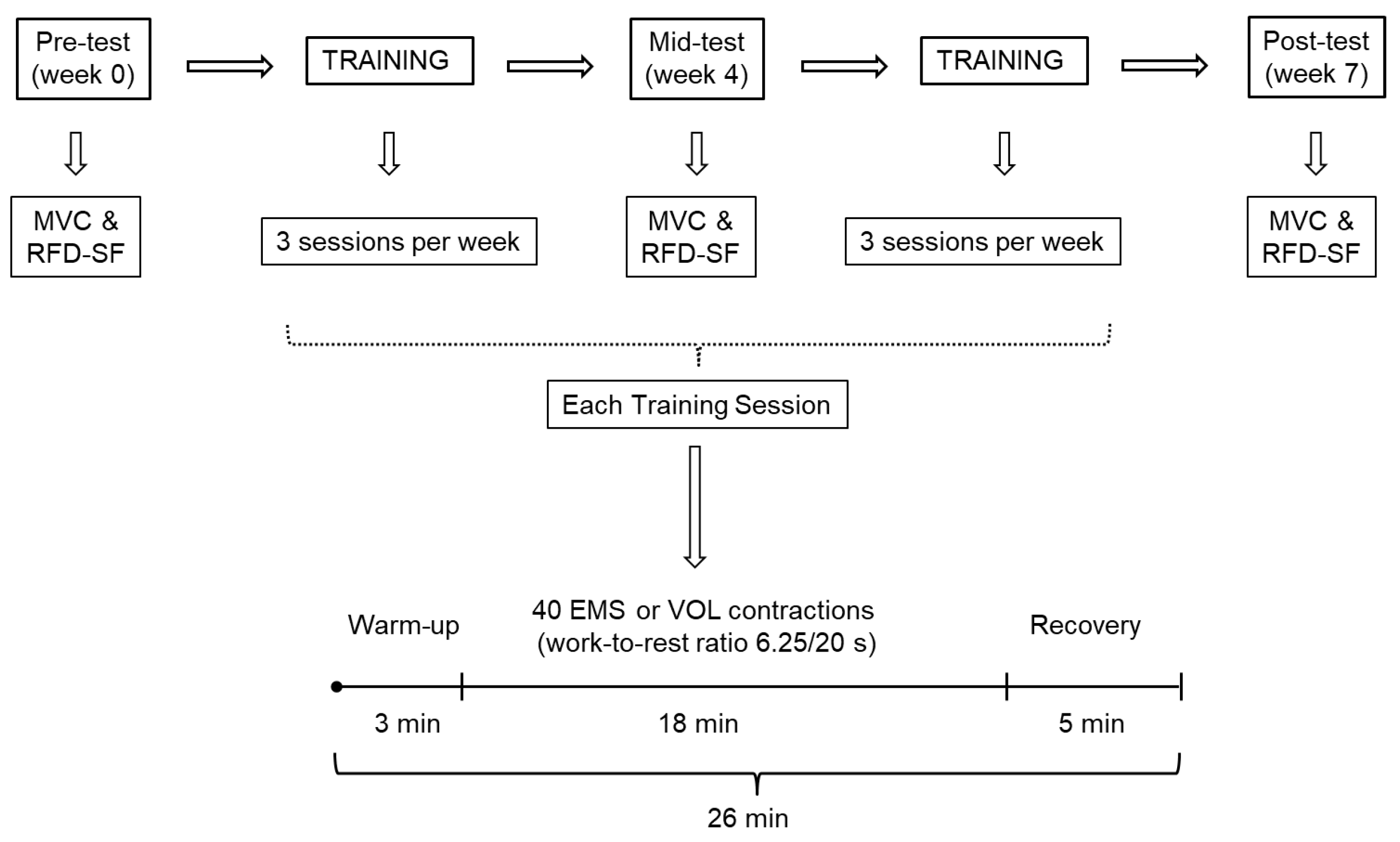
2.1. Study Design

This study is an interventional clinical trial with factorial assignment (registered within ClinicalTrials.gov, identifier: NCT04624438). For the sake of simplicity, a pre–post design with two random parallel groups (both experimental) was used. Quadriceps femoris (QF) function was assessed by MVC and RTD-SF on three occasions: prior to the intervention (pre-test), after 3 weeks of training (which served as the adjustment of exercise threshold; mid-test), and after 6 weeks of training (post-test; Figure 1). Isometric training involved either electromyostimulation (EMS) or the voluntary (VOL) exercises of QF. One week prior to intervention, all subjects participated in one practice session to familiarize themselves with stimulation parameters and training protocols and to determine the intensity of EMS needed to achieve 60% of MVC. Prior to the pre-test, body mass and percent of body fat were assessed using a bioelectric impedance method (In Body 720; InBody Co., Ltd. InBody Bldg, 625, Eonju-ro, Gangnam-gu, Seoul, 06106 Korea) and body height with a standard stadiometer. After the pre-test, the participants were randomly assigned into either an EMS or VOL group. Neither participants nor evaluators were blinded to the intervention or the assessment.
Both groups of participants performed unilateral isometric strength training three times per week for six weeks. Training density and intensity were selected in line with previous recommendations and knowing the limitations of EMS [24,25,36]. Participants were advised to refrain from any type of resistance training targeting the leg muscles for the duration of the study. All sessions were performed at the same time of the day for each subject (always between 8 a.m. and 2 p.m.) and under similar environmental conditions (~22 °C and ~60% humidity).

2.2. Subjects

The subjects were recruited by word-of-mouth. Twenty-six physical education students (14 males and 12 females) were randomly assigned to either the EMS or VOL group (7 males and 6 females in each group; mean ± SD: age: 21 ± 1.5 years (range: 20–25 years); body mass: 75 ± 12 kg, body fat: 18.4 ± 5.5%; body height: 175 ± 14 cm in EMS group, and mean ± SD: age: 21 ± 2.5 years (range: 20–25 years); body mass: 77 ± 12 kg, body fat: 16.9 ± 6.1%; body height: 177 ± 12 cm in VOL group). There were no significant between-group differences in any of subjects’ characteristics before and after the intervention (all p > 0.41). All participants were regularly physically active through their standard academic curriculum that included 6 to 8 h of physical activity per week (both moderate and high intensity). Besides being physically active, the participants were required to be free of muscle–skeletal or neurological disease or medication intake and to refrain from changing daily routines with respect to training and sleeping. All participants gave written informed consent that was in accordance with the Declaration of Helsinki and approved by the University Institutional Review Board (02-672-2/09-04-2015).

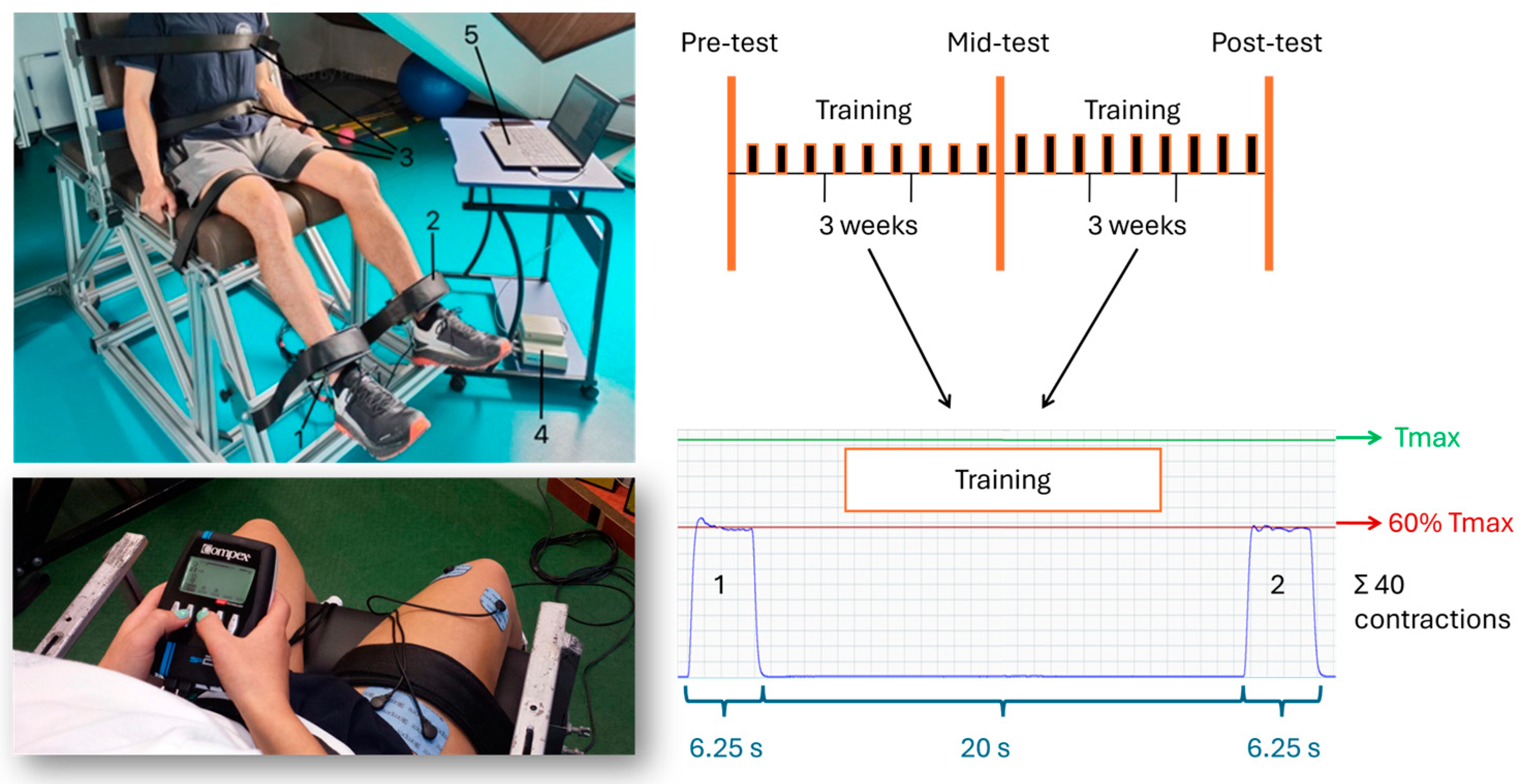
2.3. Testing Procedures

All sessions were conducted on workdays and began with a standardized warm-up consisting of stationary cycling for 5 min and 5 min of active stretching. Following the warm-up, the participants were seated in a custom-made chair with knee and hip angles fixed at 120° and 100°, respectively (Figure 2, upper left panel). The chair was adjusted for every participant with respect to their body size. The participant’s trunk, waist, and thighs were firmly strapped to the chair and the distal parts of their legs were affixed to strain gauge force transducers (Dongguan South China Sea Electronic Co., Ltd., Dongguan, China; load cell S-Type CZL302; range 2 kN) using rigid cuffs. The cuffs were secured with hook and loop fasteners 2 cm above the malleolus lateralis.
All testing procedures were performed separately for each leg. Maximal voluntary contraction (MVC) was always performed prior to RTD-SF testing to determine the greatest value of torque during the performed task [1]. Participants were instructed to extend the knee ‘as fast and as hard as possible’ and each maximal contraction was sustained for approximately 3 s [28]. Three MVCs were completed for each leg with a 60 s rest between successive trials [3,4]. The MVC with the highest value of torque (Tmax) over the three consecutive trials was used for further analysis as well as to prescribe the subsequent submaximal fast contractions that were expressed as a percentage of Tmax.
Following a 5 min rest, the RTD-SF testing protocol was conducted. Subjects completed ~15 submaximal rapid contractions for familiarization purposes. They were instructed to contract their QF muscle as quickly as possible and to relax instantly. Three sets of ≥20 contractions were completed for experimental purposes. In each set, there were 5 consecutive contractions at 4 different intensities presented in ascending order (20%, 40%, 60%, 80% of Tmax, i.e., MVC) [7,8]. The rest time between the sets was 60 s [1]. The experimenter gave a voice command to perform a new trial every 4–5 s. Visual feedback of torque as a target line was provided to the subjects on a computer monitor, but subjects were advised not to pay attention to accuracy.
Data were collected and sampled at 1000 Hz using the commercially available software Isometrics (version 4.0.0, ‘Sports Medical Solutions’, Belgrade, Serbia). Signals were filtered with a low-pass (5 Hz), second-order Butterworth filter. The software automatically calculated the Tmax (peak value on the torque–time trace after reaching the plateau) and RTDmax (peak of first derivative of the torque–time signal overlapping 0.1 s intervals). The peak torque and peak RTD from the isometric torque pulses were also automatically calculated as the maximal value during the 1 s interval of each repetition and as a maximum of the first derivative of the torque–time curve, respectively. Each recording was inspected to ensure that no countermovement was present as this would influence RFD.
Peak torque and peak RTD were expressed as a percentage of Tmax and plotted to obtain a regression line (slope and intercept) and R2 values for each participant, respectively. The slope of the regression line provides the RTD-SF (for more details, please see Figure 2). The R2 represents consistency in the performance rapid muscular contractions [1,12].

2.4. Training Procedures

The training procedures were applied unilaterally over the QF with lower MVC torque at the pre-test, regardless of leg dominance. As a note, there was a significant between-leg difference in Tmax in both groups (10 ± 7%; p < 0.01 in EMS, and 12.5 ± 5%; p < 0.01 in VOL group). Participants’ positioning during training sessions was identical to that during testing procedures. The participant’s trunk, waist, and thighs were firmly strapped to the chair with knee and hip angles fixed at 120° and 100°, respectively. The distal part of the trained leg was affixed to a strain gauge force transducer (Figure 2, upper left panel).
Each EMS and VOL training session consisted of 40 contractions each lasting 6.25 s and separated by 20 s inter-contraction intervals. The number of contractions and contraction-to-rest ratio (6.25/20 s) were in accordance with the work-to-rest pattern produced by the muscle stimulation device (Figure 1 and Figure 2—upper and lower right panels). The stimulation intensity was determined during the familiarization when EMS was delivered at the individually maximal tolerable dose and was kept close to identical among the session, averaging in total ~60% MVC per subject [24]. Specifically, EMS intensity varied between 58% and 63% across sessions and subjects, depending on their daily pain tolerance. Due to anticipated positive exercise effects on Tmax, participants were re-tested after the third week of intervention (mid-test), and these data served to adjust the exercise threshold (please see Figure 2, upper right panel).
The subjects were instructed to stay relaxed during EMS sessions, thus stimulation was not superimposed onto voluntary contractions. A portable battery-powered commercially available stimulator (Compex SP2.0, Medicompex SA, Ecublens, Switzerland) was used in the EMS group. The motor points were identified by stimulating the skin surface with a pen electrode and a large reference electrode placed over the skin. Stimulatory current was gradually increased until a clear muscle twitch was observed. The positions of the motor points were marked with water-resistant markers, so the electrodes were applied at the same sites throughout the intervention period. The positive electrodes, measuring 25 cm2 (5 cm × 5 cm), which had membrane-depolarizing properties, were placed as close as possible to motor points of vastus medialis and vastus lateralis muscles. The negative electrode, measuring 50 cm2 (10 cm × 5 cm), was placed near the proximal insertion of rectus femoris muscle. The stimulator discharged biphasic rectangular pulses lasting 400 μs. The stimulation frequency and duty cycle were 75 Hz and 6.25 s of stimulation followed by a pause lasting 20 s (duty cycle, 24%) [25,37]. The selected stimulation parameters correspond to a maximum strength development program. The stimulation intensity was monitored on-line and determined by the subject at the start of each EMS session according to their pain threshold and to produce a torque corresponding to at least 60% of the pre-test Tmax score [24].
Regarding the VOL group, participants were required to reach the prescribed torque level only through voluntary activation of QF. To attain the same number of contractions and contraction-to-rest ratio as in EMS, automated audible signals were delivered in accordance with the contraction–rest pattern produced by the muscle stimulation device.
Depending on the type of exercise, the subjects were asked to increase the current (please see Figure 2, lower left panel) or voluntary activation intensity throughout the training session to maintain target torque level of ~60% MVC. The exerted torque during each contraction was measured with the same force transducer and software as described for testing procedures, and real-time feedback along with the individual 60% MVC threshold was provided on a computer screen. All training sessions were supervised by at least one of the researchers who conducted the experiment.

2.5. Statistical Analyses
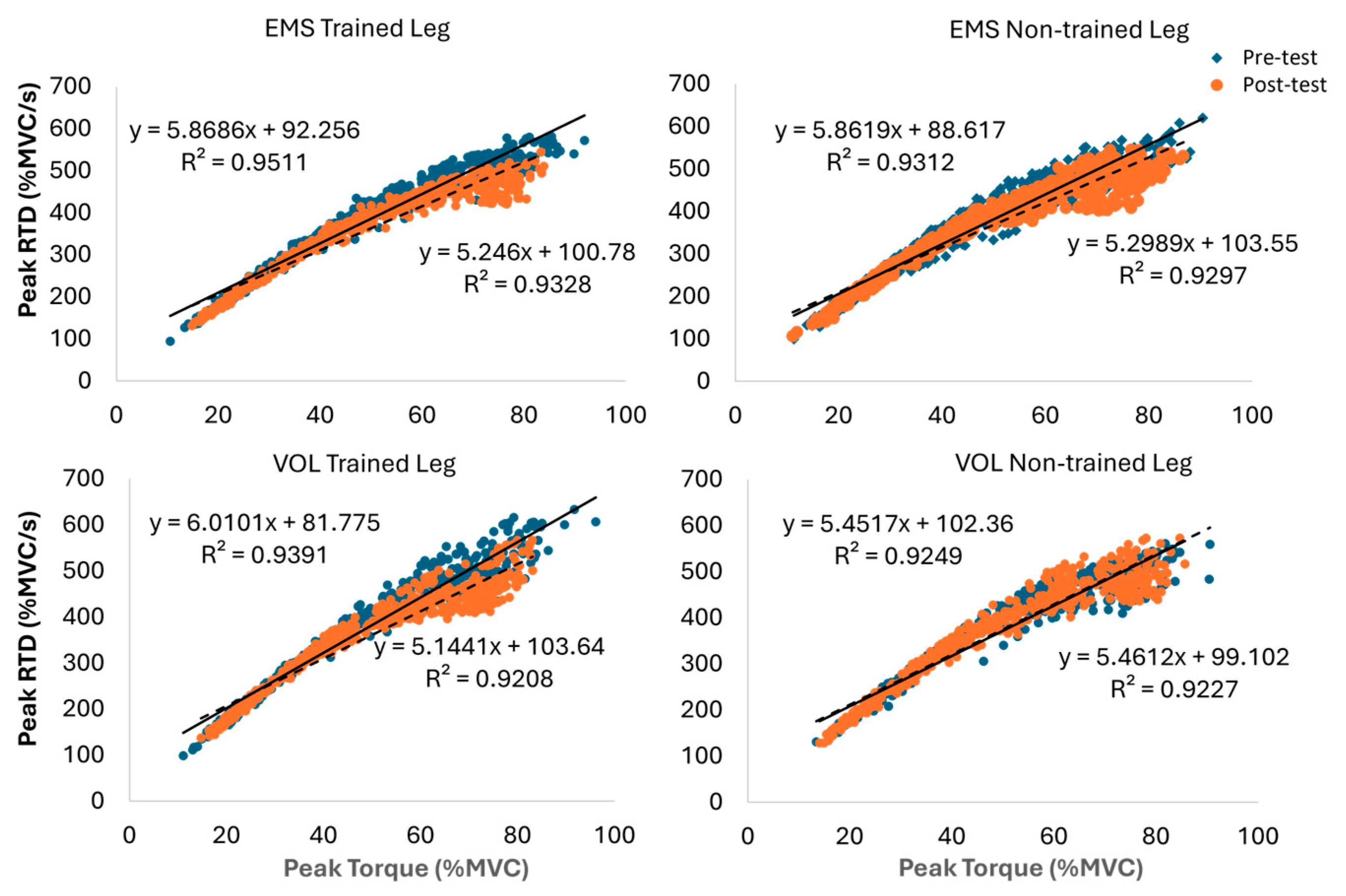
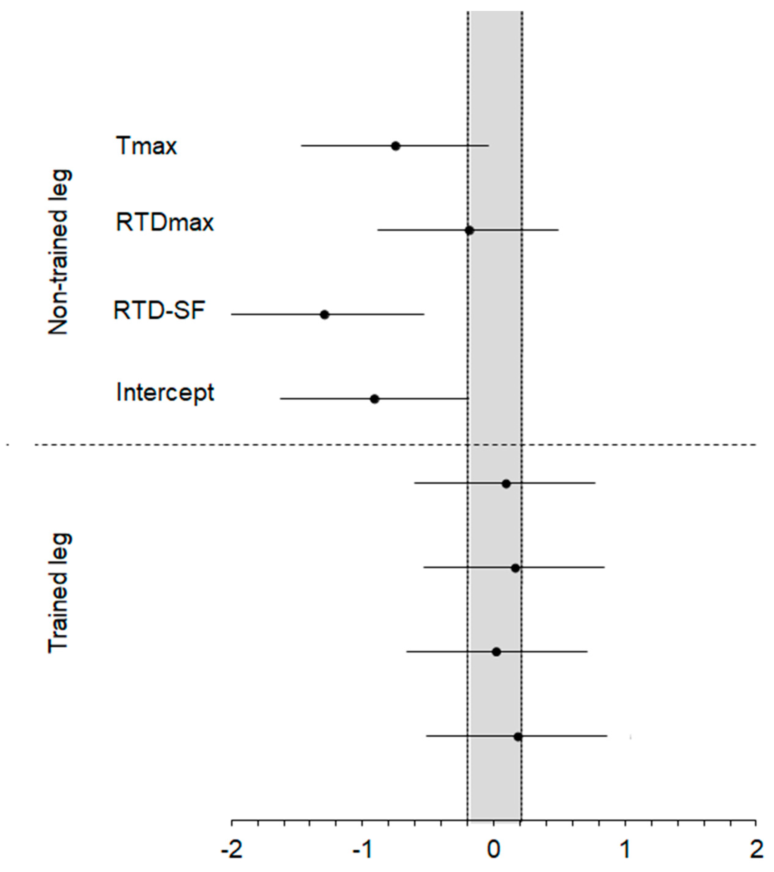
Prior to statistical analyses, Tmax and RTDmax data were normalized with participants’ individual body mass. Normality of the dependent variables was confirmed using a Shapiro–Wilk test (p > 0.05). Descriptive statistics (mean and standard deviation) were calculated for all variables (Tmax, RTDmax, RTD-SF, and Intercept). A 2 × 2 analysis of variance (ANOVA) with time (pre-test and post-test) as within-subject factor and group (EMS and VOL) as between-subject factor was applied to each dependent variable separately for the trained and non-trained leg, followed by the Bonferroni post hoc test. The Cohen’s effect size (d) was used to quantify the within-group differences as d < 0.2 (trivial or no effect), d = 0.2–0.5 (small), d = 0.5–0.8 (moderate), d = 0.8–1.3 (large), and d > 1.3 (very large). Cohen’s d effect size (ES) and 90% confidence interval (CI) were computed using the harmonic mean of the SD of the compared conditions and an ES of 0.20 was considered as the minimal value of practical importance. When the 90% CI overlapped substantial positive and negative values, the effect was deemed unclear; otherwise, effects were deemed clear [38]. p-values < 0.05 were considered statistically significant. The magnitude comparison analysis was performed using a custom Excel spreadsheet (available from: https://www.sportsci.org/jour/03/wghtrials.htm, accessed on 18 September 2023), while other statistical analyses were performed using the software package SPSS (IBM SPSS version 20.0, Chicago, IL, USA).

3. Results

Consistency in the performance of rapid muscular contractions represented by the coefficient of determination of the RTD-SF regression line remained high, from pre-test to post-test (median R2 = 0.951, range 0.936–0.959). Figure 3 depicts the relationship of peak torque relative to Tmax and the respective peak RTD (%Tmax) for trained and non-trained legs in EMS and VOL groups, respectively.
Table 1 depicts the changes in the dependent variables from pre-test and post-test. Regarding the trained leg, ANOVA revealed significant main effect of time for all dependent variables, with generally large effect sizes observed both within EMS and VOL groups (0.78–2.2). The largest change was observed for Tmax (29%), followed by RTDmax (~13%), RTD-SF (~−15%), and Intercept. The main effect of group factor and interaction of factors were not significant, indicating that EMS and VOL exercises induced similar changes over a six-week period, as supported by the analysis of the standardized differences (Figure 3; lower panel), since the ES always ranged from −0.20 to 0.20 (i.e., trivial differences).
For the non-trained leg (Table 1), time factor and group×time interaction were significant only for RTD-SF, whereas a subsequent post hoc test revealed that significant change was observed only within EMS group. However, note that although differences between training modalities reached significance only for RTD-SF, Figure 4 (upper panel) indicates that differences for RTD-SF, Intercept, and Tmax are substantially greater for the EMS than VOL group (ES < −0.20). This is also supported by moderate within-group effect sizes (0.60–0.76), whereas Cohen’s d was trivial across the VOL group.

4. Discussion

The aim of this study was to investigate the changes in the quadriceps femoris muscle rate of torque development scaling factor (RTD-SF) following unilateral isometric training. The training intervention included either electrostimulation or voluntary exercises (EMS and VOL, respectively). The main findings regarding our first hypothesis are as follows: (1) both interventions induced similar decrements in RTD-SF (approximately 15%); (2) Intercept was increased following both training modalities (22 and 17% for EMS and VOL, respectively) but due to the high variability the change was significant only following EMS; (3) both interventions increased Tmax by ~30%; and (4) comparable increments, although of lower magnitude (14%), were observed for RTDmax. Our second hypothesis was not confirmed because the cross-education effect to the non-trained leg was observed only for the EMS group.
As was hypothesized, the applied training interventions had a significant effect on the RTD-SF. In fact, to our surprise, both EMS and VOL induced a decrease in RTD-SF, which could be partly explained by a higher relative increase in Tmax than in RTDmax. Although previous studies revealed that the RTD-SF protocol could be used to discriminate between participants of different ages [14], sexes [13], fatigue or physical activity levels [11,15], or inter-limb asymmetries [7,39], currently no published data are available regarding how strength training interventions affect the RTD-SF. The study that investigated changes in RTD-SF following exercise intervention was conducted on active older adults and it involved a 6-week high-intensity interval cycling intervention [12]. Hence, the authors reported a 34% improvement in RTD-SF (i.e., a higher slope) of QF in the exercise group but not in controls. This is not surprising since robust improvements in explosive capacities are expected following both high- and low-speed dynamic exercises and ballistic isometric exercises performed against lower load (e.g., <60% of maximum torque) and with the intent of rapid torque production [5]. In the current study, it seems that rapid torque generation was unaffected by training intervention for intensities < 50% MVC (Figure 2). However, a visible decrease in rapid production can be observed for higher intensities (50–80% MVC) that we suspect resulted in lower RTD-SF when compared to pre-test values. Considering that isometric exercises used in this study were not as ballistic as in the RFD-SF assessment protocol, the applied training modalities had a significant negative impact on neuromuscular quickness assessed through RTD-SF. This finding could be of importance for researchers investigating muscle function and for coaches working with speed-trained athletes who may consider using moderate-intensity isometric contractions in their training or rehabilitation routines as well.
Intercept is the variable that has mostly stayed unreported or not discussed as it was considered to provide no additional information [1,2,6,12]. A study that investigated how cognitive fatigue affects RFD-SF presented rarely reported results on Intercept that remained unaffected [16]. Kozinc et al. [2] speculated that a higher Intercept together with the same RFD-SF could indicate superior quickness across the range of contraction intensities, which is only partly the case in our study since Intercept increased while the slope, i.e., RFD-SF, declined. Nevertheless, more studies are needed to answer the question on the physiological and potentially clinical relevance of the Intercept.
Regarding maximal strength and RTD gains, it should be noted that the level of the electrically evoked torque achieved in this study ranged between 58% and 63% MVC (the average intensity across participants was 60% MVC). The intensity was selected following previous recommendations and knowing the limitations of EMS [24,25,36]. Specifically, to promote strength gains, EMS was previously most commonly applied at intensities of ~60% MVC [24]. To ensure a comparable training volume between the two training modalities we matched the contraction intensity and duration for the EMS and VOL groups. Although higher voluntary exercise loads are recommended to promote muscle hypertrophy and strength gains [40], significant strength gains could be achieved even when using medium loads (50–60% MVC) [25,41,42,43,44]. In fact, Szeto et al. [44] demonstrated a 31% increase in MVC following isometric VOL training at 50% MVC, while Filipovic et al. [24] reported gains in maximum strength of 32.6 ± 17.6% in trained subjects and of 32 ± 15.6% in elite athletes following isometric EMS. Nonetheless, the results obtained in the current study are in line with those reported previously [24,25,42,43,44] and show no substantial difference in MVC torque gains between VOL and EMS training modalities.
While MVC Tmax was among the most investigated variables in EMS studies [23,24], changes in RTDmax have been scarcely reported. For example, Speicher et al. [45] demonstrated significant increases of up to 16% in the QF muscle during the early time sections of RTD (100–200 ms from the contraction onset) but not in maximal RTD after dynamic whole-body combination EMS training [24]. That is not surprising since it is well known that performing isometric exercises in a ballistic manner can maximize the improvement of RTD [40]. Nevertheless, as shown in the current study, an increase in RTD max (15% for both training modalities) is warranted even when isometric exercises are performed on a single muscle group and in a non-ballistic manner.
Our second hypothesis and our final findings concern the cross-education to the non-trained leg. A significant change in RTD-SF of the non-trained leg was obtained only following EMS training, suggesting potential central adaptations. Moderate, non-significant change was also observed for the Intercept. Although the cross-education effect is well described in the literature [18,19,26,32], this is the first study to investigate this phenomenon concerning RTD-SF and strength training. In the aforementioned study of Bellumori et al. [12], the authors reported that their lower limb speed-cycling training program had a positive cross-education effect on RFD-SF of non-trained upper limbs. Those who compared the effects of voluntary and stimulated isometric exercises on the studied phenomenon report no difference in strength gains between the two training modalities [26,32] or greater gains following EMS than VOL exercises [34]. In fact, similar strength gains in the non-trained leg were reported both after low-intensity (i.e., 20% MVC [32]) and medium-intensity EMS and VOL training (i.e., 65% MVC [26]). However, the size of the increase may depend upon the type of muscle action. Namely, Hortobágyi et al. [19] reported greater strength gains in contralateral limbs with EMS than with VOL exercises, but their study investigated eccentric contractions. Based on Cohen’s d, our study indicates that changes in Tmax could be larger after EMS than VOL training. To our surprise and contrary to previously published studies [20,26,30,32], it appears that training intensity of isometric VOL exercise applied in the current study (60%) was below the necessary threshold for altering functional properties of corticospinal pathways [19], unlike in the EMS group. Interestingly, a study similar to ours used five sets of eight contractions at 65% of MVC over a 4-week period and reported a similar cross-education effect (21%) following isometric and EMS exercises [20]. We can only speculate the reasons for such findings. One may be that our sample consisted of young adults who were moderately to highly physically active unlike in other studies with young non-active or non-trained [20,32] or habitually active but not specifically trained [26] participants. In addition, the non-trained leg of our participants was the one that had better Tmax at pre-test regardless of dominance, so it is plausible that the selected intensity was not strong enough to induce cross-education in those assigned to the VOL group.
This study is not without limitations. Firstly, we have no EMG recordings to provide information on changes in motor unit recruitment and muscle activation. Also, ultrasound recordings would help investigate potential changes in muscle cross-sectional area. Secondly, it may be of interest to correlate changes in RTD-SF and data on corticospinal excitability, or from other performance tests (jumping, sprinting, kicking, etc.) to accompany the obtained findings. In addition, the derivative of the MVC torque–time curve was not processed in the way that RTD at different time intervals is calculated but rather traditional RTDmax was used to track changes in explosive strength. Finally, the fact that we used a relatively limited sample that not only consisted of males and females but was also physically active may explain why some hypothesized effects were not observed, which may also influence the generalization of our findings.

5. Conclusions

When performed at equivalent workloads (frequency, intensity, volume, and work-to-rest ratio) EMS and VOL isometric exercises may be complementary with respect to changes in muscle function of the trained leg. RTD-SF as a measure of neuromuscular quickness is sensitive to medium-intensity unilateral isometric strength training (60% MVC). Specifically, RFD-SF was negatively affected by applied interventions, although it appears that rapid torque generation was unaffected for intensities < 50% MVC, while a decrease was observed for higher intensities (50–80% MVC). Regarding the cross-education effect on the non-trained leg, the applied training intensity induced a significant decrease in the RTD-SF following the EMS exercise, whereas such changes did not reach significance in the VOL exercise group. These findings can be of interest both for researchers and coaches when using moderate-intensity isometric training in athletes, as well as for rehabilitation specialists working with individuals recovering from sports-related injuries and other conditions where submaximal and maximal intensities should be avoided.

Author Contributions

O.M.K. and D.M.M. conceived and designed the research. O.M.K. and N.M. conducted experiments. S.D.M. and S.R.F. supervised outcome assessment and contributed equipment and analytical tools. A.G.-R. and N.Š. analyzed data. O.M.K. wrote the manuscript. All authors have read and agreed to the published version of the manuscript.

Funding

This work was supported by institutional support from the Ministry of Education, Science, and Technological Development of the Republic of Serbia under the contract no. 451-03-68/2022-14/200154 (O.M.K. and D.M.M.) and under the contract no. 451-03-9/2022-14/200015 (S.R.F. and S.D.M.).

Institutional Review Board Statement

The study was conducted in accordance with the Declaration of Helsinki and approved by the Institutional Review Board of University of Belgrade (02-672-2/09-04-2015).

Informed Consent Statement

Informed consent was obtained from all subjects involved in the study.

Data Availability Statement

The data that support the findings of this study are available on request from the corresponding author (O.M.K.).

Conflicts of Interest

Author Nejc Šarabon was employed by the company Science to Practice, Ltd. The remaining authors declare that the re-search was conducted in the absence of any commercial or financial relationships that could be construed as a potential conflict of interest.

References

  1. Bellumori, M.; Jaric, S.; Knight, C.A. The Rate of Force Development Scaling Factor (RFD-SF): Protocol, Reliability, and Muscle Comparisons. Exp. Brain Res. 2011, 212, 359–369. [Google Scholar] [CrossRef] [PubMed]
  2. Kozinc, Ž.; Smajla, D.; Šarabon, N. The Rate of Force Development Scaling Factor: A Review of Underlying Factors, Assessment Methods and Potential for Practical Applications. Eur. J. Appl. Physiol. 2022, 122, 861–873. [Google Scholar] [CrossRef] [PubMed]
  3. Knezevic, O.M.; Mirkov, D.M.; Kadija, M.; Nedeljkovic, A.; Jaric, S. Asymmetries in Explosive Strength Following Anterior Cruciate Ligament Reconstruction. Knee 2014, 21, 1039–1045. [Google Scholar] [CrossRef]
  4. Mirkov, D.M.; Knezevic, O.M.; Maffiuletti, N.A.; Kadija, M.; Nedeljkovic, A.; Jaric, S. Contralateral Limb Deficit after ACL-Reconstruction: An Analysis of Early and Late Phase of Rate of Force Development. J. Sports Sci. 2017, 35, 435–440. [Google Scholar] [CrossRef] [PubMed]
  5. Blazevich, A.J.; Wilson, C.J.; Alcaraz, P.E.; Rubio-Arias, J.A. Effects of Resistance Training Movement Pattern and Velocity on Isometric Muscular Rate of Force Development: A Systematic Review with Meta-Analysis and Meta-Regression. Sports Med. 2020, 50, 943–963. [Google Scholar] [CrossRef] [PubMed]
  6. Uygur, M.; de Freitas, P.B.; Barone, D.A. Rate of Force Development and Relaxation Scaling Factors Are Highly Sensitive to Detect Upper Extremity Motor Impairments in Multiple Sclerosis. J. Neurol. Sci. 2020, 408, 116500. [Google Scholar] [CrossRef] [PubMed]
  7. Smajla, D.; Knezevic, O.M.; Mirkov, D.M.; Šarabon, N. Interlimb Asymmetries and Ipsilateral Associations of Plantar Flexors and Knee Extensors Rate-of-Force Development Scaling Factor. Symmetry 2020, 12, 1522. [Google Scholar] [CrossRef]
  8. Šarabon, N.; Knezevic, M.O.; Mirkov, M.D.; Smajla, D. Introduction of Dynamic Rate-of-Force Development Scaling Factor in Progressive Drop Jumps. J. Biomech. 2020, 110, 109980. [Google Scholar] [CrossRef] [PubMed]
  9. Casartelli, N.C.; Lepers, R.; Maffiuletti, N.A. Assessment of the Rate of Force Development Scaling Factor for the Hip Muscles. Muscle Nerve 2014, 50, 932–938. [Google Scholar] [CrossRef]
  10. Mathern, R.M.; Anhorn, M.; Uygur, M. A Novel Method to Assess Rate of Force Relaxation: Reliability and Comparisons with Rate of Force Development across Various Muscles. Eur. J. Appl. Physiol. 2019, 119, 291–300. [Google Scholar] [CrossRef]
  11. Stefanović, Ž.; Kukić, F.; Knežević, O.M.; Šarabon, N.; Mirkov, D.M. Evaluation of the Reduced Protocol for the Assessment of Rate of Force Development Scaling Factor. Symmetry 2023, 15, 1590. [Google Scholar] [CrossRef]
  12. Bellumori, M.; Uygur, M.; Knight, C.A. High-Speed Cycling Intervention Improves Rate-Dependent Mobility in Older Adults. Med. Sci. Sports Exerc. 2017, 49, 106–114. [Google Scholar] [CrossRef] [PubMed]
  13. Corrêa, T.G.C.; Donato, S.V.S.; Lima, K.C.A.; Uygur, M.; Pereira, R.V.; de Freitas, P.B. Age- And Sex-Related Differences in the Maximum Muscle Performance and Rate of Force Development Scaling Factor of Precision Grip Muscles. Motor Control 2020, 24, 274–290. [Google Scholar] [CrossRef] [PubMed]
  14. Bellumori, M.; Jaric, S.; Knight, C.A. Age-Related Decline in the Rate of Force Development Scaling Factor. Motor Control 2013, 17, 370–381. [Google Scholar] [CrossRef] [PubMed]
  15. Boccia, G.; Dardanello, D.; Brustio, P.R.; Tarperi, C.; Festa, L.; Zoppirolli, C.; Pellegrini, B.; Schena, F.; Rainoldi, A. Neuromuscular Fatigue Does Not Impair the Rate of Force Development in Ballistic Contractions of Submaximal Amplitudes. Front. Physiol. 2018, 9, 1503. [Google Scholar] [CrossRef] [PubMed]
  16. Jacquet, T.; Poulin-Charronnat, B.; Bard, P.; Lepers, R. Effect of Mental Fatigue on Hand Force Production Capacities. PLoS ONE 2024, 19, e0298958. [Google Scholar] [CrossRef] [PubMed]
  17. Whittaker, J.L.; Culvenor, A.G.; Juhl, C.B.; Berg, B.; Bricca, A.; Filbay, S.R.; Holm, P.; Macri, E.; Urhausen, A.P.; Ardern, C.L.; et al. OPTIKNEE 2022: Consensus Recommendations to Optimise Knee Health after Traumatic Knee Injury to Prevent Osteoarthritis. Br. J. Sports Med. 2022, 56, 1393–1405. [Google Scholar] [CrossRef] [PubMed]
  18. Hortobágyi, T.; Maffiuletti, N. Neural Adaptations to Electrical Stimulation Strength Training. Eur. J. Appl. Physiol. 2011, 111, 2439–2449. [Google Scholar] [CrossRef]
  19. Hortobágyi, T.; Scott, K.; Lambert, J.; Hamilton, G.; Tracy, J. Cross-Education of Muscle Strength Is Greater with Stimulated than Voluntary Contractions. Motor Control 1999, 3, 205–219. [Google Scholar] [CrossRef]
  20. Zhou, S.; Oakman, A.; Davie, A.J. Effects of Unilateral Voluntary and Electromyostimulation Training on Muscular Strength on the Contralateral Limb. Hong Kong J. Sports Med. Sports Sci. 2002, XIV, 1–11. [Google Scholar]
  21. Sonnery-Cottet, B.; Saithna, A.; Quelard, B.; Daggett, M.; Borade, A.; Ouanezar, H.; Thaunat, M.; Blakeney, W.G. Arthrogenic Muscle Inhibition after ACL Reconstruction: A Scoping Review of the Efficacy of Interventions. Br. J. Sports Med. 2019, 53, 289–298. [Google Scholar] [CrossRef] [PubMed]
  22. Colson, S.S.; Martin, A.; Van Hoecke, J. Effects of Electromyostimulation versus Voluntary Isometric Training on Elbow Flexor Muscle Strength. J. Electromyogr. Kinesiol. 2009, 19, e311–e319. [Google Scholar] [CrossRef] [PubMed]
  23. Filipovic, A.; Kleinode, H.; Dorman, U.; Meste, J. Electromyostimulation-a Systematic Review of the Influence of Training Regimens and Stimulation Parameters on Effectiveness in Electromyostimulation Training of Selected Strength Parameters. J. Strength Cond. Res. 2011, 25, 3218–3238. [Google Scholar] [CrossRef] [PubMed]
  24. Filipovic, A.; Kleinöder, H.; Dörmann, U.; Mester, J. Electromyostimulation-a Systematic Review of the Effects of Different Electromyostimulation Methods on Selected Strength Parameters in Trained and Elite Athletes. J. Strength Cond. Res. 2012, 26, 2600–2614. [Google Scholar] [CrossRef] [PubMed]
  25. Natsume, T.; Ozaki, H.; Kakigi, R.; Kobayashi, H.; Naito, H. Effects of Training Intensity in Electromyostimulation on Human Skeletal Muscle. Eur. J. Appl. Physiol. 2018, 118, 1339–1347. [Google Scholar] [CrossRef] [PubMed]
  26. Oakman, A.; Zhou, S.; Davie, A. Cross-Education Effect Observed in Voluntary and Electromyostimulation Strength Training. In Proceedings of the 17 International Symposium on Biomechanics in Sports, Perth, Australia, 30 June–6 July 1999; pp. 401–404. [Google Scholar]
  27. Aagaard, P.; Simonsen, E.B.; Andersen, J.L.; Magnusson, P.; Dyhre-Poulsen, P. Increased Rate of Force Development and Neural Drive of Human Skeletal Muscle Following Resistance Training. J. Appl. Physiol. 2002, 93, 1318–1326. [Google Scholar] [CrossRef] [PubMed]
  28. Andersen, L.L.; Aagaard, P. Influence of Maximal Muscle Strength and Intrinsic Muscle Contractile Properties on Contractile Rate of Force Development. Eur. J. Appl. Physiol. 2006, 96, 46–52. [Google Scholar] [CrossRef] [PubMed]
  29. Mastalerz, A.; Sadowski, J. The Effect of Unilateral Training on Contralateral Limb Power in Young Women and Men. Biol. Sport 2020, 37, 443. [Google Scholar] [CrossRef] [PubMed]
  30. Lee, M.; Carroll, T. Cross Education: Possible Mechanisms for the Contralateral Effects of Unilateral Resistance Training. Sports Med. 2007, 37, 1–14. [Google Scholar] [CrossRef]
  31. Benito-Martínez, E.; Senovilla-Herguedas, D.; de la Torre-Montero, J.; Martínez-Beltrán, M.; Reguera-García, M.; Alonso-Cortés, B. Local and Contralateral Effects after the Application of Neuromuscular Electrostimulation in Lower Limbs. Int. J. Environ. Res. Public Health 2020, 17, 9028. [Google Scholar] [CrossRef]
  32. Kadri, M.A.; Noé, F.; Nouar, M.B.; Paillard, T. Effects of Training Programs Based on Ipsilateral Voluntary and Stimulated Contractions on Muscle Strength and Monopedal Postural Control of the Contralateral Limb. Eur. J. Appl. Physiol. 2017, 117, 1799–1806. [Google Scholar] [CrossRef] [PubMed]
  33. Gondin, J.; Cozzone, P.; Bendahan, D. Is High-Frequency Neuromuscular Electrical Stimulation a Suitable Tool for Muscle Performance Improvement in Both Healthy Humans and Athletes? Eur. J. Appl. Physiol. 2011, 111, 2473–2487. [Google Scholar] [CrossRef] [PubMed]
  34. Green, L.A.; Gabriel, D.A. The Effect of Unilateral Training on Contralateral Limb Strength in Young, Older, and Patient Populations: A Meta-Analysis of Cross Education. Phys. Ther. Rev. 2018, 23, 238–249. [Google Scholar] [CrossRef]
  35. Hester, G.M.; Magrini, M.A.; Colquhoun, R.J.; Barrera-Curiel, A.; Estrada, C.A.; Olmos, A.A.; Bailly, A.R.; Ha, P.L.; Defreitas, J.M. Cross-Education: Effects of Age on Rapid and Maximal Voluntary Contractile Characteristics in Males. Eur. J. Appl. Physiol. 2019, 119, 1313–1322. [Google Scholar] [CrossRef] [PubMed]
  36. Maffiuletti, N.A. Physiological and Methodological Considerations for the Use of Neuromuscular Electrical Stimulation. Eur. J. Appl. Physiol. 2010, 110, 223–234. [Google Scholar] [CrossRef] [PubMed]
  37. Gondin, J.; Guette, M.; Ballay, Y.; Martin, A. Electromyostimulation Training Effects on Neural Drive and Muscle Architecture. Med. Sci. Sports Exerc. 2005, 37, 1291–1299. [Google Scholar] [CrossRef]
  38. Hopkins, W.G.; Marshall, S.W.; Batterham, A.M.; Hanin, J. Progressive Statistics for Studies in Sports Medicine and Exercise Science. Med. Sci. Sports Exerc. 2009, 41, 3–12. [Google Scholar] [CrossRef] [PubMed]
  39. Kozinc, Ž.; Šarabon, N. Inter-Limb Asymmetries in Volleyball Players: Differences between Testing Approaches and Association with Performance. J. Sports Sci. Med. 2020, 19, 745. [Google Scholar] [PubMed]
  40. Lum, D.; Barbosa, T. Brief Review: Effects of Isometric Strength Training on Strength and Dynamic Performance. Int. J. Sports Med. 2019, 40, 363–375. [Google Scholar] [CrossRef]
  41. Rhea, M.R.; Alvar, B.A.; Burkett, L.N.; Ball, S.D. A Meta-Analysis to Determine the Dose Response for Strength Development. Med. Sci. Sports Exerc. 2003, 35, 456–464. [Google Scholar] [CrossRef]
  42. Kanehisa, H.; Nagareda, H.; Kawakami, Y.; Akima, H.; Masani, K.; Kouzaki, M.; Fukunaga, T. Effects of Equivolume Isometric Training Programs Comprising Medium or High Resistance on Muscle Size and Strength. Eur. J. Appl. Physiol. 2002, 87, 112–119. [Google Scholar] [CrossRef] [PubMed]
  43. Ullrich, B.; Holzinger, S.; Soleimani, M.; Pelzer, T.; Stening, J.; Pfeiffer, M. Neuromuscular Responses to 14 Weeks of Traditional and Daily Undulating Resistance Training. Int. J. Sports Med. 2015, 36, 554–562. [Google Scholar] [CrossRef] [PubMed]
  44. Szeto, G.; Strauss, G.; Domenico, G.; Lai, H. The Effect of Training Intensity on Voluntary Isometric Strength Improvement. Aust. J. Physiother. 1989, 35, 210–217. [Google Scholar] [CrossRef] [PubMed]
  45. Speicher, U.; Kleinöder, H. Effektitvitätsprüfung Klassischer Und Moderner Krafttrainingsverfahren—Differentielle Kraftdiagnostik Und Moderne Trainingsregulation. F.I.T-Wissenschaftsmag. Dtsch. Sporthochsch. Köln 2009, 14, 20–23. [Google Scholar]
Figure 1. An overview of the study.
Applsci 14 06485 g001
Figure 2. Testing and training set-up (upper left panel; 1: force transducer; 2: shanks; 3: rigid straps; 4: acquisition and analog-to-digital conversion unit; 5: monitor with visual feedback). Note that only trained legs were affixed to force transducer during the training sessions. Lower left panel shows positioning of EMS electrodes and subject increasing the current to maintain targeted training intensity. Upper and lower right panels depict training procedure across weeks and visual feedback given during the training sessions (first two contractions out of 40 are marked as “1” and “2”).
Applsci 14 06485 g002
Figure 3. Pre-test and post-test group relationships between peak force relative to Tmax and the respective peak RTD (%Tmax/s) for EMS and VOL, as well as for trained and non-trained leg, respectively.
Applsci 14 06485 g003
Figure 4. Standardized mean differences (90% confidence intervals) for all dependent variables between the EMS and VOL group (ES = EMS mean - VOL mean/SD both). The shaded area represents a trivial difference (ES from -0.20 to 0.20). Tmax—maximum torque; RTDmax—maximum rate of torque development; RTD-SF—rate of torque development scaling factor.
Applsci 14 06485 g004
Table 1. Descriptive data for dependent variables shown as mean ± standard deviation. Mean relative changes between pre-test and post-test (% change) are given as raw values. Within-group effect size is presented with Cohen’s d.
EMSVOLANOVA
Pre-TestPost-Test% ChangeCohen’s dPre-TestPost-Test% ChangeCohen’s dF-Value
Time
F-Value
Group
F-Value
Time × Group
Trained leg
Tmax (Nm/kg)15.7 ± 220.2 ± 3.529.32.1216.6 ± 2.221.4 ± 3.428.82.1182.0 **0.90.0
RTD (Nm/kg/s)83 ± 1393 ± 1712.30.9285 ± 1897 ± 1516.61.1016.4 **0.10.2
RTD-SF6.0 ± 0.65.1 ± 0.7−14.61.105.9 ± 0.85.0 ± 0.6−14.91.0125.1 **0.10.0
Intercept0.9 ± 0.181.07 ± 0.2122.50.780.86 ± 0.291.08 ± 0.1724.90.7212.5 **0.00.1
Non-trained leg
Tmax (Nm/kg)17 ± 1.919 ± 3.611.90.7619.2 ± 2.819.4 ± 2.92.20.203.281.42.1
RTD (Nm/kg/s)87 ± 1689 ± 2020.3496 ± 1894 ± 18−0.60.010.040.90.4
RTD-SF5.9 ± 0.65.1 ± 0.5 −12.60.765.5 ± 0.65.4 ± 0.7−0.10.206.35 *0.05.5 *
Intercept0.94 ± 0.251.10 ± 0.1716.60.601.02 ± 0.170.99 ± 0.24−2.20.251.130.02.7
* Main effect or factor interaction significant at p < 0.05; ** main effect or factor interaction significant at p < 0.01; post hoc significant at p < 0.01.
Disclaimer/Publisher’s Note: The statements, opinions and data contained in all publications are solely those of the individual author(s) and contributor(s) and not of MDPI and/or the editor(s). MDPI and/or the editor(s) disclaim responsibility for any injury to people or property resulting from any ideas, methods, instructions or products referred to in the content.

