
Skin Absorbed Dose Coefficients for Human Legs from Beta Radiation as a Function of Height


Abstract
:1. Introduction
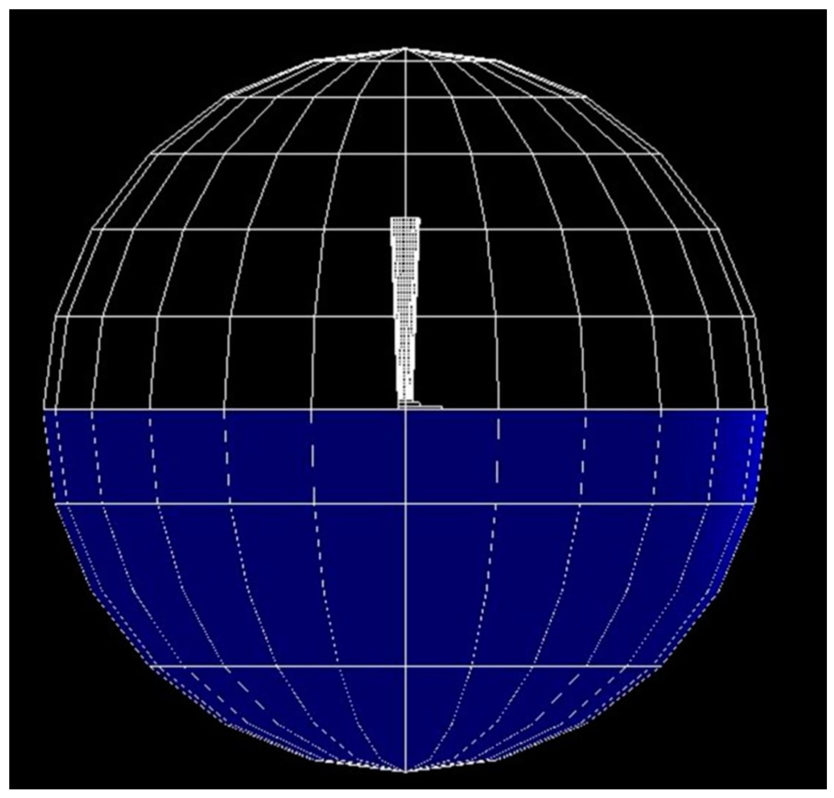
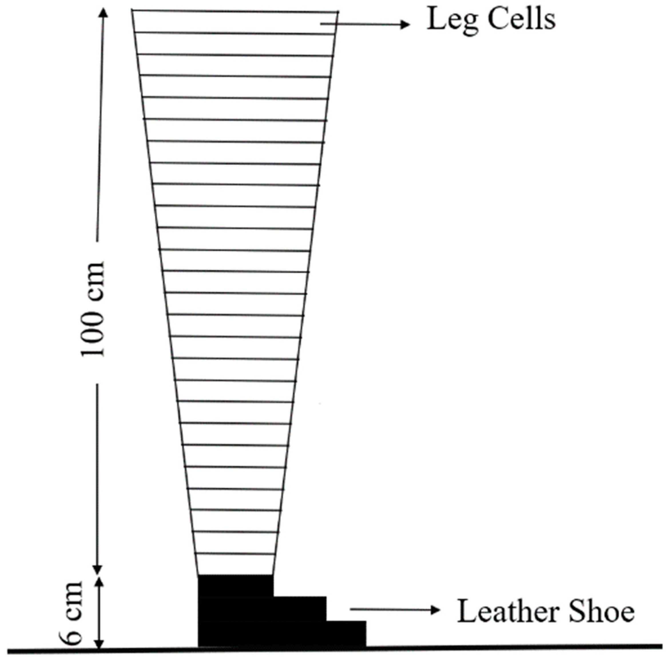
2. Materials and Methods
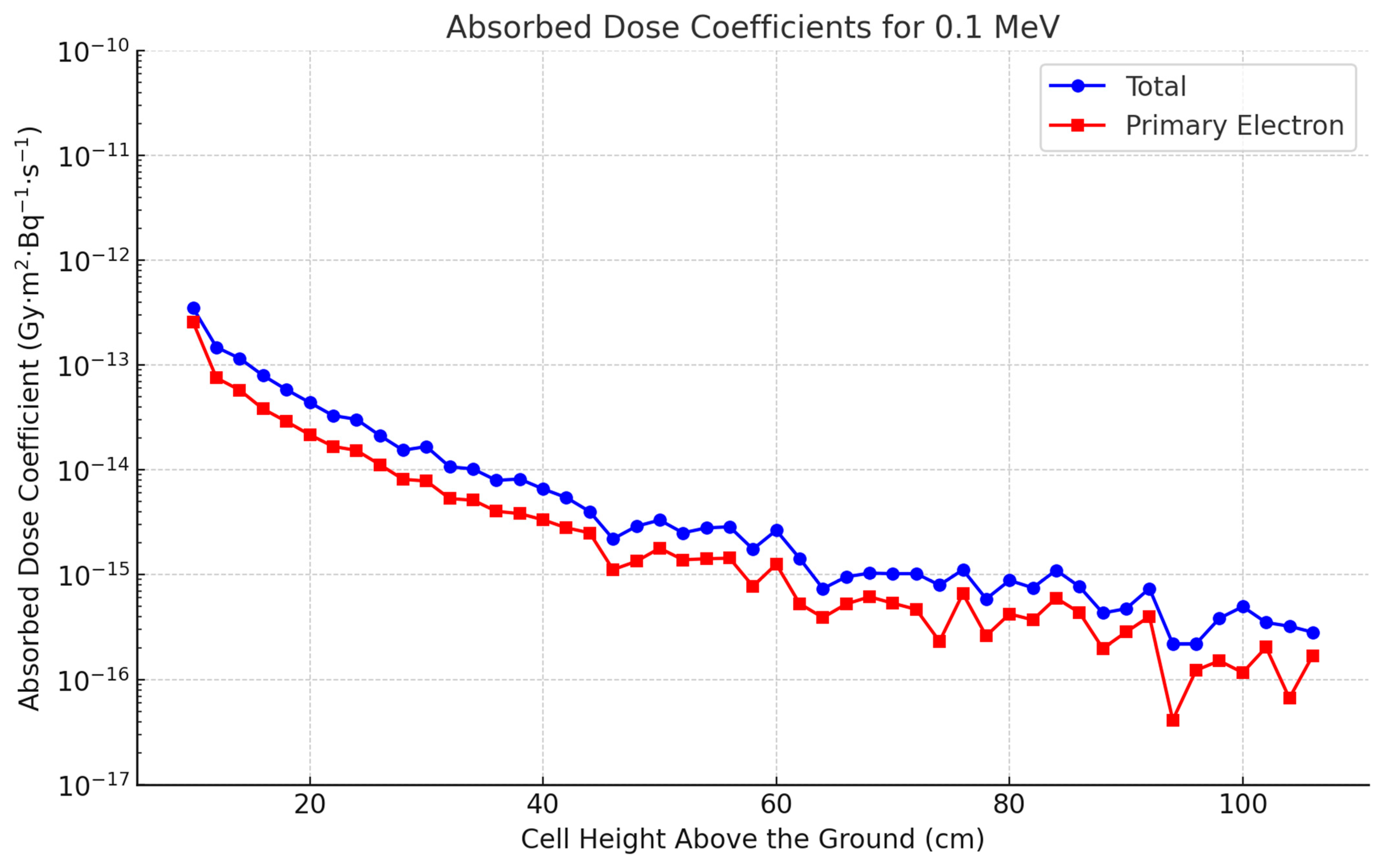
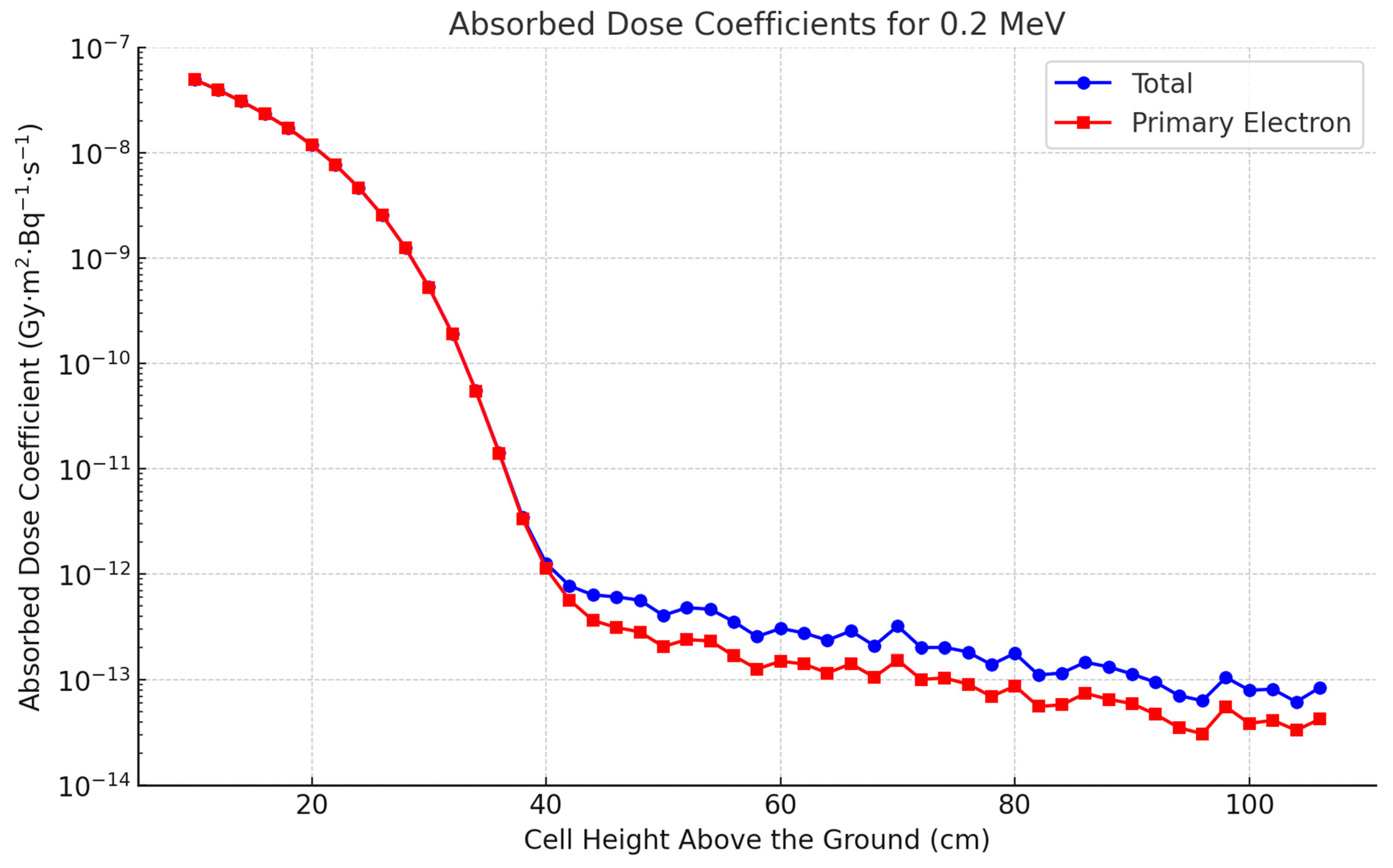
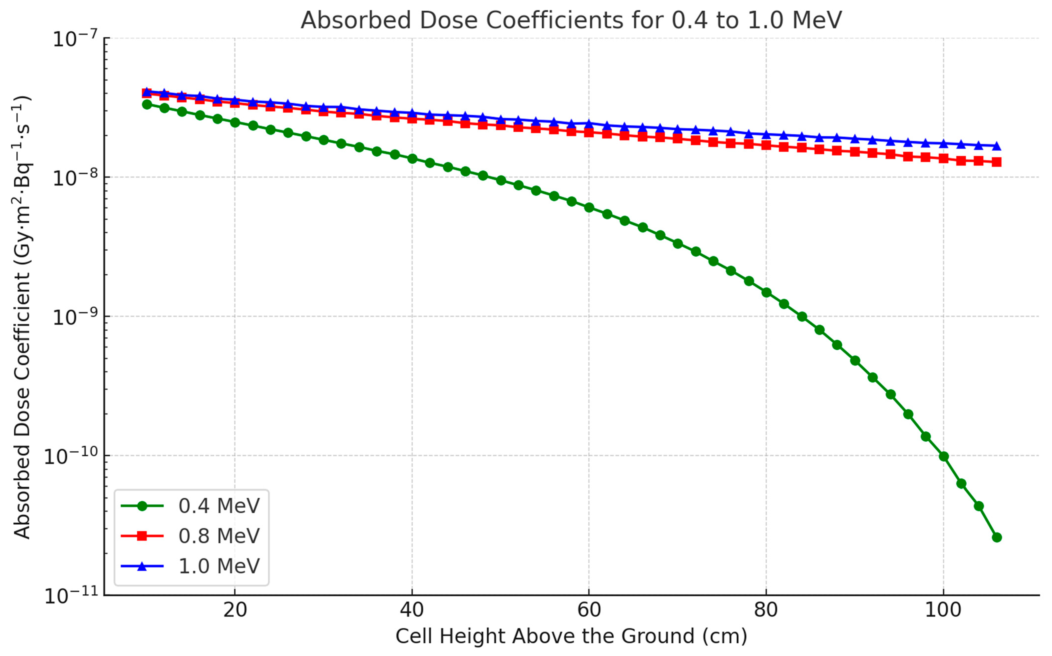
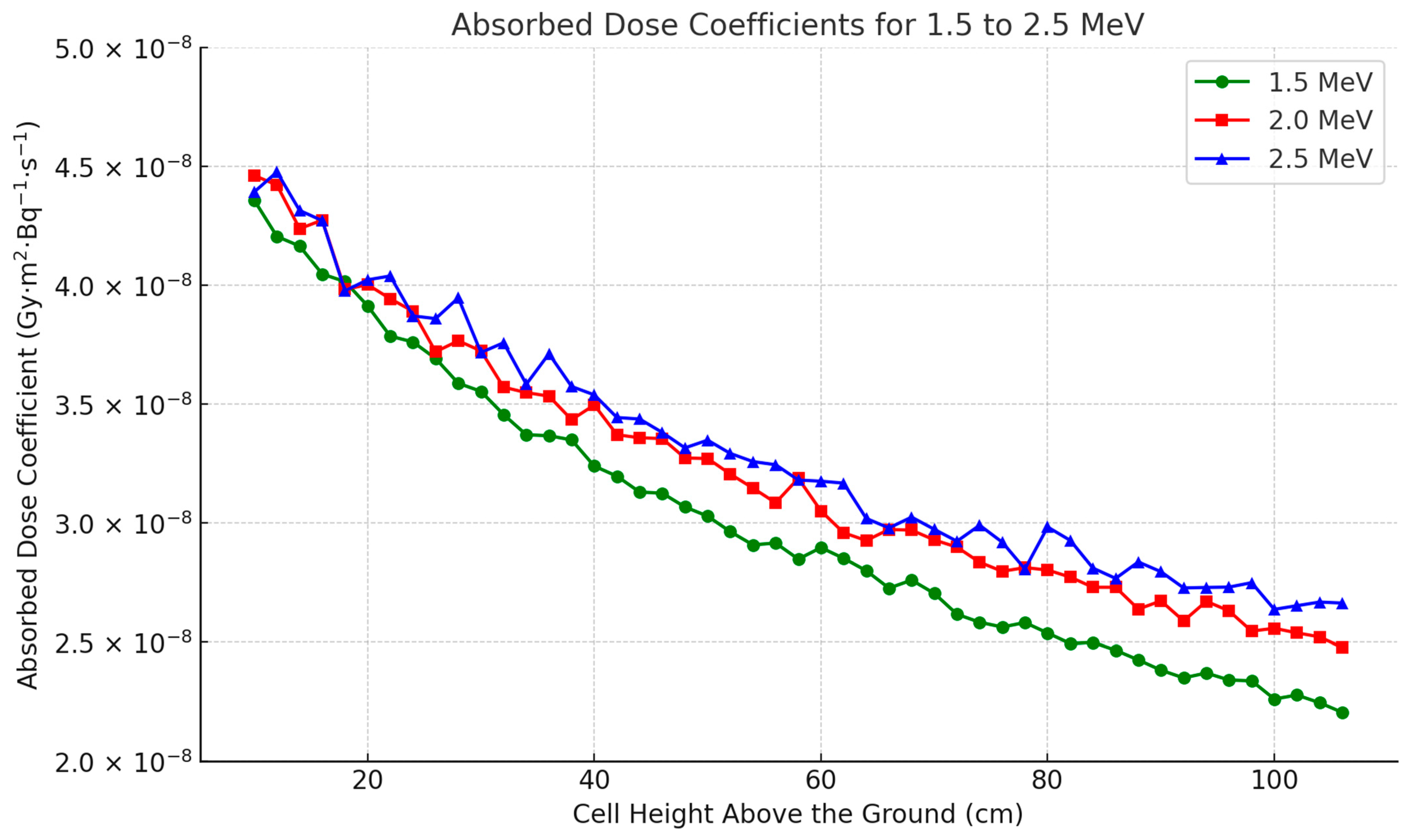
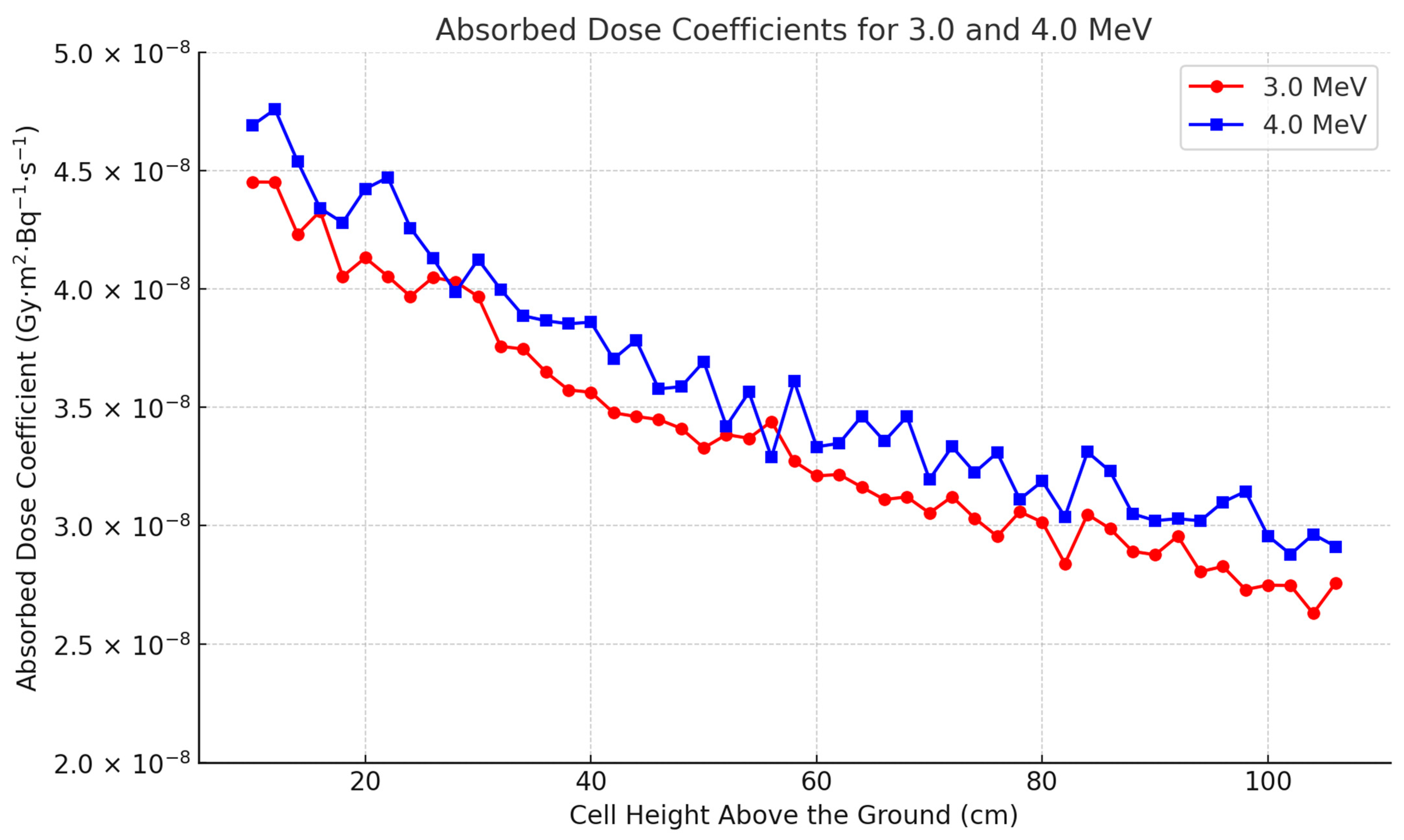
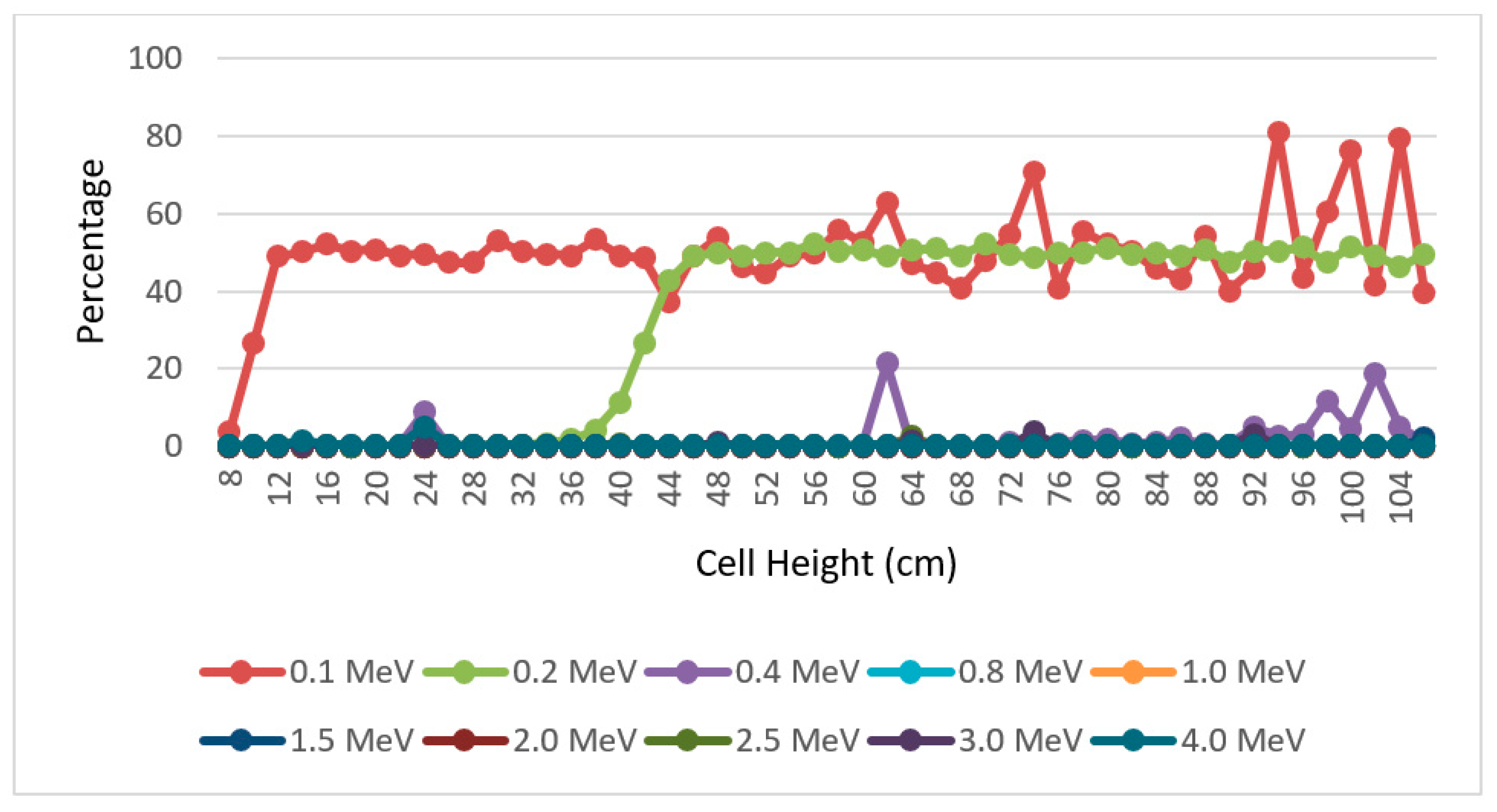
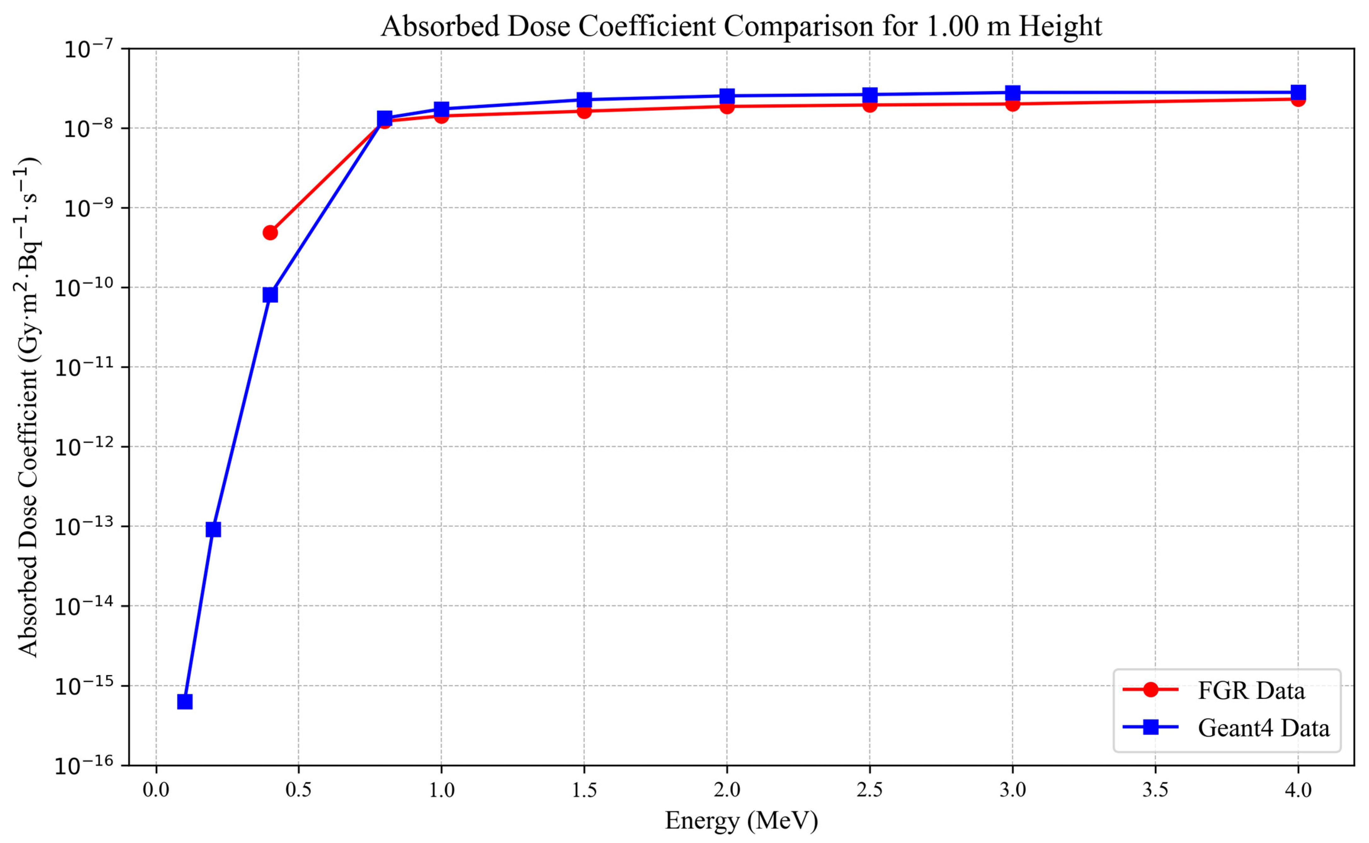
3. Results and Discussion
4. Conclusions
Author Contributions
Funding
Institutional Review Board Statement
Informed Consent Statement
Data Availability Statement
Conflicts of Interest
Appendix A

| Cell Height (cm) | SADC 0.1 MeV (Total) | SADC 0.1 MeV (Electron Only) | SADC 0.2 MeV (Total) | SADC 0.2 MeV (Electron Only) | |||||
|---|---|---|---|---|---|---|---|---|---|
| 8 | 6.27556 × 10⁻¹² | 6.04969 × 10⁻¹² | 5.17971 × 10⁻⁸ | 5.17873 × 10⁻⁸ | |||||
| 10 | 3.49948 × 10⁻¹³ | 2.5631 × 10⁻¹³ | 4.97467 × 10⁻⁸ | 4.97367 × 10⁻⁸ | |||||
| 12 | 1.48091 × 10⁻¹³ | 7.54308 × 10⁻¹⁴ | 3.98042 × 10⁻⁸ | 3.97957 × 10⁻⁸ | |||||
| 14 | 1.15671 × 10⁻¹³ | 5.76044 × 10⁻¹⁴ | 3.09343 × 10⁻⁸ | 3.09269 × 10⁻⁸ | |||||
| 16 | 7.94832 × 10⁻¹⁴ | 3.80907 × 10⁻¹⁴ | 2.34543 × 10⁻⁸ | 2.34479 × 10⁻⁸ | |||||
| 18 | 5.82612 × 10⁻¹⁴ | 2.89362 × 10⁻¹⁴ | 1.72147 × 10⁻⁸ | 1.72092 × 10⁻⁸ | |||||
| 20 | 4.38622 × 10⁻¹⁴ | 2.1619 × 10⁻¹⁴ | 1.18875 × 10⁻⁸ | 1.18829 × 10⁻⁸ | |||||
| 22 | 3.29422 × 10⁻¹⁴ | 1.67262 × 10⁻¹⁴ | 7.76607 × 10⁻⁹ | 7.76232 × 10⁻⁹ | |||||
| 24 | 3.03609 × 10⁻¹⁴ | 1.53336 × 10⁻¹⁴ | 4.68356 × 10⁻⁹ | 4.68057 × 10⁻⁹ | |||||
| 26 | 2.12978 × 10⁻¹⁴ | 1.12308 × 10⁻¹⁴ | 2.57619 × 10⁻⁹ | 2.57389 × 10⁻⁹ | |||||
| 28 | 1.54008 × 10⁻¹⁴ | 8.12283 × 10⁻¹⁵ | 1.253 × 10⁻⁹ | 1.25135 × 10⁻⁹ | |||||
| 30 | 1.6672 × 10⁻¹⁴ | 7.81634 × 10⁻¹⁵ | 5.30977 × 10⁻¹⁰ | 5.29859 × 10⁻¹⁰ | |||||
| 32 | 1.06824 × 10⁻¹⁴ | 5.31659 × 10⁻¹⁵ | 1.92356 × 10⁻¹⁰ | 1.91631 × 10⁻¹⁰ | |||||
| 34 | 1.01953 × 10⁻¹⁴ | 5.12833 × 10⁻¹⁵ | 5.52993 × 10⁻¹¹ | 5.48955 × 10⁻¹¹ | |||||
| 36 | 7.93504 × 10⁻¹⁵ | 4.02001 × 10⁻¹⁵ | 1.42506 × 10⁻¹¹ | 1.40187 × 10⁻¹¹ | |||||
| 38 | 8.1733 × 10⁻¹⁵ | 3.82112 × 10⁻¹⁵ | 3.47246 × 10⁻¹² | 3.3317 × 10⁻¹² | |||||
| 40 | 6.57543 × 10⁻¹⁵ | 3.33496 × 10⁻¹⁵ | 1.26596 × 10⁻¹² | 1.12702 × 10⁻¹² | |||||
| 42 | 5.43535 × 10⁻¹⁵ | 2.79618 × 10⁻¹⁵ | 7.73613 × 10⁻¹³ | 5.67011 × 10⁻¹³ | |||||
| 44 | 3.99769 × 10⁻¹⁵ | 2.504 × 10⁻¹⁵ | 6.33219 × 10⁻¹³ | 3.62858 × 10⁻¹³ | |||||
| 46 | 2.19007 × 10⁻¹⁵ | 1.11618 × 10⁻¹⁵ | 6.07855 × 10⁻¹³ | 3.11515 × 10⁻¹³ | |||||
| 48 | 2.88994 × 10⁻¹⁵ | 1.33791 × 10⁻¹⁵ | 5.65247 × 10⁻¹³ | 2.82757 × 10⁻¹³ | |||||
| 50 | 3.3146 × 10⁻¹⁵ | 1.78671 × 10⁻¹⁵ | 4.05524 × 10⁻¹³ | 2.05199 × 10⁻¹³ | |||||
| 52 | 2.5046 × 10⁻¹⁵ | 1.38472 × 10⁻¹⁵ | 4.80034 × 10⁻¹³ | 2.39384 × 10⁻¹³ | |||||
| 54 | 2.79076 × 10⁻¹⁵ | 1.41675 × 10⁻¹⁵ | 4.64174 × 10⁻¹³ | 2.32038 × 10⁻¹³ | |||||
| 56 | 2.85994 × 10⁻¹⁵ | 1.4356 × 10⁻¹⁵ | 3.54047 × 10⁻¹³ | 1.68791 × 10⁻¹³ | |||||
| 58 | 1.74978 × 10⁻¹⁵ | 7.73286 × 10⁻¹⁶ | 2.55205 × 10⁻¹³ | 1.2679 × 10⁻¹³ | |||||
| 60 | 2.64676 × 10⁻¹⁵ | 1.25784 × 10⁻¹⁵ | 3.05163 × 10⁻¹³ | 1.5015 × 10⁻¹³ | |||||
| 62 | 1.42919 × 10⁻¹⁵ | 5.28574 × 10⁻¹⁶ | 2.76536 × 10⁻¹³ | 1.41442 × 10⁻¹³ | |||||
| 64 | 7.34606 × 10⁻¹⁶ | 3.89442 × 10⁻¹⁶ | 2.35564 × 10⁻¹³ | 1.16183 × 10⁻¹³ | |||||
| 66 | 9.52596 × 10⁻¹⁶ | 5.24889 × 10⁻¹⁶ | 2.88768 × 10⁻¹³ | 1.41663 × 10⁻¹³ | |||||
| 68 | 1.03313 × 10⁻¹⁵ | 6.14273 × 10⁻¹⁶ | 2.09216 × 10⁻¹³ | 1.06419 × 10⁻¹³ | |||||
| 70 | 1.02064 × 10⁻¹⁵ | 5.33462 × 10⁻¹⁶ | 3.21689 × 10⁻¹³ | 1.53746 × 10⁻¹³ | |||||
| 72 | 1.0211 × 10⁻¹⁵ | 4.65846 × 10⁻¹⁶ | 2.00937 × 10⁻¹³ | 1.00923 × 10⁻¹³ | |||||
| 74 | 7.93887 × 10⁻¹⁶ | 2.31195 × 10⁻¹⁶ | 2.01728 × 10⁻¹³ | 1.04154 × 10⁻¹³ | |||||
| 76 | 1.11199 × 10⁻¹⁵ | 6.59262 × 10⁻¹⁶ | 1.82569 × 10⁻¹³ | 9.09997 × 10⁻¹⁴ | |||||
| 78 | 5.86184 × 10⁻¹⁶ | 2.60717 × 10⁻¹⁶ | 1.39463 × 10⁻¹³ | 6.95489 × 10⁻¹⁴ | |||||
| 80 | 8.80337 × 10⁻¹⁶ | 4.21701 × 10⁻¹⁶ | 1.78755 × 10⁻¹³ | 8.73919 × 10⁻¹⁴ | |||||
| 82 | 7.48739 × 10⁻¹⁶ | 3.70453 × 10⁻¹⁶ | 1.11334 × 10⁻¹³ | 5.59451 × 10⁻¹⁴ | |||||
| 84 | 1.10088 × 10⁻¹⁵ | 5.97302 × 10⁻¹⁶ | 1.1604 × 10⁻¹³ | 5.79167 × 10⁻¹⁴ | |||||
| 86 | 7.67459 × 10⁻¹⁶ | 4.3404 × 10⁻¹⁶ | 1.46857 × 10⁻¹³ | 7.44679 × 10⁻¹⁴ | |||||
| 88 | 4.30537 × 10⁻¹⁶ | 1.97844 × 10⁻¹⁶ | 1.32768 × 10⁻¹³ | 6.54113 × 10⁻¹⁴ | |||||
| 90 | 4.73508 × 10⁻¹⁶ | 2.83608 × 10⁻¹⁶ | 1.13431 × 10⁻¹³ | 5.94924 × 10⁻¹⁴ | |||||
| 92 | 7.37274 × 10⁻¹⁶ | 3.98758 × 10⁻¹⁶ | 9.47813 × 10⁻¹⁴ | 4.70627 × 10⁻¹⁴ | |||||
| 94 | 2.18959 × 10⁻¹⁶ | 4.14658 × 10⁻¹⁷ | 7.08436 × 10⁻¹⁴ | 3.51179 × 10⁻¹⁴ | |||||
| 96 | 2.1946 × 10⁻¹⁶ | 1.23618 × 10⁻¹⁶ | 6.3413 × 10⁻¹⁴ | 3.08134 × 10⁻¹⁴ | |||||
| 98 | 3.8332 × 10⁻¹⁶ | 1.51884 × 10⁻¹⁶ | 1.05418 × 10⁻¹³ | 5.54133 × 10⁻¹⁴ | |||||
| 100 | 4.95157 × 10⁻¹⁶ | 1.17279 × 10⁻¹⁶ | 7.97614 × 10⁻¹⁴ | 3.86298 × 10⁻¹⁴ | |||||
| 102 | 3.49371 × 10⁻¹⁶ | 2.04285 × 10⁻¹⁶ | 8.14804 × 10⁻¹⁴ | 4.13479 × 10⁻¹⁴ | |||||
| 104 | 3.22285 × 10⁻¹⁶ | 6.69031 × 10⁻¹⁷ | 6.17878 × 10⁻¹⁴ | 3.32707 × 10⁻¹⁴ | |||||
| 106 | 2.82602 × 10⁻¹⁶ | 1.70301 × 10⁻¹⁶ | 8.42694 × 10⁻¹⁴ | 4.23136 × 10⁻¹⁴ | |||||
| SADC 0.4 MeV (Total) | SADC 0.4 MeV (Electron Only) | SADC 0.8 MeV (Total) | SADC 0.8 MeV (Electron Only) | SADC 1.0 MeV (Total) | SADC 1.0 MeV (Electron Only) | ||||
| 3.14306 × 10⁻⁸ | 3.14236 × 10⁻⁸ | 3.71331 × 10⁻⁸ | 3.71206 × 10⁻⁸ | 3.87557 × 10⁻⁸ | 3.87402 × 10⁻⁸ | ||||
| 3.35304 × 10⁻⁸ | 3.35219 × 10⁻⁸ | 3.98432 × 10⁻⁸ | 3.98315 × 10⁻⁸ | 4.13645 × 10⁻⁸ | 4.13495 × 10⁻⁸ | ||||
| 3.15434 × 10⁻⁸ | 3.15365 × 10⁻⁸ | 3.86493 × 10⁻⁸ | 3.86363 × 10⁻⁸ | 4.02664 × 10⁻⁸ | 4.02502 × 10⁻⁸ | ||||
| 2.97547 × 10⁻⁸ | 2.97475 × 10⁻⁸ | 3.73593 × 10⁻⁸ | 3.7347 × 10⁻⁸ | 3.88599 × 10⁻⁸ | 3.88451 × 10⁻⁸ | ||||
| 2.79693 × 10⁻⁸ | 2.79623 × 10⁻⁸ | 3.62962 × 10⁻⁸ | 3.6283 × 10⁻⁸ | 3.82832 × 10⁻⁸ | 3.82679 × 10⁻⁸ | ||||
| 2.63882 × 10⁻⁸ | 2.63822 × 10⁻⁸ | 3.49538 × 10⁻⁸ | 3.4942 × 10⁻⁸ | 3.66882 × 10⁻⁸ | 3.66742 × 10⁻⁸ | ||||
| 2.49211 × 10⁻⁸ | 2.49149 × 10⁻⁸ | 3.40601 × 10⁻⁸ | 3.4049 × 10⁻⁸ | 3.60246 × 10⁻⁸ | 3.60108 × 10⁻⁸ | ||||
| 2.34914 × 10⁻⁸ | 2.34861 × 10⁻⁸ | 3.2964 × 10⁻⁸ | 3.2953 × 10⁻⁸ | 3.49379 × 10⁻⁸ | 3.49242 × 10⁻⁸ | ||||
| 2.2119 × 10⁻⁸ | 2.40647 × 10⁻⁸ | 3.21937 × 10⁻⁸ | 3.2183 × 10⁻⁸ | 3.44987 × 10⁻⁸ | 3.44853 × 10⁻⁸ | ||||
| 2.08857 × 10⁻⁸ | 2.08806 × 10⁻⁸ | 3.14558 × 10⁻⁸ | 3.14448 × 10⁻⁸ | 3.37317 × 10⁻⁸ | 3.37177 × 10⁻⁸ | ||||
| 1.96906 × 10⁻⁸ | 1.96857 × 10⁻⁸ | 3.0552 × 10⁻⁸ | 3.05417 × 10⁻⁸ | 3.25048 × 10⁻⁸ | 3.24921 × 10⁻⁸ | ||||
| 1.85601 × 10⁻⁸ | 1.85561 × 10⁻⁸ | 2.95743 × 10⁻⁸ | 2.95642 × 10⁻⁸ | 3.1991 × 10⁻⁸ | 3.19789 × 10⁻⁸ | ||||
| 1.74731 × 10⁻⁸ | 1.7469 × 10⁻⁸ | 2.89703 × 10⁻⁸ | 2.89606 × 10⁻⁸ | 3.19365 × 10⁻⁸ | 3.1924 × 10⁻⁸ | ||||
| 1.65135 × 10⁻⁸ | 1.65095 × 10⁻⁸ | 2.84946 × 10⁻⁸ | 2.84851 × 10⁻⁸ | 3.07177 × 10⁻⁸ | 3.07057 × 10⁻⁸ | ||||
| 1.5453 × 10⁻⁸ | 1.54487 × 10⁻⁸ | 2.75534 × 10⁻⁸ | 2.75434 × 10⁻⁸ | 3.00535 × 10⁻⁸ | 3.00419 × 10⁻⁸ | ||||
| 1.461 × 10⁻⁸ | 1.46061 × 10⁻⁸ | 2.68904 × 10⁻⁸ | 2.68806 × 10⁻⁸ | 2.93964 × 10⁻⁸ | 2.93847 × 10⁻⁸ | ||||
| 1.36546 × 10⁻⁸ | 1.36511 × 10⁻⁸ | 2.62921 × 10⁻⁸ | 2.62825 × 10⁻⁸ | 2.89998 × 10⁻⁸ | 2.89882 × 10⁻⁸ | ||||
| 1.26872 × 10⁻⁸ | 1.26832 × 10⁻⁸ | 2.58724 × 10⁻⁸ | 2.58633 × 10⁻⁸ | 2.81526 × 10⁻⁸ | 2.81412 × 10⁻⁸ | ||||
| 1.18984 × 10⁻⁸ | 1.18952 × 10⁻⁸ | 2.53118 × 10⁻⁸ | 2.53028 × 10⁻⁸ | 2.78858 × 10⁻⁸ | 2.78741 × 10⁻⁸ | ||||
| 1.10589 × 10⁻⁸ | 1.10561 × 10⁻⁸ | 2.44213 × 10⁻⁸ | 2.44132 × 10⁻⁸ | 2.76 × 10⁻⁸ | 2.75892 × 10⁻⁸ | ||||
| 1.02906 × 10⁻⁸ | 1.02877 × 10⁻⁸ | 2.3911 × 10⁻⁸ | 2.39023 × 10⁻⁸ | 2.71368 × 10⁻⁸ | 2.71264 × 10⁻⁸ | ||||
| 9.50825 × 10⁻⁹ | 9.50546 × 10⁻⁹ | 2.35808 × 10⁻⁸ | 2.35723 × 10⁻⁸ | 2.61447 × 10⁻⁸ | 2.61337 × 10⁻⁸ | ||||
| 8.75714 × 10⁻⁹ | 8.75464 × 10⁻⁹ | 2.28001 × 10⁻⁸ | 2.27923 × 10⁻⁸ | 2.59255 × 10⁻⁸ | 2.59152 × 10⁻⁸ | ||||
| 8.06402 × 10⁻⁹ | 8.06152 × 10⁻⁹ | 2.24159 × 10⁻⁸ | 2.24083 × 10⁻⁸ | 2.53628 × 10⁻⁸ | 2.53527 × 10⁻⁸ | ||||
| 7.37938 × 10⁻⁹ | 7.37738 × 10⁻⁹ | 2.19578 × 10⁻⁸ | 2.195 × 10⁻⁸ | 2.50483 × 10⁻⁸ | 2.50381 × 10⁻⁸ | ||||
| 6.7417 × 10⁻⁹ | 6.73961 × 10⁻⁹ | 2.13707 × 10⁻⁸ | 2.13629 × 10⁻⁸ | 2.42554 × 10⁻⁸ | 2.42455 × 10⁻⁸ | ||||
| 6.06397 × 10⁻⁹ | 6.06218 × 10⁻⁹ | 2.10356 × 10⁻⁸ | 2.10278 × 10⁻⁸ | 2.44312 × 10⁻⁸ | 2.4421 × 10⁻⁸ | ||||
| 5.4638 × 10⁻⁹ | 6.62567 × 10⁻⁹ | 2.05911 × 10⁻⁸ | 2.05836 × 10⁻⁸ | 2.35574 × 10⁻⁸ | 2.35477 × 10⁻⁸ | ||||
| 4.8893 × 10⁻⁹ | 4.88762 × 10⁻⁹ | 2.00101 × 10⁻⁸ | 2.0003 × 10⁻⁸ | 2.31366 × 10⁻⁸ | 2.31273 × 10⁻⁸ | ||||
| 4.37482 × 10⁻⁹ | 4.37343 × 10⁻⁹ | 1.95563 × 10⁻⁸ | 1.95495 × 10⁻⁸ | 2.29404 × 10⁻⁸ | 2.29308 × 10⁻⁸ | ||||
| 3.83669 × 10⁻⁹ | 3.8354 × 10⁻⁹ | 1.92875 × 10⁻⁸ | 1.92804 × 10⁻⁸ | 2.26207 × 10⁻⁸ | 2.26112 × 10⁻⁸ | ||||
| 3.3598 × 10⁻⁹ | 3.35854 × 10⁻⁹ | 1.88643 × 10⁻⁸ | 1.88577 × 10⁻⁸ | 2.2127 × 10⁻⁸ | 2.21175 × 10⁻⁸ | ||||
| 2.92522 × 10⁻⁹ | 2.90008 × 10⁻⁹ | 1.83822 × 10⁻⁸ | 1.83754 × 10⁻⁸ | 2.19156 × 10⁻⁸ | 2.19063 × 10⁻⁸ | ||||
| 2.49481 × 10⁻⁹ | 2.48722 × 10⁻⁹ | 1.79079 × 10⁻⁸ | 1.79012 × 10⁻⁸ | 2.16257 × 10⁻⁸ | 2.16162 × 10⁻⁸ | ||||
| 2.13793 × 10⁻⁹ | 2.12477 × 10⁻⁹ | 1.75758 × 10⁻⁸ | 1.75693 × 10⁻⁸ | 2.13088 × 10⁻⁸ | 2.12997 × 10⁻⁸ | ||||
| 1.80001 × 10⁻⁹ | 1.77208 × 10⁻⁹ | 1.73691 × 10⁻⁸ | 1.73626 × 10⁻⁸ | 2.05987 × 10⁻⁸ | 2.05897 × 10⁻⁸ | ||||
| 1.49755 × 10⁻⁹ | 1.5238 × 10⁻⁹ | 1.69167 × 10⁻⁸ | 1.69104 × 10⁻⁸ | 2.03303 × 10⁻⁸ | 2.03218 × 10⁻⁸ | ||||
| 1.23233 × 10⁻⁹ | 1.24209 × 10⁻⁹ | 1.65557 × 10⁻⁸ | 1.65498 × 10⁻⁸ | 2.00776 × 10⁻⁸ | 2.00692 × 10⁻⁸ | ||||
| 1.00011 × 10⁻⁹ | 9.91292 × 10⁻¹⁰ | 1.62468 × 10⁻⁸ | 1.62404 × 10⁻⁸ | 1.97985 × 10⁻⁸ | 1.97902 × 10⁻⁸ | ||||
| 8.01972 × 10⁻¹⁰ | 8.18378 × 10⁻¹⁰ | 1.58755 × 10⁻⁸ | 1.58696 × 10⁻⁸ | 1.93064 × 10⁻⁸ | 1.9298 × 10⁻⁸ | ||||
| 6.26823 × 10⁻¹⁰ | 6.30403 × 10⁻¹⁰ | 1.54783 × 10⁻⁸ | 1.54723 × 10⁻⁸ | 1.92712 × 10⁻⁸ | 1.92627 × 10⁻⁸ | ||||
| 4.83981 × 10⁻¹⁰ | 4.82477 × 10⁻¹⁰ | 1.52549 × 10⁻⁸ | 1.52493 × 10⁻⁸ | 1.88957 × 10⁻⁸ | 1.88879 × 10⁻⁸ | ||||
| 3.66043 × 10⁻¹⁰ | 3.83834 × 10⁻¹⁰ | 1.49319 × 10⁻⁸ | 1.49263 × 10⁻⁸ | 1.862 × 10⁻⁸ | 1.86122 × 10⁻⁸ | ||||
| 2.75858 × 10⁻¹⁰ | 2.6864 × 10⁻¹⁰ | 1.46126 × 10⁻⁸ | 1.46071 × 10⁻⁸ | 1.82247 × 10⁻⁸ | 1.8217 × 10⁻⁸ | ||||
| 1.99562 × 10⁻¹⁰ | 1.93692 × 10⁻¹⁰ | 1.40584 × 10⁻⁸ | 1.40531 × 10⁻⁸ | 1.79156 × 10⁻⁸ | 1.79077 × 10⁻⁸ | ||||
| 1.38245 × 10⁻¹⁰ | 1.2183 × 10⁻¹⁰ | 1.39193 × 10⁻⁸ | 1.39139 × 10⁻⁸ | 1.76165 × 10⁻⁸ | 1.76088 × 10⁻⁸ | ||||
| 9.95857 × 10⁻¹¹ | 1.04006 × 10⁻¹⁰ | 1.36202 × 10⁻⁸ | 1.3615 × 10⁻⁸ | 1.75089 × 10⁻⁸ | 1.75014 × 10⁻⁸ | ||||
| 6.3325 × 10⁻¹¹ | 7.51687 × 10⁻¹¹ | 1.31612 × 10⁻⁸ | 1.3156 × 10⁻⁸ | 1.72524 × 10⁻⁸ | 1.72449 × 10⁻⁸ | ||||
| 4.36845 × 10⁻¹¹ | 4.576 × 10⁻¹¹ | 1.3083 × 10⁻⁸ | 1.3078 × 10⁻⁸ | 1.69807 × 10⁻⁸ | 1.69733 × 10⁻⁸ | ||||
| 2.60868 × 10⁻¹¹ | 2.57599 × 10⁻¹¹ | 1.28412 × 10⁻⁸ | 1.28362 × 10⁻⁸ | 1.68421 × 10⁻⁸ | 1.68348 × 10⁻⁸ | ||||
| SADC 1.5 MeV (Total) | SADC 1.5 MeV (Electron Only) | SADC 2.0 MeV (Total) | SADC 2.0 MeV (Electron Only) | SADC 2.5 MeV (Total) | SADC 2.5 MeV (Electron Only) | ||||
| 4.02822 × 10⁻⁸ | 4.02612 × 10⁻⁸ | 4.22774 × 10⁻⁸ | 4.22438 × 10⁻⁸ | 4.28428 × 10⁻⁸ | 4.2798 × 10⁻⁸ | ||||
| 4.35781 × 10⁻⁸ | 4.35533 × 10⁻⁸ | 4.46211 × 10⁻⁸ | 4.45885 × 10⁻⁸ | 4.39318 × 10⁻⁸ | 4.38886 × 10⁻⁸ | ||||
| 4.20559 × 10⁻⁸ | 4.20323 × 10⁻⁸ | 4.42184 × 10⁻⁸ | 4.41868 × 10⁻⁸ | 4.47688 × 10⁻⁸ | 4.47216 × 10⁻⁸ | ||||
| 4.16521 × 10⁻⁸ | 4.16298 × 10⁻⁸ | 4.23838 × 10⁻⁸ | 4.23528 × 10⁻⁸ | 4.31496 × 10⁻⁸ | 4.31075 × 10⁻⁸ | ||||
| 4.04743 × 10⁻⁸ | 4.04523 × 10⁻⁸ | 4.27443 × 10⁻⁸ | 4.27131 × 10⁻⁸ | 4.27223 × 10⁻⁸ | 4.26751 × 10⁻⁸ | ||||
| 4.0158 × 10⁻⁸ | 4.01361 × 10⁻⁸ | 3.98282 × 10⁻⁸ | 3.97955 × 10⁻⁸ | 3.97683 × 10⁻⁸ | 3.97307 × 10⁻⁸ | ||||
| 3.91374 × 10⁻⁸ | 3.91137 × 10⁻⁸ | 4.00275 × 10⁻⁸ | 3.99957 × 10⁻⁸ | 4.02321 × 10⁻⁸ | 4.01864 × 10⁻⁸ | ||||
| 3.786 × 10⁻⁸ | 3.78401 × 10⁻⁸ | 3.94357 × 10⁻⁸ | 3.9405 × 10⁻⁸ | 4.0393 × 10⁻⁸ | 4.03481 × 10⁻⁸ | ||||
| 3.76174 × 10⁻⁸ | 3.75969 × 10⁻⁸ | 3.89091 × 10⁻⁸ | 3.88786 × 10⁻⁸ | 3.87127 × 10⁻⁸ | 3.8673 × 10⁻⁸ | ||||
| 3.69208 × 10⁻⁸ | 3.69009 × 10⁻⁸ | 3.72031 × 10⁻⁸ | 3.71758 × 10⁻⁸ | 3.85987 × 10⁻⁸ | 3.85558 × 10⁻⁸ | ||||
| 3.58793 × 10⁻⁸ | 3.58614 × 10⁻⁸ | 3.76659 × 10⁻⁸ | 3.76393 × 10⁻⁸ | 3.94718 × 10⁻⁸ | 3.9432 × 10⁻⁸ | ||||
| 3.55385 × 10⁻⁸ | 3.55182 × 10⁻⁸ | 3.72394 × 10⁻⁸ | 3.72079 × 10⁻⁸ | 3.71736 × 10⁻⁸ | 3.71339 × 10⁻⁸ | ||||
| 3.45592 × 10⁻⁸ | 3.45396 × 10⁻⁸ | 3.57131 × 10⁻⁸ | 3.5686 × 10⁻⁸ | 3.75834 × 10⁻⁸ | 3.75445 × 10⁻⁸ | ||||
| 3.37135 × 10⁻⁸ | 3.3695 × 10⁻⁸ | 3.54866 × 10⁻⁸ | 3.5458 × 10⁻⁸ | 3.58372 × 10⁻⁸ | 3.57988 × 10⁻⁸ | ||||
| 3.36703 × 10⁻⁸ | 3.36508 × 10⁻⁸ | 3.53362 × 10⁻⁸ | 3.53083 × 10⁻⁸ | 3.71197 × 10⁻⁸ | 3.70825 × 10⁻⁸ | ||||
| 3.34957 × 10⁻⁸ | 3.34771 × 10⁻⁸ | 3.43453 × 10⁻⁸ | 3.43203 × 10⁻⁸ | 3.57416 × 10⁻⁸ | 3.57036 × 10⁻⁸ | ||||
| 3.2392 × 10⁻⁸ | 3.23734 × 10⁻⁸ | 3.4965 × 10⁻⁸ | 3.49368 × 10⁻⁸ | 3.53907 × 10⁻⁸ | 3.51735 × 10⁻⁸ | ||||
| 3.1969 × 10⁻⁸ | 3.19518 × 10⁻⁸ | 3.37118 × 10⁻⁸ | 3.36833 × 10⁻⁸ | 3.44374 × 10⁻⁸ | 3.4398 × 10⁻⁸ | ||||
| 3.1301 × 10⁻⁸ | 3.12826 × 10⁻⁸ | 3.35855 × 10⁻⁸ | 3.356 × 10⁻⁸ | 3.4374 × 10⁻⁸ | 3.43377 × 10⁻⁸ | ||||
| 3.12536 × 10⁻⁸ | 3.12355 × 10⁻⁸ | 3.35501 × 10⁻⁸ | 3.35249 × 10⁻⁸ | 3.38086 × 10⁻⁸ | 3.37751 × 10⁻⁸ | ||||
| 3.06733 × 10⁻⁸ | 3.06543 × 10⁻⁸ | 3.27337 × 10⁻⁸ | 3.2707 × 10⁻⁸ | 3.31598 × 10⁻⁸ | 3.31261 × 10⁻⁸ | ||||
| 3.02872 × 10⁻⁸ | 3.02702 × 10⁻⁸ | 3.27121 × 10⁻⁸ | 3.26868 × 10⁻⁸ | 3.34841 × 10⁻⁸ | 3.34462 × 10⁻⁸ | ||||
| 2.96346 × 10⁻⁸ | 2.96192 × 10⁻⁸ | 3.20571 × 10⁻⁸ | 3.20331 × 10⁻⁸ | 3.29261 × 10⁻⁸ | 3.28933 × 10⁻⁸ | ||||
| 2.90777 × 10⁻⁸ | 2.90615 × 10⁻⁸ | 3.14669 × 10⁻⁸ | 3.14416 × 10⁻⁸ | 3.2582 × 10⁻⁸ | 3.25497 × 10⁻⁸ | ||||
| 2.91561 × 10⁻⁸ | 2.91385 × 10⁻⁸ | 3.08501 × 10⁻⁸ | 3.08261 × 10⁻⁸ | 3.24511 × 10⁻⁸ | 3.2419 × 10⁻⁸ | ||||
| 2.84802 × 10⁻⁸ | 2.84642 × 10⁻⁸ | 3.18758 × 10⁻⁸ | 3.18523 × 10⁻⁸ | 3.18086 × 10⁻⁸ | 3.1778 × 10⁻⁸ | ||||
| 2.89623 × 10⁻⁸ | 2.89453 × 10⁻⁸ | 3.05075 × 10⁻⁸ | 3.04845 × 10⁻⁸ | 3.17572 × 10⁻⁸ | 3.17235 × 10⁻⁸ | ||||
| 2.85121 × 10⁻⁸ | 2.84956 × 10⁻⁸ | 2.95877 × 10⁻⁸ | 2.95623 × 10⁻⁸ | 3.16766 × 10⁻⁸ | 3.16454 × 10⁻⁸ | ||||
| 2.79915 × 10⁻⁸ | 2.79752 × 10⁻⁸ | 2.92609 × 10⁻⁸ | 2.92387 × 10⁻⁸ | 3.01892 × 10⁻⁸ | 2.94404 × 10⁻⁸ | ||||
| 2.72598 × 10⁻⁸ | 2.7244 × 10⁻⁸ | 2.97286 × 10⁻⁸ | 2.97057 × 10⁻⁸ | 2.97902 × 10⁻⁸ | 2.97587 × 10⁻⁸ | ||||
| 2.75966 × 10⁻⁸ | 2.75811 × 10⁻⁸ | 2.96975 × 10⁻⁸ | 2.96749 × 10⁻⁸ | 3.02458 × 10⁻⁸ | 3.02159 × 10⁻⁸ | ||||
| 2.70389 × 10⁻⁸ | 2.70235 × 10⁻⁸ | 2.92857 × 10⁻⁸ | 2.92619 × 10⁻⁸ | 2.97321 × 10⁻⁸ | 2.9697 × 10⁻⁸ | ||||
| 2.61642 × 10⁻⁸ | 2.61485 × 10⁻⁸ | 2.89833 × 10⁻⁸ | 2.89606 × 10⁻⁸ | 2.92305 × 10⁻⁸ | 2.9199 × 10⁻⁸ | ||||
| 2.58267 × 10⁻⁸ | 2.58111 × 10⁻⁸ | 2.83458 × 10⁻⁸ | 2.83223 × 10⁻⁸ | 2.99053 × 10⁻⁸ | 2.98729 × 10⁻⁸ | ||||
| 2.56272 × 10⁻⁸ | 2.56117 × 10⁻⁸ | 2.79668 × 10⁻⁸ | 2.79438 × 10⁻⁸ | 2.91888 × 10⁻⁸ | 2.9159 × 10⁻⁸ | ||||
| 2.58161 × 10⁻⁸ | 2.5801 × 10⁻⁸ | 2.81231 × 10⁻⁸ | 2.80993 × 10⁻⁸ | 2.80607 × 10⁻⁸ | 2.80304 × 10⁻⁸ | ||||
| 2.53749 × 10⁻⁸ | 2.53607 × 10⁻⁸ | 2.80203 × 10⁻⁸ | 2.79989 × 10⁻⁸ | 2.98294 × 10⁻⁸ | 2.97998 × 10⁻⁸ | ||||
| 2.49378 × 10⁻⁸ | 2.4923 × 10⁻⁸ | 2.77227 × 10⁻⁸ | 2.76993 × 10⁻⁸ | 2.92661 × 10⁻⁸ | 2.92378 × 10⁻⁸ | ||||
| 2.49891 × 10⁻⁸ | 2.49737 × 10⁻⁸ | 2.72965 × 10⁻⁸ | 2.72749 × 10⁻⁸ | 2.81004 × 10⁻⁸ | 2.8071 × 10⁻⁸ | ||||
| 2.46617 × 10⁻⁸ | 2.46473 × 10⁻⁸ | 2.72949 × 10⁻⁸ | 2.72716 × 10⁻⁸ | 2.7669 × 10⁻⁸ | 2.76403 × 10⁻⁸ | ||||
| 2.42575 × 10⁻⁸ | 2.42437 × 10⁻⁸ | 2.63802 × 10⁻⁸ | 2.6361 × 10⁻⁸ | 2.83591 × 10⁻⁸ | 2.83309 × 10⁻⁸ | ||||
| 2.3826 × 10⁻⁸ | 2.38118 × 10⁻⁸ | 2.67228 × 10⁻⁸ | 2.67015 × 10⁻⁸ | 2.79499 × 10⁻⁸ | 2.79198 × 10⁻⁸ | ||||
| 2.35034 × 10⁻⁸ | 2.34895 × 10⁻⁸ | 2.58826 × 10⁻⁸ | 2.58609 × 10⁻⁸ | 2.72681 × 10⁻⁸ | 2.72404 × 10⁻⁸ | ||||
| 2.37087 × 10⁻⁸ | 2.36953 × 10⁻⁸ | 2.67065 × 10⁻⁸ | 2.66863 × 10⁻⁸ | 2.72896 × 10⁻⁸ | 2.72609 × 10⁻⁸ | ||||
| 2.34147 × 10⁻⁸ | 2.33999 × 10⁻⁸ | 2.63057 × 10⁻⁸ | 2.62846 × 10⁻⁸ | 2.73034 × 10⁻⁸ | 2.72769 × 10⁻⁸ | ||||
| 2.33797 × 10⁻⁸ | 2.3367 × 10⁻⁸ | 2.54557 × 10⁻⁸ | 2.54354 × 10⁻⁸ | 2.7489 × 10⁻⁸ | 2.7459 × 10⁻⁸ | ||||
| 2.26179 × 10⁻⁸ | 2.26052 × 10⁻⁸ | 2.55735 × 10⁻⁸ | 2.55528 × 10⁻⁸ | 2.63682 × 10⁻⁸ | 2.6339 × 10⁻⁸ | ||||
| 2.27819 × 10⁻⁸ | 2.27676 × 10⁻⁸ | 2.53802 × 10⁻⁸ | 2.53602 × 10⁻⁸ | 2.65219 × 10⁻⁸ | 2.64937 × 10⁻⁸ | ||||
| 2.24591 × 10⁻⁸ | 2.24455 × 10⁻⁸ | 2.52146 × 10⁻⁸ | 2.51934 × 10⁻⁸ | 2.6675 × 10⁻⁸ | 2.66459 × 10⁻⁸ | ||||
| 2.20536 × 10⁻⁸ | 2.16054 × 10⁻⁸ | 2.47716 × 10⁻⁸ | 2.47535 × 10⁻⁸ | 2.66325 × 10⁻⁸ | 2.66055 × 10⁻⁸ | ||||
| SADC 3.0 MeV (Total) | SADC 3.0 MeV (Electron Only) | SADC 4.0 MeV (Total) | SADC 4.0 MeV (Electron Only) | ||||||
| 4.20511 × 10⁻⁸ | 4.1998 × 10⁻⁸ | 4.58802 × 10⁻⁸ | 4.57834 × 10⁻⁸ | ||||||
| 4.45277 × 10⁻⁸ | 4.4472 × 10⁻⁸ | 4.69229 × 10⁻⁸ | 4.68391 × 10⁻⁸ | ||||||
| 4.45175 × 10⁻⁸ | 4.4463 × 10⁻⁸ | 4.75976 × 10⁻⁸ | 4.75095 × 10⁻⁸ | ||||||
| 4.23205 × 10⁻⁸ | 4.22706 × 10⁻⁸ | 4.53908 × 10⁻⁸ | 4.46769 × 10⁻⁸ | ||||||
| 4.32902 × 10⁻⁸ | 4.32353 × 10⁻⁸ | 4.3412 × 10⁻⁸ | 4.33252 × 10⁻⁸ | ||||||
| 4.05345 × 10⁻⁸ | 4.04797 × 10⁻⁸ | 4.28062 × 10⁻⁸ | 4.27183 × 10⁻⁸ | ||||||
| 4.13216 × 10⁻⁸ | 4.1269 × 10⁻⁸ | 4.42347 × 10⁻⁸ | 4.41496 × 10⁻⁸ | ||||||
| 4.05335 × 10⁻⁸ | 4.04834 × 10⁻⁸ | 4.47187 × 10⁻⁸ | 4.46321 × 10⁻⁸ | ||||||
| 3.96911 × 10⁻⁸ | 3.96404 × 10⁻⁸ | 4.25847 × 10⁻⁸ | 4.4726 × 10⁻⁸ | ||||||
| 4.04927 × 10⁻⁸ | 4.04379 × 10⁻⁸ | 4.12922 × 10⁻⁸ | 4.12035 × 10⁻⁸ | ||||||
| 4.03101 × 10⁻⁸ | 4.02574 × 10⁻⁸ | 3.986 × 10⁻⁸ | 3.97829 × 10⁻⁸ | ||||||
| 3.96804 × 10⁻⁸ | 3.96315 × 10⁻⁸ | 4.12521 × 10⁻⁸ | 4.11665 × 10⁻⁸ | ||||||
| 3.7577 × 10⁻⁸ | 3.75301 × 10⁻⁸ | 3.99838 × 10⁻⁸ | 3.99013 × 10⁻⁸ | ||||||
| 3.74613 × 10⁻⁸ | 3.7418 × 10⁻⁸ | 3.88705 × 10⁻⁸ | 3.87953 × 10⁻⁸ | ||||||
| 3.64923 × 10⁻⁸ | 3.64333 × 10⁻⁸ | 3.86586 × 10⁻⁸ | 3.85915 × 10⁻⁸ | ||||||
| 3.57357 × 10⁻⁸ | 3.56946 × 10⁻⁸ | 3.853 × 10⁻⁸ | 3.84546 × 10⁻⁸ | ||||||
| 3.5634 × 10⁻⁸ | 3.55886 × 10⁻⁸ | 3.85997 × 10⁻⁸ | 3.85212 × 10⁻⁸ | ||||||
| 3.47664 × 10⁻⁸ | 3.47146 × 10⁻⁸ | 3.70396 × 10⁻⁸ | 3.69637 × 10⁻⁸ | ||||||
| 3.46103 × 10⁻⁸ | 3.45674 × 10⁻⁸ | 3.78387 × 10⁻⁸ | 3.77595 × 10⁻⁸ | ||||||
| 3.44836 × 10⁻⁸ | 3.4439 × 10⁻⁸ | 3.5778 × 10⁻⁸ | 3.57084 × 10⁻⁸ | ||||||
| 3.41062 × 10⁻⁸ | 3.37894 × 10⁻⁸ | 3.58706 × 10⁻⁸ | 3.58017 × 10⁻⁸ | ||||||
| 3.32799 × 10⁻⁸ | 3.32387 × 10⁻⁸ | 3.69322 × 10⁻⁸ | 3.6855 × 10⁻⁸ | ||||||
| 3.38445 × 10⁻⁸ | 3.38002 × 10⁻⁸ | 3.42048 × 10⁻⁸ | 3.41373 × 10⁻⁸ | ||||||
| 3.36898 × 10⁻⁸ | 3.36469 × 10⁻⁸ | 3.56404 × 10⁻⁸ | 3.55644 × 10⁻⁸ | ||||||
| 3.44036 × 10⁻⁸ | 3.43601 × 10⁻⁸ | 3.28853 × 10⁻⁸ | 3.282 × 10⁻⁸ | ||||||
| 3.27176 × 10⁻⁸ | 3.26751 × 10⁻⁸ | 3.61148 × 10⁻⁸ | 3.60441 × 10⁻⁸ | ||||||
| 3.21002 × 10⁻⁸ | 3.20555 × 10⁻⁸ | 3.33296 × 10⁻⁸ | 3.32635 × 10⁻⁸ | ||||||
| 3.21583 × 10⁻⁸ | 3.21148 × 10⁻⁸ | 3.3475 × 10⁻⁸ | 3.34041 × 10⁻⁸ | ||||||
| 3.16253 × 10⁻⁸ | 3.12094 × 10⁻⁸ | 3.46265 × 10⁻⁸ | 3.45599 × 10⁻⁸ | ||||||
| 3.10999 × 10⁻⁸ | 3.1059 × 10⁻⁸ | 3.35655 × 10⁻⁸ | 3.34982 × 10⁻⁸ | ||||||
| 3.12104 × 10⁻⁸ | 3.11717 × 10⁻⁸ | 3.46085 × 10⁻⁸ | 3.45394 × 10⁻⁸ | ||||||
| 3.05309 × 10⁻⁸ | 3.04933 × 10⁻⁸ | 3.19524 × 10⁻⁸ | 3.18923 × 10⁻⁸ | ||||||
| 3.12222 × 10⁻⁸ | 3.11797 × 10⁻⁸ | 3.33362 × 10⁻⁸ | 3.3277 × 10⁻⁸ | ||||||
| 3.03118 × 10⁻⁸ | 2.92382 × 10⁻⁸ | 3.22444 × 10⁻⁸ | 3.21825 × 10⁻⁸ | ||||||
| 2.95433 × 10⁻⁸ | 2.95025 × 10⁻⁸ | 3.30793 × 10⁻⁸ | 3.3009 × 10⁻⁸ | ||||||
| 3.05812 × 10⁻⁸ | 3.05413 × 10⁻⁸ | 3.11085 × 10⁻⁸ | 3.10506 × 10⁻⁸ | ||||||
| 3.01425 × 10⁻⁸ | 3.01027 × 10⁻⁸ | 3.1875 × 10⁻⁸ | 3.1818 × 10⁻⁸ | ||||||
| 2.83938 × 10⁻⁸ | 2.83573 × 10⁻⁸ | 3.03722 × 10⁻⁸ | 3.02523 × 10⁻⁸ | ||||||
| 3.04715 × 10⁻⁸ | 3.04315 × 10⁻⁸ | 3.3109 × 10⁻⁸ | 3.30421 × 10⁻⁸ | ||||||
| 2.98686 × 10⁻⁸ | 2.98263 × 10⁻⁸ | 3.2297 × 10⁻⁸ | 3.22284 × 10⁻⁸ | ||||||
| 2.89078 × 10⁻⁸ | 2.88681 × 10⁻⁸ | 3.04955 × 10⁻⁸ | 3.04358 × 10⁻⁸ | ||||||
| 2.87648 × 10⁻⁸ | 2.87283 × 10⁻⁸ | 3.0208 × 10⁻⁸ | 3.01498 × 10⁻⁸ | ||||||
| 2.95522 × 10⁻⁸ | 2.86641 × 10⁻⁸ | 3.02883 × 10⁻⁸ | 3.02293 × 10⁻⁸ | ||||||
| 2.80486 × 10⁻⁸ | 2.80137 × 10⁻⁸ | 3.02007 × 10⁻⁸ | 3.01423 × 10⁻⁸ | ||||||
| 2.82728 × 10⁻⁸ | 2.82342 × 10⁻⁸ | 3.09922 × 10⁻⁸ | 3.09288 × 10⁻⁸ | ||||||
| 2.7294 × 10⁻⁸ | 2.72597 × 10⁻⁸ | 3.14406 × 10⁻⁸ | 3.13808 × 10⁻⁸ | ||||||
| 2.74851 × 10⁻⁸ | 2.74505 × 10⁻⁸ | 2.9558 × 10⁻⁸ | 2.95043 × 10⁻⁸ | ||||||
| 2.74629 × 10⁻⁸ | 2.74276 × 10⁻⁸ | 2.87861 × 10⁻⁸ | 2.87294 × 10⁻⁸ | ||||||
| 2.62886 × 10⁻⁸ | 2.6256 × 10⁻⁸ | 2.96314 × 10⁻⁸ | 2.95688 × 10⁻⁸ | ||||||
| 2.75767 × 10⁻⁸ | 2.75428 × 10⁻⁸ | 2.91121 × 10⁻⁸ | 2.90592 × 10⁻⁸ | ||||||
References
- Rojavin, Y.; Seamon, M.J.; Tripathi, R.S.; Papadimos, T.J.; Galwankar, S.; Kman, N.; Cipolla, J.; Grossman, M.D.; Marchigiani, R.; Stawicki, S.P. Civilian nuclear incidents: An overview of historical, medical, and scientific aspects. J. Emergencies Trauma Shock 2011, 4, 260. [Google Scholar]
- Dent, R.A. Management of Casualties from Radiation Events. Eur. Burn J. 2023, 4, 584–595. [Google Scholar] [CrossRef]
- Molé, D.M. Introduction to nuclear and radiological disasters. In Ciottone’s Disaster Medicine; Elsevier: Amsterdam, The Netherlands, 2024; pp. 647–652. [Google Scholar]
- Hatch, M.; Ron, E.; Bouville, A.; Zablotska, L.; Howe, G. The Chernobyl disaster: Cancer following the accident at the Chernobyl nuclear power plant. Epidemiol. Rev. 2005, 27, 56–66. [Google Scholar] [CrossRef]
- Luckey, T.D. Radiation Hormesis; CRC Press: Boca Raton, FL, USA, 2020. [Google Scholar]
- Barabanova, A.V. Significance of beta-radiation skin burns in Chernobyl patients for the theory and practice of radiopathology. Vojnosanit. Pregl. 2006, 63, 477–480. [Google Scholar] [CrossRef] [PubMed]
- Yamamoto, S.; Ishibashi, H. Development of a three-layer phoswich alpha–beta–gamma imaging detector. Nucl. Instrum. Methods Phys. Res. Sect. A Accel. Spectrometers Detect. Assoc. Equip. 2015, 785, 129–134. [Google Scholar] [CrossRef]
- Obrador, E.; Salvador-Palmer, R.; Villaescusa, J.I.; Gallego, E.; Pellicer, B.; Estrela, J.M.; Montoro, A. Nuclear and radiological emergencies: Biological effects, countermeasures and biodosimetry. Antioxidants 2022, 11, 1098. [Google Scholar] [CrossRef]
- L’annunziata, M.F. Radioactivity: Introduction and History, from the Quantum to Quarks; Elsevier: Amsterdam, The Netherlands, 2016. [Google Scholar]
- L’Annunziata, M.F. Handbook of Radioactivity Analysis; Academic Press: Cambridge, MA, USA, 2012. [Google Scholar]
- L’Annunziata, M. Gamma-and X-Radiation—Photons. In Radioactivity: Introduction and History, 1st ed.; Elsevier: Amsterdam, The Netherlands, 2007; pp. 187–215. [Google Scholar]
- Airey, P.; Hinton, T.; Twining, J. The Scientific Basis. In Radioactivity in the Environment; Elsevier: Amsterdam, The Netherlands, 2012; Volume 18, pp. 1–57. [Google Scholar]
- Vergados, J.; Ejiri, H.; Šimkovic, F. Theory of neutrinoless double-beta decay. Rep. Prog. Phys. 2012, 75, 106301. [Google Scholar] [CrossRef]
- Jenkins, D. Radiation Detection for Nuclear Physics; IOP Publishing: Bristol, UK, 2020. [Google Scholar]
- Iddins, C.J.; DiCarlo, A.L.; Ervin, M.D.; Herrera-Reyes, E.; Goans, R.E. Cutaneous and local radiation injuries. J. Radiol. Prot. 2022, 42, 011001. [Google Scholar] [CrossRef]
- Talapko, J.; Talapko, D.; Katalinić, D.; Kotris, I.; Erić, I.; Belić, D.; Vasilj Mihaljević, M.; Vasilj, A.; Erić, S.; Flam, J. Health Effects of Ionizing Radiation on the Human Body. Medicina 2024, 60, 653. [Google Scholar] [CrossRef]
- Ghazwani, M.; Hani, U.; Alqarni, M.H.; Alam, A. Beta caryophyllene-loaded nanostructured lipid carriers for topical management of skin disorders: Statistical optimization, in vitro and dermatokinetic evaluation. Gels 2023, 9, 550. [Google Scholar] [CrossRef]
- Fujisawa, M.; Haga, Y.; Sota, M.; Abe, M.; Kaga, Y.; Inaba, Y.; Suzuki, M.; Meguro, T.; Hosoi, Y.; Chida, K. Evaluation of lens doses among medical staff involved in nuclear medicine: Current eye radiation exposure among nuclear-medicine staff. Appl. Sci. 2023, 13, 9182. [Google Scholar] [CrossRef]
- Tanzifi, G.; Koshki, A.A. Translation of Radiation Protection in Nuclear Medicine. Eurasian J. Chem. Med. Pet. Res. 2024, 3, 775–781. [Google Scholar]
- Eckerman, K.; Endo, A. ICRP Publication 107. Nuclear decay data for dosimetric calculations. Ann. ICRP 2008, 38, 7–96. [Google Scholar] [PubMed]
- Kang, H.; Min, S.; Seo, B.; Roh, C.; Hong, S.; Cheong, J.H. Low energy beta emitter measurement: A review. Chemosensors 2020, 8, 106. [Google Scholar] [CrossRef]
- Cross, W.; Ing, H.; Freedman, N. A short atlas of beta-ray spectra. Phys. Med. Biol. 1983, 28, 1251. [Google Scholar] [CrossRef]
- Hsu, H.-H.; Chen, J.; Ing, H.; Clifford, E.; McLean, T. Skin dose measurement with microspec-2TM. Nucl. Instrum. Methods Phys. Res. Sect. A Accel. Spectrometers Detect. Assoc. Equip. 1998, 412, 155–160. [Google Scholar] [CrossRef]
- Faw, R.E. Absorbed doses to skin from radionuclide sources on the body surface. Health Phys. 1992, 63, 443–448. [Google Scholar] [CrossRef]
- Hamby, D.M.; Mangini, C.D.; Caffrey, J.A.; Tang, M. VARSKIN 5: A Computer Code for Skin Contamination Dosimetry; United States Nuclear Regulatory Commission, Office of Nuclear Regulatory: Rockville, MD, USA, 2014. [Google Scholar]
- Sherbini, S.; DeCicco, J.; Gray, A.T.; Struckmeyer, R. Verification of the VARSKIN beta skin dose calculation computer code. Health Phys. 2008, 94, 527–538. [Google Scholar] [CrossRef]
- Amato, E.; Italiano, A. Evaluation of skin absorbed doses during manipulation of radioactive sources: A comparison between the VARSKIN code and Monte Carlo simulations. J. Radiol. Prot. 2018, 38, 262. [Google Scholar] [CrossRef] [PubMed]
- Misdaq, M.; Harrass, H.; Saikouk, H.; Matrane, A. Dose to Medical Personnel. Health Phys. 2020, 118, 129–135. [Google Scholar] [CrossRef]
- Agostinelli, S.; Allison, J.; Amako, K.a.; Apostolakis, J.; Araujo, H.; Arce, P.; Asai, M.; Axen, D.; Banerjee, S.; Barrand, G. GEANT4—A simulation toolkit. Nucl. Instrum. Methods Phys. Res. Sect. A Accel. Spectrometers Detect. Assoc. Equip. 2003, 506, 250–303. [Google Scholar] [CrossRef]
- Kotroumpelou, C.; Kyriakou, I.; Ivanchenko, V.; Incerti, S.; Emfietzoglou, D. Electron Absorbed Fractions and S Factors for Intermediate Size Target Volumes: Comparison of Analytic Calculations and Monte Carlo Simulations. Appl. Sci. 2024, 14, 2275. [Google Scholar] [CrossRef]
- Chow, J.C.; Santiago, C.A. DNA damage of iron-gold nanoparticle heterojunction irradiated by kV photon beams: A monte carlo study. Appl. Sci. 2023, 13, 8942. [Google Scholar] [CrossRef]
- Kadri, O.; Alfuraih, A. Buildup Factor Computation and Percentage Depth Dose Simulation of Tissue Mimicking Materials for an External Photon Beam (0.15–15 MeV). Appl. Sci. 2022, 12, 4250. [Google Scholar] [CrossRef]
- Frosio, T.; Bertreix, P.; Menaa, N.; Thomas, S. Calculation and benchmark of fluence-to-local skin equivalent dose coefficients for neutrons with FLUKA, MCNP, and GEANT4 Monte-Carlo codes. J. Radiol. Prot. 2021, 41, 564. [Google Scholar] [CrossRef] [PubMed]
- Berger, M.J. Improved point kernels for electron and beta-ray dosimetry. Rep. NBSIR 1973, 107, 73–107. [Google Scholar]
- Eckerman, K. External Exposure to Radionuclides in Air, Water, and Soil [Microform]; Oak Ridge National Laboratory: Oak Ridge, TN, USA, 1993. [Google Scholar]
- Bellamy, M.; Dewji, S.; Leggett, R.; Hiller, M.; Veinot, K.; Manger, R.; Eckerman, K.; Ryman, J.; Easterly, C.; Hertel, N. External Exposure to Radionuclides in Air, Water, and Soil; Federal Guidance Report NO. 15; Oak Ridge National Laboratory: Oak Ridge, TN, USA, 2019. [Google Scholar]
- Arce, P.; Bolst, D.; Bordage, M.C.; Brown, J.; Cirrone, P.; Cortés-Giraldo, M.A.; Cutajar, D.; Cuttone, G.; Desorgher, L.; Dondero, P. Report on G4-Med, a Geant4 benchmarking system for medical physics applications developed by the Geant4 Medical Simulation Benchmarking Group. Med. Phys. 2021, 48, 19–56. [Google Scholar] [CrossRef]
- Dhakal, R.; Yosofvand, M.; Moussa, H. Development and Application of MAGIC-f Gel in Cancer Research and Medical Imaging. Appl. Sci. 2021, 11, 7783. [Google Scholar] [CrossRef]
- Loffredo, F.; Vardaci, E.; Bianco, D.; Di Nitto, A.; Quarto, M. Protons interaction with Nomex target: Secondary Radiation from a Monte Carlo simulation with Geant4. Appl. Sci. 2022, 12, 2643. [Google Scholar] [CrossRef]
- Berger, M.J.; Seltzer, S.M. Stopping Powers and Ranges of Electrons and Positrons, 2nd ed.; Center for Radiation Research, National Bureau of Standards: Washington, DC, USA, 1982. [Google Scholar]
- Usta, M. Continuous slowing-down approximation ranges of biological materials for 0.05–10 MeV alpha particles by using different approach methods. Appl. Radiat. Isot. 2021, 178, 109951. [Google Scholar] [CrossRef]
- Turner, J.E. Atoms, Radiation, and Radiation Protection; John Wiley & Sons: Hoboken, NJ, USA, 2008. [Google Scholar]
- Powell, C.J.; Jablonski, A. The NIST electron effective-attenuation-length database. J. Surf. Anal. 2002, 9, 322–325. [Google Scholar] [CrossRef]
- Sjövall, P.; Skedung, L.; Gregoire, S.; Biganska, O.; Clément, F.; Luengo, G.S. Imaging the distribution of skin lipids and topically applied compounds in human skin using mass spectrometry. Sci. Rep. 2018, 8, 1–14. [Google Scholar] [CrossRef] [PubMed]
- de Oliveira, N.F.P.; de Souza, B.F.; de Castro Coêlho, M. Uv radiation and its relation to DNA methylation in epidermal cells: A review. Epigenomes 2020, 4, 23. [Google Scholar] [CrossRef]
- Veltri, A.; Lang, C.; Lien, W.-H. Concise review: Wnt signaling pathways in skin development and epidermal stem cells. Stem Cells 2018, 36, 22–35. [Google Scholar] [CrossRef] [PubMed]
- i Garau, M.M.; Calduch, A.L.; López, E.C. Radiobiology of the acute radiation syndrome. Rep. Pract. Oncol. Radiother. 2011, 16, 123–130. [Google Scholar] [CrossRef] [PubMed]
- White, S.C.; Pharoah, M.J. Oral radiology-E-Book: Principles and interpretation; Elsevier Health Sciences: Amsterdam, The Netherlands, 2014. [Google Scholar]
- Shih, B.B.; Farrar, M.D.; Cooke, M.S.; Osman, J.; Langton, A.K.; Kift, R.; Webb, A.R.; Berry, J.L.; Watson, R.E.; Vail, A. Fractional sunburn threshold UVR doses generate equivalent vitamin D and DNA damage in skin types I–VI but with epidermal DNA damage gradient correlated to skin darkness. J. Investig. Dermatol. 2018, 138, 2244–2252. [Google Scholar] [CrossRef] [PubMed]
- Watt, T.C.; Inskip, P.D.; Stratton, K.; Smith, S.A.; Kry, S.F.; Sigurdson, A.J.; Stovall, M.; Leisenring, W.; Robison, L.L.; Mertens, A.C. Radiation-related risk of basal cell carcinoma: A report from the Childhood Cancer Survivor Study. J. Natl. Cancer Inst. 2012, 104, 1240–1250. [Google Scholar] [CrossRef] [PubMed]
- Petoussi-Henss, N.; Bolch, W.; Eckerman, K.; Endo, A.; Hertel, N.; Hunt, J.; Pelliccioni, M.; Schlattl, H.; Zankl, M. Conversion coefficients for radiological protection quantities for external radiation exposures. Ann. ICRP 2010, 40, 1–257. [Google Scholar] [CrossRef] [PubMed]
- Hopewell, J. The skin: Its structure and response to ionizing radiation. Int. J. Radiat. Biol. 1990, 57, 751–773. [Google Scholar] [CrossRef] [PubMed]
- Whitton, J.T.; Everall, J. The thickness of the epidermis. Br. J. Dermatol. 1973, 89, 467–476. [Google Scholar] [CrossRef]
- El-Ouardi, Y.; Dadouch, A.; Aknouch, A.; Mouhib, M.; Maghnouj, A.; Didi, A. Comparative Study Between Geant4, MCNP6 and Experimental Results Against Gamma Radiation Comes from a Cobalt-60 Source. Mosc. Univ. Phys. Bull. 2020, 75, 507–511. [Google Scholar] [CrossRef]

| Material | H | C | N | O | Na | Al | Si | P | S | Cl | K | Ca | Fe | Ar | Density (g/cm3) |
|---|---|---|---|---|---|---|---|---|---|---|---|---|---|---|---|
| Tissue | 10 | 20.4 | 4.2 | 64.5 | 0.2 | 0 | 0 | 0.1 | 0.2 | 0.3 | 0.1 | 0 | 0 | 0 | 1.09 |
| Leather | 9.08 | 19.14 | 4.8 | 67 | 0 | 0 | 0 | 0 | 0 | 0 | 0 | 0 | 0 | 0 | 1.15 |
| Soil | 2.1 | 1.6 | 0 | 57.7 | 0 | 5 | 27.1 | 0 | 0 | 0 | 1.3 | 4.1 | 1.1 | 0 | 1.60 |
| Air | 0.064 | 0.014 | 75.4 | 23.2 | 0 | 0 | 0 | 0 | 0 | 0 | 0 | 0 | 0 | 1.3 | 0.0012 |
| Energy (MeV) | CSDA Range (cm) | Energy (MeV) | CSDA Range (cm) |
|---|---|---|---|
| 0.1 | 13.5 | 1.5 | 660 |
| 0.2 | 42 | 2.0 | 910 |
| 0.4 | 121 | 2.5 | 1150 |
| 0.8 | 310 | 3.0 | 1380 |
| 1.0 | 410 | 4.0 | 1840 |
Disclaimer/Publisher’s Note: The statements, opinions and data contained in all publications are solely those of the individual author(s) and contributor(s) and not of MDPI and/or the editor(s). MDPI and/or the editor(s) disclaim responsibility for any injury to people or property resulting from any ideas, methods, instructions or products referred to in the content. |
© 2024 by the authors. Licensee MDPI, Basel, Switzerland. This article is an open access article distributed under the terms and conditions of the Creative Commons Attribution (CC BY) license (https://creativecommons.org/licenses/by/4.0/).
Share and Cite
Yosofvand, M.; Dhakal, R.; Nejat, A.; Moussa, H. Skin Absorbed Dose Coefficients for Human Legs from Beta Radiation as a Function of Height. Appl. Sci. 2024, 14, 7363. https://doi.org/10.3390/app14167363
Yosofvand M, Dhakal R, Nejat A, Moussa H. Skin Absorbed Dose Coefficients for Human Legs from Beta Radiation as a Function of Height. Applied Sciences. 2024; 14(16):7363. https://doi.org/10.3390/app14167363
Chicago/Turabian StyleYosofvand, Mohammad, Rabin Dhakal, Ali Nejat, and Hanna Moussa. 2024. "Skin Absorbed Dose Coefficients for Human Legs from Beta Radiation as a Function of Height" Applied Sciences 14, no. 16: 7363. https://doi.org/10.3390/app14167363