Article

Self-Etch Adhesive-Loaded ZrO2/Ag3PO4 Nanoparticles on Caries-Affected Dentin: A Tensile Bond Strength, Scanning Electron Microscopy, Energy Dispersive X-ray Spectroscopy, Survival Rate Assessment of S. mutans, and Degree of Conversion Analysis

by
Fahad Alkhudhairy
* and
Mohammad H. AlRefeai
Restorative Dental Sciences Department, College of Dentistry, King Saud University, Riyadh 11421, Saudi Arabia
*
Author to whom correspondence should be addressed.
Appl. Sci. 2024, 14(2), 563; https://doi.org/10.3390/app14020563
Submission received: 15 November 2023 / Revised: 30 December 2023 / Accepted: 31 December 2023 / Published: 9 January 2024
(This article belongs to the Section Applied Dentistry and Oral Sciences)

Abstract

:
Aim: To incorporate different concentrations of zirconia/silver phosphate (ZrO2/Ag3PO4) nanoparticles (NPs) in self-etch (SE) adhesive. Surface characterization, elemental analysis, survival rate assessment of S. mutans, micro-tensile bond strength (μTBS), and the degree of conversion (DC) of composite bonded to caries-affected dentin (CAD) were determined. Material and Methods: This study employed a comprehensive methodological approach, incorporating a variety of analytical techniques, including scanning electron microscopy (SEM) combined with energy-dispersive X-ray spectroscopy (EDX), Fourier transform infrared (FTIR) spectroscopy, and μTBS testing. Eighty human third molars that had caries advancement up to the middle third of the dentin were included. Samples were distributed into four groups based on concentration of ZrO2/Ag3PO4 NPs in the primers of the two-step SE adhesive: Group 1 (Control): unmodified SE adhesive; Group 2: 0.15 wt% ZrO2/Ag3PO4 + SE adhesive; Group 3: 0.25 wt% ZrO2/Ag3PO4 + SE adhesive; Group 4: 0.5 wt% ZrO2/Ag3PO4 + SE adhesive. SEM was employed to investigate the morphological characteristics of ZrO2/Ag3PO4 NPs. For elemental distribution EDX spectroscopy and to assess the of cured and uncured adhesive with changed concentrations of NPs, FTIR spectroscopy were performed. Antibacterial efficacy was calculated in adhesives with different concentrations of ZrO2/Ag3PO4 using the pour plate method. For μTBS assessment, a compressive force was applied at the material–dentin interface at a crosshead speed of 0.5 mm/min. The debonding process of each specimen was measured in MegaPascals (MPa). One-way analysis of variance (ANOVA) and Tukey’s post hoc test were used to compare the means and standard deviation (SD) between groups. Results: The samples from Group 4, which were applied with 0.5 wt% ZrO2/Ag3PO4 + SE, displayed the lowest survival rate (0.12 ± 0.01 CFU/mL) of S. mutans. The strongest bond of composite to the CAD surface was observed in Group 4 (0.5 wt% ZrO2/Ag3PO4 + SE) (20.12 ± 0.79 MPa). The highest DC was observed in the control group (unmodified SE (69.85 ± 8.37)). Conclusion: The self-etch adhesive modified with ZrO2/Ag3PO4 nanoparticles showed a favorable effect on micro-tensile bond strength (μTBS) and demonstrated enhanced antibacterial efficacy against S. mutans.

1. Introduction

The practice of restoring teeth following partial caries removal (PCR) has become widely accepted in modern dentistry [1]. The ideas of minimal intervention dentistry are founded upon the concept of selectively removing infected dentin while maintaining caries-affected dentin (CAD) [2]. Streptococcus mutans (S. mutans) is the primary bacterium implicated in the development of dental caries [2]. The significance of the bonding process in restorative dentistry has led to extensive research to examine the bond integrity at the resin–dentin interface [3]. Insufficient bond strength and microleakage are two critical variables that exert a substantial influence on the longevity of adhesive restorations, ultimately leading to undesirable restorative failures [2]. It is important to acknowledge that these failures are usually due to the tendency of resin-based materials to experience considerable dimension change and the development of internal tension during the polymerization process [4]. The utilization of self-etch adhesives (SE) has gained significant popularity as a result of their ability to effectively minimize post-operative irritation. SE adhesives involve the incorporation of acidic functional monomer which serves to enhance chemical adhesion and bond durability [5]. However, it is important to note that this method does not exhibit superior bond strength when compared to the phosphoric acid etch-and-rinse adhesive technique [6]. Therefore, it was considered imperative to develop a nano-engineered solution to address the inherent flaws of the etch-and-dry technique and improve the antimicrobial potency of adhesive restorations. In the past, efforts have been made to include drugs in dental materials to improve their microbiological properties while preserving existing mechanical and physical benefits [7,8].
Recently, the combination of zirconia/silver phosphate (ZrO2/Ag3PO4) nanoparticles (NPs) has piqued the interest of researchers, primarily because of the synergistic benefits that silver and zirconium NPs offer [9]. In the realm of medical and pharmaceutical nano-engineering, silver nanoparticles (AgNPs) have garnered considerable attention due to their remarkable ability to effectively combat a wide spectrum of bacteria while exhibiting relatively low cytotoxicity when compared to other metal nanoparticles [10,11,12,13]. The available scientific literature has underscored the potential of Ag3PO4 (silver-phosphate) to eliminate both bacteria and fungi. However, it has been proposed that the mechanical and antibacterial efficacy of Ag3PO4 can be significantly enhanced through its integration with other types of nanoparticles [14,15]. Zirconium oxide (ZrO2) boasts commendable characteristics, including high strength, exceptional toughness, excellent resistance to corrosion, low toxicity, compatibility with biological systems, and inherent antibacterial properties [16]. The inclusion of ZrO2 in various materials or matrices has demonstrated its ability to improve fracture toughness, flexural strength, shear bond strength, and optical properties [17]. Nonetheless, there is a noticeable dearth of research on the impact of ZrO2/Ag3PO4 NP-modified self-etch (SE) adhesives concerning their antibacterial potential against S. mutans and their micro-tensile bond strength (μTBS) to CAD surfaces, necessitating further investigation in this area.
Based on the existing body of indexed literature, it was observed that data related to the effect of different concentrations of ZrO2/Ag3PO4 NPs incorporated in SE adhesive against S. mutans and μTBS have not been reported yet. Therefore, the present research encompasses a thorough examination that integrates multiple analytical techniques, including the utilization of scanning electron microscopy (SEM) in conjunction with energy-dispersive X-ray spectroscopy (EDX), Fourier transform infrared (FTIR) spectroscopy, and μTBS testing along with microbial analysis. It was hypothesized that there would be no significant difference in the μTBS of SE adhesive modified with ZrO2/Ag3PO4 loaded in three different concentrations (0.15 wt%, 0.25 wt%, and 0.5 wt%) compared to unmodified SE adhesive. Furthermore, it was also anticipated that there would be no significant difference in the antibacterial efficacy or FTIR spectroscopy of ZrO2/Ag3PO4 NP-modified SE adhesive compared to unmodified SE adhesive. Thus, this study aimed to evaluate the impact of ZrO2/Ag3PO4 NPs in 0.15 wt%, 0.25 wt%, and 0.5 wt% added-in adhesive. Surface characterization, elemental analysis, survival rate assessment of S. mutans, μTBS, and the DC of composite bonded to CAD were determined.

2. Materials and Methods

The present study was carried out following CRIS (Checklist for reporting in vitro study) guidelines. The study was approved by the Ethical Committee of King Saud University. In determining the sample size, it was decided that 20 teeth per group would be adequate, taking into account a significance level of 0.05 and a study power of 0.80 for the detection of a significant difference [18,19]. Eighty human third molars that had caries advancement up to the middle 1/3rd of the dentin confirmed by bitewing radiography were included. After selecting the specimens, a dental scaler was used to remove the attached soft tissues from the tooth surfaces. All teeth were disinfected by immersing them in distilled water mixed with 0.4% thymol solution. The entire infected dentin surface was removed and the CAD surface was obtained using visual, tactile, and caries-detecting dye. A diamond saw (Isomet, Buehler, Lake Bluff, IL, USA) was utilized to obtain a flat mid-coronal dentin surface.

2.1. ZrO2/Ag3PO4 Nanoparticles Synthesis

All of the chemicals used in the present investigation were purchased from Sigma Aldrich St. Louis, MO, USA. In the beginning, 25 mL of ethylene glycol was used to dissolve 0.6 g of zirconyl chloride while being continually stirred. Following that, 0.2 g PVP and 0.3 g sodium hydroxide were added to the solution and sonicated for 40 min. The product underwent centrifugation followed by subsequent washing with water and ethanol and was, thereafter, subjected to overnight drying. To synthesize ZrO2/Ag3PO4 nanoparticles, a dispersion of 0.3 g ZrO2 in 30 mL of water was prepared. Subsequently, 0.5 g of silver nitrate was introduced to the dispersion while maintaining continuous stirring. Afterward, 10 mL of 0.3 g disodium hydrogen phosphate (Na2HPO4) was added to the above-formed solution. This was then mixed for another 5 min. The mixture was sonicated for 60 min. Precipitate was washed with distilled water and absolute ethanol several times and dried at 70 °C for 5 h [20].

2.2. SEM Analysis

SEM was employed to investigate the morphological characteristics of ZrO2/Ag3PO4 nanoparticles. The specimens underwent fixation by immersion in a solution containing 2.5% glutaraldehyde and 0.2 Molar cacodylate buffer for 12 h at a temperature of 4 °C. Following fixation, samples were subjected to washing in 0.2 M cacodylate buffer for 1 h with two exchanges. Subsequently, the samples underwent a brief wash in distilled water and were then immersed in a 2% NaClO solution for 10 min. This step was followed by another rinse in distilled water for the same duration. The dehydration process involved a series of steps where the specimens were sequentially immersed in ethyl alcohol solutions with increasing concentrations of 50%, 75%, 90%, 95%, and finally 100%. Each immersion step lasted for 20 min, culminating in a final immersion in absolute alcohol for 1 h. The dehydrated sections were then placed in an oven set at 37 °C for 24 h. A limited quantity of dried nanoparticles were adhered on aluminum stubs (15 mm) that had been coated with a thin layer of gold. Subsequently, these samples were examined using a scanning electron microscope (Jeol JSM-IT-100, Jeol, Osaka, Japan). The investigations involved the utilization of an accelerating voltage of 30 kV, and different magnifications were employed to capture detailed micrographs of the samples under inquiry [18,19,20].

2.3. EDX Assessment

EDX spectroscopy was employed to examine the elemental distribution and composition of the ZrO2/Ag3PO4 nanoparticles [21,22,23].

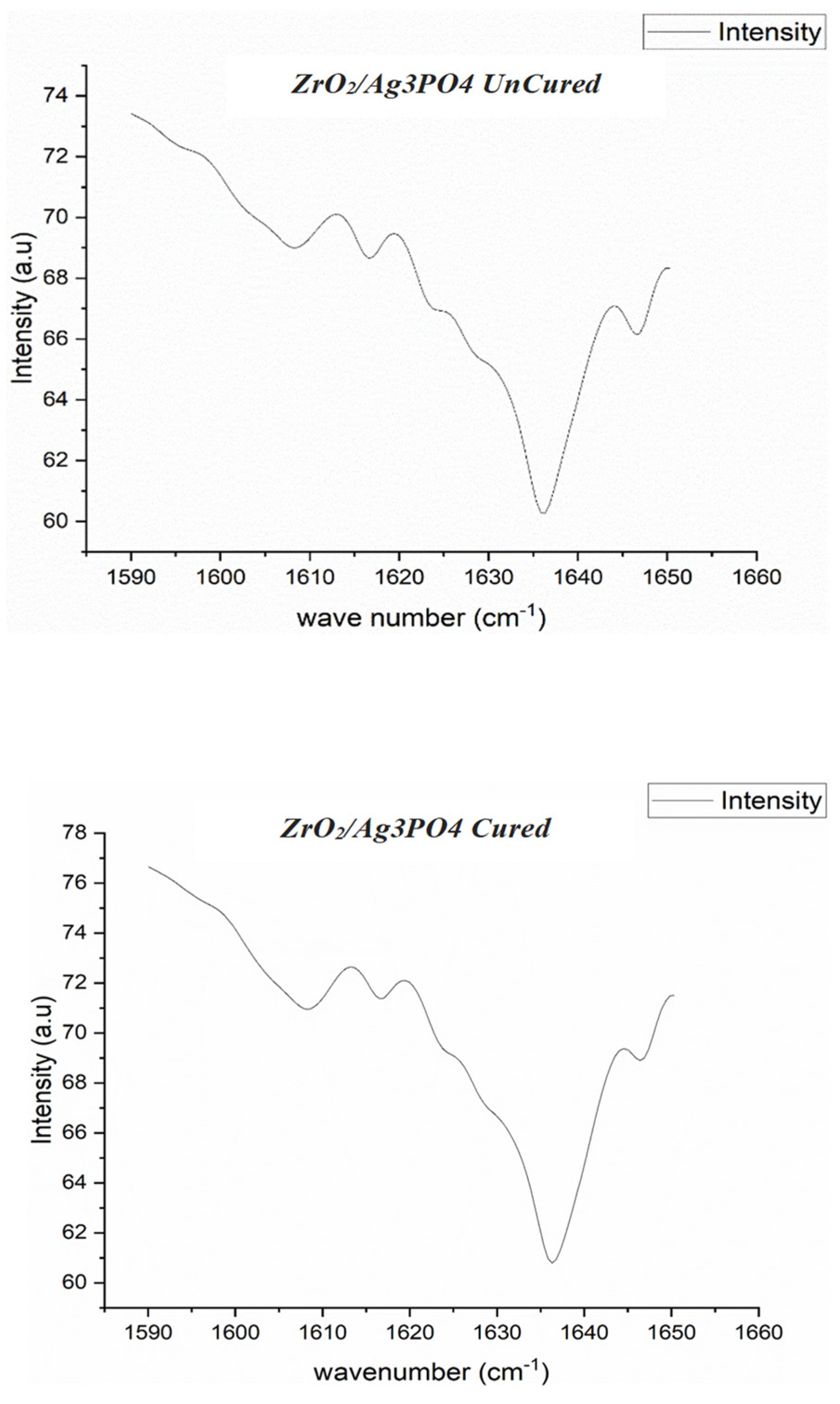
2.4. FTIR Spectroscopy and Degree of Conversion

The degree of conversion (DC) for unmodified self-etch (SE) adhesive (control) was assessed along with that of SE adhesives with varying concentrations of ZrO2/Ag3PO4 nanoparticles (cured and uncured). SE adhesives with the addition of 0.15%, 0.25%, and 0.5% ZrO2/Ag3PO4 (cured and uncured) were examined using FTIR spectroscopy. Initially, absorbance peaks associated with C–C double bonds were recorded. Subsequently, the FTIR spectra were collected for the uncured resin over 40 s. The aromatic C–C and aliphatic absorbance peaks related to C=C were identified at 1607 and 1638 cm−1, respectively [16,24].

2.5. Bacterial Culture

The antibacterial efficacy of adhesives with different concentrations of NPs (0.15%, 0.25%, and 0.5%) was assessed using the pour plate method. S. Mutans specimens obtained from Becton–Dickinson (BD Diagnostics-Difco, Franklin Lakes, NJ, USA) were cultured for 24 h in brain heart infusion broth (BHI) (BHI, Difco, Sparks, MD, USA) under conditions of 5% CO2 and a temperature of 37 °C. The culture medium was enriched with a 2% sucrose solution (weight/volume). Approximately 1 mL of the diluted S. mutans culture was carefully pipetted into the center of a Petri dish. Following a 24 h incubation period, the samples containing adherent biofilms were replenished with fresh media and subjected to an additional 24 h incubation period. Over two days, the culture displayed the development of biofilms that had reached a relatively advanced level of maturity. Survival rate assessment: S. mutans cultures underwent bacterial survival rate analysis. To achieve the desired objective, a tenfold dilution was performed with a hockey-shaped spreader. The next step was to produce aliquots and distribute them uniformly throughout the MRS plate. The dish was then placed in a 37 °C incubator. The following formula was used to determine the percentage of bacteria that survived [15,25]:
Survival rates = Colony forming units (CFUs) of each experiment
group/CFU count of the control.

2.6. Random Allocation of Samples

The specimens were distributed into four different groups via random allocation based on the percentage of ZrO2/Ag3PO4 NPs added in the primer of SE adhesive (n = 20):
Group 1: Unmodified SE (Control)
In this group, no ZrO2/Ag3PO4 NPs were added to the SE adhesive primer;
Group 2: 0.15 wt% ZrO2/Ag3PO4 + SE adhesive
The prepared ZrO2/Ag3PO4 NPs were silanized using 1.0 vol % metha-cryloxy-propyl-tri methoxy silane (MPS, Sigma Aldrich, St. Louis, MO, USA). After silanization, ZrO2/Ag3PO4 NPs were added in primer at 0.15 wt% incrementally. The primer was then applied and left for 20 s followed by gentle air blowing for 5 s;
Group 3: 0.25 wt% ZrO2/Ag3PO4 + SE adhesive
Similar steps were followed to modify and apply the primer as in Group 2 but the concentration of ZrO2/Ag3PO4 NPs used was set to 0.25 wt%;
Group 4: 0.5 wt% ZrO2/Ag3PO4 + SE adhesive
Similar steps were followed to modify and apply the primer as in Group 2 but the concentration of ZrO2/Ag3PO4 NPs used was set to 0.5 wt%.

2.7. Restoration Bonding

The remaining forty samples had bonding agents applied, and the film was gently blown with air to achieve uniformity followed by 10 s of light curing. The composite restoration was then built on a CAD surface using a Tygon tube (Norton Inc., Seattle, WA, USA) with a 0.9 mm internal diameter and a 2 mm height. A plugger was used to condense the A2 color of Z350XT composite resin (made by 3M ESPE, in St. Paul, MN, USA) into the tube. The composite was exposed to a curing light (Demi Plus; Kerr, Los Angeles, CA, USA) that produced 1100 mW/cm2 of light for 20 s. The teeth were kept in an incubator for 24 h at 37 °C and 100% humidity [24,26].

2.8. μTBS and Failure Mode Analysis

The attachment of each specimen was accomplished by affixing its ends to a custom-designed Ciucchi’s jig with the utilization of cyanoacrylate adhesive. The final component was securely attached to a universal testing machine (UTM) (Lloyd Instruments, LR 5K, Sheffield, UK). A compressive force was applied at the material–dentin interface at a crosshead speed of 0.5 mm/min. The load necessary for the debonding process of each specimen was measured in units of MegaPascals (MPa). After the debonding of specimens, the debonded tooth surface was examined under a stereomicroscope (Nikon Model, DSD230, Nikon Co., Tokyo, Japan) at 40× magnification [1,26,27].

2.9. Statistical Analysis

The assessment of data normality was conducted using Levene’s Test. One-way analysis of variance (ANOVA) and Tukey’s post hoc test were used to compare the means and standard deviations (SD) between groups at a p-value of 0.05 using SPSS, version 22.0 (SPSS Inc., Chicago, IL, USA).

3. Results

3.1. SEM EDX of ZrO2/Ag3PO4 Synthesized Nanoparticles

The SEM image exhibits a clustered micrograph of ZrO2/Ag3PO4. The particles seen in the image exhibit a wide variety of morphologies, and when observed at lower levels of magnification, there is an uneven dispersion of particle sizes. Upon conducting a more detailed analysis with increased magnification, it becomes evident that the ZrO2/Ag3PO4 nanoparticles exhibit a consistently asymmetrical pattern, albeit with some degree of size variation (Figure 1). Elemental spectroscopy is employed to ascertain the distinct elemental compositions present in ZrO2/Ag3PO4 nanoparticles, namely zirconium, silver, oxides, and phosphate (Figure 2).

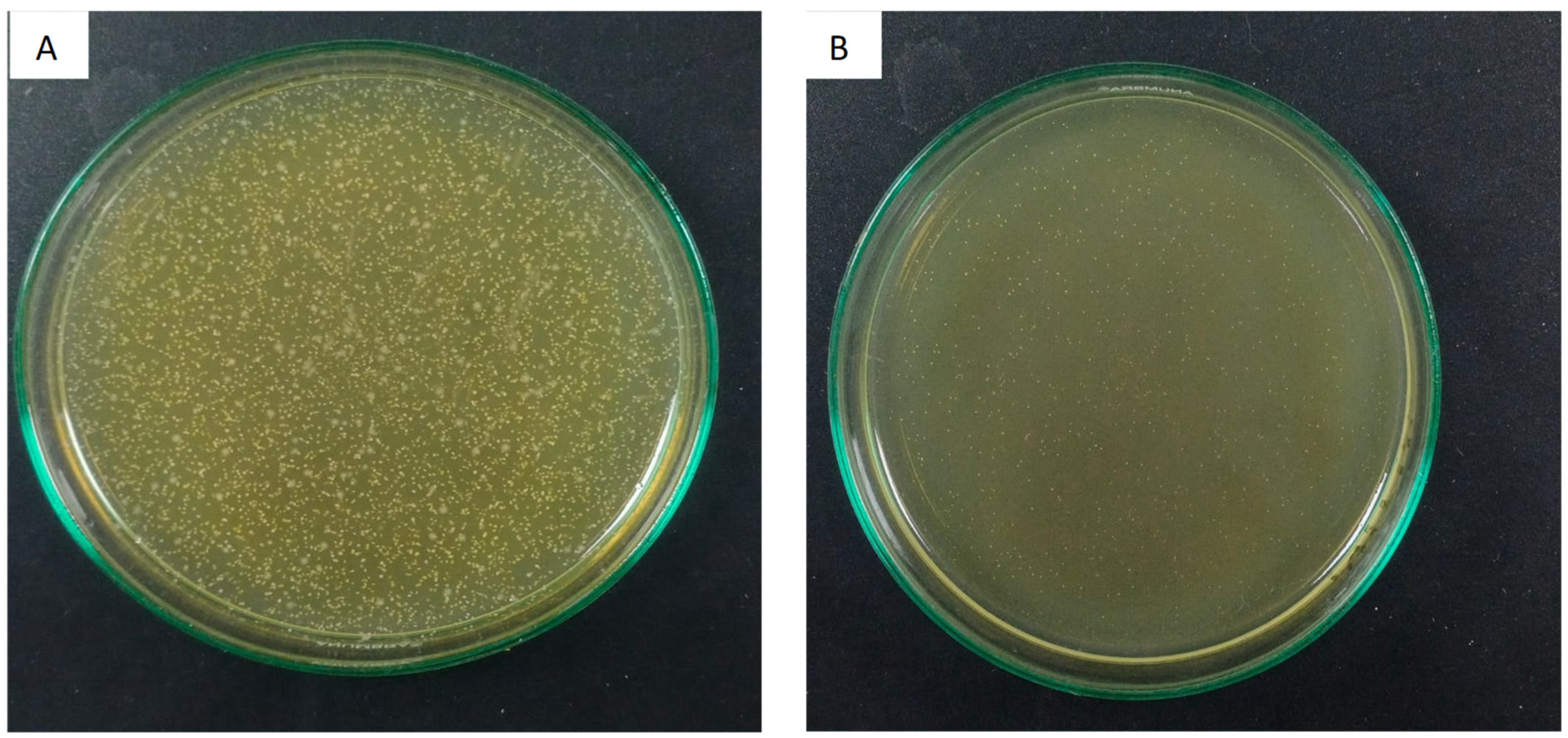
3.2. Antimicrobial Evaluation

The survival rates of S. mutans following the application of modified adhesives containing ZrO2/Ag3PO4 NPs onto the CAD surface are presented in Table 1. The findings of the present study revealed that the samples from Group 4, which were applied with 0.5 wt% ZrO2/Ag3PO4 + SE, displayed the lowest survival rate (0.12 ± 0.01 CFU/mL) of S. mutans. Nevertheless, the study specimens from Group 1 that were applied with unmodified SE adhesive (Control) established the highest survival rate of microorganisms (0.40 ± 0.09 CFU/mL). Intergroup comparison analysis revealed that Group 2 (0.15 wt% ZrO2/Ag3PO4 + SE) (0.19 ± CFU/mL), Group 3 (0.25 wt% ZrO2/Ag3PO4 + SE) (19.85 ± 0.57 CFU/mL), and Group 4 samples displayed comparable outcomes in their efficacy as an antimicrobial agent against S. mutans (p > 0.05). However, Group 1 exhibited significantly lower outcomes of S. mutans survival (Figure 3)

3.3. μTBS, Degree of Conversion, and Failure Analysis

The μTBS values at the CAD restoration surface following the application of modified SE adhesives containing ZrO2/Ag3PO4 NPs are shown in Table 2. The strongest bond of composite to the CAD surface was observed in Group 4 (0.5 wt% ZrO2/Ag3PO4 + SE) (20.12 ± 0.79 MPa). However, the μTBS was weakest in Group 1 (unmodified SE) (15.28 ± 0.22 MPa) samples. Intergroup comparison analysis demonstrated that Group 2 (0.15 wt% ZrO2/Ag3PO4 + SE) (19.16 ± 0.45 MPa), Group 3 (0.25 wt% ZrO2/Ag3PO4 + SE) (19.85 ± 0.57 MPa), and Group 4 demonstrated no significant differences in their attained bond integrity values. (p > 0.05). However, Group 1 exhibited significantly lower outcomes of bond strength. The failure mode among the experimental groups is presented in Table 3. The major forms of failure identified in Groups 2, 3, and 4 were cohesive, whereas Group 1 exhibited predominantly adhesive failure patterns (Table 3).
The FTIR spectra displayed characteristic peaks associated with the conversion of double bonds in both the unmodified self-etch (SE) adhesive (control) and the SE adhesive formulations containing 0.15%, 0.25%, and 0.5% ZrO2/Ag3PO4. DC was estimated in SE adhesive loaded with 0.5% by weight ZrO2/Ag3PO4 (Figure 4); the peak height ratios were measured between the absorbance levels of the aliphatic C=C peak at 1638 cm−1 and a reference internal peak of the aromatic C=C at 1607 cm−1. These measurements were taken during the curing process and then compared to those obtained from the unpolymerized adhesive. The highest DC was observed in the control group (unmodified SE (69.85 ± 8.37)), whereas DC was comparable in 0.15 wt% ZrO2/Ag3PO4 + SE adhesive (41.89 ± 8.11), 0.25 wt% ZrO2/Ag3PO4 + SE adhesive (43.74 ± 6.91), and 0.5 wt% ZrO2/Ag3PO4 + SE adhesive (49.68 ± 6.59) formulations (p > 0.05) (Table 4).

4. Discussion

This study was initially based on the hypothesis that there would be no significant difference in the μTBS of SE adhesives when modified with different concentrations (0.15 wt%, 0.25 wt%, and 0.5 wt%) of ZrO2/Ag3PO4 compared to an unmodified SE adhesive. Additionally, it was anticipated that there would be no significant difference in the antibacterial efficacy or FTIR spectroscopy of ZrO2/Ag3PO4 NP-modified SE adhesive compared to unmodified SE adhesive. However, the results of this study revealed that all the groups in which the adhesive was modified using ZrO2/Ag3PO4 nanoparticles showed improved performance in terms of μTBS and antimicrobial effectiveness against S. mutans. Nevertheless, there was a rejection of the hypothesis since the DC of the adhesive without nanoparticles was found to be higher compared to the adhesive loaded with ZrO2/Ag3PO4 nanoparticles.
Numerous research studies have been conducted to explore the impact of integrating nanofillers into polymers. The primary objective behind the use of various engineered nanoparticles is to enhance the antibacterial effectiveness and bond strength of adhesives employed in restorative dentistry [28,29]. The primary mechanism underlying the antibacterial activity of NPs is attributed to their interaction with the bacterial cell wall, facilitating their subsequent penetration of the cell membrane due to their tiny size [30]. In the present study, outcomes show that ZrO2/Ag3PO4 NPs, when used in different concentrations, display effectiveness against S. mutans. This can be explained on behalf of previous studies which state that silver–phosphate glass particles possess bactericidal properties against various pathogens, such as Staphylococcus epidermidis, Enterococcus faecalis, and S. mutans [31,32,33]. Reports have highlighted that the release of silver ions from nanoparticle surfaces plays a significant role in their detrimental effects. The liberated silver ions can penetrate bacterial cells, where they disrupt various enzymatic processes within the cellular environment. This interference ultimately culminates in the demise of the bacterial cells [34,35]. However, the combination of ZrO2 NPs with Ag3PO4 works in synergy to improve the antibacterial potential of Ag3PO4 by forming reactive oxygen species (ROSs) that lead to the destruction of bacterial cells. The increased levels of ROS kill bacteria by the process of lipid peroxidation. This is in agreement with the findings of a study conducted by Ayanwale and coworkers [36]. Nonetheless, this is the first study to assess the influence of ZrO2/Ag3PO4 nanoparticles on the survival of S. mutans, and further investigation is still required to gain a comprehensive understanding of the results and their implications.
Regarding μTBS, it was determined that all ZrO2/Ag3PO4-modified experimental SE adhesives showed significantly higher bond strength values than the control. The findings of the current investigation align with prior research, indicating that the incorporation of nanofillers resulted in a notable enhancement in the adhesive strength of dentin [37,38]. The nano-scale size of the two inorganic fillers employed in the present investigation could be considered a crucial aspect of this discovery. It is widely recognized that inorganic NPs possess a significant surface area which enhances the adhesion capacity, improving bond strength. It was also revealed that silver phosphate NPs are capable of forming apatite crystals as well as causing noticeable alterations in the calcium/phosphate ratio of dentin substrate [37]. Furthermore, an expected synergistic effect arises from ZrO2 nanoparticles, and this can be reasonably elucidated by the heightened wettability of the adhesive resin. Zirconium-based nanoparticles showcase excellent dispersion characteristics within a material, thereby enhancing its biocompatibility. The authors anticipate that these alterations collectively contribute to the augmentation of resin tags and the formation of a hybrid layer within the irregularities induced by acid etching. Consequently, this improvement is presumed to positively impact bond integrity scores [30,39].
The results have also demonstrated that an increase in the concentration of ZrO2/Ag3PO4 nanoparticles in SE adhesive leads to a linear improvement in both antimicrobial potency and micro-tensile bond strength (μTBS). It is critical to emphasize that the release rate of silver ions is determined by both the size and surface properties of nanoparticles. The dimensions of nanoparticles impact the extent of contact and interaction with the surrounding medium, while the charge and surface composition determine the stability of the nanoparticles. Smaller nanoparticles have been found to dissolve more quickly in different substances, resulting in the release of silver ions. This phenomenon has the potential to make a substantial contribution to the antibacterial efficacy of nanoparticles [40,41,42]. Existing works in the literature have often suggested that increasing nanoparticle concentrations in adhesives may have a detrimental effect on the physical and mechanical properties of materials [21,43]. However, in the current study, it is evident that a higher concentration of ZrO2/Ag3PO4 nanoparticles has a positive impact on the adhesive’s bond strength. Nonetheless, it is important to note that the influence of ZrO2/Ag3PO4 nanoparticles on the mechanical properties at higher concentrations, beyond those investigated in this research, remains a subject for future investigation.
Concerning outcomes associated with DC, one plausible explanation for the observed decrease in DC following the inclusion of ZrO2/Ag3PO4 nanoparticles is that it interferes with the propagation of curing light within the adhesive substance. This interference hinders the transformation of monomers into polymers during the curing process [24]. The potential consequence of reduced light penetration is a drop in DC, as it may impede the efficient polymerization of all monomers during the curing phase [44]. Regarding failure mode, it was observed that ZrO2/Ag3PO4 NP-modified adhesives displayed a cohesive type of failure, whereas unmodified bonding agents predominantly reported adhesive failure patterns. Several reasons for cohesive failure can be attributed to factors such as stress concentration, material properties, material aging and defects, chemical reactions, fatigue, thermal change, etc. [26,45].
The results of the current study show promise; however, readers are advised to interpret them cautiously, as the prospective implications are confined to the specific adhesive used in this investigation. It is important to note that different proportions of ZrO2/Ag3PO4 nanoparticles, not identical to those employed in this study, may exhibit distinct characteristics. Additional studies should encompass an assessment of how these nanoparticles influence other mechanical properties, including flexural strength, microhardness, and surface roughness. The current study did not include an aging process for the samples, which could potentially impact μTBS values and the occurrence of bond failure. The authors acknowledge that further investigation is required to explore the alterations occurring on the CAD surface after the application of primers modified with ZrO2/Ag3PO4 nanoparticles. It is crucial to evaluate the size distribution and zeta potential of ZrO2/Ag3PO4 nanoparticles using Dynamic Light Scattering (DLS) and zeta potential techniques, along with determining the minimum inhibitory values of ZrO2/Ag3PO4 against S. mutans. Additionally, the use of remineralizing agents such as fluoride should be tested in combination with ZrO2/Ag3PO4 nanoparticles in future reports to evaluate their mutual effects [46,47,48]. A more comprehensive understanding of the material’s surface properties and its interactions with the enhanced adhesive and CAD may be attained by incorporating Atomic Force Microscopy (AFM) into the investigation.

5. Conclusions

The self-etch adhesive, when modified with ZrO2/Ag3PO4 nanoparticles, exhibited a positive influence on micro-tensile bond strength (μTBS) and displayed improved antibacterial efficacy against S. mutans. Notably, the study observed that augmenting the concentration of ZrO2/Ag3PO4 nanoparticles did not adversely affect μTBS. However, it was noted that the inclusion of ZrO2/Ag3PO4 led to a reduction in the degree of conversion (DC) in the self-etch adhesive.

Author Contributions

Conceptualization; Methodology; Software, Validation, Formal analysis, Investigation data curation, writing—original draft preparation, and writing—review and editing, visualization, supervision, project administration, and funding acquisition were performed by F.A. and M.H.A. All authors have read and agreed to the published version of the manuscript.

Funding

The authors are grateful to the researchers supporting project at King Saud University for funding through the project (RSPD2023R815), Riyadh, Saudi Arabia.

Institutional Review Board Statement

The study was conducted in accordance with the Declaration of Helsinki, and approved by the Ethical Committee of King Saud University (protocol code number is FC#256-25 and the approval date is 12 June 2023).

Informed Consent Statement

Informed consent was obtained from all subjects involved in the study.

Data Availability Statement

The raw data supporting the conclusions of this article will be made available by the authors upon request.

Conflicts of Interest

The authors declare no conflicts of interest.

References

  1. Alrahlah, A.; Naseem, M.; Tanveer, S.A.; Abrar, E.; Charania, A.; AlRifaiy, M.Q.; Vohra, F. Influence of Disinfection of Caries Effected Dentin with Different Concentration of Silver Diamine Fluoride, Curcumin and Er, Cr:YSGG on Adhesive Bond Strength to Resin Composite. Photodiagnosis Photodyn. Ther. 2020, 32, 102065. [Google Scholar] [CrossRef]
  2. Al Ahdal, K.; Maawadh, A.M.; Al Deeb, L.; Alshamrani, A.S.; Almohareb, T.; Alrahlah, A. Effect of Malachite Green, Ocimum Sanctum, and Er, Cr: YSGG Laser on Antimicrobial Activity against S. mutans and CAD Disinfection Bonded to Resin Restoration. Photodiagnosis Photodyn. Ther. 2023, 42, 103571. [Google Scholar] [CrossRef] [PubMed]
  3. AlSheikh, R.; Abduldaiem, O.Y.; Alkhalifa, M.S.; Jillani, M.S.; Dehailan, L.A.; Barakat, A.; Alazmah, A.; Hameed, M.S.; Niazi, F. Different Cavity Disinfectant Efficacy against S. mutans and Shear Bond Strength of Caries Affected Dentin Bonded to Resin Restoration. Photodiagnosis Photodyn. Ther. 2023, 42, 103560. [Google Scholar] [CrossRef] [PubMed]
  4. Alfaawaz, Y.F. Disinfection of Caries Affected Dentin Using Rose Bengal, Titanium Sapphire Laser; Ammonium Hexa-Fluorosilicate, and Ozonated Water on Resin Dentin Bond Strength. Photodiagnosis Photodyn. Ther. 2022, 39, 102912. [Google Scholar] [CrossRef] [PubMed]
  5. Mandava, D.; Ajitha, P.; Narayanan, L.L. Comparative Evaluation of Tensile Bond Strengths of Total-Etch Adhesives and Self-Etch Adhesives with Single and Multiple Consecutive Applications: An in Vitro Study. J. Conserv. Dent. 2009, 12, 55. [Google Scholar] [CrossRef] [PubMed]
  6. Vieira, B.R.; Dantas, E.L.D.A.; Cavalcanti, Y.W.; Santiago, B.M.; Sousa, F.B. De Comparison of Self-Etching Adhesives and Etch-and-Rinse Adhesives on the Failure Rate of Posterior Composite Resin Restorations: A Systematic Review and Meta-Analysis. Eur. J. Dent. 2022, 16, 258–265. [Google Scholar]
  7. Costa, J.; Bettencourt, A.; Madeira, A.; Nepomuceno, L.S.; Portugal, J.; Neves, C.B. Surface Properties after Chemical Aging of Chlorhexidine Delivery Systems Based on Acrylic Resin. Rev. Port. Estomatol. Med. Dent. E Cir. Maxilofac. 2019, 60, 155–162. [Google Scholar] [CrossRef]
  8. Costa, J.V.; Portugal, J.; Neves, C.B.; Bettencourt, A.F. Should Local Drug Delivery Systems Be Used in Dentistry? Drug Deliv. Transl. Res. 2022, 12, 1395–1407. [Google Scholar] [CrossRef]
  9. Targino, A.G.R.; Flores, M.A.P.; Dos Santos, V.E.; De Godoy Bené Bezerra, F.; De Luna Freire, H.; Galembeck, A.; Rosenblatt, A. An Innovative Approach to Treating Dental Decay in Children. A New Anti-Caries Agent. J. Mater. Sci. Mater. Med. 2014, 25, 2041–2047. [Google Scholar] [CrossRef]
  10. Naik, S.V.; Sugandhan, S.; Ramasetty, P.A.; Tripathi, A.P.; Deepak, B.M. Nanotechnology in Dentin Disinfection: Can We Preserve the Bond? Int. J. Clin. Pediatr. Dent. 2018, 11, 468–473. [Google Scholar] [CrossRef]
  11. Li, X.; Robinson, S.M.; Gupta, A.; Saha, K.; Jiang, Z.; Moyano, D.F.; Sahar, A.; Riley, M.A.; Rotello, V.M. Functional Gold Nanoparticles as Potent Antimicrobial Agents against Multi-Drug-Resistant Bacteria. ACS Nano 2014, 8, 10682–10686. [Google Scholar] [CrossRef] [PubMed]
  12. Giráldez-Pérez, R.M.; Grueso, E.M.; Carbonero, A.; Álvarez Márquez, J.; Gordillo, M.; Kuliszewska, E.; Prado-Gotor, R. Synergistic Antibacterial Effects of Amoxicillin and Gold Nanoparticles: A Therapeutic Option to Combat Antibiotic Resistance. Antibiotics 2023, 12, 1275. [Google Scholar] [CrossRef] [PubMed]
  13. Giráldez-Pérez, R.M.; Grueso, E.M.; Jiménez-Aguayo, R.; Carbonero, A.; González-Bravo, M.; Kuliszewska, E.; Prado-Gotor, R. Use of Nanoparticles to Prevent Resistance to Antibiotics—Synthesis and Characterization of Gold Nanosystems Based on Tetracycline. Pharmaceutics 2022, 14, 1941. [Google Scholar] [CrossRef] [PubMed]
  14. Bruna, T.; Maldonado-Bravo, F.; Jara, P.; Caro, N. Silver Nanoparticles and Their Antibacterial Applications. Int. J. Mol. Sci. 2021, 22, 7202. [Google Scholar] [CrossRef] [PubMed]
  15. Melo, M.A.S.; Cheng, L.; Zhang, K.; Weir, M.D.; Rodrigues, L.K.A.; Xu, H.H.K. Novel Dental Adhesives Containing Nanoparticles of Silver and Amorphous Calcium Phosphate. Dent. Mater. 2013, 29, 199–210. [Google Scholar] [CrossRef] [PubMed]
  16. Aljamhan, A.S.; Alrefeai, M.H.; Alhabdan, A.; Alzehiri, M.H.; Naseem, M.; Vohra, F.; Alkhudhairy, F. Interaction of Zirconium Oxide Nanoparticle Infiltrated Resin Adhesive with Dentin Conditioned by Phosphoric Acid and Er, Cr: YSGG Laser. J. Appl. Biomater. Funct. Mater. 2022, 20, 22808000221087349. [Google Scholar] [CrossRef] [PubMed]
  17. Hatta, M.; Shinya, A.; Yokoyama, D.; Gomi, H.; Vallittu, P.K.; Shinya, A. The Effect of Surface Treatment on Bond Strength of Layering Porcelain and Hybrid Composite Bonded to Zirconium Dioxide Ceramics. J. Prosthodont. Res. 2011, 55, 146–153. [Google Scholar] [CrossRef] [PubMed]
  18. Bin-Shuwaish, M.S.; Maawadh, A.M.; Al-Hamdan, R.S.; Alresayes, S.; Ali, T.; Almutairi, B.; Vohra, F.; Abduljabbar, T. Influence of Graphene Oxide Filler Content on the Dentin Bond Integrity, Degree of Conversion and Bond Strength of Experimental Adhesive. A SEM, Micro-Raman, FTIR and Microtensile Study. Mater. Res. Express 2020, 7, 115403. [Google Scholar] [CrossRef]
  19. Vohra, F.; Alamri, R.R.; Almohsen, F.O.; El Mourad, A.M.; Farooq, I.; Alsaif, R. Fiber Post Bonding with Beta-Tricalcium Phosphate Incorporated Root Dentin Adhesive. SEM, EDX, FTIR, Rheometric and Bond Strength Study. Microsc. Res. Tech. 2023, 86, 762–772. [Google Scholar] [CrossRef]
  20. Khan, A.S.; Alhamdan, Y.; Alibrahim, H.; Almulhim, K.S.; Nawaz, M.; Ahmed, S.Z.; Aljuaid, K.; Ateeq, I.S.; Akhtar, S.; Ansari, M.A.; et al. Analyses of Experimental Dental Adhesives Based on Zirconia/Silver Phosphate Nanoparticles. Polymers 2023, 15, 2614. [Google Scholar] [CrossRef]
  21. Alsunbul, H.; Alfawaz, Y.F.; Alhamdan, E.M.; Farooq, I.; Vohra, F.; Abduljabbar, T. Influence of Carbon and Graphene Oxide Nanoparticle on the Adhesive Properties of Dentin Bonding Polymer: A SEM, EDX, FTIR Study. J. Appl. Biomater. Funct. Mater. 2023, 21, 22808000231159238. [Google Scholar] [CrossRef]
  22. Risnes, S.; Saeed, M.; Sehic, A. Scanning Electron Microscopy (SEM) Methods for Dental Enamel. In Methods in Molecular Biology; Humana Press Inc.: Totowa, NJ, USA, 2019; Volume 1922, pp. 293–308. [Google Scholar]
  23. Alrefeai, M.H.; Aljamhan, A.S.; Alhabdan, A.; Alzehiri, M.H.; Naseem, M.; Alkhudhairy, F. Influence of Methylene Blue, Riboflavin, and Indocyanine Green on the Bond Strength of Caries Affected Dentin When Bonded to Resin-Modified Glass Ionomer Cement. Photodiagnosis Photodyn. Ther. 2022, 38, 102792. [Google Scholar] [CrossRef] [PubMed]
  24. Alhenaki, A.M.; Attar, E.A.; Alshahrani, A.; Farooq, I.; Vohra, F.; Abduljabbar, T. Dentin Bond Integrity of Filled and Unfilled Resin Adhesive Enhanced with Silica Nanoparticles—An Sem, Edx, Micro-Raman, Ftir and Micro-Tensile Bond Strength Study. Polymers 2021, 13, 1093. [Google Scholar] [CrossRef] [PubMed]
  25. Elgamily, H.M.; El-Sayed, H.S.; Abdelnabi, A. The Antibacterial Effect of Two Cavity Disinfectants against One of Cariogenic Pathogen: An in Vitro Comparative Study. Contemp. Clin. Dent. 2018, 9, 457–462. [Google Scholar] [CrossRef] [PubMed]
  26. Alkhudhairy, F.; Vohra, F.; Naseem, M. Influence of Er,Cr:YSGG Laser Dentin Conditioning on the Bond Strength of Bioactive and Conventional Bulk-Fill Dental Restorative Material. Photobiomodulation Photomed. Laser Surg. 2020, 38, 30–35. [Google Scholar] [CrossRef] [PubMed]
  27. Al Deeb, L.; Bin-Shuwaish, M.S.; Abrar, E.; Naseem, M.; Al-Hamdan, R.S.; Maawadh, A.M.; Al Deeb, M.; Almohareb, T.; Al Ahdal, K.; Vohra, F.; et al. Efficacy of Chlorhexidine, Er Cr YSGG Laser and Photodynamic Therapy on the Adhesive Bond Integrity of Caries Affected Dentin. An In-Vitro Study. Photodiagnosis Photodyn. Ther. 2020, 31, 101875. [Google Scholar] [CrossRef] [PubMed]
  28. Makvandi, P.; Gu, J.T.; Zare, E.N.; Ashtari, B.; Moeini, A.; Tay, F.R.; Niu, L.N. Polymeric and inorganic nanoscopical antimicrobial fillers in dentistry. Acta Biomater. 2020, 101, 69–101. [Google Scholar] [CrossRef] [PubMed]
  29. Almutairi, B.; Kattan, H.F.; BinMahfooz, A.M.; Qutub, O.A.; Basunbul, G.; ArRejaie, A.S.; Farooq, I.; Vohra, F.; Abduljabbar, T. Synergistic Effect of Graphene Oxide/Calcium Phosphate Nanofiller in a Dentin Adhesive on Its Dentin Bond Integrity and Degree of Conversion. A Scanning Electron Microscopy, Energy Dispersive X-Ray Spectroscopy, Fourier Transform Infrared, Micro-Raman, An. Microsc. Res. Tech. 2021, 84, 2082–2094. [Google Scholar] [CrossRef]
  30. Al-Saleh, S.; Alateeq, A.; Alshaya, A.H.; Al-Qahtani, A.S.; Tulbah, H.I.; Binhasan, M.; Shabib, S.; Farooq, I.; Vohra, F.; Abduljabbar, T. Article Influence of TiO2 and ZrO2 Nanoparticles on Adhesive Bond Strength and Viscosity of Dentin Polymer: A Physical and Chemical Evaluation. Polymers 2021, 13, 3794. [Google Scholar] [CrossRef]
  31. Lee, M.J.; Seo, Y.B.; Seo, J.Y.; Ryu, J.H.; Ahn, H.J.; Kim, K.M.; Kwon, J.S.; Choi, S.H. Development of a Bioactive Flowable Resin Composite Containing a Zinc-Doped Phosphate-Based Glass. Nanomaterials 2020, 10, 2311. [Google Scholar] [CrossRef]
  32. Szliszka, E.; Czuba, Z.P.; Domino, M.; Mazur, B.; Zydowicz, G.; Krol, W. Ethanolic Extract of Propolis (EEP) Enhances the Apoptosis- Inducing Potential of TRAIL in Cancer Cells. Molecules 2009, 14, 738–754. [Google Scholar] [CrossRef] [PubMed]
  33. Wang, Y.Y.; Chatzistavrou, X.; Faulk, D.; Badylak, S.; Zheng, L.; Papagerakis, S.; Ge, L.; Liu, H.; Papagerakis, P. Biological and Bactericidal Properties of Ag-Doped Bioactive Glass in a Natural Extracellular Matrix Hydrogel with Potential Application in Dentistry. Eur. Cells Mater. 2015, 29, 342–355. [Google Scholar] [CrossRef] [PubMed]
  34. Nair, M.G.; Nirmala, M.; Rekha, K.; Anukaliani, A. Structural, Optical, Photo Catalytic and Antibacterial Activity of ZnO and Co Doped ZnO Nanoparticles. Mater. Lett. 2011, 65, 1797–1800. [Google Scholar] [CrossRef]
  35. Salar, R.; Kumar, N.; Sharma, P. Enhanced Antibacterial Activity of Streptomycin against Some Human Pathogens Using Green Synthesized Silver Nanoparticles. Resour. Technol. 2015, 1, 106–115. [Google Scholar] [CrossRef]
  36. Ayanwale, A.P.; Ruíz-Baltazar, A.d.J.; Espinoza-Cristóbal, L.; Reyes-López, S.Y. Bactericidal Activity Study of ZrO2-Ag2O Nanoparticles. Dose-Response 2020, 18, 3. [Google Scholar] [CrossRef]
  37. Lohbauer, U.; Wagner, A.; Belli, R.; Stoetzel, C.; Hilpert, A.; Kurland, H.D.; Grabow, J.; Müller, F.A. Zirconia Nanoparticles Prepared by Laser Vaporization as Fillers for Dental Adhesives. Acta Biomater. 2010, 6, 4539–4546. [Google Scholar] [CrossRef] [PubMed]
  38. Almutairi, B.; Binhasan, M.; Shabib, S.; Al-Qahtani, A.S.; Tulbah, H.I.; Al-Aali, K.A.; Vohra, F.; Abduljabbar, T. Adhesive Bond Integrity of Silanized Zirconia Nanoparticles in Polymeric Resin Dentin Bonding Agent. An FTIR, SEM, and Micro-Tensile Experiment. Int. J. Adhes. Adhes. 2022, 114, 103069. [Google Scholar] [CrossRef]
  39. Khurana, C.; Sharma, P.; Pandey, O.P.; Chudasama, B. Synergistic Effect of Metal Nanoparticles on the Antimicrobial Activities of Antibiotics against Biorecycling Microbes. J. Mater. Sci. Technol. 2016, 32, 524–532. [Google Scholar] [CrossRef]
  40. Le Ouay, B.; Stellacci, F. Antibacterial Activity of Silver Nanoparticles: A Surface Science Insight. Nano Today 2015, 10, 339–354. [Google Scholar] [CrossRef]
  41. Ivask, A.; Kurvet, I.; Kasemets, K.; Blinova, I.; Aruoja, V.; Suppi, S.; Vija, H.; Käkinen, A.; Titma, T.; Heinlaan, M.; et al. Size-Dependent Toxicity of Silver Nanoparticles to Bacteria, Yeast, Algae, Crustaceans and Mammalian Cells in Vitro. PLoS ONE 2014, 9, e102108. [Google Scholar] [CrossRef]
  42. Raza, M.A.; Kanwal, Z.; Rauf, A.; Sabri, A.N.; Riaz, S.; Naseem, S. Size- and Shape-Dependent Antibacterial Studies of Silver Nanoparticles Synthesized by Wet Chemical Routes. Nanomaterials 2016, 6, 74. [Google Scholar] [CrossRef] [PubMed]
  43. Aljamhan, A.S.; Alrefeai, M.H.; Alhabdan, A.; Alhusseini, S.A.; Farooq, I.; Vohra, F.; Naseem, M.; Alkhudhairy, F. Influence of ER-CR-YSGG Laser and Photodynamic Therapy on the Dentin Bond Integrity of Nano-Hydroxyapatite Containing Resin Dentin Adhesive: SEM-EDX. Polymers 2021, 13, 1903. [Google Scholar] [CrossRef] [PubMed]
  44. Fathima, J.B.; Pugazhendhi, A.; Venis, R. Synthesis and Characterization of ZrO2 Nanoparticles-Antimicrobial Activity and Their Prospective Role in Dental Care. Microb. Pathog. 2017, 110, 245–251. [Google Scholar] [CrossRef] [PubMed]
  45. Vohra, F.; Bukhari, I.A.; Sheikh, S.A.; Naseem, M.; Hussain, M. Photodynamic Activation of Irrigation (Using Different Laser Prototypes) on Push out Bond Strength of Fiber Posts. Photodiagnosis Photodyn. Ther. 2020, 30, 101716. [Google Scholar] [CrossRef] [PubMed]
  46. Zampetti, P.; Scribante, A. Historical and Bibliometric Notes on the Use of Fluoride in Caries Prevention. Eur. J. Paediatr. Dent. 2020, 21, 148–152. [Google Scholar] [CrossRef]
  47. Desai, S.; Rao, D.; Panwar, S.; Kothari, N.; Gupta, S. An in Vitro Comparative Evaluation of Casein Phosphopeptide-Amorphous Calcium Phosphate Fluoride, Tricalcium Phosphate and Grape Seed Extract on Remineralization of Artificial Caries Lesion in Primary Enamel. J. Clin. Pediatr. Dent. 2022, 46, 72–80. [Google Scholar] [CrossRef]
  48. Butera, A.; Pascadopoli, M.; Pellegrini, M.; Trapani, B.; Gallo, S.; Radu, M.; Scribante, A. Biomimetic Hydroxyapatite Paste for Molar–Incisor Hypomineralization: A Randomized Clinical Trial. Oral Dis. 2023, 29, 2789–2798. [Google Scholar] [CrossRef]
Figure 1. (A) The SEM image (×1200) presents a clustered micrograph of ZrO2/Ag3PO4 ranging from 100 nm to 500 nm. The particles exhibit a heterogeneous shape, and there is an uneven random distribution of particle sizes when observed at low magnification. (B) When examined at high magnification (×10,000), the ZrO2/Ag3PO4 nanoparticles display a more uniform partially asymmetrical pattern, with variability in sizes.
Applsci 14 00563 g001
Figure 2. Elemental spectroscopy demonstrates the different compositions of elements in ZrO2/Ag3PO4 nanoparticles, i.e., zirconium, silver, oxides, and phosphate.
Applsci 14 00563 g002
Figure 3. ZrO2/Ag3PO4 nanoparticles at various concentrations against S. mutans, the pour plate method was utilized. (A) presents the colony-forming units (CFU) of S. mutans when treated with Uniformed Self-Etch (Control). In (B), the CFU of S. mutans is depicted when exposed to a 0.5% weight (wt%) of ZrO2/Ag3PO4 nanoparticles.
Applsci 14 00563 g003
Figure 4. The FTIR spectra comprise two sets: cured 0.5% ZrO2/Ag3PO4 and uncured 0.5% ZrO2/Ag3PO4. The spectral analysis was focused on the specific region between 1608 cm−1 and 1636 cm−1 for both the cured and uncured samples. The ratios of (C=C&C–C) absorbance intensities (percentage of unreacted double bonds) before and after polymerization were identified.
Applsci 14 00563 g004
Table 1. The survival rates of S. mutans following the application of modified adhesives containing ZrO2/Ag3PO4 NPs onto the CAD surface.
Experimental GroupsSurvival Rate CFU/mLStandard Deviation (SD)
Group 1: Unmodified SE (Control)0.40 A0.09
Group 2: 0.15 wt% ZrO2/Ag3PO4 + SE adhesive0.19 B0.05
Group 3: 0.25 wt% ZrO2/Ag3PO4 + SE adhesive0.17 B0.04
Group 4: 0.5 wt% ZrO2/Ag3PO4 + SE adhesive0.12 B0.01
Self-etch (SE), zirconia/silver phosphate (ZrO2/Ag3PO4). The different superscript capital letters denote statistically significant differences (p < 0.05).
Table 2. Micro-tensile bond strength (μTBS) at the CAD restoration interface following the application of modified self-etch adhesives containing ZrO2/Ag3PO4 NPs.
Investigated GroupsMean ± SD (MPa)p-Value
Group 1: Unmodified SE (Control)15.28 ± 0.22 a<0.05
Group 2: 0.15 wt% ZrO2/Ag3PO4 + SE adhesive19.16 ± 0.45 b
Group 3: 0.25 wt% ZrO2/Ag3PO4 + SE adhesive19.85 ± 0.57 b
Group 4: 0.5 wt% ZrO2/Ag3PO4 + SE adhesive20.12 ± 0.79 b
Self-etch (SE), zirconia/silver phosphate (ZrO2/Ag3PO4). Different superscript characters denote statistically significant differences.
Table 3. Percentage distribution of modes of failure.
Failure TypeGroup 1Group 2 Group 3Group 4
Adhesive20%10%10%10%
Cohesive40%60%70%80%
Admixed40%30%20%10%
Table 4. Degree of conversion of unmodified SE (Control) and adhesives loaded with ZrO2/Ag3PO4 at different percentages.
Experimental GroupsDegree of Conversion % *
Group 1: Unmodified SE (Control)69.85 ± 8.37 B
Group 2: 0.15 wt% ZrO2/Ag3PO4 + SE adhesive41.89 ± 8.11 A
Group 3: 0.25 wt% ZrO2/Ag3PO4 + SE adhesive43.74 ± 6.91 A
Group 4: 0.5 wt% ZrO2/Ag3PO4 + SE adhesive49.68 ± 6.59 A
* ANOVA. Dissimilar superscript capital alphabets denote significant differences among groups.
Disclaimer/Publisher’s Note: The statements, opinions and data contained in all publications are solely those of the individual author(s) and contributor(s) and not of MDPI and/or the editor(s). MDPI and/or the editor(s) disclaim responsibility for any injury to people or property resulting from any ideas, methods, instructions or products referred to in the content.

