Article

A Long-Term Split-Mouth Randomized Controlled Trial to Assess Implant Treatment Outcome Using Implants with a Different Surface Roughness

1
Department of Periodontology and Oral Implantology, Faculty of Medicine and Health Sciences, Oral Health Sciences, Ghent University, C. Heymanslaan 10, B-9000 Ghent, Belgium
2
Department of Reconstructive Dentistry, Faculty of Medicine and Health Sciences, Oral Health Sciences, Ghent University, C. Heymanslaan 10, B-9000 Ghent, Belgium
3
Faculty of Medicine and Health Sciences, Oral Health Sciences, Ghent University, C. Heymanslaan 10, B-9000 Ghent, Belgium
4
Department of Periodontology, Academic Centre for Dentistry Amsterdam (ACTA), Gustav Mahlerlaan 3004, 1081 LA Amsterdam, The Netherlands
*
Author to whom correspondence should be addressed.
Appl. Sci. 2024, 14(4), 1658; https://doi.org/10.3390/app14041658
Submission received: 11 January 2024 / Revised: 9 February 2024 / Accepted: 14 February 2024 / Published: 19 February 2024
(This article belongs to the Special Issue Advances in Dental Implants)

Abstract

:
The influence of implant surface roughness on implant treatment outcome is still under debate. A rougher implant surface improves osseointegration but yields greater plaque accumulation and biofilm formation. Hybrid implants have a minimally rough component coronally and a moderately rough implant body. The aim of this split-mouth study is to evaluate the long-term outcome of treatment with hybrid and moderately rough implants after 6 years. As a secondary aim, Oral Health Quality of Life (OHQoL) was assessed after one and six years. Fully edentulous patients received an overdenture supported by two implants. One implant had a hybrid surface (MSC) and one implant had a moderately rough surface (DCC). Radiographic crestal bone loss (CBL), plaque score, bleeding on probing (BoP) and periodontal probing depth (PPD) were determined at one and six years. OHQoL was evaluated using the Oral Health Impact Profile-14 (OHIP-14) questionnaire and a Visual Analogue Scale (VAS). Twenty-one patients with 42 implants were evaluated after 6 years. No implants failed and a mean CBL of 0.26 mm (SD: 0.42) for the MSC group and 0.17 mm (SD: 0.29) for the DCC group was observed, which was not statistically significant. Periodontal parameters were comparable after 6 years and a significant improvement in OHQoL was observed. This randomized controlled trial concludes that hybrid implants are a predictable treatment alternative for moderately rough implants in patients with an overdenture supported by two implants.

1. Introduction

Lack of retention is a common problem in edentulous patients with conventional complete dentures. This could negatively affect their oral health-related quality of life (OHQoL) [1]. Nowadays, it is generally accepted that a two-implant-supported mandibular overdenture is the minimum that edentulous patients should be offered as a first treatment plan [2]. Osseointegrated implants minimize the rate of bone resorption, improving the stability and retention of an implant-supported prosthesis [3], which has a positive impact on oral function and on OHQoL [4]. Osseointegration is enhanced by implant surface roughness. Due to surface enlargement, the mechanical interlocking between bone tissue and the implant surface is improved [5]. According to Albrektsson and Wennerberg’s classification, implants with an Sa-value between 0.5 and 1.5 µm are considered minimally rough, while implants with a value between 1 and 2 µm are considered moderately rough [6]. Several studies comparing minimally and moderately rough implants have shown increased bone-to-implant contact for moderately rough implants [7,8,9]. On the other hand, a rougher surface facilitates greater plaque accumulation and formation of a biofilm, which could play an important role in peri-implant soft-tissue health [10,11].
Initial crestal bone loss can represent normal remodeling in response to surgery or it can be a consequence of the re-establishment of the peri-implant biological width [12,13]. Non-optimal implant components, poor clinical handling (e.g., traumatic surgery), restrictive prosthodontics (e.g., cement residues) or compromised patient characteristics (e.g., poor oral hygiene, smoking and drugs) could contribute to crestal bone loss occurring in the years thereafter [14]. Over time, this progressive crestal bone loss can lead to peri-implantitis, soft-tissue recession and, finally, implant failure. This occurs in approximately 5% of patients [13,15]. Windael et al., (2021) found that early crestal bone loss is a significant predictor of future peri-implantitis development. Crestal bone loss of 0.5 mm or more during the first year of function is associated with 5.43 times higher odds of developing peri-implantitis [16].
Hybrid implants could be a possible solution for the previously described issues. This type of implant has a minimally rough component coronally and a moderately rough implant body apically [17]. A systematic review by Doornewaard et al., (2017) suggests that implants with a minimally rough coronal part had statistically significantly less crestal bone loss in comparison to implants with a moderately rough and rough coronal part [13]. A hybrid design could possibly ensure better mucosal health and reduce the risk of peri-implant diseases, whilst the implant body could ensure osseointegration.
The present split-mouth study aims to compare the long-term implant outcome between hybrid and moderately rough implants in patients with a bar-supported mandibular overdenture. The primary outcome is crestal bone loss (CBL). Peri-implant soft-tissue health, defined by plaque score, bleeding on probing (BoP) and periodontal probing depth (PPD), is the secondary outcome. Additionally, this study compares OHQoL before treatment to OHQoL 1 year and 6 years after connection of an implant-supported overdenture.

2. Material and Methods

2.1. Patient Selection

Fully edentulous patients were enrolled in the department of dentistry at Ghent University Hospital between 2015 and 2017. Inclusion and exclusion criteria are listed below.
Inclusion criteria:
  • Totally edentulous for at least four months [18];
  • Presence of sufficient residual bone volume to place two implants of 4 mm in diameter and 9–11 mm in length.
Exclusion criteria:
  • <21 years old [19];
  • Smoking [20];
  • General contra-indications for oral surgery, e.g., full-dose head and neck radiation, intravenously administrated bisphosphonates and current chemotherapy.
All patients signed an informed consent form declaring agreement to participation. The study was conducted with the approval of the ethical committee of Ghent University Hospital (EC/2014/1231).
Sample size calculation was performed using SAS Power and Sample Size Calculator using an effect size of 1 mm and a standard deviation of 0.6 mm, with the level of significance set at 0.05 and β = 0.80. These parameters were based on Vervaeke et al., 2014 [12] and led to a sample size of a minimum of 14 patients (28 implants). To counter the effects of possible dropouts, 24 patients (48 implants) were enrolled, of which 21 (11 females, 10 males; mean age: 71) were present at the 1- and 6-year follow-up visits [14] (mean: 73 months; range: 61–88 months). Three patients were lost to follow-up due to following reasons: one patient died, one patient was not reachable and one patient was no longer able to attend the visits due to health reasons. Prior to implant placement, every patient received new upper and lower full dentures to ensure a correct vertical dimension, smile line and occlusion.

2.2. Surgical Procedure

As described by Glibert et al., (2018), the following surgical procedure was performed. A local anesthetic, Xylocaine–Adrenaline (Dentsply, York, PA, USA), was administered prior to the surgery. Two deep conical cylindrical implants (Southern implants, Irene, South Africa) were placed on the canine positions after elevation of a mucoperiosteal flap. A one-stage or two-stage approach was performed, depending on the primary stability of the implant. One MSC implant with a hybrid surface moderately sandblasted on the mid- and apical section (Sa: 1.3 μm, Sdr: 60%, minimally rough machined surface on the coronal 3 mm similar to the machined surface of the original Brånemark implants; Sa: 0.9 μm, Sdr: 34%) [6,21] and one implant with a moderately rough sandblasted surface overall (DCC; Sa: 1.3 μm, Sdr: 60%) [21] were placed (Figure 1). Besides the surface roughness, the two implants were identical: a thread pitch of 0.6 mm, an integrated platform-shift with a smooth implant bevel, an internal connection and microthreads on the implant neck were present. The implant position was randomized using a coin flip, i.e., randomization occurred within subjects. A final abutment (Compact Conical Abutment, Southern implants, Irene, South Africa) of 4 mm height was installed at implant placement or at second-stage surgery. The baseline was as follows. Following implant placement, an intra-oral radiograph was taken using a Rinn-Set XCP (Dentsply Rinn, York, PA, USA). Antibiotics were prescribed (Amoxicillin 2 g/day for four days) [22] and the patients were instructed to use a mouth rinse (containing 0.12% chlorhexidine) and to eat soft food [17].

2.3. Prosthetic Procedure

After implant placement, the denture was grinded to avoid contact with the healing caps and a soft reliner (Coe Soft, GC Europe, Leuven, Belgium) was inserted in the existing mandibular denture. The reliner needed to be replaced every two to three weeks to avoid overload of the implants. After a healing period of three months following implant placement [18], impressions were taken at abutment level. The current denture served as the impression tray (pickup technique). After the impression was taken, a titanium bar was CAD/CAM designed and milled (Proscan, Zonhoven, Belgium). Afterwards, the prosthesis was connected to the implants [17].

2.4. Crestal Bone Loss

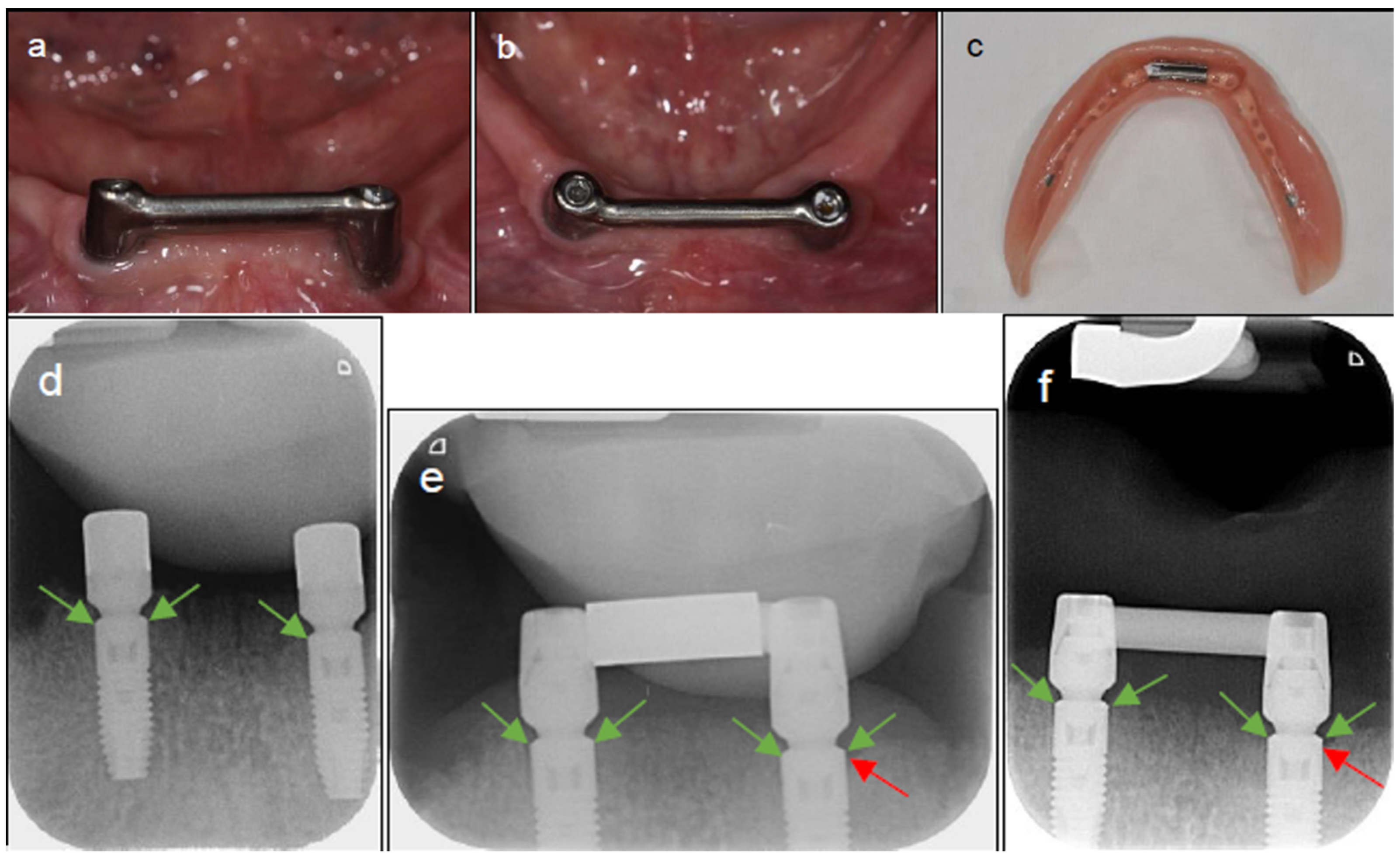
At baseline and follow-up, peri-apical intra-oral radiographs were taken using a Rinn-Set XCP. Bone level measurements were performed using Mediadent software (version number: V7) (Corilus, Ghent, Belgium). All radiographs were calibrated using implant length or tread pitch as a reference. Bone level was defined as the distance between the implant bevel and the bone-to-implant contact. Measurements were performed mesial and distal from each implant on the baseline radiographs (implant placement), after 1 year and after 6 years of loading (Figure 2) [17,22]. The first twenty measurements (ten implants) were performed by two observers (AVL and AS) and the intraclass correlation coefficient (ICC) was calculated. A favorable ICC of 0.845 (ICC > 0.750) was obtained; thus, the other measurements were completed by one observer. The measurements were conducted blinded. The mean of the mesial and distal measurements was used for further analysis. Crestal bone loss (CBL) was calculated by comparing the mean bone level at 1 and 6 years with the bone level at baseline [14].

2.5. Peri-Implant Health

Peri-implant health was assessed by measuring the following parameters at the 6-year follow-up visit: plaque score, BoP, PPD and suppuration. The following protocol was implemented during these visits. First, plaque presence or absence (mesial, distal, buccal and lingual) was indicated on both implants. Next, the PPDs were measured with a periodontal probe (mesial, distal, buccal and lingual). After probing, the presence or absence of bleeding and suppuration were evaluated (mesial, distal, buccal and lingual). Plaque and BoP were calculated as the sum of the mesial, distal, buccal and lingual measurements divided by four (the number of measurement sites). PPD was calculated as the mean of the mesial, distal, buccal and lingual measurements [23].

2.6. Prosthetic Functioning and Oral Health Quality of Life

During the 6-year follow-up visit, the bar-supported mandibular overdenture was evaluated for fit, stability and connection by an independent researcher (CM). An intra-oral examination to check for any ulcers, candida, pain or other anomalies was performed. The upper full denture was also checked for lack of retention. The patients were asked to complete an Oral Health Impact Profile (OHIP-14) questionnaire and a chewing ability-related Visual Analogue Scale (VAS) [24,25,26,27]. All maintenance interventions which occurred after the connection of the prosthesis to the implants were noted.

2.7. Statistical Analysis

The data collected in this study were analyzed using SPSS statistics 28 (IBM, North Castle, NY, USA). To compare the mean CBL between an MSC and DCC implant at the different timepoints, linear mixed models were used with fixed effects including time (6Y vs. 1Y), group (MSC vs. DCC) and a two-way interaction between time and group. The estimated marginal means were computed by time and group. All hypothesis testing was performed at the two-sided 5% significance level.
Mean crestal bone loss might not be fully representative of the actual clinical situation. Therefore, CBL was transformed from a continuous variable into a binary variable. Taking into account the study by Windael et al., (2021), 0.5 mm was taken as a cut-off value for clinically relevant bone loss [16], i.e., ≤0.5 mm bone loss equated to the binary value 0 (no CBL). The difference in the percentage of clinically relevant CBL (>0.5 mm) after 6 years was evaluated by applying a binary logistic GEE (Generalized Estimating Equations).
Differences in plaque, BoP and PPD between the MSC and DCC groups after 6 years were also analyzed using GEE models for a negative binomial distribution with a log-link function. Differences in OHQoL were compared between baseline (when the new complete denture was placed without connection to the implants), 1 year and 6 years after connection. Wilcoxon matched-pairs signed-rank tests were applied for the OHIP-14 and paired Student’s t-tests were applied for the VAS. These tests were performed for each of the three paired time points and only patients present at both paired time points were included. Patient files were checked and maintenance interventions which occurred after the installment of the final prosthesis were summarized and described by their absolute frequencies. A fixed sequential procedure was incorporated to correct for multiple testing.

3. Results

3.1. Crestal Bone Loss

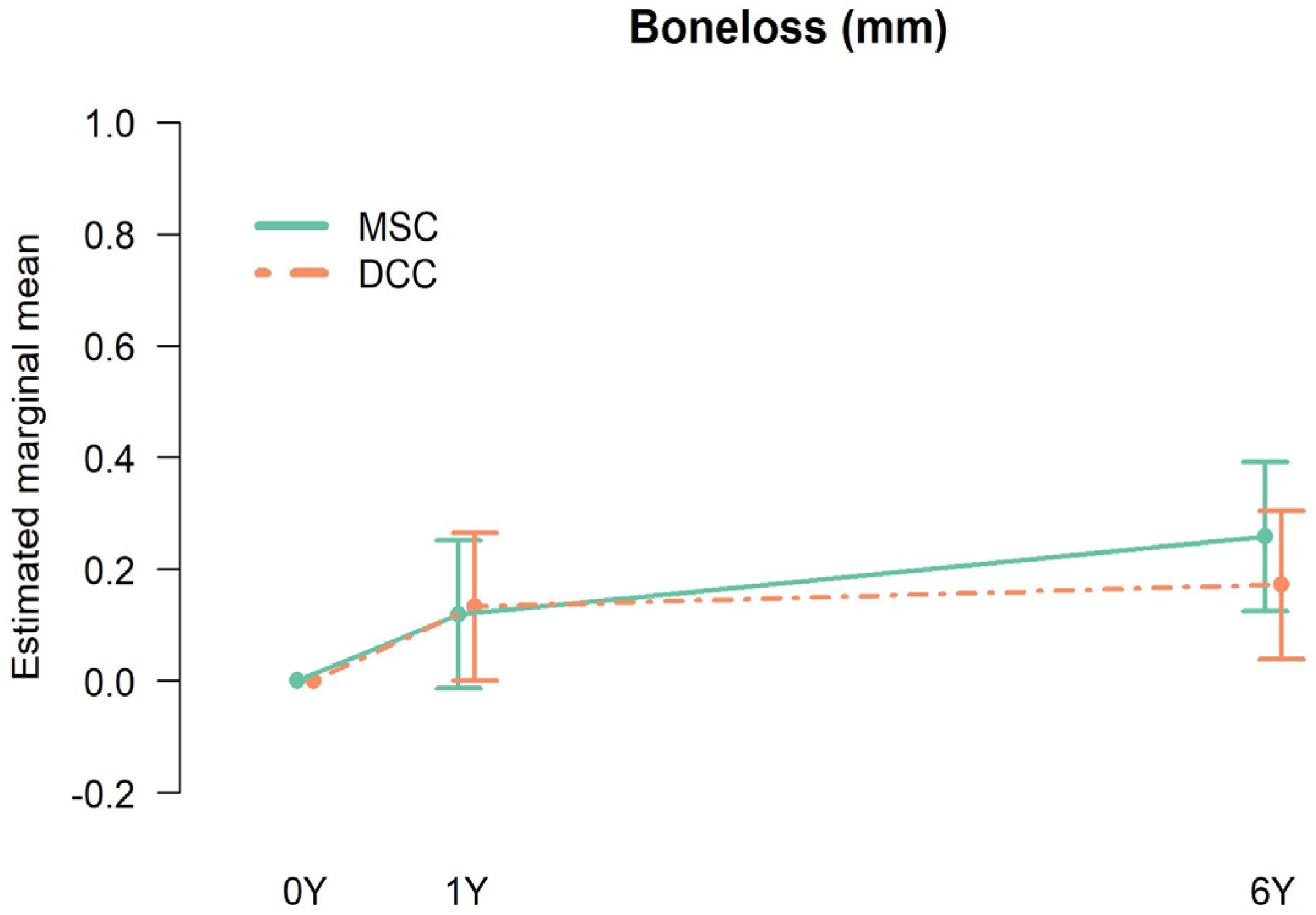
At the 1-year follow-up, 21 patients were evaluated and the DCC implants yielded a mean CBL of 0.13 mm (SD: 0.23; range: 0–0.75) and a mean CBL of 0.12 mm (SD: 0.24; range: 0–0.79) was measured at the MSC implants. After 6 years, a mean CBL of 0.26 mm (SD: 0.42; range: 0–1.79) for the MSC group and 0.17 mm (SD: 0.29; range: 0–1.00) for the DCC group was observed (Table 1 and Figure 3). At 6 years (21 patients), a mean difference in CBL of 0.09 mm between MSC and DCC was estimated, which is not statistically significant (p = 0.30; 95% CI [−0.08, 0.25]) (Table 2).
After 6 years, six implants (four MSC and two DCC implants) had bone loss >0.5 mm, but a statistically significant difference in risk of bone loss >0.5 mm could not be demonstrated between the MSC and DCC groups (p = 0.14; 95% CI [−0.03, 0.22]). Peri-implantitis was diagnosed in one MSC implant [16].

3.2. Peri-Implant Health

The estimated means, estimated mean differences and p-values for plaque, BoP and PPD are shown in Table 3. For plaque, BoP and PPD, the estimated mean differences were not statistically significant (p > 0.05).

3.3. Quality of Life

The observed mean values for OHIP-14 and VAS after 6 years are shown in Table 4. For both OHIP-14 and VAS, a statistically significant difference was observed between the baseline and the 1-year follow-up (p < 0.001), as well as between the baseline and the 6-year follow-up (p < 0.001). Between the 1-year and 6-year follow-ups, no statistically significant difference was found for OHIP-14 and VAS (p = 0.11, p = 0.73, respectively).
In addition to the evaluation of the OHQoL, the five most common maintenance interventions that occurred from the moment of connection until the 6-year follow-up were tracked and are shown in Table 5. Ulcers in the mandible were most common, followed by a loose-fitting upper denture, calculus, need for a rebasing of the upper denture and need for repair of the upper denture.

4. Discussion

The main purpose of this split-mouth study was to compare the implant outcome of hybrid and moderately rough implants after 6 years in patients with a bar-supported mandibular overdenture. A limited number of studies have examined the survival rates of hybrid implants, which range from 95.8% to 100% within a follow-up period of 12 to 60 months [28]. This is in line with the survival rates reported in the present study after a follow-up period that ranged from 61 to 88 months. Radiographic evaluation of the crestal bone level is a key criterion in evaluating the clinical outcome. This study observed relatively stable bone levels at 6 years, consistent with the 100% implant survival rate.
The influence of implant surface roughness on CBL is still under debate. A study by Raes et al. (2018), comparing 48 minimally and moderately rough implants, has shown statistically significantly more bone loss for the moderately rough implants [7]. Note that the patient population had a history of severe periodontitis, which could explain the more pronounced crestal bone loss around moderately rough surface implants. In the current study, no significant difference in CBL, plaque, BoP and PPD could be observed between implants with a minimally rough and moderately rough coronal part.
Currently, only a limited number of studies have reported on the clinical outcomes of hybrid implants compared to moderately rough implants. In a clinical trial conducted by Zetterqvist et al., (2010), 139 hybrid implants (with a minimally rough surface coronal part) were compared to 165 fully etched implants. After 5 years, a statistically significant difference was found in terms of CBL, which was less for the fully etched group [10]. This outcome is possibly related to the very low Sa value of the coronal part of the hybrid implants, which was 0.18 µm. This value is closer to that of a polished surface rather than a minimally rough surface, such as that in the current study, where the Sa value was 0.9 µm. In another study by Camarda et al., (2021), a microtextured implant with a 3.6 mm long machined collar was compared to two implants which both had a 1.2 mm collar (one collar was microtextured and one was machined) after 25 years. Implants with a longer machined collar showed statistically significantly more bone loss than those with a shorter collar. This outcome is possibly related to the different macrodesign of the implants, whereas, in the current study, the two implants were identical except for the surface roughness. It is also interesting that the implants with shorter machined collars showed less bone loss than the shorter microtextured collars; nonetheless, this was not statistically significant [29]. A study by Spinato et al., (2017) compared 37 sandblasted and double-etched (SDE) implants to 38 hybrid implants with an apical SDE surface and a coronal machined surface. After one year, no difference was seen in CBL between these two implants, which is consistent with the results of the current study [30]. Similarly, Rothamel et al., (2022) conducted a randomized, controlled multicenter study, in which 58 patients were included. Each patient received one implant with a machined shoulder and one implant with a rough shoulder. Except for the shoulders, both implants were identical. After a follow-up period of 2 years, no statistically significant difference was found in terms of CBL and soft-tissue response [31]. In terms of the current long-term RCT, it can be concluded that the MSC implants perform equally well to the DCC implants with respect to mean CBL and there was no increased risk of progressive bone loss (>0.5 mm) between the two types of implants. Recently, it has been suggested that implants with increased CBL at early stages are at risk of bone loss progression, compromising their clinical outcome [32,33]. The latter has been confirmed by Windael et al., (2021). In a large prospective clinical study, 407 patients with 1482 implants were included and 175 were diagnosed with peri-implantitis after a 10-year follow-up. From this study, it was concluded that implants with CBL ≥ 0.5 mm during the first year of function have a higher risk of developing future peri-implantitis [16]. In the present study, while four implants had CBL > 0.5 mm after 1 year and six implants had CBL > 0.5 mm after 6 years, peri-implantitis was only diagnosed in one implant after 6 years. It should be noted that, in the present study, patient selection was performed at intake, the patient population was regularly seen for a check-up and the follow-up period is relatively short to diagnose peri-implantitis. A limitation of the current study might be the strict selection criteria whereby high peri-implantitis risk patients such as smokers are excluded from this study [20]. Therefore, one must be cautious- to extrapolate the results of the present study to the general patient population.
A secondary aim of this study was to compare the OHQoL of patients at baseline with a conventional complete denture with that of patients at 1 year and 6 years after connection with an implant-supported overdenture. Sivaramakrishnan et al., (2016) performed a meta-analysis comparing conventional dentures to implant-supported overdentures. According to the results of an OHIP-14 questionnaire, the implant-supported overdentures performed better than the conventional dentures in terms of patient satisfaction [34]. This is in line with the results of the present study, in which a clear improvement in OHQoL was observed at the 1-year and the 6-year follow-ups in comparison to baseline (before connection of the prosthesis to the implants). Between 1 year and 6 years, no statistically significant difference was recorded, which may indicate that the greatest improvement in OHQoL happens shortly after connection to the implants. In the present study, a decrease in the number of ulcers was observed between the moment of connection and the 6-year follow-up. As suggested by Yen et al., (2015), this could possibly be responsible for the increase in OHQoL [35]. Nonetheless, the OHIP-14 and VAS scores are standardized measuring tools to examine the effect of a dental treatment on OHQoL; they do not give full coverage of the latter. OHQoL is also affected by psychosocial impact, orofacial pain and oral function; therefore, the results of the present study should be interpreted with care [24].

5. Conclusions

This 6-year follow-up study concludes that there is no difference in crestal bone loss and peri-implant health between hybrid implants (MSC) and moderately rough implants (DCC) in patients with an implant-supported mandibular overdenture. A bar-supported overdenture could contribute to a improvement in OHQoL in comparison to a conventional complete denture and most improvement tends to happen shortly after connection to the implants.

Author Contributions

Conceptualization, M.G., C.M. and H.D.B.; Methodology, M.G., C.M. and H.D.B.; Validation, M.G., C.M. and H.D.B.; Formal analysis, M.G., C.M. and H.D.B.; Investigation, M.G., C.M. and H.D.B.; Resources, M.G., C.M. and H.D.B.; Data curation, C.M., A.V.L. and A.S.; Writing—original draft, A.V.L. and A.S.; Writing—review & editing, M.G.; Visualization, M.G.; Supervision, C.M. and H.D.B.; Project administration, M.G.; Funding acquisition, H.D.B. All authors have read and agreed to the published version of the manuscript.

Funding

This study was funded by means of material support and Hugo De Bruyn holds a research collaboration agreement on behalve of Ghent University with Southern Implants. The submission fee was also funded by Southern Implants.

Institutional Review Board Statement

This study was ethically approved by the ethical committee of the Ghent University Hospital nr: EC/2014/1231 (28 November 2014).

Informed Consent Statement

Informed consent was obtained from all subjects involved in the study.

Data Availability Statement

Dataset available on request from the authors.

Conflicts of Interest

The authors declare no conflict of interest.

References

  1. Burns, D.R. The mandibular complete overdenture. Dent. Clin. N. Am. 2004, 48, 603–623. [Google Scholar] [CrossRef]
  2. British Society for the Study of Prosthetic Dentistry. The York consensus statement on implant-supported overdentures. Eur. J. Prosthodont. Restor. Dent. 2009, 17, 164–165. [Google Scholar]
  3. Lee, D.J.; Saponaro, P.C. Management of Edentulous Patients. Dent. Clin. N. Am. 2019, 63, 249–261. [Google Scholar] [CrossRef]
  4. Sharma, A.; Nagrath, R.; Lahori, M. A comparative evaluation of chewing efficiency, masticatory bite force, and patient satisfaction between conventional denture and implant-supported mandibular overdenture: An in vivo study. J. Indian Prosthodont. Soc. 2017, 17, 361–372. [Google Scholar] [CrossRef]
  5. Ogle, O.E. Implant Surface Material, Design, and Osseointegration. Dent. Clin. N. Am. 2015, 59, 505–520. [Google Scholar] [CrossRef] [PubMed]
  6. Wennerberg, A.; Albrektsson, T. On implant surfaces: A review of current knowledge and opinions. Int. J. Oral Maxillofac. Implant. 2010, 25, 63–74. [Google Scholar]
  7. Raes, M.; D’hondt, R.; Teughels, W.; Coucke, W.; Quirynen, M. A 5-year randomized clinical trial comparing minimally with moderately rough implants in patients with severe periodontitis. J. Clin. Periodontol. 2018, 45, 711–720. [Google Scholar] [CrossRef]
  8. Arnhart, C.; Dvorak, G.; Trefil, C.; Huber, C.; Watzek, G.; Zechner, W. Impact of implant surface topography: A clinical study with a mean functional loading time of 85 months. Clin. Oral Implant. Res. 2013, 24, 1049–1054. [Google Scholar] [CrossRef]
  9. Vandeweghe, S.; Ferreira, D.; Vermeersch, L.; Mariën, M.; De Bruyn, H. Long-term retrospective follow-up of turned and moderately rough implants in the edentulous jaw. Clin. Oral Implant. Res. 2016, 27, 421–426. [Google Scholar] [CrossRef] [PubMed]
  10. Zetterqvist, L.; Feldman, S.; Rotter, B.; Vincenzi, G.; Wennström, J.L.; Chierico, A.; Stach, R.M.; Kenealy, J.N. A Prospective, Multicenter, Randomized-Controlled 5-Year Study of Hybrid and Fully Etched Implants for the Incidence of Peri-Implantitis. J. Periodontol. 2010, 81, 493–501. [Google Scholar] [CrossRef] [PubMed]
  11. Teughels, W.; Van Assche, N.; Sliepen, I.; Quirynen, M. Effect of material characteristics and/or surface topography on biofilm development. Clin. Oral Implants Res. 2006, 17, 68–81. [Google Scholar] [CrossRef]
  12. Vervaeke, S.; Dierens, M.; Besseler, J.; De Bruyn, H. The influence of initial soft tissue thickness on peri-implant bone remodeling. Clin. Implant. Dent. Relat. Res. 2014, 16, 238–247. [Google Scholar] [CrossRef]
  13. Doornewaard, R.; Christiaens, V.; De Bruyn, H.; Jacobsson, M.; Cosyn, J.; Vervaeke, S.; Jacquet, W. Long-Term Effect of Surface Roughness and Patients’ Factors on Crestal Bone Loss at Dental Implants. A Systematic Review and Meta-Analysis. Clin. Implant. Dent. Relat. Res. 2017, 19, 372–399. [Google Scholar] [CrossRef]
  14. Albrektsson, T.; Chrcanovic, B.; Östman, P.; Sennerby, L. Initial and long-term crestal bone responses to modern dental implants. Periodontology 2000 2017, 73, 41–50. [Google Scholar] [CrossRef] [PubMed]
  15. Smeets, R.; Stadlinger, B.; Schwarz, F.; Beck-Broichsitter, B.; Jung, O.; Precht, C.; Kloss, F.; Gröbe, A.; Heiland, M.; Ebker, T. Impact of Dental Implant Surface Modifications on Osseointegration. BioMed Res. Int. 2016, 2016, 6285620. [Google Scholar] [CrossRef] [PubMed]
  16. Windael, S.; Collaert, B.; De Buyser, S.; De Bruyn, H.; Vervaeke, S. Early peri-implant bone loss as a predictor for peri-implantitis: A 10-year prospective cohort study. Clin. Implant. Dent. Relat. Res. 2021, 23, 298–308. [Google Scholar] [CrossRef]
  17. Glibert, M.; Matthys, C.; Maat, R.; De Bruyn, H.; Vervaeke, S. A randomized controlled clinical trial assessing initial crestal bone remodeling of implants with a different surface roughness. Clin. Implant. Dent. Relat. Res. 2018, 20, 824–828. [Google Scholar] [CrossRef] [PubMed]
  18. Donos, N.; Van Asche, N.; Akbar, A.N.; Francisco, H.; Gonzales, O.; Gotfredsen, K.; Haas, R.; Happe, A.; Leow, N.; Navarro, J.M.; et al. Impact of timing of dental implant placement and loading: Summary and consensus statements of group 1—The 6th EAO Consensus Conference 2021. Clin. Oral Implant. Res. 2021, 32 (Suppl. 21), 85–92. [Google Scholar] [CrossRef]
  19. Bernard, J.P.; Schatz, J.P.; Christou, P.; Belser, U.; Kiliaridis, S. Long-term vertical changes of the anterior maxillary teeth adjacent to single implants in young and mature adults. J. Clin. Periodontol. 2004, 31, 1024–1028. [Google Scholar] [CrossRef]
  20. dos Reis, I.N.R.; Amaral, G.C.L.S.D.; Hassan, M.A.; Villar, C.C.; Romito, G.A.; Spin-Neto, R.; Pannuti, C.M. The influence of smoking on the incidence of peri-implantitis: A systematic review and meta-analysis. Clin. Oral Implant. Res. 2023, 34, 543–554. [Google Scholar] [CrossRef]
  21. Svanborg, L.M.; Andersson, M.; Wennerberg, A. Surface characterization of commercial oral implants on the nanometer level. J. Biomed. Mater. Res. Part B Appl. Biomater. 2010, 92, 462–469. [Google Scholar] [CrossRef]
  22. De Bruyn, H.; Vandeweghe, S.; Ruyffelaert, C.; Cosyn, J.; Sennerby, L. Radiographic evaluation of modern oral implants with emphasis on crestal bone level and relevance to peri-implant health. Periodontology 2000 2013, 62, 256–270. [Google Scholar] [CrossRef] [PubMed]
  23. Salvi, G.E.; Lang, N.P. Diagnostic parameters for monitoring peri-implant conditions. Int. J. Oral Maxillofac. Implant. 2004, 19, 116–127. [Google Scholar]
  24. Campos, L.A.; Peltomäki, T.; Marôco, J.; Campos, J.A.D.B. Use of Oral Health Impact Profile-14 (OHIP-14) in Different Contexts. What Is Being Measured? Int. J. Environ. Res. Public Health 2021, 18, 13412. [Google Scholar] [CrossRef]
  25. Slade, G.D. Derivation and validation of a short-form oral health impact profile. Community Dent. Oral Epidemiol. 1997, 25, 284–290. [Google Scholar] [CrossRef] [PubMed]
  26. Celebić, A.; Knezović-Zlatarić, D. A comparison of patient’s satisfaction between complete and partial removable denture wearers. J. Dent. 2003, 31, 445–451. [Google Scholar] [CrossRef] [PubMed]
  27. Dikicier, S.; Atay, A.; Korkmaz, C. Health-related quality of life in edentulous patients. J. Med. Life 2021, 14, 683–689. [Google Scholar] [CrossRef]
  28. Lee, C.; Tran, D.; Jeng, M.; Shen, Y. Survival rates of hybrid rough surface implants and their alveolar bone level alterations. J. Periodontol. 2018, 89, 1390–1399. [Google Scholar] [CrossRef]
  29. Camarda, A.J.; Durand, R.; Benkarim, M.; Rompré, P.H.; Guertin, G.; Ciaburro, H. Prospective randomized clinical trial evaluating the effects of two different implant collar designs on peri-implant healing and functional osseointegration after 25 years. Clin. Oral Implant. Res. 2021, 32, 285–296. [Google Scholar] [CrossRef]
  30. Spinato, S.; Bernardello, F.; Sassatelli, P.; Zaffe, D. Hybrid and fully-etched surface implants in periodontally healthy patients: A comparative retrospective study on marginal bone loss. Clin. Implant. Dent. Relat. Res. 2017, 19, 663–670. [Google Scholar] [CrossRef]
  31. Rothamel, D.; Heinz, M.; Ferrari, D.; Eissing, A.; Holtmann, H.; Schorn, L.; Fienitz, T. Impact of machined versus structured implant shoulder designs on crestal bone level changes: A randomized, controlled, multicenter study. Int. J. Implant. Dent. 2022, 8, 31. [Google Scholar] [CrossRef] [PubMed]
  32. Albrektsson, T.; Buser, D.; Sennerby, L. Crestal Bone Loss and Oral Implants. Clin. Implant. Dent. Relat. Res. 2012, 14, 783–791. [Google Scholar] [CrossRef] [PubMed]
  33. Galindo-Moreno, P.; León-Cano, A.; Ortega-Oller, I.; Monje, A.; O’Valle, F.; Catena, A. Marginal bone loss as success criterion in implant dentistry: Beyond 2 mm. Clin. Oral Implant. Res. 2015, 26, e28–e34. [Google Scholar] [CrossRef] [PubMed]
  34. Sivaramakrishnan, G.; Sridharan, K. Comparison of implant supported mandibular overdentures and conventional dentures on quality of life: A systematic review and meta-analysis of randomized controlled studies. Aust. Dent. J. 2016, 61, 482–488. [Google Scholar] [CrossRef]
  35. Yen, Y.-Y.; Lee, H.-E.; Wu, Y.-M.; Lan, S.-J.; Wang, W.-C.; Du, J.-K.; Huang, S.-T.; Hsu, K.-J. Impact of removable dentures on oral health-related quality of life among elderly adults in Taiwan. BMC Oral Health 2015, 15, 1. [Google Scholar] [CrossRef]
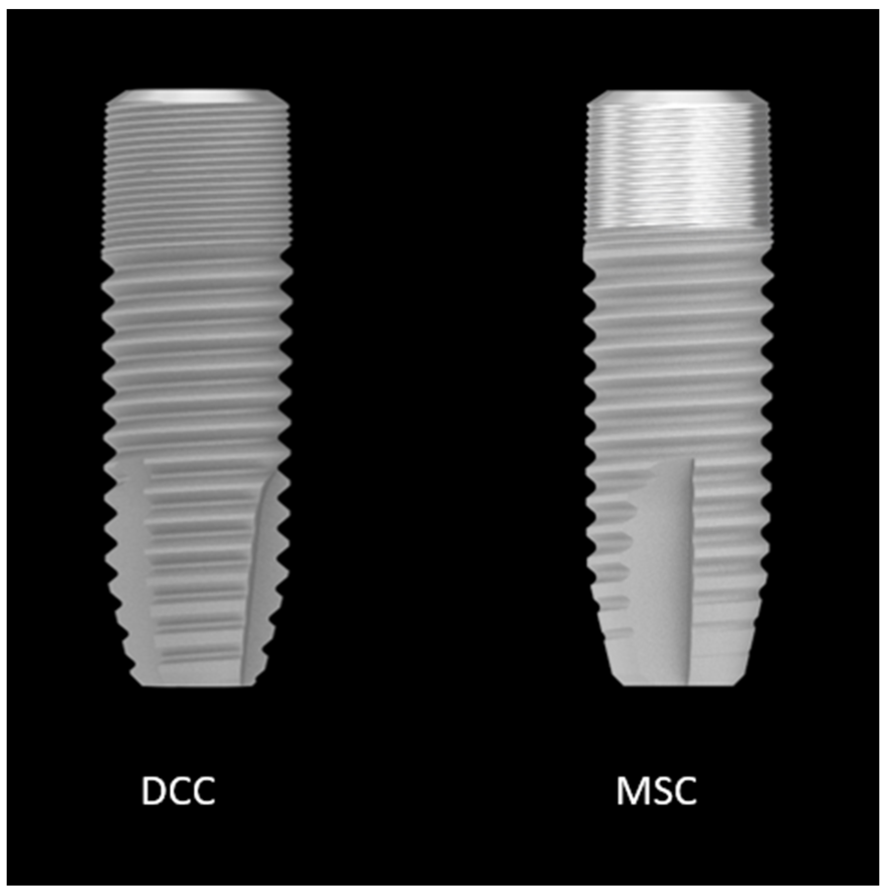
Figure 1. A conventional implant with a full moderately rough surface (DCC) and a hybrid surface implant with a 3 mm minimally rough coronal part (MSC).
Applsci 14 01658 g001
Figure 2. Implant-supported bar structure in the mandible (a,b) and mandibular overdenture (c) after 6 years of function. Intra-oral radiographs at baseline (d), 1 year (e) and 6 years (f). The reference point (green) and the measured bone level (red) are indicated.
Applsci 14 01658 g002
Figure 3. Estimated mean CBL at baseline, 1 year and 6 years in mm. 95% CIs are presented.
Applsci 14 01658 g003
Table 1. Estimated mean bone loss (mm) by time and type of implant.
TimeTypeMean (mm)Lower Bound 95% CIUpper Bound 95% CI
1 YearDCC0.130.000.27
1 YearMSC0.12−0.010.25
6 YearDCC0.170.040.31
6 YearMSC0.260.130.40
Table 2. Comparisons of estimated mean bone loss (mm) by time and type of implant. The estimated mean change in bone loss over time is not significantly different between types (p value for interaction for Type III test of fixed effect = 0.324).
TestReferenceAt/WithEstimated Mean DifferenceLower Limit 95% CIUpper Limit 95% CIp Value
MSC DCC 1 Year−0.014−0.1570.1280.844
MSC DCC 6 Year0.086−0.0560.2290.231
6 Year1 YearDCC 0.039−0.1030.1820.586
6 Year1 YearMSC 0.139−0.0030.2820.055
Table 3. Estimated means and mean differences inclusive p-values for plaque, bleeding on probing (BoP) and periodontal probing depth (PPD) after 6 years (range for plaque, BoP and PPD in mm). MSC: hybrid implant; DCC: moderately rough implant.
Estimated MeanEstimated Mean Differencep-Value
PlaqueDCC0.52; 95% CI [0.43, 0.64]0.01; 95% CI [−0.10, 0.12]0.84
MSC0.51; 95% CI [0.41, 0.63]
BopDCC0.44; 95% CI [0.30, 0.64]0.02; 95% CI [−0.13, 0.17]0.76
MSC0.42; 95% CI [0.29, 0.60]
PPDDCC2.28; 95% CI [1.93, 2.68]0.23; 95% CI [−0.13, 0.58]0.21
MSC2.50; 95% CI [2.04, 3.05]
Table 4. Observed median Oral Health Impact Profile index (OHIP-14) and observed mean Visual Analogue Scale (VAS) at baseline at the 1-year and 6-year follow-up.
Median baselineMedian 1 yearMedian 6 year
OHIP-1419.50
(IQR: 9.5–24.3)
4.0
(IQR: 2.0–8.0)
3.79
(IQR: 0.0–5.0)
Median baselineMedian 1 yearMedian 6 year
VAS32.90
(SD: 9.95;
range: 6.7–48.5)
17.21
(SD: 11.68;
range: 1.7–43.7)
16.29
(SD: 12.92;
range: 0–38.8)
Table 5. Number of the 5 most common maintenance interventions from the moment of connection (CD) until the 6-year follow-up, which are ulcers in the mandible (ulcer L), loose fitting of the upper prosthesis (loose U), calculus on the upper and/or lower prosthesis (calculus), rebasing of the upper prosthesis (rebasing U) and repairs of the upper prosthesis (repair U).
Ulcer LLoose UCalculusRebasing URepair U
CD3743--
CD-1Y348-3
1Y-6Y8105159
Total4818161512
Disclaimer/Publisher’s Note: The statements, opinions and data contained in all publications are solely those of the individual author(s) and contributor(s) and not of MDPI and/or the editor(s). MDPI and/or the editor(s) disclaim responsibility for any injury to people or property resulting from any ideas, methods, instructions or products referred to in the content.