Article

A Comprehensive Study on the Antibacterial Activities of Carbon Quantum Dots Derived from Orange Juice against Escherichia coli

1
Faculty of Fundamental Sciences, Hue University of Medicine and Pharmacy, Hue University, Hue 530000, Vietnam
2
Institute of Research and Development, Duy Tan University, Da Nang 550000, Vietnam
3
Faculty of Natural Sciences, Duy Tan University, Da Nang 550000, Vietnam
4
Institute of Physics, Vietnam Academy of Science and Technology, Ha Noi 100000, Vietnam
5
Faculty of Natural Sciences, Phu Yen University, Tuy Hoa City 56000, Vietnam
6
Institute of Theoretical and Applied Research, Duy Tan University, Ha Noi 100000, Vietnam
*
Authors to whom correspondence should be addressed.
Appl. Sci. 2024, 14(6), 2509; https://doi.org/10.3390/app14062509
Submission received: 24 January 2024 / Revised: 8 March 2024 / Accepted: 9 March 2024 / Published: 16 March 2024

Abstract

:
Carbon quantum dots (CQDs) are known for their intriguing optical properties, low toxicity, and high biocompatibility, which make them promising for biomedical applications. In this study, CQDs were synthesized by subjecting orange juice to microplasma as a carbon source at atmospheric pressure and low temperatures. The resulting CQDs exhibited a narrow size distribution, with an average diameter of approximately 4.5 nm and a pH value of 5.67. These CQDs exhibited strong blue emission characteristics. The antibacterial properties of the CQDs against Escherichia coli (E. coli) strains were evaluated using minimum inhibitory concentration assays. The study revealed that an effective inhibition of E. coli was achieved at a minimum inhibitory concentration of 0.1 ppm, while the minimum bactericidal concentration for this bacterial strain was 1 ppm, resulting in an average antibacterial efficacy of 57%. Notably, the antibacterial effects of the CQDs were observed without the need for additional light or oxidants, demonstrating the applicability of CQDs in combating bacterial strains.

1. Introduction

Advancements in nanotechnology have significantly impacted various application fields, including drug delivery, imaging, and therapeutic agents [1]. The diversification of biological and chemical properties in nanomaterials has paved the way for their application in nanomedicine, offering reduced immunogenicity, prolonged circulation, improved solubilization, and enhanced safety. Nanoparticles have demonstrated potential antibacterial properties due to their capacity to penetrate and disrupt bacterial cells, presenting promising strategies to combat the growing threat of bacterial resistance [2]. Several types of nanoparticles have been explored for antibacterial applications, including Ag, Cu, Au, and Fe, which have shown considerable effectiveness [2,3,4]. However, their clinical utility poses challenges. Moreover, it is crucial to consider the potential toxicity of these nanoparticles on cells, which cannot be overlooked.
Carbon quantum dots (CQDs) have emerged as significant photoluminescent nanomaterials in recent years, devoid of metals. These materials comprise a carbon core enveloped within an amorphous carbon structure, offering notable advantages, such as photostability, high biocompatibility, water dispersibility, low toxicity, and straightforward synthesis methods [1,5]. Their exceptional fluorescence and minimal cytotoxicity make them ideal for optoelectronics, sensing, and photocatalysis [6,7]. In the biomedical realm, CQDs have gained recognition for diverse theranostic applications like cellular imaging, drug delivery, and antibacterial activity [1,8,9]. Structured as nanoparticles, these minuscule carbon-based materials exhibit versatile traits applicable across domains encompassing biomedical imaging, drug delivery, sensors, catalysis, electronics, energy storage, water purification, nanocomposite materials, and photocatalysis. The adaptability of CQDs to nanoparticle forms positions them as promising candidates in nanotechnology-driven systems.
However, transitioning from theoretical potential to practical application poses significant challenges. Addressing scalable production, synthesis reproducibility, and long-term safety concerns is vital for realizing the extensive potential of CQDs. This emphasizes the crucial need for innovation, scalability, reproducibility, and safety considerations across various scientific disciplines and industrial sectors [10]. Additionally, CQDs can be categorized into two distinct types, namely hydrophilic and hydrophobic CQDs [11]. Hydrophilic CQDs, with an affinity for water, excel in aqueous applications like biomedicine and water purification [12]. In contrast, hydrophobic CQDs, repelling water, find utility in non-aqueous environments for applications such as photothermal therapy, energy storage, and surface coatings [13]. Understanding their specific properties enables targeted applications across scientific and industrial domains.
Diverse approaches have been employed for the synthesis of CQDs, encompassing both (i) bottom-up methods, such as hydrothermal and solvothermal syntheses, microwave synthesis, and plasma treatments, and (ii) top-down techniques, including electrochemical exfoliation, laser ablation, and chemical oxidation [1,14,15]. However, it is essential to acknowledge that some of these methods involve the use of strong acids, high processing temperatures, and complex multi-step processes, which can pose disadvantages when scaling up production or considering medical applications, potentially introducing hazardous effects due to the presence of toxic chemicals. As a result, there remains a pressing need for greener chemistry routes in the synthesis of nanoparticles, aiming for eco-friendly and cost-effective production. Notably, recent efforts have explored the use of natural precursors, such as juice from Citrus medica [16], orange juice [17], honey [18], and grape seeds [8], for CQD synthesis. These products derived from natural sources are considered “green” and cost-effective, making them suitable for bulk product development. Researchers have highlighted the advantages of synthesizing CQDs from fruit juices, particularly citrus fruits, which are predominantly acidic and rich in carboxyl groups, resulting in higher yields compared to other fruits [16].
Plasma is a fundamental state of matter that comprises photons, as well as charged, molecular, atomic, and radical species. Cold plasmas can be generated without the need for expensive vacuum systems or pumps, making them applicable in various fields, such as plasma sintering, water desalination, nanomaterial synthesis, surface modification, material deposition, and bacterial inactivation [19]. Xu et al. utilized cold plasmas to synthesize CQDs, employing chloroplatinic acid and ethanol as precursors, resulting in a single-emission wavelength [20]. Kumar et al. achieved the creation of graphene quantum dots enveloped in gold nanoparticles through DC microplasma [21]. Blue emissive CQDs have been successfully synthesized via plasma treatment using a range of carbon precursors, including citric acid, D-fructose, sodium dodecyl sulfate, chitosan, hydrocarbons, and folic acid [21,22,23,24,25]. Notably, CQD synthesis has proven successful in both acidic and basic reaction environments. However, it is worth noting that the use of fruit juice as a precursor for CQD synthesis through the plasma method remains an unexplored area of research.
The excessive use of antibiotics, both in agricultural and clinical settings, has resulted in widespread antibiotic resistance, necessitating the urgent development of new antibacterial agents. According to the World Health Organization (WHO), Gram-negative (GN) bacterial pathogens are particularly resistant to antibacterial treatments [26]. The structure of GN bacteria inherently provides greater resistance compared to Gram-positive (GP) bacteria. GN bacteria feature a slim, peptidoglycan-covered outer layer with multiple thin membrane layers that do not readily absorb external materials, making them more challenging to combat with various antibacterial agents. Escherichia coli (E. coli), a representative GN bacteria, has been studied for its resistance to antimicrobial cationic surfactants [27]. CQDs with positive surface charges act as potent antibacterial agents against negatively charged bacteria, impairing the surfaces of bacterial cells. Consequently, CQDs emerge as promising candidates for photo-activated antibacterials capable of effectively preventing and controlling pathogenic bacteria. Notably, the environmental and health hazards posed by antibacterial chemicals, such as hydrogen peroxide and sodium hypochlorite [28], are mitigated by utilizing CQDs, offering a safer alternative to chemical antimicrobial toxicity.
In this study, eco-friendly CQDs were synthesized from orange juice, serving as a green precursor through a plasma method. The optimal CQD was subjected to antibacterial testing, with the primary goal of investigating and determining the average antibacterial rate of the CQDs against E. coli strains. The results indicated that CQDs exhibit potential as novel antibacterial nanoparticles.

2. Materials and Methods

2.1. Synthesis of CQDs

The microplasma system illustrated in Figure 1 was used to synthesize CQDs. The setup included a graphite electrode, a plasma nozzle, a DC high-voltage source, and a glass beaker containing the precursor solution. The plasma electrode consisted of a tungsten rod with a diameter of approximately 1.5 mm connected to the negative polarity through a 100 kΩ resistor.
To synthesize CQDs, 30 mL of orange juice solution was placed in a glass beaker, as depicted in Figure 1. The plasma treatment was carried out for 20 min at atmospheric pressure and low temperatures ranging from 35 to 60 °C. The operating current was maintained at a constant operating current of 5 mA with a high-voltage source typically around 5 kV. After treatment, the solution was filtered through filter paper with pore sizes smaller than 2 μm in diameter and a diameter of 125 mm to remove any residues. Then, the solution was centrifuged to separate the deposit. The resulting redispersed product was mixed with water, resulting in a pure CQD solution with a light-yellow color.

2.2. Characterization

The synthesized CQDs underwent comprehensive characterization using various techniques. UV-vis absorption spectroscopy was carried out in the range of 200–700 nm with a Jasco V-570 instrument (The Jasco Corporation, Tokyo, Japan). To assess their emission behavior, photoluminescence studies were performed using fluorescence spectroscopy with a Fluorog 3 (HORIBA New Jersey Optical Spectroscopy Center, Piscataway, NJ, USA), both analyses employing quartz cells with a 1 cm path length. Particle size and uniformity were analyzed using a transmission electron microscope (TEM) and high-resolution transmission electron microscopy (HRTEM) (JEOL 2100, JEOL Ltd., Tokyo, Japan), providing insights into their structural features. TEM samples were prepared by dispersing the powdered sample in water, then drip-drying on a copper grid coated with a carbon film. The functional groups of carbon quantum dots (CQDs), including hydroxyl (OH–) and carbonyl (C=O), were examined using Fourier-transform infrared spectroscopy (FTIR) with an FTIR Cary 630 (Agilent Technologies, Santa Clara, CA, USA) over a wavenumber range from 400 to 4000 cm−1. The shifting of functional group peak readings was observed, and FTIR measurements were conducted by preparing a solution of the sample and placing a drop of the solution onto the measurement disk for analysis. All these measurements were conducted at room temperature to ensure consistency in the characterization conditions.

2.3. Antibacterial Activity Assay

In this study, we assessed the antibacterial efficacy of CQDs against E. coli through minimum inhibitory concentration (MIC) assays. The MIC represented the concentration at which no visible growth was observed. The procedure involved streaking and culturing E. coli strains on a nutrient agar medium, followed by an 18 to 24 h incubation at 37 °C. Subsequently, a bacterial suspension was prepared using 0.9% physiological saline to achieve a bacterial concentration of 0.5 McFarland, equivalent to 108 colony-forming units per milliliter (CFU/mL). Following this, 50 μL of CQDs was applied to the agar disk previously inoculated with E. coli, and the mixture was incubated at 37 °C for 24 h. After cultivation, the number of bacterial colonies on each dish was recorded, and visual inspection for bacterial growth (turbidity) was conducted to determine the MICs of the CQD sample.
MBC Determination: Following incubation, the agar plates were inspected to verify the presence or absence of bacterial colonies. Absence of colonies indicated that the CQD concentration in that aliquot was adequate to eliminate at least 99.9% of the bacterial population, and that concentration was then deemed as the MBC.
The antimicrobial activity was assessed based on surviving colony-forming unit (CFU) counts, and the antibacterial rate was determined using Equation (1). In the bacterial preparation process, a suspension of E. coli was meticulously created with a concentration of 106 CFU/mL, utilizing sterile 0.9% NaCl solution. The resulting inoculum was formed by combining 1.5 mL Muller–Hinton broth with 50 µL of the suspension, and this inoculum underwent a 24 h incubation period at 25 °C under natural light conditions. After incubation, the E. coli suspension was serially diluted in 1:100 steps, resulting in concentrations of 10−2, 10−4, 10−6, 10−7, and 10−8. Three final dilution concentration levels were chosen, and 100 µL from each level was plated onto separate nutrient agar plates. Subsequent incubation at 37 °C for 24 h allowed for the analysis of colony counts. From these counts, a specific dilution level was chosen for further stages of the experiment, representing the inoculum size for subsequent investigations into the antibacterial activity of the substance under scrutiny.
Antibacterial   rate   ( % ) = Control   group   CFU E x p e r i m e n t a l   g r o u p   C F U Control   group   CFU × 100 %
In this context, the control group denoted bacterial growth without CQDs, whereas the experimental group denoted bacterial growth in the presence of CQDs. The CFU values served as a quantitative measure of the antibacterial effectiveness of CQDs.

3. Results

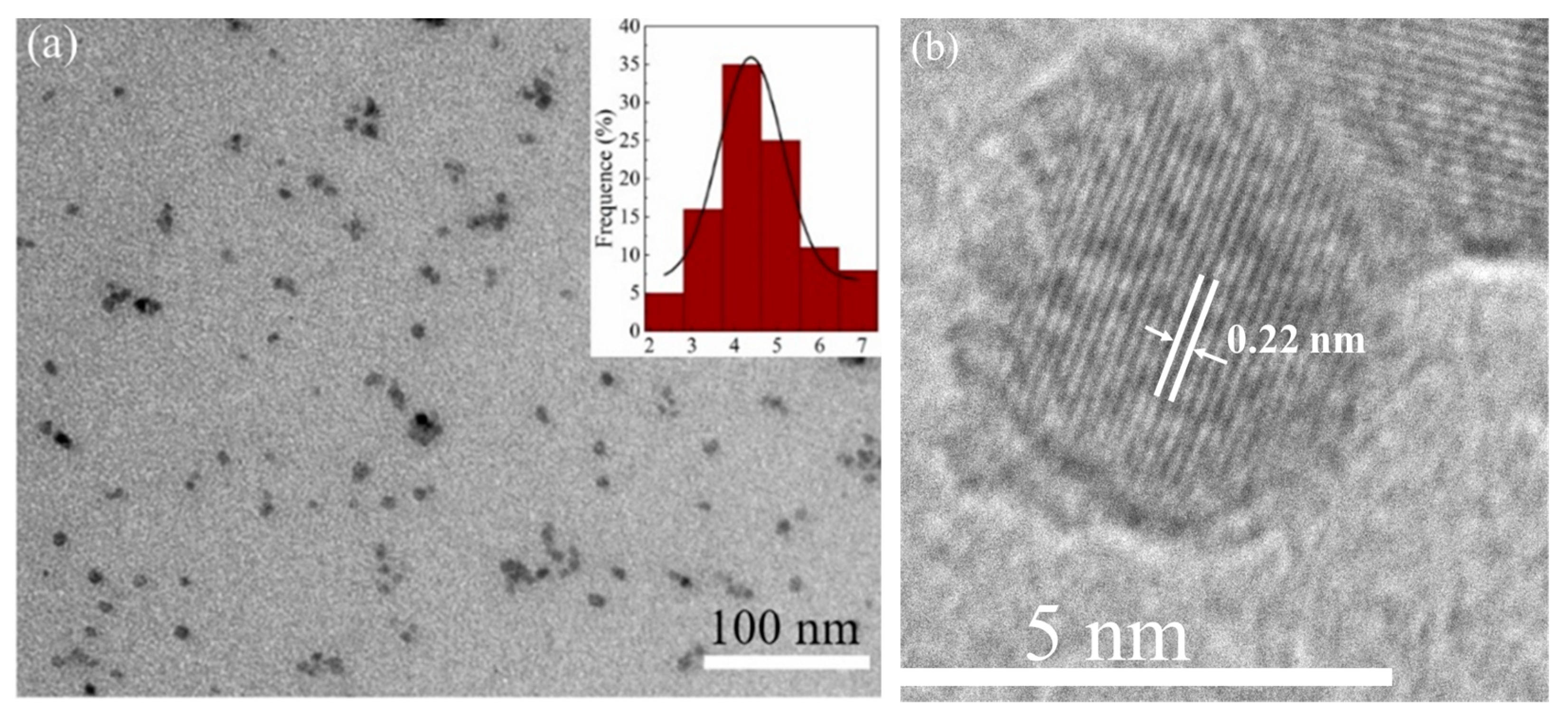
3.1. Structural Characterisation

The morphological analysis of CQDs utilized both TEM and HRTEM, as illustrated in Figure 2. The TEM image in Figure 2a reveals quasi-spherical CQDs with an average size of 4.4 nm and a narrow size distribution, as emphasized in the inset. Figure 2b provides HRTEM images, further confirming the uniformity and excellent crystalline structure of the CQDs. The majority of CQDs observed in HRTEM align with sizes from the TEM analysis, indicating consistent dimensions. The high-magnification HRTEM image displays distinct lattice fringes measuring 0.22 nm, similar to the (100) facet of sp2 graphitic carbon [29], highlighting a well-defined crystalline structure within the CQDs. The combined TEM and HRTEM analyses offer a comprehensive view of the morphology of CQDs. The quasi-spherical shape, narrow size distribution, and good crystallinity affirm the precision and quality of the synthesized CQDs. These findings are crucial for understanding the structural characteristics of CQDs and informing their potential applications in various scientific and technological fields.
Figure 3 presents the FTIR spectrum of CQDs. The peak at 3250 cm−1 in the synthesized CQDs is attributed to the adsorption of O-H stretching vibration [30]. Peaks in the range of 1000–1131 cm−1 are associated with C-H stretching [30]. Peaks at 1641 cm−1 and 1412 cm−1 can be attributed to the C=O and -C-OH groups, indicating plasma treatment that leads to the formation of sp2 domains. These groups originate from small precursor molecules like sugar and citric acid. As a result, there are oxygen, hydroxyl, and carboxyl groups present in the sp3 carbon area [31]. These functional groups contribute to the aqueous solubility of CQDs after carbonization by facilitating the formation of bonds with surrounding oxygen groups [32,33].

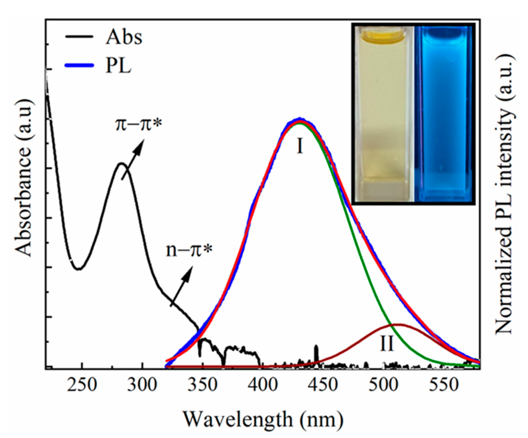
3.2. Optical Properties

The use of the UV–vis absorption spectrum, a straightforward evaluation technique for CQD products, is presented in Figure 4. This characterization was carried out within a wavelength range of 200–600 nm. The UV–vis absorption spectrum exhibits strong and sharp absorption bands at 283 nm, likely attributed to the π–π* transition of the C=C bond. Additionally, a weak and broad band at 335 nm is observed, corresponding to the n–π* transition in the C=O bond [34]. This outcome confirms the formation of carbon nanoparticles following the reaction, aligning with reported experimental results [35,36]. Additionally, the results demonstrate that CQDs exhibit blue fluorescence properties under UV light at 254 nm (inset in Figure 4), which is attributed to the quantum confinement effect and edge defects [34]. In orange juice, compounds such as citric acid, sucrose, and glucose are present [37]. Plasma generated at this interface produces a dense population of electrons that interact with the sample solution, leading to the dissociation of solution precursors and the production of C2 and CH species [38]. These species facilitate carbon nucleation and carbon dot formation.
The normalized photoluminescent (PL) spectrum of CQDs is presented in Figure 4. The maximum PL emission wavelength of CQDs was recorded at an excitation wavelength of 300 nm. The PL peak fitting in Figure 4 illustrates the PL spectrum composed of two peaks: (I) the main peak centered at 430.3 nm (green line curve) with a full-width half maximum (FWHM) of 94 nm and (II) another shoulder peak at 511 nm (wine line curve) with an FWHM of 73 nm, representing the emission centers denoting the surface states of the samples. CQDs exhibit multiple emission centers due to the presence of different surface chemical groups on CQDs [39]. The distribution of emission centers on CQDs can be inferred from their PL spectra.

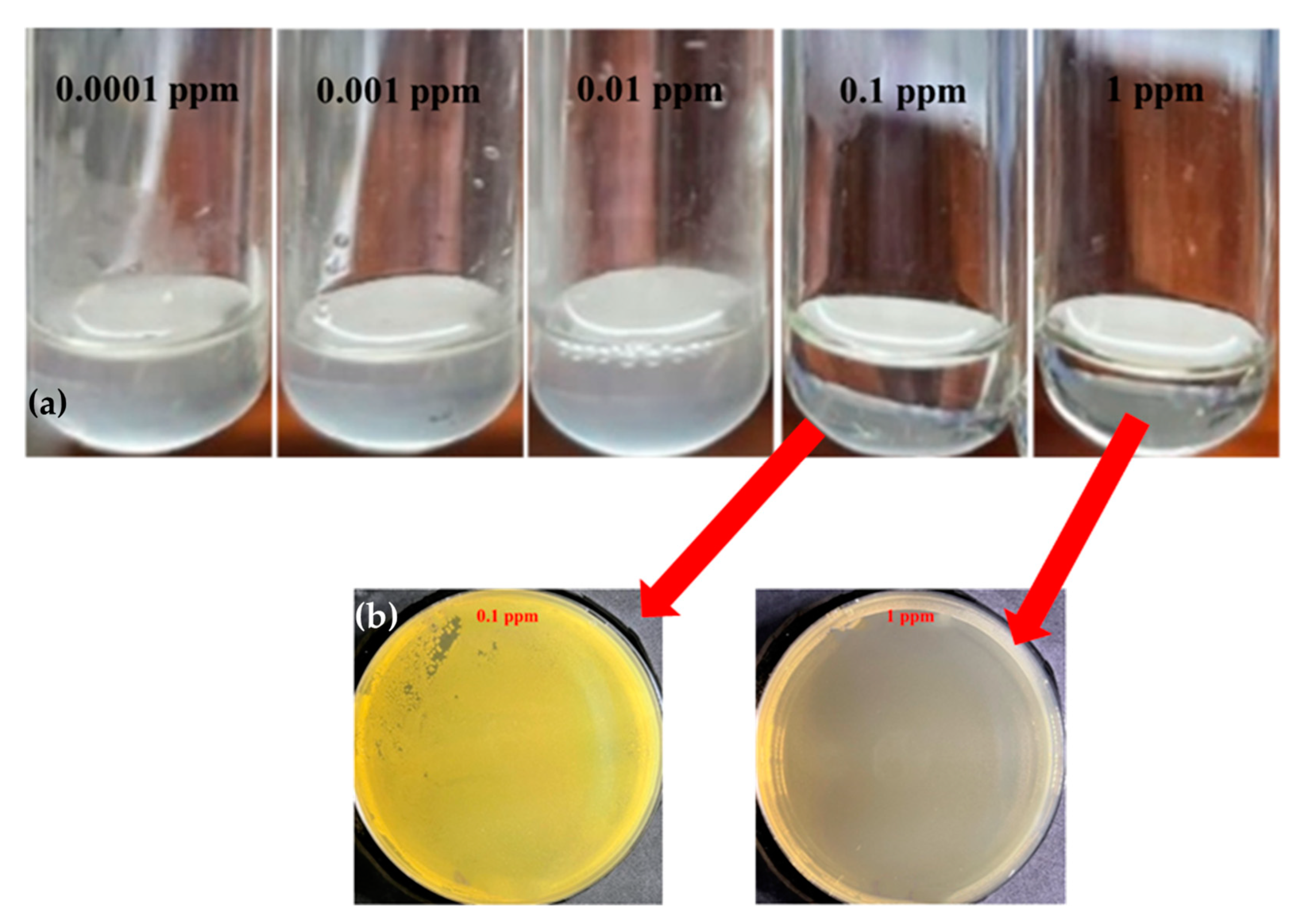
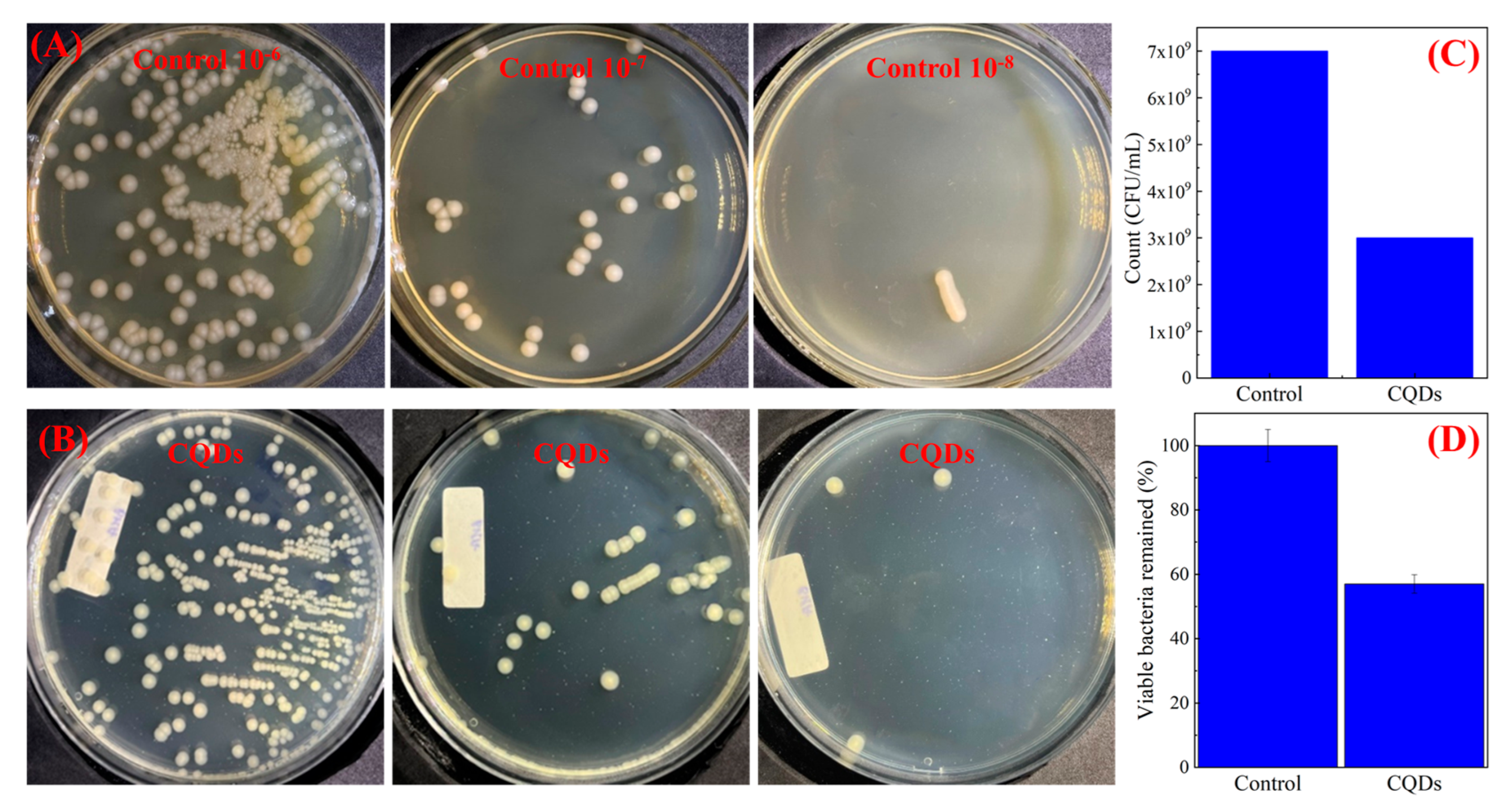
3.3. Antibacterial Properties

Antibacterial efficacy is a crucial parameter for assessing the potential of positively charged CQDs as antibacterial agents in biomedical applications. In this study, E. coli was utilized as a model to evaluate the antimicrobial properties against Gram-negative bacteria. The images in Figure 5a depict the antibacterial results of E. coli with the MICs of CQDs. MICs were visually assessed through tube turbidity before and after incubation with bacteria. The MICs of QCQD exhibited observable bacterial growth at various concentrations of CQDs (0.0001, 0.001, 0.01, 0.1, and 1 ppm). The results indicate that concentrations below 0.1 ppm maintained a clear and transparent solution, while concentrations of 0.1 ppm and 1 ppm displayed no observable bacterial growth. Therefore, it can be inferred that the MIC of CQDs for E. coli in our experiment was 1 ppm.
After determining the minimum inhibitory concentrations (MICs), a pivotal phase in our analysis involved streaking and culturing samples on nutrient agar using the smoothing method. Subsequently, these cultures underwent a meticulous 24 h incubation at 37 °C, providing a comprehensive exploration of the minimal bacterial concentration (MBC). The detailed findings illustrated in Figure 5b offer profound insights into the efficacy of CQDs against E. coli. The observed MBC of CQDs for E. coli, established at 1 ppm in our experimental context, serves as a crucial indicator of their inhibitory potential. This result underscores that, even at a low concentration of 1 ppm, CQDs effectively halt the growth of E. coli bacteria. A systematic approach involving nutrient agar and the incubation process ensures a robust assessment of bacterial concentration thresholds, reinforcing the reliability and significance of our findings.
In Figure 6, the photograph illustrates the quantity of viable E. coli bacterial cells after treatment, as evaluated through a disk-diffusion test employing CQDs, as shown in Figure 6A,B. The antibacterial rate of the CQDs against E. coli is 57%, corresponding to statistical histograms after treatment with control and CQDs samples at a concentration of 10−8, as assessed by determining the number of surviving bacteria in the culture dish, as seen in Figure 6C,D. Further details regarding the evaluation of antibacterial efficacy were elucidated using the standard plate count method. The remarkable antibacterial effectiveness of CQDs is intricately linked to their inherent physicochemical and functional properties. This efficacy is mainly attributed to the induction of oxidative stress initiated by the generation of reactive oxygen species (ROS) during photoactivation [40,41]. These ROS, produced through this photoactivation process, exhibit versatile capabilities, including deactivating proteins, inducing nucleic acid damage, and initiating lipid peroxidation. This antibacterial mechanism acts as a robust disinfection process, specifically targeting bacteria. The consequences of this oxidative stress cascade extend to the disruption of bacterial cell walls and the deterioration of infective biofilm matrices. The insights from Figure 6 highlight the compelling antibacterial activity of CQDs, emphasizing their potential as effective agents in disinfection strategies against E. coli.
The observed increase in the measured inhibition diameter of bacteria after exposure to CQDs emphasizes their efficacy in hindering bacterial growth and biofilm formation. Our study was specifically designed to directly investigate the antibacterial properties inherent in the CQD sample when challenged with the E. coli bacterial strain. It is noteworthy that CQDs consistently demonstrated the ability to inhibit E. coli growth across various exposure times, aligning with established findings in the scientific literature [42,43].
The observed differences in performance, as compared to certain studies, result from our deliberate choice of a direct testing method. We intentionally excluded active ingredients or external influences such as light or ultraviolet irradiation [44,45,46]. While this approach may lead to a relatively lower recorded performance, it ensures a more authentic evaluation of the intrinsic antibacterial capabilities of the CQDs. One significant finding from our research is that a mere 30 s contact time between bacteria and the CQD sample is enough to inhibit the growth of E. coli. This rapid onset of antibacterial activity sets our study apart from investigations involving hydrophobic carbon quantum dots/polydimethylsiloxane nanocomposites. In those studies, bacterial growth was effectively suppressed after a longer 15 min irradiation period [47,48]. The highlighted speed and effectiveness of the CQD sample in inhibiting bacterial growth make it a potentially rapid and efficient strategy, particularly when used against the E. coli bacterial strain. The estimated values indicate that these results are slightly lower compared to some reported findings [49,50]. However, this outcome could be explained by the experiment using CQDs directly without the addition of any other active substances such as doped or additional various amide groups, as well as the absence of external factors such as exposure to light or ultraviolet radiation [14,51], resulting in a lower observed efficiency.

4. Conclusions

This study successfully synthesized CQDs from orange juice using a novel microplasma method under ambient conditions. The synthesized CQDs exhibited a narrow size distribution, with an average diameter of 4.4 nm, and demonstrated strong blue emission at a wavelength of 449.5 nm, with an additional shoulder peak at 509 nm. Notably, these CQDs displayed remarkable antibacterial activity against E. coli strains, achieving a minimum inhibitory concentration of 0.1 ppm and a minimum bactericidal concentration of 1 ppm. This translates to an average antibacterial efficacy of 57% against E. coli, highlighting their potential as potent antibacterial agents. Furthermore, the antibacterial effect was observed without requiring additional light or oxidants, offering a potentially simpler and less resource-intensive approach compared to conventional methods. The tunable optical properties, low toxicity, and excellent dispersibility of these CQDs, coupled with their demonstrated antibacterial activity, make them promising candidates for various applications in the fields of biomedicine and environmental remediation.

Author Contributions

V.D.P. and M.H.N. conceived the experiment. H.M.P., H.T.D., D.T.L. and A.T.L. conducted the experiments and analyzed the data. All authors participated in the discussion of the results. T.B.V., V.D.P., T.B.N. and M.H.N. wrote the initial draft of the paper. T.B.N., V.D.P., D.T.L., T.B.V. and M.H.N. supervised the project. All authors have read and agreed to the published version of the manuscript.

Funding

This research was funded by the Physics Program at the Vietnam Academy of Science and Technology under Project No. KHCBVL.05/21-22 and Project No. NVCC05.03/22-23.

Institutional Review Board Statement

Not applicable.

Informed Consent Statement

Not applicable.

Data Availability Statement

All data that support the findings of this study are available from the corresponding author upon reasonable request.

Acknowledgments

The authors are grateful for the use of the facilities of the joint optics and photonics laboratory at the Institute of Physics (IOP, VAST).

Conflicts of Interest

The authors declare no conflicts of interest.

References

  1. Gagic, M.; Kociova, S.; Smerkova, K.; Michalkova, H.; Setka, M.; Svec, P.; Pribyl, J.; Masilko, J.; Balkova, R.; Heger, Z.; et al. One-Pot Synthesis of Natural Amine-Modified Biocompatible Carbon Quantum Dots with Antibacterial Activity. J. Colloid Interface Sci. 2020, 580, 30–48. [Google Scholar] [CrossRef]
  2. Schröfel, A.; Kratošová, G.; Šafařík, I.; Šafaříková, M.; Raška, I.; Shor, L.M. Applications of Biosynthesized Metallic Nanoparticles—A Review. Acta Biomater. 2014, 10, 4023–4042. [Google Scholar] [CrossRef] [PubMed]
  3. Lewinski, N.; Colvin, V.; Drezek, R. Cytotoxicity of Nanoparticles. Small 2008, 4, 26–49. [Google Scholar] [CrossRef]
  4. Huang, Y.W.; Cambre, M.; Lee, H.J. The Toxicity of Nanoparticles Depends on Multiple Molecular and Physicochemical Mechanisms. Int. J. Mol. Sci. 2017, 18, 2702. [Google Scholar] [CrossRef] [PubMed]
  5. Shen, L.M.; Liu, J. New Development in Carbon Quantum Dots Technical Applications. Talanta 2016, 156–157, 245–256. [Google Scholar] [CrossRef]
  6. Ren, W.; Chen, S.; Li, S.; Zhang, Y.; Liu, J.; Guan, M.; Yang, H.; Li, N.; Han, C.; Li, T.; et al. Photoluminescence Enhancement of Carbon Dots by Surfactants at Room Temperature. Chem. A Eur. J. 2018, 24, 15806–15811. [Google Scholar] [CrossRef]
  7. Baker, S.N.; Baker, G.A. Luminescent Carbon Nanodots: Emergent Nanolights. Angew. Chemie Int. Ed. 2010, 49, 6726–6744. [Google Scholar] [CrossRef]
  8. Parvathy, C.R.; Praseetha, P.K. Evaluation of Anti-Diabetic Potential of Anti-Microbial Carbon Quantum Dots from Vitis Vinifera Seeds. Nano Biomed. Eng. 2023, 15, 28–35. [Google Scholar] [CrossRef]
  9. Wu, Y.; Li, C.; van der Mei, H.C.; Busscher, H.J.; Ren, Y. Carbon Quantum Dots Derived from Different Carbon Sources for Antibacterial Applications. Antibiotics 2021, 10, 623. [Google Scholar] [CrossRef]
  10. Yang, J.; Zhang, X.; Ma, Y.H.; Gao, G.; Chen, X.; Jia, H.R.; Li, Y.H.; Chen, Z.; Wu, F.G. Carbon Dot-Based Platform for Simultaneous Bacterial Distinguishment and Antibacterial Applications. ACS Appl. Mater. Interfaces 2016, 8, 32170–32181. [Google Scholar] [CrossRef]
  11. Varisco, M.; Zufferey, D.; Ruggi, A.; Zhang, Y.; Erni, R.; Mamula, O. Synthesis of Hydrophilic and Hydrophobic Carbon Quantum Dots from Waste of Wine Fermentation. R. Soc. Open Sci. 2017, 4, 170900. [Google Scholar] [CrossRef]
  12. Guo, H.; Wen, S.; Li, W.; Li, M.; Wang, L.; Chang, Q.; Zhang, J.; Lai, J.; Vajtai, R.; Ajayan, P.M.; et al. A Universal Strategy to Separate Hydrophilic Hybrid-Light Carbon Quantum Dots Using Pure Water as Eluent. Appl. Mater. Today 2020, 18, 100528. [Google Scholar] [CrossRef]
  13. Kováčová, M.; Špitalská, E.; Špitálský, Z. Light-Activated Polymer Nanocomposites Doped with a New Type of Carbon Quantum Dots for Antibacterial Applications. In Urinary Stents: Current State and Future Perspectives; Springer International Publishing: Cham, Switzerland, 2022; pp. 315–324. [Google Scholar] [CrossRef]
  14. Mahat, N.A.; Nor, N.S.M.; Shamsudin, S.A. Effects of Positive Carbon Quantum Dots on Gram-Negative Bacteria as an Antimicrobial Agent. J. Inorg. Organomet. Polym. Mater. 2022, 32, 2428–2440. [Google Scholar] [CrossRef]
  15. Abu Rabe, D.I.; Al Awak, M.M.; Yang, F.; Okonjo, P.A.; Dong, X.; Teisl, L.R.; Wang, P.; Tang, Y.; Pan, N.; Sun, Y.P.; et al. The Dominant Role of Surface Functionalization in Carbon Dots’ Photo-Activated Antibacterial Activity. Int. J. Nanomed. 2019, 14, 2655–2665. [Google Scholar] [CrossRef]
  16. Selvaraju, N.; Ganesh, P.S.; Palrasu, V.; Venugopal, G.; Mariappan, V. Evaluation of Antimicrobial and Antibiofilm Activity of Citrus Medica Fruit Juice Based Carbon Dots against Pseudomonas Aeruginosa. ACS Omega 2022, 7, 36227–36234. [Google Scholar] [CrossRef]
  17. Sahu, S.; Behera, B.; Maiti, T.K.; Mohapatra, S. Simple One-Step Synthesis of Highly Luminescent Carbon Dots from Orange Juice: Application as Excellent Bio-Imaging Agents. Chem. Commun. 2012, 48, 8835–8837. [Google Scholar] [CrossRef] [PubMed]
  18. Surendran, P.; Lakshmanan, A.; Priya, S.S.; Balakrishnan, K.; Rameshkumar, P.; Kannan, K.; Geetha, P.; Hegde, T.A.; Vinitha, G. Bioinspired Fluorescence Carbon Quantum Dots Extracted from Natural Honey: Efficient Material for Photonic and Antibacterial Applications. Nano-Struct. Nano-Objects 2020, 24, 100589. [Google Scholar] [CrossRef]
  19. Chiang, W.H.; Mariotti, D.; Sankaran, R.M.; Eden, J.G.; Ostrikov, K. Microplasmas for Advanced Materials and Devices. Adv. Mater. 2020, 32, e1905508. [Google Scholar] [CrossRef] [PubMed]
  20. Lin, L.; Zhang, Z.; Min, Y. Microfluidic Plasma: Novel Process Intensification Strategy. Green Process. Synth. 2022, 11, 1064–1071. [Google Scholar] [CrossRef]
  21. Rathore, S.; Patel, D.K.; Thakur, M.K.; Haider, G.; Kalbac, M.; Kruskopf, M.; Liu, C.I.; Rigosi, A.F.; Elmquist, R.E.; Liang, C.T.; et al. Highly Sensitive Broadband Binary Photoresponse in Gateless Epitaxial Graphene on 4H–SiC. Carbon 2021, 184, 72–81. [Google Scholar] [CrossRef]
  22. Orriere, T.; Kurniawan, D.; Chang, Y.C.; Pai, D.Z.; Chiang, W.H. Effect of Plasma Polarity on the Synthesis of Graphene Quantum Dots by Atmospheric-Pressure Microplasmas. Nanotechnology 2020, 31, 485001. [Google Scholar] [CrossRef] [PubMed]
  23. Weerasinghe, J.; Scott, J.; Deshan, A.D.K.; Chen, D.; Singh, A.; Sen, S.; Sonar, P.; Vasilev, K.; Li, Q.; Ostrikov, K. Monochromatic Blue and Switchable Blue-Green Carbon Quantum Dots by Room-Temperature Air Plasma Processing. Adv. Mater. Technol. 2022, 7, 2100586. [Google Scholar] [CrossRef]
  24. Zheng, P.; Liu, R.; Wang, J.; Luo, Y.; Zhao, H.; Mao, X.; Lai, C. Cathode and Hollow Metal Anode with Miniature Argon Gas Flow. J. Appl. Spectrosc. 2022, 89, 141–149. [Google Scholar] [CrossRef]
  25. Yang, J.S.; Pai, D.Z.; Chiang, W.H. Microplasma-Enhanced Synthesis of Colloidal Graphene Quantum Dots at Ambient Conditions. Carbon 2019, 153, 315–319. [Google Scholar] [CrossRef]
  26. Breijyeh, Z.; Jubeh, B.; Karaman, R. Resistance of Gram-Negative Bacteria to Current Antibacterial Agents and Approaches to Resolve It. Molecules 2020, 25, 1340. [Google Scholar] [CrossRef] [PubMed]
  27. Dong, X.; Awak, M.A.; Tomlinson, N.; Tang, Y.; Sun, Y.P.; Yang, L. Antibacterial Effects of Carbon Dots in Combination with Other Antimicrobial Reagents. PLoS ONE 2017, 12, e0182324. [Google Scholar] [CrossRef] [PubMed]
  28. Fernando, K.A.S.; Sahu, S.; Liu, Y.; Lewis, W.K.; Guliants, E.A.; Jafariyan, A.; Wang, P.; Bunker, C.E.; Sun, Y.P. Carbon Quantum Dots and Applications in Photocatalytic Energy Conversion. ACS Appl. Mater. Interfaces 2015, 7, 8363–8376. [Google Scholar] [CrossRef] [PubMed]
  29. Dong, Y.; Pang, H.; Yang, H.B.; Guo, C.; Shao, J.; Chi, Y.; Li, C.M.; Yu, T. Carbon-Based Dots Co-doped with Nitrogen and Sulfur for High Quantum Yield and Excitation-Independent Emission. Angew. Chemie 2013, 125, 7954–7958. [Google Scholar] [CrossRef]
  30. Gu, J.; Zhang, X.; Pang, A.; Yang, J. Facile Synthesis and Photoluminescence Characteristics of Blue-Emitting Nitrogen-Doped Graphene Quantum Dots. Nanotechnology 2016, 27, 164704. [Google Scholar] [CrossRef]
  31. Zhu, S.; Zhang, J.; Tang, S.; Qiao, C.; Wang, L.; Wang, H.; Liu, X.; Li, B.; Li, Y.; Yu, W.; et al. Surface Chemistry Routes to Modulate the Photoluminescence of Graphene Quantum Dots: From Fluorescence Mechanism to up-Conversion Bioimaging Applications. Adv. Funct. Mater. 2012, 22, 4732–4740. [Google Scholar] [CrossRef]
  32. Jamaludin, N.; Tan, T.L.; Zaman, A.S.K.; Sadrolhosseini, A.R.; Rashid, S.A. Acid-Free Hydrothermal-Extraction and Molecular Structure of Carbon Quantum Dots Derived from Empty Fruit Bunch Biochar. Materials 2020, 13, 3356. [Google Scholar] [CrossRef]
  33. Qin, X.; Lu, W.; Asiri, A.M.; Al-Youbi, A.O.; Sun, X. Microwave-Assisted Rapid Green Synthesis of Photoluminescent Carbon Nanodots from Flour and Their Applications for Sensitive and Selective Detection of Mercury(II) Ions. Sens. Actuators B Chem. 2013, 184, 156–162. [Google Scholar] [CrossRef]
  34. Yadav, P.K.; Chandra, S.; Kumar, V.; Kumar, D.; Hasan, S.H. Carbon Quantum Dots: Synthesis, Structure, Properties, and Catalytic Applications for Organic Synthesis. Catalysts 2023, 13, 422. [Google Scholar] [CrossRef]
  35. Zhao, S.; Chen, X.; Zhang, C.; Zhao, P.; Ragauskas, A.J.; Song, X. Fluorescence Enhancement of Lignin-Based Carbon Quantum Dots by Concentration-Dependent and Electron-Donating Substituent Synergy and Their Cell Imaging Applications. ACS Appl. Mater. Interfaces 2021, 13, 61565–61577. [Google Scholar] [CrossRef] [PubMed]
  36. Zhao, S.; Lan, M.; Zhu, X.; Xue, H.; Ng, T.W.; Meng, X.; Lee, C.S.; Wang, P.; Zhang, W. Green Synthesis of Bifunctional Fluorescent Carbon Dots from Garlic for Cellular Imaging and Free Radical Scavenging. ACS Appl. Mater. Interfaces 2015, 7, 17054–17060. [Google Scholar] [CrossRef] [PubMed]
  37. Kelebek, H.; Selli, S. Determination of Volatile, Phenolic, Organic Acid and Sugar Components in a Turkish Cv. Dortyol (Citrus Sinensis L. Osbeck) Orange Juice. J. Sci. Food Agric. 2011, 91, 1855–1862. [Google Scholar] [CrossRef] [PubMed]
  38. Wang, C.S.; Chen, H.C.; Shih, W.C.; Cheng, H.F.; Lin, I.N. Effect of H2/Ar Plasma on Growth Behavior of Ultra-Nanocrystalline Diamond Films: The TEM Study. Diam. Relat. Mater. 2010, 19, 138–142. [Google Scholar] [CrossRef]
  39. Liu, M.L.; Chen, B.B.; Li, C.M.; Huang, C.Z. Carbon Dots: Synthesis, Formation Mechanism, Fluorescence Origin and Sensing Applications. Green Chem. 2019, 21, 449–471. [Google Scholar] [CrossRef]
  40. Lu, S.; Sui, L.; Liu, J.; Zhu, S.; Chen, A.; Jin, M.; Yang, B. Near-Infrared Photoluminescent Polymer–Carbon Nanodots with Two-Photon Fluorescence. Adv. Mater. 2017, 29, 1603443. [Google Scholar] [CrossRef]
  41. Long, P.; Feng, Y.; Li, Y.; Cao, C.; Li, S.; An, H.; Qin, C.; Han, J.; Feng, W. Solid-State Fluorescence of Fluorine-Modified Carbon Nanodots Aggregates Triggered by Poly(Ethylene Glycol). ACS Appl. Mater. Interfaces 2017, 9, 37981–37990. [Google Scholar] [CrossRef]
  42. Ftekan, A.K.; Alobaidi, Y.M.; Hamza, A.M. Antibacterial Activities of Carbon Quantum Dots Derived from Lemon Juice. AIP Conf. Proc. 2022, 2437, 020099. [Google Scholar] [CrossRef]
  43. Yadav, P.; Nishanthi, S.T.; Purohit, B.; Shanavas, A.; Kailasam, K. Metal Free Visible Light Photocatalytic Carbon Nitride Quantum Dots as Efficient Antibacterial Agents: An Insight Study. Carbon 2019, 152, 587–597. [Google Scholar] [CrossRef]
  44. Savaedi, S.; Soheyli, E.; Zheng, G.; Lou, Q.; Sahraei, R.; Shan, C. Excitation-Independent Deep-Blue Emitting Carbon Dots with 62% Emission Quantum Efficiency and Monoexponential Decay Profile for High-Resolution Fingerprint Identification. Nanotechnology 2022, 33, 445601. [Google Scholar] [CrossRef]
  45. Molaei, M.J. The Optical Properties and Solar Energy Conversion Applications of Carbon Quantum Dots: A Review. Sol. Energy 2020, 196, 549–566. [Google Scholar] [CrossRef]
  46. Chakravarty, A.; Ahmad, I.; Singh, P.; Ud Din Sheikh, M.; Aalam, G.; Sagadevan, S.; Ikram, S. Green Synthesis of Silver Nanoparticles Using Fruits Extracts of Syzygium Cumini and Their Bioactivity. Chem. Phys. Lett. 2022, 795, 139493. [Google Scholar] [CrossRef]
  47. Marković, Z.M.; Kováčová, M.; Humpolíček, P.; Budimir, M.D.; Vajďák, J.; Kubát, P.; Mičušík, M.; Švajdlenková, H.; Danko, M.; Capáková, Z.; et al. Antibacterial Photodynamic Activity of Carbon Quantum Dots/Polydimethylsiloxane Nanocomposites against Staphylococcus Aureus, Escherichia Coli and Klebsiella Pneumoniae. Photodiagnosis Photodyn. Ther. 2019, 26, 342–349. [Google Scholar] [CrossRef] [PubMed]
  48. Stanković, N.K.; Bodik, M.; Šiffalovič, P.; Kotlar, M.; Mičušik, M.; Špitalsky, Z.; Danko, M.; Milivojević, D.D.; Kleinova, A.; Kubat, P.; et al. Antibacterial and Antibiofouling Properties of Light Triggered Fluorescent Hydrophobic Carbon Quantum Dots Langmuir-Blodgett Thin Films. ACS Sustain. Chem. Eng. 2018, 6, 4154–4163. [Google Scholar] [CrossRef]
  49. Bhatt, S.; Bhatt, M.; Kumar, A.; Vyas, G.; Gajaria, T.; Paul, P. Green Route for Synthesis of Multifunctional Fluorescent Carbon Dots from Tulsi Leaves and Its Application as Cr(VI) Sensors, Bio-Imaging and Patterning Agents. Colloids Surf. B Biointerfaces 2018, 167, 126–133. [Google Scholar] [CrossRef] [PubMed]
  50. Shahshahanipour, M.; Rezaei, B.; Ensafi, A.A.; Etemadifar, Z. An Ancient Plant for the Synthesis of a Novel Carbon Dot and Its Applications as an Antibacterial Agent and Probe for Sensing of an Anti-Cancer Drug. Mater. Sci. Eng. C 2019, 98, 826–833. [Google Scholar] [CrossRef] [PubMed]
  51. Ashok Kumar, S.; Dheeraj Kumar, M.; Saikia, M.; Renuga Devi, N.; Subramania, A. A Review on Plant Derived Carbon Quantum Dots for Bio-Imaging. Mater. Adv. 2023, 4, 3951–3966. [Google Scholar] [CrossRef]
Figure 1. Schematic of the microplasma system used to synthesize the CQDs and its compact antibacterial activity.
Applsci 14 02509 g001
Figure 2. (a) TEM and (b) HRTEM images of CQDs (inset of Figure 2a: histogram showing the size distribution of CQDs).
Applsci 14 02509 g002
Figure 3. FTIR spectrum of CQDs.
Applsci 14 02509 g003
Figure 4. UV–vis absorption and emission spectrum of CQDs (peak fitting of PL spectrum with two peaks (I and II)) (inset: optical images under daylight and UV light at 254 nm).
Applsci 14 02509 g004
Figure 5. (a) Minimum inhibitory concentration (MIC) and (b) minimum bactericidal concentration (MBC) of CQDs against E. coli.
Applsci 14 02509 g005
Figure 6. (A,B) Typical photographs of E. coli colonies treated with control and CQDs at varying concentrations. (C) Bacterial colony counts from dilutions; (D) statistical post-treatment histograms at a concentration of 10−8.
Applsci 14 02509 g006
Disclaimer/Publisher’s Note: The statements, opinions and data contained in all publications are solely those of the individual author(s) and contributor(s) and not of MDPI and/or the editor(s). MDPI and/or the editor(s) disclaim responsibility for any injury to people or property resulting from any ideas, methods, instructions or products referred to in the content.

