Review

A Systematic Review of Osteotomies to Correct Hallux Valgus in the First Metatarsal

by
M. Santos
1,
L. Roseiro
1,2,3,
E. Cortesão Seiça
4 and
A. M. Amaro
1,*
1
University of Coimbra, Centre for Mechanical Engineering, Materials and Processes (CEMMPRE-ARISE), Department of Mechanical Engineering, 3030-788 Coimbra, Portugal
2
Polytechnique Institute of Coimbra, Coimbra Institute of Engineering, 3030-199 Coimbra, Portugal
3
Research Centre for Natural Resources Environment and Society (CERNAS), Polytechnic Institute of Coimbra, Bencanta, 3045-601 Coimbra, Portugal
4
Figueira da Foz District Hospital, 3094-001 Figueira da Foz, Portugal
*
Author to whom correspondence should be addressed.
Appl. Sci. 2024, 14(7), 3043; https://doi.org/10.3390/app14073043
Submission received: 8 March 2024 / Revised: 27 March 2024 / Accepted: 2 April 2024 / Published: 4 April 2024
(This article belongs to the Special Issue Biomechanics and Sport Engineering: Latest Advances and Prospects)

Abstract

:

Featured Application

This article aims to analyze and contextualize osteotomies to treat hallux valgus pathology to find gaps in this area where engineering can significantly help.

Abstract

Hallux valgus is one of the most common deformities of the forefoot. When addressing hallux valgus, surgical management plays a central role, with the majority of techniques consisting of cutting (osteotomy) and reshaping the overall anatomy of the metatarsal and phalanx. Understanding and analyzing the results of many osteotomies to treat hallux valgus is essential. This systematic review aims to summarize a structure of the different practices used through osteotomies to correct hallux valgus in the metatarsal, the results obtained, and the methodologies used to provide a follow-up. This systematic search was carried out using three databases: the National Library of Medicine, Science Direct, and Sage Journals. This research mainly focused on analyzing the outcomes and post-operative results of the various osteotomies of the first metatarsal manuscripts that addressed more than just the procedure analysis or its results, which were excluded, as in the case of combined pathologies. Fifteen manuscripts were included for full-text analysis based on comparative/explanatory studies of surgical procedures for hallux valgus metatarsal osteotomies and their clinical outcomes. The clinical results were analyzed in two aspects: the dimensional analysis of the metatarsal before and after the surgical procedure and the technologies used to obtain the follow-up. This review will guide future research into the more comprehensive use of the metatarsal osteotomy process and where engineering can and should intervene and make the procedures more precise and straightforward.

1. Introduction

The hallux valgus (HV) deformity is caused by a combination of factors that include genetic predisposition, a shortened first metatarsal, dorsiflexion of the first metatarsal, flexible or rigid forefoot varus, rigid or flexible pes planovalgus, gastrocnemius equinus, aberrant foot mechanics, and joint hypermobility [1]. Certain arthritic conditions like gouty arthritis and psoriatic arthritis can also lead to HV deformity; there is evidence that rheumatoid arthritis may also be a factor. Additionally, HV deformity is more common in individuals with connective tissue disorders, such as Marfan syndrome and Ehlers-Danlos syndrome, and in those with Down syndrome [2]. Muscular imbalances in the foot caused by various conditions can also contribute to HV deformity. While tight footwear and high heels are often blamed for this condition, it is worth noting that individuals from indigenous populations may experience pronounced HV deformity, while those wearing shoes that compress their feet extensively may not. This suggests that footwear may exacerbate an underlying skeletal abnormality rather than being the primary cause [2,3]. The pathophysiology of HV is complex. However, it is generally believed that an imbalance exists between the extrinsic and intrinsic muscles of the foot, along with the involvement of the ligaments. The alignment of the first metatarsal is maintained by the tension exerted by the peroneus longus muscle laterally and the abductor hallucis muscle medially. Collateral ligaments function to restrict movement along the transverse plane at the first metatarsophalangeal (MTP) joint. If there is heightened pressure at the head of the first metatarsal, it will initiate medial-dorsal movement. This action increases the angle of the hallux, which is further exacerbated by muscle stabilization during walking. As a result, these forces act to push the first metatarsal medially and the hallux laterally, placing strain on the medial collateral ligament and the medial capsule, which can potentially lead to eventual rupture. In the absence of medial stabilizing structures, the lateral structures, such as the adductor hallucis muscle and collateral/lateral joint capsule ligaments, worsen this deformity [3,4].
Figure 1 schematically shows the main factors that can lead to HVâ.
The first metatarsophalangeal joint (MTPJ) can become imbalanced, leading to the development of a deformity called hallux valgus, commonly known as a bunion. This condition can cause subluxation of the articular surface of the proximal phalanx over the head of the first MT, resulting in a visible bunion. To classify the severity of this deformity and help with surgical planning, healthcare professionals use two angles—hallux valgus angle (HVâ) and intermetatarsal angle (IMâ). These angles help to determine the severity of the deformity, which is classified as mild (HVâ angles < 30º and IMâ < 13º), moderate (HVâ angles 30–40º and IMâ 13–20º), or severe (with HVâ > 40º and IMâ > 20º). Radiographic measurement is used to analyze the deformity and anticipate the necessary impact of surgical intervention on hallux realignment, which is essential in selecting the appropriate procedure or combination of techniques [5,6].
Shuhei Nozaki et al. [7,8] presented a methodology to quantify the hallux valgus angle (HVA) used to measure the severity of the hallux valgus deformity. This methodology involves obtaining a 3D visualization of the foot and defining the highest and lowest points of the proximal and distal articular surfaces of the first metatarsal (1MT) (Figure 2). A longitudinal axis for the 1MT is defined by drawing lines between the points. This process is repeated for the first proximal phalanx, defining the highest and lowest points on the proximal and distal articular surfaces. A longitudinal axis for the first proximal phalanx is created by drawing a line between the midpoints. The angle between the longitudinal axes of the 1MT and the first proximal phalanx is measured on a plane defined by the lowest points of the heads of the first and fifth metatarsals and the calcaneal tuberosity. This angle quantifies the hallux valgus deformity.
Figure 3 visually represents the sequential steps typically involved in implementing the rotation and translation of the first metatarsal (MT) and hallucal in hallux valgus deformity and its anatomical consequences.
Treatment for HVâ deformity often requires surgical intervention. The most effective surgical procedures involve osteotomies, an orthopedic surgical technique that intentionally cuts a bone to correct deformities, realign bone structures, and resolve misalignments or joint instability issues. Several types of osteotomies are commonly used to treat HV pathology, including basal closing wedge osteotomy, distal Chevron metatarsal osteotomy, proximal metatarsal osteotomy, proximal oblique-domed osteotomy, proximal spherical metatarsal osteotomy, rotational scarf osteotomy, and Mitchell osteotomy.
This systematic review is the initial stage of a project aimed at developing mechanical devices to assist surgical procedures used to treat hallux valgus. This review has three primary objectives: 1. Identify the surgical methods described in the literature. 2. Identify and comprehend the monitoring methodologies used for patients recovering from Hallux Valgus surgery. 3. Understand the degree of involvement of orthopedic biomechanics in pre- and post-surgical identification and patient recovery monitoring.

2. Materials and Methods
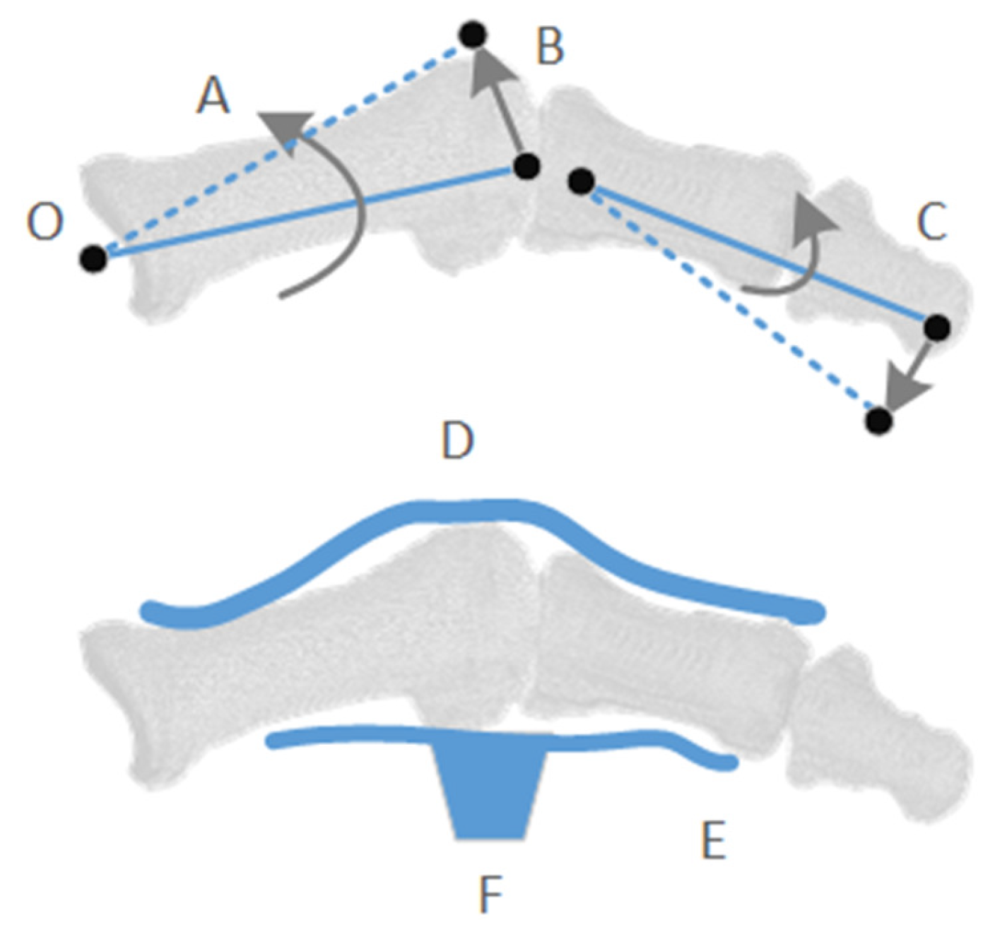
A systematic review was carried out based on the PRISMA guidelines (Preferred Reporting Items for Systematic Reviews and Meta-Analyses) to conduct and report the results of systematic reviews [11]. To search for relevant literature, three electronic databases, namely the National Library of Medicine, Science Direct, and Sage Journals, were used with the help of keywords mentioned in Table 1.
The manuscripts considered for inclusion had to be written in English and had to reference the osteotomy used for HVâ. Only manuscripts published in scientific journals from selected databases and relevant to the question posed were considered eligible. According to the authors’ knowledge, all studies identified to date and published in the selected academic databases were evaluated. Eligibility criteria were established to select titles obtained during the literature search independently. After eliminating non-relevant manuscripts, the remaining manuscripts were compiled, and the full text was read to assess eligibility for inclusion. A flow diagram of the medical records screening selection is provided in Figure 4.
The features of the studies that were taken into consideration are presented in Table 2.

3. Results

The procedures and results obtained in the pre-selected articles (Table 2) are described in the following subchapters, explaining these seven distinct osteotomies reported in the literature. For a better understanding, firstly, the type of surgical procedure is defined with the support of a schematic figure. The methodology and conclusions reported by the authors are described, and a particular focus is made concerning the follow-up of the technique and the biomechanical analysis made.

3.1. Basal Closing Wedge Osteotomy

3.1.1. Procedure and Analysis

According to Hans et al. [12], the basal closing wedge is a surgical technique for treating foot deformities. The procedure involves making a 4 cm long cut on the dorsal surface of the foot and manually adjusting the toe’s position. Using an osteotome, a medial incision is then made to expose the MT (metatarsal bone) and remove the medial eminence. Based on pre-operative radiographs, the size of the lateral base wedge is determined, which is then removed using an oscillating saw 1 cm distal to the metatarsocuneiform joint. The toe is then checked for alignment and fixed with a navicular screw. Figure 5 provides a schematic illustration of the procedure.

3.1.2. Follow-up and Biomechanical Analysis

As per Hans et al. [12], there are significant issues associated with the osteotomy type, one of which is the shortening of the first metatarsal bone. On average, 5 mm of shortening is observed. The incidence of dorsal angulation is another common issue, affecting 25% of the cases analyzed. Transfer metatarsalgia is also a common problem, affecting 25% of the feet studied. Besides being a challenging technique, determining the exact dimensions from pre-operative radiographs is often difficult. However, the average reduction in the intermetatarsal angle (IMâ) of 16.1º in the pre-operative period increased to 6.7º in the final follow-up, representing a good correction. The authors [12] opine that the results were not as good as those reported for other procedures and, therefore, recommend using alternative methods, such as crescent basal osteotomy or Chevron basal osteotomy.
In the literature, no recorded follow-up studies involving biomechanical methodologies are associated with this surgical technique.

3.2. Distal Chevron Metatarsal Osteotomy

3.2.1. Procedure and Analysis

Distal Chevron metatarsal osteotomy (DCMO) was described by Jaehyuong et al. [13]. First, a lateral incision is made to perform the initial lateral release of the first MT. After this, adductor tenotomy and the release of the MT-sesamoid capsule are completed for lateral soft tissue relief, followed by a varus brisement. A varus stress test of the first MTPJ is conducted to determine the appropriate extent of the lateral release, with around 20° of varus shift considered optimal. Next, a T-shaped incision is made at the medial capsule using the medial approach. Depending on the severity of the deformity, a 5–8 mm broad vertical limb of the capsule is excised. An oscillating microsaw is used to perform a bunionectomy, cutting parallel to the medial aspect of the first MT shaft. The 60° Chevron osteotomy’s apex is positioned around 5–7 mm from the articular surface at the center of the first MT head. After the osteotomy, the MT head is laterally translated, as shown in Figure 6.
In cases where there is a noticeable angle between the distal metatarsal bone and the articular surface (DMAâ), the displaced fragment is corrected by rotating it medially. Two 1.1 mm Kirschner wires are inserted from a point 2 cm above the osteotomy site to fix the displaced fragment, and any remaining bony prominence is removed. If there is a defect in the cancellous bone, a bone cyst, or low cancellous bone density around the osteotomy site, an autologous bone graft is used, which is obtained from the bunionectomy bone or the removed fragments of the proximal bony prominence.
Capsulorrhaphy is performed with the pronated toe in supination. If there is still deformity after distal Chevron MT osteotomy (DCMO) and accompanying soft tissue procedures, akin phalangeal osteotomy is performed based on the surgeon’s judgment. The osteotomy is performed 7 mm below the proximal phalangeal base. Patients with no residual hallux pronation may undergo 1–3 mm medial closing wedge osteotomy, attempting to greenstick the lateral cortex. A complete osteotomy is performed for patients with residual toe pronation, followed by derotation.

3.2.2. Follow-up and Biomechanical Analysis

A multicenter retrospective study involving four hospitals was carried out by Jae-Hyung et al. [13]. The authors analyzed 45 feet from 38 patients who received DCMO for HVâ with metatarsus adductus (MA) and had at least one year of post-operative follow-up. The HVâ and the IMâ were measured, which improved by 24.5º and 7.3º, respectively. The MA angle (MAâ) and the degree of the lateral sesamoid were maintained, with no differences in the pre-and post-operative periods between the two groups created with mild MA (18° ≤ MAâ < 20°) and moderate (20° ≤ MAâ). Only the mean post-operative IMâ showed a difference between the two groups. Foot function index (FFI) and visual analogue scale (VAS) were recorded, and both improved significantly. When the extent of improvement was compared between the two groups, no significant differences existed in any category. The total recurrence rate (HVâ ≥ 20°) was 11.1% (5/45), and although the moderate group (4/29, 13.8%) had a higher proportion than the mild group (1/16, 6.3%), this was not statistically significant. In conclusion, DCMO for HV patients with MA had satisfactory radiographic and clinical results with minimal recurrence; Makabe et al. [14] reinforce this conclusion, stating that it is a less invasive procedure with good clinical prospects and positive results. Except in cases of severe combined deformity, they were recommending isolated DCMO without any procedure or manipulation of the other metatarsals as a viable treatment option.
Presently, plantar pressure distribution measurement has revealed itself as essential for detecting and diagnosing specific pathologies and plantar deformities. This methodology can help to study the structures and pathologies of the foot, aiding in diagnosis, but can also help in choosing treatment, prevention, and rehabilitation. By studying plantar pressure distribution, it is possible to develop solutions to improve the distribution of these pressures in the patient’s daily life. Peak pressure (PP) is the most commonly used plantar pressure variable to express foot loading. This value represents the maximum load in an area under the foot during one step [27,28,29]. However, several other variables can be obtained from this type of equipment. The market currently offers various equipment that allows the analysis of plantar pressure distribution. There are two main types of methodology for evaluation of plantar pressure distribution: (a) plantar pressure distribution under the foot, obtained with an instrumented fixed platform; (b) plantar pressure distribution inside footwear, obtained with instrumented insoles. Figure 7 shows an example of left and right foot plantar pressure distribution with seven anatomical masks to allow separate observation of different foot regions.
Kernozek et a. [15] used EMED-SF Pedography Analyzer,™ (Novel GMBH, Munich, Germany). The pressure platform comprised a 32 × 62 sensor matrix with two sensors per cm2 resolution. The sampling frequency was fixed at 70 Hz and auto-triggered to enable first contact with the platform to commence collection. The threshold for collection was 10 Kpa. The data were then stored. Saro et al. [16] used Pedar system insoles (Novel GmbH, Munich, Germany). These are 2.5 mm thick and contain 99 capacitive pressure sensors distributed across the insole. The insoles were regularly calibrated throughout the study period according to the manufacturer’s guidelines.
According to the authors, PP for the central forefoot (CFF) increased post-surgically. However, pressure time integral, force time integral, and contact time were unchanged in the CFF region. Peak pressure, pressure time integral, and contact time all decreased post-surgically for the MT region. Contact area in the lateral toe (LT) region was increased post-surgically.
The participants were instructed to walk along a 10 m walkway at their natural pace, with data collection focused on the middle 6 m to minimize the impact of acceleration and deceleration. The average walking speed was determined based on the time recorded by photocells positioned along the 6 m data collection area.
Each participant underwent two trials during each examination, with the second trial results used for analysis in all cases. All participants, including healthy individuals and patients, wore the same types of shoes (Klaveness Skofabrikk AS, Sandefjord, Norway) during the plantar pressure measurements. This shoe model was chosen due to its reputation for comfort and stability. It features a deep toe box providing ample space for the toes and wide shapes, allowing custom insole fitting. These shoes offered adequate cushioning and stability for the entire foot, ensuring consistent placement of the plantar structures on the insole sensors throughout the trials. Although slight variations were observed in pressure distribution between healthy participants’ left and right feet, data from both feet were analyzed. Figure 5 represents the definitions of anatomical masks.
The findings of this research [15,16] indicate that Chevron osteotomy appears to be effective in managing moderate hallux valgus deformity. It reduced pain levels during walking and maintained satisfactory joint mobility for 12 months post-surgically. According to these authors, no significant alterations were observed in various plantar load metrics (such as peak force, force–time integral, peak pressure, pressure–time integral, or contact time) for the MFF or MT regions.
Although there were improvements in the initial plantar flexion of the metatarsophalangeal joint after surgery, there was no noticeable enhancement in force generation in the MT region. The reasons behind the unchanged peak force despite decreases in peak pressure, pressure–time integral, and contact time are not readily explainable. Additionally, there were no changes in the contact area. However, these results suggest that the capacity for force production in the hallux and first MT may still be compromised even a year following surgery.
The reduction in load related explicitly to the hallux region in this study can be attributed to two main factors. Firstly, surgical intervention may weaken the intrinsic musculoskeletal structures surrounding the first metatarsophalangeal joint, altering the hallux’s mechanical function.
Similar levels of force–time integral, pressure–time integral, and contact time were observed both before and after surgery in the CFF region in this study. According to these findings, limited evidence suggests improvements in plantar loading variables 12 months post-surgery compared to pre-operative values. These changes in loading are unlikely to be attributed to variations in walking speed since contact time did not differ between pre-operative and post-operative conditions. The Chevron procedure may not significantly alter functional foot mechanics, as reflected in the outcomes of plantar loading. Instead, it might modify foot shape for cosmetic purposes and facilitate fitting into footwear.
Further investigations are needed to assess plantar loading patterns following surgical correction for hallux valgus and verify if these observations remain consistent. These alterations in plantar loading appear unrelated to improvements in foot function. Based on this study’s results, the Chevron osteotomy’s effectiveness may lie in its ability to alleviate pain, correct the alignment of the first MT and hallux, and enhance cosmesis rather than restore plantar-loading characteristics.

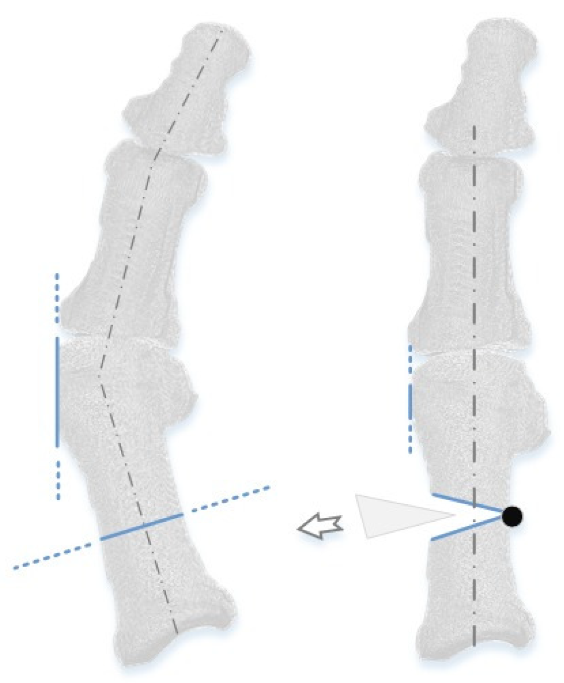
3.3. Proximal Metatarsal Osteotomy

3.3.1. Procedure and Analysis

According to Haddad et al. [17], for this procedure, exostosis of the head of the first MT is excised, the medial capsule and abductor hallucis undergo advancement, and the release of the lateral capsule and adductor tendon as well as open wedge osteotomy at the base of the first MT with insertion of a bone graft occur (Figure 8).
No nonunions of the osteotomy have occurred during the recent three years, and satisfactory maintenance of the corrections has occurred; according to Haddad et al. [17], this method is preferable to distal MT osteotomy. Choi et al. [18] consider this procedure very effective, and it has a low incidence of persistent pain in the area after surgery. Nyska et al. [19] indicate that the average pain scale before surgery was 2.5 points and 1.17 points after surgery. The scale for difficulty wearing shoes decreased from 2.68 to 1.65 points.

3.3.2. Follow-up and Biomechanical Analysis

Foot pressure measurements were conducted using an EMED-SF platform (EMED-SF Pedography Analyzer (Novel GMBH, Munich, Germany). The pressure platform, measuring 400 × 240 (mm), was seamlessly integrated into a 5 m walkway. Patients were instructed to walk barefoot at their usual pace across the platform. Data from three typical steps collected before and after surgery were analyzed using EMED software. Additionally, a control group comprising ten healthy women matched for age walked across the platform at their natural pace three times, and their data were compared [19].
Analysis of the steps involved delineating seven regions of interest: heel, midfoot, lateral, medial forefoot, toes 2-5, and the hallux. Variables utilized in the analysis included area, maximal forces, peak pressures, duration of contact phases, pressure–time and force–time integrals, and the timing of PP and force for each region of interest. The study developed by Nyska et al. [19] concluded that based on the plantar foot pressure data, patients with hallux valgus deformity have a typical medial and CFF loading pattern.
Kraus et al. [20] studied five fresh-frozen cadaveric below-knee specimens with hallux valgus deformities. These specimens underwent loading up to 400 N on a servo-hydraulic load frame. The study aimed to analyze joint contact characteristics at the first MT joint, employing a Tekscan pressure sensor (Model 6900, 1100 psi; Tekscan Inc., Boston, MA) with varying opening wedge sizes of 3, 5, and 7 mm, both with and without a distal soft tissue release (DSTR). Analysis of variance and post hoc multiple comparisons were utilized to compare contact force, area, and peak contact stress among different groups compared to untreated specimens. The findings revealed that untreated specimens exhibited a mean contact force of 47.7 ± 33.5 N. This force increased progressively with the size of the opening wedge and demonstrated statistical significance for a 7 mm opening wedge (129.7 ± 62.3 N) and a 7 mm wedge with DSTR (134.8 ± 60.5 N).
The mean peak contact stress for untreated specimens was measured at 2.8 ± 1.3 MPa, which escalated incrementally with wedge size to 5.7 ± 3.0 MPa for a 7 mm wedge alone and 5.6 ± 2.5 MPa for a 7 mm wedge with DSTR. Although contact area increased with corrections, none of these changes reached statistical significance.
In summary, the study concluded that as the size of the opening wedge increased, so did the loading on the first MT joint. Joint stresses exceeding 4.7 MPa were identified as chondrogenic, potentially increasing the likelihood of arthritic joint development in patients.

3.4. Proximal Oblique-Domed Osteotomy

3.4.1. Procedure and Analysis

According to Takao et al. [21], the procedure involved cutting the adductor hallucis tendon at the insertion of the proximal phalanx and the sesamoid bone. The osteotomy was then executed 3 cm dorso-distal to the metatarsocuneiform joint to transfer the approximately 5 mm distal fragment plantarly and rotate it laterally, thereby reducing the first IMâ by 5 degrees—representation in Figure 9.

3.4.2. Follow-up and Biomechanical Analysis

Takao et al. [21] conclude that significant improvements were noted in the HVÂ angle, the first-second IMâ, the first-fifth IMâ, talus height, and plantar calcaneal angle from before surgery to one-year post-surgery.
Flat foot and MT primus varus are the primary contributors to HVâ, emphasizing the importance of correcting these deformities to prevent the recurrence of the condition. This study [21] presents a clinical and radiological evaluation of the correction of HVâ, MT primus varus, and pes planus following proximal oblique dome osteotomy of the MT, coupled with distal soft tissue reconstruction. Twenty-seven feet from twenty-two patients with moderate or severe HVâ who underwent proximal oblique dome osteotomies were examined.
This study suggests that proximal oblique MT osteotomy, as a surgical intervention for moderate or severe HVâ with a flat foot, may be recommended to correct both the longitudinal arch of the foot and the first-second IMâ.

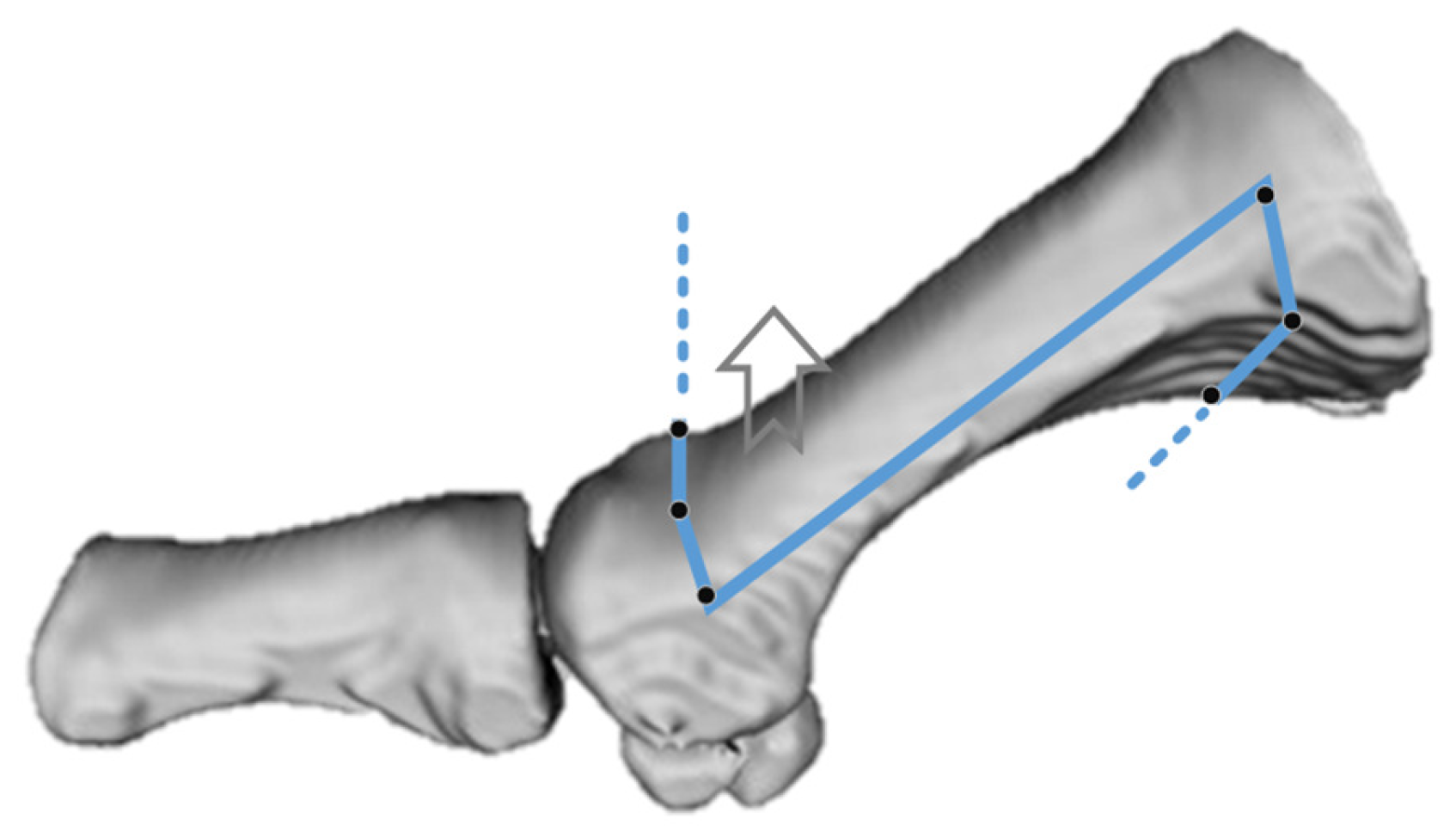
3.5. Proximal Spherical Metatarsal Osteotomy

3.5.1. Procedure and Analysis

Tanaka et al. [22] developed spherical osteotomy. The lateral soft tissue procedure was performed in the standard manner, followed by a spherical cut of the first MT from distal to proximal. The distal fragment was rotated in the lateral and plantar directions. They aimed to achieve 3 degrees of plantar flexion at the osteotomy site, measuring the inclination angle of the first MT formed between the plantar line and the first MT axis.
The plantar line was a line between the lowest points of the calcaneus and the medial sesamoid. The medial sesamoid is usually more inferior in the lateral view, as the sesamoid complex is pronounced in HVÂ. The axis of the first MT was determined by connecting points dividing the width of the proximal and distal metaphysis. Intraoperatively, three degrees of plantar flexion at the osteotomy site were verified using a protractor—representation of the procedure in Figure 10.

3.5.2. Follow-up and Biomechanical Analysis

The osteotomy site was fixed with small screws and Kirschner wires. Medial soft tissue reconstruction was performed at the same time. Post-operatively, a dressing was applied without plaster immobilization. Weight bearing on the heel was allowed one day after surgery. Although 81% of patients expressed satisfaction with the results, it was found that 50% of those with a pre-operative HVâ angle of 50 degrees or greater had a post-operative HVâ angle of 20 degrees or greater. In these cases, the correction of the primus varus metatarsus was adequate, but the valgus deviation of the hallux was only reasonable. The average correction towards plantar flexion was 1.5 degrees, and no increase in arch height was observed.
The effectiveness of the treatment was assessed using the AOFAS Hallux Metatarsophalangeal-Interphalangeal Scale. Pain levels, specifically at the first metatarsophalangeal joint and metatarsalgia, were examined independently using the pain subscale of the AOFAS scale, categorized as ‘no pain’, ‘mild pain’, ‘moderate pain’, or ‘severe pain’. The active and passive motion ranges at the first MTP joint were measured following the AOFAS guidelines. Satisfaction levels were gauged utilizing Glynn’s methodology. Results were classified as ‘excellent’ if complete alleviation of symptoms and deformity was achieved, as assessed by both the patient and surgeons. ‘Good’ outcomes were reported when the patient was content but experienced mild symptoms. Conversely, outcomes were deemed ‘unsatisfactory’ if a significant flaw persisted, such as metatarsalgia, notable recurrence, or hallux varus. Additionally, patients were surveyed regarding their willingness to undergo the procedure again.
Proximal spherical osteotomy has consistently demonstrated satisfactory results for patients with HVâ angles less than 50 degrees. However, corrections were less effective in more severe cases of deformities. Additionally, plantar flexion in the osteotomy area was compensated for by the displacement of the first tarsometatarsal joint.

3.6. Rotational Scarf Osteotomy

3.6.1. Procedure and Analysis

Larholt et al. [23] describe a surgical procedure where a longitudinal incision is made along the medial aspect of the foot, extending from the first metatarsocuneiform joint to the hallux interphalangeal joint. Following this, a semi-elliptical capsular excision was performed, involving the resection of the medial eminence and intra-articular release of the lateral sesamoid. Additionally, the adductor tendon was transected to the proximal phalanx.
A blade was maneuvered through the joint, positioned superior to the adductor tendon, and then directed plantarly to section the adductor tendon. The same blade was further moved along the lateral aspect of the MT head to sever the sesamoid suspensory ligament on the lateral side of the joint. Reese’s osteotomy guide ensured the scarf cuts were made in a single plane.
The horizontal cut was initiated from the distal dorsal aspect of the MT head, proceeding plantar proximally to a point just distal to the proximal tubercle of the first MT. At this juncture, two-thirds of the MT were dorsal to the osteotomy, and one-third was below. A dorsal distal cut was then made perpendicular to the second MT to preserve the length of the first MT. The proximal plantar cut was executed at a 45-degree angle from proximal medial to distal lateral.
The dorsal and plantar fragments of the MT were separated, and a beaver blade was introduced between the fragments to divide any remaining intact periosteum and the first lateral attachments of the capsule. The head of the first MT was rotated relative to the dorsal fragment, creating a bony protrusion indicating the extent of correction and rotation. One millimeter of protrusion corresponded to 1 degree of angle correction, measured intraoperatively using a depth gauge—represented in Figure 11.
It was ensured that the tibial sesamoid was directly below the medial sesamoid groove of the head of the first MT. The osteotomy was then internally fixed with two 2.0 mm cortical screws. The medial protrusion of the first MT bone was resected, and an Akin osteotomy was performed and fixed with a 1.2 mm threaded Kirschner wire. The capsule was closed with 2-0 Vicryl, and the skin was closed subcuticularly with 5-0 Vicryl. The wound was dressed with a non-adherent dressing, gauze, and a crepe bandage. The patient was encouraged to perform active hallux flexion movements within the bandage. Wound repair was conducted one week post-operatively, and for the subsequent two weeks, the patient wore surgical shoes and walked on heels with elbow crutch support.
After two weeks, the bandages were removed, and the patient transitioned to running shoes or sneakers with laces to compress foot swelling, initiating post-operative exercises. The angular reduction achieved in the IMâ and HVâ between the pre-operative and post-operative periods is 10.1º and 21.8º, respectively.

3.6.2. Follow-up and Biomechanical Analysis

Dhukaram et al. [24] analyzed the procedure based on a score, considering a healthy foot without pathology and a foot with hallux valgus as usual. These authors used a Musgrave pressure plate (Preston Communications Ltd., Dublin, Ireland), a platform-based vertical pressure measurement system installed seamlessly within a 762 mm walkway at the Gait Study Center. This floor-mounted platform has 642 × 297 × 38 (mm) dimensions, with an active area measuring 194 × 394 (mm). It comprises a grid of 2,048 sensors capable of sampling pressure data at a frequency of 55.6 Hz, with each sensor covering an area of 5 mm × 5 mm (0.25 cm2). The pressure range per unit area spans 0.3 to 15 kg/cm. The collected data are processed using a 400 MHz Pentium II microcomputer (Intel, Santa Clara, CA, USA) running Winfoot software (Preston Communications Ltd., Dublin, Ireland).
The authors [24] found several limitations in the study. Firstly, they did not have pre-operative clinical and pedobarographic assessment data for comparison. Both osteotomies did not influence the pre-operative pressure pattern. However, the authors aimed to investigate whether the aforementioned corrective procedures for hallux valgus restore plantar pressure distribution to the usual pattern and to discern their differences.
Furthermore, the groups were comparable in demographic characteristics, pre-operative deformity, and post-operative regimen, except for follow-up time. The mean follow-up time for the Scarf group was shorter than that of the Mitchell group, which could have affected the result with longer follow-ups.
The Mitchell and Scarf osteotomies were performed by different surgical teams, which potentially influenced the outcome based on the expertise of each team.
Although the authors aimed to examine post-hallux valgus correction pedobarography and its clinical relevance, correlating this with radiological results would have been advantageous. However, they did not seek final follow-up radiographs.
Furthermore, additional analysis within study groups, comparing unilateral and bilateral procedures and symptomatic and asymptomatic feet, was limited due to insufficient sample size to achieve statistical significance.
Patient inclusion criteria were based on subjective assessment.
Finally, they did not perform null hypothesis testing on the AOFAS scores between the two intervention groups.
The authors concluded that the osteotomy failed to restore the hallux load-bearing function, determining the procedure’s outcome.

3.7. Mitchell Osteotomy

3.7.1. Procedure and Analysis

According to Heerspink et al. [25], Mitchell osteotomy is a stepped osteotomy performed proximal to the sesamoids, leaving a lateral bone bridge measuring 4 to 5 mm. Two holes were placed approximately 4 mm distal and 10 mm proximal to the cut. The second cut was a complete osteotomy approximately 2 mm proximal and parallel to the distal cut. A wire passing through the two holes stabilized the osteotomy. Moreover, the capsular tissues were plicated—a representation of the procedure is in Figure 12.

3.7.2. Follow-up and Biomechanical Analysis

Chevron group. The mean shortening of the first MT in the Chevron group was 4.4 mm versus 6.6 mm in the Mitchell group. No significant differences were found in post-operative HVA or IMA between the two groups. The mean HVA correction was significantly greater after a Mitchell osteotomy than a Chevron osteotomy. The post-operative length of the first MT was significantly shorter after Mitchell’s osteotomy. Yamamoto et al. [26] also agree that the procedure causes a significant shortening of the first MT and thus relieves tension in soft tissues, such as the adductor hallucis.

4. Discussion

4.1. Analysis of Procedures and Results

After an initial examination of the procedures and their scope, it is noteworthy that there are considerable differences in exposure areas. This is a crucial consideration because a smaller procedural area tends to result in a shorter recovery time, increasing patient satisfaction.
Basal closing wedge osteotomy involves a manipulation of the MT in two positions, requiring a large working area. Although radiological results show a positive correction of the deformity, with initial mean HVâ angle of 38.9º reducing to 19.9º, IMâ angle from 16.1º to 6.7º, and sesamoid position changing from 2.6 to 0.9, a significant disadvantage is the shortening of the first MT, potentially leading to irregular positioning of the toes.
Precision is crucial in Chevron distal MT osteotomy, but the procedure focuses on a limited MT area, reducing incisions and potentially shortening the post-operative recovery period. The average improvement in the HVâ angle was notably better than basal closing wedge osteotomy, decreasing from 35.1º to 10.6º. Although positive, the mean IMâ angle did not exceed that obtained in the basal closing wedge osteotomy and did not result in the MT shortening. Although specific results are not provided for proximal MT osteotomy, the procedure appears to require a wide soft tissue cut, indicating a longer recovery time, similar to basal closing wedge osteotomy. Despite a minor incision procedure, like distal MT Chevron osteotomy, proximal oblique dome osteotomy may require a second procedure to minimize recurrences. It presents an average HVâ recovery of 28.5º, surpassing basal and distal Chevron closing wedge osteotomy, and an average IMâ of 8.4º, surpassing distal Chevron closing wedge osteotomy, but it is inferior to the closing wedge.
Regarding proximal spherical MT osteotomy, it states significantly higher recovery averages of 34º for HVâ and 13.1º for IMâ compared to other procedures. Notably, patient satisfaction tends to decrease for HVâ angles above 50°, and it is a more minor incision procedure. Rotational scarf osteotomy is a comprehensive procedure that can pose challenges post-operatively—an average HVâ of 21.4º and IMâ of 9.9º present median values for the other procedures analyzed. Finally, the Mitchell osteotomy is a procedure that shortens the MT but can relieve tension in the soft tissues.
The results of the load application study on five-frozen cadaveric below-knee specimens were very interesting. They corroborated the idea that surgical procedures must be carried out with a prior assessment of the need for invasion of the procedure and always with the perspective of minimizing the intrusion to not weaken the affected area any further than it already is. The more invasive the surgical procedure, the more significant the long-term implications for bone health and the loads applied. Furthermore, from this perspective, engineering can significantly help adjust and regulate the type of procedures and dimensions to treat deformation in the least invasive and harmful way possible for the bone structure itself, with equipment and devices to assist surgeons.
Traditionally, studies concerning the evaluation of pathology treatment have primarily focused on X-ray imaging, pain identification, and visual perception of walking patterns. However, a pertinent question arises regarding the adequacy of these approaches in providing a comprehensive evaluation of treatment efficacy, as the outcomes often need more significant and tangible differences.
One intriguing avenue to consider in exploring alternative evaluation methods is thermographic analysis. This technique offers a non-invasive means to assess physiological changes associated with pathology treatment. By detecting variations in skin temperature, thermography can provide valuable insights into underlying physiological processes, potentially offering a more holistic understanding of treatment outcomes.
Moreover, incorporating engineering methodologies presents another promising opportunity to enhance treatment evaluation protocols. For instance, the complete gait load distribution analysis offers a nuanced perspective on how treatment interventions impact biomechanical function. Engineering techniques can facilitate precise measurement and analysis of gait symmetry and load distribution across each limb, thereby enabling a more comprehensive assessment of treatment efficacy.
Furthermore, advancements in engineering can contribute to developing specialized tools and technologies tailored to the specific needs of pathology treatment evaluation. By leveraging engineering expertise, innovative solutions can be devised to address existing limitations and enhance the accuracy and reliability of treatment assessment methodologies.
In conclusion, while traditional evaluation approaches have their merits, there is a growing recognition of the need for more sophisticated and comprehensive assessment methods in pathology treatment. Incorporating techniques such as thermographic analysis and engineering-based approaches holds significant promise in enriching our understanding of treatment outcomes and ultimately improving patient care.
Most studies compare pre- and post-surgical hallux deformation and analyze patient satisfaction and pain level; however, some focus on plantar pressure. After analyzing the results based on plantar pressure distribution on the foot, we identified the evaluation models, fixed platforms, and plantar pressure insoles. The platforms offer high-resolution measurements and are accurate, but they run the risk of providing unreliable information, as walking can change without apparent patient awareness. It is possible to carry out static and dynamic measurements and have the platform directly in contact with the skin of the foot. Tests are carried out barefoot, while insoles are used in dynamic studies. Furthermore, insoles generally allow for more reliable and precise measurements of plantar pressure centers. This is because contact with the foot is much more organic, with the disadvantage of requiring more care due to slipping that can occur inside the shoe, as well as foot perspiration that can compromise the sensors. Thus, it is an analysis process that requires more care and can be more expensive as the insole has to be personalized, but it is more reliable if used well. This can be concluded by understanding the use of reported studies of four fixed platforms and one with plantar pressure insoles. By analyzing the values of peak pressures, we were able to analyze the tables and see that there are no discrepancies that the authors consider significant in the foot’s structure. They unanimously consider that the procedures provide quality of life to patients due to the foot’s aesthetics and shape and because they no longer feel pain. However, once altered, the foot’s structure does not change significantly after the surgical procedure. It is concluded that the minor the procedure in terms of incision in the bone, the better.
In summary, all procedures are viable, but considering the balance between optimal results, lower risks of errors, and smaller incisions, basal closing wedge osteotomy and distal Chevron osteotomy are recommended.
Focusing on the fact that this is a systematic review article, the content presented corresponds only to what the articles primarily present. A more significant explanation of some procedures in some articles and discrepancies in biomechanical studies would be advantageous, allowing a critical approach to the advantages and disadvantages of each approach, as well as their restrictions and contraindications. A more in-depth analysis of some articles would be essential, allowing a more detailed and consistent analysis of the results and a more effective comparison of surgical procedures, namely Mitchell osteotomy, proximal oblique dome osteotomy, and basal closing wedge osteotomy, in addition to standardizing the analysis of acquired results to improve comparative analysis.
An interesting approach in future work involves a comparative discussion around the environmental impact of each surgical technique [30].

4.2. Limitations on Review

Limitations must be considered when interpreting the manuscripts included in this review. The search was limited to three databases without integrating reference lists and manual examinations to identify other relevant manuscripts. The choice of search terms and inclusion criteria also determines the results of this review (the use of different words and criteria may have changed the number of manuscripts included).

5. Conclusions

This manuscript aims to highlight the existing processes for the treatment of hallux valgus pathology and to identify its characteristics, the technologies used in pre-and post-operative monitoring of patients, and the results obtained; to compare them; and to try to analyze which ones provide the best results regardless of the technique used. All procedures have limitations, and there are always specific cases in which the surgeon, through their experience, can determine which procedure is most appropriate, regardless of its difficulty. However, in most cases, it is worth simplifying and providing better quality and less time for recovery for the patient, as well as ensuring lasting and visible results.

Author Contributions

Conceptualization, M.S., A.M.A. and L.R.; methodology, A.M.A. and L.R.; validation, M.S., A.M.A., E.C.S. and L.R.; formal analysis, M.S., A.M.A., E.C.S. and L.R.; investigation, M.S.; writing—preparation of the original draft, M.S., A.M.A. and L.R.; writing—review and editing, M.S., A.M.A., E.C.S. and L.R.; supervision, A.M.A. and L.R. All authors have read and agreed to the published version of the manuscript.

Funding

This research received no external funding.

Acknowledgments

The authors acknowledge FCT—Fundação para a Ciência e Tecnologia, under the projects UIDB/00285/2020 and LA/P/0112/2020.

Conflicts of Interest

The authors declare no conflicts of interest.

References

  1. Lee, S.-S.; Kim, J.H.; Kim, S.; Jung, E.Y.; Ryu, D.J.; Lee, D.K.; Wang, J.H. Avoiding Overcorrection to Increase Patient Satisfaction After Open Wedge High Tibial Osteotomy. Am. J. Sports Med. 2022, 50, 2453–2461. [Google Scholar] [CrossRef] [PubMed]
  2. Nix, S.; Smith, M.; Vicenzino, B. Prevalence of hallux valgus in the general population: A systematic review and meta-analysis. J. Foot Ankle Res. 2010, 3, 21. [Google Scholar] [CrossRef] [PubMed]
  3. Perera, A.; Mason, L.; Stephens, M. The pathogenesis of hallux valgus. J. Bone Jt. Surg. 2011, 93, 1650–1661. [Google Scholar] [CrossRef] [PubMed]
  4. Alkhalifah, K.M.; Almotiri, M.M.; Alharbi, A.E.; Alrashidi, A.; Aldhali, I.K.; Alsaqry, O.S.; Alharbi, K.H.; Almogbil, I. A Systematic Review and Meta-Analysis of the Prevalence of Hallux Valgus in the General Population. Cureus 2023, 15, e42739. [Google Scholar] [CrossRef]
  5. Nguyen, J.K.; Sullivan, M.; Alpuerto, B.B., 2nd; Mueller, S.; Sly, N. A radiographic analysis of the abnormal hallux interphalangeus angle range: Considerations for surgeons performing Akin osteotomies. J. Orthop. Surg. 2019, 27, 2309499019841093. [Google Scholar] [CrossRef] [PubMed]
  6. Flaherty, A.; Chen, J. Minimally Invasive Chevron Akin Osteotomy for Hallux Valgus Correction. JBJS Essent. Surg. Tech. 2024, 14, e22. [Google Scholar] [CrossRef] [PubMed]
  7. Nozaki, S.; Watanabe, K.; Katayose, M.; Yamatsu, K.; Teramoto, A.; Ogihara, N. Three-dimensional morphological variations in the calcaneus and talus in relation to the hallux valgus angle. Ann. Anat. Anat. Anz. 2023, 247, 152053. [Google Scholar] [CrossRef] [PubMed]
  8. Yokozuka, M.; Okazaki, K. Characteristics of hindfoot morphology and ankle range of motion in young women with hallux valgus. J. Foot Ankle Res. 2023, 16, 64. [Google Scholar] [CrossRef]
  9. Sharma, J.; Aydogan, U. Algorithm for Severe Hallux Valgus Associated with Metatarsus Adductus. Foot Ankle Int. 2015, 36, 1499–1503. [Google Scholar] [CrossRef] [PubMed]
  10. Steadman, J.; Barg, A.; Saltzman, C.L. First Metatarsal Rotation in Hallux Valgus Deformity. Foot Ankle Int. 2021, 42, 510–522. [Google Scholar] [CrossRef] [PubMed]
  11. Rethlefsen, M.L.; Kirtley, S.; Waffenschmidt, S.; Ayala, A.P.; Moher, D.; Page, M.J.; Koffel, J.B.; PRISMA-S Group. PRISMA-S: An extension to the PRISMA Statement for Reporting Literature Searches in Systematic Reviews. Syst. Rev. 2021, 10, 1–19. [Google Scholar] [CrossRef] [PubMed]
  12. Trnka, H.-J.; Mühlbauer, M.; Zembsch, A.; Hungerford, M.; Ritschl, P.; Salzer, M. Basal Closing Wedge Osteotomy for Correction of Hallux Valgus and Metatarsus Primus Varus: 10- To 22-Year Follow-up. Foot Ankle Int. 1999, 20, 171–177. [Google Scholar] [CrossRef] [PubMed]
  13. Lee, J.; Lee, H.S.; Jeong, J.-J.; Seo, D.-K.; Kee, T.; So, S.; Choi, Y.R. Distal chevron metatarsal osteotomy is a viable treatment option for hallux valgus with metatarsus adductus—Multicentre retrospective study. Int. Orthop. 2021, 45, 2261–2270. [Google Scholar] [CrossRef] [PubMed]
  14. Makabe, K.; Matsumoto, T.; Tanaka, S.; Uchida, Y.; Nagase, Y.; Nishikawa, T. Comparison of short-term outcomes after distal metatarsal telescoping osteotomy and scarf osteotomy for hallux valgus in patients with rheumatoid arthritis. Foot Ankle Surg. Tech. Rep. Cases 2023, 3, 100254. [Google Scholar] [CrossRef]
  15. Kernozek, T.W.; Sterriker, S.A. Chevron (Austin) distal metatarsal osteotomy for hallux valgus: Comparison of pre- and post-surgical characteristics. Foot Ankle Int. 2002, 23, 503–508. [Google Scholar] [CrossRef] [PubMed]
  16. Saro, C.; Andrén, B.; Felländer-Tsai, L.; Lindgren, U.; Arndt, A. Plantar pressure distribution and pain after distal osteotomy for hallux valgus: A prospective study of 22 patients with 12-month follow-up. Foot 2007, 17, 84–93. [Google Scholar] [CrossRef]
  17. Haddad, R.J., Jr. Hallux valgus and metatarsus primus varus treated by bunionectomy and proximal metatarsal osteotomy. South. Med. J. 1975, 68, 684–686. [Google Scholar] [CrossRef] [PubMed]
  18. Choi, J.Y.; Suh, Y.M.; Yeom, J.W.; Suh, J.S. Comparison of Postoperative Height Changes of the Second Metatarsal among 3 Osteotomy Methods for Hallux Valgus Deformity Correction. Foot Ankle Int. 2017, 38, 20–26. [Google Scholar] [CrossRef]
  19. Nyska, M.; Liberson, A.; McCabe, C.; Linge, K.; Klenerman, L. Plantar foot pressure distribution in patients with Hallux valgus treated by distal soft tissue procedure and proximal metatarsal osteotomy. Foot Ankle Surg. 1998, 4, 35–41. [Google Scholar] [CrossRef]
  20. Kraus, J.; Ziegele, M.J.; Wang, M.; Law, B. First Tarsometatarsal Joint Loading After Sequential Correction of Hallux Valgus Using a Proximal Opening Wedge Metatarsal Osteotomy and Distal Soft Tissue Procedure. Foot Ankle Orthop. 2021, 6, 24730114211026934. [Google Scholar] [CrossRef] [PubMed]
  21. Takao, M.; Komatsu, F.; Oae, K.; Miyamoto, W.; Uchio, Y.; Ochi, M.; Matsushita, T. Proximal oblique-domed osteotomy of the first metatarsal for the treatment of hallux valgus associate with flat foot: Effect to the correction of the longitudinal arch of the foot. Arch. Orthop. Trauma Surg. 2007, 127, 685–690. [Google Scholar] [CrossRef] [PubMed]
  22. Tanaka, Y.; Takakura, Y.; Kumai, T.; Sugimoto, K.; Taniguchi, A.; Hattori, K. Proximal spherical metatarsal osteotomy for the foot with severe hallux valgus. Foot Ankle Int. 2008, 29, 1025–1030. [Google Scholar] [CrossRef] [PubMed]
  23. Larholt, J.; Kilmartin, T.E. Rotational scarf and akin osteotomy for correction of hallux valgus associated with metatarsus adductus. Foot Ankle Int. 2010, 31, 220–228. [Google Scholar] [CrossRef]
  24. Dhukaram, V.; Hullin, M.G.; Kumar, C.S. The Mitchell and Scarf Osteotomies for Hallux Valgus Correction: A Retrospective, Comparative Analysis Using Plantar Pressures. J. Foot Ankle Surg. 2006, 45, 400–409. [Google Scholar] [CrossRef] [PubMed]
  25. Heerspink, F.O.L.; Verburg, H.; Reininga, I.; van Raaij, T. Chevron versus Mitchell osteotomy in hallux valgus surgery: A comparative study. J. Foot Ankle Surg. 2015, 54, 361–364. [Google Scholar] [CrossRef] [PubMed]
  26. Yamamoto, K.; Imakiire, A.; Katori, Y.; Masaoka, T.; Koizumi, R. Clinical results of modified Mitchell’s osteotomy for hallux valgus augmented with oblique lesser metatarsal osteotomy. J. Orthop. Surg. 2005, 13, 245–252. [Google Scholar] [CrossRef] [PubMed]
  27. Melai, T.; Ijzerman, T.H.; Schaper, N.C.; de Lange, T.L.; Willems, P.J.; Meijer, K.; Lieverse, A.G.; Savelberg, H.H. Calculation of plantar pressure time integral, an alternative approach. Gait Posture 2011, 34, 379–383. [Google Scholar] [CrossRef] [PubMed]
  28. Arin-Bal, G.; Livanelioglu, A.; Leardini, A.; Belvedere, C. Correlations between plantar pressure and postural balance in healthy subjects and their comparison according to gender and limb dominance: A cross-sectional descriptive study. Gait Posture 2024, 108, 124–131. [Google Scholar] [CrossRef] [PubMed]
  29. Wilczyński, J.; Paprocki, M. Correlations between foot defects and balance reactions among young school-children. Med. Stud. Stud. Med. 2020, 36, 265–272. [Google Scholar]
  30. National Institute for Health; Care Research Global Health Research Unit on Global Surgery. Reducing the environmental impact of surgery on a global scale: Systematic review and co-prioritization with healthcare workers in 132 countries. Br. J. Surg. 2023, 110, 804–817. [Google Scholar] [CrossRef]
Figure 1. Scheme of the main factors that can lead to HVâ.
Applsci 14 03043 g001
Figure 2. Quantification of hallux valgus angle (HVA).
Applsci 14 03043 g002
Figure 3. Visual representation of the rotation and translation of the first metatarsal: A—Initial pronation of the first MT, indicating a medial shift or tilting of the bone. B—Varus translation, indicating a deviation of the bone towards the body’s midline, along with significant rotation of the head of the first MT. This rotation exacerbates the deformity. C—Rotation and progression of valgus translation of the hallux, where the big toe deviates from the body’s midline. D—Plantar rotation of the abductor hallucis tendon. E—Rotation of insertions of the hallux flexor and extensor tendons, causing the proximal end of the distal phalanx (the bone of the big toe) to be pulled into the valgus, exacerbating the deviation. F—Rotation of the insertion of the adductor hallucis longus tendon, which adds pronation-promoting force to the base of the hallux, further exacerbating the deformity. These sequential changes ultimately lead to osteoarthritis of the metatarsophalangeal joint (MTPJ) and compression-induced degradation of adjacent toes [9,10].
Applsci 14 03043 g003
Figure 4. Flow diagram of study selection.
Applsci 14 03043 g004
Figure 5. Basal closing wedge osteotomy procedure. Left—the laterally based bony wedge is removed with an oscillating saw, taking care to preserve the medial cortex. Right—the resulting osteotomy is closed and fixed with a navicular screw.
Applsci 14 03043 g005
Figure 6. Distal Chevron metatarsal osteotomy. Representation of the lateral translation after angular sawing.
Applsci 14 03043 g006
Figure 7. Example of plantar pressure distribution with seven anatomical masks, obtained from an emed® platform (EMED-SF Pedography Analyzer (Novel GMBH, Munich, Germany): 1—heel; 2—MFF; 3—LFF; 4—CFF; 5—medial forefoot; 6—MT; 7—toes.
Applsci 14 03043 g007
Figure 8. Sketch of an X-ray showing the medial angulation of the first MT and the procedure, comparing the pre- and post-operative periods [17].
Applsci 14 03043 g008
Figure 9. Proximal oblique-domed psteotomy representation.
Applsci 14 03043 g009
Figure 10. Proximal spherical metatarsal osteotomy representation.
Applsci 14 03043 g010
Figure 11. Rotational scarf osteotomy representation.
Applsci 14 03043 g011
Figure 12. Mitchell osteotomy representation.
Applsci 14 03043 g012
Table 1. Searched databases and associated search terms used.
DatabaseSearch Terms
PubMed
Science Direct
Sage Journals
(“pressure distribution”[All Fields] AND (“osteotomy$”[Title] AND “hallux valgus”[Title])) OR ((“foot”[Title] OR “metatarsus”[Title]) AND “osteotomy$”[Title] AND “hallux valgus”[Title])data
Table 2. Characteristics of the included studies.
Ref. Number of Participants/Cases Follow-up Duration Procedure Summary
[12] Forty-two patients (60 feet) ≥10 years Basal Closing Wedge Osteotomy The article analyzes the results obtained with the procedure for at least ten years and the disadvantages and complications of the long-term process for correcting HVâ and metatarsus primus varus.
[13] Thirty-eight patients (45 feet) ≥1 years Distal Chevron Metatarsal Osteotomy The article analyzes whether the procedure is viable for treating HVâ with metatarsus adductus.
[14]Forty-five patients (47 feet)------Telescopic Osteotomy, and Scarf OsteotomyThe article compares the short-term results after telescopic osteotomy of the distal MT and scarf osteotomy for hallux valgus in patients with rheumatoid arthritis.
[15]Twenty-five patients≥1 yearsChevron Distal Metatarsal OsteotomyThe article compares pre- and post-surgical characteristics of Chevron distal MT osteotomy for hallux valgus.
[16]Twenty-two patients≤1 yearDistal OsteotomyThe article analyzes plantar pressure distribution and pain after distal osteotomy for hallux valgus.
[17] Sixteen feet =3 years Proximal Metatarsal Osteotomy The article analyzes the procedure and results for treating HVâ and metatarsus primus varus.
[18]------≥1 yearsProximal Chevron Metatarsal Osteotomy (PCMO), Scarf Osteotomy, and Distal Chevron Metatarsal Osteotomy (DCMO)The article compares post-operative changes in the height of the second MT between three osteotomy methods for correcting hallux valgus deformity.
[19]Twenty patients (29 feet)------Proximal Metatarsal OsteotomyThe article analyzes foot plantar pressure distribution in patients with hallux valgus treated by distal soft tissue procedure and proximal MT osteotomy.
[20]Five fresh-frozen cadaveric below-knee specimens------Proximal Opening Wedge Metatarsal OsteotomyThe article reviews the first tarsometatarsal joint loading after sequential hallux valgus correction using a proximal opening wedge MT osteotomy and distal soft tissue procedure.
[21] Twenty-two
patients (27 feet)
≥2 years Proximal Oblique Domed Osteotomy The article analyzes the procedure for treating HVâ associated with a flat foot.
[22] Thirty-seven patients (48 feet) >2 years <8 Proximal Spherical Metatarsal Osteotomy The article reviews the procedure for treating severe HVâ.
[23] Twenty-seven
patients (38 feet)
=5 years Rotational Scarf Osteotomy The article analyzes the procedure for correcting HVâ associated with the MT adductus.
[24]Twenty-eight patients>1 years <5Mitchell and Scarf OsteotomiesThe article takes a retrospective, comparative analysis using plantar pressures from the Mitchell and scarf osteotomies for hallux valgus correction.
[25]Eighty-four patients------Chevron and Mitchell OsteotomyThe article compares Chevron versus Mitchell osteotomy in hallux valgus surgery.
[26]Fifty-three patients (93 feet)≥5 yearsModified Mitchell Osteotomy and Oblique Lesser Metatarsal OsteotomyThe article presents the clinical results of modified Mitchell osteotomy for augmented hallux valgus with oblique lesser MT osteotomy.
Disclaimer/Publisher’s Note: The statements, opinions and data contained in all publications are solely those of the individual author(s) and contributor(s) and not of MDPI and/or the editor(s). MDPI and/or the editor(s) disclaim responsibility for any injury to people or property resulting from any ideas, methods, instructions or products referred to in the content.

