Article

Pesticidal Potential of Essential Oil Obtained from a New Variety of Marigold (Tagetes patula L., fam. Asteraceae)

by
Catalina Tudora
1,*,
Florin Nenciu
1,*,
Adriana Muscalu
1,
Floarea Burnichi
2,
Florentina Gatea
3,
Oana Alina Boiu-Sicuia
4 and
Florentina Israel-Roming
4
1
Testing Department, National Institute of Research-Development for Machines and Installations Designed for Agriculture and Food Industry-INMA, 6 Ion Ionescu de la Brad Avenue, 013813 Bucharest, Romania
2
Vegetable Research and Development Station Buzau, 23 Mesteacanului Street, 120024 Buzau, Romania
3
National Institute for Biological Sciences, 296, Splaiul Independentei, 060031 Bucharest, Romania
4
Department of Plant Sciences, University of Agronomic Sciences and Veterinary Medicine of Bucharest, 59 Marasti Blvd., 011464 Bucharest, Romania
*
Authors to whom correspondence should be addressed.
Appl. Sci. 2024, 14(8), 3159; https://doi.org/10.3390/app14083159
Submission received: 28 February 2024 / Revised: 1 April 2024 / Accepted: 6 April 2024 / Published: 9 April 2024

Abstract

:
Essential oils (EOs) extracted from various medicinal plants offer a promising alternative to non-selective chemical substances commonly employed in conventional agriculture. Their chemical composition includes several classes of chemical compounds with beneficial properties, such as monoterpenes, sesquiterpenes, and phenylpropanoids, which can selectively control microbiological elements in soil and plants. The aim of the present study was to evaluate the essential oils and floral waters obtained from a new variety of marigold (Tagetes patula L., fam. Asteraceae, “Nanuk” variety) across various parameters, including biochemical characterization using GC-MS, antioxidant activity evaluated under three methods (DPPH, ABTS, FRAP), antimicrobial properties (for three G bacteria: Perctobacterium carotovorum, Pseudomonas marginalis, Pseudomonas syringae and against three phytopathogenic fungi: Rhizoctonia solani, Fusarium oxysporum, Botrytis cinerea), and insecticidal activity. The results showed that when applied in high concentrations, marigold essential oil has a potential bactericidal effect on P. carotovorum, as well as a potential fungicidal effect on B. cinerea.

1. Introduction

Essential oils (EOs) are complex mixtures of volatile, bioactive substances and compounds [1,2], recognized for their properties and benefits, including proven antimicrobial activity, against a large number of phytopathogenic strains [3,4]. They offer significant benefits over synthetic agrochemical products, serving as an environmentally friendly alternative. These compounds leave few residues in the environment and have demonstrated low toxicity to mammals [5]. However, EOs are susceptible to degradation by environmental factors such as heat, moisture, oxygen, UV radiation, and light. The main disadvantages of EOs are their easy volatilization, difficult handling, and low solubility in water due to their hydrophobicity [6]. These characteristics contribute to the challenge of applying essential oils, particularly when there is a desire to use them across various agricultural systems [7]. On the other hand, many EOs have been described as having phytotoxic characteristics [8,9]. They have also been studied for their efficacy in weed control due to their bioherbicidal potential [10].
EOs obtained from Tagetes spp. present a strong, sweet, fruity, citrus-like aroma. They are yellow to red amber in color and they have medium viscosity. They can become thick like gel when exposed to air for a long time due to polymerization. EOs are rich in monoterpene hydrocarbons (e.g., ocimene, limonene, terpinene, myrcene, etc.) and acyclic monoterpene ketones (e.g., tagetone, dihydrotagetone, tagetenone), which are considered the primary compounds. In addition, they have smaller amounts of sesquiterpene hydrocarbons and oxygenated compounds [10,11]. Among plant varieties, the amount and type of compounds may vary. These variations arise from biological factors such as temperature, soil differences, weather conditions, light exposure, and other environmental factors. This implies that even among botanically identical plants, there can be variations in their chemical compositions [12].
In the search for new control strategies based on natural products, a series of recent papers have highlighted the antimicrobial activity of essential oils obtained from Tagetes spp. Thus, in the study conducted by [2], encapsulating EOs obtained from Thymus vulgaris and Tagetes minuta resulted in a reduced yield of potato tubers (10.14 g and 10.29 g tuber weight/plant, respectively), while in vitro tests showed bacteriostatic activity against the G+ bacterium Streptomyces scabies, making them a promising tool for combating common scab in potatoes, an economically significant disease.
Research in the field of biopesticides has shown that most of the marigold species (Tagetes spp.) contain phytochemical substances with insecticidal activity [13,14]. Unfortunately, many of these compounds have limited practical use due to their volatility and weak persistence under field conditions.
A research study [15] has shown that there is quantitative variation in the bioactive compounds in Tagetes erecta depending on variety, geographical area, extraction method, environmental factors, and plant organs being processed. Additionally, the results support the hypothesis that antifungal capacity and cytotoxic activity can be attributed to the lipophilic nature and low molecular weight of the compounds in marigold essential oils. A study [16] proved that Tagetes spp. extracts can be utilized for their biopesticidal potential. However, before applying marigold extracts as biopesticides in agriculture, the active compounds responsible for this effect should be thoroughly analyzed, and their mode of action should also be better understood. Moreover, further studies are required to assess the phytochemical residues of marigolds on soil arthropod communities and human health before the commercial use of marigold-based biopesticides as an alternative to conventional chemicals [16]. Some pathogens have been identified in the literature as having the potential to be effectively controlled using essential oils. The pathogens tested in this study are described below.
P. marginalis is a bacterium with phytopathogenic potential for vegetable plants and some ornamental plants [17,18]. The infection symptoms may appear during vegetation, after harvest, or during storage [19].
P. syringae, is a phytopathogenic bacterium with an extremely varied host range. Due to this aspect, the species are used as a model organism in numerous studies [20] to understand the pathogenicity mechanisms encountered by the bacteria. More than 50 pathological varieties have been identified for these bacterial species; these pathovars can infect almost all plants of economic interest.
Pectobacterium carotovorum infects many vegetable plants, such as carrots, onions, potatoes, tomatoes, lettuce etc., but also decorative plants, like tulips, irises, calla lilies, etc. [21,22].
F. oxysporum comprises more than 120 special forms and resistance breeds, and most of them are pathogenic to plants of agricultural and horticultural interest, with a very wide range of host plants. Sometimes it can also develop saprophytically, on plant debris and in the soil, or as an asymptomatic endophyte, harmless to the host. R. solani is a cosmopolitan soil-borne fungi with a large spectrum of host plants, whereas B. cinerea is responsible for gray mold, which can infect more than 200 plant species [23].
The aim of the present paper was to determine the chemical composition and the pesticidal effects of essential oils and floral waters extracted from a new variety of marigold (T. patula L., fam. Asteraceae). In the first phase, determinations of the composition of the oils and floral waters were carried out utilizing GC-MS analysis. The assessment of antioxidant capacity was performed using DPPH (2,2-Diphenyl-1-picrylhydrazyl), ABTS (2,2′-azinobis (3-ethylbenzothiazoline-6-sulfonic acid)), and FRAP (ferric ion reducing antioxidant power) methods. The antimicrobial potential of the essential oils was investigated against three Gram-negative phytopathogenic bacteria (P. carotovorum, P. marginalis, P. syringae) and three phytopathogenic fungi (R. solani, F. oxysporum, B. cinerea). To evaluate insecticidal activity, a formulation was created by combining essential oil with the entomopathogenic fungus Beauveria brongniartii, with the aim of targeting and controlling the pest insect named Sitophilus granarius.

2. Materials and Methods

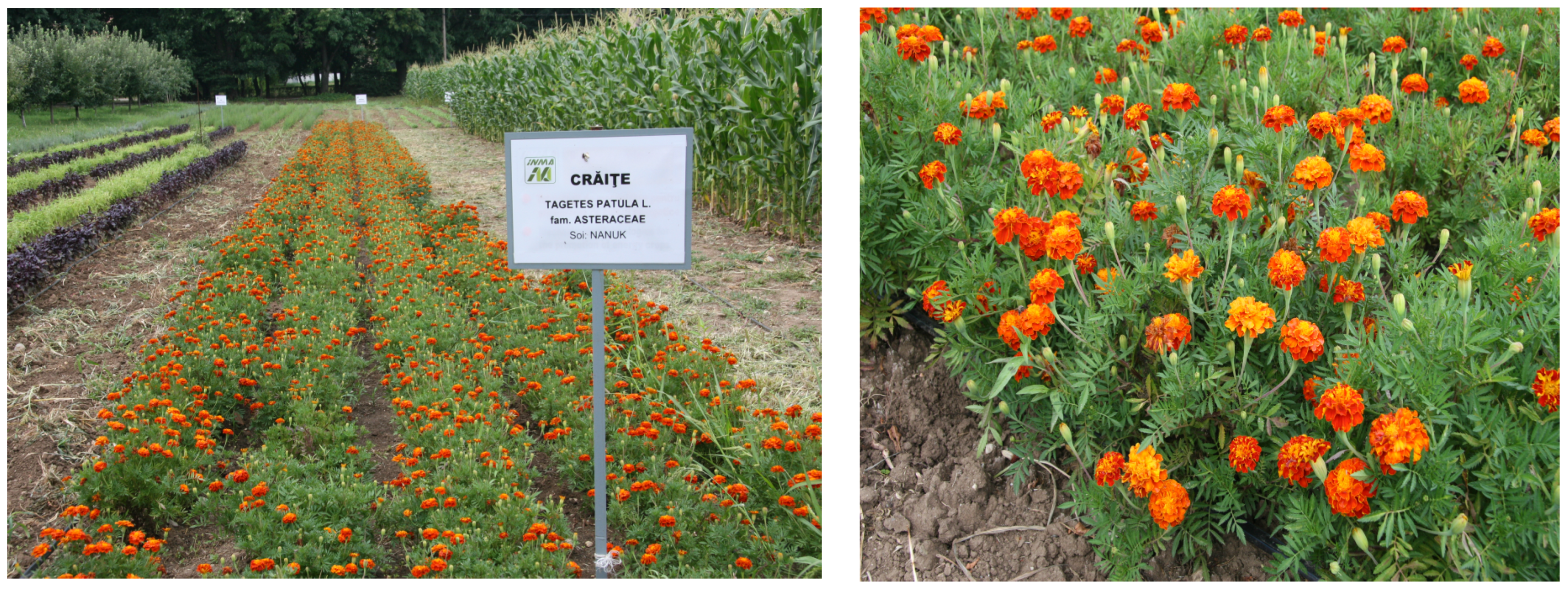
2.1. Experimental Cultures and Plant Samples

The experiments were carried out using a new variety of marigold (T. patula L., fam. Asteraceae), namely, the “Nanuk” variety (certificate no. 10003/2014), a semi-late variety with a well-defined genetic constitution that is very well adapted to the climatic conditions existing in Romania. Essential oils and floral waters were obtained by processing the plant material collected from the novel variety in the 2018–2020 growing season (Figure 1).
The “Nanuk” marigold variety was obtained at S.C.D.L. Buzau and is characterized by reddish-brown flowers and a bush height of 40–42 cm. The flowering period is between July and the occurrence of the first frosts.
The cultivation complied with the specific recommended technological operations (land preparation, planting seedlings, culture maintenance works, harvesting). Two harvests/year were obtained, during June to September 2018–2020, and the production varied depending on the climatic conditions, with on average approx. 900 kg ha−1 of green vegetable raw material.
The raw material required for processing was produced in one of the experimental fields belonging to INMA Bucharest Institute, situated in the Băneasa area (44°30′01″ N; 26°04′19″ E, altitude 90 m). The climate in this region is characterized as transitional continental temperate, and the experimental lands are dominated by reddish-brown soils. Throughout the growing season, the average temperature ranged from 19.4 °C in June to 21.7 °C in August, and the average precipitation was recorded as 83.63 mm. There was a peak in precipitation in June, reaching 155.9 mm, but deficits were noted in August (34.2 mm) and in September (26.3 mm).

2.2. The Extraction Process of Essential Oils and Floral Waters

The marigold plants were harvested during the flowering stage in order to maximize the quantity of oils and floral water. Extraction was completed using hydrodistillation technology, processing series of 10 kg of green plants per batch, represented by selected inflorescences and sprout tips. The process uses steam separation to obtain the hydrolate (a mixture of essential oils and floral waters), subsequently employing techniques based on decantation, filtration, and density differences to separate the two products. The extraction processing was set to 2.5 h per series. The equipment used for extraction was an Aura distillateur, featuring a 130 L tank. Precise steam control was achieved through the use of an electric steam generator of the MA 15–18 kW type, producing a constant 0.1 bar. After separation using a 10 L Florentine vessel, the resulting oils and floral waters were stored in opaque bottles and maintained at 4 °C until chemical evaluation and characterization [24].
The quantity of oil extracted from the vegetable material was determined using Equation (1):
O i l ( % v / w e t   b a s e ) = O b s e r v e d   v o l u m e   o f   o i l   ( m L ) W e i g h t   o f   s a m p l e   ( m L ) × 100

2.3. Evaluation of Essential Oils and Floral Waters

Gas chromatography coupled with mass spectrometry (GC-MS) was used to identify the chemical composition and concentration of the main volatile compounds. A 7890 A-Agilent Technologies gas chromatograph, in conjunction with the 5975 C Mass Selective detector MS manufactured by Agilent Technologies, California, USA, and a Macrogol Column 20,000 R (30 m × 0.25 mm ID, bonded 0.50 µm), were the instruments utilized for the examination. Helium was used as the carrier gas, with a flow rate of 1.5 mL/min. The temperature range was from −250 °C (10 degrees/min) to 280 °C (const. 5.5 min). The injector and detector temperatures were set at 220 °C and 235 °C, respectively. The mobile phase consisted of 1 mL/min and the injector was split (split ratio: 1:100). The sample was injected automatically, and 1 mL of essential oil was utilized for the analysis. Before injection, 100 times the EO was dissolved in n-hexane. Fifteen milliliters of undiluted FW were extracted into ten milliliters of n-hexane, and the mixture was dried over anhydrous sodium sulfate R. An interval of 0–70 min was established as the scan range for the GC-MS analysis. For each EO and FW, one sample was examined. The elements were identified in the chromatograms for each testing probe using the retention times and spectra of the reference solutions. By comparing the retention indices of the individual constituents to those of compounds reported in the literature, the constituents were determined. The Wiley Registry 10th Edition/ NIST Standard Reference Database 1A library served as the basis for identification [24].

2.4. Evaluation of the EO and FW Antioxidant Activity

An improved depiction of the antioxidant activity of marigold EO and FW can be obtained by utilizing three different assessment techniques (DPPH, ABTS, FRAP). The basic ideas behind each technique and their synergy allow for a more precise evaluation of the compounds [24].
A.
The scavenger activity of the DPPH radical (2,2-diphenyl-1-picrylhydrazyl) is based on the ability of antioxidants to reduce the DPPH radical. The rate of DPPH remaining in the solution is determined using Formula (2):
% D P P H = A c o n t r o l   s a m p l e A s a m p l e A c o n t r o l   s a m p l e × 100
where Acontrol sample is the absorbance of the control sample and Asample is the absorbance of the sample.
The abbreviation “IC50” refers to the quantity of samples needed to decrease DPPH absorbance by 50%. Each sample was tested at five different concentrations in triplicate in order to determine its associated IC50.
B.
The scavenger activity of the ABTS radical (2,2′-azinobis (3-ethylbenzothiazoline-6-sulfonic acid)): Because the expression of the extract antioxidant capacity can be related to Trolox equivalents, the technique is known as TEAC (Trolox equivalent antioxidant capacity). Three Trolox standard calibration curves were used to express the antioxidant capacity in milligrams (mg) of Trolox.
C.
Ferric ion reducing antioxidant power (FRAP): This relies on the capacity of antioxidants to reduce the yellow-colored tripyridyltriazine Fe3+ (Fe (III)-TPTZ) complex to the blue-colored tripyridyltriazine Fe2+ (Fe (II)-TPTZ) complex by the action of electron release by antioxidants. The evaluations were tested in triplicate, and the FRAP values of each sample were expressed in mM Trolox g-1 for EOs and L-1h for FWs.

2.5. Evaluation of Microbial Strains and Growth Conditions

The three G- bacterial strains used were P. carotovorum, P. marginalis, and P. syringae. All strains are natural isolates with high plant pathogenic activity. Bacterial inoculum was obtained from fresh cultures and prepared in Luria Bertani (LB) broth at 28 °C. Three strains of fungal phytopathogens, R. solani, F. oxysporum, and B. cinerea, which can produce high economic losses in agriculture, were selected. For R. solani and F. oxysporum, the fungal inoculum was prepared as mycelia plugs 8 mm in diameter collected from 14-day-old cultures obtained on potato dextrose agar. In the case of B. cinereal, the fungal inoculum was prepared as mycelia plugs 8 mm in diameter collected from 14-day-old cultures obtained on potato dextrose agar [24].

2.6. Assessment of Essential Oil Emulsions

Emulsions were formulated in a solution containing 10% DMSO and supplemented with 0.5% Tween 80. This solvent demonstrated no impact on microbial growth [25,26]. The tests performed for the EO of marigold were in C1 = 100% (undiluted), in C2 = 75% (three quarters EO and the rest solvent), in C3 = 50% (half EO and half solvent), and in C4 = 25% (one quarter EO and the remaining three quarters solvent).

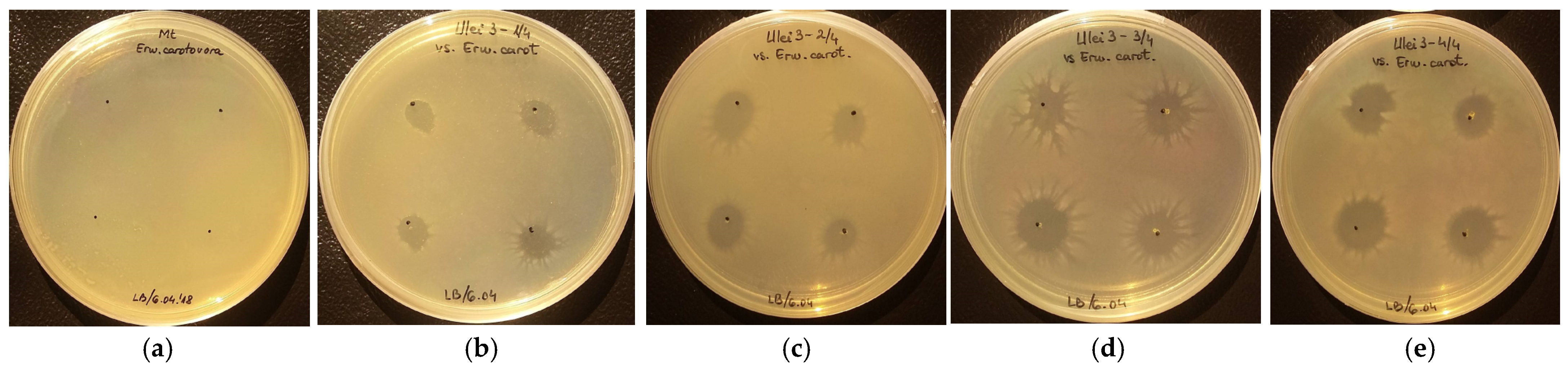
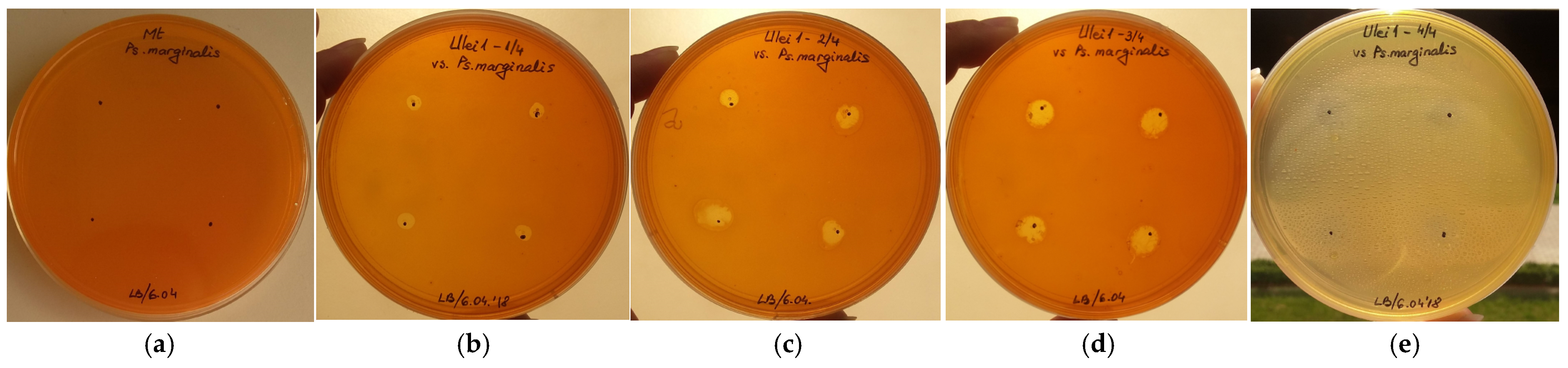
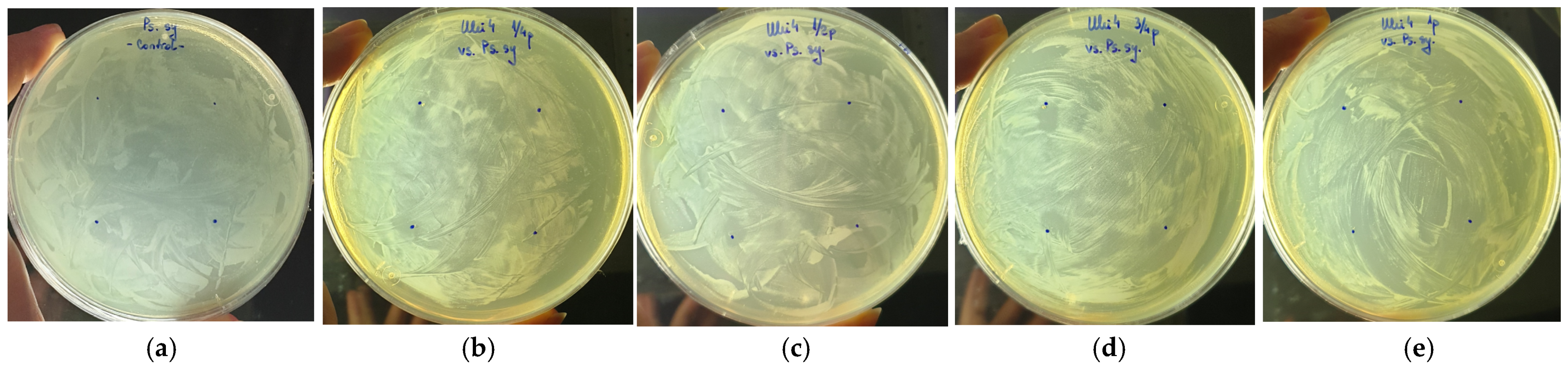
2.7. Antibacterial Assay

The antibacterial potential of the essential oils was assessed under in vitro conditions using non-ventilated, sterile polypropylene Petri dishes. Each dish was filled with 20 mL of LB agar and inoculated with a fresh bacterial suspension containing 108 colony-forming units per milliliter (CFU/mL). The essential oils were evenly distributed and spotted (100 µL/spot) at equidistant points on each plate, with four replicates prepared for each concentration of the oils being tested. Positive controls, lacking EOs, were also set up for the phytopathogenic bacterium. All plates were sealed with parafilm and then incubated at 28 °C. For each pathogen, two control plates were prepared: one containing only the test bacteria (without the solvent), and another where the test bacteria were cultured with the solvent (a mixture of 10% DMSO with 0.5% Tween 80 in water). Biometric measurements were taken after 24 h and again after 7 days of inoculation (bacterial colony diameter). Antibacterial activity was assessed based on the clear areas where the pathogen failed to colonize the growth substrate [24].

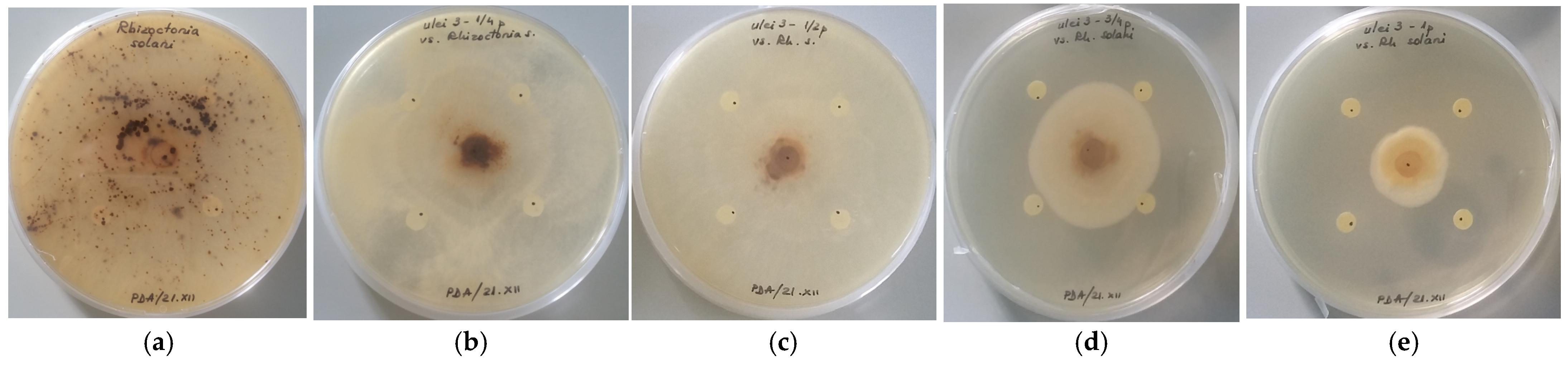
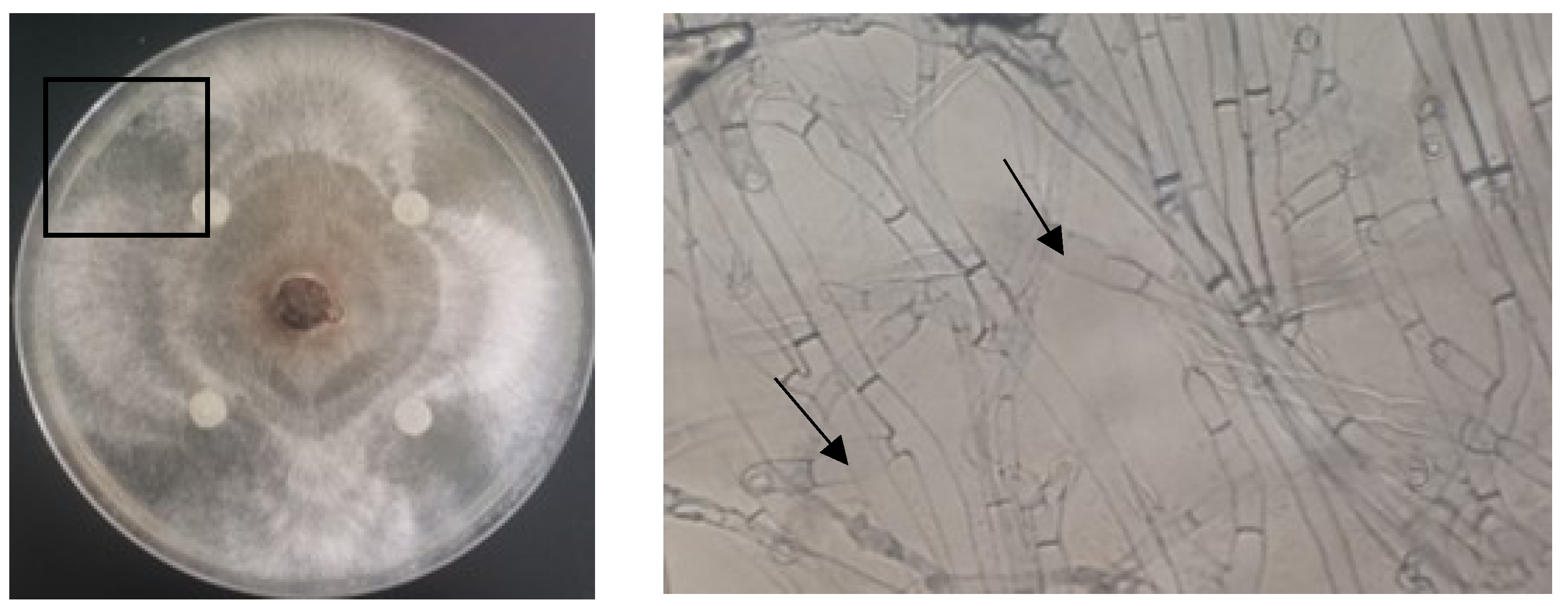
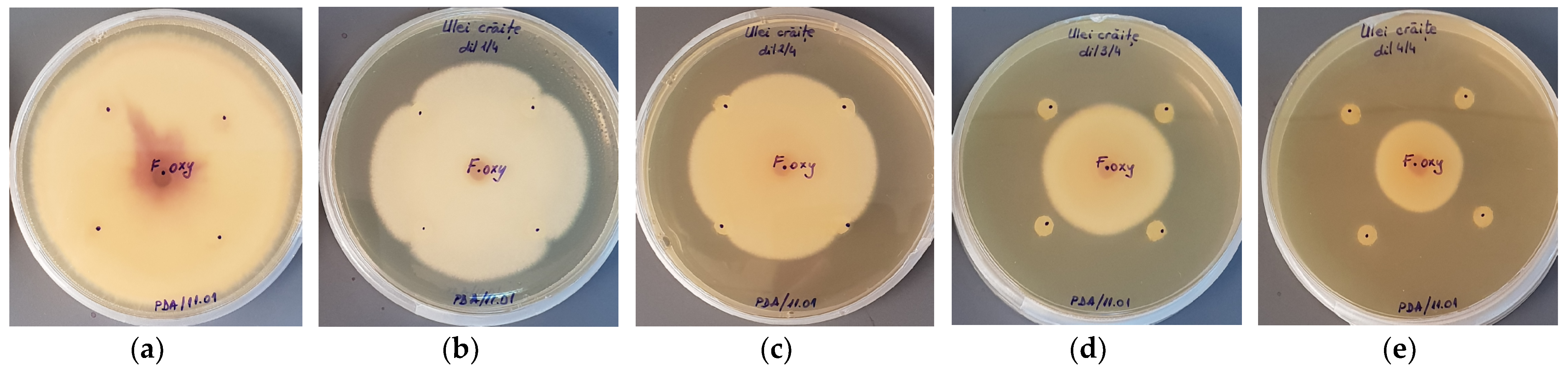
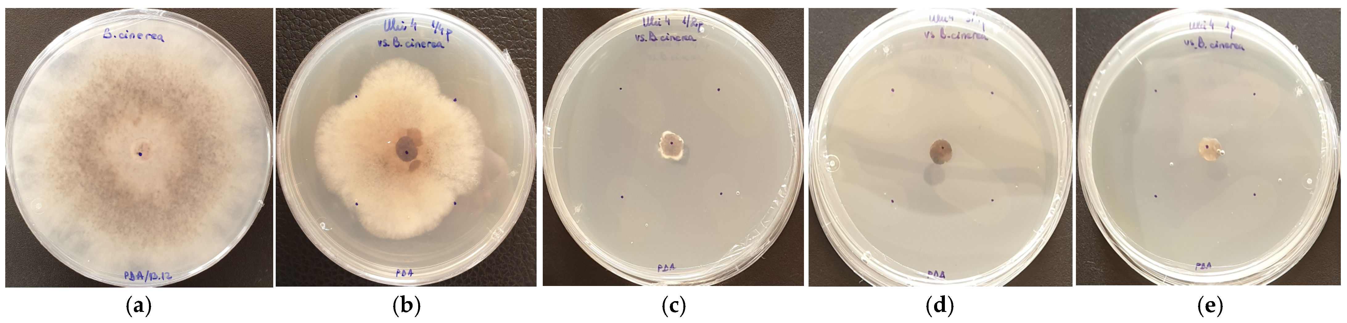
2.8. Antifungal Assay

The antifungal assay was carried out under similar conditions as in the previous test. However, PDA medium was chosen to maintain the fungal growth. Mycelia plugs with a diameter of 8 mm were used to inoculate the central region of the plates. Four sterile paper disks with a diameter of 5 mm were positioned two centimeters apart and equally spaced from the fungus inoculum. An EO emulsion volume of 10 µL was placed in each disk. Four replicates (Petri dishes) per EO concentration were prepared. Additionally, negative controls lacking EO were made for every plant pathogenic fungus. For the first ten days following inoculation, plates were parafilm sealed and incubated between 26 and 28 °C, and daily analyses were performed. During this period, the fungus was able to fully colonize the growth medium’s surface on the control plates. This represents the highest level of active growth that can be measured using biometric techniques. To assess the marigold EO’s antifungal properties, biometric tests were performed on the fungal growth. The mycelial growth was measured after 3, 5, 7, and 10 days after the fungus was placed in Petri dishes and compared to the fungal growth in the control. To assess the marigold EO antifungal properties, biometric tests were performed on the fungal growth after 10 days. Fungal inhibition efficacy (E, %) was determined using Equation (3), proposed by [25]:
E = R c R T R c × 100
where Rc = the radius of the fungal colony in the control plates, and RT = the fungal radius in the test plates.
Light microscopy examinations were conducted on the microbial growth in both the control and test plates to identify any potential anomalies related to cells and mycelia [24].

2.9. Insecticidal Assay

A 0.5 mL volume of conidial suspension, obtained from an 18-day-old sporulated culture of B. brongniartii (strain BbgMm1a/09), was used to inoculate Petri dishes containing potato dextrose agar (PDA) medium. Around 2 h post-inoculation, medium discs with a 7 mm diameter were excised from these plates. Subsequently, using a microbiological loop, each disc was transferred to the center of a Petri dish containing PDA medium. Additionally, a disc of sterile filter paper saturated with marigold essential oil in 7 different concentrations (1, 2, 5, 10, 20, 50, and 100 μL l−1 air) was affixed to the inner side of the dish lid. The dilution of essential oil and the concentration preparation were carried out in a sterile 0.2% water–agar solution, with pipetting performed while being constantly stirred. The plates were immediately sealed with parafilm and kept in an incubator at 23 °C after application. Three duplicates of each concentration were tested. Over an interval of seven days, the colony size was measured in two perpendicular directions. The following formula was used to calculate the inhibition of mycelial growth in relation to the size of the control colony (4):
I M G = ( ( D c D s ) / D c ) × 100
where IMG = inhibition of mycelial growth, Dc = diameter of control colony, and Ds = diameter of sample colony.
To assess the insecticidal effect of the essential oil on the insect S. granarius, adult specimens were utilized in this study. These adults were maintained in darkness at room temperature of (22 ± 2 °C). Filter papers were saturated with 10 µL of essential oil and positioned at the base of a 250 mL Berzelius beaker. Subsequently, 100 g of wheat were added to the beaker, and 30 adult insects were released over the filter paper. The beakers were placed in 5 L glass jars and covered. Treatments were performed in the dark, with t = 20 °C and relative humidity RH = 55%. There was no EO used in the control treatment. The number of dead S. granarius adults was determined at 2, 4, and 7 days after initiating the treatment [24].

2.10. Statistical Processing of Experimental Data

The purpose of the analysis was to determine the statistical average temporal efficacy for the EO, depending on the tested concentrations, against F. oxysporum and B. cinerea. The insecticidal activity was determined using two-way ANOVA followed by Bonferroni analysis, while the data interpretation was achieved using GraphPadPrism 5.01. software. For each reading across time, four repetitions of the essential oils were conducted, and the results were analyzed using simple statistical estimators, including arithmetic means, medians, and quartiles.

3. Results

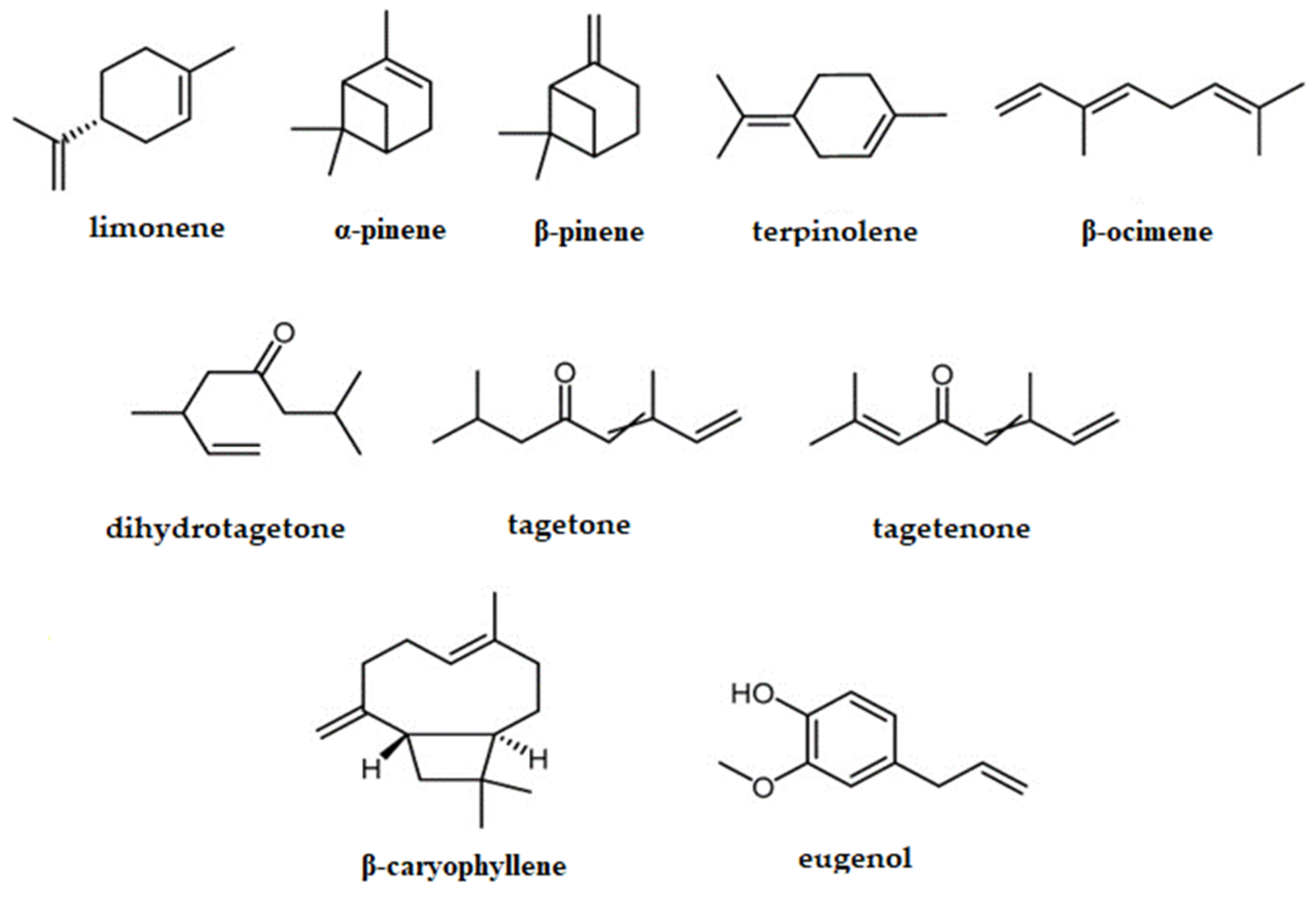
3.1. EO and FW Chemical Composition Evaluation with GC/MS

The chemical composition of the EOs obtained from the “Nanuk” marigold variety in all three years is presented in Table 1.
For the floral waters obtained from the dwarf marigold variety, the chemical composition is shown in Table 2.
The analyses conducted with GC-MS over three years (2018–2020) on FWs identified, on average, 16 compounds, representing between 99.99% and 100% of the total compounds separated. Carene (9.93–15.45%), caryophyllene (3.45–13.38%), piperitenone (6.56–8.73%), piperitone (1.36–9.01%), and elemene (2.53–4.20%) were the main compounds identified. Additionally, other compounds were identified in smaller quantities, such as D-limonene (4.42–7.83%), β-ocimene (5.28–11.73%), etc. (Table 1).
Regarding the compound classes, the following were identified: monoterpenes (65.69–81.36%), especially oxygenated monoterpenes (40.36–48.88%), along with sesquiterpenes (7.78–29.85%), particularly sesquiterpene hydrocarbonate (6.50–24.97%). Additionally, diterpenes were identified, more notably in 2019 (4.55%).

3.2. Antioxidant Activity of the EOs and FWs

The comparative antioxidant evaluation of both essential oils (EOs) and floral waters (FWs) obtained during 2018–2020 is illustrated in Table 3.
For marigold essential oil, the result of the “Nanuk” variety obtained in the three years (2018–2020) show that it exhibited high antioxidant activity, especially through the DPPH method, for the samples related to the year 2018 (0.20 ± 0.00 g l−1). The lower the IC50 value, the higher the antioxidant capacity of the analyzed sample. In the case of testing through the ABTS method, the highest antioxidant capacity was recorded for the sample from the year 2019 (0.24 ± 0.01 g−1), followed by the year 2020 (0.17 ± 0.00 g−1), compared to the year 2018 (0.09 ± 0.01 g−1). The year 2019 showed a significant difference in the antioxidant activity, influenced by the high humidity levels from the flowering period. The results obtained through the FRAP method for marigold EO, “Nanuk” variety, show high antioxidant capacity for the samples from the year 2019 (42.05 ± 3.88 g−1) and the year 2020 (35.04 ± 2.18 g−1). There was a significant increase in antioxidant capacity for the marigold essential oil sample from the year 2018 (0.20 ± 0.00 l−1), as well as a substantial increase in the values obtained through both the ABTS and the FRAP methods.

3.3. Antibacterial Activity of the EO

EO obtained by hydrodistillation of marigold inflorescences and shoot tips was tested against three G-phytopathogenic bacteria: P. carotovorum, P. marginalis, and P. syringae. The EO expressed wider inhibition areas when applied in c > 50% against the tested bacteria. It is assumed that, at this concentration, the emulsion contained sufficient solvent to ensure a good dispersion of the active ingredient and sufficient essential oil for bacterial inhibition.
When tested against P. carotovorum, a correlation of the inhibitory effect with the concentration was observed (after 24 h and 7 days of incubation, respectively) in the EOs extracted from the “Nanuk” varieties after measuring the inhibition zones (Figure 2, Table 4) of bacterial growth. These zones were slightly diminished, mainly by 0.3 ÷ 0.4 cm. Longer incubation times provided the opportunity for viable bacteria cells to multiply and colonize the area spotted with oil sample, starting from the edge and going toward the center. However, no colonies developed starting from inside the treated areas. This indicate bactericidal activity only when the essential oils are in direct contact with the bacterial cells; otherwise, the effect is bacteriostatic.
Similar analysis carried out on P. marginalis showed a bacteriostatic effect of the EO against this pathogen (Figure 3, Table 5).
The results obtained on P. syringae show that marigold EO reduced the density of bacterial growth on the oil spot footprint in all four tested concentrations, with the effect being bacteriostatic (Figure 4, Table 6).

3.4. Antifungal Activity of the EO

The EO obtained by hydrodistillation were tested against the pathogenic fungi R. solani (Figure 5 and Figure 6 and Table 7), F. oxysporum (Figure 7 and Table 8), and B. cinerea (Figure 8 and Figure 9 and Table 9). Observations on fungal growth in the presence and absence of the essential oil were made after 3, 5, and 7 days of incubation at 28 °C. Subsequently, to determine whether marigold EO exhibits fungicidal or fungistatic activity, the same plates were also analyzed after 10 days of incubation (Figure 5, Figure 6, Figure 7, Figure 8 and Figure 9).
After 10 days of incubation, EO had an efficacy of 77.6% in inhibiting the growth of R. solani when applied undiluted and 57.0% at a concentration of C3 = 75% oil in emulsion. The last concentrations tested (C1 = 25% and C2 = 50% in emulsion) did not inhibit the colonization ability of R. solani, although the hyphae were less abundant compared to the untreated control. Optical microscopy studies showed that the morphology of R. solani colony in the presence of the oil at C1 = 25% underwent changes. Near the oil-impregnated discs, the cells were shorter and some of them were slightly swollen and thickened (Figure 6, arrow).
For the phytopathogenic fungus F. oxysporum, it should be noted that marigold EO (Figure 7) did not have fungicidal activity, only fungistatic, and was able to delay mycelial growth, with the degree of inhibition depending on the concentration of the oil used and its composition.
For the “Nanuk” variety of marigold EO after 10 days of incubation, for C1 = 25%, B. cinerea showed delayed growth in the region where the oil was placed (Figure 8), indicating that the inhibitory effect was also due to other non-volatile compounds. As a general observation, for marigold EO at high concentrations (C > 50%), mycelial growth was completely suppressed compared to the control, and at C1 = 25%, mycelial growth was only delayed by 3 days.
Optical microscopy data show that in the oil spot area, the fungus had very limited contact with the agar surface; perforations in the cell wall and leakage of cytoplasm from the mycelium were observed (Figure 9).

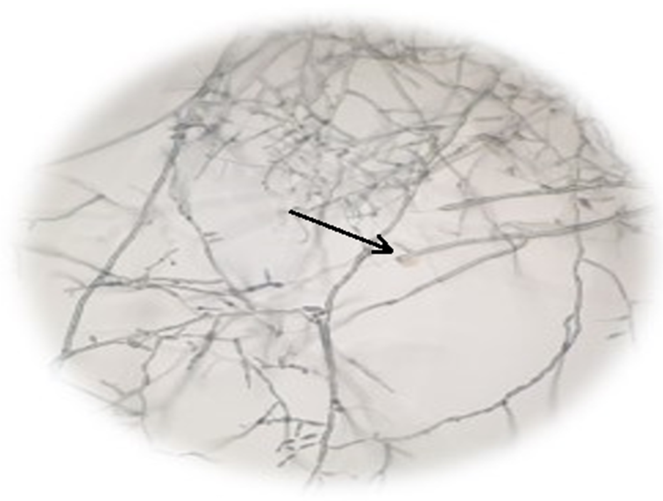
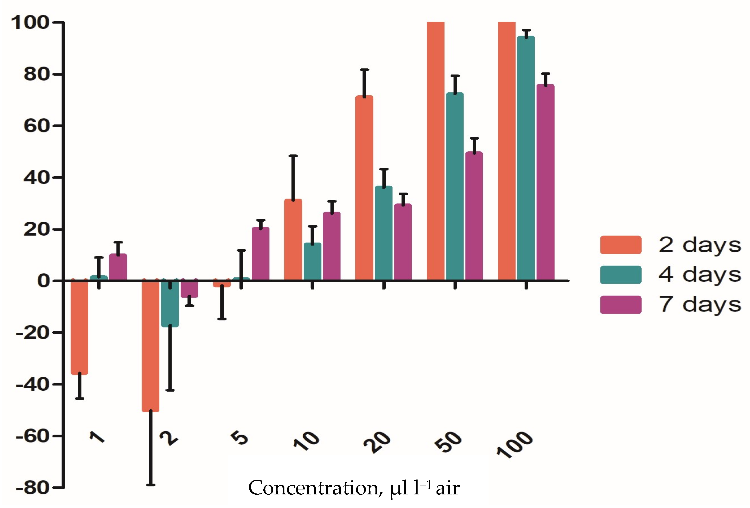
3.5. Insecticidal Activity of the EO

This study aimed to test the effects of this EO, with the goal of obtaining a potential product that incorporates in its formula both marigold EO and the entomopathogenic fungus B. brongniartii to collectively combat the storage insect S. granarius. The results obtained for the tested marigold EO showed a fungistatic effect of 100% at a concentration of c = 100 μL L−1 air two days after inoculation. It was also observed that in the first two days, at concentrations of c = 1 μL L−1 air and c = 2 μL L−1 air, marigold EO had a stimulating effect on fungal mycelium growth. The inhibitory effect is positively correlated with the tested concentration (Figure 10). The two-way ANOVA test showed that the factors, treatment (df = 3, F = 10.76, p = 0.003), and concentrations (df = 6, F = 198.1, p < 0.0001) had a highly significant influence on the fungal mycelium growth.
Also, this EO did not exhibit satisfactory fumigant activity against adult S. granarius, as no mortality was recorded during the testing period. This could be related to the low concentration of EO in the air and the low incubation temperature. The initial objective was to combine the attributes of marigold essential oil with those of the entomopathogenic fungus B. brongniartii, aiming for an enhanced insecticidal effectiveness against the insect S. granarius. However, given the essential oil’s lack of efficacy against the insect, it was decided not to proceed with the experiment involving the mixture of the two solutions. The two control solutions (fungi and essential oil) were tested only separately against the insect.

4. Discussion

4.1. EO and FW Chemical Composition Evaluation

Diterpenes are not usually present in EOs but are sometimes encountered as minor, insignificant constituents [26]. The marigold EO was rich in monoterpenic hydrocarbons (ocimene, limonene, terpinene, myrcene, etc.) and acyclic monoterpenic ketones (tagetone, dihydrotagetone, tagetenone), which are the primary compounds, in addition to smaller amounts of sesquiterpenic hydrocarbons and oxygenated compounds. Within these groups, the chemical diversity is quite high. Figure 11 shows the main chemical structures of the compounds identified in the marigold EO, which were also found in many other studies [27,28].
There are studies that mention the impact of geographical origin on the chemical diversity [29] of the EOs obtained from T. patula. In the studies [30,31], limonene, α-terpinolene, 4-vinylguaiacol, and γ-terpinene are mentioned as the main compounds; however, there is a lack of evaluation of some compounds, such as β-ocimene, β-caryophyllene, piperitone, and piperitenone.
Two studies conducted on the EO obtained from marigolds [31,32] found that the main identified compounds were limonene, (Z)-β-ocimene, α-terpinolene, (E)-tagetone, (Z)-tagetone, piperitenone, piperitone, and β-caryophyllene in variable amounts, confirming the results obtained in the present work.
Marotti et al. mention that in Italian essential oils obtained from marigold inflorescences, the main compounds are piperitone (28.9%), terpinolene (5.8%), β-caryophyllene (3.8%), limonene (3.5%), linalool (2.7%), myrcene (1.8%), and terpinen-4-ol (1.1%) [32]. In the case of the “Nanuk” variety, some of these compounds were identified, but in different quantities (usually higher)—for example: piperitones 1.36–9.01%, β-caryophyllenes 3.45–13.38%, limonenes 4.42–7.83%, and linalool 2.34–4.27%.
As in the investigated case, other studies [33,34] also obtained significant variations egarding the compounds obtained during the 3 years (2018–2020), depending on the year. Therefore, the primary factor influencing these variations is likely to be the fluctuation in climatic conditions. Regarding the compound classes, in marigold FW (“Nanuk” variety), mainly monoterpene compounds were identified (55.10–95.80%), especially oxygenated ones (95.80%), followed by sesquiterpenes (37.61% in 2019) and diterpenes in smaller amounts (1.67–5.23%).
It should be noted that the chemical composition of EO and FW largely depends on a series of endogenous and exogenous factors, including genetic traits of the plant/variety, plant organs from which extraction is performed (roots, leaves, stems, capitula), growth conditions, drying and storage, and stress factors (weather conditions during the cultivation year, disease, and pest attacks) affecting the plant. The chemical composition of the EO is influenced by extraction methods and solvents used, extract standardization, etc. [33].

4.2. Antioxidant Activity of the EOs and FWs

The comparative values of antioxidant activity for FWs obtained during the period 2018–2020 through the three methods (DPPH, ABTS, FRAP) showed significant variations in antioxidant capacity values. It is noticeable that for all the IC50 values of marigold FWs there was a doubling of the value (over the three years), which correlates with a lower antioxidant capacity compared to that of the EOs. This could suggest a modification in their chemical composition due to a decrease in the concentration of compounds with antioxidant activity.
The results in this study show the ability of EOs obtained from the “Nanuk” variety to eliminate three different radicals, suggesting their usefulness as potent antioxidant agents for further investigations. Additionally, the variation in climatic conditions over the three years (2018–2020) has influenced the chemical composition of EOs and FWs, indicating a potential modification in their chemical composition by decreasing or increasing the concentration of compounds with antioxidant activity.
The study conducted on EOs obtained from the aerial parts of T. ellipitica exhibited moderate antioxidant activity [35]. The antioxidant properties can be attributed to a high content of ketones (acyclic monoterpenes), including cis- and trans-tagetenone and tagetone, found in the composition of the EOs, as well as the synergistic action among various major and minor compounds [36]. However, the mechanism by which the compounds in EOs exert their antioxidant effect is not yet fully understood. Several mechanisms have been proposed, primarily their redox properties, which play a significant role in the absorption and neutralization of free radicals, as well as the decomposition of peroxides [37].

4.3. Antibacterial and Antifungal Activity of the EO of Marigold

Based on the experimental results obtained, the hypothesis that this EO could have a bactericidal effect against P. carotovorum is not excluded, and a correlation is observed between the inhibitory effect and the concentration used. The recorded bacterial growth resulted from the colonization and expansion of colonies only at the periphery of the EO spots. No isolated colonies were observed on the EO footprint, supporting the hypothesis that bacteria that came into direct contact with the EO lost their viability and could not proliferate further. EOs contain various active compounds that can disrupt multiple targets in bacterial cells, and one of the most important is the cytoplasmic membrane [38]. Some compounds in EOs increase the permeability of the cell membrane, leading to its loss of viability, a phenomenon associated with ion homeostasis and the electron transport chain [39].
The results obtained in the tests performed on P. marginalis (bacteriostatic effect) confirm a series of experimental findings. Although after 24 h of incubation good inhibitory efficiency was observed, in line with the increase in essential oil concentration, after 7 days of incubation, it was noticed that in the variants where a solvent was used, the colony density was lower in the previously clear area. Colonies developed in that region were rarer compared to plates where undiluted EO was tested. Colony density, however, could not be assessed differentially between test dilutions. Marigold EO used undiluted maintained a clear area of inhibition of bacterial growth even after 7 days of incubation. This aspect can be justified by the fact that this oil, being denser, may have evaporated more slowly compared to the others. However, taking into account the fact that after 7 days bacterial colonies were also observed in the initially clear area (after 24 h), the EO effect was bacteriostatic. Tests conducted by [40], showed that hyssop essential oil was almost inactive against certain Gram-negative bacteria (P. aeruginosa, E. coli, Salmonella typhi). Romagnoli et al. [41] extracted the EO from dried T. patula flowers and investigated its antifungal effect on Penicillium digitatum and B. cinerea strains. The EO showed remarkable activity in both fungi, reaching 100% inhibition, even at the lowest concentrations. Flower extracts of T. patula exhibited toxicity against soil-borne fungus F. oxysporum f.sp. lycopersici, causing wilt disease in tomato plant [41].
Thembo et al. [42] used the aerial parts from T. minuta against isolates from four fungi species of agricultural and clinical importance: F. verticillioides, F. proliferatum, Aspergillus flavus, and A. parasiticus. The extraction solvents used were hexane, dichloromethane, methanol, and water. The concentration of the extracts was 10 mg/mL. The drug amphotericin B and the agricultural fungicide Cantus were used as positive controls.
Despite the promising in vitro results, in-depth studies are required to understand the mechanisms of action of EOs obtained from Tagetes spp. for their potential use in biotechnology [43,44]. Compounds in these EOs, especially terpenoids (dihydrotagetones, tagetones, ocimenones), are responsible for the identified antimicrobial activity [43]. In the future, the goal is to identify the active compounds in these EOs by fractionating them and determining the antimicrobial activity for each compound individually, considering both synergistic and antagonistic antimicrobial interactions [45].

4.4. Insecticidal Activity of the EO of Marigold

Previous studies have shown that the insecticidal activity of marigold EO correlates with the major compounds it contains (Table 1). The present study showed that marigold essential oil exhibited complete inhibition of an entomopathogenic fungus, but it did not demonstrate any activity against the insect. Consequently, it is not considered a viable alternative as an insecticide. However, other studies have shown that extracts with similar compounds could have effects in certain conditions. Zoubiri and Baaliouamer documented the high effectiveness of EOs of T. minuta against Anopheles gambiae mosquitoes, which are responsible for malaria transmission [44]. Insecticidal investigation of T. erecta leaf oil against the white termite of sugarcane fields (Odontotermes obesus Rhamb.) showed that it conferred 100% mortality at 6 µL/Petri plate dose after 24 h of exposure, whereas at lower doses and shorter exposures, it showed diminished mortality rates [45,46].
For the EO obtained from T. lucida, repellent activity against Sitophilus zea mais was observed. The main compounds in this EO were oxygenated monoterpenes and phenolic compounds [47].

5. Conclusions

The essential oil obtained in the 2020 production showed the best chemical composition in terms of both the compounds obtained and their quantities.
The antifungal activity demonstrated the most significant efficacy against the phytopathogenic fungus B. cinerea, with treatment effectiveness exceeding 90% at concentrations above 50%.
The EO obtained from the “Nanuk” variety of marigold did not exhibit any insecticidal effects on S. granarius.
The floral water of this variety was solely analyzed for its chemical composition and antioxidant activity, as the substances of interest were detected in significantly low amounts, making them unsuitable for other purposes.

Author Contributions

Conceptualization, C.T., F.B., F.I.-R. and O.A.B.-S.; methodology, C.T., F.B., F.I.-R. and F.N.; software, C.T.; validation, C.T., F.N., F.B. and F.I.-R.; formal analysis, F.G.; investigation, C.T., F.G., C.T. and O.A.B.-S.; resources, A.M., F.B. and F.I.-R.; writing—original draft preparation, C.T., F.B. and F.I.-R.; writing—review and editing, visualization, C.T., F.N., F.B. and F.I.-R.; supervision, C.T., F.I.-R., F.B. and F.N.; project administration, C.T. and A.M. All authors have read and agreed to the published version of the manuscript.

Funding

This work was supported by the NUCLEU Program, carried out with the support of ANCSI, Project PN 9N/01.01.2023, PN 23 04 02 05—Innovative technology for the superior utilization of inflorescences and seeds of medicinal plants, and PN 23 04 02 01—Innovative technology for the production of biofertilizers, aimed at restoring soil biodiversity and reducing the effects of drought on agricultural land.

Institutional Review Board Statement

Not applicable.

Informed Consent Statement

Not applicable.

Data Availability Statement

The raw data supporting the conclusions of this article will be made available by the authors on request.

Conflicts of Interest

The authors declare no conflicts of interest.

Abbreviations

MAPs—medicinal and aromatic plants; EO—essential oil of Marigold, “Nanuk” variety; FW—floral water of Marigold, “Nanuk” variety.

References

  1. Burt, S. Essential oils: Their antibacterial properties and potential applications in foods—A review. Int. J. Food Microbiol. 2004, 94, 223–253. [Google Scholar] [CrossRef] [PubMed]
  2. Prieto, M.C.; Camacho, N.M.; Inocenti, F.D.; Mignolli, F.; Lucini, E.; Palma, S.; Bima, P.; Grosso, N.R.; Asensio, C.M. Microencapsulation of Thymus vulgaris and Tagetes minuta essential oils: Volatile release behavior, antibacterial activity and effect on potato yield. J. Saudi Soc. Agric. Sci. 2023, 3, 195–204. [Google Scholar] [CrossRef]
  3. Prieto, M.C.; Lapaz, M.I.; Lucini, E.I.; Pianzzola, M.J.; Grosso, N.R.; Asensio, C.M. Thyme and suico essential oils: Promising natural tools for potato common scab control. Plant Biol. 2020, 22, 81–89. [Google Scholar] [CrossRef] [PubMed]
  4. Hajian-Maleki, H.; Baghaee-Ravari, S.; Moghaddam, M. Herbal essential oils exert a preservative effect against the potato soft rot disease. Sci. Hortic. 2021, 285, 110192. [Google Scholar] [CrossRef]
  5. Isman, M.B. Plant essential oils for pest and disease management. Crop Prot. 2000, 19, 603–608. [Google Scholar] [CrossRef]
  6. Asensio, C.M.; Paredes, A.J.; Martin, M.P.; Alemandi, D.A.; Nepote, V.; Grosso, N.R. Antioxidant stability study of oregano essential oil microcapsules prepared by spray-drying. J. Food Sci. 2017, 82, 2864–2872. [Google Scholar] [CrossRef]
  7. Ayala-Zavala, J.F.; Soto-Valdez, H.; Gonzáles-León, A.; Alvarez-Parrilla, E.; Martin Belloso, O.; Gonzalez-Aguilar, G.A. Microencapsulation of cinnamon leaf (Cinnamomum zeylanicum) and garlic (Allium sativum) oils in beta-cyclodextrin. J. Incl. Phenom. Macrocycl. Chem. 2008, 60, 359–368. [Google Scholar] [CrossRef]
  8. Alipour, M.; Saharkhiz, M.J.; Niakousari, M.; Damyeh, M.S. Phytotoxicity of encapsulated essential oil of rosemary on germination and morphophysiological features of amaranth and radish seedlings. Sci. Hortic. 2019, 243, 131–139. [Google Scholar] [CrossRef]
  9. Taban, A.; Saharkhiz, M.J.; Kavoosi, G. Development of pre-emergence herbicide based on Arabic gum-gelatin, apple pectin and savory essential oil nano-particles: A potential green alternative to metribuzin. Int. J. Biol. Macromol. 2021, 167, 756–765. [Google Scholar] [CrossRef]
  10. Wanjal, W.C.; Wanzala, W. Chapter 90-Tagetes (Tagetes minuta) oils. In Essential Oils in Food Preservation, Flavor and Safety; Academic Press: London, UK, 2016. [Google Scholar]
  11. Salehi, B.; Valussi, M.; Bezerra Morais-Braga, M.F.; Pereira, C.J.N.; Borges Leal, A.L.A.; Melo Coutinho, H.D.; Vitalini, S.; Kregiel, D.; Antolak, H.; Sharifi-Rad, M.; et al. Tagetes spp. Essential Oils and other extracts: Chemical Characterization and Biological Activity. Molecules 2018, 23, 2847. [Google Scholar] [CrossRef]
  12. Gakuubi, M.M.; Wanzala, W.; Wagacha, J.M.; Dossaji, S.F. Bioactive properties of Tagetes minuta L. (Asteraceae) essential oils: A review. AJONP Am. J. Essent. Oils Nat. Prod. 2016, 4, 27–36. [Google Scholar]
  13. Fabrick, J.A.; Yool, A.J.; Spurgeon, D.W. Insecticidal activity of marigold Tagetes patula plants and foliar extracts against the hemipteran pests, Lygus hesperus and Bemisia tabaci. PLoS ONE 2020, 15, e0233511. [Google Scholar] [CrossRef] [PubMed]
  14. Ravikumar, P. Chemical examination and insecticidal properties of Tagetes erecta and Tagetes patula. Asian J. Biol. Sci. 2010, 5, 29–31. [Google Scholar]
  15. Safar, A.A.; Anwar Othman Ghafoor, A.O.; Dastan, D. Screening of chemical characterization, antifungal and cytotoxic activities of essential oil constituents of Tagetes erecta L. from Erbil, Kurdistan Region-Iraq. Pol. J. Environ. Stud. 2020, 29, 2317–2326. [Google Scholar] [CrossRef]
  16. Bakshi, L.; Gosh, R. Marigold biopesticide as an alternative to conventional chemical pesticides. J. Adv. Sci. Res. 2022, 13, 26–33. [Google Scholar] [CrossRef]
  17. Wright, P.J.; Hale, C.N. A field and storage rot of onion caused by Pseudomonas marginalis. N. Z. J. Crop Hortic. Sci. 1992, 20, 435–438. [Google Scholar] [CrossRef]
  18. Krejzar, V.; Mertelík, J.; Pánková, I. Pseudomonas marginalis Associated with Soft Rot of Zantedeschia spp. Plant Prot. Sci. 2008, 44, 85–90. [Google Scholar] [CrossRef]
  19. Kůdela, V.; Krejzar, V.; Pánková, I. Pseudomonas corrugata and Pseudomonas marginalis associated with the collapse of tomato plants in Rockwool slab hydroponic culture. Plant Protect. Sci. 2010, 46, 1–11. [Google Scholar] [CrossRef]
  20. Xin, X.F.; Kvitko, B.; He, S.Y. Pseudomonas syringae: What it takes to be a pathogen. Nature reviews. Microbiology 2018, 16, 316–328. [Google Scholar] [CrossRef]
  21. Boyraz, N.; Bastas, K.K.; Maden, S.; Yasar, A. Bacterial leaf and peduncle soft rot caused by Pectobacterium carotovorum on tulips in Konya, Turkey. Phytoparas 2006, 34, 272–280. [Google Scholar] [CrossRef]
  22. Raymond, G.A.T.; Roland, T.V.F. Diseases of Temperate Horticultural Plants; CAB International: New York, NY, USA, 2014; 488p. [Google Scholar]
  23. Lahlali, R.; Hijri, M. Screening, identification and evaluation of potential biocontrol fungal endophytes against Rhizoctonia solani AG3 on potato plants. FEMS Microbiol. Lett. 2010, 311, 152–159. [Google Scholar] [CrossRef] [PubMed]
  24. Stan (Tudora), C.; Neneciu, F.; Muscalu, A.; Vladut, V.N.; Burnichi, F.; Popescu, C.; Gatea, F.; Boiu-Sicuia, O.A.; Israel-Roming, F. Chemical composition, antioxidant and antimicrobial effects of essential oils extracted from two new Ocimum basilicum L. varietes. Diversity 2022, 14, 1048. [Google Scholar] [CrossRef]
  25. Chouhan, S.; Sharma, K.; Guleria, S. Antimicrobial Activity of Some Essential Oils-Present Status and Future Perspectives. Medicines 2017, 4, 58. [Google Scholar] [CrossRef] [PubMed]
  26. Koul, O.; Suresh, W.; Dhaliwal, G.S. Essential oils as green pesticides: Potential and constraints. Biopest. Intern. 2008, 4, 63–84. [Google Scholar]
  27. Salachna, P.; Łopusiewicz, Ł.; Wesołowska, A.; Meller, E.; Piechocki, R. Mushroom waste biomass alters the yield, total phenolic content, antioxidant activity and essential oil composition of Tagetes patula L. Inds. Crops Prod. 2021, 171, 113961. [Google Scholar] [CrossRef]
  28. Abbas, A.; Tabanka, N.; Amin, E.; Demirci, B.; Khan, I.A. Chemical composition and biting deterrent activity essential oil of Tagetes patula (Marigold) against Aedes aegypti. NPC Nat. Prod. Commun. 2016, 11, 1535–1538. [Google Scholar] [CrossRef]
  29. Politi, F.A.; De Souza-Moreira, T.M.; Rodrigues, E.R.; De Queiroz, G.M.; Figueira, G.M.; Januario, A.H.; Berenger, J.M.; Socolovschi, C.; Parola, P.; Pietro, R.C. Chemical characterization and acaricide potential of essential oil from aerial parts of Tagetes patula L. (Asteraceae) against engorged adult females of Rhipicephalus sanguineus (Latreille, 1806). Parasit. Res. 2013, 12, 2261–2268. [Google Scholar] [CrossRef] [PubMed]
  30. Safar, A.A.; Ghafoor, A.O.; Dastan, D. Chemical composition, antibacterial and antioxidant activities of Tagetes patula L. essential oil raised in Erbil, Iraq. J. Rep. Pharm. Sci. 2020, 9, 59–67. [Google Scholar] [CrossRef]
  31. Singh, P.; Krishna, A.; Kumar, V.; Krishna, S.; Singh, K.; Gupta, M.; Singh, S. Chemistry and biology of industrial crop Tagetes species: A review. J. Essent. Oil Res. 2015, 28, 1–14. [Google Scholar] [CrossRef]
  32. Marotti, M.; Piccaglia, R.; Biavati, B.; Marotti, I. Characterization and yield evaluation of essential oils from different Tagetes species. J. Essent. Oil Res. 2004, 16, 440–444. [Google Scholar] [CrossRef]
  33. Shetty, L.J.; Sakr, M.F.; Al-Obaidy, K.; Patel, J.M.; Shareef, H. A brief review on medicinal plant Tagetes erecta Linn. J. Appl. Pharm. Sci. 2015, 5, 91–95. [Google Scholar] [CrossRef]
  34. Isiaka, A.O.; Nureni, O. The essential oil from the leaves and flowers of ‘African Marigold’, Tagetes erecta L. J. Essent. Oil Res. 2006, 18, 366–368. [Google Scholar] [CrossRef]
  35. Cerrón-Mercado, F.; Perez-Alvarez, J.A.; Nolazco-Cama, D.; Salva-Ruíz, B.; Tellez-Monzon, L.; Fernández-López, J.; Viuda-Martos, M. Chemical Composition, Antioxidant and Antibacterial Activities of Essential Oil Obtained from Chincho (Tagetes elliptica) Leaves Grown in the Peruvian Andes. Foods 2023, 12, 894. [Google Scholar] [CrossRef] [PubMed]
  36. Barra, A. Factors Affecting Chemical Variability of Essential Oils: A Review of Recent Developments. Nat. Prod. Commun. 2009, 4, 1147–1154. [Google Scholar] [CrossRef] [PubMed]
  37. Zielińska-Błajet, M.; Feder-Kubis, J. Monoterpenes and their derivatives-recent development in biological and medical applications. Int. J. Mol. Sci. 2020, 21, 7078. [Google Scholar] [CrossRef] [PubMed]
  38. Jugreet, B.S.; Suroowan, S.; Rengasamy, R.R.K.; Mahomoodally, M.F. Chemistry, bioactivities, mode of action and industrial applications of essential oils. Trends Food Sci. Technol. 2020, 101, 89–105. [Google Scholar] [CrossRef]
  39. Bakkali, F.; Averbeck, S.; Averbeck, D.; Idaomar, M. Biological effects of essential oils-A review. Food Chem. Toxicol. 2008, 46, 446–475. [Google Scholar] [CrossRef] [PubMed]
  40. Mazzanti, G.; Battinelli, L.; Salvatore, G. Antimicrobial properties of the linalol-rich essential oil of Hyssopus officinalis L. var decumbens (Lamiaceae). Flavour Fragr. J. 1998, 13, 289–294. [Google Scholar] [CrossRef]
  41. Romagnoli, C.; Bruni, R.; Andreotti, E.; Rai, M.K.; Vicentini, C.B.; Mares, D. Chemical characterization and antifungal activity of essential oil of capitula from wild indian Tagetes patula L. Protoplasma 2005, 225, 57–65. [Google Scholar] [CrossRef]
  42. Thembo, K.M.; Vismer, H.F.; Nyazema, N.Z.; Gelderblom, W.C.; Katerere, D.R. Antifungal activity of four weedy plant extracts against selected mycotoxigenic fungi. J. Appl. Microbiol. 2010, 109, 1479–1486. [Google Scholar] [CrossRef]
  43. Santos, D.C.D.; Schneider, L.R.; da Silva Barboza, A.; Diniz Campos, Â.; Lund, R.G. Systematic review and technological overview of the antimicrobial activity of Tagetes minuta and future perspectives. J. Ethnopharmacol. 2017, 208, 8–15. [Google Scholar] [CrossRef] [PubMed]
  44. Zoubiri, S.; Baaliouamer, A. Potentiality of plants as source of insecticide principles. J. Saudi Chem. Soc. 2014, 18, 925–938. [Google Scholar] [CrossRef]
  45. Singh, G.; Singh, O.P.; De Lampasona, M.P.; Catalán, C.A.N. Studies on essential oils. Part 35: Chemical and biocidal investigations on Tagetes erecta leaf volatile oil. Flavour Fragr. J. 2003, 18, 62–65. [Google Scholar] [CrossRef]
  46. Laznik, Z.; Vidrih, M.; Trdan, S. Efficacy of four essential oils against Sitophilus granarius (L.) adults after short-term exposure. Afr. J. Agric. Res. 2012, 7, 3175–3181. [Google Scholar] [CrossRef]
  47. Nerio, L.S.; Olivero-Verbel, J.; Stashenko, E.E. Repellent activity of essential oils from seven aromatic plants grown in Colombia against Sitophilus zeamais Motschulsky (Coleoptera). J. Stored Prod. Res. 2009, 45, 212–214. [Google Scholar] [CrossRef]
Figure 1. Marigold (T. patula L., fam. Asteraceae), “Nanuk” variety.
Applsci 14 03159 g001
Figure 2. Growth of P. carotovorum bacterium after one day of incubation in the presence of different concentrations of marigold EO. (a) Control without EO, (b) C1 = 25% EO; (c) C2 = 50% EO; (d) C3 = 75% EO; (e) C4 = 100% EO.
Applsci 14 03159 g002
Figure 3. Growth of P. marginalis bacterium after one day of incubation in the presence of different concentrations of marigold EO. (a) Control without EO, (b) C1 = 25% EO; (c) C2 = 50% EO; (d) C3 = 75% EO; (e) C4 = 100% EO.
Applsci 14 03159 g003
Figure 4. Growth of P. syringae bacterium after one day of incubation in the presence of different concentrations of marigold. (a) Control without EO, (b) C1 = 25% EO; (c) C2 = 50% EO; (d) C3 = 75% EO; (e) C4 = 100% EO.
Applsci 14 03159 g004
Figure 5. Growth of the phytopathogenic fungus R. solani after 10 days of incubation in the presence of different concentrations of marigold EO. (a) Control without EO, (b) C1 = 25% EO; (c) C2 = 50% EO; (d) C3 = 75% EO; (e) C4 = 100% EO.
Applsci 14 03159 g005
Figure 6. Microscopic images of the mycelium R. solani in the presence of the EO of marigold (C1 = 25%), “Nanuk” variety. The box depicts the magnified studied area, and the arrows highlight the EO action on the cells.
Applsci 14 03159 g006
Figure 7. Growth of the phytopathogenic fungus F. oxysporum after 10 days of incubation in the presence of different concentrations of marigold EO. (a) Control without EO, (b) C1 = 25% EO; (c) C2 = 50% EO; (d) C3 = 75% EO; (e) C4 = 100% EO.
Applsci 14 03159 g007
Figure 8. Growth of the phytopathogenic fungus B. cinerea after 10 days of incubation in the presence of different concentrations of marigold EO. (a) Control without EO, (b) C1 = 25% EO; (c) C2 = 50% EO; (d) C3 = 75% EO; (e) C4 = 100% EO.
Applsci 14 03159 g008
Figure 9. Inhibitory activity of marigold EO, “Nanuk” variety, against B. cinerea. The arrow highlight the EO action on the cells (leakage of cytoplasm from the mycelium).
Applsci 14 03159 g009
Figure 10. The fumigant effect of marigold EO, “Nanuk” variety, on mycelial growth in B. brongniartii at different concentrations (after 2, 4, and 7 days).
Applsci 14 03159 g010
Figure 11. Chemical structures of the main compounds identified in Tagetes spp. EOs [11].
Applsci 14 03159 g011
Table 1. Chemical composition of the EOs (period 2018–2020) isolated from the aerial plant part of the marigold (T. patula L., fam. Asteraceae).
Compound
Name
Molecular
Formula
RI2018 *
(Area %)
2019 *
(Area %)
2020 *
(Area %)
D-LimoneneC10H1614.014.425.357.83
β-OcimeneC10H1615.995.285.5711.73
CareneC10H1617.549.9311.2115.45
OxiraneC2H4O23.513.02nd6.25
β-PinocamphoneC10H16O24.633.73ndnd
Ciclohexene methanolC7H14O24.663.08nd3.13
cis-TagetoneC10H16O24.71ndnd2.41
trans-TagetoneC10H16O25.13nd2.714.61
α-PinocamphoneC10H16O25.387.90ndnd
LinaloolC10H18O26.02nd4.272.34
AzuleneC10H826.93nd3.10nd
CaryophylleneC15H2426.7412.6913.383.45
EstragoleC10H12O28.98nd13.584.09
cis-VerbenoneC10H14O29.135.444.23nd
GermacreneC15H2429.446.126.97nd
trans-VerbenoneC10H14O29.5810.18nd9.81
EudesmadieneC15H2430.89nd3.09nd
PiperitoneC10H16O29.863.779.011.36
ElemeneC15H2430.004.202.533.05
BerbenoneC10H14O34.279.37ndnd
PiperitenoneC10H14O34.078.496.568.73
3-EicosyneC20H3834.55nd4.55nd
PhytolC20H40O35.00ndnd1.48
ElemolC15H26O37.222.38ndnd
CadinolC15H26O39.30nd4.881.28
Total of major compounds 25 compounds identified, representing over 99.99%
Classes
Monoterpene hydrocarbons19.6325.2335.01
Oxygenated monoterpenes 48.8740.3646.34
Monoterpenes68.5065.5981.35
Sesquiterpene hydrocarbons23.0124.976.50
Oxygenated sesquiterpenes 2.384.881.28
Sesquiterpenes25.3929.857.78
Diterpene hydrocarbons04.550
Oxygenated diterpenes001.48
Diterpenes04.551.48
Others6.1009.38
* Essential oils of the “Nanuk” marigold variety obtained during the period 2018–2020; RI—retention index; area of the peak—the values were expressed as (area percentage); nd—not detected.
Table 2. Chemical composition of the FWs (period 2018–2020) isolated from the aerial plant part of the marigold (T. patula L., fam. Asteraceae).
Compound
Name
Molecular FormulaRI2018 *
(Area %)
2019 *
(Area %)
2020 *
(Area %)
D-LimoneneC10H1614.43nd4.33nd
α-PineneC10H1616.40nd4.28nd
α-TerpineolC10H18O17.551.167.84nd
OxiraneC2H4O23.521.54ndnd
cis-TagetoneC10H16O24.611.341.720.99
trans-PinocamphoneC10H16O24.642.00ndnd
trans-TagetoneC10H16O24.781.352.631.24
cis-PinocamphoneC10H16O25.404.56ndnd
LinaloolC10H18O26.031.452.231.78
Terpinen-4-olC10H18O27.24ndnd0.69
AzuleneC10H826.94nd3.40nd
CaryophylleneC15H2426.752.3412.10nd
EstragoleC10H12O28.98nd11.640.67
GermacreneC15H2429.76nd6.61nd
CitralC10H16O29.972.11nd2.39
cis-VerbenoneC10H14O29.524.874.296.48
BorneoleC10H18O29.55ndnd0.73
trans-VerbenoneC10H14O29.595.43nd8.19
α-HumuleneC15H2429.91nd3.17nd
PiperitoneC10H16O30.1614.182.9010.96
CadinenesC15H2430.26nd4.89nd
EudesmadieneC15H2430.97nd2.00nd
ElemeneC15H2430.91nd3.31nd
Phenylethyl acetateC10H12O32.19ndnd0.95
CarvoneC10H14O32.68ndnd0.79
ThymolC10H14O32.881.20nd0.70
PiperitenoneC10H16O34.0850.949.8460.19
3-EicosyneC20H3834.57nd5.23nd
PhytolC20H40O34.281.67ndnd
XylenolC8H10O35.13ndnd1.53
CadinolC15H26O39.31nd5.53nd
Acetyl cresolC9H10O239.453.86ndnd
Hydroxy-methylacetophenoneC9H10O239.781.72nd1.72
EthanoneC2H2O39.89nd2.06nd
Total of major compounds 34 compounds identified, representing 100%
Classes
Monoterpene hydrocarbons012.010
Oxygenated monoterpenes90.5943.0995.80
Monoterpenes90.5955.1095.80
Sesquiterpene hydrocarbons2.3432.080
Oxygenated sesquiterpenes05.530
Sesquiterpenes2.3437.610
Diterpene hydrocarbons05.230
Oxygenated diterpenes1.6700
Diterpenes1.675.230
Others5.402.064.20
* Floral waters of the “Nanuk” marigold variety, obtained during the period 2018–2020; RI—retention index; area of the peak—the values were expressed as (area percentage); nd—not detected.
Table 3. Antioxidant capacity of EOs and FWs obtained from new variety of marigold (T. patula L., fam. Asteraceae).
Methods EOs *FWs *
201820192020201820192020
DPPH a (IC50 L−1)0.20 ± 0.000.61 ± 0.020.72 ± 0.02352.76 ± 2.21125.50 ± 0.14754.87 ± 2.19
ABTS a (mM Trolox g−1)0.09 ± 0.010.24 ± 0.010.17 ± 0.000.96 ± 0.021.58 ± 0.300.27 ± 0.06
FRAP (mM Trolox g−1)10.51 ± 0.2742.05 ± 3.8835.04 ± 2.180.05 ± 0.010.32 ± 0.000.08 ± 0.00
* EO—essential oil of marigold, “Nanuk” variety; * FW—floral water of marigold, “Nanuk” variety; 2018–2020—testing period; a—values are expressed as average ± SD (n = 3).
Table 4. The action of marigold EO on the bacterium P. carotovorum.
SampleConcentrationInhibition Diameter (cm)
%24 h7 Days
Control sample-00
Marigold EO,
Nanuk” variety
1001.301.08
751.281.13
501.051.00
250.830.73
Table 5. The action of marigold EO on the bacterium P. marginalis.
SampleConcentrationInhibition Diameter (cm)
%24 h7 Days
Control sample-00
Marigold EO,
Nanuk” variety
1000.80The bacteria were not influenced by the EO anymore, and small isolated colonies were identified.
750.75
500.55
250.53
Table 6. The action of marigold EO on the bacterium P. syringae.
SampleConcentrationInhibition Diameter (cm)
%24 h7 Days
Control sample-00
Marigold EO,
Nanuk” variety
1000.50The bacteria were not influenced by the EO anymore, and small isolated colonies were identified.
750.40
500.30
250.24
Table 7. The action of marigold EO on the phytopathogenic fungus R. solani.
SampleConcentrationInhibition Diameter (cm)Efficiency
%3 Days5 Days7 Days10 Days%
Control sample03.903.903.903.90/
Marigold EO,
Nanuk” variety
10000.030.130.8877.58
7500.180.431.6857.00
500.232.053.483.900
251.803.403.903.900
Table 8. The action of marigold EO on the phytopatogenic fungus F. oxysporum.
SampleConcentrationInhibition Diameter (cm)Efficiency
%3 Days5 Days7 Days10 Days%
Control sample01.232.423.204.12/
Marigold EO,
Nanuk” variety
1000.120.100.451.0574.51
750.100.501.001.8056.31
500.200.701.502.1747.33
250.751.602.373.2521.12
Table 9. The action of marigold EO on the phytopatogenic fungus B. cinerea.
SampleConcentrationInhibition Diameter (cm)Efficiency
%3 Days5 Days7 Days10 Days%
Control sample01.402.603.453.80/
Marigold EO,
Nanuk” variety
1000000100
750000100
500000.3590.79
2500.441.242.1443.75
Disclaimer/Publisher’s Note: The statements, opinions and data contained in all publications are solely those of the individual author(s) and contributor(s) and not of MDPI and/or the editor(s). MDPI and/or the editor(s) disclaim responsibility for any injury to people or property resulting from any ideas, methods, instructions or products referred to in the content.

