Article

Use of Selected Plant Extracts in Controlling and Neutralizing Toxins and Sporozoites Associated with Necrotic Enteritis and Coccidiosis

1
Department of Animal Sciences, North Carolina Agricultural and Technical State University, Greensboro, NC 27411, USA
2
Department of Pathology and Laboratory Medicine, Legorreta Cancer Center, Brown University, Providence, RI 02903, USA
3
Animal Biosciences and Biotechnology Laboratory, Agricultural Research Services, United States Department of Agriculture, Beltsville, MD 20705, USA
4
Department of Agriculture, University of Arkansas at Pine Bluff, Pine Bluff, AR 71601, USA
5
Food and Nutritional Sciences, Department of Family and Consumer Sciences, North Carolina Agricultural and Technical State University, Greensboro, NC 27411, USA
*
Author to whom correspondence should be addressed.
Appl. Sci. 2024, 14(8), 3178; https://doi.org/10.3390/app14083178
Submission received: 1 March 2024 / Revised: 26 March 2024 / Accepted: 6 April 2024 / Published: 10 April 2024
(This article belongs to the Special Issue Applied Microbial Biotechnology for Poultry Science)

Abstract

:
Due to increasing concerns about the contamination of animal food products with antibiotic-resistant bacteria and their byproducts, phytogenic feed additives in animal diets have been explored as antibiotic alternatives. In this study, we investigated the effect of ginger root extract (GRE), green tea extract (GTEC caffeinated and GTED decaffeinated), and onion peel combined (OPEC) on the activity of C. perfringens toxin genes and Eimeria tenella sporozoites. To this end, two Clostridium perfringens strains, CP19 and CP240 (Rollins Diagnostic Lab, Raleigh, NC, USA), were cultured (three replicates per treatment) as follows: without additives (Control), with Bacitracin Methylene Disalicylate (BMD), with GRE, with GTEC, with GTED, and, finally, with OPEC for 0, 2, 4, 6, 8, and 24 h. RNA was extracted to determine the expression of tpeL, alpha toxin (α-toxin), and NetB and we measured the protein concentration of NetB-positive C. perfringens toxin. Also, we evaluated the cytotoxic effect of green tea and ginger extracts on E. tenella sporozoites. Results show that phytogenic extracts, GRE, GTEC, and GTED, significantly reduced (p < 0.05) the level of expression of α-toxin gene compared to control; however, BMD treatment showed much less effect. Furthermore, NetB and tpeL encoding gene expression was significantly (p < 0.05) reduced by GRE and GTED, as well as BMD treatment, compared to the control. In contrast, GTEC treatment did not change the expression levels of these genes and was similar to control. With the CP240 strain, all the selected phytogenic extracts significantly reduced (p < 0.05) the expression of selected genes, except for OPEC, which was similar to control. GRE, GTEC, and GTED all reduced the viability of concentration of E. tenella sporozoites. Overall, our data show that these selected phytogenic extracts reduced the level of expression of toxin encoding genes associated with necrotic enteritis and decreased the viability of sporozoites which cause coccidiosis in broiler chicken.

1. Introduction

Clostridia are anaerobic, Gram-positive, and spore-forming rod-shaped bacteria that are found in the gastrointestinal tract of wild and domestic birds [1]. Clostridium perfringens (C. perfringens) is associated with four economically significant diseases: botulism, ulcerative enteritis, necrotic enteritis (NE), and gangrenous dermatitis. To cause lesions or a diseased state in the host, C. perfringens releases toxins [2]. In chickens, the NetB gene, encoding necrotic enteritis B-like (NetB) toxin, has been recently shown to play a leading role in the virulence of NE in the United States [3,4], and studies have demonstrated that tpeL and alpha toxin (α-toxin) also play a role in NE virulence [5,6]. Therefore, a strain of C. perfringens that is NetB-positive has been used to induce NE [7]. Similarly, Eimeria tenella (E. tenella) is a protozoan that causes coccidiosis in many animals, including poultry. Coccidiosis leads to hemorrhagic lesions of varying severity depending on the age of bird and strain and the dose of infecting pathogen [8]. Moreover, the coinfection of Eimeria tenella and bacterial pathogens such as C. perfringens can also worsen the impact of coccidiosis and NE in poultry [9].
To reduce the economic losses resulting from NE and coccidiosis which cause weight reduction, decreased feed conversion efficiency, enhanced morbidity, and mortality associated with weakened digestion and absorption, various growth-promoting and disease-preventing in-feed antibiotics have been administered in commercial poultry production [10,11,12,13]. However, with the potential concerns about the increasing antibiotic resistance in humans and the associated risk of antibiotic residues in animal meat products, most growth-promoting antibiotics have been regulated [14]. In recent years, several novel antibiotic alternative strategies have been developed to enhance animal production [15], and phytochemicals have been used as natural growth-promoters in the ruminants, swine, and poultry industries [16]. To this end, it is important that novel antibiotic alternatives, such as phytogenic extracts, or phytogenic feed additives, such as ginger, green tea, and onion peel extracts, are explored as in-feed antibiotics. While ginger has been reported to contain gingerol, shogaols, gingerdiol, and gingerdione [17], green tea consists of catechins, alkaloids, and polysaccharide [18], and onion peel contains quercetin and isorhamnetin [19,20]. Overall, these phytogenic extracts have been shown to have characteristic bioactive constituents which exhibit antimicrobial, antioxidant, anti-inflammatory, immune-modulatory, and digestion-stimulating properties [21,22].
The objective of this study was to investigate the effect of phytogenic extracts such as ginger root extract (GRE), green tea extract caffeinated (GTEC), green tea extract decaffeinated (GTED), and onion peel extract combined (OPEC) on the activity of C. perfringens toxins. Furthermore, the effects of these selected phytochemicals, especially green tea and ginger extracts, on E. tenella sporozoites were investigated to develop phytogenic-based feed additives to replace antibiotics in promoting intestinal health and preventing NE and coccidiosis in broiler production.

2. Materials and Methods

2.1. Experimental Design

Two Clostridium perfringens strains, CP19 and CP240 (Rollins Diagnostic Lab, Raleigh, NC, USA), were cultured (3 replicates per treatment) as follows: without additives (Control), with bacitracin methylene disalicylate (BMD), with GRE, with GTEC, with GTED, and with OPEC, making it 6 treatments. GRE, GTE, GTEC, GTED, and OPEC extracts were obtained from Sabinsa Corporation, East Windsor, NJ, USA. GRE has been reported to contain gingerol, shogaols, gingerdiol, and gingerdione [17], while GTE consists of catechins, alkaloids, and polysaccharide [18], and OPEC contains quercetin and isorhamnetin [19,20].

2.2. Clostridium Inoculum and Phytogenic Extract Preparation

To prepare the inoculum, 10 μL of the NetB-positive C. perfringens Strains stock culture was placed in a tube containing fluid thioglycolate broth followed by an overnight anaerobic incubation at 37 °C. A total of 10 μL of Clostridium strain was streaked on Perfringens TSC Agar Base and incubated for 48 h at 37 °C anaerobically. A single colony was subsequently transferred to a tube containing sterile TSC. Thereafter, the suspension containing C. perfringens was transferred into fresh sterile TSC broth. All extracts and antibiotic (additives) were processed as follows: 20 mg of the additive tested was dissolved in 2 mL of water to obtain a concentration of 10 mg/mL. Then, the stock solution prepared of each additive was diluted to 2.5 mg/mL, 1.25 mg/mL, 0.625 mg/mL, 0.3125 mg/mL, 0.165 mg/mL, and 0.0825 mg/mL concentrations. Then, 100 μL of the inoculum was added to the varied concentration of additive dilutions resulting from treatments as follows: OD of sterile TSC (blank) and Clostridium culture containing no extracts or antibiotic (Control), or containing 2.5 mg/mL, 1.25 mg/mL, 0.625 mg/mL, 0.3125 mg/mL, 0.165 mg/mL, and 0.0825 mg/mL was taken using a spectrophotometer set at wavelength to 600 nm at 0, 2, 4, 6, 8, and 24 h incubation time point at 37 °C. The same procedure was performed for BMD, GRE, GTEC, GTED, and OPEC. Each of the control and plant extract concentrations was triplicated.
The bacterial concentration of each inoculum (CFU/mL) was determined by plating 100 μL of the inoculum culture on TSC plates, followed by anaerobic incubation at 37 °C for 11.30 h and counting the numbers of colonies. Minimum inhibitory concentration (MIC) is determined visually as the lowest concentration that leads to growth inhibition.

2.3. Preparation of Green Tea Powder and Ginger Extracts Used

Initially, a green tea powder was dissolved in sterile distilled water (1 g/10 mL), then the mixture was filtered using Whatman filter paper No. 2. The final solution was diluted 1:10 in 10% RPMI-1640 medium (Sigma, St. Louis, MO, USA). The ginger extracts (200 mg/mL) were initially diluted 1:1 in sterile distilled water, and a final solution was diluted 1:20 in 10% RPMI-1640 medium (Sigma, USA).

2.4. E. tenella Sporozoites Purification

Sporozoites of E. tenella were obtained by excystation of sporulated oocyst, as described previously [23]. Briefly, sporulated oocysts were cleaned from the potassium dichromate through centrifugation at 2000× g for 10 min at room temperature (RT). The sporocyst was isolated from freshly sporulated oocysts using a Mini-bead beater (BioSpec Products, Bartlesville, OK, USA) with 0.5 mm grass bead for 25 s. The released sporocysts were filtered through a sterile 10 µm filter (Pluriselect, Leipzig, Germany) on a clean 50 mL tube. After filtration, the filter was washed 3 times with sterile PBS (pH 7.0) in order to recover the sporocysts. The collected sporocysts were centrifuged at 2000× g for 10 min at RT, and supernatant was discarded. The collected pellets were treated with 0.25% trypsin and 0.014 M taurocholic acid (Sigma, USA) at 41 °C for 1 h. The sporozoites were collected by centrifugation at 200× g for 10 min and resuspended to 5 × 10⁵/ mL in 10% RPMI-1640 medium (Sigma).

2.5. E. tenella Sporozoites Killing Assay Using Phytochemicals

The Sporozoites were cultured at 5 × 10⁵ in triplicate in 96-well flat-bottomed plates with 100 µL/well of green tea extracts (10 mg/mL to 1 mg/mL) or ginger solutions (10 mg/mL to 1 mg/mL, or 10 µg/mL to 1 µg/mL) at 41 °C for 6 h. Medium was used only as nontreated control, DW (v/v) was used as a vehicle control, and 100 µg/mL of cNK-2 synthetic peptides was used as a positive control. After incubation, the sporozoites viability was measured by Cellometer (Nexcelom Bioscience, Lawrence, MA, USA) with AOPI staining solution (v/v) (Nexcelom Bioscience, MA, USA).

2.6. RNA Extraction and Quantitative Real-Time RT-PCR

The total RNA of the clostridium culture samples with or without phytogenic extract or BMD was extracted using an Aurum Total RNA Mini Kit (Biorad, Hercules, CA, USA). The optical density of the bacteria was determined by adding 50 µL of the culture to 950 µL of the growth medium and the optical density was measured at 600 nm using a microplate reader. An equivalent of 3 OD/mL bacterial culture was transferred into a 2 mL capped micro-centrifuge tube and centrifuged at 13,500× rpm for 10 min; thereafter, RNA extraction was performed according to the manufacturer’s instruction. Likewise, extracted RNA was reverse-transcribed using iScript master mix (Biorad, USA) according to the manufacturer’s recommendations. A real-time RT-PCR was carried out using primers specific to NetB, tpeL, and α-toxin genes, as well as 16S RNA housekeeping gene, as listed in Table 1. Each analysis was performed in triplicate and relative expressions of target genes were normalized to the expression of 16S RNA and calculated by the 2−∆∆Ct method.

2.7. Enzyme-Linked Immunosorbent Assay (ELISA)

Frozen clostridium culture samples with or without phytogenic extract or BMD were thawed on ice and subjected to an enzyme-linked immunosorbent assay (ELISA) procedure for the determination of antibody levels to recombinant NetB C. Perfringens protein according to the manufacturer’s instruction (Creative Diagnostics, Shirley, NY, USA). Antigen capture assay for NetB detection was carried out with antigen-capture ELISA as described [24]. All samples were tested in triplicate. Results were detected by measuring optical density at 450 nm (OD450) using a microplate reader.

2.8. Data Analysis

All data were subjected to one-way ANOVA using the general linear models (GLM) procedure of SAS Version 9.4 (SAS Inc., Cary, NC, USA, 2004). Significant differences among means were determined using the Duncan option of the GLM procedure as a post hoc test, and statements of statistical significance were based upon p < 0.05. Data are presented as means ± SEM.

3. Results

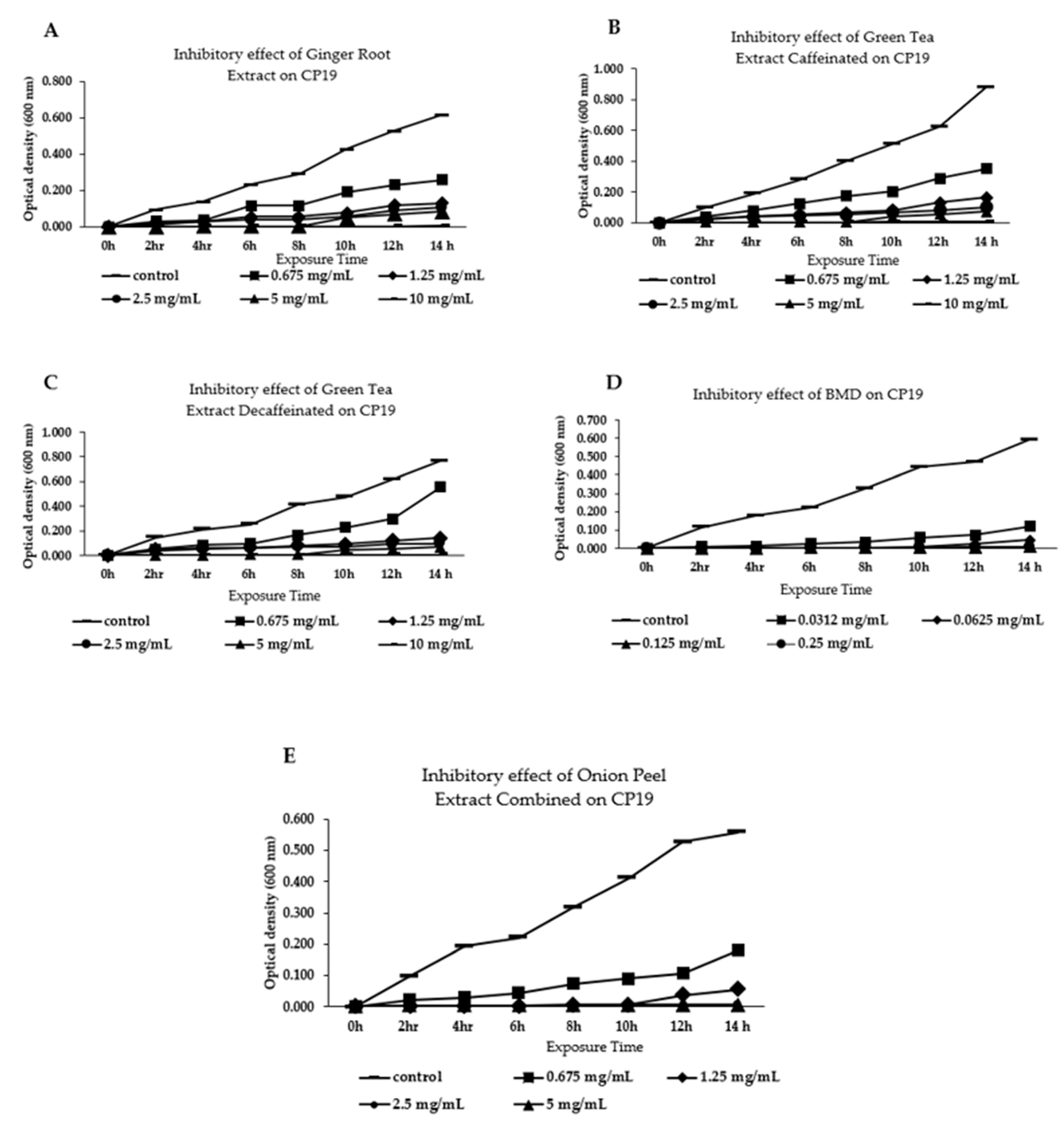
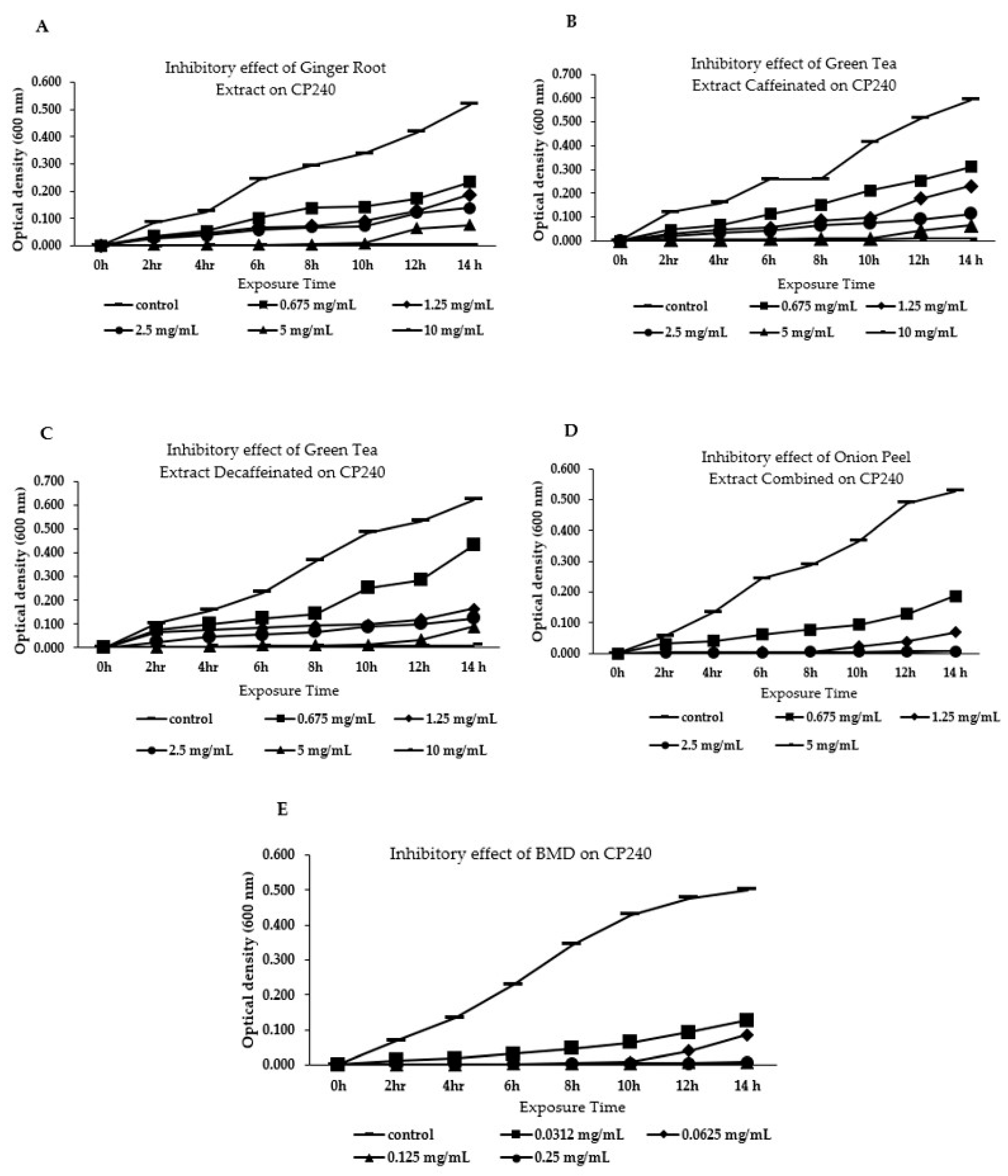
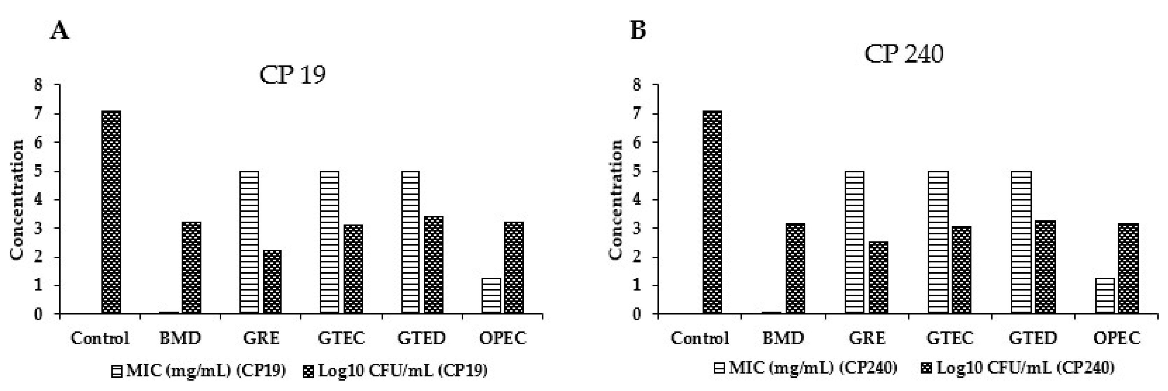
3.1. Effect of Different Phytogenic Extracts on Bacteria Growth Parameters

MIC was determined visually as the lowest concentration that inhibited bacterial growth. Results showed that the MIC-CP19 for GRE, GTEC, and GTED was 5 mg/mL, while that of OPEC was 1.25 mg/mL, and that of bacitracin methylene disalicylate (BMD) was 0.0625 mg/mL at the 8 h time point (Figure 1A–E). Correspondingly, CFU results supported our findings since the CFU/mL at the aforementioned concentrations showed an ideal reduction in the number of bacterial colonies with GRE (2.24 CFU/mL), GTEC (3.11 CFU/mL), GTED (3.39 CFU/mL), OPEC (3.22 CFU/mL), and BMD (3.21 CFU/mL). Likewise, the MIC-CP240 for GRE, GTEC, and GTED was 5 mg/mL, and BMD was 0.0625 mg/mL at the 10 h time point, while OPEC was 1.25 mg/mL at the 8 h time point (Figure 2A–E). Correspondingly, CFU results support our findings, as the CFU/mL at the aforementioned concentrations showed an ideal reduction in the number of bacteria colonies with GRE (2.53 CFU/mL), GTEC (3.04 CFU/mL), GTED (3.27 CFU/mL), OPEC (2.87 CFU/mL), and BMD (2.29 CFU/mL) (Figure 3A,B).

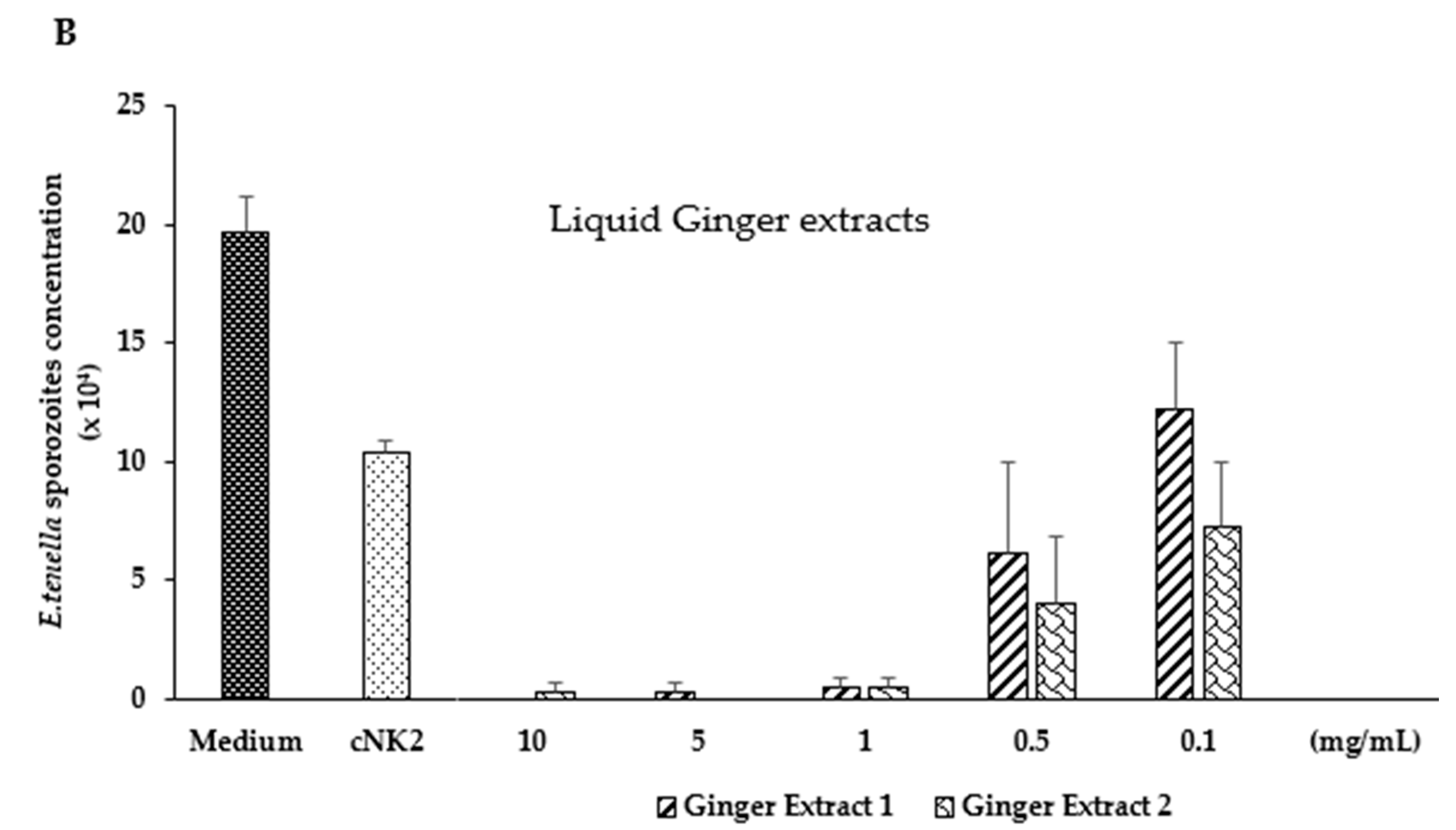
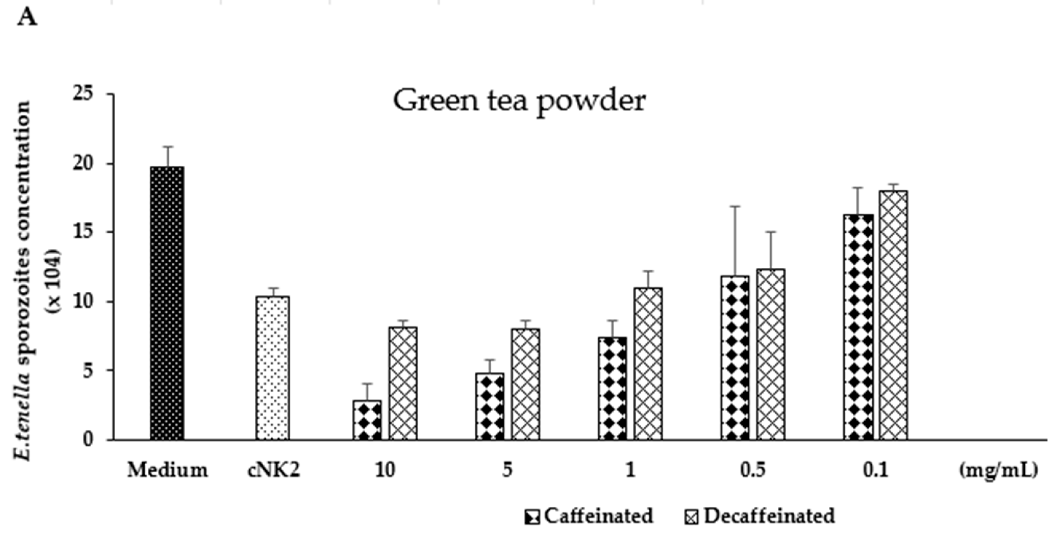
3.2. In Vitro Evaluation of Green Tea and Ginger Extracts on Killing Action on E. tenella Sporozoites

Phytogenic extracts such as green tea (GTEC and GTED) and ginger reduced the concentration of viable E. tenella sporozoites in vitro. GTEC at 5 to 10 mg/mL decreased the concentration of viable sporozoites compared to cNK2, while at the same dosage, the killing action was similar to cNK2 in GTED (Figure 4A). In addition, GRE reduced the concentration of viable E. tenella sporozoites at a dosage of 1 to 10 μg/mL, compared to 100 µg/mL of cNK-2 synthetic peptides used as a positive control (Figure 4B).

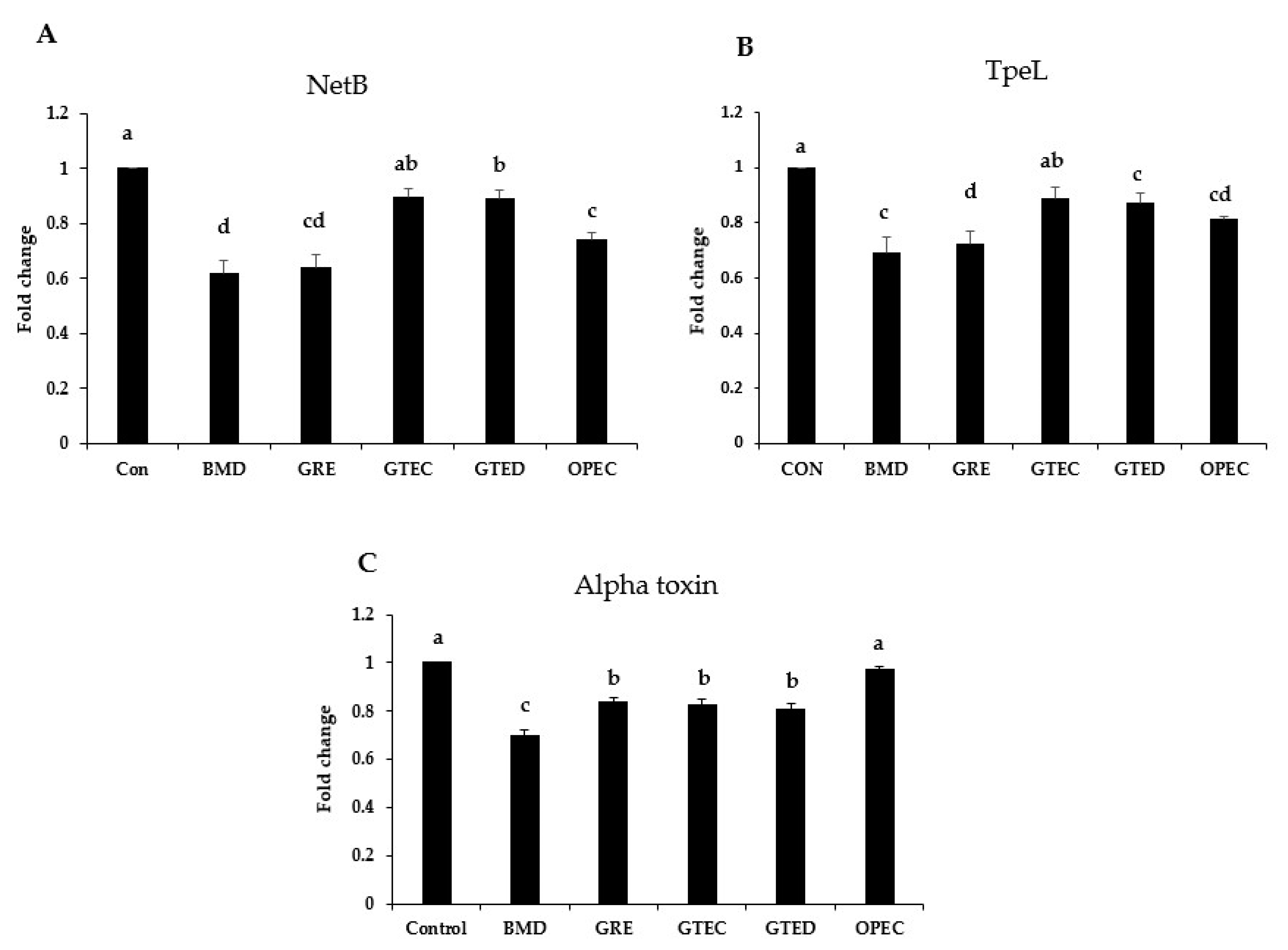
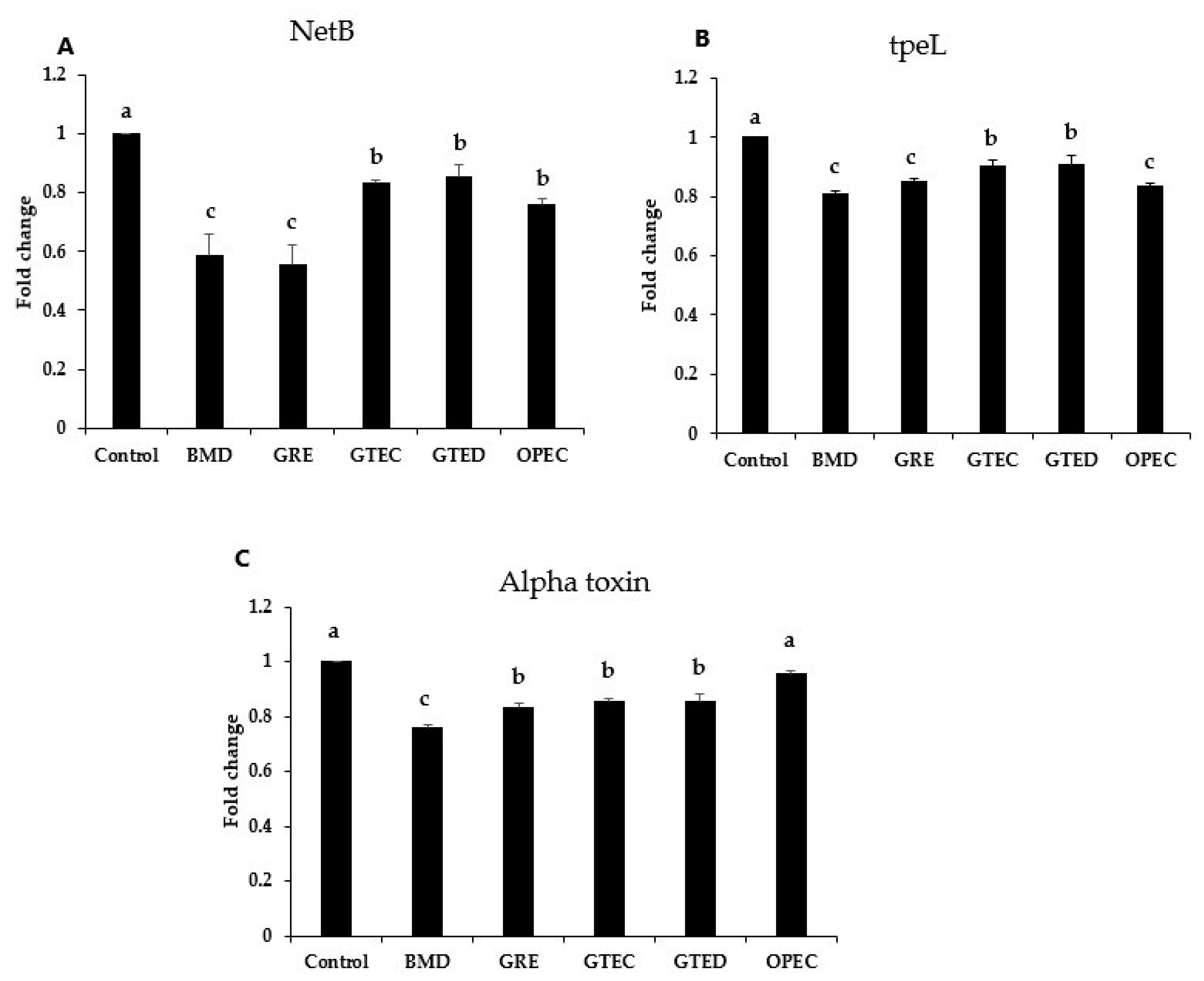
3.3. Effect of Phytogenic Extracts on C. perfringens Toxin Gene Expression

To understand the effect of phytogenic extracts on C. perfringens toxins, we evaluated the expression of C. perfringens (CP19 and CP240 strains) toxin encoding genes such as tpeL, Alpha toxin, and NetB. For the CP19 Alpha toxin gene, phytogenic extracts GRE, GTEC, and GTED reduced (p < 0.05) the level of expression of Alpha toxin gene compared to control and OPEC; however, BMD reduced Alpha toxin gene expression compared to GRE, GTEC, and GTED. Furthermore, NetB toxin gene expression was reduced significantly (p < 0.05) upon treatment with GRE, GTED, and OPEC, as well as BMD, compared to the control. However, NetB gene expression for GTEC was similar to the control. In addition, BMD, GRE, GTED, and OPEC reduced (p < 0.05) the level of expression of tpeL compared to the control. However, the level of expression of tpeL for GTEC was similar to the control (Figure 5A–C). For the CP240 strain Alpha toxin encoding gene, all the selected phytogenic extracts reduced (p < 0.05) the expression of NetB gene, with BMD having the lowest level of expression. Similarly, all the selected phytogenic extracts reduced (p < 0.05) the expression of the NetB gene. However, only GRE was comparable to BMD in its potency to reduce the expression of NetB gene. Additionally, tpeL expression was reduced (p < 0.05) by the selected phytogenic extracts, with GRE and OPEC showing similar values to BMD (Figure 6A–C).

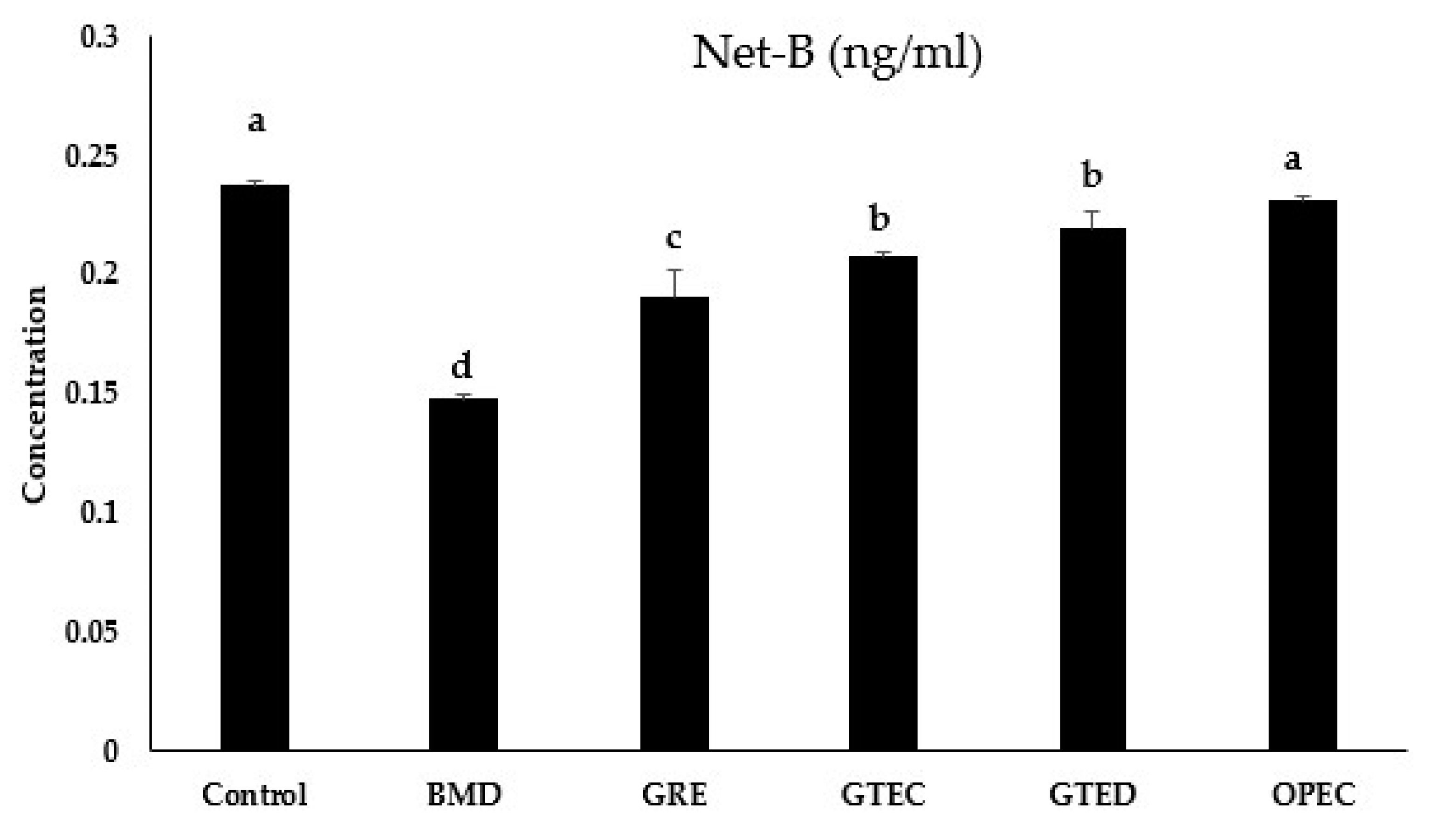
3.4. Effect of Selected Phytogenic Extracts on Protein Concentration of NetB Toxin

The concentration of NetB toxin was significantly reduced (p < 0.05) by GRE, GTEC, and GTE; however, the value for OPEC was similar to the control. Furthermore, BMD had the lowest concentration for NetB toxin (Figure 7).

4. Discussion

With the increasing governmental regulation on the use of in-feed antibiotics in many commercial agricultural food production, various antibiotic alternative strategies have been developed and are being used in animal agriculture [15]. Among novel antibiotic strategies, phytochemicals have shown many benefits to promote growth, enhance immunity, and reduce oxidative stress in animal production and in disease management in recent years [16,25]. Studies show that phytogenic extracts supplemented into the diet of broiler chicken not only improved digestibility and absorption [26,27] but also enhanced immunity, reduced oxidative stress caused by pathogens, modulated gut microbiota, and improved animal health [16,28].
This study investigated selected phytogenic extracts such as GRE, GTEC, GTED, and OPEC for their ability to prevent bacteria proliferation, kill protozoa (E. tenella), and mitigate the effect of disease-causing toxins associated with C. perfringens in vitro. Our MIC and CFU results showed that selected phytogenic extracts GRE, GTEC, GTED, and OPEC mitigated the growth of C. perfringens but at a higher concentration compared to BMD. The inhibitory action of phytogenic extracts including GRE, GTEC, and GTED on bacteria growth has been associated with their inherent antimicrobial properties [29,30]. Therefore, our results agree with the findings of [31] which state that phytogenic product-based extracts altered the abundance of microorganisms in caecum cultures. In a similar manner, BMD increased the growth performance of broiler chickens and mitigated bacteria growth by increasing the diversity or richness of the intestinal microbiota [32]. This suggests that selected phytogenic extracts used in this study and BMD may be preventing the proliferation of pathogenic bacteria while promoting the growth of beneficial bacteria [33]. Phytogenic extracts containing catechins have been shown to bind to the bacterial lipid bilayer cell membrane, causing damage to it [34]. Likewise, the exposure of bacteria to flavonoids like quercetin and kaempferol found on onion skin degrades or inhibits bacteria growth [35].
In addition, in vitro evaluation of green tea and ginger extracts in E. tenella sporozoites killing assay showed that GRE, GTEC, and GTED exert antimicrobial properties against coccidia. In this study, we used cNK-2, the core α-helical region of cNK-lysin, as positive control, since it has been established that cNK2 notably exhibits a high antimicrobial activity and can kill Eimeria sporozoites through the disruption of their membrane [36]. Our results show that 5 to 10 mg/mL of GTEC decreased the concentration of viable sporozoites compared to 100 µg/mL of cNK2. However, GTED had similar killing action at the same dosage compared to cNK2. The differences in the killing action of GTEC and GTED may be attributed to caffeine’s ability to inhibit a wide variety of pathogenic microorganisms by passing through the cell wall of bacteria, where it inhibits DNA synthesis [37,38]. Similarly, GRE reduced the concentration of sporozoites at a dosage of 1 to 10 mg/mL, compared to 100 µg/mL of cNK2. This aligns with the findings of a previous study that demonstrated the therapeutic properties of green tea to include anticoccidial properties [22]. Moreover, Majid et al. [39] reported that ginger containing phenols and other compounds derived, such as gingerdiol, shogaols, gingerol, and gingerdione, may be responsible for the coccidiostatic effect of ginger when fed to broiler chickens. Overall, these results show that these selected phytogenic extracts have antimicrobial property.
To further understand the effect of these selected phytogenic extracts on toxin encoding genes associated with C. perfringens, we analyzed the effect of these extracts on the expression of NetB, tpeL and Alpha toxin genes, as well as NetB production, by C. perfringens bacteria in vitro. Our qRT-PCR and ELISA data demonstrated that these selected phytogenic extracts decreased the expression of these genes in the CP19 and CP240 strains as well as the protein concentration of the NetB toxin. However, OPEC and GTEC did not have any effects on the expression of Alpha toxin and tpeL genes for the CP19 strain, but to a larger extent, BMD had a superior impact in decreasing the level of expression of these genes. Moreover, studies have shown that plant extracts containing bioactive ingredients such as terpenoids, phenolics, glycosides, and alkaloids can replace the use of antibiotics in the treatment of NE [40]. Likewise, studies have shown that garlic, a phytogenic extract containing similar bioactive compounds as in GRE, OPEC, and GTE, has been reported to ameliorate the negative effects of C. perfringens challenge in broiler chickens [41,42]. Overall, these findings show that GRE, GTEC, GTED, and OPEC reduce the concentration of E. tenella sporozoites as well as the virulence of other NE-causing clostridia strains such as CP19 and CP240. The beneficial effects of phytochemicals can be attributed to their antimicrobial and antioxidant properties. Detailed mechanisms of beneficial phytochemicals and their effects on the intestinal microbiota to improve performance will be another important beneficial effect of dietary phytochemicals [16]. At the optimal concentration using selected phytochemicals, one can inhibit parasite reproduction and toxin generation in NE and reduce coccidiosis-caused gut damages, as well as reduce toxin production by C. perfringens, which causes necrotic enteritis. Effective disease mitigation strategies using various antibiotic alternatives such as phytochemicals would reduce environmental contamination of antibiotics, with eventual reduction of AMR risk to humans.

5. Conclusions

This study provided insights into the beneficial effects of using selected phytochemicals as feed additives that show the inhibitory effects of NE-causing organisms and coccidiosis-causing E. tenella parasites. In conclusion, our studies showed that phytogenic extracts that include GRE, GTEC, GTED, and OPEC reduced C. perfringens proliferation. OPEC, GRE, GTED, and GTEC were the ranked order in reducing the proliferation of C. perfringens. The optimal dose for GRE, GTEC, and GTD was 5 mg/mL, with OPEC at 1.25 mg/mL for CP19 and CP240. Our study showed that the efficacy of GRE, GTEC, GTD, and OPEC was comparable to BMD, albeit at a higher concentration. Based on their anti-Clostridial and anti-coccidial effects, the results of these studies clearly support using well-studied, selected phytochemicals in mitigating disease responses for agricultural animals such as poultry. With the risk associated with the use of antibiotics in the prevention and treatment of NE, the phytogenic extracts that we described in this paper have the potential to serve as alternatives to antibiotic uses in broiler chicken production. However, further studies are required to understand the mechanisms by which these phytogenic extracts mitigate the impact of these disease-causing toxins and gut-damaging parasites.

Author Contributions

Authors contributed equally to this work. Study concept and design: Y.O.F., M.M.K., H.S.L., Y.L. and H.W.K.; acquisition of laboratory data: M.M.K. and Y.L.; data analysis and interpretation: M.M.K., Y.L., Y.O.F., H.S.L. and A.O.A.; drafting of the manuscript and/or critical revision for important intellectual content: A.O.A., P.C.O. and Y.O.F. All authors have read and agreed to the published version of the manuscript.

Funding

This research was financially supported by the USDA/NIFA SAS award 2020-69012-31823; accession number 12023193, and partly by the National Institute for Food and Agriculture of the United States Department of Agriculture, Project No. NC.X-329-5-20-120-1, in the Agricultural Research Program, North Carolina Agricultural and Technical State University.

Institutional Review Board Statement

Not applicable.

Informed Consent Statement

Not applicable.

Data Availability Statement

The raw data supporting the conclusions of this article will be made available by the authors on request.

Acknowledgments

The authors thank members of the Poultry Ethnomedicine Research Laboratory at North Carolina A&T State University (Greensboro, NC) for their technical support. We also thank the reviewers for the time and effort that they invested in the review of our manuscript.

Conflicts of Interest

The authors declare no conflicts of interest.

References

  1. Hassan, K.A.; Elbourne, L.D.; Tetu, S.G.; Melville, S.B.; Rood, J.I.; Paulsen, I.T. Genomic analyses of Clostridium perfringens isolates from five toxinotypes. Microbiol. Res. 2015, 166, 255–263. [Google Scholar] [CrossRef]
  2. Uzal, F.A.; Freedman, J.C.; Shrestha, A.; Theoret, J.R.; Garcia, J.; Awad, M.M.; Adams, V.; Moore, R.J.; Rood, J.I.; McClane, B.A. Towards an understanding of the role of Clostridium perfringens toxins in human and animal disease. Future Microbiol. 2014, 9, 361–377. [Google Scholar] [CrossRef] [PubMed]
  3. Keyburn, A.L.; Boyce, J.D.; Vaz, P.; Bannam, T.L.; Ford, M.E.; Parker, D.; Di Rubbo, A.; Rood, J.I.; Moore, R.J. NetB, a new toxin that is associated with avian necrotic enteritis caused by Clostridium perfringens. PLoS Pathog. 2008, 4, e26. [Google Scholar] [CrossRef]
  4. Lee, K.W.; Lillehoj, H.S. Role of Clostridium perfringens necrotic enteritis B-like toxin in disease pathogenesis. Vaccines 2022, 10, 61. [Google Scholar] [CrossRef]
  5. Yang, W.Y.; Chou, C.H.; Wang, C. Characterization of toxin genes and quantitative analysis of netB in necrotic enteritis (NE)-producing and non-NE-producing Clostridium perfringens isolated from chickens. Anaerobe 2018, 54, 115–120. [Google Scholar] [CrossRef]
  6. Duff, A.F.; Vuong, C.N.; Searer, K.L.; Briggs, W.N.; Wilson, K.M.; Hargis, B.M.; Bergham, L.R.; Bielke, L.R. Preliminary studies on development of a novel subunit vaccine targeting Clostridium perfringens mucolytic enzymes for the control of necrotic enteritis in broilers. Poult. Sci. 2019, 98, 6319–6325. [Google Scholar] [CrossRef] [PubMed]
  7. Cooper, K.K.; Songer, J.G. Virulence of Clostridium perfringens in an experimental model of poultry necrotic enteritis. Vet. Microbiol. 2010, 142, 323–328. [Google Scholar] [CrossRef]
  8. Johnson, J.; Reid, W.M. Anticoccidial drugs: Lesion scoring techniques in battery and floor-pen experiments with chickens. Exp. Parasitol. 1970, 28, 30–36. [Google Scholar] [CrossRef] [PubMed]
  9. Moore, R.J. Necrotic enteritis predisposing factors in broiler chickens. Avian Pathol. 2016, 45, 275–281. [Google Scholar] [CrossRef]
  10. Abudabos, A.M.; Alyemni, A.H.; Swilam, E.O.; Al-Ghadi, M. Comparative anticoccidial effect of some natural products against Eimeria spp. infection on performance traits, intestinal lesion and occyte number in broiler. Pak. J. Zool. 2017, 49, 1989–1995. [Google Scholar] [CrossRef]
  11. Blue, C.E.C.; Jababu, Y.; Ibrahim, S.A.; Minor, R.C.; Williams, L.L.; Adetunji, A.O.; Ali, R.; Young, L.S.; Fasina, Y.O. Spray-Dried Plasma Promotes Broiler Chick Growth by Enhancing Immune Surveillance. Animal 2023, 13, 1436. [Google Scholar] [CrossRef] [PubMed]
  12. Emami, N.K.; Dalloul, R.A. Centennial Review: Recent developments in host-pathogen interactions during necrotic enteritis in poultry. Poult. Sci. 2021, 100, 101330. [Google Scholar] [CrossRef] [PubMed]
  13. Adetunji, A.; Casey, T.; Franco, J.; Shah, D.; Fasina, Y. Proteomic Analysis of the Effect of Salmonella Challenge on Broiler Chicken. Molecules 2022, 27, 7277. [Google Scholar] [CrossRef] [PubMed]
  14. Babaei, S.; Rahimi, S.; Torshizi, M.A.K.; Tahmasebi, G.; Khaleghi Miran, S.N. Effects of propolis, royal jelly, honey and bee pollen on growth performance and immune system of Japanese quails. Vet. Res Vet. Q. 2016, 7, 13–20. [Google Scholar]
  15. Wickramasuriya, S.S.; Park, I.; Lee, K.; Lee, Y.; Kim, W.H.; Nam, H.; Lillehoj, H.S. Role of physiology, immunity, microbiota, and infectious diseases in the gut health of poultry. Vaccines 2022, 10, 172. [Google Scholar] [CrossRef] [PubMed]
  16. Lillehoj, H.; Liu, Y.; Calsamiglia, S.; Fernandez-Miyakawa, M.E.; Chi, F.; Cravens, R.L.; Oh, S.; Gay, C.G. Phytochemicals as antibiotic alternatives to promote growth and enhance host health. Vet. Res. 2018, 49, 76. [Google Scholar] [CrossRef] [PubMed]
  17. Dosu, G.; Obanla, T.; Zhang, S.; Sang, S.; Adetunji, A.O.; Fahrenholz, A.O.; Ferket, P.R.; Nagabhushanam, K.; Fasina, Y.O. Supplementation of ginger root extract into broiler chicken diet: Effects on growth performance and immunocompetence. Poult. Sci. 2023, 102, 102897. [Google Scholar] [CrossRef]
  18. Chandravanshi, A.; Naik, M.G.; Chandravanshi, P.; Rathore, S.S. Camellia sinensis (Green tea) as feed additive enhanced immune response and disease resistance of Cyprinus carpio (common carp) against Aeromonas hydrophila infection. J. Exp. Zool. 2020, 23, 1383–1390. [Google Scholar]
  19. Khaki, A.; Fathiazad, F.; Nouri, M.; Khaki, A.A.; Khamenehi, H.J.; Hamadeh, M. Evaluation of androgenic activity of Allium cepa on spermatogenesis in the rat. Folia Morphol. 2009, 68, 45–51. [Google Scholar]
  20. Khaki, A.; Farnam, A.; Badie, A.D.; Nikniaz, H. Treatment effects of onion (Allium cepa) and ginger (Zingiber officinale) on sexual behavior of rat after inducing an antiepileptic drug (lamotrigine). Balkan Med. J. 2012, 29, 236–246. [Google Scholar] [CrossRef]
  21. Settle, T.; Leonard, S.S.; Falkenstein, E.; Fix, N.; Van Dyke, K.; Klandorf, H. Effects of a Phytogenic Feed Additive Versus an Antibiotic Feed Additive on Oxidative Stress in Broiler Chicks and a Possible Mechanism Determined by Electron Spin Resonance. Int. J. Poult. Sci. 2014, 13, 62–69. [Google Scholar] [CrossRef] [PubMed]
  22. Jang, S.I.; Jun, M.H.; Lillehoj, H.S.; Dalloul, R.A.; Kong, I.K.; Kim, S.; Min, W. Anticoccidial effect of green tea-based diets against Eimeria maxima. Vet. Parasitol. 2007, 144, 172–175. [Google Scholar] [CrossRef] [PubMed]
  23. Lee, K.W.; Kim, W.H.; Li, C.; Lillehoj, H.S. Detection of necrotic enteritis B–like toxin secreted by Clostridium perfringens using capture enzyme-linked immunosorbent assay. Avian Dis. 2020, 64, 490–495. [Google Scholar] [CrossRef] [PubMed]
  24. Hong, Y.H.; Lillehoj, H.S.; Siragusa, G.R.; Bannerman, D.D.; Lillehoj, E.P. Antimicrobial activity of chicken NK-lysin against. Eimeria sporozoites. Avian Dis. 2008, 52, 302–305. [Google Scholar] [CrossRef]
  25. Anand, U.; Jacobo-Herrera, N.; Altemimi, A.; Lakhssassi, N.A. Comprehensive Review on Medicinal Plants as Antimicrobial Therapeutics: Potential Avenues of Biocompatible Drug Discovery. Metabolites 2019, 9, 258. [Google Scholar] [CrossRef]
  26. Afsharmanesh, M.; Sadaghi, B. Effects of dietary alternatives (probiotic, green tea powder, and Kombucha tea) as antimicrobial growth promoters on growth, ileal nutrient digestibility, blood parameters, and immune response of broiler chickens. Comp. Clin. Pathol. 2014, 23, 717–724. [Google Scholar] [CrossRef]
  27. Al-Khalaifah, H.; Al-Nasser, A.; Al-Surrayai, T.; Sultan, H.; Al-Attal, D.; Al-Kandari, R.; Al-Saleem, H.; Al-Holi, A.; Dashti, F. Effect of Ginger Powder on Production Performance, Antioxidant Status, Hematological Parameters, Digestibility, and Plasma Cholesterol Content in Broiler Chickens. Animal 2022, 12, 901. [Google Scholar] [CrossRef] [PubMed]
  28. Pérez-Burillo, S.; Hinojosa-Nogueira, D.; Pastoriza, S.; Rufián-Henares, J.A. Plant extracts as natural modulators of gut microbiota community structure and functionality. Heliyon 2020, 6, e05474. [Google Scholar] [CrossRef]
  29. Reygaert, W.C. The antimicrobial possibilities of green tea. Front. Microbiol. 2014, 5, 434. [Google Scholar] [CrossRef]
  30. Sagar, N.A.; Pareek, S. Antimicrobial assessment of polyphenolic extracts from onion (Allium cepa L.) skin of fifteen cultivars by sonication-assisted extraction method. Heliyon 2020, 6, e05478. [Google Scholar]
  31. Bajagai, Y.S.; Alsemgeest, J.; Moore, R.J.; Van, T.T.H.; Stanley, D. Phytogenic products, used as alternatives to antibiotic growth promoters, modify the intestinal microbiota derived from a range of production systems: An in vitro model. Appl. Microbiol. Biotechnol. 2020, 104, 10631–10640. [Google Scholar] [CrossRef]
  32. Crisol-Martinez, E.; Stanley, D.; Geier, M.S.; Hughes, R.J.; Moore, R.J. Understanding the mechanisms of zinc bacitracin and avilamycin on animal production: Linking gut microbiota and growth performance in chickens. Appl. Microbiol. Biotechnol. 2017, 101, 4547–4559. [Google Scholar] [CrossRef] [PubMed]
  33. Qorbanpour, M.; Fahim, T.; Javandel, F.; Nosrati, M.; Paz, E.; Seidavi, A.; Ragni, M.; Laudadio, V.; Tufarelli, V. Effect of Dietary Ginger (Zingiber officinale Roscoe) and Multi-Strain Probiotic on Growth and Carcass Traits, Blood Biochemistry, Immune Responses and Intestinal Microflora in Broiler Chickens. Animal 2018, 8, 117. [Google Scholar] [CrossRef] [PubMed]
  34. Sirk, T.W.; Brown, E.F.; Friedman, M.; Sum, A.K. Molecular binding of catechins to biomembranes: Relationship to biological activity. J. Agric. Food Chem. 2009, 57, 6720–6728. [Google Scholar] [CrossRef]
  35. Duan, Y.; Jin, D.H.; Kim, H.S.; Seong, J.H.; Lee, Y.G.; Kim, D.S.; Jang, S.H. Analysis of total phenol, flavonoid content and antioxidant activity of various extraction solvents extracts from onion (Allium cepa L.) peels. J. Korean Appl. Sci. Technol. 2015, 32, 418–426. [Google Scholar] [CrossRef]
  36. Kim, W.; Lillehoj, H.; Min, W. Evaluation of the Immunomodulatory Activity of the Chicken NK-Lysin-Derived Peptide cNK-2. Sci. Rep. 2017, 7, 45099. [Google Scholar] [CrossRef]
  37. Chen, J.; Zhang, S.; Yang, X. Control of brown rot on nectarines by tea polyphenol combined with tea saponin. J. Crop Prot. 2013, 45, 29–35. [Google Scholar] [CrossRef]
  38. Nonthakaew, A.; Matan, N.; Aewsiri, T.; Matan, N. Caffeine in foods and its antimicrobial activity. Int. Food Res. J. 2015, 22, 9–14. [Google Scholar]
  39. Majid, A.; Naila, C.; Rifat, K.; Shabana, N.; Sina, G. Anticoccidial effect of garlic (Allium sativum) and ginger (Zingiber officinale) against experimentally induced coccidiosis in broiler chickens. J. Appl. Anim. Res. 2019, 47, 79–84. [Google Scholar]
  40. Diaz Carrasco, J.M.; Redondo, L.M.; Redondo, E.A.; Dominguez, J.E.; Chacana, A.P.; Fernandez Miyakawa, M.E. Use of Plant Extracts as an Effective Manner to Control Clostridium perfringens Induced Necrotic Enteritis in Poultry. Biomed Res. Int. 2016, 2016, 3278359. [Google Scholar] [CrossRef]
  41. Ibrahim, D.; Ismail, T.A.; Khalifa, E.; Abd El-Kader, S.A.; Mohamed, D.I.; Mohamed, D.T.; Shahin, S.E.; Abd El-Hamid, M.I. Supplementing Garlic Nanohydrogel Optimized Growth, Gastrointestinal Integrity and Economics and Ameliorated Necrotic Enteritis in Broiler Chickens Using a Clostridium perfringens Challenge Model. Animal 2021, 11, 2027. [Google Scholar] [CrossRef] [PubMed]
  42. Jimoh, A.; Ibitoye, E.; Dabai, Y.; Garba, S. In vivo antimicrobial potentials of garlic against Clostridium perfringens and its promotant effects on performance of broiler chickens. Pak. J. Biol. Sci. 2013, 16, 1978–1984. [Google Scholar] [CrossRef] [PubMed]
Figure 1. Inhibitory effect of selected plant extracts on CP19: (A) ginger root extract; (B) green tea extract caffeinated; (C) green tea extract decaffeinated; (D) BMD; (E) onion peel extract.
Applsci 14 03178 g001
Figure 2. Inhibitory effect of plant extracts on CP240: (A) ginger root extract; (B) green tea extract caffeinated; (C) green tea extract decaffeinated; (D) onion peel extract; (E) BMD.
Applsci 14 03178 g002
Figure 3. Comparison between MIC and corresponding CFU of phytogenic extracts and BMD for (A) CP19 (B) and CP240. The data are expressed as means, with n > 3 per treatment.
Applsci 14 03178 g003
Figure 4. In vitro evaluation of (A) green tea and (B) ginger for their killing action on E. tenella sporozoites. Each sample was analyzed in triplicate at different concentrations. The medium was used only as a vehicle control, and 100 µg/mL of cNK2 peptide was used as a positive control. The data are expressed as means ± SEM.
Applsci 14 03178 g004aApplsci 14 03178 g004b
Figure 5. Effect of phytogenic extracts on CP19 toxin encoding genes (A) NetB, (B) tpeL, (C) α-toxin. The data are expressed as means ± SEM, with n > 3 per treatment; a, b, ab, c, d, cd means bars not sharing a common superscript are significantly different among treatments (p < 0.05).
Applsci 14 03178 g005
Figure 6. Effect of phytogenic extracts on CP240 toxin encoding genes: (A) NetB, (B) tpeL, (C) α-toxin. The data are expressed as means ± SEM, with n > 3 per treatment; a, b, c means bars not sharing a common superscript are significantly different among treatments (p < 0.05).
Applsci 14 03178 g006
Figure 7. Effect of phytogenic extract on NetB toxin encoding gene using CP19 strain. The data are expressed as means ± SEM, with n > 3 per treatment; a, b, c, d means bars not sharing a common superscript are significantly different among treatments (p < 0.05).
Applsci 14 03178 g007
Table 1. List of primers.
Gene Name Primer Sequence
Necrotic enteritis toxin B (NetB)Forward5′-AGTGTAATTAGTACAAGCC-3′
Necrotic enteritis toxin B (NetB)Reverse5′-GGCCATTTCATTTTTCCGTAA-3′
Alpha toxin (α/cpa)Forward5′-AGTCTACGCTTGGGATGGAA-3′
Alpha toxin (α/cpa)Reverse5′-TTTCCTGGGTTGTCCATTTC-3′
16SrRNAForward5′-CCTTACCTACACTTGACATCCC-3′
16SrRNAReverse5′-GGACTTAACCCAACATCTCACG-3′
tpeLForward5′-GCCTGGATTTGCCTGTAGGA-3′
tpeLReverse5′-GCCAGACATTATGCCACTCC-3′
Disclaimer/Publisher’s Note: The statements, opinions and data contained in all publications are solely those of the individual author(s) and contributor(s) and not of MDPI and/or the editor(s). MDPI and/or the editor(s) disclaim responsibility for any injury to people or property resulting from any ideas, methods, instructions or products referred to in the content.

