Article

1-Deoxynojirimycin Attenuates High-Glucose-Induced Oxidative DNA Damage via Activating NRF2/OGG1 Signaling

1
The Center for Ion Beam Bioengineering & Green Agriculture, Hefei Institutes of Physical Science, Chinese Academy of Sciences, Hefei 230031, China
2
Science Island Branch of the Graduate School, University of Science and Technology of China, Hefei 230031, China
3
Zhongke Taihe Experimental Station, Fuyang 236626, China
*
Author to whom correspondence should be addressed.
Appl. Sci. 2024, 14(8), 3186; https://doi.org/10.3390/app14083186
Submission received: 22 February 2024 / Revised: 2 April 2024 / Accepted: 8 April 2024 / Published: 10 April 2024
(This article belongs to the Special Issue Plant-Based Compounds or Extractions for Medical Applications)

Abstract

:
1-Deoxynojirimycin (DNJ) is a type of alkaloid that mainly exists in mulberry fruit and leaves. DNJ inhibits α-glucosidase, reduces the absorption of sugar, and suppresses after-meal hyperglycemia. It was reported that DNJ functions in attenuating cellular oxidative stress. However, the mechanisms remain largely unknown. In this study, we firstly confirmed that 5 µmol/L DNJ treatment mitigated the oxidative DNA damage and cell senescence in human umbilical vein endothelial cells (HUVEC) cultured in medium containing 50 mmol/L glucose. Next, we found that DNJ treatment stimulates the expression of anti-oxidative response regulator, Nuclear factor (erythroid-derived 2)-like 2 (NRF2) by around 50% in cells cultured with high glucose. In addition, 8-oxoguanine DNA glycosylase (OGG1) was upregulated by over 15% after DNJ treatment to mitigate high-glucose-induced oxidative DNA damage, and it was identified as a downstream target of NRF2. Further, DNJ treatment promoted the phosphorylation and activation of AKT (ser473) by around 50% in cells cultured with high glucose, and AKT inhibitor treatment abrogated DNJ-induced upregulation of NRF2 and OGG1. Taken together, our results indicate that DNJ is an effective natural antioxidant in mitigating high-glucose-induced oxidative stress in HUVEC via activating the AKT-NRF2-OGG1 anti-oxidative response.

1. Introduction

Morus alba is a deciduous tree cultivated in various tropical, subtropical, and temperate countries, including China, Japan, Korea, Thailand, and others. M. alba contains abundant bioactive compounds; for example, mulberry fruit and leaves contain alkaloids, flavones, sterols, volatile oils, and polysaccharides [1]. These natural bioactive compounds exhibit potent physiological functions such as anti-diabetic, anti-oxidative, anti-obesity, and anti-microbial activities [2]. Mulberry is considered as a suitable ingredient of functional foods for its nutrition and health benefits, and mulberry products can widely be found in the market, including mulberry powder, dried fruit, juices, jams, desserts, and candies. In addition, mulberry fruit and leaves are incorporated into a variety of traditional medicines due to their potential of improving diseases [3]. In ancient China, diabetes was called XiaoKe syndrome, with comprehensive symptoms such as polyuria, polydipsia, overeating, weight loss, and fatigue. According to the Compendium of Materia Medica, drinking decocted mulberry leaves to treat diabetes had a long history in ancient China. Recent research also indicates that mulberry fruit and leaves are beneficial for blood sugar levels [4,5]. Among the bioactive compounds synthesized in mulberry, 1-Deoxynojirimycin (DNJ), a natural alkaloid azasugar and iminopyranose, has been reported as an antihyperglycemic agent [6,7,8]. Compared to oligosaccharides, DNJ has a higher affinity for glycosidase so it can inhibit glycosidase from combining with oligosaccharides to produce glucose, thereby achieving blood sugar lowering [9]. In addition, DNJ was reported to be an effective antioxidant. Ma et al. found that in patients with stable angina pectoris, 4-week treatment with DNJ significantly increased the serum superoxide dismutase (SOD) level, which the authors thought was associated with the improvement of stable angina pectoris symptoms [10]. Wang et al. reported that mulberry leaf DNJ treatment improved the antioxidant and anti-inflammatory properties and serum lipid profile in heart disease patients. DNJ reduces carotid intima-media thickness, and the serum levels of DNJ showed a strong relationship with intima-media thickness values [11]. Gao et al. found in their mulberry juice fermentation study that with the increase in DNJ and quercetin contents, the ABTS+ free-radical-scavenging activity of mulberry beverages was notably enhanced [12]. These previous studies indicated the antioxidant potential of DNJ.
In addition to being a cause of diabetes, high blood glucose also can lead to other health problems, such as cell aging and oxidative injuries [13,14,15]. Zhang et al. reported that mulberry leaf extract, which contains DNJ, flavonoid, and polysaccharide, enhanced insulin sensitivity and alleviated the oxidative damage in a type 2 diabetes rat model by increasing the activities of anti-oxidative enzymes such as SOD, catalase, and glutathione peroxidase [16]. He et al. found that in a type 2 diabetes mouse model, DNJ treatment significantly lowered the serum level of glucose and increased the serum SOD activity [17]. These studies proposed the multi-faceted roles of DNJ in its improvement of diabetes. In response to oxidative stress, anti-oxidative signaling pathways will be stimulated to protect cells. Nuclear factor (erythroid-derived 2)-like 2 (NRF2) is a master regulator of anti-oxidative responses in mammalian cells and regulates the expression of series of proteins which function to maintain cellular oxidative status equilibrium. Under normal conditions, NRF2 is sequestered in the cytoplasm and degraded via the ubiquitin-mediated pathway. When cellular oxidative stress increases, NRF2 translocates from the cytoplasm into the nucleus where it transcriptionally activates its target genes. Most NRF2 targets reported to date are proteins involved in decreasing the cellular oxidative level or anti-inflammation, such as NAD(P)H quinone oxidoreductase 1 (NQO1) and heme oxygenase-1 (HO-1) [18,19,20,21,22]. Proteins involved in other physiological processes such as DNA damage repair are also targets of NRF2, for example, 53BP1 and 8-oxoguanine DNA glycosylase (OGG1) [23,24]. In a recently published animal study, Wang et al. reported that a diet supplemented with DNJ extract from mulberry leaves increased the anti-oxidative index in the plasma and intestinal tissues of hens. Their cell studies, using hen intestine epithelial cells, indicated that DNJ increased the level of NRF2 mRNA, and enhanced the expression of reactive species scavenging enzymes including SOD and catalase [25]. Yu et al. observed that mulberry extract as a food supplement upregulated NRF2 protein in the liver tissue of inflammation model rats challenged with lipopolysaccharide [26]. These studies indicated that in addition to inhibiting glycosidase, DNJ can activate NRF2-mediated anti-oxidative responses.
It is well-known that high concentrations of glucose induce the production of reactive oxygen species (ROS), which further lead to cellular disorders and cell death. Given the facts that DNJ treatment potentiates NRF2 anti-oxidative signaling [25,26], in this study, we investigated whether the NRF2 antioxidative response was stimulated by DNJ treatment in high-glucose-challenged cells. Our results showed that DNJ could ameliorate high-glucose-induced oxidative DNA damage and senescence through activating NRF2. We further identified that DNJ activated AKT to stimulate NRF2 anti-oxidative signaling and led to the upregulation of OGG1, which facilitated the repair of oxidative DNA damage in high-glucose-challenged cells. This study provided new insight into the mechanism of DNJ in mitigating cellular damage caused by a high concentration of glucose.

2. Materials and Methods

2.1. Chemicals and Materials

DNJ was purchased from Shanghai Acmec Biochemical Technology Co., Ltd. (Shanghai, China). The purity of DNJ was shown to be 98%. A CCK-8 cell viability kit was purchased from GlpBio (Montclair, CA, USA). 9-Fluorenylmethyl chloroformate (FMOC-Cl) was purchased from Merck (Darmstadt, Germany). NRF2 inhibitor ML385 was purchased from MedChemExpress (Shanghai, China)

2.2. Cell Line and Culture Condition

Human Umbilical Vein Endothelial Cells (HUVEC) were purchased from MBC (Shanghai, China) and cultured in RPMI-1640 medium (Solarbio, Beijing, China) supplemented with endothelial cell growth supplement with heparin, and 10% FBS at 37 °C and 5% CO2 in a humidified incubator. In this study, HUVEC were seeded as the following four groups: 11 mmol/L glucose, 11 mmol/L glucose + 5 μmol/L DNJ, 50 mmol/L glucose, 50 mmol/L glucose + 5 μmol/L DNJ.

2.3. High-Performance Liquid Chromatography (HPLC) Analysis of DNJ

Mulberry extract was purchased from Anhui Xiantian Biological Science and Technology Co., Ltd. (Fuyang, China) and stored at −20 °C. Mulberry extract was frozen overnight at −80 °C and dried using a freeze-drying machine. Then, DNJ (for preparing the standard curve) and the dried mulberry extract were dissolved in distilled water. The volume of 140 µL of sample solution was mixed with 169 µL of borate buffer (pH8.5) and 250 µL of 5 mmol/L FMOC-Cl (dissolved in 50% acetonitrile). This mixture was incubated at 25 °C for 20 min. Subsequently, 25 µL of 0.1 mmol/L glycine solution was added to stop the reaction, and then 66 µL of 1% acetic acid solution was added. Finally, 57 µL of distilled water was added. After thorough mixing, the solution was centrifuged at 10,000× g for 3 min at 10 °C.
The FMOC-Cl-derived DNJ or DNJ in mulberry extract were detected using HPLC equipped with a reversed-phase Waters Acquity C18 column (Shanghai, China). The mobile phases used were as follows: acetonitrile (A) and 0.1% acetic acid (B). The C18 column was pre-equilibrated with A:B = 1:9. After sample injection, the following chromatography steps were used: Step 1, A:B = 1:9 for the first 10 min, Step 2, linear change of A:B = 1:9 to 6:4 in the next 5 min, and Step 3, A:B = 6:4 for 5 min. The chromatography was performed at 30 °C and the optical absorbance at 254 nm was recorded for quantitative analysis. Each sample was analyzed 3 times.

2.4. Cytoxicity of DNJ

The cytoxicity of DNJ was tested using the CCK-8 cell viability kit according to the manual provided by the manufacturer. HUVEC were trypsinized, counted, and seeded into a 96-well cell culture plate at the concentration of 4000 cells per well. After incubation for 24 h at 37 °C, the cell culture medium was replaced with 100 μL of fresh medium containing DNJ with a final concentration from 0 to 300 μmol/L. The cells were incubated for 48 h, and then 10 μL of CCK-8 reagent was added into each well. Four hours later, optical absorbance values at 450 nm were recorded using a microplate reader (PERLONG 9602, Beijing, China). Three replicates were set for each concentration of DNJ, and the experiment was performed 4 times.

2.5. Immunofluorescence Staining

To assess the level of cellular DNA oxidant damage, the levels of 8-OH-dG in cells were detected via immunofluorescence staining. HUVEC were grown on the round coverslips. After washing with PBS two times, cells were fixed by 4% paraformaldehyde for 30 min. After washing with PBS two times again, 0.3% Triton-X100 was used to treat the fixed cells for 20 min. Subsequently, cells were blocked by using PBST (PBS with 0.1% Triton-X100) containing 1% BSA at room temperature for 1.5 h. Then, the cells were incubated with anti-8-OH-dG antibody (1:100; sc-66036; Santa Cruz, Shanghai, China) diluted in blocking buffer at 4 °C overnight. After washing with PBST three times, Alexa-Fluor conjugated secondary antibody (Jackson ImmunoResearch Laboratories, Inc., West Grove, PA, USA) diluted in blocking buffer was added, and the samples were incubated for 2 h at room temperature in a dark box. After washing with PBST three times, DAPI was added to counterstain the cell nuclei for 10 min. Then, the samples were washed with PBS. Fluorescent images were captured under an Olympus fluorescence microscope (Tokyo, Japan). The analysis of fluorescence intensity was conducted by the use of ImageJ (version 1.43) software National Institute of Health, Bethesda, MD, USA). Three replicates were set for each sample, and the experiment was performed 3 times.

2.6. Senescence-Associated β-Galactosidase (SA-β-gal) Staining

A SA-β-gal staining kit purchased from Beyotime Biotechnology (Shanghai, China) was used to evaluate cell senescence, according to the user manual. Briefly, HUVEC were seeded on polypropylene membrane and cultured. After treatment, the cells were washed with PBS and fixed with the fixation solution provided in the kit for 15 min at room temperature. Then, the cells were washed with PBS three times, and freshly mixed working solution was added. The samples were stored at 37 °C overnight. The working solution was replaced with PBS, and the stained cells were counted under an Olympus microscope (Tokyo, Japan). Three replicates were set for each sample, and the experiment was performed 3 times.

2.7. Western Blot Assay

After culturing, HUVEC were washed with cold PBS. Then, RIPA buffer containing a protease inhibitor cocktail (Roche Diagnostics GmbH, Mannheim, Germany) was used to lyse the cells, and the whole cell lysate was harvested. Protein concentrations were quantified using the BCA kit (Sangon Biotech, Shanghai, China). After boiling denaturation, protein samples supplemented with loading buffer were separated using 10–12% SDS-PAGE and subsequently transferred to the polyvinylidene fluoride (PVDF) membrane (Roche Diagnostics GmbH, Mannheim, Germany). The PVDF membrane was blocked with TBST (Tris buffered saline with 0.05% Tween-20) containing 5% non-fat milk for 1.5 h at room temperature and then incubated with primary antibody on a 4 °C shaking table overnight. The membrane was washed with TBST three times to remove excess nonspecific binding. And then, secondary antibody (Jackson ImmunoResearch Laboratories, Inc., West Grove, PA, USA) was incubated with the PVDF membrane for 1 h at room temperature on a shaking table. After being washed with TBST three times, the PVDF membrane was reacted with ECL (Boster Biological Technology, Ltd., Wuhan, China) in a dark environment. Protein bands were detected using a chemiluminescence image analyzer (Protein Simple, Inc., Shanghai, China). NIH ImageJ software (National Institute of Health, Bethesda, MD, USA) was used for the quantitative analysis of protein bands. The antibodies used in this study were the following: NRF2 (1:1000; R1312-8; Huabio, Hangzhou, China), β-actin (1:1000; AB0033; Abways, Shanghai, China), Heme Oxygenase-1 (1:1000; 10701-1-AP; ProteinTech, Wuhan, China), OGG1 (1:1000; 15125-1-AP; ProteinTech, Wuhan, China), p-AKT (1:1000; 4060S; Cell Signal Technology, Danvers, MA, USA). The ratio of target protein to its corresponding endogenous control β-actin was used to analyze the relative expression level. Three replicates were set for each sample, and each Western blot analysis was performed 3 times.

2.8. Statistical Analysis

All experiments were independently repeated at least 3 times and all statistical data are presented as means ± standard error. The statistical significance of differences was determined by using the one-way ANOVA with Tukey test with GraphPad Prism version 8.0 (Graphpad Software, Inc., San Diego, CA, USA).

3. Results

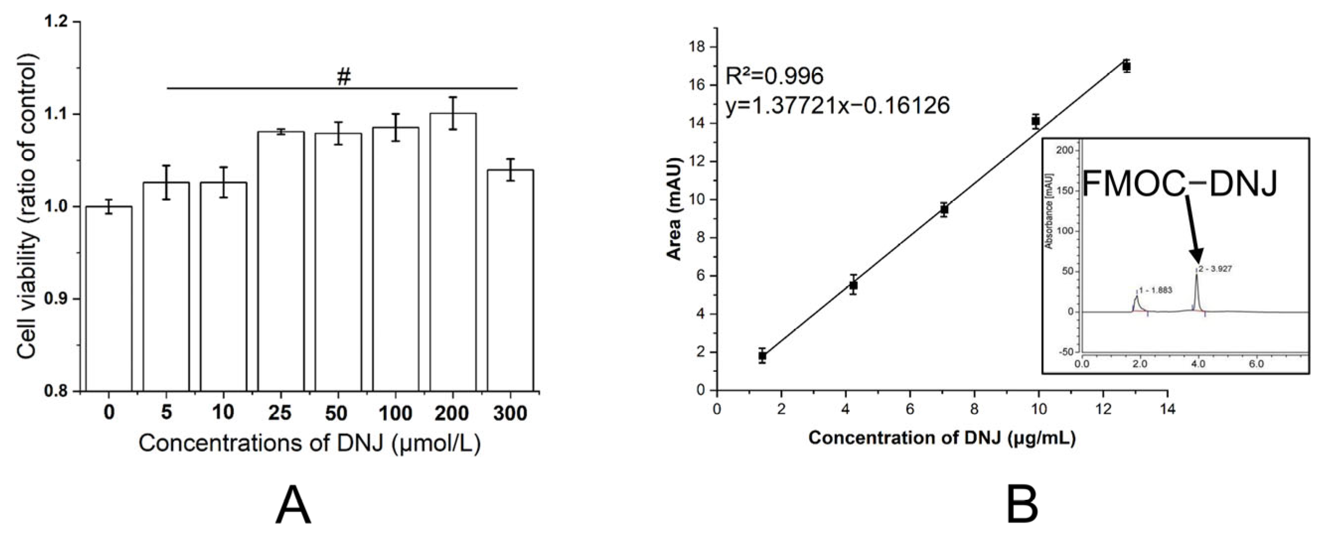
3.1. Cytoxicity of DNJ

To assess the cytotoxicity of DNJ, a CCK-8 cell viability assay was performed and the tested final concentrations of DNJ were 0, 5, 10, 25, 50, 100, 200, and 300 μmol/L. As shown in Figure 1A, the addition of DNJ slightly enhanced the viabilities of HUVEC. To determine the appropriate DNJ concentration to be used in our subsequent studies, we further detected the content of DNJ in mulberry fruit. Following extraction and derivatization using FMOC-Cl, the content of DNJ was measured using the HPLC method. According to the standard curve (Figure 1B), the concentration of DNJ in mulberry fruit was around 1.57 μg/mL, which is equal to 9.6 μmol/L. We therefore used a final concentration of 5 μmol/L DNJ in the following experiments.

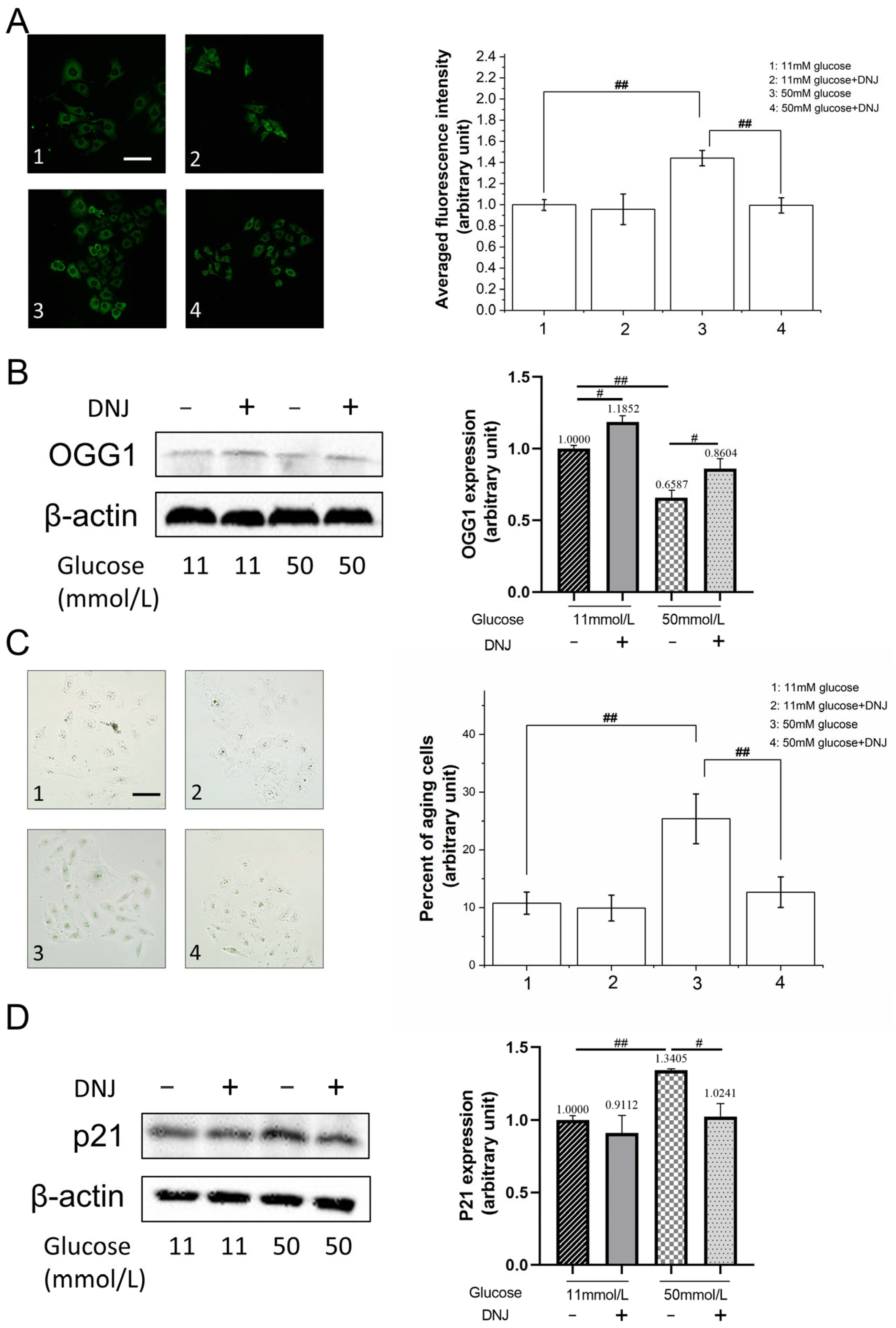
3.2. DNJ Alleviates High-Glucose-Induced Oxidative DNA Damage

High glucose leads to increased cellular oxidative stress. To investigate this, we examined whether DNJ treatment protected cells against oxidative DNA damage. The levels of 8-OHdG were used to assess DNA oxidative damage levels. HUVEC were cultured in medium containing 11 mmol/L glucose for 24 h. Then, the cells were further cultured in medium containing 11 mmol/L or 50 mmol/L glucose for 48 h. The levels of 8-OHdG were detected via immunofluorescence staining. As shown in Figure 2A, the level of 8-OHdG in the cells cultured in medium containing 50 mmol/L glucose showed an elevation of 48% as compared to that in the cells cultured in medium containing 11 mmol/L glucose, showing that high glucose resulted in exacerbated oxidative DNA damage. The addition of 5 μmol/L DNJ diminished the elevation of 8-OHdG caused by 50 mmol/L glucose, indicating that DNJ plays a role in mitigating oxidative DNA damage. OGG1 is an enzyme responsible for the repair of oxidative DNA damage. As shown in Figure 2B, its expression levels were enhanced by the addition of DNJ. In the 50 mmol/L glucose treatment group, a 30% increase in OGG1 expression was observed with the addition of DNJ. This result indicated that DNJ relieved oxidative DNA damage via enhancing the repair thereof.
Cell senescence is closely associated with both elevated DNA damage and high glucose exposure. Since DNJ treatment mitigated high-glucose-induced oxidative DNA damage, we next detected whether DNJ prevents high-glucose-induced senescence using a SA-β-gal assay. As shown in Figure 2C, the ratio of senescent HUVEC increased from around 10% in the 11 mmol/L glucose exposure group to around 25% in the 50 mmol/L glucose exposure group. Treatment with DNJ decreased 50 mmol/L glucose-induced senescence to a ratio of 12%. In addition, cell senescence marker p21 was detected via Western blotting. As shown in Figure 2D, compared with 11 mmol/L glucose-treated cells, the expression of p21 in 50 mmol/L glucose-treated cells increased by 33%, while the addition of DNJ restored the expression of p21. These results indicate that DNJ treatment mitigates high-glucose-induced oxidative DNA damage and senescence.

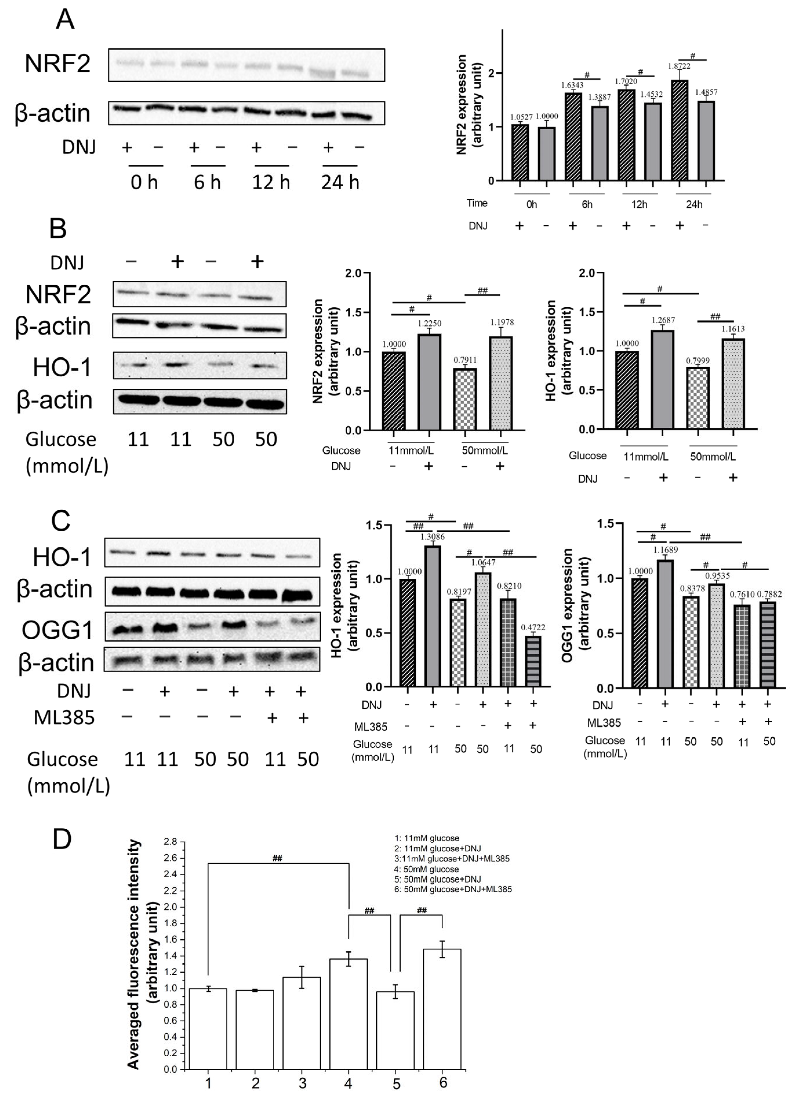
3.3. DNJ Promotes Oxidative DNA Damage Repair via Stimulating NRF2/OGG1

NRF2 is a master regulator of cellular oxidative equilibrium. As mentioned above, DNJ ameliorates high-glucose-induced oxidative damage. We therefore examined whether DNJ stimulated the NRF2 anti-oxidative response in HUVEC cultured in the medium containing 11 mmol/L glucose. As shown in Figure 3A, the induction of NRF2 expression in HUVEC was observed at different time intervals post the 5 µmol/L DNJ treatment. At 24 h post DNJ treatment, the NRF2 expression increased by around 26%. We next detected the expression of NRF2 in cells cultured in the medium containing 11 mmol/L glucose and 50 mmol/L glucose after DNJ treatment, respectively. As shown in Figure 3B, the addition of DNJ increased the expression of NRF2 by around 23% in cells cultured in the medium containing 11 mmol/L, and by around 50% in cells cultured in the medium containing 50 mmol/L glucose. Heme oxygenase 1 (HO-1) is a downstream target of NRF2 [18]. To confirm the activation of NRF2 by DNJ, the expression of HO-1 was detected. As shown in Figure 3B, the addition of DNJ enhanced the expression of HO-1 in cells cultured in media containing 11 mmol/L and 50 mmol/L glucose. These results indicate that DNJ stimulates the NRF2 anti-oxidative response.
OGG1 was reported as another downstream target of NRF2 [24]. Since we have shown that DNJ increases the expression of OGG1, we next used ML385, a specific chemical inhibitor of NRF2, to study whether DNJ treatment enhances OGG1 expression via NRF2. As shown in Figure 3C, 10 µmol/L ML385 inhibited the induced expression of HO-1 and OGG1 by DNJ. In addition, oxidative DNA damage levels under the application of 10 µmol/L ML385 were detected via immuno-staining of 8-OHdG. As shown in Figure 3D, the effect of DNJ in ameliorating 50 mmol/L glucose-induced 8-OHdG formation was blocked by the use of ML385 treatment. These results indicate that DNJ treatment decreases high-glucose-induced oxidative DNA damage through activating NRF2 and its downstream target, OGG1.

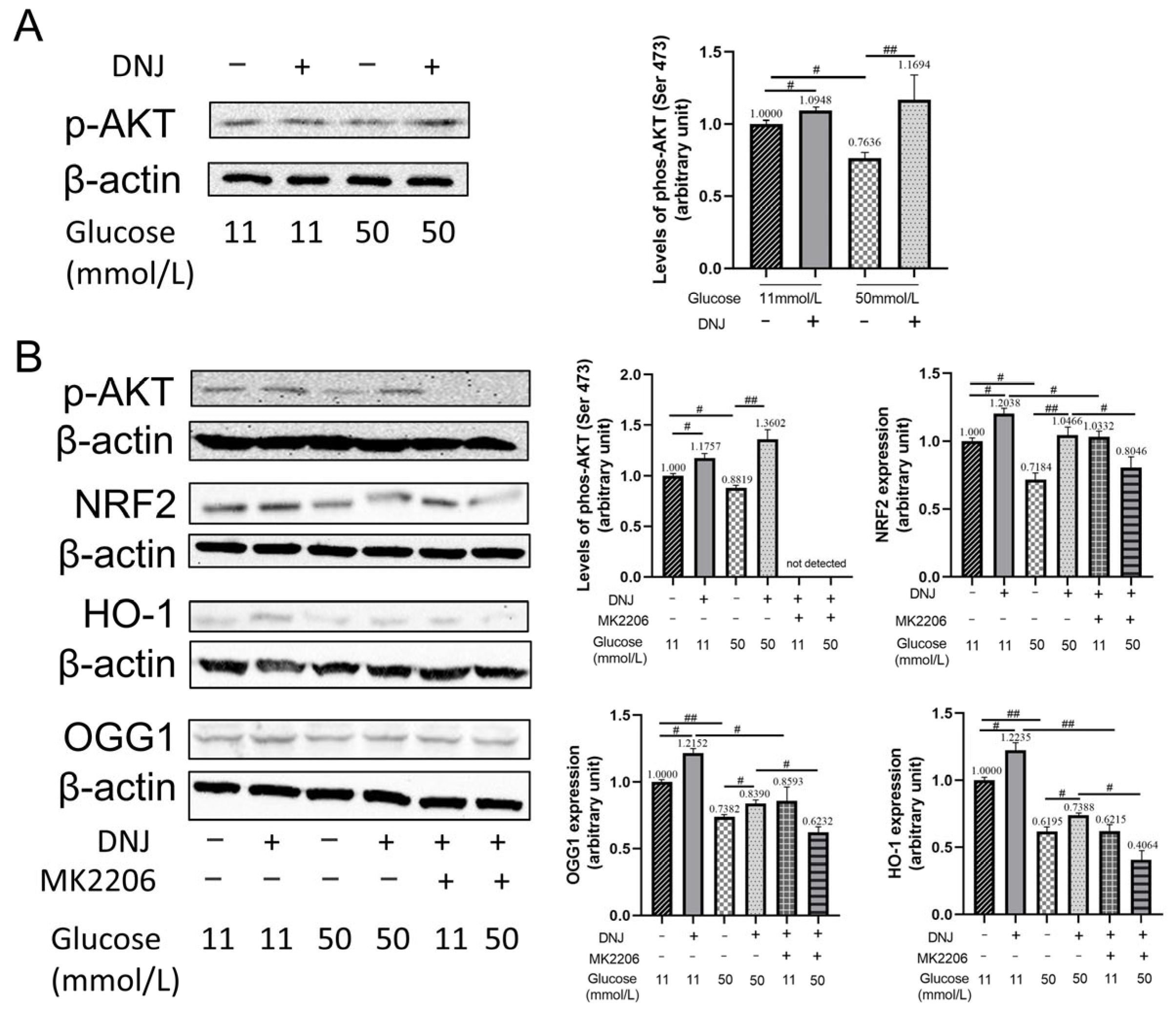
3.4. DNJ Enhances AKT Activation to Stimulate NRF2

Protein kinase AKT was reported to be activated by various natural products to protect cells against exterior insults [27]. Herein, we examined whether DNJ treatment enhances AKT activation. As shown in Figure 4A, 50 mmol/L glucose decreased the level of phosphorylated AKT (Ser 473) compared to 11 mmol/L glucose. And DNJ treatment increased the phosphorylation of AKT at Ser473 in cells cultured in media containing 11 mmol/L and 50 mmol/L glucose. These results showed that high glucose inhibited AKT activation. However, DNJ stimulated AKT activation, especially in cells cultured in 50 mmol/L glucose. Next, we checked whether AKT activation contributes to the enhancement of the NRF2 anti-oxidative response due to DNJ treatment. AKT inhibitor MK2206 (final concentration 10 µmol/L) treatment suppressed AKT phosphorylation (Figure 4B). In addition, the induced expression of NRF2 and HO-1 by DNJ was significantly inhibited by MK2206, indicating that DNJ stimulates NRF2 signaling through enhancing the activation of AKT. Furthermore, the application of MK2206 inhibited the expression of OGG1 in cells treated with DNJ. Altogether, these results show that DNJ treatment promotes the activation of AKT, and leads to the stimulation of NRF2 signaling, which upregulates the expression of OGG1 to mitigate oxidative DNA damage caused by high glucose.

4. Discussion

Hyperglycemia, which occurs during diabetes (both type 1 and type 2) and, to a lesser extent, during insulin resistance, causes oxidative stress [28]. Traditional Chinese medicine is highly favored for its natural, healthy, and environmentally friendly characteristics and has always been an important research object in antioxidant research [29]. The herbal compound, DNJ, is a representative iminopyranose produced by the mulberry tree [6,7,8]. The content of DNJ from different parts of the mulberry tree varies a lot. Rodríguez-Sánchez et al. reported that DNJ levels of the mulberry tree were 4–268 mg/100 g dried weight for leaves, 27–475 mg/100 g for branches, and 11–27 mg/100 g for its fruit [30]. And for mulberry fruit, the content of DNJ differs. Shreelakshmi et al. detected DNJ levels in mulberry fruit at different ripening stages, and found that fruit collected at the mid-ripe stage contained the highest level of DNJ, of around 2.91 mg/100 g of fruit weight [31]. Previous reports showed that DNJ also can be synthesized by microorganisms including Streptomyces sp. and Bacillus sp. Wu et al. reported the production of 296.56 mg/L DNJ in Streptomyces lavendulae [32]. Li et al. achieved a 1632.50 mg/L yield of DNJ in Bacillus amyloliquefaciens via modifying its metabolic paths [33]. The α-glucosidase activity inhibitor property makes DNJ a potential compound for the treatment of diabetes. In addition, previous studies showed that DNJ is effective in anti-inflammatory, anti-obesity, and anti-oxidative applications [2]. The results from E et al. showed that DNJ treatment slightly stimulates the viability of HUVEC at concentrations of up to 200 µmol/L. Our cell viability test showed that in the concentration range from 5 µmol/L to 200 µmol/L, DNJ treatment stimulated the viability of HUVEC, consistent with the results of Shuang et al. [15]. When the DNJ concentration was increased to 300 µmol/L, its cell viability stimulation effect was weak compared to that observed at 200 µmol/L. Wang et al. observed that 50 µg/L (306 µmol/L) and 100 µg/L DNJ treatment slightly decreased the viability of B16F10 cells, and the inhibition of matrix metalloproteinases (MMPs) by using a high concentration of DNJ was one reason [34]. This indicated that DNJ affects different physiological processes, and dose-response studies are essential for the development of DNJ in various applications. It was reported that DNJ reduces high-glucose-induced ROS generation and senescence in HUVEC [15]. Similar results were observed in our study. Treatment using 5 µmol/L DNJ effectively protected cells from excessive oxidative DNA damage and senescence in HUVEC cultured with high glucose, confirming the anti-oxidant role of DNJ. Previous animal studies indicated that DNJ treatment attenuates cellular oxidative stress by upregulating the expression of detoxification enzymes with the capability to scavenge ROS, for example, superoxide dismutase (SOD), catalase (CAT), and glutathione peroxidase (GSH-Px) [35]. Wang et al. found that DNJ treatment upregulated the mRNA levels of NRF2 in chicken intestinal epithelial cells [25]. NRF2 is an important regulator of cellular oxidative status. As well as DNJ, various natural products have been found to stimulate the NRF2 antioxidative response, such as chlorogenic acid, geniposidic acid, and glucoraphanin [36,37,38], which were thought to be of value to health. HO-1 is a well-known downstream target of NRF2. It catalyzes the degradation of heme into carbon monoxide, ferrous iron, and biliverdin, and is used as an indicator of the stimulation of NRF2 antioxidative signaling [18]. In addition to upregulating detoxification enzymes, NRF2 was found to have other physiological functions. MDM2 is an E3 ubiquitin-protein ligase that mediates the ubiquitination of p53. NRF2 positively regulates the expression of MDM2 and influences checkpoints upon extracellular stress conditions arising [39]. Yang et al. reported that NRF2 directly binds the antioxidant response elements in the promoter of TIGAR (TP53-induced glycolysis and apoptosis regulator) and upregulates its expression. The enhanced expression of TIGAR accelerates glucose metabolism toward the pentose phosphate pathway [40]. Kim et al. found that NRF2 transcriptionally regulates the expression of 53BP1, an important mediator of DNA double-strand break repair, supporting the idea that NRF2 is a candidate for tumor therapy [23]. In this study, given that DNJ treatment ameliorated high-glucose-induced 8-OHdG formation, and upregulated NRF2/HO-1 expression, we detected the connection of NRF2 with OGG1, an DNA glycosylase involved in repairing 8-oxoguanine. Our results show that DNJ treatment increases the expression of OGG1. If the transcriptional activity of NRF2 is abrogated by ML385, the enhancement of OGG1 expression is inhibited, and 8-OHdG formation is increased. This indicates that under high glucose, DNJ can protect cells by accelerating the repair of oxidized DNA through activating the NRF2/OGG1 signal. Several other natural products, such as geniposidic acid, resveratrol, and triphlorethol-A were reported to show cell protective roles by upregulating NRF2/OGG1 to alleviate oxidative stress [37,41,42]. Oxidative DNA damage leads to the accumulation of DNA mutations, and is an important reason for aging. Our results show that DNJ treatment alleviates cell senescence caused by high glucose, and supports the antioxidant function of DNJ. Shang et al. reported in their recent study that NRF2 positively regulates OGG1 expression in leukemia cells. A chromatin immunoprecipitation assay revealed NRF2 bound to the promoter of OGG1, providing evidence to confirm that OGG1 is a transcriptional target of NRF2 [43].
The expression of NRF2 can be regulated by different protein kinases, such as PKC (protein kinase C) and ERK 1/2 (extracellular signal regulated kinase 1/2) [19]. AKT is another kinase that can regulate the activation of the NRF2 antioxidative response. Fu et al. reported that rehmannioside A improved cognitive impairment after cerebral ischemia by activating PI3K/AKT/NRF2 and inhibiting ferroptosis [44]. Hou et al. reported that flavonoids from a deep-sea-derived Arthrinium sp. protected HUVEC against ox-LDL-induced oxidative stress through activating the AKT/NRF2 pathway [45]. AKT is a master regulator of multiple physiological processes, including glucose metabolism. In cells exposed to DNJ, AKT activation was observed. Liu et al. found that in a mice hyperglycemia model, the intravenous application of DNJ improved insulin sensitivity via activating PI3K/AKT signaling [46]. A similar result was observed in the study conducted by Kang et al. [47]. In the study of Chan et al., they investigated whether DNJ showed effects on the development of atherosclerosis, and found that DNJ inhibited the migration of A7r5 vascular smooth muscle cells (VSMCs) under hyperglycemic conditions, mimicking diabetes. The antimigratory effects of DNJ on VSMC involved the decreased phosphorylation of AKT [48]. Wang et al. reported that DNJ significantly inhibited the adipogenesis of porcine intramuscular adipocytes. Interestingly, DNJ sharply inhibited the phosphorylation of ERK1/2, but did not change the phosphorylation of AKT in intramuscular adipocytes [49]. These previous studies indicate that DNJ affects the activation of AKT in a manner which is dependent on the origin of cells. Our study showed that DNJ stimulates the activation of AKT in HUVEC challenged by high glucose, and leads to the upregulation of NRF2 and OGG1 to protect cells, providing a mechanistic explanation for the antioxidant role of DNJ (Figure 5).
Ren et al. performed a study to investigate the combined effects of DNJ with morin on ameliorating insulin resistance and lipid accumulation in prediabetic mice. They found that the combination of a 5 mg/kg dose of DNJ and 25 mg/kg dose of morin effectively hindered the progression of diabetes mellitus type 2 (T2DM) by 87.56%, which was achieved via inhibition of suppressors of cytokine signaling 3 (SOCS3) and the promotion of PPARγ as well as SOCS2 expression. The combination with morin reduced the dose of DNJ from 20 mg/kg to 5 mg/kg for the similar control level of T2DM, thereby eliminating the potential adverse effect of high doses of DNJ [50]. He et al. reported that the feeding of T2DM model mice with DNJ containing water (10% DNJ, 200 mg/kg·day) significantly decreased the concentration of blood glucose and increased SOD activity which contributed to the ameliorated hepatic fibrosis [17]. Kimura et al. reported that food-grade mulberry powder enriched with DNJ suppressed the elevation of postprandial blood glucose in humans. They showed that single oral administration of 0.8 and 1.2 g of DNJ-enriched powder (corresponding to 12 mg and 18 mg of DNJ) significantly suppressed the elevation of postprandial blood glucose, indicating that a dietary supplement of DNJ-rich powder may be a potential way to prevent diabetes mellitus [51]. Aramwit et al. reported that in mild dyslipidemia patients, daily uptake of three tablets of 280 mg of mulberry leaf powder (DNJ content was 0.367 mg/tablet) three times a day before meals for 12 weeks slightly increased the average erythrocyte glutathione peroxidase activity and significantly decreased the mean serum 8-isoprostane level of patients [52]. These previous clinical and animal studies indicated that DNJ and DNJ-containing mulberry extracts are promising in the treatment and prevention of diabetes or diabetes-associated diseases. Besides DNJ, phenolics and flavonoids are rich in mulberry and show various pharmacological effects [1]. More and more studies have confirmed that mulberry extracts are effective in anti-cancer, anti-inflammation, and the prevention of cardiometabolic risks [6,53,54]. The synergistic effects of DNJ with other mulberry natural products, the underlying mechanisms, and the optimal doses for specific medical aims need to be extensively studied.

5. Conclusions

Previous studies indicate that DNJ, which is known for its anti-hyperglycemia capability, have the function of mitigating cellular oxidative stress. However, the underlying mechanisms remain to be elucidated. NRF2 is considered as the master regulator of cellular oxidative equilibrium. In the current study, we investigated whether NRF2 is involved in the anti-oxidant function of DNJ in high-glucose-challenged HUVEC. Our data show that 5 µmol/L DNJ is effective in stimulating NRF2 signaling. Meanwhile, DNJ mitigates high-glucose-induced oxidative DNA damage via enhancing the expression of the oxidative DNA damage repair protein, OGG1, under the regulation of NRF2. We further demonstrated that DNJ activates AKT in high-glucose-challenged cells and results in the stimulation of the NRF2/OGG1 pathway to attenuate oxidative DNA damage. Briefly summarized, we identified that the activation of AKT/NRF2/OGG1 signaling way is one of the mechanisms through which DNJ mitigates oxidative stress in high-glucose-treated cells. Elevated oxidative stress is associated with certain types of health risks. Uncovering the antioxidant mechanisms supports the development of DNJ for therapeutic use, and as a dietary supplement. Besides being a regulator of cellular oxidative equilibrium, NRF2 is involved in the regulation of inflammation and metabolism. Therefore, the exploration of the related new functions and mechanisms of DNJ will be important research topics.

Author Contributions

Conceptualization, J.W. and Y.C.; methodology, Y.C. and J.W.; writing—original draft preparation, Y.C.; writing—review and editing, J.W.; funding acquisition, J.W. All authors have read and agreed to the published version of the manuscript.

Funding

This research was funded by Anhui Provincial Major Science and Technology Project “202103a06020017”.

Data Availability Statement

Data are contained within the article.

Conflicts of Interest

The authors declare no conflicts of interest.

References

  1. Chen, C.; Mohamad, R.; Saikim, F.; Mahyudin, A.; Mohd, N. Morus alba L. Plant: Bioactive Compounds and Potential as a Functional Food Ingredient. Foods 2021, 10, 689. [Google Scholar] [CrossRef] [PubMed]
  2. Zhang, W.; Mu, M.; Wu, H.; Liang, Z. An overview of the biological production of 1-deoxynojirimycin: Current status and future perspective. Appl. Microbiol. Biotechnol. 2019, 103, 9335–9344. [Google Scholar] [CrossRef] [PubMed]
  3. Maqsood, M.; Saeed, R.; Sahar, A.; Khan, M. Mulberry plant as a source of functional food with therapeutic and nutritional applications: A review. J. Food Biochem. 2022, 46, e14263. [Google Scholar] [CrossRef] [PubMed]
  4. Song, H.; Lai, J.; Tang, Q.; Zheng, X. Mulberry ethanol extract attenuates hepatic steatosis and insulin resistance in high-fat diet-fed mice. Nutr. Res. 2016, 36, 710–718. [Google Scholar] [CrossRef] [PubMed]
  5. Chen, S.; Xi, M.; Gao, F.; Li, M.; Dong, T.; Geng, Z.; Liu, C.; Huang, F.; Wang, J.; Li, X.; et al. Evaluation of mulberry leaves’ hypoglycemic properties and hypoglycemic mechanisms. Front. Pharmacol. 2023, 14, 1045309. [Google Scholar] [CrossRef] [PubMed]
  6. Fongsodsri, K.; Thaipitakwong, T.; Rujimongkon, K.; Kanjanapruthipong, T.; Ampawong, S.; Reamtong, O.; Aramwit, P. Mulberry-Derived 1-Deoxynojirimycin Prevents Type 2 Diabetes Mellitus Progression via Modulation of Retinol-Binding Protein 4 and Haptoglobin. Nutrients 2022, 14, 4538. [Google Scholar] [CrossRef] [PubMed]
  7. Qiao, Y.; Ito, M.; Kimura, T.; Ikeuchi, T.; Takita, T.; Yasukawa, K. Inhibitory effect of Morus australis leaf extract and its component iminosugars on intestinal carbohydrate-digesting enzymes. J. Biosci. Bioeng. 2021, 132, 226–233. [Google Scholar] [CrossRef] [PubMed]
  8. Marchetti, L.; Truzzi, E.; Frosi, I.; Papetti, A.; Cappellozza, S.; Saviane, A.; Pellati, F.; Bertelli, D. In vitro bioactivity evaluation of mulberry leaf extracts as nutraceuticals for the management of diabetes mellitus. Food Funct. 2022, 13, 4344–4359. [Google Scholar] [CrossRef] [PubMed]
  9. Iftikhar, M.; Lu, Y.; Zhou, M. An overview of therapeutic potential of N-alkylated 1-deoxynojirimycin congeners. Carbohydr. Res. 2021, 504, 108317. [Google Scholar] [CrossRef]
  10. Ma, Y.; Lv, W.; Gu, Y.; Yu, S. 1-Deoxynojirimycin in Mulberry (Morus indica L.) Leaves Ameliorates Stable Angina Pectoris in Patients with Coronary Heart Disease by Improving Antioxidant and Anti-inflammatory Capacities. Front. Pharmacol. 2019, 10, 569. [Google Scholar] [CrossRef]
  11. Wang, Y.; Yu, Z.; Jiang, J.; Li, Y.; Yu, S. Mulberry leaf attenuates atherosclerotic lesions in patients with coronary heart disease possibly via 1-Deoxynojirimycin: A placebo-controlled, double-blind clinical trial. J. Food Biochem. 2021, 45, e13573. [Google Scholar] [CrossRef] [PubMed]
  12. Gao, T.; Chen, J.; Xu, F.; Wang, Y.; Zhao, P.; Ding, Y.; Han, Y.; Yang, J.; Tao, Y. Mixed Mulberry Fruit and Mulberry Leaf Fermented Alcoholic Beverages: Assessment of Chemical Composition, Antioxidant Capacity In Vitro and Sensory Evaluation. Foods 2022, 11, 3125. [Google Scholar] [CrossRef] [PubMed]
  13. Zhang, C.; Gu, L.; Xie, H.; Liu, Y.; Huang, P.; Zhang, J.; Luo, D.; Zhang, J. Glucose transport, transporters and metabolism in diabetic retinopathy. Biochim. Biophys. Acta Mol. Basis. Dis. 2023, 1870, 166995. [Google Scholar] [CrossRef] [PubMed]
  14. Mohd Nor, N.; Budin, S.; Zainalabidin, S.; Jalil, J.; Sapian, S.; Jubaidi, F.; Mohamad Anuar, N. The Role of Polyphenol in Modulating Associated Genes in Diabetes-Induced Vascular Disorders. Int. J. Mol. Sci. 2022, 23, 6396. [Google Scholar] [CrossRef] [PubMed]
  15. Shuang, E.; Kijima, R.; Honma, T.; Yamamoto, K.; Hatakeyama, Y.; Kitano, Y.; Kimura, T.; Nakagawa, K.; Miyazawa, T.; Tsuduki, T. 1-Deoxynojirimycin attenuates high glucose-accelerated senescence in human umbilical vein endothelial cells. Exp. Gerontol. 2014, 55, 63–69. [Google Scholar]
  16. Zhang, Y.; Li, L.; Chai, T.; Xu, H.; Du, H.; Jiang, Y. Mulberry leaf multi-components exert hypoglycemic effects through regulation of the PI-3K/Akt insulin signaling pathway in type 2 diabetic rats. J. Ethnopharmacol. 2024, 319 (Pt 3), 117307. [Google Scholar] [CrossRef]
  17. He, X.; Sun, Z.; Ma, K.; Mei, Y. 1-deoxynojirimycin alleviates liver fibrosis induced by type 2 diabetes in mice. Nan Fang Yi Ke Da Xue Xue Bao 2021, 41, 1342–1349. [Google Scholar]
  18. Tonelli, C.; Chio, I.; Tuveson, D. Transcriptional Regulation by Nrf2. Antioxid. Redox Signal. 2018, 29, 1727–1745. [Google Scholar] [CrossRef]
  19. He, F.; Ru, X.; Wen, T. NRF2, a Transcription Factor for Stress Response and Beyond. Int. J. Mol. Sci. 2020, 21, 4777. [Google Scholar] [CrossRef]
  20. Xu, Q.; Zhang, P.; Han, X.; Ren, H.; Yu, W.; Hao, W.; Luo, B.; Khan, M.; Chen, N. Role of ionizing radiation activated NRF2 in lung cancer radioresistance. Int. J Biol. Macromol. 2023, 241, 124476. [Google Scholar] [CrossRef]
  21. Niture, S.; Khatri, R.; Jaiswal, A. Regulation of Nrf2-an update. Free. Radic. Biol. Med. 2014, 66, 36–44. [Google Scholar] [CrossRef] [PubMed]
  22. Lee, J.; Jang, J.; Park, S.; Yang, S. An Update on the Role of Nrf2 in Respiratory Disease: Molecular Mechanisms and Therapeutic Approaches. Int. J. Mol. Sci. 2021, 22, 8406. [Google Scholar] [CrossRef] [PubMed]
  23. Kim, S.; Pandita, R.; Eskiocak, U.; Ly, P.; Kaisani, A.; Kumar, R.; Cornelius, C.; Wright, W.; Pandita, T.; Shay, J. Targeting of Nrf2 induces DNA damage signaling and protects colonic epithelial cells from ionizing radiation. Proc. Natl. Acad. Sci. USA 2012, 109, E2949–E2955. [Google Scholar] [CrossRef] [PubMed]
  24. Singh, B.; Chatterjee, A.; Ronghe, A.; Bhat, N.; Bhat, H. Antioxidant-mediated up-regulation of OGG1 via NRF2 induction is associated with inhibition of oxidative DNA damage in estrogen-induced breast cancer. BMC Cancer 2013, 13, 253. [Google Scholar] [CrossRef] [PubMed]
  25. Wang, M.; Feng, Y.; Li, T.; Zhao, C.; Barcenas, A.; Serrano, B.; Qu, L.; Shen, M.; Zhao, W. The Effects of 1-Deoxynojirimycin from Mulberry on Oxidative Stress and Inflammation in Laying Hens and the Direct Effects on Intestine Epithelium Cells In Vitro. Animals 2023, 13, 2830. [Google Scholar] [CrossRef] [PubMed]
  26. Yu, Y.; Chen, Y.; Shi, X.; Ye, C.; Wang, J.; Huang, J.; Zhang, B.; Deng, Z. Hepatoprotective effects of different mulberry leaf extracts against acute liver injury in rats by alleviating oxidative stress and inflammatory response. Food Funct. 2022, 13, 8593–8604. [Google Scholar] [CrossRef] [PubMed]
  27. Sarkar, F.; Li, Y.; Wang, Z.; Kong, D. Cellular signaling perturbation by natural products. Cell Signal. 2009, 21, 1541–1547. [Google Scholar] [CrossRef] [PubMed]
  28. King, G.; Loeken, M. Hyperglycemia-induced oxidative stress in diabetic complications. Histochem. Cell Biol. 2004, 122, 333–338. [Google Scholar] [CrossRef]
  29. Xue, H.; Zhang, P.; Zhang, C.; Gao, Y.; Tan, J. Research progress in the preparation, structural characterization, and biological activities of polysaccharides from traditional Chinese medicine. Int. J. Biol. Macromol. 2024, 262 (Pt 1), 129923. [Google Scholar] [CrossRef]
  30. Rodríguez-Sánchez, S.; Hernández-Hernández, O.; Ruiz-Matute, A.; Sanz, M. A derivatization procedure for the simultaneous analysis of iminosugars and other low molecular weight carbohydrates by GC–MS in mulberry (Morus sp.). Food Chem. 2011, 126, 353–359. [Google Scholar] [CrossRef]
  31. Shreelakshmi, S.; Nazareth, M.; Kumar, S.; Giridhar, P.; Prashanth, K.; Shetty, N. Physicochemical Composition and Characterization of Bioactive Compounds of Mulberry (Morus indica L.) Fruit during Ontogeny. Plant Foods Hum. Nutr. 2021, 76, 304–310. [Google Scholar] [CrossRef] [PubMed]
  32. Wu, H.; Guo, Y.; Chen, L.; Chen, G.; Liang, Z. A Novel Strategy to Regulate 1-Deoxynojirimycin Production Based on Its Biosynthetic Pathway in Streptomyces lavendulae. Front. Microbiol. 2019, 10, 1968. [Google Scholar] [CrossRef] [PubMed]
  33. Li, X.; Zhang, M.; Lu, Y.; Wu, N.; Chen, J.; Ji, Z.; Zhan, Y.; Ma, X.; Chen, J.; Cai, D.; et al. Metabolic engineering of Bacillus amyloliquefaciens for efficient production of α-glucosidase inhibitor1-deoxynojirimycin. Synth. Syst. Biotechnol. 2023, 8, 378–385. [Google Scholar] [CrossRef] [PubMed]
  34. Wang, R.; Yang, C.; Hu, M. 1-Deoxynojirimycin inhibits metastasis of B16F10 melanoma cells by attenuating the activity and expression of matrix metalloproteinases-2 and -9 and altering cell surface glycosylation. J. Agric. Food Chem. 2010, 58, 8988–8993. [Google Scholar] [CrossRef] [PubMed]
  35. Piao, X.; Li, S.; Sui, X.; Guo, L.; Liu, X.; Li, H.; Gao, L.; Cai, S.; Li, Y.; Wang, T.; et al. 1-Deoxynojirimycin (DNJ) Ameliorates Indomethacin-Induced Gastric Ulcer in Mice by Affecting NF-kappaB Signaling Pathway. Front. Pharmacol. 2018, 9, 372. [Google Scholar] [CrossRef] [PubMed]
  36. Ouyang, H.; Du, A.; Zhou, L.; Zhang, T.; Lu, B.; Wang, Z.; Ji, L. Chlorogenic acid improves diabetic retinopathy by alleviating blood-retinal-barrier dysfunction via inducing Nrf2 activation. Phytother. Res. 2022, 36, 1386–1401. [Google Scholar] [CrossRef] [PubMed]
  37. Cheng, S.; Jia, H.; Zhang, Y.; Zhou, J.; Chen, X.; Wu, L.; Wang, J. Geniposidic Acid from Eucommia ulmoides Oliver Staminate Flower Tea Mitigates Cellular Oxidative Stress via Activating AKT/NRF2 Signaling. Molecules 2022, 27, 8568. [Google Scholar] [CrossRef] [PubMed]
  38. McWalter, G.; Higgins, L.; McLellan, L.; Henderson, C.; Song, L.; Thornalley, P.; Itoh, K.; Yamamoto, M.; Hayes, J. Transcription factor Nrf2 is essential for induction of NAD(P)H: Quinone oxidoreductase 1, glutathione S-transferases, and glutamate cysteine ligase by broccoli seeds and isothiocyanates. J. Nutr. 2004, 134 (Suppl. 12), 3499S–3506S. [Google Scholar] [CrossRef]
  39. You, A.; Nam, C.; Wakabayashi, N.; Yamamoto, M.; Kensler, T.; Kwak, M. Transcription factor Nrf2 maintains the basal expression of Mdm2: An implication of the regulation of p53 signaling by Nrf2. Arch. Biochem. Biophys. 2011, 507, 356–364. [Google Scholar] [CrossRef]
  40. Yang, C.; Cui, X.; Ding, Z.; Jiang, T.; Feng, X.; Pan, Y.; Lin, Y.; Shang, T.; Wang, Q.; Pan, J.; et al. Gankyrin and TIGAR cooperatively accelerate glucose metabolism toward the PPP and TCA cycle in hepatocellular carcinoma. Cancer Sci. 2022, 113, 4151–4164. [Google Scholar] [CrossRef]
  41. Singh, B.; Shoulson, R.; Chatterjee, A.; Ronghe, A.; Bhat, N.; Dim, D.; Bhat, H. Resveratrol inhibits estrogen-induced breast carcinogenesis through induction of NRF2-mediated protective pathways. Carcinogenesis 2014, 35, 1872–1880. [Google Scholar] [CrossRef] [PubMed]
  42. Kim, K.; Lee, I.; Kang, K.; Piao, M.; Ryu, M.; Kim, J.; Lee, N.; Hyun, J. Triphlorethol-A from Ecklonia cava up-regulates the oxidant sensitive 8-oxoguanine DNA glycosylase 1. Mar. Drugs 2014, 12, 5357–5371. [Google Scholar] [CrossRef] [PubMed]
  43. Shang, Q.; Pan, C.; Zhang, X.; Yang, T.; Hu, T.; Zheng, L.; Cao, S.; Feng, C.; Hu, X.; Chai, X.; et al. Nuclear factor Nrf2 promotes glycosidase OGG1 expression by activating the AKT pathway to enhance leukemia cell resistance to cytarabine. J. Biol. Chem. 2023, 299, 102798. [Google Scholar] [CrossRef] [PubMed]
  44. Fu, C.; Wu, Y.; Liu, S.; Luo, C.; Lu, Y.; Liu, M.; Wang, L.; Zhang, Y.; Liu, X. Rehmannioside A improves cognitive impairment and alleviates ferroptosis via activating PI3K/AKT/Nrf2 and SLC7A11/GPX4 signaling pathway after ischemia. J. Ethnopharmacol. 2022, 289, 115021. [Google Scholar] [CrossRef] [PubMed]
  45. Hou, J.; Wang, Y.; Zhong, Y.; Che, T.; Hu, Y.; Bao, J.; Meng, N. Protective Effect of Flavonoids from a Deep-Sea-Derived Arthrinium sp. against ox-LDL-Induced Oxidative Injury through Activating the AKT/Nrf2/HO-1 Pathway in Vascular Endothelial Cells. Mar. Drugs 2021, 19, 712. [Google Scholar] [CrossRef]
  46. Liu, Q.; Li, X.; Li, C.; Zheng, Y.; Peng, G. 1-Deoxynojirimycin Alleviates Insulin Resistance via Activation of Insulin Signaling PI3K/AKT Pathway in Skeletal Muscle of db/db Mice. Molecules 2015, 20, 21700–21714. [Google Scholar] [CrossRef] [PubMed]
  47. Kang, C.; Park, M.; Lee, H. Mulberry (Morus alba L.) Leaf Extract and 1-Deoxynojirimycin Improve Skeletal Muscle Insulin Resistance via the Activation of IRS-1/PI3K/Akt Pathway in db/db Mice. Life 2022, 12, 1630. [Google Scholar] [CrossRef] [PubMed]
  48. Chan, K.; Lin, M.; Huang, C.; Chang, W.; Wang, C. Mulberry 1-deoxynojirimycin pleiotropically inhibits glucose-stimulated vascular smooth muscle cell migration by activation of AMPK/RhoB and down-regulation of FAK. J. Agric. Food Chem. 2013, 61, 9867–9875. [Google Scholar] [CrossRef] [PubMed]
  49. Wang, G.; Zhu, L.; Ma, M.; Chen, X.; Gao, Y.; Yu, T.; Yang, G.; Pang, W. Mulberry 1-Deoxynojirimycin Inhibits Adipogenesis by Repression of the ERK/PPARγ Signaling Pathway in Porcine Intramuscular Adipocytes. J. Agric. Food Chem. 2015, 63, 6212–6220. [Google Scholar] [CrossRef]
  50. Ren, X.; Guo, Q.; Jiang, H.; Han, X.; He, X.; Liu, H.; Xiu, Z.; Dong, Y. Combinational application of the natural products 1-deoxynojirimycin and morin ameliorates insulin resistance and lipid accumulation in prediabetic mice. Phytomedicine 2023, 121, 155106. [Google Scholar] [CrossRef]
  51. Kimura, T.; Nakagawa, K.; Kubota, H.; Kojima, Y.; Goto, Y.; Yamagishi, K.; Oita, S.; Oikawa, S.; Miyazawa, T. Food-grade mulberry powder enriched with 1-deoxynojirimycin suppresses the elevation of postprandial blood glucose in humans. J. Agric. Food Chem. 2007, 55, 5869–5874. [Google Scholar] [CrossRef] [PubMed]
  52. Aramwit, P.; Supasyndh, O.; Siritienthong, T.; Bang, N. Mulberry leaf reduces oxidation and C-reactive protein level in patients with mild dyslipidemia. Biomed. Res. Int. 2013, 2013, 787981. [Google Scholar] [CrossRef] [PubMed]
  53. Yamamoto, K.; Sakamoto, Y.; Mizowaki, Y.; Iwagaki, Y.; Kimura, T.; Nakagawa, K.; Miyazawa, T.; Tsuduki, T. Intake of mulberry 1-deoxynojirimycin prevents colorectal cancer in mice. J. Clin. Biochem. Nutr. 2017, 61, 47–52. [Google Scholar]
  54. Thaipitakwong, T.; Numhom, S.; Aramwit, P. Mulberry leaves and their potential effects against cardiometabolic risks: A review of chemical compositions, biological properties and clinical efficacy. Pharm. Biol. 2018, 56, 109–118. [Google Scholar] [CrossRef] [PubMed]
Figure 1. (A) Cytotoxicity test of DNJ at different concentrations using a CCK−8 assay. Three replicates were set for each concentration of DNJ, and the experiment was performed four times. (B) Measurement of FMOC-derived DNJ via high-performance liquid chromatography, representative picture, and the standard curve. Each sample was analyzed three times. # means the p value < 0.05. One-way ANOVA with Tukey test was used to analyze the significance.
Applsci 14 03186 g001
Figure 2. (A) Immuno-staining of 8-OHdG. HUVEC were cultured in 11 mmol/L glucose (1), 11 mmol/L glucose and DNJ (2), 50 mmol/L glucose (3), and 50 mmol/L glucose and DNJ. Scale bar is 50 µm. (B) Western blotting detection of the expression levels of OGG1 in cells cultured in 11 mmol/L and 50 mmol/L glucose with or without DNJ treatment. (C) SA-β-gal staining of senescent cells. HUVEC were cultured in 11 mmol/L glucose (1), 11 mmol/L glucose and DNJ (2), 50 mmol/L glucose (3), and 50 mmol/L glucose and DNJ. Scale bar is 50 µm. (D) Western blotting detection of the expression levels of p21 in cells cultured in 11 mmol/L and 50 mmol/L glucose with or without DNJ treatment. Three replicates were set for each sample, and each experiment was performed three times. # means the p value < 0.05, ## means the p value < 0.01. One-way ANOVA with Tukey test was used to analyze the significance.
Applsci 14 03186 g002
Figure 3. (A) Western blotting detection of NRF2 expression levels in HUVEC 0, 6, 12, 24 h post the treatment using DNJ. (B) Western blotting detection of the expression levels of NRF2 and HO-1 in HUVEC cultured in media containing 11 mmol/L and 50 mmol/L glucose, with or without DNJ treatment. (C) Western blotting detection of the expression levels of HO-1 and OGG1 in HUVEC. The cells were cultured in media containing 11 mmol/L and 50 mmol/L glucose. DNJ and ML385 treatments are shown in the figure. (D) Immuno-fluorescence staining of 8-OHdG in HUVEC. The cells were cultured in media containing 11 mmol/L and 50 mmol/L glucose. DNJ and ML385 treatments are indicated in the figure. Three replicates were set for each sample, and each experiment was performed three times. # means the p value < 0.05, ## means the p value < 0.01. One-way ANOVA with Tukey test was used to analyze the significance.
Applsci 14 03186 g003
Figure 4. (A) Western blotting detection of the expression levels of phosphorylated AKT (ser473) in cells cultured in media containing 11 mmol/L and 50 mmol/L glucose, with or without DNJ treatment. (B) Western blotting detection of the expression levels of phosphorylated AKT (ser473), NRF2, HO-1, and OGG1 in HUVEC. The cells were cultured in media containing 11 mmol/L and 50 mmol/L glucose. DNJ and MK2206 treatments are indicated in the figure. Three replicates were set for each sample, and each experiment was performed three times. # means the p value < 0.05, ## means the p value < 0.01. One-way ANOVA with Tukey test was used to analyze the significance.
Applsci 14 03186 g004
Figure 5. A graphical summary of main results of this study. ARE is the abbreviation for anti-oxidative response elements.
Applsci 14 03186 g005
Disclaimer/Publisher’s Note: The statements, opinions and data contained in all publications are solely those of the individual author(s) and contributor(s) and not of MDPI and/or the editor(s). MDPI and/or the editor(s) disclaim responsibility for any injury to people or property resulting from any ideas, methods, instructions or products referred to in the content.

