Abstract
Background: The aim of this study was to propose a new 3D printing method for attachment production and compare the reproduction accuracy of traditional attachments with the proposed 3D-printed attachments. Methods: A standardized 3D model attachment was created with the dimensions of 3, 2, and 2 mm for the apico-coronal, mesio-distal, and vestibulo-lingual dimensions, respectively. A 3D ideal model of the maxillary arch was used to apply four standardized attachments on the vestibular surface of selected teeth. The obtained model with placed attachments was used to reproduce composite attachments via the conventional method. A transfer template was used to bond with the flow composite resin 3D-printed attachment on a new arch model without attachments. The models with traditional attachments and 3D-printed attachments were scanned and overlapped with the original CAD model with attachments. To assess the attachment precision, vertical and horizontal cutting planes were used on the overlapped models. The outcome selection focused on puff analysis (excess composite material evaluation) and shape analysis (attachment accuracy evaluation). Results: The results indicated that the 3D-printed attachments showed significant differences (p < 0.05) compared to the traditional attachments. The descriptive statistics showed the higher discrepancies compared to the CAD model of the traditionally created attachments in the shape (0.85 mm) and puff dimension (1.02 mm). Conclusion: Custom 3D-printed attachment production is an effective method for achieving greater attachment precision.
1. Introduction
In recent years, there has been an increase in patients needing aesthetic orthodontic treatment such as clear aligner therapy. The influence of traditional orthodontic appliances, such as brackets, on patients’ aesthetic perceptions and quality of life has been widely studied. For example, Barrera-Chaparro and colleagues examined the orthodontic treatment need, types of brackets, and oral health-related quality of life, highlighting the importance of considering these factors in treatment planning [1].
In addition to the aesthetic impact of orthodontic appliances, patients increasingly seek optimal aesthetic results by focusing on, for example, the incisor position and arch symmetry; therefore, the current orthodontic research focuses on improving the performance of clear aligners during orthodontic treatment, which is also associated with the use of mini-screws to address complex treatments in order to combine the aesthetic need during treatment and the need for an optimal aesthetic result [2,3].
In addition to the cosmetic aspect, the literature has also focused on the effects of aligner treatment on tooth structures by comparing it to conventional treatment with special attention paid to root resorption; some studies have shown minimal or insignificant differences in root resorption between the two treatment groups, while others have suggested that patients treated with clear aligners may have slightly lower levels of root resorption than those treated with fixed brackets [4,5,6,7].
The literature has also addressed the advantages of aligner removability. In fact, although the ability to remove the aligner is an advantage because it makes it less invasive for patients by allowing better oral hygiene than traditional fixed braces [8], it can also be a disadvantage because it requires patient cooperation [9].
The performance of aligner therapy is related to aligner and attachment characteristics [10].
To improve the performance of aligners in managing tooth movement, the use of attachments was introduced [11].
Attachments are composite buttons that are transferred to the tooth surface using an adhesive technique through a template. They serve as a force transfer device from the aligner to the tooth, enhance retention, and improve specific tooth movements [11,12].
Attachments can have different designs, with various shapes and sizes, chosen according to the planned tooth movement. These supports are virtually placed and subsequently bonded on the tooth surfaces using specific placement templates [13,14,15].
The clinical reproduction of the attachment shape is influenced by various clinical variables, such as the transfer template material [10,16], the attachment construction material [17], and the type of polymerization [18].
This study’s aim is to propose a new technique for attachment reproduction. It involves the creation of attachments with 3D printing technology and the subsequent bonding of pre-made attachments with assisted clear templates. Moreover, this study aims to compare traditional attachment fabrication and the originally proposed technique in terms of the reproduction accuracy.
2. Materials and Methods
The research protocol was reviewed and approved by the Ethics Committee of the University of Messina (Prot.33-20 obtained on 4 March 2020). The following experimental study was conducted according to the guidelines of the Declaration of Helsinki.
The sample for this experimental study included an STL file of the dental cast records of 30 subjects (mean age 30.9 ± 7.0 y.o.), including 15 males (mean age 31.2 ± 7.7 y.o.) and 15 females (mean age 30.7 ± 5.2 y.o.) selected from the digital archive of the Orthodontic Clinic of the University Polyclinic of Messina.
The selection criteria for the included patients were as follows:
- -
- Permanent dentition.
- -
- Patients with Class I malocclusion.
The exclusion criteria were as follows:
- -
- Patients with dental caries.
- -
- Gingival recessions.
- -
- Crown or periodontal abnormalities.
- -
- Presence of dental crowding.
For each selected patient, the upper model was imported into the Meshmixer software (Autodesk Inc., San Francisco, CA, USA). A geometric tridimensional object with a parallelepiped shape was imported into the software interface and used as the attachment for the aligner therapy. The parallelepiped presented the following dimensions: 3, 2, and 2 mm for the apico-coronal, mesio-distal and vestibulo-lingual dimensions, respectively. This attachment was duplicated three times in order to obtain four identical attachments.
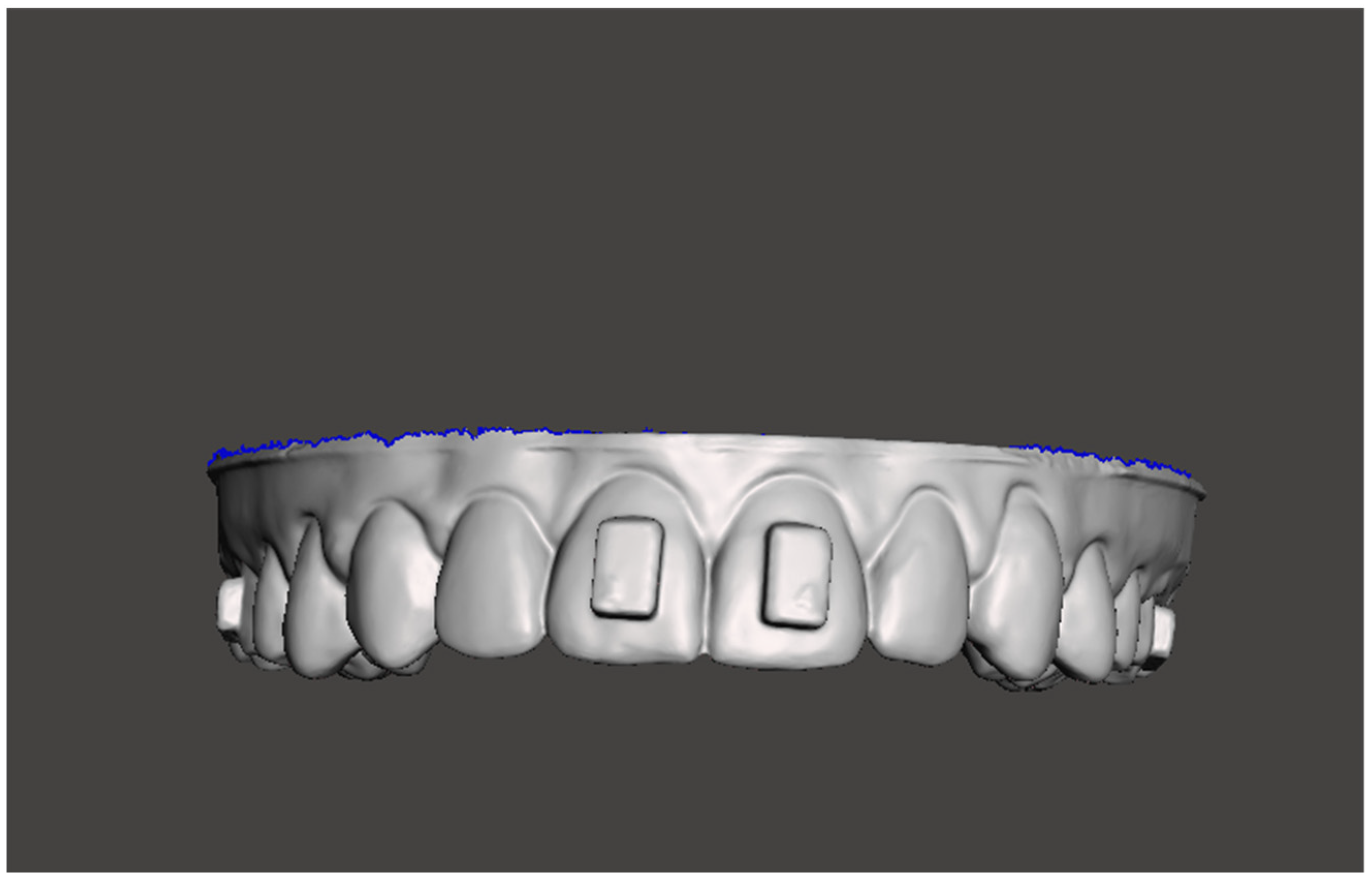
The parallelepiped attachments created were then placed on the vestibular faces of 1.1, 2.1, 1.6 and 2.6 and consequently overlapped, moving the attachment 0.3 mm in the direction of the vestibular surface.
The attachments were merged along with the upper model to constitute a single STL file (stereolithographic file format), named Model Master (MM) (Figure 1).
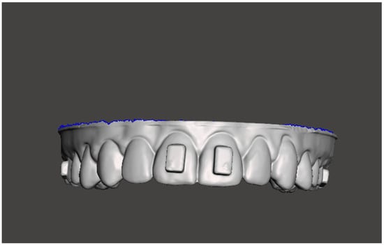
Figure 1.
Model Master (MM).
The Boolean operation was performed by subtracting the model from the attachment shape, thus obtaining attachments with a customized base plate according to the dental anatomy.
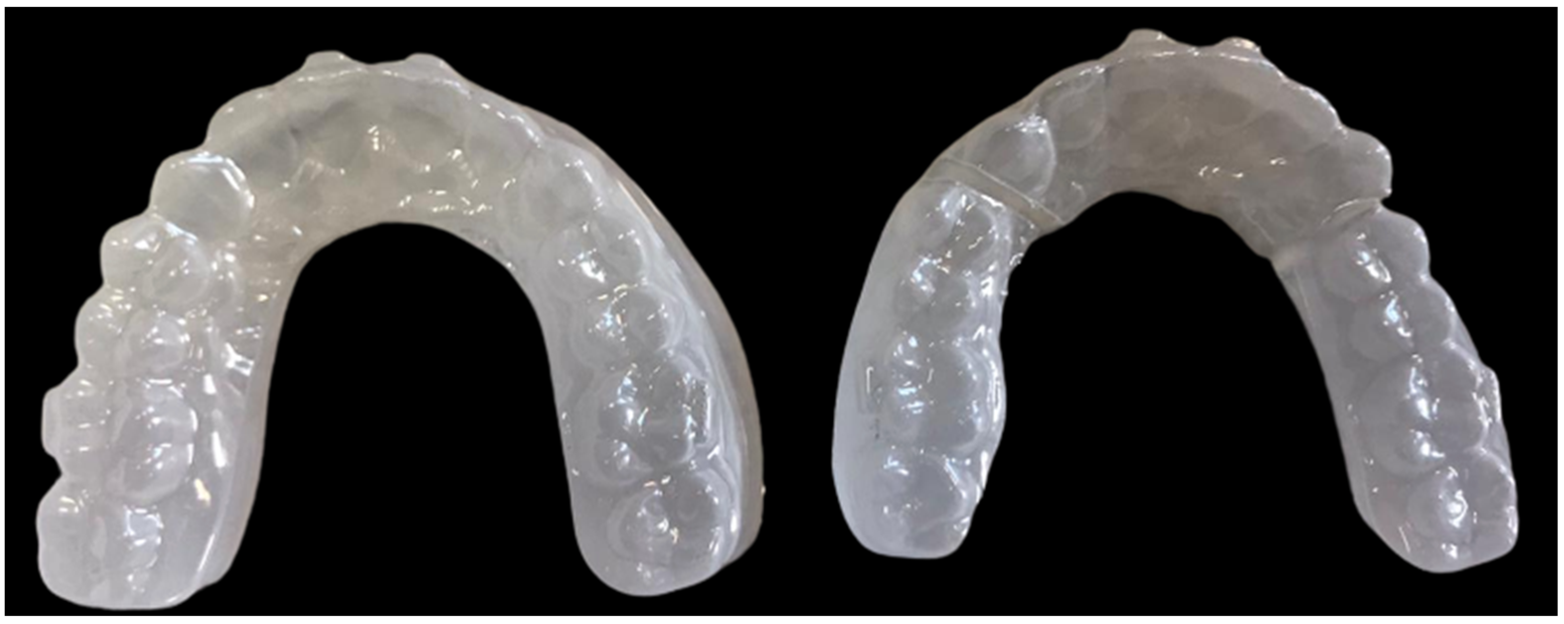
Subsequently, the created custom attachments (CA), the MM model, and two models without attachments (M1, M2) were produced by a 3D printing process using a Liquid Crystal Precision 1.5 3D printer (Photocentric Inc., Avondale, AZ, USA) with Daylight Precision Dental Model White Resin (Photocentric Inc., Avondale, AZ, USA) (Figure 2).

Figure 2.
Printing plate on the CHITUBOX program: 2 models without attachment, 1 with attachment (MM), and custom 4 attachments.
The MM model was used to fabricate, via the thermoforming process (Erkoform 3D Motion, Erkodent Erich Kopp GmbH, Pfalzgrafenweiler, Germany), the 0.8 mm glycol-modified polyethylene terephthalate transfer template (Pet-G-Erkodur, Erkodent Erich Kopp GmbH, Pfalzgrafenweiler, Germany) used to support the clinician in the attachment-bonding process.
This template will be used for both the clinical formation of the traditional attachments (TA) and the transfer of the 3D-printed attachments (CA) (Figure 3).
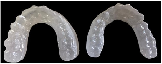
Figure 3.
Thermoformed transfer templates were used for the two bonding protocols.
2.1. Attachment Transfer: Operating Procedure
After obtaining the MM model, two transfer templates were created using two sheets of 0.8 mm PET-G to facilitate the two bonding procedures:
- -
- Bonding of 3D-printed attachments (CA)
- -
- Bonding of traditional attachment (TA)
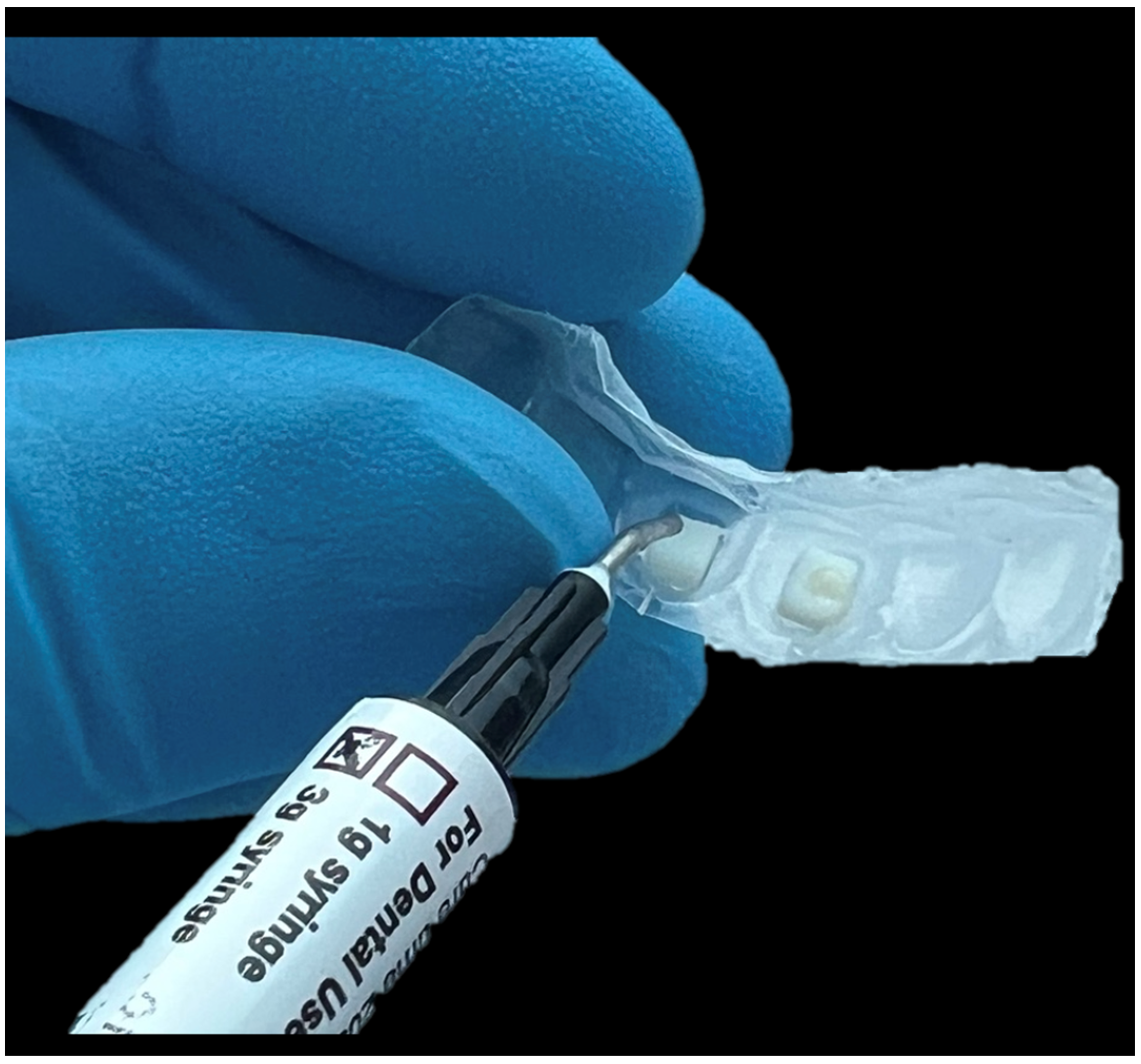
The bonding procedure for the 3D-printed attachments was carried out by dividing the transfer template into three pieces. The custom 3D-printed attachment was inserted into the transfer template and a standardized (a drop of flow composite that covered about 50% of the attachment surface) amount of flow composite (Enaflow-Micerium Spa, Avegno, Genoa, Italy) was applied for bonding. Adhesive was then applied into the model, and each part of the template was inserted into the model and cured with a UV Grand Valo lamp (Ultradent, 505 West Ultradent Drive South Jordan, UT 84095, USA) (Figure 4).
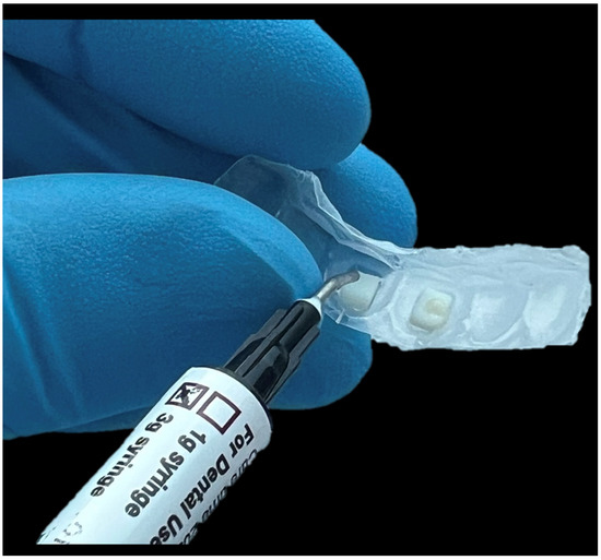
Figure 4.
Three-dimensional printed bonding attachment protocol: Placement of the attachment in the template and positioning of the flow composite.
The traditional transfer procedure involved the creation of a direct-flow attachment (Enaflow-Micerium Spa, Avegno, Genoa, Italy). Inside the special slot created in the transfer template, the flow composite was inserted, then the adhesive was applied in the model, and finally, through the transfer template inserted in the model the attachments were transferred and cured. The attachments were created with flow composite according to the previously standardized procedure.
The models with the conventional attachments and the model with the bonded 3D-printed attachment were both scanned with the Maestro 3D MDS500 Desktop Scanner (AGE Solutions S.r.l., Pisa, Italy; www.maestro3d.com).
Each model with attachments (CA and TA) was digitally superimposed into the Master model (MM) using exocad 2.2 Valletta (DentalCAD; exocad GmbH, Darmstadt, Germany) software to assess the precision of the attachment fabrication through the two distinct procedures.
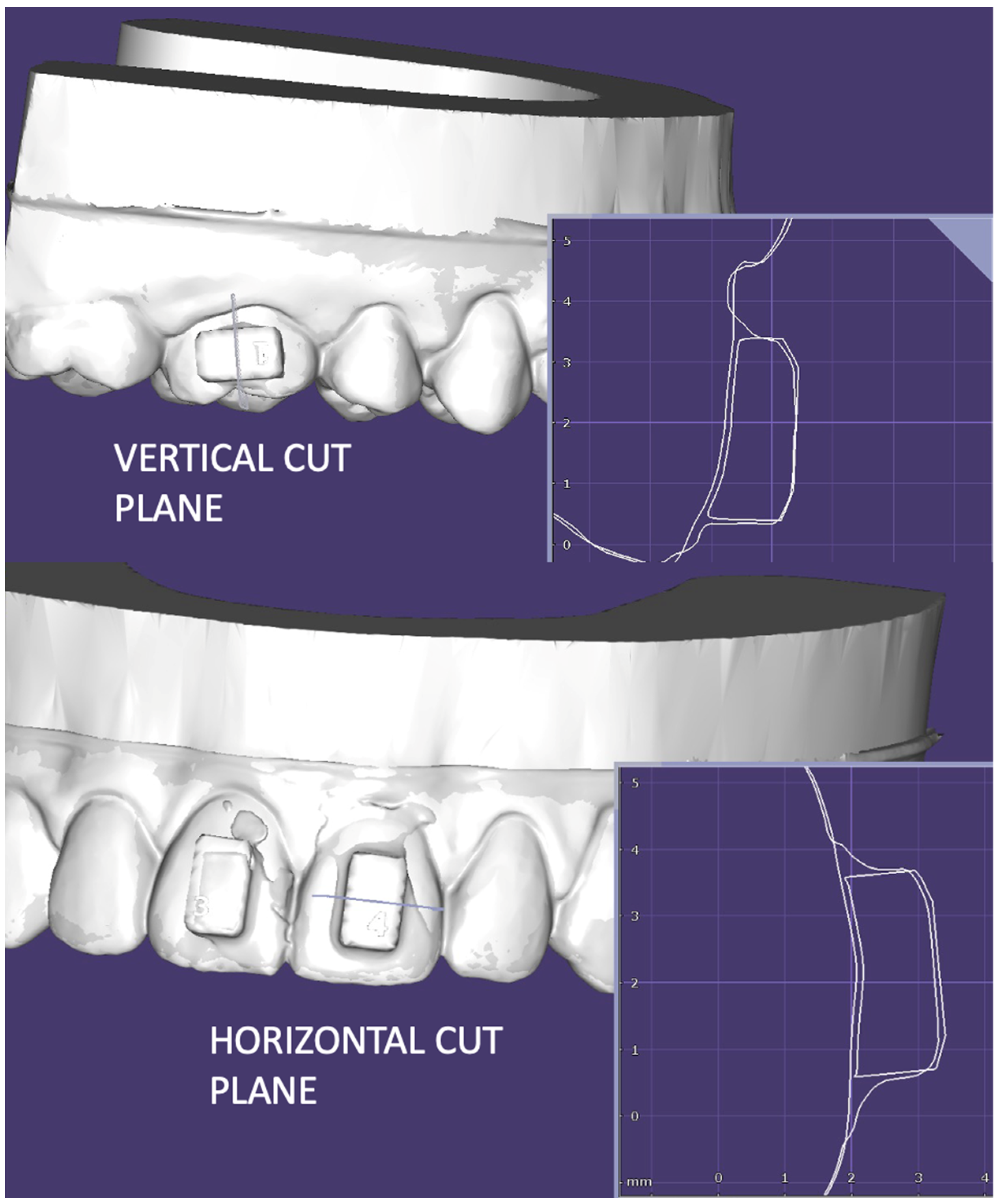
The discrepancies between the overlapping models were evaluated using the following two cutting planes:
- -
- Vertical (following the long axis of the tooth).
- -
- Horizontal (perpendicular to the long axis of the tooth) (Figure 5).
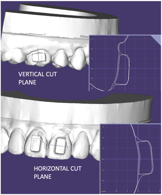 Figure 5. Reference planes used to assess discrepancies: The vertical cutting plane (Ver) and the horizontal cutting plane (Hor).
Measurements were performed between the two attachment profiles.
In the vertical cutting plane, the comparison of the superimposed models involved the assessment of two key outcomes:
- -
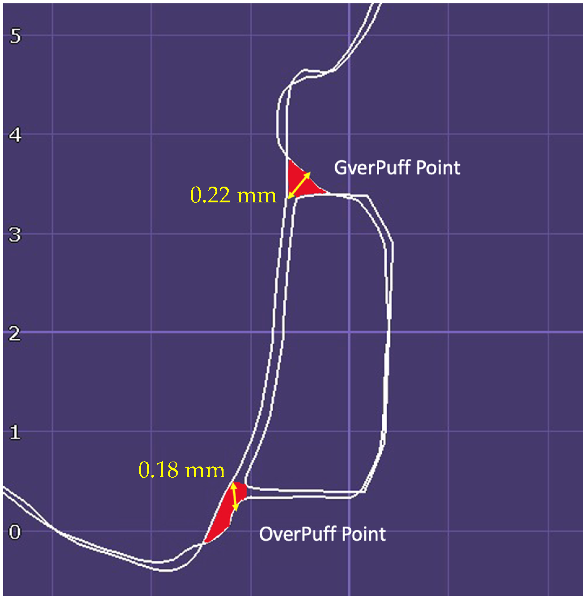
- Puff analysis
The amount of excess composite material at the level of the attachment–model interface was measured. The excess composite was measured in the most gingival portion (GverpuffPoint) and the occlusal portion (OverpuffPoint), and the greatest model profile discrepancy was estimated (Figure 6).
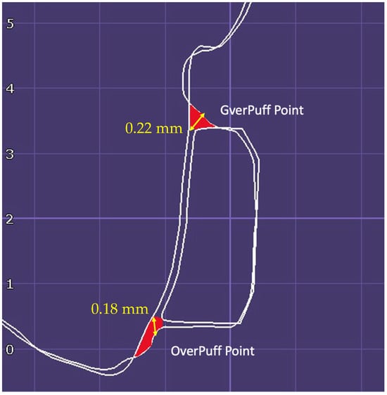
Figure 6.
Measuring points used for the puff analysis in the vertical cutting plane.
- -
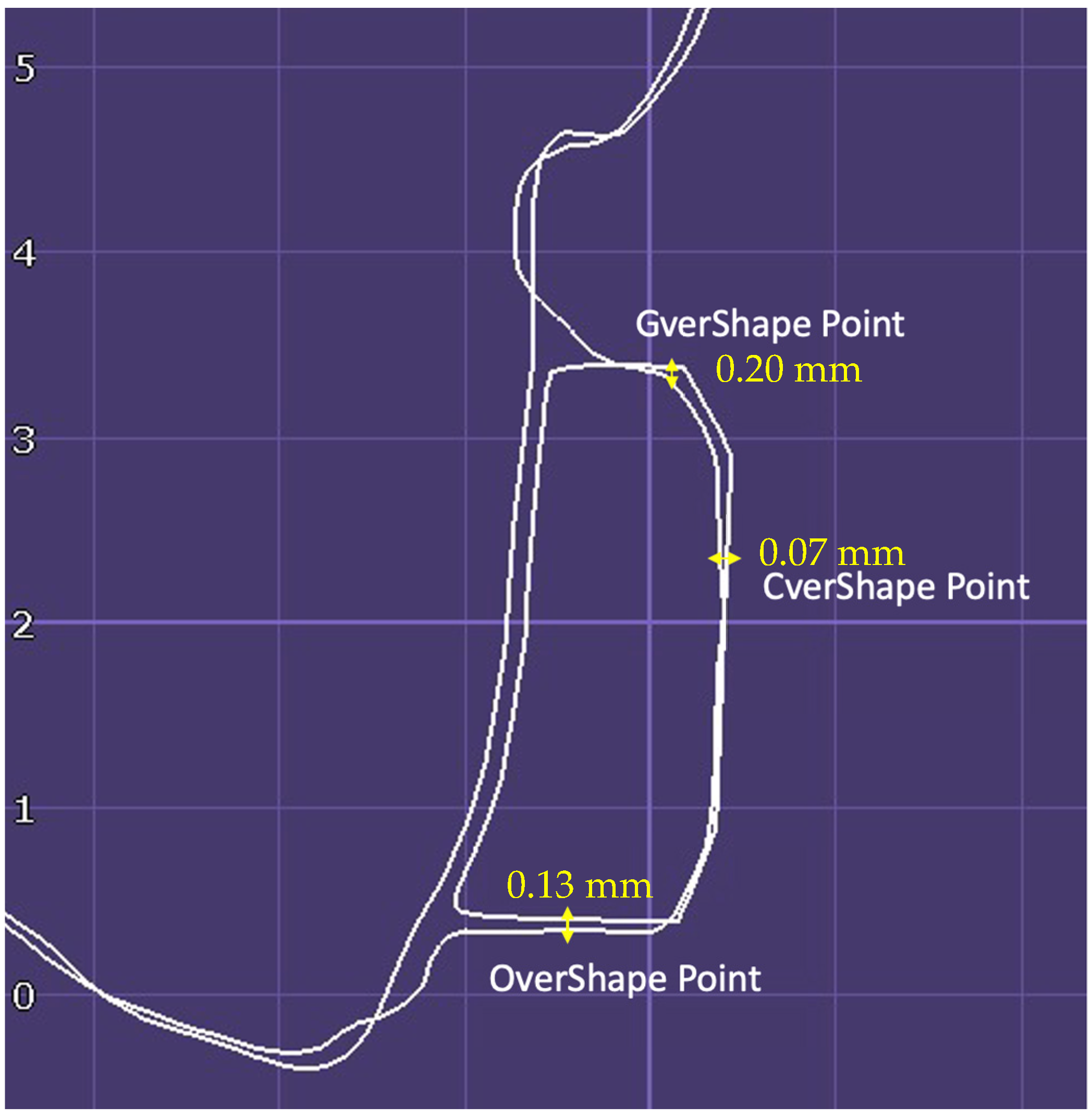
- Shape analysis
The analysis focused on the accuracy of the shape of the attachment. The shape analysis evaluated the average discrepancy of the two considered attachment profiles at three different levels: the gingival level (GvershapePoint), central level (CvershapePoint), and occlusal level of the attachment (OvershapePoint) (Figure 7).
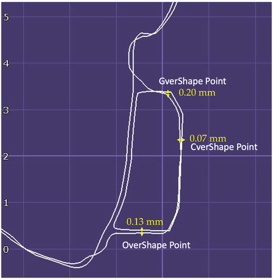
Figure 7.
Measuring points used for the shape analysis in the vertical cutting plane.
On the horizontal shear plane, the same assessment was performed:
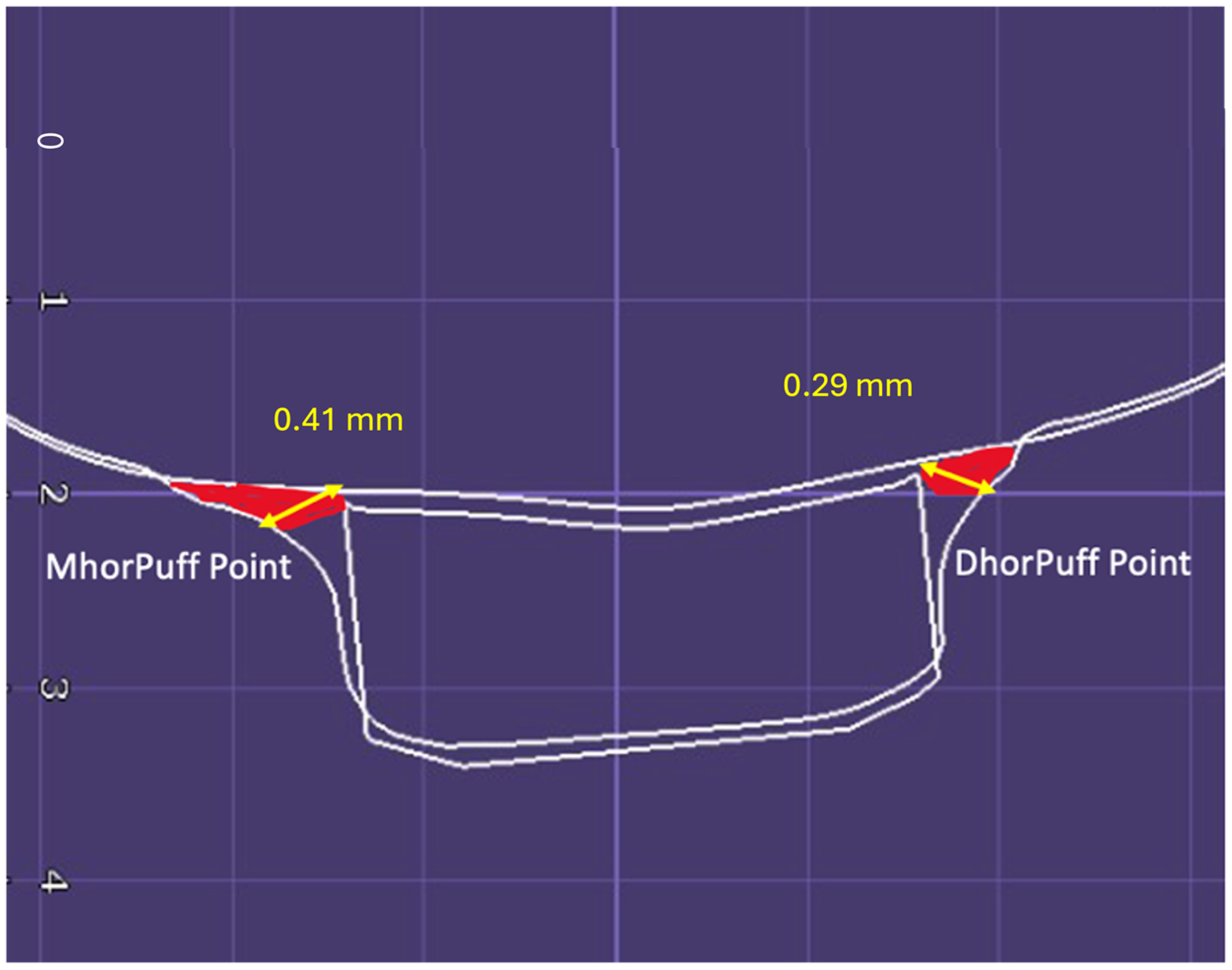
- Puff Analysis
The assessment was carried out at the points of maximum mesial (MhorpuffPoint) and distal discrepancies along the attachment–model interface (DhorpuffPoint) (Figure 8).
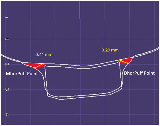
Figure 8.
Measuring points used for the puff analysis in the horizontal cutting plane.
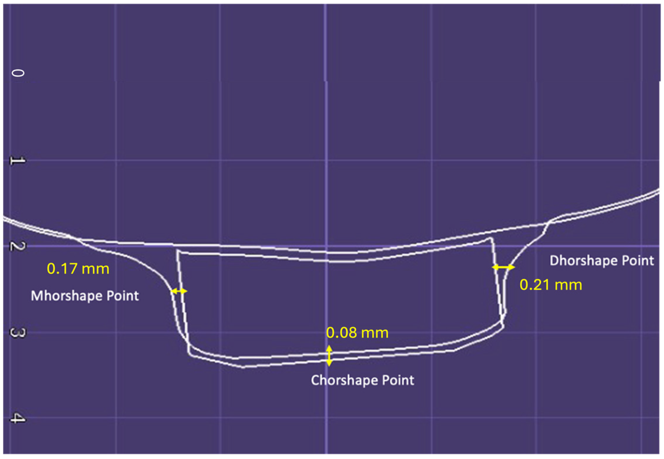
- Shape Analysis
The accuracy of the shape of the attachment at the midpoint of the discrepancy was assessed at the mesial (MhorshapePoint), central (ChorshapePoint), and distal profiles of the attachment (DhorshapePoint) (Figure 9).
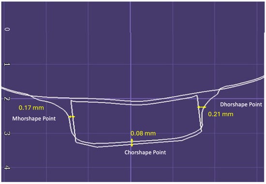
Figure 9.
Measuring points used for the shape analysis in the horizontal cutting plane.
For each attachment, 16 cutting sections were evaluated and 40 measurements were performed.
2.2. Statistical Analysis
The statistical analyses were carried out using SPSS statistical software, version 25.0 (IBM Corporation, Armonk, NY, USA). The significance levels were set at p < 0.05. The normality of the data distribution was evaluated using the Shapiro–Wilk test, and Levene’s test was applied to assess the homogeneity of the variance. Parametric ANOVA multiple comparison tests were utilized for the inferential statistics.
- Methodological Error Assessment:
To evaluate the methodological error associated with the scanning and model overlapping procedures, a comparison was performed between the outcomes of the MA-CA and MA-TA models. The intra-operator reliability was assessed using paired T-tests and the intraclass correlation coefficient (ICC). The magnitude of the random error was determined using Dahlberg’s formula. No statistically significant differences (p < 0.05) were observed between the two assessments, indicating high reliability. A preliminary power analysis was performed on the first five patients enrolled according to the methodology described above.
The analysis was conducted by considering the preliminary mean difference values between the results evaluating the relative linear discrepancy GvershapePoint MM vs. GvershapePoint CA and GvershapePoint MM vs. GvershapePoint TA. The value obtained between the averages was 0.51 mm and the standard deviation (SD) was 0.38 mm. These values were used as the results to perform the power analysis calculation.
The analysis was performed with a power of 80% and the significance level was set at 0.05.
The analysis showed a sample size of 27 cases. Enrollment was set at 30 patients to minimize the risk of false negatives.
3. Results
The findings of this study indicate that the discrepancies in the attachment shape between the planned design and the actual attachment were more pronounced for the traditionally created attachments compared to those produced through 3D printing. Specifically, descriptive statistics revealed larger disparities in shape (0.85 mm) (Table 1) and puff (Table 2) (1.02 mm) for the traditionally created attachments, particularly at the gingival level.

Table 1.
Vertical and horizontal outcomes considered in the shape analysis.

Table 2.
Vertical and horizontal outcomes considered in the puff analysis.
Furthermore, inferential statistics were employed to assess which type of attachment, traditional or 3D-printed, demonstrated fewer deviations from the planned design. The results revealed that the 3D-printed attachments exhibited greater accuracy in terms of the shape reproduction and displayed less puff at the attachment–model interface compared to traditionally created attachments (Table 3).

Table 3.
Inferential statistics and multiple comparisons (univariate ANOVA). a means significant differences with p < 0.05 were detected; b means no significant differences were detected.
These findings suggest that utilizing 3D printing technology for attachment creation may lead to improved accuracy and reduced discrepancies between the planned and actual attachment shapes, particularly when compared to traditional attachment fabrication methods.
4. Discussion
The attachments used in clear aligner therapy are intended to enhance the clinical performance of orthodontic treatment. The inadequate design or placement of these attachments could hinder their ability to exert the necessary force to achieve the desired dental positioning [11]. Another factor influencing the effectiveness of clear aligner treatment and its clinical results is the accuracy of replicating the shape of the attachments [17,19].
Many studies have assessed the effects of various forms of attachments on dental movement [13,18,20,21].
However, only a limited number of studies have evaluated the accuracy of reproducing the programmed shape required for the desired tooth displacement [17,19].
To the best of our knowledge, this is the first study in the literature that proposes 3D-printed custom attachments and evaluates their reproduction accuracy of the planned shape and the presence of blowouts after bonding compared with traditional attachments created from composite flow.
The accuracy of attachment replication can be affected by various factors, including the material of the transfer template, the material composition of the attachment, and the intraoperative skill of the operator.
A recent study [19] assessed the accuracy and reproducibility of the attachment shape by comparing the attachment transfer with two different types of templates: Pet-G (glycol-modified polyethylene terephthalate template) and PE (polyethylene template); the study concluded that Pet-G allows more accurate reproduction of the attachment shape than PE. In addition, these authors also evaluated the influence of composite resin materials with different viscosities in reproducing the shape of the attachments, revealing that the accuracy of high-viscosity and low-viscosity composites is equivalent.
Other authors carried out a comparative analysis of flowable and packable composite materials [22], concluding that flowable composite materials necessitated less time for preparation and showing no statistically significant differences in terms of damage when compared to packable composites at the one-year evaluation. More authors have shown that 3D-printed materials offer good accuracy and reproducibility of details, including composite attachments; however, they may show lower strength than traditional materials, especially in terms of the resistance to forces applied during orthodontic treatment [23]. Based on data from the previous literature [19,22], a PET-G template was used in the present study to transfer both the traditional and 3D-printed attachments; in addition, the use of a flow composite was chosen to create the traditional attachments.
The results of this study show statistically significant differences (p < 0.05) in the accuracy between the 3D-printed custom attachments and the traditional flow-made attachments.
On average, the 3D-printed custom attachments exhibited over 50% higher accuracy, with the most significant discrepancies observed on the gingival side. The greater inaccuracy in transferring the attachments at the gingival level (0.78 mm) can be due to the “tenting effect” that occurs during the thermoforming process. The tenting effect is when the thermoformed material tends to pull upward along the gingival as it cools [24].
This phenomenon can lead to challenges in achieving the optimal adherence of the thermoformed tray to the teeth, especially in the gingival area, resulting in a greater inaccuracy in the shape reproduced in the template and, consequently, in the final attachment shape. These results are consistent with the study conducted by Park SY and colleagues, in which the median gap of thermoformed transparent aligners was evaluated using micro CT and a spectrophotometer, showing that the median gap at the gingival level consistently exceeded that at the occlusal surface [25].
Another factor contributing to the greater inaccuracy of traditional bindings made from flow composite is the difficulty of standardizing the amount of composite material used during the creation of the binding, which can lead to an inconsistency between the programmed shape and the actual shape. On the contrary, the use of custom 3D-printed attachments allows for better standardization of the composite amount flow during the bonding procedure by using a standardized amount of composite.
The findings of this research indicate the potential benefits of customized 3D-printed attachments in the clear aligner therapy. However, additional investigations are necessary to assess the clinical impact of different amounts of attachment accuracy reproduction on the efficacy of clear aligners when it comes to achieving different types of dental movement. The limitation of this study is that the reliability of the reproducibility of the attack in relation to the different possible forms is not evaluated. In the future, well-designed clinical studies with a control group will be able to better evaluate these aspects of clear aligner therapy.
5. Conclusions
The presence of puffs in 3D-printed custom attachments is significantly reduced compared to the puffs present at the attachment–model interface of traditional attachments.
In the future, custom attachments made by 3D printing could be a valid alternative to traditional attachments made to improve attachment reproduction.
Author Contributions
Conceptualization, A.M.B., E.C., L.C., S.B., R.N., A.M.B., E.C., L.C. and S.B.; methodology, A.M.B., E.C., L.C., S.B. and R.N.; software, A.M.B., E.C. and L.C.; validation, A.M.B., E.C. and L.C.; formal analysis, A.M.B. and E.C.; investigation, A.M.B., E.C., L.C. and S.B. resources, A.M.B., E.C., L.C. and S.B.; data curation, A.M.B., E.C., L.C. and S.B.; writing—original draft preparation, A.M.B. and R.N.; writing—review and editing, A.M.B. and R.N.; visualization, A.M.B. and R.N.; supervision, A.M.B. and R.N.; project administration, A.M.B. and R.N.; funding acquisition, A.M.B., E.C., L.C., S.B. and R.N. All authors have read and agreed to the published version of the manuscript.
Funding
This research received no external funding.
Institutional Review Board Statement
This study was conducted in accordance with the Declaration of Helsinki and approved by the Institutional Review Board (or Ethics Committee) of the University of Messina (protocol code 33-2020, 04/03/2020).
Informed Consent Statement
Informed consent was obtained from all the subjects involved in the study.
Data Availability Statement
The data presented in this study are available on request from the corresponding author.
Conflicts of Interest
The authors declare no conflicts of interest.
References
- Barrera-Chaparro, J.P.; Plaza-Ruíz, S.P.; Parra, K.L.; Quintero, M.; Velasco, M.D.P.; Molinares, M.C.; Álvarez, C. Orthodontic treatment need, the types of brackets and the oral health-related quality of life. Dent. Med. Probl. 2023, 60, 287–294. [Google Scholar] [CrossRef] [PubMed]
- Putrino, A.; Barbato, E.; Galluccio, G. Clear Aligners: Between Evolution and Efficiency—A Scoping Review. Int. J. Environ. Res. Public Health 2021, 18, 2870. [Google Scholar] [CrossRef] [PubMed]
- Muro, M.P.; Caracciolo, A.C.A.; Patel, M.P.; Feres, M.F.N.; Roscoe, M.G. Effectiveness and predictability of treatment with clear orthodontic aligners: A scoping review. Int. Orthod. 2023, 21, 100755. [Google Scholar] [CrossRef] [PubMed]
- Fang, X.; Qi, R.; Liu, C. Root resorption in orthodontic treatment with clear aligners: A systematic review and meta-analysis. Orthod. Craniofacial Res. 2019, 22, 259–269. [Google Scholar] [CrossRef] [PubMed]
- Li, Y.; Deng, S.; Mei, L.; Li, Z.; Zhang, X.; Yang, C.; Li, Y. Prevalence and severity of apical root resorption during orthodontic treatment with clear aligners and fixed appliances: A cone beam computed tomography study. Prog. Orthod. 2020, 21, 1. [Google Scholar] [CrossRef] [PubMed]
- Nucera, R.; Giudice, A.L.; Matarese, G.; Artemisia, A.; Bramanti, E.; Crupi, P.; Cordasco, G. Analysis of the characteristics of slot design affecting resistance to sliding during active archwire configurations. Prog. Orthod. 2013, 14, 35. [Google Scholar] [CrossRef]
- Cordasco, G.; Farronato, G.; Festa, F.; Nucera, R.; Parazzoli, E.; Grossi, G.B. In vitro evaluation of the frictional forces between brackets and archwire with three passive self-ligating brackets. Eur. J. Orthod. 2009, 31, 643–646. [Google Scholar] [CrossRef]
- Rouzi, M.; Zhang, X.; Jiang, Q.; Long, H.; Lai, W.; Li, X. Impact of Clear Aligners on Oral Health and Oral Microbiome during Orthodontic Treatment. Int. Dent. J. 2023, 73, 603–611. [Google Scholar] [CrossRef]
- Kravitz, N.D.; Kusnoto, B.; BeGole, E.; Obrez, A.; Agran, B. How well does Invisalign work? A prospective clinical study evaluating the efficacy of tooth movement with Invisalign. Am. J. Orthod. Dentofac. Orthop. 2009, 135, 27–35. [Google Scholar] [CrossRef]
- Paradowska-Stolarz, A.; Wezgowiec, J.; Malysa, A.; Wieckiewicz, M. Effects of Polishing and Artificial Aging on Mechanical Properties of Dental LT Clear® Resin. J. Funct. Biomater. 2023, 14, 295. [Google Scholar] [CrossRef]
- Barreda, G.J.; Dzierewianko, E.A.; Muñoz, K.A.; Piccoli, G.I. Surface wear of resin composites used for Invisalign® attachments. Acta Odontol. Latinoam. 2017, 30, 90–95. [Google Scholar] [PubMed]
- Jedliński, M.; Mazur, M.; Greco, M.; Belfus, J.; Grocholewicz, K.; Janiszewska-Olszowska, J. Attachments for the Orthodontic Aligner Treatment—State of the Art—A Comprehensive Systematic Review. Int. J. Environ. Res. Public Health 2023, 20, 4481. [Google Scholar] [CrossRef] [PubMed]
- Gomez, J.P.; Peña, F.M.; Martínez, V.; Giraldo, D.C.; Cardona, C.I. Initial force systems during bodily tooth movement with plastic aligners and composite attachments: A three-dimensional finite element analysis. Angle Orthod. 2015, 85, 454–460. [Google Scholar] [CrossRef] [PubMed]
- Nucera, R.; Dolci, C.; Bellocchio, A.M.; Costa, S.; Barbera, S.; Rustico, L.; Farronato, M.; Militi, A.; Portelli, M. Effects of Composite Attachments on Orthodontic Clear Aligners Therapy: A Systematic Review. Materials 2022, 15, 533. [Google Scholar] [CrossRef] [PubMed]
- Weckmann, J.; Scharf, S.; Graf, I.; Schwarze, J.; Keilig, L.; Bourauel, C.; Braumann, B. Influence of attachment bonding protocol on precision of the attachment in aligner treatments. J. Orofac. Orthop. 2020, 81, 30–40. [Google Scholar] [CrossRef] [PubMed]
- Valeri, C.; Aloisio, A.; Mummolo, S.; Quinzi, V. Performance of Rigid and Soft Transfer Templates Using Viscous and Fluid Resin-Based Composites in the Attachment Bonding Process of Clear Aligners. Int. J. Dent. 2022, 2022, 1637594. [Google Scholar] [CrossRef] [PubMed]
- D’antò, V.; Muraglie, S.; Castellano, B.; Candida, E.; Sfondrini, M.F.; Scribante, A.; Grippaudo, C. Influence of Dental Composite Viscosity in Attachment Reproduction: An Experimental in Vitro Study. Materials 2019, 12, 4001. [Google Scholar] [CrossRef] [PubMed]
- Gazzani, F.; Bellisario, D.; Quadrini, F.; Danesi, C.; Alberti, A.; Cozza, P.; Pavoni, C. Light-curing process for clear aligners’ attachment reproduction: Comparison between two nanocomposites cured by the auxiliary of a new tool. BMC Oral Health 2022, 22, 376. [Google Scholar] [CrossRef] [PubMed]
- Bellocchio, A.M.; Portelli, M.; Ciraolo, L.; Ciancio, E.; Militi, A.; Peditto, M.; Barbera, S.; Nucera, R. Evaluation of the Clinical Variables Affecting Attachment Reproduction Accuracy during Clear Aligner Therapy. Materials 2023, 16, 6811. [Google Scholar] [CrossRef] [PubMed]
- Ferlias, N.; Dalstra, M.; Cornelis, M.A.; Cattaneo, P.M. In Vitro Comparison of Different Invisalign® and 3Shape® Attachment Shapes to Control Premolar Rotation. Front. Bioeng. Biotechnol. 2022, 10, 840622. [Google Scholar] [CrossRef]
- Laohachaiaroon, P.; Samruajbenjakun, B.; Chaichanasiri, E. Initial Displacement and Stress Distribution of Upper Central Incisor Extrusion with Clear Aligners and Various Shapes of Composite Attachments Using the Finite Element Method. Dent. J. 2022, 10, 114. [Google Scholar] [CrossRef] [PubMed]
- Lin, S.; Huang, L.; Li, J.; Wen, J.; Mei, L.; Xu, H.; Zhang, L.; Li, H. Assessment of preparation time and 1-year Invisalign aligner attachment survival using flowable and packable composites. Angle Orthod. 2021, 91, 583–589. [Google Scholar] [CrossRef] [PubMed]
- Cole, D.; Bencharit, S.; Carrico, C.K.; Arias, A.; Tüfekçi, E. Evaluation of fit for 3D-printed retainers compared with thermoform retainers. Am. J. Orthod. Dentofac. Orthop. 2019, 155, 592–599. [Google Scholar] [CrossRef] [PubMed]
- Takahashi, M.; Satoh, Y.; Iwasaki, S. Effect of thermal shrinkage during thermoforming on the thickness of fabricated mouthguards: Part 2 pressure formation. Dent. Traumatol. 2017, 33, 106–109. [Google Scholar] [CrossRef]
- Park, S.Y.; Choi, S.-H.; Yu, H.-S.; Kim, S.-J.; Kim, H.; Kim, K.B.; Cha, J.-Y. Comparison of translucency, thickness, and gap width of thermoformed and 3D-printed clear aligners using micro-CT and spectrophotometer. Sci. Rep. 2023, 13, 10921. [Google Scholar] [CrossRef]
Disclaimer/Publisher’s Note: The statements, opinions and data contained in all publications are solely those of the individual author(s) and contributor(s) and not of MDPI and/or the editor(s). MDPI and/or the editor(s) disclaim responsibility for any injury to people or property resulting from any ideas, methods, instructions or products referred to in the content. |
© 2024 by the authors. Licensee MDPI, Basel, Switzerland. This article is an open access article distributed under the terms and conditions of the Creative Commons Attribution (CC BY) license (https://creativecommons.org/licenses/by/4.0/).

