Article

Exergaming-Based Rehabilitation for Lateral Trunk Flexion in Parkinson’s Disease: A Pilot Study

Department of Medical, Surgical, and Health Sciences, University of Trieste, 34149 Trieste, Italy
*
Author to whom correspondence should be addressed.
Appl. Sci. 2025, 15(4), 1745; https://doi.org/10.3390/app15041745
Submission received: 4 January 2025 / Revised: 30 January 2025 / Accepted: 5 February 2025 / Published: 8 February 2025
(This article belongs to the Special Issue Exercise, Fitness, Human Performance and Health: 2nd Edition)

Abstract

:
(1) Background: Axial postural deformities represent a more common disabling motor complication in Parkinson’s disease. This study aims to investigate the clinical and neurophysiological effect of a rehabilitation treatment based on exergaming. (2) Methods: A pilot observational study was conducted on nine subjects affected by Parkinson’s disease and lateral trunk flexion, as well as on nine healthy controls with regard to some clinical and neurophysiological outcomes (3) Results: Statistically significant improvements were observed in all clinical assessment outcomes taken in to consideration: Berg balance scale (p = 0.0078), timed up and go tests (p = 0.03), degrees of lateral trunk inclination (p = 0.0039), and anterior/posterior trunk inclination (p = 0.0039). Regarding neurophysiological outcomes, the pressure pain threshold was enhanced and statistically significant in all areas assessed. Moreover, tensiomyography highlighted a statistically significant improvement in the maximal radial displacement of the ipsilateral erector spinae muscles. (4) Conclusions: The clinical and neurophysiological outcomes suggest both peripheral and central effects of exergaming. Peripherally, exergaming seems to lead to a postural trunk correction through a reduction in muscle stiffness in the ipsilateral erector spinae. Centrally, exergaming seems to lead to a central pain modulation through an upregulation of cortical connectivity associated with cognitive tasks. Taken together, these results also indicate that exergaming can be a feasible and enjoyable complement to traditional rehabilitation, potentially enhancing patients’ motivation and adherence.

1. Introduction

Axial postural deformities are a characteristic motor feature of Parkinson’s disease (PD) [1]. Although it is difficult to identify a clear prevalence due to the lack of a comprehensive classification in previous years, lateral trunk flexion (LTF) and Pisa syndrome (PS) are the most common postural deformities in PD, with an estimated prevalence of approximately 8.8% [1,2,3,4,5,6,7]. Pisa syndrome is clinically defined as a reversible lateral flexion of the trunk of at least 10°, which can be reduced through passive mobilization or supine positioning, while it worsens in sitting, standing, or walking [7,8]. In contrast, lateral trunk flexion is clinically defined as an involuntary lateral bending of the trunk with an angle ranging from 5° to 10°, and can be considered as a milder form of Pisa syndrome [1,2,3,4,6,7].
LTF significantly reduces quality of life. In fact, increased lateral bending is related with limitations in activities of daily living (ADLs), motor impairment, increased back pain intensity, imbalance, and increased risk of falling [5]. The cut off that identifies severe motor impairment and high disability is a lateral bending angle exceeding 10.5° [5]. Consequently, the treatment of these correlated symptoms is pivotal in order to prevent progression and worsening of LTF.
These correlated symptoms are usually managed with pharmacological approaches such as onabotulinumtoxin-A [9,10], non-pharmacological interventions such as rehabilitation [11], or a combination of both [12]. However, rehabilitation programs are heterogeneous, and there is ongoing debate about the best intervention type and for how long it should last [13].
Exergaming, which combines physical exercise with interactive video games, has emerged as a promising tool in rehabilitation. From a psychological point of view, its engaging nature has the potential to both enhance motivation and improve therapy adherence. By incorporating gamification elements such as rewards, challenges, and real-time feedback, exergaming transforms traditionally monotonous rehabilitation exercises into appealing activities. Exergaming significantly increases intrinsic motivation in patients by fostering a sense of autonomy, competence, and relatedness [14]. Exergames have also improved balance and mobility in older adults [15], provided higher adherence rates and greater enjoyment compared to standard care in cancer patients [16], and improved exercise capacity and quality of sleep in heart failure patients [17]. This growing body of literature indicates that exergaming can boost motivation through enjoyable, interactive modalities while simultaneously promoting adherence to therapy regimens.
With regard to exergaming in PD, today, rehabilitation protocols based on virtual reality and exergaming have emerged as a valid training strategy to improve balance, mobility, and gait in Parkinson’s disease. These approaches provide individuals with PD the opportunity to experience more sensory stimulation and motor tasks while receiving real-time feedback [18,19,20,21,22,23,24]. However, up to now, there has been no detailed investigation addressing the use of a virtual reality or exergaming approach for managing lateral trunk flexion in Parkinson’s disease.
The first aim of this study is to investigate the clinical effects of a rehabilitation treatment based on exergaming on clinical outcomes such as motor impairment, imbalance, and risk of falling. The second aim is to assess the neurophysiological effect of an exergaming-based rehabilitation treatment on the contractile properties and on the pressure pain thresholds of specific trunk muscles.

2. Materials and Methods

The pilot observational study was conducted between December 2022 and October 2023, involving nine participants affected by Parkinson’s disease (PD) and lateral trunk flexion (LTF), prodromal phase of Pisa syndrome (PS), along with nine healthy controls of similar age. The research project was approved by the Ethics Committee of the University of Trieste (protocol code 115885 10.07.2024), and all participants signed the informed consent. The participants with PD and LTF were recruited from the Neurological Clinic, adhering to the Movement Disorders Society’s clinical diagnostic criteria [25]. Pisa syndrome is clinically diagnosed according to the following criteria: lateral trunk inclination of at least 10°, ipsilateral trunk axial rotation, worsening of the postural imbalance in standing position and gait, and nearly complete resolution of the postural disorder upon passive mobilization or supine positioning. LTF shares the same features, but involves trunk inclinations below 10° [1,3,8].
The inclusion criteria for the lateral trunk flexion group were as follows: age between 65 and 85; diagnosis of PD and LTF; Hoehn and Yahr score less than or equal to 3 [26,27]; maximum lateral trunk flexion of 10°; and signed informed consent. The exclusion criteria were as follows: neurological disorders other than PD; cognitive impairments; orthopedic or rheumatologic disorders; and changes in the drug therapeutic plan during the study. On the other side, regarding the healthy controls, the inclusion criteria were as follows: age between 55 and 85 and signed informed consent. The exclusion criteria were as follows: diagnosis of PD or LTF, other kinds of neurological disorders, cognitive impairments, and orthopedic or rheumatic conditions.
All participants underwent three assessments before and after the treatment protocol. The assessments were performed by one neurologist and two physiotherapists experienced in PD. The evaluations included the clinical history, the clinical assessments, and the instrumental assessments. All the assessments, clinical and instrumental, were conducted during the ON-phase of dopaminergic therapy.

2.1. Clinical History

The collected clinical history regarded the drug therapy plan, years of PD diagnosis, previous botulinum toxin treatment, effectiveness of previous physiotherapy treatments, daily life difficulties due to PD, and episodes of falls or gait freezing.

2.2. Clinical Assessment

The clinical assessment was performed using the Berg balance scale (BBS) and the timed up and go test (TUG).
The BBS was used to assess a patient’s balance abilities, in both static tasks that require the maintenance of standing and sitting positions, and in dynamic tasks requiring postural steps. The Berg scores correlate with motor functioning (unified Parkinson’s disease rating scale—III—UPDRS-III), disease stage (modified Hoehn and Yahr scale), and ADL independence (modified Schwab and England capacity for daily living scale—S&E ADL Scale) [28].
The TUG test was used to assess motor performance, and is correlated to the functional mobility, gait speed, and risk of falls [29].
The same assessments were conducted at the end of the treatment sessions.

2.3. Instrumental Assessment

2.3.1. Pressure Pain Threshold (PPT)

Patients affected by Parkinson’s disease suffer from pain [30] due to the role of basal ganglia in pain processing [30,31], the neurodegeneration of primary nociceptive cutaneous efferent [30], or the dopaminergic deficit that could inhibit descending pain inhibition [31]. Thus, pain assessment in people with PD is an interesting variable to measure as a possible outcome of treatment. A Somedic pressure algometer was chosen for its reliability and validity to measure PPT in neurological patients [32,33,34]. PPT was assessed on transverse processes (L3, T6, and C3) bilaterally. For each of the three processes, three measurements were taken, with one minute of interval. The applied pressure increased in steps of approximately 30 KPa/s, and the patients had to press a stop remote control when pressure became pain. The software recorded the KPa values and then calculated the average of the three measurements [32,33,34].

2.3.2. Tensiomyography (TMG)

Tensiomyography (TMG) is a non-invasive validated and objective technique that assesses in vivo specific skeletal muscle characteristics like contractile properties through displacement–time parameters [35,36]. TMG measurements were evaluated bilaterally during maximal isometric contractions electrically evoked in selected trunk muscles: upper trapezius, middle trapezius, latissimus dorsi, and erector spinae [37,38,39,40]. A single 1 ms maximal monophasic electrical impulse was applied to induce belly muscle oscillations due to a twitch contraction. To record these oscillations, a sensitive digital displacement sensor (TMG-BMC Ltd., Ljubljana, Slovenia) was placed on the selected muscles [37,38,39,40].
The amplitude of stimulation was initially applied just above the threshold. Then, it was gradually enhanced to elicit the radial twitch Dm (in millimeters) and did not further increase. The electrical pulses were between 85 and 110 mA at a constant voltage of 30 V. Moreover, a 10–15 s interstimulation interval was used. The final analysis included the average of two maximal responses [37,38,39,40].
The TMG parameters were:
Contraction time (Tc): time from 10% to 90% of the maximal radial displacement (Dm);
Delay time (Td): time from electrical pulse and 10% of Dm;
Half-relaxation time (Tr): time from 90% to 50% of Dm in the decreasing curve;
Maximal radial displacement (Dm), or peak radial displacement: the spatial transverse deformation of the muscle, which increases proportionally to muscle trophy, and decreases in the presence of muscle stiffness or tension.
Sustain time (Ts): time from 50% of contraction time (Tc) and 50% of relaxation time (Tr).

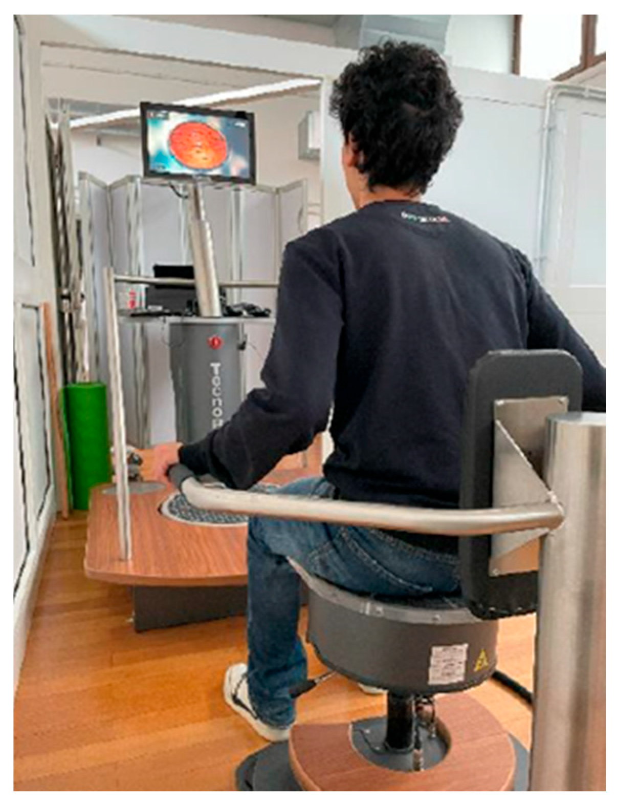
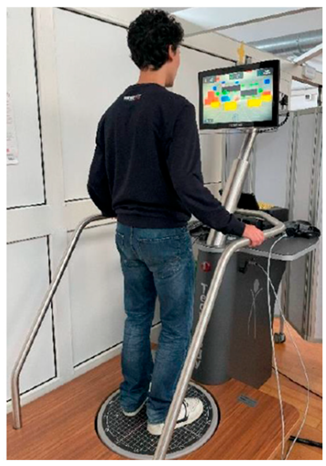
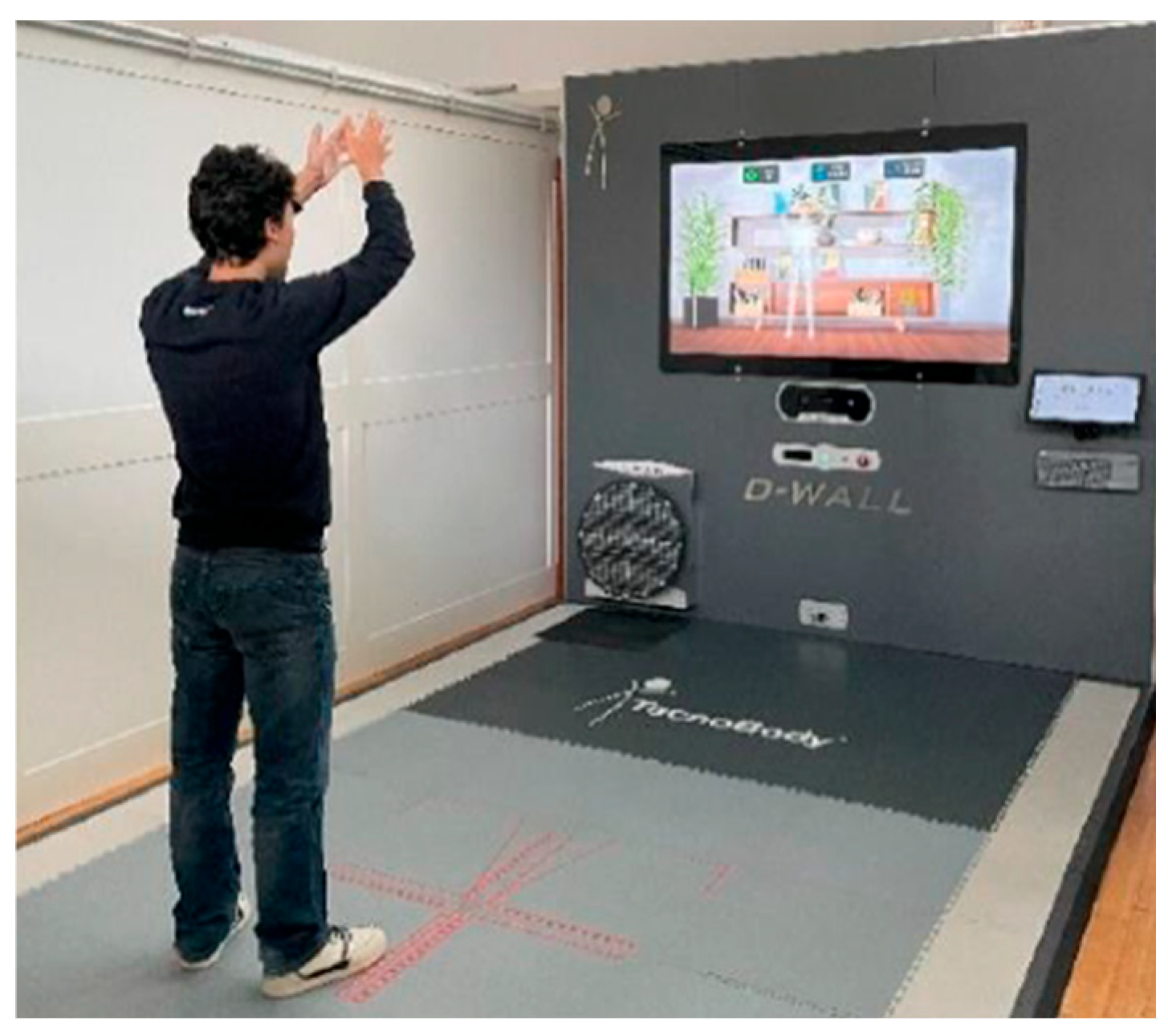
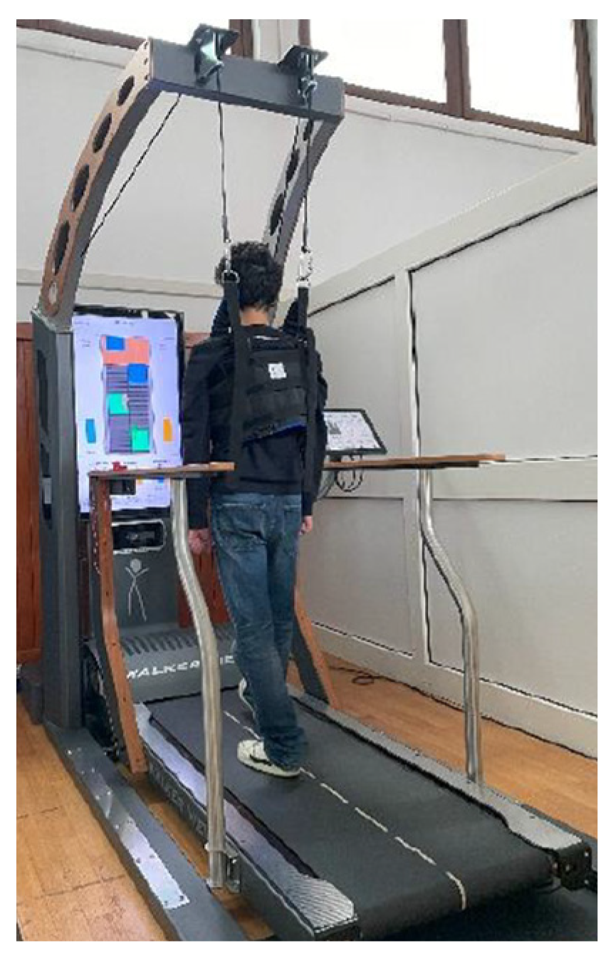
2.4. Treatment Protocol

The rehabilitation protocol was based on exergaming and virtual reality. Participants completed 16 individual sessions (60 min each, twice a week for eight weeks) carried out by two expert physiotherapists. Tecnobody® devices (ProKin, D-Wall, and WalkerView Treadmill) were used [41,42].
Rehabilitation sessions were divided in 4 phases.
In the first phase, the ProKin 252® exergaming software in Trunk Module was used. The LTF group had to sit on the “trunk balance seat” (Figure 1), a mobile table with lateral supports for the upper limbs. The goal was to improve the endurance and symmetry of trunk functions in the sitting position. More specifically, to perform the exergaming, the LTF group had to integrate pelvic anteversion/retroversion movements and lateral/diagonal weight shifting with attention and cognitive strategies. The most used exergaming strategies were “Equilibrium” (guiding a ball along a route to a target point, in increasing levels) and “Shooting range” (moving the viewfinder to hit the most targets possible within two minutes).
In the second phase, the ProKin 252® exergaming software in the standing position was used. The LTF group stood on a platform (Figure 2) with their feet 15 cm apart. The goal was to manage load shifting on the feet, and to control the trunk in the standing position. Exergaming was first performed in the static platform modality, and then in the dynamic platform modality.
In the third phase, exergaming was conducted with the D-Wall® system (Figure 3). The D-Wall® system has a 3D infrared camera that captures movements in real time. The goal was to improve forward-pass load movements according to the real-time feedback of the shift of the center of pressure (CoP) recorded by the force platform. “Shelf” and “Library” exergaming required the participants to move objects in different positions by using either one or two hands.
The fourth phase involved the use of the WalkerView® treadmill (Figure 4). The goal was to correct the walking dynamics by opening the coxofemoral angle. The WalkerView treadmill “Gait trainer” began with a load reduction of 50% of each participant’s body weight, and progressed gradually, adding load in the following sessions.

2.5. Statistical Analysis

Data were analyzed using GraphPad InStat 3.06. The Wilcoxon non-parametric test was used for pre- and post-treatment of the BBS and TUG test evaluations. A Kruskal–Wallis test (non-parametric ANOVA) was applied to compare the variance among LTF t2, LTF t2, and HC groups. The statistical significance level was ɑ 95% (p < 0.05).

3. Results

A total of nine participants with Parkinson’s disease (PD) and lateral trunk flexion (ltf) (three women, mean age 75.8, SD ± 4.6) and nine healthy controls (HC) (four women, mean age 75.4, SD ± 4.3) were enrolled in the study. There was no significant difference in age between the LTF and HC groups (p = 0.9). Among the PD group, the mean disease duration was 9.0 (SD ± 6.1) years, and the mean LTF duration was 4.3 (SD ± 3.2) years. All PD patients had a Hoehn and Yahr (H&Y) score of two (SD ± 0). At baseline (t1), the average lateral trunk inclination in the LTF group (four patients had left-side inclination) was 5.8° (SD ± 3.2°), while anterior/posterior inclination averaged 9.9° (SD ± 7.3°).

3.1. Clinical Assessment

The assessed clinical outcomes included the Berg balance scale (BBS), timed up and go test (TUG), and trunk inclination measurements (both lateral and anterior/posterior). With regard to BBS, it showed a statistically significant improvement (p = 0.0078; Cis 95% +38.4/+50.7), with BBS scores increasing from 44.5 (SD ± 8) to 49.1 (SD ± 5.7). TUG test scores decreased from 13.7 (SD ± 4) to 12.5 (SD ± 4.3), indicating a performance improvement; this reduction is statistically significant (p = 0.03; Cis 95% +10.7/+16.7). As regards the lateral trunk inclination, it decreased from 5.8° (SD ± 2.4°) to 3.1° (SD ± 1.6°), showing a statistically significant reduction (p = 0.0039; Cis 95% +3.9/+7.6). Finally, the anterior/posterior inclination of the trunk reduced from 9.9° (SD ± 7.3°) to 6° (6.5°), also a statistically significant improvement (p = 0.0039; Cis 95% +4.2/+15.5).

3.2. Neurophysiological Assessment

3.2.1. Pressure Pain Threshold (PPT)

Table 1 provides an overview of all PPT parameters before and after exergaming rehabilitation treatments in the lateral trunk flexion group (LTF) compared to the healthy control group (HC).

3.2.2. Tensiomyography (TMG)

Regarding TMG, the variation among groups in the maximal radial displacement of the erector spinae ipsilateral was statistically significant (p = 0.0088). Significant differences were also found between both the LTF group and HC at baseline (t1 vs. HC: 9.778 * p < 0.05), and between the LTF group after treatment and HC (t2 vs. HC: 9.889 * p < 0.055). However, the change within the LTF group from baseline to post-treatment was not statistically significant (t1 vs. t2: 0.1111 ns p > 0.05). Table 2 summarizes all TMG parameters for the LTF group before and after exergaming rehabilitation treatments, along with comparisons to the HC group.

4. Discussion

Prior studies have described the role of exergaming in some rehabilitation outcomes in people with Parkinson’s disease (PD). Our study, for the first time, evaluated the effects of exergaming on lateral trunk flexion (LTF). The main findings of this study revealed statistically significant improvements in both neurophysiological and clinical outcomes after an exergaming rehabilitation protocol. First, the clinical outcomes of an exergaming rehabilitation protocol are shown by statistically significant improvements in the Berg balance scale (BBS), timed up and go test (TUG), and degrees of lateral and anterior–posterior trunk inclination.
Regarding clinical parameters, the results of the present research show the potential of an exergaming rehabilitation protocol to positively impact both balance and postural stability in PD patients with LTF. These results are consistent with previous studies highlighting the benefits and effectiveness of this type of exercise—with respect to traditional treatments)—on some motor symptoms in PD. These include a reduction in the risk of falling and increased mobility and static dynamic balance, but also improved cognitive functions and adherence to rehabilitation due to a greater motivation in exercise performance [21,22,23,24,43,44,45,46]. It seems that the greater improvement in cognitive and motor functions with respect to traditional treatments is not only due to a greater motivation in exercise performance, but also due to receiving real-time feedback like visual or auditory cues that facilitates motor learning [45].
As regards the neurophysiological outcomes, improvements were found in the PPT of all areas taken into consideration and in the maximal radial displacement of the erector spinae. This study demonstrated, for the first time, statistically significant increases in PPT across L3, T6, and C3 vertebrae bilaterally after an exergaming rehabilitation protocol in patients with PD and LTF. A possible explanation of this result may be related to the exercise-induced desensitization effect, both peripherally (by suppression of pain signals) and centrally (by pain modulation) [47]. Notably, exergaming can be considered as a dual task training system, combining active exercise and cognitive training in an interactive virtual game environment [43,48]. This association seems to be more effective in terms of neuroplasticity than active exercise or cognitive training alone [47,49]. Indeed, it may induce an up-regulation of cortical connectivity associated with cognitive tasks (divided attention, inhibition of response, sustained attention) [48], and it could normalize GABA–glutamate neurotransmission [47], which in turn is related to pain modulation [50,51]. Moreover, exergaming could lead to functional changes in brain areas, such as the anterior insula, anterior and posterior cingulate cortex, and basal ganglia, which are involved in pain modulation [30,31,52].
A second neurophysiological outcome of this study was the tensiomyography (TMG), which was used to assess the mechanical properties of the muscles, allowing us to detect changes and asymmetries [37,38,39,40]. No previous study used TMG to evaluate the effect of treatments in PD, nor in LTF. The results of our study revealed a statistically significant improvement in the maximal radial displacement (Dm) of the ipsilateral erector spinae after an exergaming rehabilitation protocol. The Dm parameters of TMG represent the muscles’ spatial transverse deformation; thus, a reduced Dm is related to an increased muscle stiffness. In our study, before the exergaming treatment, the LTF group showed a statistically significant reduction in Dm in the ipsilateral erector spinae compared to the HC group, but it was normalized at the end of treatments. In fact, no differences were found between the LTF and HC groups at t2 in the side of the lateral trunk deviation, which can be associated with a clinical improvement of both the lateral and the anterior/posterior inclination of the trunk.
Taken together, these findings suggest that exergaming operates through both peripheral and central mechanisms: peripherally, it seems to lead to a trunk postural correction through a decrease in muscle stiffness in the ipsilateral erector spinae, whilst centrally, it seems to enhance pain modulation through dual-task training, leading to an upregulation of the cortical connectivity associated to cognitive tasks. From a psychological perspective, exergaming also appears to enhance motivation and adherence to therapy. Prior research has shown that in patients with neurodegenerative disorders, gamified rehabilitation strategies increase engagement, provide positive feedback, and help distract from the discomfort of treatment, leading to better health outcomes [53].
As regards the limitations of the present work, first, the small sample size may have not allowed us to highlight differences in tensiomyography before and after the exergaming rehabilitation protocol, nor between the LFT and HC groups. However, our study was a pilot study which aimed to investigate the feasibility of a rehabilitation protocol with exergaming in a population with PD and LTF. Second, a long-term follow-up would be beneficial to assess the neurophysiological and clinical effects of the interventions over time. Third, comparing exergaming with another type of rehabilitation protocol could provide a more comprehensive understanding of its effectiveness in the clinical rehabilitation management of LTF. Despite these limitations, the study introduces several novel contributions: (1) it is the first to investigate an exergaming rehabilitation protocol in people with PD and LTF; (2) it evaluates, for the first time, both neurophysiological and clinical outcomes of an exergaming rehabilitation protocol in people with PD and LTF; and (3) it introduces, for the first time, objective assessments of skeletal muscle characteristics and of pain perception in this population.

5. Conclusions

To summarize, this pilot study suggests that an exergaming rehabilitation protocol could be a useful and promising approach for a comprehensive clinical and neurophysiological management of later trunk flexion in individuals with Parkinson’s disease.
Exergaming not only enhances clinical and neurophysiological outcomes but also addresses the challenge of low engagement often associated with traditional rehabilitation. By integrating gamified elements, it fosters motivation and adherence, making therapy more enjoyable and effective. However, individual preferences and the alignment of specific games with rehabilitation objectives should be carefully considered.
Future research, including large randomized controlled trials, is needed to further validate these findings and explore potential differences between treatment approaches. Moreover, studies with a longer follow-up period would be beneficial to assess the sustainability of the intervention’s effects over time, and also to different PD and LTF populations and settings.
Such studies would also clarify its broader applicability in diverse rehabilitation contexts and across different patient populations.

Author Contributions

Conceptualization, L.M., A.G. and P.M.; methodology L.M., A.G. and P.M.; software, L.M., E.Z., S.T., M.M., R.S., A.G. and P.M.; validation L.M., E.Z., S.T., M.M., R.S., A.G. and P.M.; investigation, L.M., E.Z., S.T., R.S. and M.M.; data curation, L.M., E.Z., S.T., R.S. and M.M.; writing—original draft preparation, L.M., E.Z., S.T., M.M., R.S. and A.G.; writing—review and editing, L.M., A.G., R.S., E.Z. and P.M.; visualization, L.M., A.G., M.M., S.T. and P.M.; supervision, L.M., A.G. and P.M.; project administration, L.M., A.G. and P.M. All authors have read and agreed to the published version of the manuscript.

Funding

This research received no external funding.

Institutional Review Board Statement

The study was conducted in accordance with the Declaration of Helsinki, and was approved by the Institutional Review Board of University of Trieste (protocol code 115885 10.07.2024).

Informed Consent Statement

Informed consent was obtained from all subjects involved in the study.

Data Availability Statement

The principal author takes full responsibility for the data presented in this study, analysis of the data, conclusions, and conduct of the research. The dataset page analyzed during the current study containing the authors’ details is available from the corresponding author on reasonable request.

Acknowledgments

The authors are thankful to the people with Parkinson’s disease and the healthy controls for their participation.

Conflicts of Interest

The authors declare no conflicts of interest.

Abbreviations

The following abbreviations are used in this manuscript:
PDParkinson’s Disease
HCHealthy control
LTFLateral Trunk Flexion
BBSBerg Balance Scale
TMGTensiomyography
PPTPressure Pain Threshold
TUGTime Up andGo test
UPDRS-IIIUnified Parkinson’s Disease Rating Scale—III
H&YModified Hoehn and Yahr Scale
S&E ADL ScaleModified Schwab and England Capacity for Daily Living Scale

References

  1. Tinazzi, M.; Gandolfi, M.; Ceravolo, R.; Capecci, M.; Andrenelli, E.; Ceravolo, M.G.; Bonanni, L.; Onofrj, M.; Vitale, M.; Catalan, M.; et al. Postural Abnormalities in Parkinson’s Disease: An Epidemiological and Clinical Multicenter Study. Mov. Disord. Clin. Pract. 2019, 6, 576–585. [Google Scholar] [CrossRef] [PubMed]
  2. Tinazzi, M.; Geroin, C.; Bhidayasiri, R.; Bloem, B.R.; Capato, T.; Djaldetti, R.; Doherty, K.; Fasano, A.; Tibar, H.; Lopiano, L.; et al. Task Force Consensus on Nosology and Cut-Off Values for Axial Postural Abnormalities in Parkinsonism. Mov. Disord. Clin. Pract. 2022, 9, 594–603. [Google Scholar] [CrossRef]
  3. Tinazzi, M.; Fasano, A.; Geroin, C.; Morgante, F.; Ceravolo, R.; Rossi, S.; Thomas, A.; Fabbrini, G.; Bentivoglio, A.; Tamma, F.; et al. Pisa Syndrome in Parkinson Disease: An Observational Multicenter Italian Study. Neurology 2015, 85, 1769–1779. [Google Scholar] [CrossRef] [PubMed]
  4. Matteo, A.D.; Fasano, A.; Squintani, G.; Ricciardi, L.; Bovi, T.; Fiaschi, A.; Barone, P.; Tinazzi, M. Lateral Trunk Flexion in Parkinson’s Disease: EMG Features Disclose Two Different Underlying Pathophysiological Mechanisms. J. Neurol. 2011, 258, 740–745. [Google Scholar] [CrossRef] [PubMed]
  5. Geroin, C.; Artusi, C.A.; Gandolfi, M.; Zanolin, E.; Ceravolo, R.; Capecci, M.; Andrenelli, E.; Ceravolo, M.G.; Bonanni, L.; Onofrj, M.; et al. Does the Degree of Trunk Bending Predict Patient Disability, Motor Impairment, Falls, and Back Pain in Parkinson’s Disease? Front. Neurol. 2020, 11, 207. [Google Scholar] [CrossRef]
  6. Cao, S.; Cui, Y.; Jin, J.; Li, F.; Liu, X.; Feng, T. Prevalence of Axial Postural Abnormalities and Their Subtypes in Parkinson’s Disease: A Systematic Review and Meta-Analysis. J. Neurol. 2023, 270, 139–151. [Google Scholar] [CrossRef] [PubMed]
  7. Artusi, C.A.; Geroin, C.; Imbalzano, G.; Camozzi, S.; Aldegheri, S.; Lopiano, L.; Tinazzi, M.; Bombieri, N. Assessment of Axial Postural Abnormalities in Parkinsonism: Automatic Picture Analysis Software. Mov. Disord. Clin. Pract. 2023, 10, 636–645. [Google Scholar] [CrossRef] [PubMed]
  8. Doherty, K.M.; van de Warrenburg, B.P.; Peralta, M.C.; Silveira-Moriyama, L.; Azulay, J.P.; Gershanik, O.S.; Bloem, B.R. Postural Deformities in Parkinson’s Disease. Lancet Neurol. 2011, 10, 538–549. [Google Scholar] [CrossRef]
  9. Ledda, C.; Panero, E.; Dimanico, U.; Parisi, M.; Gandolfi, M.; Tinazzi, M.; Geroin, C.; Marchet, F.; Massazza, G.; Lopiano, L.; et al. Longitudinal Assessment of Botulinum Toxin Treatment for Lateral Trunk Flexion and Pisa Syndrome in Parkinson’s Disease: Real-Life, Long-Term Study. Toxins 2023, 15, 566. [Google Scholar] [CrossRef]
  10. Gandolfi, M.; Artusi, C.A.; Imbalzano, G.; Camozzi, S.; Crestani, M.; Lopiano, L.; Tinazzi, M.; Geroin, C. Botulinum Toxin for Axial Postural Abnormalities in Parkinson’s Disease: A Systematic Review. Toxins 2024, 16, 228. [Google Scholar] [CrossRef]
  11. Zak, M.; Sikorski, T.; Wasik, M.; Krupnik, S.; Andrychowski, J.; Brola, W. Pisa Syndrome: Pathophysiology, Physical Rehabilitation and Falls Risk. NeuroRehabilitation 2021, 49, 363–373. [Google Scholar] [CrossRef] [PubMed]
  12. Alvisi, E.; Bossio, F.; Caremani, L.; Maestri, R.; Palamara, G.; Ferrazzoli, D.; Ortelli, P.; Frazzitta, G. Effectiveness of incobotulinumtoxinA Injection and Multidisciplinary Intensive Rehabilitation Treatment in Parkinsonian Patients with Pisa Syndrome. Toxicon 2018, 156, S3–S4. [Google Scholar] [CrossRef]
  13. Tinazzi, M.; Geroin, C.; Gandolfi, M.; Smania, N.; Tamburin, S.; Morgante, F.; Fasano, A. Pisa Syndrome in Parkinson’s Disease: An Integrated Approach from Pathophysiology to Management. Mov. Disord. 2016, 31, 1785–1795. [Google Scholar] [CrossRef] [PubMed]
  14. Deci, E.L.; Ryan, R.M. The “What” and “Why” of Goal Pursuits: Human Needs and the Self-Determination of Behavior. Psychol. Inq. 2000, 11, 227–268. [Google Scholar] [CrossRef]
  15. Pacheco, T.B.; Medeiros, C.S.; Oliveira, V.H.; Vieira, E.R.; Cavalcanti, F.A. Effectiveness of exergames for improving mobility and balance in older adults: A systematic review and meta-analysis. Syst. Rev. 2020, 9, 163. [Google Scholar] [CrossRef] [PubMed]
  16. Tough, D.; Robinson, J.; Gowling, S.; Raby, P.; Dixon, J.; Harrison, S.L. The feasibility, acceptability and outcomes of exergaming among individuals with cancer: A systematic review. BMC Cancer 2018, 18, 1151. [Google Scholar] [CrossRef] [PubMed]
  17. Klompstra, L.; Jaarsma, T.; Strömberg, A.; Evangelista, L.S.; van der Wal, M.H.L. Exercise Motivation and Self-Efficacy Vary Among Patients with Heart Failure—An Explorative Analysis Using Data from the HF-Wii Study. Patient Prefer. Adherence 2021, 15, 2353–2362. [Google Scholar] [CrossRef]
  18. Li, R.; Zhang, Y.; Jiang, Y.; Wang, M.; Ang, W.H.D.; Lau, Y. Rehabilitation Training Based on Virtual Reality for Patients with Parkinson’s Disease in Improving Balance, Quality of Life, Activities of Daily Living, and Depressive Symptoms: A Systematic Review and Meta-Regression Analysis. Clin. Rehabil. 2021, 35, 1089–1102. [Google Scholar] [CrossRef]
  19. Triegaardt, J.; Han, T.S.; Sada, C.; Sharma, S.; Sharma, P. The Role of Virtual Reality on Outcomes in Rehabilitation of Parkinson’s Disease: Meta-Analysis and Systematic Review in 1031 Participants. Neurol. Sci. 2020, 41, 529–536. [Google Scholar] [CrossRef] [PubMed]
  20. Pazzaglia, C.; Imbimbo, I.; Tranchita, E.; Minganti, C.; Ricciardi, D.; Monaco, R.L.; Parisi, A.; Padua, L. Comparison of Virtual Reality Rehabilitation and Conventional Rehabilitation in Parkinson’s Disease: A Randomised Controlled Trial. Physiotherapy 2020, 106, 36–42. [Google Scholar] [CrossRef]
  21. Barry, G.; Galna, B.; Rochester, L. The Role of Exergaming in Parkinson’s Disease Rehabilitation: A Systematic Review of the Evidence. J. NeuroEngineering Rehabil. 2014, 11, 33. [Google Scholar] [CrossRef] [PubMed]
  22. Shih, M.C.; Wang, R.Y.; Cheng, S.J.; Yang, Y.R. Effects of a Balance-Based Exergaming Intervention Using the Kinect Sensor on Posture Stability in Individuals with Parkinson’s Disease: A Single-Blinded Randomized Controlled Trial. J. NeuroEngineering Rehabil. 2016, 13, 78. [Google Scholar] [CrossRef]
  23. Skrzatek, A.; Nuic, D.; Cherif, S.; Beranger, B.; Gallea, C.; Bardinet, E.; Welter, M.L. Brain Modulation after Exergaming Training in Advanced Forms of Parkinson’s Disease: A Randomized Controlled Study. J. NeuroEngineering Rehabil. 2024, 21, 133. [Google Scholar] [CrossRef] [PubMed]
  24. Nuic, D.; van de Weijer, S.; Cherif, S.; Skrzatek, A.; Zeeboer, E.; Olivier, C.; Corvol, J.C.; Foulon, P.; Pastor, J.Z.; Mercier, G.; et al. Home-Based Exergaming to Treat Gait and Balance Disorders in Patients with Parkinson’s Disease: A Phase II Randomized Controlled Trial. Eur. J. Neurol. 2024, 31, e16055. [Google Scholar] [CrossRef] [PubMed]
  25. Postuma, R.B.; Poewe, W.; Litvan, I.; Lewis, S.; Lang, A.E.; Halliday, G.; Goetz, C.G.; Chan, P.; Slow, E.; Seppi, K.; et al. Validation of the MDS Clinical Diagnostic Criteria for Parkinson’s Disease. Mov. Disord. 2018, 33, 1601–1608. [Google Scholar] [CrossRef]
  26. Hoehn, M.M.; Yahr, M.D. Parkinsonism: Onset, Progression, and Mortality. Neurology 1967, 17, 427. [Google Scholar] [CrossRef] [PubMed]
  27. Hoehn, M.M.; Yahr, M.D. Parkinsonism: Onset, Progression, and Mortality. Neurology 2001, 57, S11–S26. [Google Scholar] [CrossRef]
  28. Qutubuddin, A.A.; Pegg, P.O.; Cifu, D.X.; Brown, R.; McNamee, S.; Carne, W. Validating the Berg Balance Scale for Patients with Parkinson’s Disease: A Key to Rehabilitation Evaluation. Arch. Phys. Med. Rehabil. 2005, 86, 789–792. [Google Scholar] [CrossRef] [PubMed]
  29. Nocera, J.R.; Stegemöller, E.L.; Malaty, I.A.; Okun, M.S.; Marsiske, M.; Hass, C.J. Using the Timed up & Go Test in a Clinical Setting to Predict Falling in Parkinson’s Disease. Arch. Phys. Med. Rehabil. 2013, 94, 1300–1305. [Google Scholar] [CrossRef]
  30. Blanchet, P.J.; Brefel-Courbon, C. Chronic Pain and Pain Processing in Parkinson’s Disease. Prog. Neuro-Psychopharmacol. Biol. Psychiatry 2018, 87, 200–206. [Google Scholar] [CrossRef]
  31. Lu, J.S.; Chen, Q.Y.; Chen, X.; Li, X.H.; Zhou, Z.; Liu, Q.; Lin, Y.; Zhou, M.; Xu, P.Y.; Zhuo, M. Cellular and Synaptic Mechanisms for Parkinson’s Disease-Related Chronic Pain. Mol. Pain 2021, 17, 1744806921999025. [Google Scholar] [CrossRef]
  32. Deodato, M.; Granato, A.; Ceschin, M.; Galmonte, A.; Manganotti, P. Algometer Assessment of Pressure Pain Threshold After Onabotulinumtoxin-A and Physical Therapy Treatments in Patients With Chronic Migraine: An Observational Study. Front. Pain Res. 2022, 3, 770397. [Google Scholar] [CrossRef]
  33. Deodato, M.; Granato, A.; Martini, M.; Sabot, R.; Buoite Stella, A.; Manganotti, P. Instrumental Assessment of Pressure Pain Threshold over Trigeminal and Extra-Trigeminal Area in People with Episodic and Chronic Migraine: A Cross-Sectional Observational Study. Neurol. Sci. 2024, 45, 3923–3929. [Google Scholar] [CrossRef] [PubMed]
  34. Deodato, M.; Granato, A.; Martini, M.; Stella, A.B.; Galmonte, A.; Murena, L.; Manganotti, P. Neurophysiological and Clinical Outcomes in Episodic Migraine Without Aura: A Cross-Sectional Study. J. Clin. Neurophysiol. 2024, 41, 388–395. [Google Scholar] [CrossRef]
  35. Čular, D.; Babić, M.; Zubac, D.; Kezić, A.; Macan, I.; Peyré-Tartaruga, L.A.; Ceccarini, F.; Padulo, J. Tensiomyography: From Muscle Assessment to Talent Identification Tool. Front. Physiol. 2023, 14, 1163078. [Google Scholar] [CrossRef]
  36. Pus, K.; Paravlic, A.H.; Šimunič, B. The Use of Tensiomyography in Older Adults: A Systematic Review. Front. Physiol. 2023, 14, 1213993. [Google Scholar] [CrossRef]
  37. Stella, A.B.; Galimi, A.; Martini, M.; Lenarda, L.D.; Murena, L.; Deodato, M. Muscle Asymmetries in the Lower Limbs of Male Soccer Players: Preliminary Findings on the Association between Countermovement Jump and Tensiomyography. Sports 2022, 10, 177. [Google Scholar] [CrossRef] [PubMed]
  38. Deodato, M.; Saponaro, S.; Šimunič, B.; Martini, M.; Murena, L.; Stella, A.B. Trunk Muscles’ Characteristics in Adolescent Gymnasts with Low Back Pain: A Pilot Study on the Effects of a Physiotherapy Intervention Including a Postural Reeducation Program. J. Man. Manip. Ther. 2024, 32, 310–324. [Google Scholar] [CrossRef]
  39. Deodato, M.; Saponaro, S.; Šimunič, B.; Martini, M.; Galmonte, A.; Murena, L.; Stella, A.B. Sex-Based Comparison of Trunk Flexors and Extensors Functional and Contractile Characteristics in Young Gymnasts. Sport Sci. Health 2024, 20, 147–155. [Google Scholar] [CrossRef]
  40. Stella, A.B.; Cargnel, A.; Raffini, A.; Mazzari, L.; Martini, M.; Ajčević, M.; Accardo, A.; Deodato, M.; Murena, L. Shoulder Tensiomyography and Isometric Strength in Swimmers Before and After a Fatiguing Protocol. J. Athl. Train. 2024, 59, 738–744. [Google Scholar] [CrossRef]
  41. Bravi, M.; Massaroni, C.; Santacaterina, F.; Di Tocco, J.; Schena, E.; Sterzi, S.; Bressi, F.; Miccinilli, S. Validity Analysis of WalkerViewTM Instrumented Treadmill for Measuring Spatiotemporal and Kinematic Gait Parameters. Sensor 2021, 21, 4795. [Google Scholar] [CrossRef]
  42. Bravi, M.; Santacaterina, F.; Bressi, F.; Morrone, M.; Renzi, A.; Di Tocco, J.; Schena, E.; Sterzi, S.; Massaroni, C. Instrumented Treadmill for Run Biomechanics Analysis: A Comparative Study. Biomed. Tech. 2023, 68, 563–571. [Google Scholar] [CrossRef]
  43. Garcia-Agundez, A.; Folkerts, A.K.; Konrad, R.; Caserman, P.; Tregel, T.; Goosses, M.; Göbel, S.; Kalbe, E. Recent Advances in Rehabilitation for Parkinson’s Disease with Exergames: A Systematic Review. J. NeuroEngineering Rehabil. 2019, 16, 17. [Google Scholar] [CrossRef]
  44. Harris, D.M.; Rantalainen, T.; Muthalib, M.; Johnson, L.; Duckham, R.L.; Smith, S.T.; Daly, R.M.; Teo, W.-P. Concurrent Exergaming and Transcranial Direct Current Stimulation to Improve Balance in People with Parkinson’s Disease: Study Protocol for a Randomised Controlled Trial. Trials 2018, 19, 387. [Google Scholar] [CrossRef] [PubMed]
  45. Chuang, C.-S.; Chen, Y.-W.; Zeng, B.-Y.; Hung, C.-M.; Tu, Y.-K.; Tai, Y.-C.; Wu, Y.-C.; Hsu, C.-W.; Lei, W.-T.; Wu, S.-L.; et al. Effects of Modern Technology (Exergame and Virtual Reality) Assisted Rehabilitation vs Conventional Rehabilitation in Patients with Parkinson’s Disease: A Network Meta-Analysis of Randomised Controlled Trials. Physiotherapy 2022, 117, 35–42. [Google Scholar] [CrossRef] [PubMed]
  46. Çetin, B.; Kılınç, M.; Çakmaklı, G.Y. The Effects of Exergames on Upper Extremity Performance, Trunk Mobility, Gait, Balance, and Cognition in Parkinson’s Disease: A Randomized Controlled Study. Acta Neurol. Belg. 2024, 124, 853–863. [Google Scholar] [CrossRef] [PubMed]
  47. Deodato, M.; Granato, A.; Stella, A.B.; Martini, M.; Marchetti, E.; Lise, I.; Galmonte, A.; Murena, L.; Manganotti, P. Efficacy of a Dual Task Protocol on Neurophysiological and Clinical Outcomes in Migraine: A Randomized Control Trial. Neurol. Sci. 2024, 45, 4015–4026. [Google Scholar] [CrossRef]
  48. Marotta, N.; Calanfiore, D.; Curci, C.; Lippi, L.; Ammendolia, V.; Ferraro, F.; Invernizzi, M.; de Sire, A. Integrating Virtual Reality and Exergaming in Cognitive Rehabilitation of Patients with Parkinson Disease: A Systematic Review of Randomized Controlled Trials. Eur. J. Phys. Rehabil. Med. 2022, 58, 818–826. [Google Scholar] [CrossRef]
  49. Perini, R.; Bortoletto, M.; Capogrosso, M.; Fertonani, A.; Miniussi, C. Acute Effects of Aerobic Exercise Promote Learning. Sci. Rep. 2016, 6, 25440. [Google Scholar] [CrossRef] [PubMed]
  50. Rea, K.; Finn, D.P. The Role of Supraspinal GABA and Glutamate in the Mediation and Modulation of Pain. In Acute Pain: Causes, Effects and Treatment; Nova Science Publishers, Inc.: New York, USA, 2009; ISBN 978-1-60741-223-6. [Google Scholar]
  51. Enna, S.J.; McCarson, K.E. The Role of GABA in the Mediation and Perception of Pain. Adv. Pharmacol. 2006, 54, 1–27. [Google Scholar]
  52. Peyron, R.; Laurent, B.; García-Larrea, L. Functional Imaging of Brain Responses to Pain. A Review and Meta-Analysis (2000). Neurophysiol. Clin. 2000, 30, 263–288. [Google Scholar] [CrossRef] [PubMed]
  53. Adlakha, S.; Chhabra, D.; Shukla, P. Effectiveness of gamification for the rehabilitation of neurodegenerative disorders. Chaos Solitons Fractals 2020, 140, 110192. [Google Scholar] [CrossRef]
Figure 1. ProKin 252® in Trunk Module.
Applsci 15 01745 g001
Figure 2. ProKin 252® in standing position.
Applsci 15 01745 g002
Figure 3. D-Wall®.
Figure 3. D-Wall®.
Applsci 15 01745 g003
Figure 4. WalkerView® treadmill.
Applsci 15 01745 g004
Table 1. Pressure pain threshold.
PPTLTFHCDifferences
t1 vs. t1 vs. HC
p-Value
L3 ipsilateralt1 365.76 (SD ± 181.12)713.30 (SD ± 216.34)t1 vs. t2−11.56<0.01 **
t2 704.17 (SD ± 134.24)t1 vs HC−10.44<0.05 *
t2 vs HC1.11 >0.05
L3 contralateralt1 438.62 (SD ± 159.25)713.30 (SD ± 216.34)t1 vs. t2−11.33<0.01 **
t2 714.61 (SD ± 157.35) t1 vs HC−10.00<0.05 *
t2 vs HC1.33>0.05
T6 ipsilateralt1 423.99 (SD ± 91.11)598.02 (SD ± 80.12)t1 vs. t2−8.78>0.05
t2 561.13 (SD ± 143.01) t1 vs. HC−10.89<0.05 *
t2 vs. HC−2.11>0.05
T6 contralateralt1 369.09 (SD ± 68.06)598.02 (SD ± 80.12) t1 vs. t2−11.78<0.01 **
t2 573.89 (SD ± 137.52) t1 vs. HC −13.22<0.01 **
t2 vs. HC −1.44>0.05
C3 ipsilateralt1 330.38 (SD ± 132.70)565.49 (SD ± 82.35)t1 vs. t2 −8.00 >0.05
t2 493.60 (SD ± 133.94) t1 vs. HC −12.33<0.01 **
t2 vs HC −4.33>0.05
C3 contralateralt1 356.61 (SD ± 136.25)565.49 (SD ± 82.35)t1 vs. t2 −4.33>0.05
t2 446.81 (SD ± 102.14) t1 vs. HC −11.17<0.01 **
t2 vs. HC −6.83>0.05
* p < 0.05; ** p < 0.01 Wilcoxon non-parametric test at the fist evaluation (T1) and at the end of each treatment (T2); Kruskal–Wallis test (non-parametric ANOVA) for the variance among t1 t2 of TLF and HC; LTF: lateral trunk flexion group; HC: healthy control group; SD: standard deviation.
Table 2. Tensiomyography parameters in lateral trunk flexion group (TLF) and in healthy controls (HC).
MuscleTLF
T1
TLF
T2
HC
(Average Right and Left Side)
p-Value
Upper Trapezius ipsilateral
   Time of contraction, ms30.21 (SD ± 18.13)37.33 (SD ± 23.24)27.21 (SD ± 10.60)0.51
   Time of delay, ms21.78 (SD ± 2.81)23.01 (SD ± 3.04)21.10 (SD ± 2.10)0.31
   Time of relaxation, ms193.44 (SD ± 229.46)134.54 (SD ± 95.87)113.51 (SD ± 50.80)0.89
   Maximal radial displacement, mm4.06 (SD± 2.21)4.67 (SD ± 2.01)2.74 (SD ± 2.16)0.15
   Time of sustain, ms505.01 (SD± 181.14)468.23 (SD ± 211.26)498.96 (SD ± 284.42)0.72
Upper Trapezius contralateral
   Time of contraction, ms35.33 (SD ± 23.23)44.52 (SD ± 26.62)27.21 (SD ± 10.60)0.14
   Time of delay, ms22.02 (SD ± 1.86)23.72 (SD ± 3.26)21.10 (SD ± 2.10)0.30
   Time of relaxation, ms135.30 (SD ± 87.78)136.01 (SD ± 61.67)113.51 (SD ± 50.80)0.88
   Maximal radial displacement, mm3.40 (SD ± 1.41)3.96 (SD ± 1.04)2.74 (SD ± 2.16)0.14
   Time of sustain, ms472.74 (SD ± 217.46)520.27 (SD ± 256.76) 498.96 (SD ± 284.42)0.92
Middle Trapezius ipsilateral
   Time of contraction, ms42.34 (SD ± 30.25)22.49 (SD ± 7.14)23.69 (SD ± 12.22)0.06
   Time of delay, ms21.17 (SD ± 5.50)19.20 (SD ± 2.27)20.30 (SD ± 2.36)0.58
   Time of relaxation, ms181.43 (SD ± 162.36)139.45 (SD ± 104.99)181.74 (SD ± 116.06)0.51
   Maximal radial displacement, mm2.26 (SD ± 1.42)2.24 (SD ± 1.08)1.27 (SD ± 0.40)0.06
   Time of sustain, ms337.53 (SD ± 236.10)365.94 (SD ± 150.03)389.75 (SD ± 168.88)0.22
Middle Trapezius contralateral
   Time of contraction, ms21.92 (SD ± 3.73)19.09 (SD ± 2.14)23.69 (SD ± 12.22)0.37
   Time of delay, ms19.88 (SD ± 1.98)18.92 (SD ± 1.31)20.30 (SD ± 2.36)0.50
   Time of relaxation, ms129.39 (SD ± 121.15)107.67 (SD ± 73.10)181.74 (SD ± 116.06)0.32
   Maximal radial displacement, mm1.96 (SD ± 1.33)2.00 (SD ± 1.03)1.27 (SD ± 0.40)0.35
   Time of sustain, ms351.02 (SD ± 164.94)376.78 (SD ± 248.09)389.75 (SD ± 168.88)0.62
Latissimus Dorsi ipsilateral
   Time of contraction, ms33.52 (SD ± 24.69)40.24 (SD ± 23.20)41.45 (SD ± 12.76)0.29
   Time of delay, ms26.7 (SD ± 7.3)27.86 (SD ± 9.06)38.30 (SD ± 23.32)0.09 *
   Time of relaxation, ms96.24 (SD ± 189.25)115.92 (SD ± 80.26)112.97 (SD ± 110.98)0.14
   Maximal radial displacement, mm1.91 (SD ± 2.63)4.70 (SD ± 3.67)1.86 (SD ± 0.919)0.12
   Time of sustain, ms262.94 (SD ± 344.42)280.60 (SD ± 202.42)205.98 (SD ± 114.26)0.35
Latissimus Dorsi contralateral
   Time of contraction, ms31.08 (SD ± 21.72)44.79 (SD ± 17.06)41.45 (SD ± 12.76)0.16
   Time of delay, ms28.7 (SD ± 10.6)33.01 (SD ± 4.69)38.30 (SD ± 23.32)0.23
   Time of relaxation, ms207.12 (SD ± 278.98)109.82 (SD ± 83.22)112.97 (SD ± 110.98)>0.99
   Maximal radial displacement, mm1.29 (SD ± 1.03)3.53 (SD ± 2.70)1.86 (SD ± 0.91)0.15
   Time of sustain, ms281.39 (SD ± 285.85)234.65 (SD ± 119.47)205.98 (SD ± 114.26)0.92
Erector Spinae ipsilateral
   Time of contraction, ms22.00 (SD ± 25.13)33.36 (SD ± 40.58)60.86 (SD ± 49.80)0.16
   Time of delay, ms11.72 (SD ± 11.63)12.46 (SD ± 12.26)24.67 (SD ± 9.10)0.05
   Time of relaxation, ms48.21 (SD ± 69.09)39.45 (SD ± 79.20)168.98 (SD ± 242.02)0.96
   Maximal radial displacement, mm0.35 (SD ± 0.53)0.37 (SD ± 0.70)1.44 (SD ± 1.05)<0.01 *
   Time of sustain, ms147.92 (SD ± 207.26)252.44 (SD ± 340.25)501.27 (SD ± 199.03)0.03
Erector Spinae contralateral
   Time of contraction, ms51.98 (SD ± 58.35)54.16 (SD ± 72.78)60.86 (SD ± 49.80)0.65
   Time of delay, ms21.38 (SD ± 23.24)23.97 (SD ± 31.73)24.67 (SD ± 9.10)0.62
   Time of relaxation, ms94.83 (SD ± 159.01)37.93 (SD ± 73.21)168.98 (SD ± 242.02)0.14
   Maximal radial displacement, mm0.81 (SD ± 1.22)0.61(SD ± 0.69)1.44 (SD ± 1.05)0.07
   Time of sustain, ms333.43 (SD ± 369.28)320.50 (SD ± 383.32)501.27 (SD ± 199.03)0.18
* p < 0.05; Wilcoxon non-parametric test at the fist evaluation (T1) and at the end of each treatment (T2); Kruskal–Wallis test (non-parametric ANOVA) for the variance among t1 t2 of TLF and HC; LTF: lateral trunk flexion group; HC: healthy control group; SD: standard deviation.
Disclaimer/Publisher’s Note: The statements, opinions and data contained in all publications are solely those of the individual author(s) and contributor(s) and not of MDPI and/or the editor(s). MDPI and/or the editor(s) disclaim responsibility for any injury to people or property resulting from any ideas, methods, instructions or products referred to in the content.

