Article

Biomechanical and Clinical Validation of a Modulus-Graded Ti-Nb-Sn Femoral Stem for Suppressing Stress Shielding in Total Hip Arthroplasty

1
Department of Orthopaedic Surgery, Tohoku University Graduate School of Medicine, Sendai 980-8574, Japan
2
Department of Radiology, Akira University of Graduate School of Medicine, Akita 010-0041, Japan
3
Department of Mechanical and System Engineering, Niigata Institute of Technology, Kashiwazaki 945-1103, Japan
*
Author to whom correspondence should be addressed.
Appl. Sci. 2025, 15(9), 4827; https://doi.org/10.3390/app15094827 (registering DOI)
Submission received: 8 April 2025 / Revised: 24 April 2025 / Accepted: 25 April 2025 / Published: 26 April 2025
(This article belongs to the Special Issue Titanium and Its Compounds: Properties and Innovative Applications)

Abstract

:
Stress shielding remains a major concern in cementless total hip arthroplasty (THA) due to the stiffness mismatch between femoral stems and surrounding bone. This study investigated the biomechanical and clinical performance of a novel Ti-33.6Nb-4Sn (Ti-Nb-Sn) alloy stem with a graded Young’s modulus achieved through localized heat treatment. A finite element model (FEM) of the Ti-Nb-Sn stem, incorporating experimentally validated Young’s modulus gradients, was constructed and implanted into a patient-specific femoral model. Stress distribution and micromotion were assessed under physiological loading conditions. Clinical validation was performed by evaluating radiographic outcomes at 1 and 3 years postoperatively in 40 patients who underwent THA using the Ti-Nb-Sn stem. FEM analysis showed low micromotion at the proximal press-fit region (4.89 μm rotational and 11.74 μm longitudinal), well below the threshold for osseointegration and loosening. Stress distribution was concentrated in the proximal region, effectively reducing stress shielding distally. Clinical results demonstrated minimal stress shielding, with no cases exceeding Grade 3 according to Engh’s classification. The Ti-Nb-Sn stem with a gradient Young’s modulus provided biomechanical behavior closely resembling in vivo conditions and showed promising clinical results in minimizing stress shielding. These findings support the clinical potential of modulus-graded Ti-Nb-Sn stems for improving implant stability in THA.

1. Introduction

Stress shielding after total hip arthroplasty (THA) shows clear radiographic features, while only a few studies detail related symptoms [1,2,3]. Engh et al. reported that patients display a lower walking score and reduced ambulation when stress shielding is present [4]. Kusano et al. noted that high-degree stress shielding accompanies a lower Harris Hip Score [5]. In contrast, most studies focus on radiographic patterns and implant factors without detailing clinical complaints [1,2]. Thus, the available evidence supports that reduced walking ability and thigh pain are the primary symptoms linked to stress shielding after THA [4,5].
Stress shielding caused by stiffness mismatch between femoral stems and surrounding bone remains a major concern in cementless THA [4,6,7,8]. Moreover, the extent and clinical impact of stress shielding have been reported to differ depending on the proximal femoral morphology. Specifically, patients with Dorr type C femora, characterized by a wide canal and thin cortical bone, are at greater risk of developing high-degree stress shielding due to reduced mechanical support around the implant [9,10,11]. Severe stress shielding has been associated with a higher incidence of implant loosening, periprosthetic fractures, and compromised long-term fixation [4,12]. These complications are thought to arise from the altered stress conduction patterns caused by the mismatch between the prosthesis stiffness and the surrounding bone, which triggers bone resorption in unloaded regions [7]. Therefore, minimizing the modulus mismatch between the implant and the host bone is critical not only for preventing radiographic changes but also for preserving mechanical stability and reducing clinical complications over time. Conventional Ti-6Al-4V alloy stems, despite their excellent mechanical strength, exhibit Young’s modulus values far exceeding that of cortical bone, often leading to periprosthetic bone resorption [13]. With a Young’s modulus of 110 GPa, Ti-6Al-4V exhibits high stiffness, which causes stress to be transferred away from the medial proximal femur. As a result, bone mineral density can decrease by as much as 45.8% over a ten-year period [14,15].
To address this, titanium-based alloys with a lower and gradable Young’s modulus, such as Ti-33.6Nb-4Sn (Ti-Nb-Sn), have been proposed by Hanada et al. [16]. They developed a novel β-type titanium alloy, Ti-Nb-Sn, characterized by an exceptionally low Young’s modulus of approximately 40 GPa. In addition to its low stiffness, a key advantage of the Ti-Nb-Sn alloy lies in its tunable mechanical properties through heat treatment. The localized heat treatment of the Ti-Nb-Sn alloy stem was designed to induce a gradual increase in Young’s modulus along the longitudinal axis. By applying controlled heating, the proximal region of the stem achieved a higher modulus to maintain mechanical strength and resist neck fracture, while the distal portion retained a lower modulus to promote physiological load transfer to the surrounding bone and reduce stress shielding. This approach enabled functional optimization without altering the alloy composition. Utilizing this feature, the researchers engineered a new cementless femoral stem exhibiting a functional gradient in both Young’s modulus and mechanical strength. Previous studies have confirmed the alloy’s safety and biocompatibility [17,18,19,20].
Unlike conventional Ti-6Al-4V alloy stems, which exhibit a high and uniform Young’s modulus (~110 GPa) and are associated with significant stress shielding, the Ti-Nb-Sn stem utilizes a novel approach to stiffness control [16,21]. Traditional functionally graded materials achieve modulus variation through compositional changes, which can introduce heterogeneity in biocompatibility and mechanical reliability. In contrast, the Ti-Nb-Sn stem attains a continuous and predictable Young’s modulus gradient solely by localized heat treatment of a homogeneous Ti-Nb-Sn alloy, maintaining consistent material composition while modulating mechanical properties. This strategy ensures both biomechanical optimization and clinical safety, offering a practical and scalable solution for reducing stress shielding in cementless total hip arthroplasty. Furthermore, to our knowledge, the Ti-Nb-Sn stem represents one of the first modulus-graded stems to be evaluated through mid-term clinical radiographic follow-up in human subjects [21,22].
However, reducing the stiffness of the femoral stem presents a trade-off with maintaining the mechanical strength necessary to support loads acting on the neck region [23]. Excessive reduction in stiffness can compromise structural integrity, making it difficult to implement. To address this issue, a novel stem has been developed that achieves both high strength at the neck and reduced stiffness distally, through a heat treatment process that creates a gradient in the Young’s modulus along the stem. The Ti-Nb-Sn stem, designed with high strength in the proximal region and a low Young’s modulus distally, is considered optimal for minimizing both fatigue-related failure and stress shielding. In Japan, the Ti-Nb-Sn stem has progressed through clinical trials and has been adopted for clinical application. Its beneficial effect in reducing stress shielding has been reported at both the one-year and three-year postoperative evaluations [21,22].
This study investigates the biomechanical performance of a Ti-Nb-Sn alloy stem with a validated Young’s modulus gradient applied across its geometry. By constructing a highly precise gradient finite element model (FEM) and performing FEM analysis using patient-specific bone density data, we aim to assess the in vivo functional performance of the Ti-Nb-Sn stem and its clinical potential in suppressing stress shielding. Furthermore, we also evaluate the actual occurrence of stress shielding based on follow-up results from clinical cases in which the Ti-Nb-Sn stem was implanted.

2. Materials and Methods

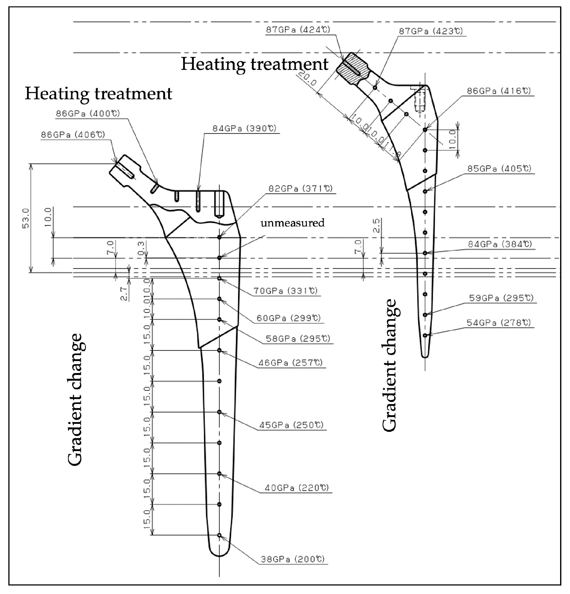
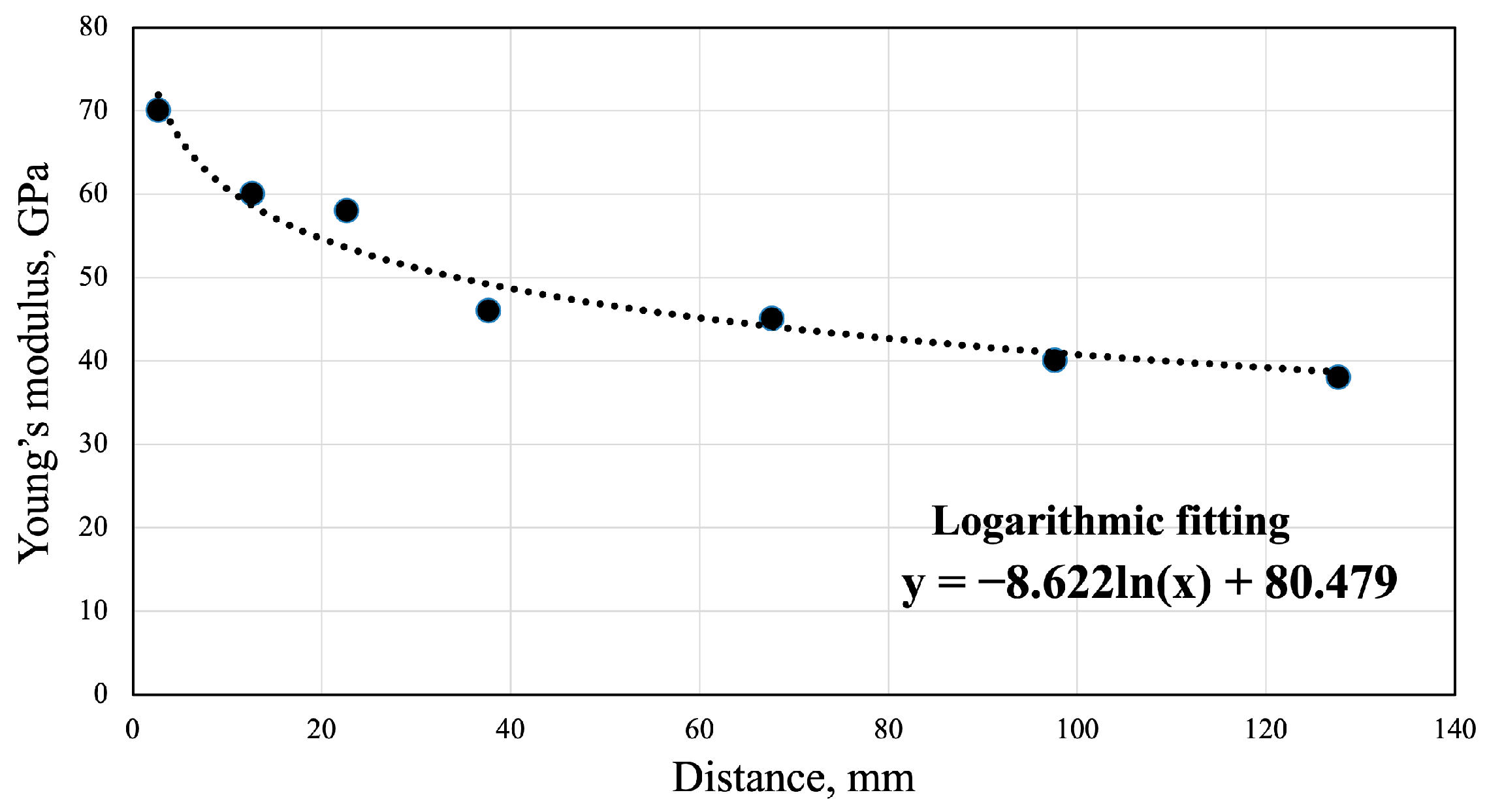
2.1. Construction of Gradient Young’s Modulus FEM

A FEM of the Ti-Nb-Sn alloy stem, which faithfully replicates the graded elasticity achieved by specialized heat treatment, was constructed and analyzed. The gradient in Young’s modulus was derived from the experimental validation data obtained using a custom-designed gradient heat treatment furnace. The localized heat treatment was performed using a custom-designed gradient furnace. The proximal region of the stem was heated at 673 K for 30 min under a vacuum atmosphere (10−3 Pa), while the distal region was protected using an insulating plate to preserve a lower Young’s modulus. This gradient treatment method enabled a continuous transition in mechanical properties without altering the alloy composition [16]. The heat-treated Ti-Nb-Sn stem with a graded Young’s modulus is shown in Figure 1. A logarithmic approximation was used to formulate the relationship between the distance from the insulating plate and the Young’s modulus, which was then assigned to each element of the meshed stem model (Figure 2).
The Ti-Nb-Sn stem was designed with a porous surface coating applied to the proximal one-third of its length to facilitate initial press-fit fixation and promote bone ingrowth. In contrast, the distal two-thirds of the stem were polished to a smooth finish to minimize stress concentration and encourage physiological load transfer to the surrounding bone. This design configuration aims to balance mechanical stability and long-term biological fixation.
The FEM was constructed using tetrahedral elements. Young’s modulus values were mapped to each element based on the formulated gradient curve, starting from the designated gradient initiation point, which was determined in consideration of the fatigue test conditions from the approval process.

2.2. Bone Mineral Density-Reflected Bone Modeling

Patient-specific femoral models were generated from computed tomography (CT) data using Mimics (Materialise, Leuven, Belgium), and bone mineral density was calculated by correlating CT values with a calibration phantom. The Young’s modulus of the bone was then estimated from the derived bone density values and integrated into the model. The stem model was implanted into a reconstructed femoral model representing a female patient in her sixties, characterized by a champagne-flute-shaped femoral canal. The inner cortical cut height was approximately 15 mm above the lesser trochanter (Figure 3). FEM analysis was conducted using Abaqus 2025 (Dassault Systèmes, Velizy Villacoublay, France), and the stem geometry was prepared in SolidWorks (Dassault Systèmes, Velizy Villacoublay, France) [24].

2.3. Boundary and Loading Conditions

Boundary conditions were defined based on physiological loading during walking (P0), and soft tissue constraints were implemented (P1 and P2) following the conditions previously reported [25]. Boundary and loading conditions are shown in Figure 4. In FEM analysis, a press-fit amount of 0.1 mm was applied, with a stem–bone friction coefficient of 0.1 for the porous surface and 0 for the inferior bottom region following the conditions previously reported [26,27,28]. The von Mises stress and micromotion were evaluated.

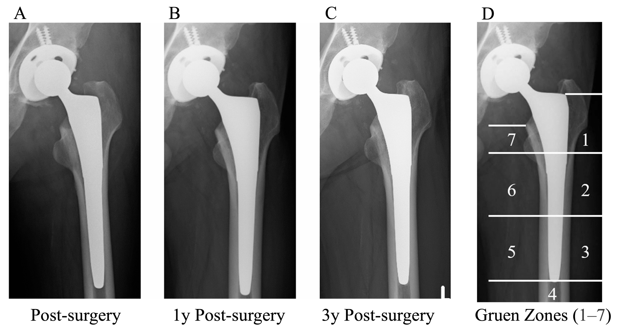
2.4. Assessment of Clinical Image

Regarding the clinical data, results from a previous study were used, with permission [22]. The clinical research methodology is described below. This study was conducted in accordance with the Declaration of Helsinki and received approval from our institutional ethics committee (Approval No. 2021-1-1059). Informed consent was obtained from all participating patients. A total of 40 patients scheduled to undergo unilateral THA with a Ti-Nb-Sn stem were enrolled in the Ti-Nb-Sn group. The inclusion criteria included patients over 20 years of age with a diagnosis of osteoarthritis, avascular necrosis, or rheumatoid arthritis. Patients were excluded if they had previously undergone surgery on the affected hip, including hip arthroplasty, osteotomy, or tenotomy, had bilateral hip disorders, or had rheumatoid arthritis classified as Charnley category C (multiple joint involvement or other conditions severely limiting mobility) [29], a history of deep venous thrombosis or pulmonary embolism, metal allergy, severe obesity (body mass index > 35.0 kg/m2), uncontrolled diabetes mellitus, and infection around the hip joint. Patients with unavailable data in medical records and poor-quality radiographs were excluded.
Anteroposterior radiographs of both hips and lateral radiographs of the affected hip were obtained preoperatively, as well as immediately after surgery, and at 1 and 3 years postoperatively for both the Ti-Nb-Sn and control groups. Stress shielding was evaluated at 1 and 3 years postoperatively using Engh’s classification [30]. Additionally, the distribution of stress shielding at the 3-year follow-up was analyzed according to Gruen zones 1–7 [31]. The radiographic evaluation of stress shielding was independently performed under blinded conditions by two orthopedic surgeons who were not involved in the implantation procedures.
The baseline demographic characteristics of the 40 patients included in the Ti-Nb-Sn group are summarized as follows: The mean age was 64.2 ± 10.7 years, with 36 females (90%) and 4 males (10%). The mean body mass index (BMI) was 24.6 ± 4.1 kg/m2. Bone mineral density (BMD) changes were not assessed in this study. No matched control group was included; thus, clinical and radiographic outcomes were evaluated independently within the Ti-Nb-Sn stem cohort.

2.5. Statistical Analysis

All results were expressed as mean ± standard deviation. Kappa coefficients were calculated for the reproducibility of scores between examiners in the grades of Engh’s classification using SPSS version 21 (IBM, Armonk, NY, USA) to assess the reliability of the measurements.

3. Results

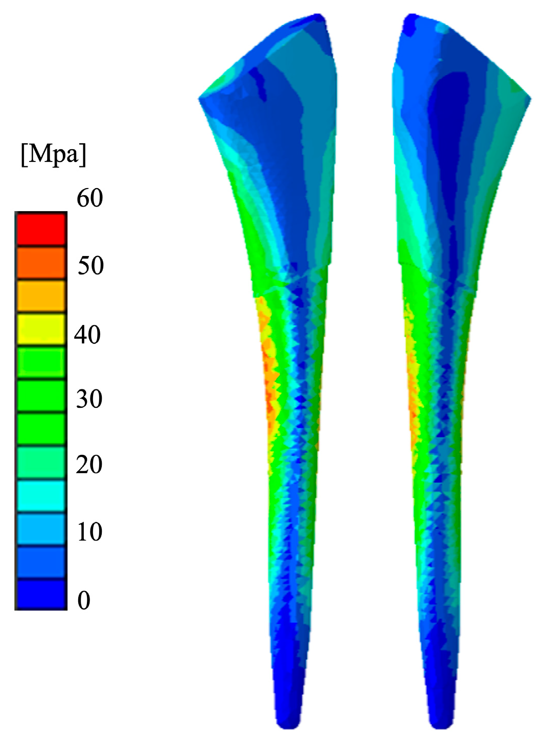
3.1. Mechanical Stress Distribution

Figure 5 shows the results of mechanical stress distribution. In the FEM of the Ti-Nb-Sn stem, the distribution of von Mises stress was high in both the medial and lateral aspects of the central portion of the stem. When comparing the proximal and distal regions, the proximal region exhibited a higher stress distribution. Furthermore, within the proximal region, the medial side showed a greater concentration of stress than the lateral side. In contrast, the stress distribution in the distal region was generally low. The maximum value of von Mises stress observed on the compression side of the Ti-Nb-Sn stem was 52.36 MPa.

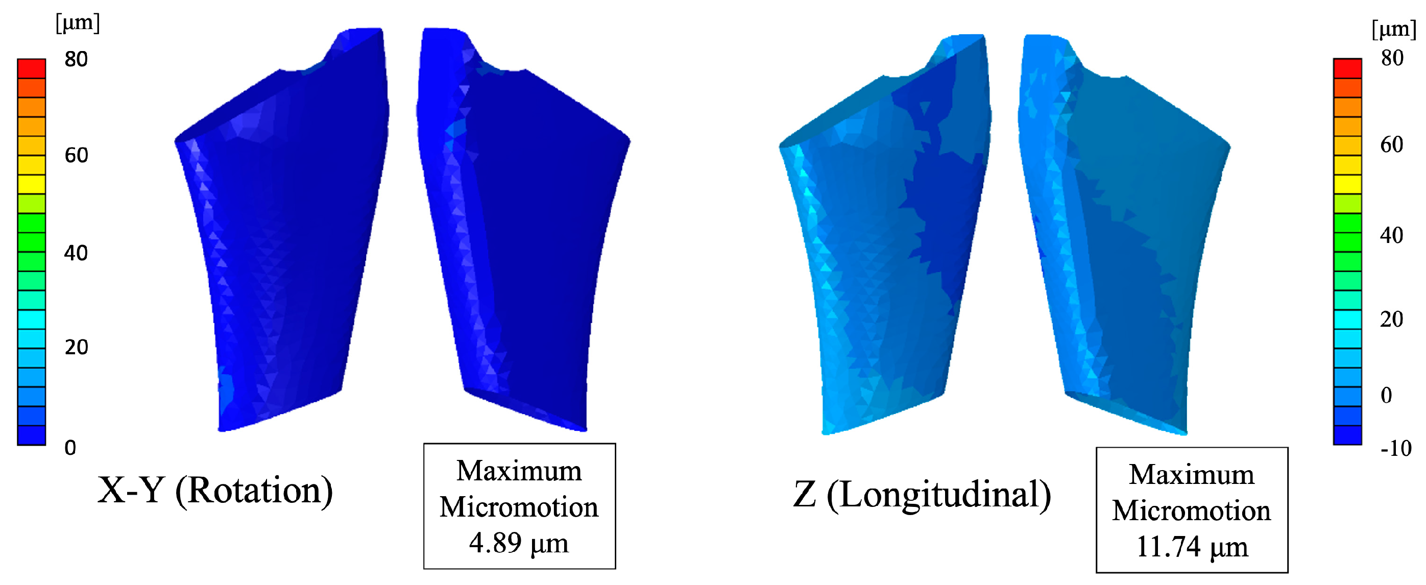
3.2. Micromotion Assessment

Figure 6 shows the distribution of micromotion obtained from the FEM analysis. The calculated maximum micromotion was 4.89 μm in the rotational direction and 11.74 μm in the longitudinal direction. Compared with the reference value of 150 μm [32], which has been reported as the threshold for the onset of implant loosening, the micromotion in both directions was clearly smaller. Furthermore, even in comparison with the more stringent reference value of 40 μm [32], which is considered favorable for bone ingrowth, the present analysis results were well within acceptable limits in both rotational and longitudinal directions.
Figure 7 shows the results of the micromotion distribution analysis at the distal region of the stem. Although the micromotion at the proximal porous press-fit region was minimal, both rotational and longitudinal micromotions tended to increase toward the distal end of the stem. The calculated maximum micromotion at the distal region was 65.38 μm in the rotational direction and 74.65 μm in the longitudinal direction.

3.3. Clinical Image Assessment

Figure 8 shows the radiographic changes associated with stress shielding. The incidence and severity of stress shielding were low in the Ti-Nb-Sn group at both the one-year and three-year follow-ups (Table 1). Interobserver reliability for the assessment of stress shielding was evaluated using kappa coefficients, which were 0.85 and 0.75 at the one-year and three-year follow-ups, respectively, indicating substantial to almost perfect agreement [33]. In addition to Engh’s classification, the interobserver agreement for stress-shielding assessment by Gruen zone was evaluated. The calculated kappa coefficient for Gruen zone-based evaluation was 0.78, indicating substantial reproducibility between observers. The results of the stress-shielding assessment by Gruen zone are presented in Table 2. Stress shielding in the Ti-Nb-Sn group was primarily observed in Zones 1 and 7.

4. Discussion

This study demonstrated that a FEM with a validated Ti-Nb-Sn Young’s modulus gradient provides a highly accurate simulation of in vivo mechanical behavior. Importantly, the proximal micromotion values suggested favorable conditions for osseointegration and long-term stability. Additionally, in the three-year clinical follow-up of Ti-Nb-Sn stem implantation, stress shielding was markedly suppressed. According to Engh’s classification, no cases exceeding Grade 3 stress shielding were reported. This supports the biomechanical findings from FEM simulations and underscores the clinical relevance of Young’s modulus gradient control.
FEM studies of hip arthroplasty indicated that the prosthesis design materially influences stress shielding and load transfer [34,35,36,37]. The reviewed papers employed diverse FEM approaches—including micro-level, CT-based, and patient-specific 3D models—to assess both proximal and distal bone responses [34,38,39]. For example, an optimized design featuring dorsal ridges and perforations achieved a 31.46% reduction in proximal stress shielding, while one comparative study noted that a short stem (Metha) retained 20% of native proximal stress, versus 6% for a standard stem [35,40]. Other investigations documented proximal strain reductions of 40–50% and a ribbed anatomic design with a 71.6% stress-shielding ratio in the posterior quadrant [38,39].
Distal load transfer also varied by design [34,38]. Increased mid-to-distal stresses were associated with some configurations, with one paper reporting distal densification and another observing improved metaphyseal loading linked to higher bone mineral density [40]. Material properties and geometric features—from variable stem rigidity to surface modifications—appeared to govern these biomechanical outcomes [36,38]. Overall, the studies substantiated that modifications in prosthesis design and material parameters are associated with quantifiable differences in stress distribution and bone remodeling after total hip arthroplasty [35,40].
This study provided critical insights from a FEM of a stem fabricated from Ti-Nb-Sn alloy, which enabled the formation of a Young’s modulus gradient through localized heat treatment. By incorporating a controlled gradient in Young’s modulus, the FEM accurately simulated the mechanical environment of the implanted stem, including micromotion and stress distribution. Moreover, the study is of high clinical relevance, as the FEM-derived predictions were validated through mid-term postoperative radiographic evaluations, demonstrating a strong correlation between computational analysis and actual clinical outcomes. This integration of material science, computational modeling, and clinical validation underscores the potential of modulus-graded implants in optimizing stress distribution and minimizing stress shielding in vivo.
Previous studies evaluating conventional Ti-6Al-4V stems have reported considerable stress shielding, with 35–50% of cases exhibiting Engh’s classification Grade 3 or higher within the first 2–3 years postoperatively [4,37]. Additionally, micromotion at the stem–bone interface for Ti-6Al-4V implants has often been reported in the range of 50–150 μm under physiological loading conditions, occasionally exceeding thresholds associated with compromised osseointegration [32]. In contrast, the Ti-Nb-Sn stem with a continuous Young’s modulus gradient demonstrated substantially reduced stress shielding, with no cases exceeding Grade 2 at three years, and achieved micromotion values well below 40 μm in the critical proximal press-fit region. These favorable outcomes highlighted the clinical advantages of using a modulus-graded Ti-Nb-Sn alloy stem to enhance load transfer, preserve proximal bone stock, and promote stable long-term fixation in cementless total hip arthroplasty.
β-type titanium alloys for orthopedic implants are primarily based on Ti-Nb-Zr-Ta systems, with occasional additions of elements, such as Sn, Si, and Sc. These compositions have been shown to offer lower Young’s moduli compared to the conventional Ti-6Al-4V alloy [41,42,43,44]. Reported modulus values ranged from 37 to 85 GPa (versus roughly 110–120 GPa for Ti-6Al-4V) [42,45]. For example, one study of a (Ti-35Nb-7Zr-5Ta)98Si2 alloy reported a Young’s modulus of 37 GPa with a yield strength of 1296 MPa and an ultimate strength of 3263 MPa, while another evaluating a Ti24Nb38Zr2Mo0.1Sc alloy noted a modulus of 65.4 GPa, yield strength of 780 MPa, and ultimate strength of 809 MPa [45,46].
Previous studies indicated that stabilizing the beta phase with niobium and tantalum led to mechanical properties suited for load-bearing applications and that fine-tuning compositions via elements such as Zr, Sn, and Si enhanced strength without sacrificing a low modulus [42,43,44]. In vitro and animal studies documented favorable biological responses—increased cell attachment, proliferation, osteogenic differentiation, and osseointegration—and noted corrosion resistance that was comparable or superior to Ti-6Al-4V [45,46,47]. Processing methods, such as cold rolling, laser deposition, and spark plasma sintering, further tailored the alloys’ microstructures and properties [46]. Although other promising β-type titanium alloys have been reported, TiNbSn alloy remains the only one that has been clinically applied in orthopedic devices, such as joint prostheses.
Several limitations of this study should be acknowledged. First, although the FEM analysis was based on a validated Young’s modulus gradient derived from experimental data and incorporated patient-specific bone properties, the simulations assumed ideal implantation conditions. In clinical practice, however, variations in stem positioning—including varus or valgus alignment—may affect the stress distribution and compromise the intended stress-shielding suppression effect. It remains unclear whether the biomechanical advantages observed in this study would be preserved under such suboptimal alignment conditions. Second, while the micromotion thresholds used to evaluate implant stability (150 μm for loosening and 40 μm for osseointegration) are widely accepted in the literature, they represent general guidelines and may not fully account for patient-specific biological responses. Additionally, the micromotion analysis did not account for dynamic loading cycles over time, which may influence long-term bone–implant interactions and the progression of stress shielding. Third, the FEM analyses in this study were conducted under the assumption of ideal neutral stem alignment. In clinical practice, deviations such as varus or valgus malalignment can occur, which may alter the stress distribution patterns around the femoral stem. Malalignment could shift the load-bearing regions, potentially exacerbating stress shielding in certain Gruen zones or increasing micromotion at the bone–implant interface. Although our FEM results demonstrated favorable biomechanical behavior under neutral conditions, future analyses incorporating surgical variability, such as different alignment scenarios, would be necessary to further validate the robustness of the modulus-graded Ti-Nb-Sn stem design. Fourth, the FEM analysis utilized a femoral morphology characterized by a champagne-flute-shaped canal, which is commonly associated with Dorr type A or B femora. This selection was based on its favorable cortical support and the widespread use of such morphologies in cementless total hip arthroplasty. However, different femoral canal shapes, such as stovepipe configurations observed in Dorr type C femora, may present distinct biomechanical environments. In particular, the reduced cortical thickness and wider canal in Dorr type C bones could affect the stress transfer mechanism and potentially exacerbate stress shielding. Therefore, future studies evaluating the modulus-graded Ti-Nb-Sn stem in a broader range of anatomical variations are warranted to ensure generalizability of the present findings. Fifth, the FEM analysis employed in this study was based on static loading conditions representing peak forces during walking. In clinical reality, implants are subjected to cyclic loading resulting from daily activities, such as walking, stair climbing, and rising from a chair. Repetitive loading could lead to cumulative micromotion at the bone–implant interface, potentially affecting long-term osseointegration and stress-shielding progression. Although our three-year clinical follow-up demonstrated favorable implant stability and minimal stress shielding, future studies incorporating cyclic loading simulations and long-term mechanical fatigue testing are warranted to comprehensively evaluate the durability of the modulus-graded Ti-Nb-Sn stem under repeated physiological stresses. Sixth, although the present study employed a FEM based on static boundary conditions and simplified muscle loading, it did not incorporate comprehensive musculoskeletal modeling. Advanced musculoskeletal simulations can better replicate physiological loading patterns by accounting for complex muscle forces, joint kinematics, and postural changes, thereby potentially improving the accuracy of implant–bone interaction assessments. Incorporating such approaches, as demonstrated in recent studies optimizing biomechanical strategies for injury prevention [48], may further enhance the predictive power of future biomechanical analyses for total hip arthroplasty. Seventh, although clinical validation was performed through radiographic assessment at one and three years postoperatively, the sample size was limited, and the follow-up duration was relatively short. Long-term follow-up studies are needed to confirm the durability of the stress-shielding suppression effect and the continued mechanical integrity of the modulus-graded Ti-Nb-Sn stem. Finally, this study focused on a single patient-specific femoral morphology—a champagne-flute-shaped canal in a female patient in her sixties. The generalizability of these findings to other morphotypes, age groups, or bone qualities (e.g., osteoporotic bone) has yet to be established.

5. Conclusions

This study demonstrated that a FEM incorporating a validated Young’s modulus gradient of a Ti-Nb-Sn alloy stem accurately replicated the mechanical behavior observed in vivo. The FEM analysis revealed favorable stress distribution and low proximal micromotion, suggesting optimal conditions for osseointegration and long-term implant stability. Importantly, these biomechanical findings were consistent with clinical outcomes, showing minimal stress shielding at both one and three years postoperatively. The ability to tailor the Young’s modulus through localized heat treatment enabled the design of stems that simultaneously achieved proximal strength and distal flexibility, thereby addressing the long-standing issue of stress shielding in cementless total hip arthroplasty. These findings support the clinical utility of modulus-graded implants and highlight the potential of Ti-Nb-Sn alloy stems as a promising solution for improving long-term fixation and bone preservation. Future studies with larger cohorts, longer follow-up periods, and diverse femoral morphologies are warranted to further validate these results and optimize implant design.

Author Contributions

Conceptualization, Y.M.; methodology, Y.M.; software, K.S.; validation, Y.M., H.T., H.K. and R.K.; formal analysis, Y.M. and K.S.; investigation, Y.M.; resources, Y.M.; data curation, Y.M., H.T., H.K., R.K. and K.S.; writing—original draft preparation, Y.M.; writing—review and editing, Y.M., H.T., H.K., R.K., N.M., K.S. and T.A.; visualization, Y.M.; supervision, T.A.; project administration, Y.M. All authors have read and agreed to the published version of the manuscript.

Funding

This research received no external funding.

Institutional Review Board Statement

The study was conducted in accordance with the Declaration of Helsinki and approved by the Institutional Review Board of Tohoku University Hospital (protocol code 2021-1-1059; approved on 8 December 2021).

Informed Consent Statement

Informed consent was obtained from all subjects involved in the study. Written informed consent has been obtained from the patient(s) to publish this paper.

Data Availability Statement

The data that support the findings of this study are available upon request from the corresponding author.

Conflicts of Interest

The authors declare no conflicts of interest.

Abbreviations

The following abbreviations are used in this manuscript:
THATotal hip arthroplasty
FEMFinite element model
Ti-Nb-SnTi-33.6%Nb-4Sn
CTComputed tomography

References

  1. Amendola, R.L.; Goetz, D.D.; Liu, S.S.; Callaghan, J.J. Two- to 4-Year Followup of a Short Stem THA Construct: Excellent Fixation, Thigh Pain a Concern. Clin. Orthop. Relat. Res. 2017, 475, 375–383. [Google Scholar] [CrossRef] [PubMed]
  2. Kanto, M.; Fukunishi, S.; Fukui, T.; Nishio, S.; Fujihara, Y.; Okahisa, S.; Takeda, Y.; Yoshiya, S.; Tachibana, T. Radiological Evaluation of the Relationship Between Cortical Hypertrophy and Stress Shielding After Total Hip Arthroplasty Using a Cementless Stem. Arthroplast. Today 2020, 6, 894–900. [Google Scholar] [CrossRef] [PubMed]
  3. Maeda, T.; Nakano, M.; Nakamura, Y.; Momose, T.; Sobajima, A.; Takahashi, J.; Nakata, K.; Nawata, M. Relationship between Stress Shielding and Optimal Femoral Canal Contact Regions for Short, Tapered-Wedge Stem Analyzed by 2D and 3D Systems in Total Hip Arthroplasty. J. Clin. Med. 2023, 12, 3138. [Google Scholar] [CrossRef] [PubMed]
  4. Engh, C.A., Jr.; Young, A.M.; Engh, C.A., Sr.; Hopper, R.H., Jr. Clinical consequences of stress shielding after porous-coated total hip arthroplasty. Clin. Orthop. Relat. Res. 2003, 417, 157–163. [Google Scholar] [CrossRef]
  5. Kusano, T.; Seki, T.; Higuchi, Y.; Takegami, Y.; Osawa, Y.; Ishiguro, N. Preoperative Canal Bone Ratio is Related to High-Degree Stress Shielding: A Minimum 5-Year Follow-Up Study of a Proximally Hydroxyapatite-Coated Straight Tapered Titanium Femoral Component. J. Arthroplast. 2018, 33, 1764–1769. [Google Scholar] [CrossRef]
  6. Huiskes, R. The various stress patterns of press-fit, ingrown, and cemented femoral stems. Clin. Orthop. Relat. Res. 1990, 261, 27–38. [Google Scholar] [CrossRef]
  7. Van Rietbergen, B.; Huiskes, R.; Weinans, H.; Sumner, D.R.; Turner, T.M.; Galante, J.O. ESB Research Award 1992. The mechanism of bone remodeling and resorption around press-fitted THA stems. J. Biomech. 1993, 26, 369–382. [Google Scholar] [CrossRef]
  8. Lindahl, H. Epidemiology of periprosthetic femur fracture around a total hip arthroplasty. Injury 2007, 38, 651–654. [Google Scholar] [CrossRef]
  9. Kim, J.T.; Jeong, H.J.; Lee, S.J.; Kim, H.J.; Yoo, J.J. Does Proximally Coated Single-Wedge Cementless Stem Work Well in Dorr Type C Femurs? Minimum 10-year Followup. Indian J. Orthop. 2019, 53, 94–101. [Google Scholar] [CrossRef]
  10. Tsubosaka, M.; Hayashi, S.; Hashimoto, S.; Takayama, K.; Kuroda, R.; Matsumoto, T. Patients with a Dorr type C femoral bone require attention for using a Summit cementless stem: Results of total hip arthroplasty after a minimum follow-up period of 5 years after insertion of a Summit cementless stem. J. Orthop. Sci. 2018, 23, 671–675. [Google Scholar] [CrossRef]
  11. Dorr, L.D.; Faugere, M.C.; Mackel, A.M.; Gruen, T.A.; Bognar, B.; Malluche, H.H. Structural and cellular assessment of bone quality of proximal femur. Bone 1993, 14, 231–242. [Google Scholar] [CrossRef] [PubMed]
  12. Huiskes, R.; Hollister, S.J. From structure to process, from organ to cell: Recent developments of FE-analysis in orthopaedic biomechanics. J. Biomech. Eng. 1993, 115, 520–527. [Google Scholar] [CrossRef] [PubMed]
  13. Rho, J.Y.; Ashman, R.B.; Turner, C.H. Young’s modulus of trabecular and cortical bone material: Ultrasonic and microtensile measurements. J. Biomech. 1993, 26, 111–119. [Google Scholar] [CrossRef]
  14. Yamako, G.; Janssen, D.; Hanada, S.; Anijs, T.; Ochiai, K.; Totoribe, K.; Chosa, E.; Verdonschot, N. Improving stress shielding following total hip arthroplasty by using a femoral stem made of β type Ti-33.6Nb-4Sn with a Young’s modulus gradation. J. Biomech. 2017, 63, 135–143. [Google Scholar] [CrossRef] [PubMed]
  15. Naghavi, S.A.; Tamaddon, M.; Garcia-Souto, P.; Moazen, M.; Taylor, S.; Hua, J.; Liu, C. A novel hybrid design and modelling of a customised graded Ti-6Al-4V porous hip implant to reduce stress-shielding: An experimental and numerical analysis. Front. Bioeng. Biotechnol. 2023, 11, 1092361. [Google Scholar] [CrossRef]
  16. Hanada, S.; Masahashi, N.; Jung, T.K.; Yamada, N.; Yamako, G.; Itoi, E. Fabrication of a high-performance hip prosthetic stem using β Ti-33.6Nb-4Sn. J. Mech. Behav. Biomed. Mater. 2014, 30, 140–149. [Google Scholar] [CrossRef]
  17. Ito, K.; Mori, Y.; Kamimura, M.; Koguchi, M.; Kurishima, H.; Koyama, T.; Mori, N.; Masahashi, N.; Hanada, S.; Itoi, E.; et al. β-type TiNbSn Alloy Plates with Low Young Modulus Accelerates Osteosynthesis in Rabbit Tibiae. Clin. Orthop. Relat. Res. 2022, 480, 1817–1832. [Google Scholar] [CrossRef]
  18. Mori, Y.; Fujisawa, H.; Kamimura, M.; Kogure, A.; Tanaka, H.; Mori, N.; Masahashi, N.; Aizawa, T. Acceleration of Fracture Healing in Mouse Tibiae Using Intramedullary Nails Composed of β-Type TiNbSn Alloy with Low Young’s Modulus. Tohoku J. Exp. Med. 2021, 255, 135–142. [Google Scholar] [CrossRef]
  19. Kogure, A.; Mori, Y.; Tanaka, H.; Kamimura, M.; Masahashi, N.; Hanada, S.; Itoi, E. Effects of elastic intramedullary nails composed of low Young’s modulus Ti-Nb-Sn alloy on healing of tibial osteotomies in rabbits. J. Biomed. Mater. Res. Part B Appl. Biomater. 2019, 107, 700–707. [Google Scholar] [CrossRef]
  20. Mori, Y.; Kamimura, M.; Ito, K.; Koguchi, M.; Tanaka, H.; Kurishima, H.; Koyama, T.; Mori, N.; Masahashi, N.; Aizawa, T. A Review of the Impacts of Implant Stiffness on Fracture Healing. Appl. Sci. 2024, 14, 2259. [Google Scholar] [CrossRef]
  21. Chiba, D.; Yamada, N.; Mori, Y.; Oyama, M.; Ohtsu, S.; Kuwahara, Y.; Baba, K.; Tanaka, H.; Aizawa, T.; Hanada, S.; et al. Mid-term results of a new femoral prosthesis using Ti-Nb-Sn alloy with low Young’s modulus. BMC Musculoskelet. Disord. 2021, 22, 987. [Google Scholar] [CrossRef] [PubMed]
  22. Baba, K.; Mori, Y.; Chiba, D.; Kuwahara, Y.; Kurishima, H.; Tanaka, H.; Kogure, A.; Kamimura, M.; Yamada, N.; Ohtsu, S.; et al. TiNbSn stems with gradient changes of Young’s modulus and stiffness reduce stress shielding compared to the standard fit-and-fill stems. Eur. J. Med. Res. 2023, 28, 214. [Google Scholar] [CrossRef]
  23. Liu, B.; Wang, H.; Zhang, N.; Zhang, M.; Cheng, C.K. Femoral Stems with Porous Lattice Structures: A Review. Front. Bioeng. Biotechnol. 2021, 9, 772539. [Google Scholar] [CrossRef] [PubMed]
  24. Koyama, T.; Mori, Y.; Kamimura, M.; Tanaka, H.; Tome, R.; Ito, K.; Koguchi, M.; Mori, N.; Aizawa, T. TiNbSn alloy plates with low Young’s modulus modulates interfragmentary movement and promote osteosynthesis in rat femur. J. Mech. Behav. Biomed. Mater. 2025, 161, 106820. [Google Scholar] [CrossRef]
  25. Heller, M.O.; Bergmann, G.; Kassi, J.P.; Claes, L.; Haas, N.P.; Duda, G.N. Determination of muscle loading at the hip joint for use in pre-clinical testing. J. Biomech. 2005, 38, 1155–1163. [Google Scholar] [CrossRef]
  26. Rancourt, D.; Shirazi-Adl, A.; Drouin, G.; Paiement, G. Friction properties of the interface between porous-surfaced metals and tibial cancellous bone. J. Biomed. Mater. Res. 1990, 24, 1503–1519. [Google Scholar] [CrossRef]
  27. Hefzy, M.S.; Singh, S.P. Comparison between two techniques for modeling interface conditions in a porous coated hip endoprosthesis. Med. Eng. Phys. 1997, 19, 50–62. [Google Scholar] [CrossRef]
  28. Muccini, R.; Baleani, M.; Viceconti, M. Selection of the best element type in the finite element analysis of hip prostheses. J. Med. Eng. Technol. 2000, 24, 145–148. [Google Scholar] [CrossRef] [PubMed]
  29. Charnley, J. The long-term results of low-friction arthroplasty of the hip performed as a primary intervention. J. Bone Joint Surg. Br. 1972, 54, 61–76. [Google Scholar] [CrossRef]
  30. Engh, C.A.; Bobyn, J.D.; Glassman, A.H. Porous-coated hip replacement. The factors governing bone ingrowth, stress shielding, and clinical results. J. Bone Joint Surg. Br. 1987, 69, 45–55. [Google Scholar] [CrossRef]
  31. Gruen, T.A.; McNeice, G.M.; Amstutz, H.C. “Modes of failure” of cemented stem-type femoral components: A radiographic analysis of loosening. Clin. Orthop. Relat. Res. 1979, 141, 17–27. [Google Scholar] [CrossRef]
  32. Sanchez, E.; de Vries, E.; Matthews, D.; van der Heide, E.; Janssen, D. The effect of coating characteristics on implant-bone interface mechanics. J. Biomech. 2024, 163, 111949. [Google Scholar] [CrossRef]
  33. Landis, J.R.; Koch, G.G. The measurement of observer agreement for categorical data. Biometrics 1977, 33, 159–174. [Google Scholar] [CrossRef]
  34. Boyle, C.; Kim, I.Y. Comparison of different hip prosthesis shapes considering micro-level bone remodeling and stress-shielding criteria using three-dimensional design space topology optimization. J. Biomech. 2011, 44, 1722–1728. [Google Scholar] [CrossRef]
  35. Ceddia, M.; Trentadue, B. Evaluation of Rotational Stability and Stress Shielding of a Stem Optimized for Hip Replacements-A Finite Element Study. Prosthesis 2023, 5, 678–693. [Google Scholar] [CrossRef]
  36. Ceddia, M.; Solarino, G.; Giannini, G.; De Giosa, G.; Tucci, M.; Trentadue, B. A Finite Element Analysis Study of Influence of Femoral Stem Material in Stress Shielding in a Model of Uncemented Total Hip Arthroplasty: Ti-6Al-4V versus Carbon Fibre-Reinforced PEEK Composite. J. Compos. Sci. 2024, 8, 254. [Google Scholar] [CrossRef]
  37. Hirata, Y.; Inaba, Y.; Kobayashi, N.; Ike, H.; Fujimaki, H.; Saito, T. Comparison of mechanical stress and change in bone mineral density between two types of femoral implant using finite element analysis. J. Arthroplast. 2013, 28, 1731–1735. [Google Scholar] [CrossRef] [PubMed]
  38. Heyland, M.; Checa, S.; Kendoff, D.; Duda, G.N. Anatomic grooved stem mitigates strain shielding compared to established total hip arthroplasty stem designs in finite-element models. Sci. Rep. 2019, 9, 482. [Google Scholar] [CrossRef]
  39. Luo, C.; Wu, X.D.; Wan, Y.; Liao, J.; Cheng, Q.; Tian, M.; Bai, Z.; Huang, W. Femoral Stress Changes after Total Hip Arthroplasty with the Ribbed Prosthesis: A Finite Element Analysis. Biomed. Res. Int. 2020, 2020, 6783936. [Google Scholar] [CrossRef]
  40. Yan, S.G.; Chevalier, Y.; Liu, F.; Hua, X.; Schreiner, A.; Jansson, V.; Schmidutz, F. Metaphyseal anchoring short stem hip arthroplasty provides a more physiological load transfer: A comparative finite element analysis study. J. Orthop. Surg. Res. 2020, 15, 498. [Google Scholar] [CrossRef]
  41. Qazi, J.I.; Rack, H.J. Metastable beta titanium alloys for orthopedic applications. Adv. Eng. Mater. 2005, 7, 993–998. [Google Scholar] [CrossRef]
  42. Kopova, I.; Strasky, J.; Harcuba, P.; Landa, M.; Janecek, M.; Bacakova, L. Newly developed Ti-Nb-Zr-Ta-Si-Fe biomedical beta titanium alloys with increased strength and enhanced biocompatibility. Mater. Sci. Eng. C 2016, 60, 230–238. [Google Scholar] [CrossRef] [PubMed]
  43. Cimpean, A.; Mitran, V.; Ciofrangeanu, C.M.; Galateanu, B.; Bertrand, E.; Gordin, D.M.; Iordachescu, D.; Gloriant, T. Osteoblast cell behavior on the new beta-type Ti-25Ta-25Nb alloy. Mater. Sci. Eng. C 2012, 32, 1554–1563. [Google Scholar] [CrossRef]
  44. Bahl, S.; Das, S.; Suwas, S.; Chatterjee, K. Engineering the next-generation tin containing β titanium alloys with high strength and low modulus for orthopedic applications. J. Mech. Behav. Biomed. Mater. 2018, 78, 124–133. [Google Scholar] [CrossRef]
  45. Sun, Y.; Liu, Q.P.; Yu, Z.L.; Ren, L.Q.; Zhao, X.; Wang, J.C. Study on Osseointegration Capability of β-Type Ti-Nb-Zr-Ta-Si Alloy for Orthopedic Implants. Materials 2024, 17, 472. [Google Scholar] [CrossRef]
  46. Tong, X.; Sun, Q.; Zhang, D.; Wang, K.; Dai, Y.; Shi, Z.; Li, Y.; Dargusch, M.; Huang, S.; Ma, J.; et al. Impact of scandium on mechanical properties, corrosion behavior, friction and wear performance, and cytotoxicity of a β-type Ti-24Nb-38Zr-2Mo alloy for orthopedic applications. Acta. Biomater. 2021, 134, 791–803. [Google Scholar] [CrossRef] [PubMed]
  47. Samuel, S.; Nag, S.; Nasrazadani, S.; Ukirde, V.; El Bouanani, M.; Mohandas, A.; Nguyen, K.; Banerjee, R. Corrosion resistance and in vitro response of laser-deposited Ti-Nb-Zr-Ta alloys for orthopedic implant applications. J. Biomed. Mater. Res. Part A 2010, 94, 1251–1256. [Google Scholar] [CrossRef]
  48. Xu, D.T.; Zhou, H.Y.; Quan, W.J.; Ma, X.; Chon, T.E.; Fernandez, J.; Gusztav, F.; Kovács, A.; Baker, J.S.; Gu, Y. New Insights Optimize Landing Strategies to Reduce Lower Limb Injury Risk. Cyborg Bionic Syst. 2024, 5, 0126. [Google Scholar] [CrossRef]
Figure 1. Construction of the FEM for the gradient functional Ti-Nb-Sn stem. Gradient information derived from experimental validation was applied to the meshed FEM. The relationship between the distance from the heat-insulating plate and Young’s modulus was formulated, and material properties were assigned to each mesh element accordingly.
Applsci 15 04827 g001
Figure 2. Young’s modulus gradient curve of the Ti-Nb-Sn stem derived from validation experiments. The relationship between the distance from the insulating plate and the Young’s modulus was formulated.
Applsci 15 04827 g002
Figure 3. Schematic representation of the FEM analysis model. The FEM of Young’s modulus-graded Ti-Nb-Sn stem was implanted into patient-specific femoral data reflecting bone density. A champagne-flute-shaped femoral canal from a female patient in her 60s was selected for the simulation.
Applsci 15 04827 g003
Figure 4. Boundary and loading conditions of the FEM analysis. The distal end was fully constrained. P0 represents the peak load during walking, while P1, P2, and P3 account for the effects of surrounding hip muscles. Von Mises stress and micromotion were evaluated under these conditions.
Applsci 15 04827 g004
Figure 5. Distribution of von Mises stress. Distribution of von Mises stress within the Ti-Nb-Sn femoral stem under physiological loading conditions. A color bar representing stress magnitude (MPa) is presented.
Applsci 15 04827 g005
Figure 6. FEM analysis of micromotion at the proximal region of the stem. The calculated maximum micromotion was 4.89 μm in the rotational direction and 11.74 μm in the longitudinal direction. A color bar representing micromotion magnitude (μm) is presented.
Applsci 15 04827 g006
Figure 7. FEM analysis of micromotion at the distal region of the stem. Although micromotion at the porous press-fit area was minimal, both rotational and longitudinal micromotion values were higher at the distal tip of the stem. A color bar representing micromotion magnitude (μm) is presented.
Applsci 15 04827 g007
Figure 8. Three-year follow-up radiographs of the Ti-Nb-Sn hip implant. (A) Immediately post-surgery, (B) one year post-surgery, and (C) three years post-surgery. (D) Schematic illustration of the Gruen zones (1–7) used for the radiographic evaluation of femoral stress shielding. No apparent stress shielding was observed. Reprinted with permission from [22].
Applsci 15 04827 g008
Table 1. Incidence of stress shielding with the Ti-Nb-Sn stem.
Engh’s Classification1-Year Follow-Up3-Year Follow-Up
n, (ratio)n, (ratio)
None19, (47.5%)14, (35%)
Grade 117, (42.5%)10, (25%)
Grade 24, (10%)16, (40%)
Grade 30, (0%)0, (0%)
Grade 40, (0%)0, (0%)
Table 2. Frequency of stress shielding by Gruen zone.
Gruen ZoneZone 1Zone 2Zone 3Zone 4Zone 5Zone 6Zone 7
Stress shielding9, (23%)0, (0%)0, (0%)0, (0%)0, (0%)1, (2.5%)21 (53%)
Disclaimer/Publisher’s Note: The statements, opinions and data contained in all publications are solely those of the individual author(s) and contributor(s) and not of MDPI and/or the editor(s). MDPI and/or the editor(s) disclaim responsibility for any injury to people or property resulting from any ideas, methods, instructions or products referred to in the content.

