Review

Chitosan-Based Nanocarriers for Nose to Brain Delivery

by
Blessing Atim Aderibigbe
* and
Tobeka Naki
Department of Chemistry, University of Fort Hare, Alice Campus, Ring Road, Alice, Eastern Cape 5700, South Africa
*
Author to whom correspondence should be addressed.
Appl. Sci. 2019, 9(11), 2219; https://doi.org/10.3390/app9112219
Submission received: 8 March 2019 / Revised: 29 March 2019 / Accepted: 2 April 2019 / Published: 30 May 2019
(This article belongs to the Special Issue Nanoscale Materials for Drug Delivery and Tissue Engineering)

Abstract

:
In the treatment of brain diseases, most potent drugs that have been developed exhibit poor therapeutic outcomes resulting from the inability of a therapeutic amount of the drug to reach the brain. These drugs do not exhibit targeted drug delivery mechanisms, resulting in a high concentration of the drugs in vital organs leading to drug toxicity. Chitosan (CS) is a natural-based polymer. It has unique properties such as good biodegradability, biocompatibility, mucoadhesive properties, and it has been approved for biomedical applications. It has been used to develop nanocarriers for brain targeting via intranasal administration. Nanocarriers such as nanoparticles, in situ gels, nanoemulsions, and liposomes have been developed. In vitro and in vivo studies revealed that these nanocarriers exhibited enhanced drug uptake to the brain with reduced side effects resulting from the prolonged contact time of the nanocarriers with the nasal mucosa, the surface charge of the nanocarriers, the nano size of the nanocarriers, and their capability to stretch the tight junctions within the nasal mucosa. The aforementioned unique properties make chitosan a potential material for the development of nanocarriers for targeted drug delivery to the brain. This review will focus on chitosan-based carriers for brain targeting.

Graphical Abstract

1. Introduction

The most complex organ in humans is the central nervous system (CNS). It controls most of the human functions. Its function controls all aspects of our behavior such as breathing, feelings, etc. [1]. Brain diseases can occur as a result of the interactions of the brain with several complex environmental factors [2]. Neurodegenerative disorders are becoming more widespread in society especially the elderly age group. Neurodegenerative disorders result in disability and death worldwide [3]. These diseases presently affect more than 1 billion people worldwide [4]. Currently, these diseases represent a major medical challenge and there is a need for therapeutics which are effective and can overcome the global burden of neurodegenerative disorders [5]. Some of the challenges associated with therapeutics used to treat neurodegenerative disorders are severe effects such as diabetes, dizziness, seizures, Stevens-Johnson syndrome, and toxicity, etc. [6]. Development of drug delivery systems is one approach which has the potential to overcome the pharmacological limitations associated with therapeutics used to treat neurodegenerative disorders [7]. Some delivery systems used for the delivery of drugs to the brain are liposomes, in situ gel, nanogels, nanoparticles, micelles, emulsions, dendrimers, etc. [8]. Most of the aforementioned systems have been effective in transporting bioactive agents across different models of blood-brain barrier (BBB) by known mechanisms in vitro and in vivo. Some of the aforementioned drug delivery systems have also been reported to display good preclinical outcomes which indicate their potential application for the management of conditions such as stroke, HIV (Human Immunodeficiency Virus), Alzheimer’s disease, brain tumors, etc. [9]. However, in the design of these drug delivery systems for brain targeting, factors to be considered are the particle size, their stability in physiological circulation, surface affinity, etc. [10]. In the application of nanoparticles for brain targeting, their drug release profiles have been reported to be sustained, slow, and controlled, thereby overcoming toxicity. Nanoparticles also target specific brain sites which indicate their capability to cross the blood–brain barrier which is attributed to their nano size [11]. Nanosized polymer-based carriers are potential systems for targeted delivery of potent drugs to the brain due to some of their properties such as the protection of the drugs from biological processes which can decrease the drug bioavailability, and their capability to cross the BBB [12]. Several targeted drug delivery systems and imaging devices developed for the treatment and diagnosis of brain diseases have been studied [13]. According to the World Health Organization (WHO), over 50 million people suffer from epilepsy globally. Globally, an estimation of approximately 2.4 million people are diagnosed with epilepsy each year [14]. Parkinson’s disease (PD) is reported to be common among the elderly [15]. Due to the high rate of people living with brain diseases, there is a need for the development of effective therapeutics that can penetrate the BBB. The development of drug delivery systems that can deliver drugs across the blood–brain barrier is a potential approach for the treatment of neurodegenerative disorders. This review article reports the efficacy of chitosan in the development of drug delivery systems for brain targeting.

2. Chitosan Properties and Biomedical Application

Chitosan is a cationic polysaccharide composed of a random composition of β (1→4) linked 2-amino-2-deoxy-D-glucose (D-glucosamine) and 2-acetamido-2-deoxy-D-glucose(N-acetyl-D-glucosamine) units [16]. It has a structure which is similar to cellulose and it also has chelating properties. It is obtained from deacetylation of a polysaccharide found in crustacean shells known as chitin (Figure 1) [17]. It exhibits a positive charge on its surface resulting from the presence of primary amino groups on its polymer backbone. Its ability to form inter and intramolecular hydrogen bonding and its polycationic surface make it suitable for the development of formulations for intranasal administration [17]. It exhibits good biocompatibility and it is suitable for the design of targeted drug delivery systems for the treatment of neurodegenerative disorders. Its good biodegradability property affects its bioactivity on the blood–brain barrier at the molecular level, which is advantageous in therapies for neurological disorders [18,19,20]. It is also hydrophilic in nature, which makes it useful for biomedical applications [21]. It is soluble in acidic media and exhibits unique properties useful in drug delivery, such as mucoadhesion, good gelling capability, hydrophilic nature, and good permeation effects [22]. There are many challenges associated with the treatment of neurodegenerative diseases. Effective drug delivery to the brain requires understanding the physiology and anatomy of the blood–brain barrier and modifying the surface of the drug delivery systems with antibodies for brain delivery. Therefore, research on brain-targeted delivery systems is a significant approach that can overcome the problems associated with therapeutics used to treat neurodegenerative disorders. A major limitation in the treatment of neurological diseases is the inability of the conventional drug molecules to cross the blood–brain barrier.

3. Brain Diseases

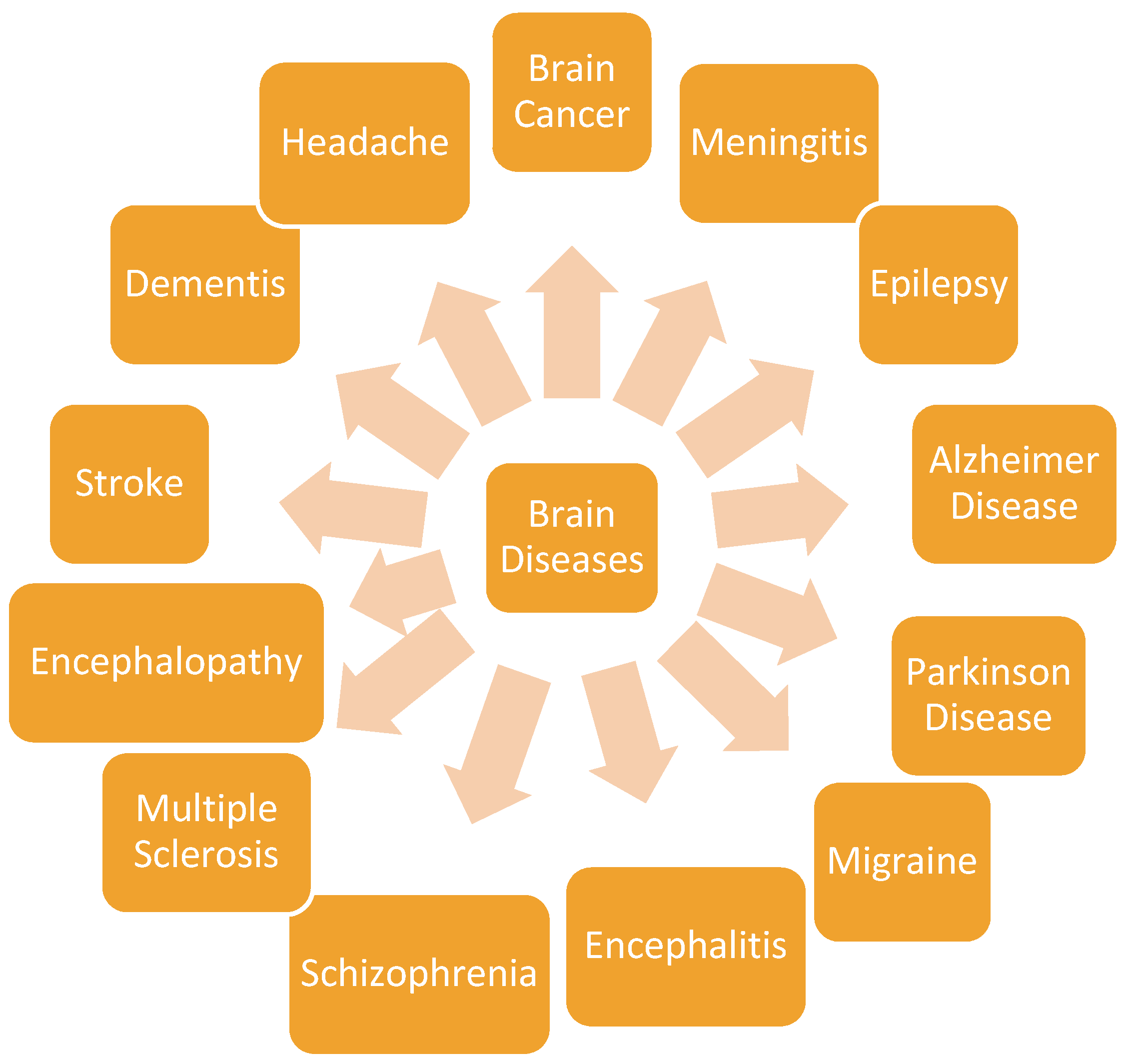
There are several diseases that affect the human brain (Figure 2). The treatment of brain diseases is challenging, resulting from several factors such as the side effects of the drugs currently used for the treatment of brain diseases, the blood–brain barrier, the association of genes, and the overlap of disease-associated genes [6]. The mechanisms behind neurological diseases are not fully understood. Some of the diseases that affect the brain include meningitis; gliomas, which are brain tumors; Alzheimer’s disease; epilepsy; Parkinson’s disease; meningitis, etc.
Meningitis is linked with the inflammation of the meninges [23]. Some risk factors that contribute to meningitis are chronic kidney failure, HIV infection, alcoholism, drug abuse, etc. Some of the medications used to treat meningitis are sulphonamides, tetracyclines, carbapenems, fluoroquinolones, glycopeptides, aminoglycosides, etc. Some of these drugs are administered intrathecally, intravenously, and orally, resulting from their poor penetration to the cerebrospinal fluid [23]. Brain tumors are abnormal growth of the tissue in the central spine or brain resulting in the disruption of the proper functioning of the brain. It can be classified as either a cancerous or non-cancerous tumor [6]. The risk factors associated with brain tumors are chemicals, genetic manipulation, viral infection, and ionizing radiation [24]. The treatment of brain tumors is very challenging and some of the drugs used for the treatment of brain tumors are carmustine wafer, lomustine, bevacizumab, and temozolomide, etc. Migraine is a headache associated with sensitivity to light, vomiting, and nausea. The factors that contribute to migraine are loud noises, anxiety, depression, overuse of medications, stress, changes in sleep patterns, etc. [25]. There is currently no cure for migraine headaches. Migraine is managed by preventing recurring migraines and treating acute attacks. Migraine headache is treated using antiepileptic drugs, beta-blockers, and triptans [25].
Stroke is a neurological disease which is classified as either ischemic or hemorrhagic stroke. Ischemic stroke results from an insufficient flow of blood to the brain due to the blockage or narrowed arteries due to conditions such as atherosclerosis, concurrent myocardial infarction, congestive heart failure, dilated cardiomyopathy, and atrial fibrillation, etc. [26]. Hemorrhagic stroke can be referred to as intracerebral hemorrhage, which originates from the rupture of weak cerebral vessels, thereby forming localized hematoma in the parenchymal cerebral space. Hemorrhagic stroke can also be classified as subarachnoid hemorrhage, which occurs outside the brain but is released into the cerebral spinal fluid. Some causes of stroke are hypertension, vascular malformation, trauma, etc. [26]. Stroke is treated using anticoagulants, antiplatelets, tissue plasminogen activator, statins, and medications for the treatment of blood pressure such as diuretics, ACE (Angiotensin converting enzyme) inhibitors, etc. Some of the medications used for the treatment of stroke suffer from side effects such as blood disorder, liver damage, muscle damage, and kidney failure [27]. Epilepsy is a brain disorder with recurrent seizures due to excess electrical discharges in the brain cells [6]. Factors which contribute to the disease are severe head injury, brain tumor, meningitis, birth-related injuries, encephalitis, etc. [28]. Some of the drugs used to treat epilepsy are brivaracetam, cannabidiol, carbamazepine, diazepam, etc. These drugs exhibit adverse effects such as hepatitis, aplastic anemia, allergic rashes, etc. [28]. The limitation of antiepileptic drugs in some patients is due to the pharmacoresistance caused by mechanisms which can be classified as disease-related, genetics, and drug-related mechanisms. The disease-related mechanism alters the pharmacological targets of antiepileptic drugs in the brains resulting in the failure of the drugs to block excitatory sodium or calcium currents [6]. In the genetic-related mechanism, poor seizure occurs due to drug efflux transporters, and the drug-related mechanism reduces the efficacy of antiepileptic drugs [29].
In Alzheimer’s disease, the brain nerve cells are destroyed leading to dementia [6]. The risk factors that contribute to Alzheimer’s disease are the damage to the neurons due to a high concentration of homocysteine levels and copper, head injury, vitamin D deficiency, etc. [30]. The disease is managed using medications such as donepezil, galantamine, rivastigmine, memantine, etc. The side effects associated with these drugs are the risk of diabetes, liver problems, depression, vomiting, headache, etc. [31]. Parkinson disease is characterized by loss of pigmented melanin-containing neurons in the midbrain and the presence of aggregates of protein alpha-synuclein in the Lewy bodies [6,32]. The treatment approach to Parkinson’s disease is symptomatic. In severe cases of Parkinson’s disease, the motor symptoms are managed by performing deep brain stimulation [33]. The classes of drugs used to treat the disease are anticholinergics, dopamine precursors, dopamine agonists, and monoamine oxidase B inhibitors [33].
Schizophrenia is a brain disorder caused by a deficiency or excess neurotransmitters. Genetic and environmental factors contribute to the disease [6,34]. It is managed with antipsychotic drugs that suffer from limitations such as hyperlipidemia, diabetes mellitus, increased risk of cardiovascular mortality, dystonia, weight gain, sexual dysfunction, etc. [35].

4. Brain Targeting

The blood–brain barrier (BBB) hinders the successful delivery of potential and potent therapeutics to the brain. There are three strategies used to deliver therapeutics to the brain [36]: (i) The first strategy is to bypass the BBB by using other routes of drug delivery such as drug-encapsulated wafers inserted in the tumor cavity during surgical resection of the brain tumors, facial intradermal injection, and drugs administered via the nasal cavity; (ii) by interrupting the BBB using surfactants and hyperosmotic agents, cell-penetrating peptides, magnetic nanoparticle-induced hyperthermia; and (iii) by the application of endogenous transporters and receptors for enhanced neural penetration of drugs in a non-invasive approach. Passive targeting is the most commonly used approach to deliver nanoparticles into brain tumors for diagnostic and therapeutic applications.
The nose-to-brain transport enhance drug targeting with reduced systemic side effects [37]. Bypassing the BBB and the direct targeting of the brain via olfactory and trigeminal neural pathways is a crucial approach for the delivery of therapeutics to the brain [38]. The transport of therapeutics via the olfactory mucosa pathway is rapid and it is achieved by drug administration via the nasal cavity. However, volume in the range of 25–200 μL can be intranasally administered resulting in the limitation of the concentration of drug that can be transported into the brain from the nasal cavity [39].
Trigeminal nerve pathway is connected to some parts of the brain such as the pons, medulla, and spinal cord. The trigeminal nerve has three branches known as the mandibular, ophthalmic, and maxillary. However, only the ophthalmic and maxillary branches have been reported to be useful for drug delivery to the brain from the nose resulting from the neurons in this branches that pass via the nasal mucosa [40]. Drug uptake via nose through the trigeminal nerve pathway is by either intracellular transport or endocytosis. When a drug is administered intranasally, it can diffuse via the nasal mucosa to the branches of trigeminal nerves present in the olfactory and respiratory regions. The drug is then transported through the brain stem to the axonal route. It has also been reported that the trigeminal nerve that passes through the cribriform plate is involved in the delivery of drugs from the nose to the forebrain [41]. Research reports also further indicated that after the absorption of the drug from nasal cavity through the mucus, several mechanisms are involved when the drug is transported through mucosa such as the paracellullar, transcellular, carrier-mediated transport, receptor-mediated transport, and transcytosis [42,43].
Drugs administered intranasally are also transported via the olfactory pathway to the olfactory mucosa which contains olfactory receptor neurons. The drug molecules are transported to the olfactory receptor neurons by paracellullar or transcellular mechanism. Factors such as the integrity of nasal epithelium, the tight junctions, space between the epithelial cells, etc., enhance drug uptake via paracellular mechanism [40]. The drug is transported via the axon and nerve bundle across the cribriform plate to the olfactory bulb on the surface of the brain. The drug transported from the olfactory nerves can enter the cerebrospinal fluid (CSF) and olfactory bulb to the brain by combining with the interstitial fluid in the brain [44]. The transportation of the drug from the nasal cavity to the brain via olfactory transport is rapid. The transportation of drug molecule via the olfactory pathway to the brain occurs by two known pathways, such as the intraneuronal and extra-neuronal pathways. In the intra-neuronal pathway, axonal transport is involved, and the drug transportation is slow. In the extra-neuronal pathway, drug transport to the brain via perineural channels and it is rapid [40].
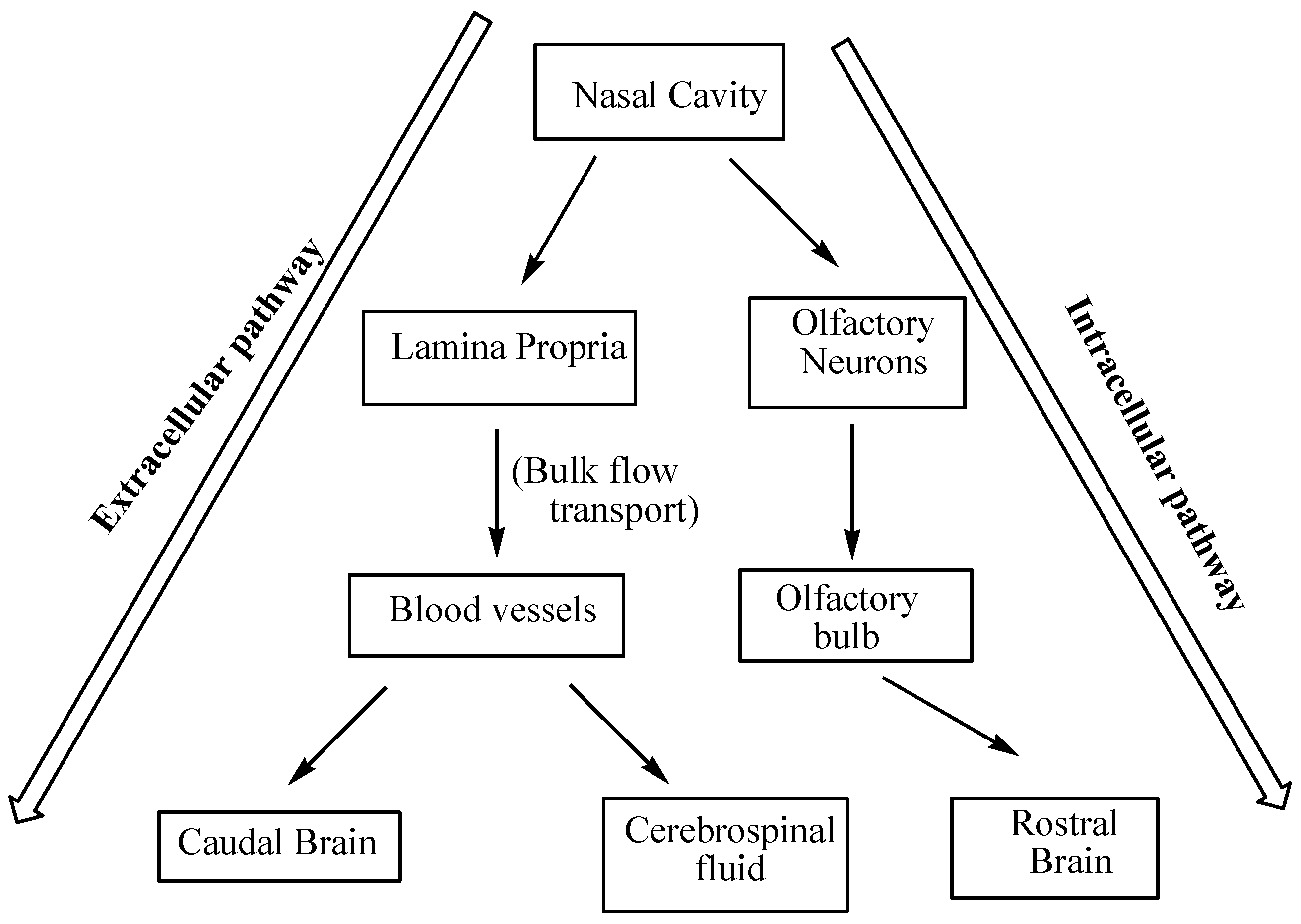
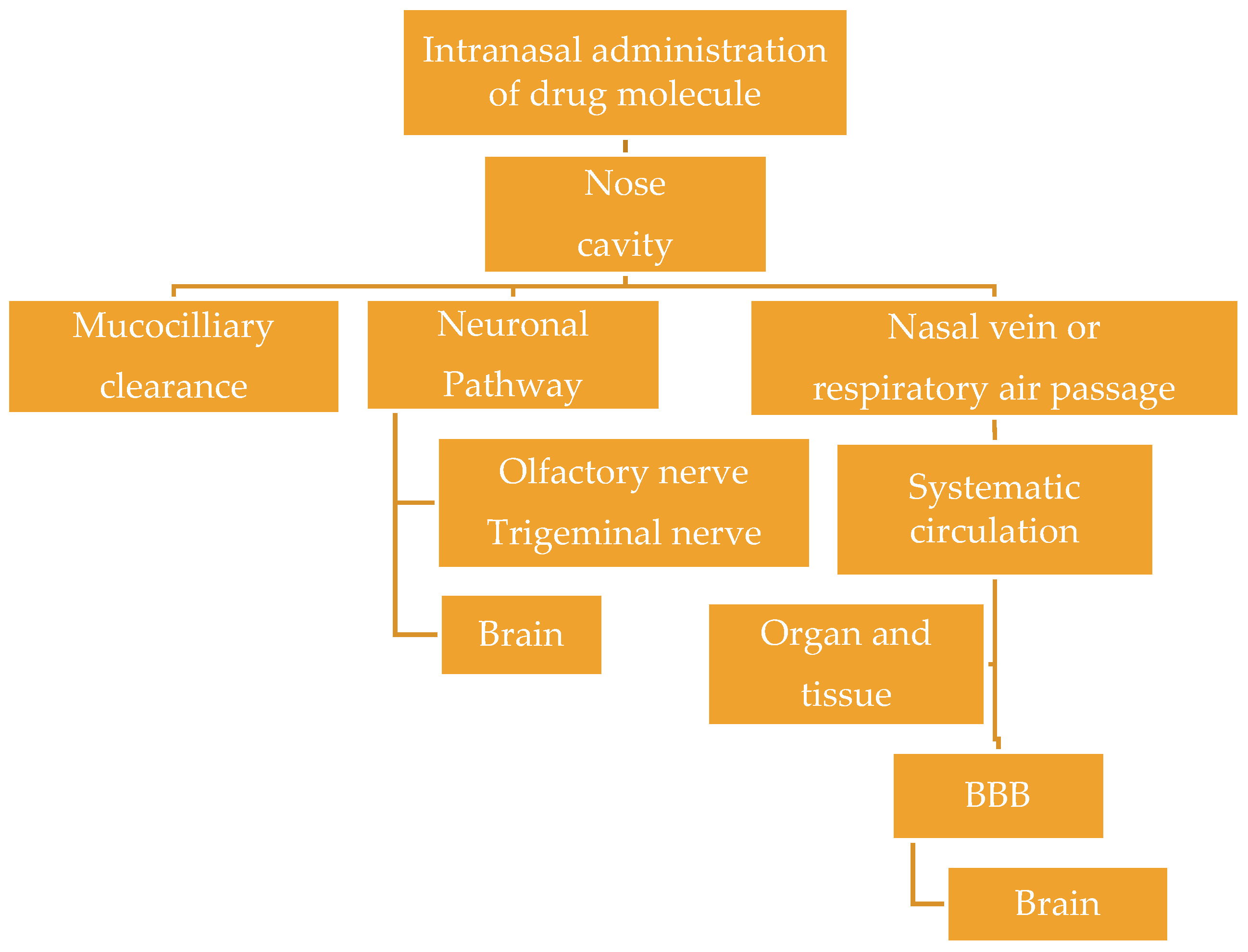
The olfactory region, the upper region of the nasal cavity, is connected to the brain (such as frontal cortex, olfactory bulb) through the olfactory nerves. The respiratory region of the nasal cavity has trigeminal sensory neurons and blood vessels. When the drug is administered intranasally, it will undergo the mucociliary clearance in the vestibular region of the nasal cavity before reaching the interior section of the nasal cavity where the drug comes in contact with the blood vessels and the neuronal network such as the olfactory and the respiratory epithelium [45,46]. Through the blood vessels, the drug undergoes systemic circulation in which the drug is transported via the BBB to the brain. The primary route of the brain drug delivery when administered intranasally is via the neuronal pathway by intracellular and extracellular transport pathways into the different regions of the brain via olfactory and trigeminal sensory neurons [45,46,47] (Figure 3). The intracellular pathway starts with the uptake of drug molecule by the olfactory neuron to the projection site of the neuron followed by release via exocytosis. In the extracellular pathway, the drug is transported across the nasal epithelium, which is composed of cells connected to the tight junctions to the lamina propria where there are neurons especially in the olfactory region [45]. Further transportation of the drug is by bulk flow processes via the neuronal axon. The uptake of the drugs via the endothelial cells in the lamina propria or the transportation of the drug from the subarachnoid CSF into the brain parenchyma indicate the capability of drugs to cross the BBB and blood–CSF barrier [46]. A diagram illustrating the nose-to-brain transportation is shown in Figure 4.
The drug molecular size is not a major factor for the permeability of drug molecules through the tight junctions. Lipophilic molecules are not affected by the tight junction and smaller hydrophilic molecules can diffuse via these barriers. However, the average lifespan of olfactory sensory neurons is 30–60 days, at which they undergo a process known as apoptosis in which the epithelial cells are replaced. During this process, there is a delay in the formation of tight junctions which enhances the permeability of drug molecules [46,48]. However, it is important to state that the exact mechanism of drug transport from the nasal cavity to the brain is not fully understood. The drug transport mechanism from the nose to the brain is influenced by other factors such as the nature of the drug and the delivery system, formulation parameters, composition, and the design of the formulation, etc. [45].

5. Chitosan Nanocarriers in Brain Targeting

The unique properties of chitosan such as good biodegradability, non-toxicity, biocompatibility, and bioadhesive nature make it useful for the formulation of systems for brain targeting. Some of the reported chitosan nanocarriers which have been designed for brain targeting are nanoparticles, in situ gels, nanoemulsions, and liposomes.

5.1. Nanoparticles (NPs)

Chitosan nanoparticles (CsNPs) are excellent nanocarriers because of their unique properties such as good biocompatibility, non-toxicity, biodegradability, and cationic nature [23,49]. Nanoparticles protect labile drugs from enzymatic degradation in the gastrointestinal tract. Chitosan nanoparticles are prepared by various methods such as sieving method, nanoprecipitation, emulsion, coacervation or precipitation, ionic gelation, and reverse micellar method, etc. [50]. CsNPs have been developed for intranasal formulations (Table 1).
Nagpal et al. optimized chitosan nanoparticles loaded with Rivastigmine for brain targeting [51]. Rivastigmine is used to treat Alzheimer’s disease. However, its poor bioavailability and limited entry into the brain resulting from its hydrophilic nature requires frequent dosing resulting in side effects such as nausea, anorexia, dyspepsia, severe bradycardia, etc. [52,53,54]. NPs loaded with rivastigmine were prepared by modified ionotropic gelation method to isolate high yields of stable nanoparticles. The particle size of the nanoparticles was influenced by the concentration of chitosan. The particle size of the NPs influenced their endocytosis rate across the brain capillary endothelial cells leading to the extended residence of the NPs in the blood and its capability to cross the BBB. The size of the prepared nanoparticles was optimized below 200 nm. The high positive ZP (Zeta Potential) value of the NPs also revealed the good stability, strong repulsion, and the absence of particle aggregation. The positive ZP is attributed to the positively charged chitosan on the surface of the particles. The high drug entrapment efficiency of the drug into the NPs was 96% due to the interaction between the drug and the chitosan matrix. Over 50% of the entrapped drug was released over a period of 4 h with a 20% initial burst release in 30 min which was influenced by the small size of drug molecules from the surface of the NP entrapped on the surface of the particles. T80-coated nanoparticles encapsulated with the drug enhanced the anti-amnesic effect of the drug significantly. The maximum tolerated dose suitable without any adverse side effects in vivo was 2.0 mg/kg. Encapsulation of the drug onto the nanoparticles increased the maximum tolerated dose of the drug by 10%. The uptake of the coated nanoparticles by the Sertoli cells was low, resulting in reduced toxicity of the formulation. The uncoated nanoparticles were toxic to Sertoli cells because they are prone to phagocytosis. The use of targeting ligand such as Tween 80® increased the cellular uptake and drug retention of the formulation. Furthermore, the significant reversal of scopolamine-induced amnesia achieved by the drug-loaded coated nanoparticles revealed that the coated NPs are a potential system for targeted brain delivery with improved efficacy for CNS disorders [51].
The chitosan nanoparticles with antibodies on the surface have the capability to cross the brain barrier and also provide neuroprotection. Chitosan-based nanoparticles were conjugated with antibodies recognizing human transferrin receptor. The particle sizes of the nanoparticles were 274 nm and 284 nm. Conjugation of the NPs with TfRmAb enhanced the affinity of the nanoparticles to the hCMEC/D3 cells modeling in vitro BBB, resulting in a good cellular uptake and cytotoxic effects in vitro. The conjugation with TfRmAb influenced endocytosis before nanoparticles aggregation, thereby increasing their transport towards cellular compartments near the nucleus when compared to the control nanoparticles which were attached to the cell membrane. The nanoparticles have the capability to cross the human blood–brain barrier cerebral microvessel endothelial cells, suggesting that the proposed cellular uptake was via the receptor-mediated endocytosis pathway. The chitosan nanoparticles without antibodies exhibited aggregation, which may have induced macropinocytosis that hindered amiloride treatment. The incorporation of antibodies onto the surface of chitosan nanoparticles was effective in overcoming the blood–brain barrier via macropinocytic and receptor-mediated endocytic pathways [55]. Chitosan-based nanoparticles with the capability to increase sufficient drug uptake into the brain thereby preventing cell death have been reported. Chitosan nanospheres were conjugated with poly(ethylene glycol) containing OX26 monoclonal antibody. The aforementioned antibody has affinity for the transferrin receptor (TfR) and can induce receptor-mediated transport across the BBB. The nanoparticles were fluorescently labeled and administered to the mice intravenously, and a significant amount of the nanoparticles was taken up into the brain tissue, outside of the intravascular compartment [56]. Chitosan nanoparticles were also loaded with thymoquinone [57]. The particle sizes of the NP ranged between 150 nm and 200 nm and was influenced by the ratio of the chitosan drug. The drug entrapment efficiency (63%) and drug-loading capacity (31%) of the nanoparticles were inversely proportional to the ratio of the drug to chitosan. Intranasal administration of the formulation resulted in a high drug concentration in the brain when compared to the drug solution which was administered intravenously. Over a period of 2–3 h after the intranasal administration of the formulation, the drug concentration in the brain was sustained. The Cmax was 2417.17 counts and the Kel was 0.0696 counts/h indicating that the low Cmax uptake of the drug solution via intranasal is due to the rapid mucociliary clearance of the instilled drug solution while the drug-loaded NPs has an intrinsic mucoadhesive property which extends the contact time of the drug-loaded NPs with the nasal mucosa, thereby increasing the AUC (Area under curve) and Cmax [57]. The uptake of the nanoparticles into the brain is also due to the nano size range of the formulation and their capability to stretch the tight junctions within the nasal mucosa, revealing it is a promising approach for brain targeting that can reduce the side effects. Yuan et al. studied the toxic effects and biodistribution of chitosan nanoparticles. Tween 80-modified chitosan nanoparticles with particles sizes of 240 nm were prepared. The NP uptake into the brain was significant in the frontal cortex and cerebellum, 30 min after systemic injection. There was a decrease in the concentration of the NPs in the two aforementioned regions of the brain over time. No significant oxidative stress damage was observed. A significant reduction in the GFAP (Glial fibrillary acidic protein) expression, which was dose-dependent, was observed in the cerebellum after exposure to TmCS-NPs (Tween-80-modified chitosan nanoparticles), suggesting the degenerative changes in the cerebellum and activated astrocytes can induce brain damage [58]. The significant reduction in the GFAP expression in the cerebellum after exposure to TmCS-NPs indicate that a thorough investigation should be done in order to evaluate the toxicity of TmCS-NPs. Reports have revealed that TmCS-NPs has toxic effects on muscle structure and the axonal development of primary and secondary motor neurons in vivo suggesting that it must be used with caution [59].
The ionic gelation method was used to prepare rivastigmine loaded chitosan nanoparticles for enhanced bioavailability and uptake of the drug to the brain via intranasal delivery [60]. The permeation enhancing activity of chitosan played an important role in drug loaded nanoparticles uptake which was high when compared to the pure drug solution [60]. The interaction of negatively charged sites of the cell membranes and tight junctions of the mucosal epithelial cells with the positively charged amino group on chitosan stimulated the opening of the tight junctions [61]. The hydrophilic nature of the pure drug influenced the reduced permeation of the pure drug solution. The brain–blood ratio of the drug after the administration of the drug via intravenous and intranasal route formulations were 0.24 and 0.79, respectively, when compared to the drug-loaded NPs which was 1.71 after intranasal administration over a period of 30 min. The low concentration of the drug in the plasma after intranasal administration was significant when compared to the intranasal administration of the free drug. The high brain–blood ratio and low plasma concentration of the drug after intranasal administration of the drug-loaded NPs indicate a nose-to-brain transport of the formulation bypassing the BBB. The brain drug concentration achieved from intranasal administration of the drug loaded chitosan NPs was Cmax (966) and AUC (247,730 ng. min/mL), which was significantly higher when compared to the intravenous and intranasal administration of the drug solution. The results revealed that NPs have good brain targeting capability and they are potential therapeutics for the treatment of Alzheimer’s disease. The drug uptake into the brain from the nasal mucosa can occur via two pathways, which are the systemic pathway, in which some of the drug reaches the brain by crossing the BBB, and by the olfactory pathway, in which the drug is transported to the brain from the nasal cavity [62].
The chitosan nanoparticles loaded with olanzapine was developed for the treatment of depression [63]. Olanzapine is a drug used for the management of neurological disorders such as schizophrenia [64,65]. It is characterized by poor bioavailability resulting from the hepatic first pass metabolism. Its low permeability results from its efflux by P-glycoprotein [66]. Olanzapine-loaded nanoparticles’ mean particle size was 183 nm with zeta potential and polydispersity index of +52.1 mV and 0.122, respectively. The entrapment efficiency and drug-loading capacity of the NPs was 72% and 26%, respectively. In vitro drug release profile of the NPs was an initial burst release followed by a sustained release mechanism. In vitro toxicity studies on RPMI 2650 human nasal epithelial cell line by MTT (3-(4,5-Dimethylthiazol-2-yl)-2,5-diphenyltetrazolium bromide) assay revealed low toxicity when compared to the free drug. Ex vivo studies on excised goat nasal mucosa further indicated the non-toxic nature of the NPs when compared to the drug solution in which the nasal mucosa displayed detachment of some cilia from the epithelial tissues indicating toxicity [63].
An ionic gelation method was used to develop chitosan nanoparticles loaded with donepezil for enhanced bioavailability and the uptake of donepezil to the brain via intranasal administration [67]. Donepezil is a drug used to treat Alzheimer disease [68]. Intranasal administration of the drug-loaded NPs in rats resulted in a high percentage of radioactivity per gram in the brain when compared to the donepezil solution. The drug-loaded NPs exhibited a high drug transport efficiency and direct transport percentages of 191.4% and 1834.5%, respectively. These results indicate that intranasal administration of chitosan nanoparticles resulted in a significant brain targeting efficiency, indicating that NPs are promising approach for the treatment of AD [67].
A spontaneous emulsification method was used to prepare chitosan nanoparticles containing rivastigmine [69]. The formulation exhibited a biphasic drug release and the mechanism of drug release from the nanoparticles was diffusion controlled. Coating the nanoparticles with 1% polysorbate 80 influenced the uptake of nanoparticles in different organs and reduced the drug release from the NPs [69]. Carbodiimide chemistry was employed for the synthesis of chitosan-valine conjugate loaded with saxagliptin, a dipeptidyl peptidase-4 enzyme inhibitor molecule used for the treatment of Alzheimer disease [70]. In vivo studies in rats indicated a good stability of the nanoparticles in the plasma with a release of only 2.5 ng/mL of the drug which is less than the Cmax of the free drug (51 ng/mL). The brain uptake studies indicated the accumulation of 53 ng/mL of the drug from the nanoparticles 24 h after the administration of the NPs formulation when compared to the free drug which was not detected. The (AUC0-t) of the drug from the NPs was over 3.42 times lower when compared to the free drug, which revealed the stability of the prepared formulation in the plasma [70]. Caban et al. prepared chitosan nanoparticles loaded with peptides for brain targeting. The drug uptake from the nanoparticles was significant in the brain when compared to the liver and spleen indicating a targeted delivery of the drug to the brain [71]. Chitosan nanoparticles are suitable for non-invasive routes of drug administration such as nasal route, which results from the absorption-enhancing effect of chitosan [72]. A conventional emulsification crosslinking method was used to prepare chitosan-based nanoparticles loaded with ropinirole hydrochloride for enhanced brain uptake. In vitro drug release from the nanoparticles indicated an initial burst release of the drug followed by a sustained drug release mechanism over a period of 10 h. The drug release was rapid from the nanoparticles with higher drug content compared to NPs with lower drug content. The drug release profile was sustained in the uncoated drug-loaded chitosan nanoparticles with a decreased drug-to-polymer ratio. The thickness of the polymeric matrix of the uncoated drug-loaded chitosan NPs influenced the drug release profile which was dominated with a slow rate of drug diffusion. The coated nanoparticles were stable over a period of three months in storage. A high drug concentration in the brain with low drug concentrations in vital organs such as the liver, kidney, and spleen after 1 h intravenous administration of the drug-loaded coated NPs when compared to the uncoated drug-loaded NPs and free drug was significant in vivo. These findings suggest that the surface-coated drug-loaded chitosan nanoparticles can improve drug uptake in the brain [73]. Jain et al. developed chitosan NPs loaded with galantamine and coated with polysorbate 80. The NPs were characterized by spherical shape and a size of 62 nm with a maximum drug entrapment of 68%. Over a period of 12 h, 97% of the drug was released. A high concentration of galantamine in the brain revealed the potential of NPs for the treatment of Alzheimer’s disease [74].
In the treatment of epilepsy, multi-drug resistance transporter and P-glycoprotein in the BBB hinder the successful transportation of antiepileptic drug into the brain, thereby hampering treatment. In order to enhance the efficacy of drugs used to treat epilepsy such as carbamazepine, it was loaded into carboxymethyl chitosan nanoparticles. Carbamazepine mode of action is via the stabilization of the inactivated state of sodium channels, thereby resulting in a fewer opening of these channels and less excitation of the brain cells. However, after oral administration of the drug, the drug concentration in the liver was high leading to a rapid drug clearance and a short half-life with adverse side effects [75].
In order to overcome the rapid drug clearance after oral administration, chitosan-based nanoparticles loaded with carbamazepine was developed for intranasal administration. The particle size of the formulation was 219 nm with 35% and 80% drug-loading and drug entrapment efficacy, respectively. The drug concentration in the plasma and brain was high after intranasal administration of the NPs formulation when compared to the administration of the drug solution. This result revealed the transportation of the drug to the brain tissue from the nasal cavity. The brain drug concentration was high when compared to the plasma concentration after nasal administration revealing the direct transportation of the drug from the nasal olfactory area into the brain. Other factors that influenced the high brain uptake of the drug are the use of mucoadhesive material in the formulation which extended the contact time of the formulation in the nasal mucosa. The encapsulation of drug into the nanoparticles enhanced the drug bioavailability and brain targeting by adhering to the mucosal surface, thereby improving the drug dissolution in the mucosa [76].
Chitosan-dextran superparamagnetic nanoparticles with a uniform diameter of 55 nm exhibited enhanced internalization in the U87, C6 glioma, and HeLa when compared to dextran-coated particles. The NPs toxicity profile up to a concentration of 10 µg/ml was acceptable and the magnetic properties of the NPs were retained after internalization into the cell. Intravenously administered NPs in orthotopic C6 gliomas in rats resulted in an accumulation of the NPs in the tumor site, suggesting that the NPs can be used to improve the tumor imaging and for targeted delivery of chemotherapeutic agents [77]. Coating the nanoparticles with chitosan increased the charge of the nanoparticles to +19.2 mV. The increased charge of the nanoparticles enhanced their high uptake by the tumor cells. The high intracellular uptake of the particles increased the cytotoxic effects of the formulation resulting from the production of reactive oxygen species (ROS) which affected mitochondrial DNA negatively. The formulation also acted as a negative MRI (Magnetic resonance imaging) contrast agent. The registered relaxation rates in the hybrid formulation were similar to the relaxation features of the commercial NPs formulations. The composite shell between the chitosan and dextran, which was cross-linked by TPP (Triphenylphosphine), did not hinder the diffusion of protons into dephasing volume around the formulation and hence, retained the MRI contrast-enhancing properties of the particles. Intravenous administration of the formulation in vivo revealed significant accumulation of the nanoparticles in the glioma cells, which significantly enhanced the contrasts of the tumor. The cytotoxicity effect of the nanoparticles on glioma C6 cell line was 30.1%. The cytotoxic effect of the NPs was influenced by the surface area and enhanced cellular uptake of the NPs. More modifications on the surface of the nanoparticles with targeting ligands have the potential to further increase the accumulation of the particles in the glioma tissue [77].
Docetaxel-loaded D-α-tocopherol polyethylene glycol 1000 succinate conjugated chitosan nanoparticles were prepared with the presence or absence of transferrin on the surface [78]. The particle size of the nanoparticles was in the size range of 130–300 nm. The incorporation of docetaxel into the NPs with targeting ligand and the presence of transferrin on the surface of NPs increased the particle size. The zeta potential of the chitosan nanoparticles was significantly positive. However, the incorporation of TPGS into the nanoparticles increased the negative charge resulting from the non-ionic nature of TPGS. The transferrin served as a receptor for specific drug delivery. The entrapment efficiency of the nanoparticles was 75%.
The drug release profile of the formulation was pH-dependent, resulting from the protonation of the chitosan’s amino groups at a low pH value. Increased electrostatic repulsion effect also induced drug diffusion from nanoparticles. The high drug release was significant at pH 5.5 and sustained at the pH of 7.4. The in vitro cytotoxicity evaluation on C6 glioma cell lines indicated that the IC50 values of the non-targeted and targeted drug-loaded NPs were 2.21 and 0.41 μg/mL, respectively, which was lower when compared to Docel™ with IC50 of 60.98 μg/mL. The conjugation of chitosan together with transferrin in the NPs resulted in a synergistic effect. In vivo pharmacokinetics studies further showed that the nanoparticles with transferrin exhibited a high AUC with a prolonged circulation in blood when compared to Docel™. The NPs is a potential treatment approach for TfR overexpressing cancer cells [78]. Methotrexate-loaded chitosan or glycol chitosan nanoparticles was coated with a layer of Tween 80 for brain delivery. Tween™ 80 coating layer of NPs is useful for brain delivery because the surfactant adsorbs apolipoproteins E and B from the blood which are useful in promoting receptor-mediated endocytosis of the NPs by the endothelial cells of the BBB [79]. The NPs were cytotoxic against C6 glioma cells line and were able to overcome the MDCKII-MDR1 (Multi-drug Resistance gene-1-Madin-Darby Canine Kidney) cell barrier. The glycol chitosan nanoparticles were the most cytotoxic NPs. A low concentration of Tween 80 of 0.1% v/v was adequate for enhanced transportation of methotrexate from the NPs across the BBB [80]. The transportation of the NPs across the MDCKII-MDR1 monolayer is believed to occur as a result of the disruption of BBB resulting in the uptake of the NPs by enhanced permeation retention effect while an adsorptive-mediated transcytosis occurred across the intact BBB. The latter transport mechanism is influenced by the electrostatic interaction between the positively charged NPs and the negatively charged membrane of the monolayer [80,81].
Nanoparticles used for the incorporation of drugs for brain targeting resulted in a high brain uptake in vivo. The high brain uptake of the drug suggests that incorporating the drugs onto the nanoparticles results in the drug’s capability to cross the BBB. Some factors such as the particle size, surface properties, and positive charge of the nanoparticles are important in the uptake of the incorporated drug into the brain. The positive charge on the surface of the nanoparticles due to chitosan induced an interaction with the negatively charged sites on the cell membranes and tight junctions of the mucosal epithelial cells, thereby opening the tight junctions. The BBB is a system of vascular cellular structures composed of tight junctions between endothelial cells, receptors, transporters, and efflux pumps of the multidrug resistance (MDR) pathway, etc., which regulate the transport of molecules into the brain. The permeability of drug to the brain is enhanced by the loosening of tight junctions between the endothelial cells [82]. Surfactant-coated nanoparticles are highly efficient in the targeted delivery of bioactive agents in therapeutic concentrations to the brain with reduced reticuloendothelial system uptake [83]. A report by Sun et al. indicated that P80 coating is important in the interaction between nanoparticles and brain micro-vessel endothelial cells and entry into CNS [84]. The increased drug transport across BBB by passive diffusion is attributed to an increase in the retention of the nanoparticles in the brain blood capillaries and its binding capability to the endothelial cell lining [85]. Nanoparticles can be taken up by the brain capillary endothelial cells via receptor-mediated endocytosis, in which the drug is released followed by diffusion into the brain where the particles undergo transcytosis [83]. On the BBB, many receptors are overexpressed such as transferrin (Tf) receptor, low-density lipoprotein receptor-related protein, etc. These receptors can bind with selected ligands and induce internalization into the cells. Decoration of NPs with receptors enhanced the transport of the NPs through the BBB [86].

5.2. In Situ Gel

Chitosan-based in situ gels are useful in biomedical applications, such as regeneration medicine and drug delivery systems. The effect of cross-linking of polymer chains in in situ gels has been reported to influence their external stimuli such as temperature and pH, and these effects have been studied extensively. Natural and synthetic polymers are used for the production of in situ gels [87]. In the last decade, in situ gels have been designed for intranasal delivery of therapeutics. Different biodegradable polymers have been used for the development of in situ gel formulations such as alginic acid, gellan gum, xyloglucan, chitosan, pectin, poly(D-lactic acid), etc. In situ gel formulations have unique advantages such as sustained drug action when compared to the conventional therapeutics [88]. Chitosan-based in situ gels have been developed for the intranasal administration of drugs (Table 2).
Chitosan/glycerophosphate-based hydrogels are effectively used in the delivery of growth factors, proteins/peptides, drugs, etc. [88,89]. Injectable thermosensitive chitosan-gelatin-β-Glycerol phosphate hydrogels loaded with ferulic acid inhibited neurological oxidative stress and was biocompatible with Neuro-2a cell [90]. The biodegradation and gelation capability of the injectable gels influenced the drug release ability of the system. Chitosan-alginate hydrogel showed an improved alternative for neural tissue engineering [91]. The hydrogel was hydrophilic with a porous structure. The cell proliferation showed that neural stem cells proliferated significantly on the chitosan-alginate hydrogel [91]. The inclusion of microsphere into pre-formed chitosan/alginate hydrogel provided the most effective delivery system, arising from the ability of the system to entrap the drug strongly, resulting in a sustained drug delivery by diffusion [92]. Thermo-sensitive injectable gels with good stability and thermosensitivity were developed from chitosan. The absence of adverse effects in vivo after the administration of the formulation was significant [93,94]. Ren et al. developed dopamine-based and polydopamine crosslinked injectable hydrogels under physiological conditions. The abundant amino groups on quaternized chitosan backbone influenced its crosslinking with polydopamine to form a stable hydrogel. The hydrogel prepared from quaternized chitosan and polydopamine was characterized by a low polymer concentration, resulting in a pore size which was large and useful for rapid drug release. The introduction of gelatin into the aforementioned hydrogel increased the biocompatibility, polymer concentration, and mechanical properties of the hydrogel. The hydrogels exhibited stable mechanical properties and good degradability. The release profile of the injectable hydrogels was sustained, revealing their capability to exhibit a long-term drug release. The release of dopamine from the hydrogels was controlled for more than 20 days, and it was influenced by the amount of the drug in the hydrogels [95].
In situ gel loaded with rivastigmine tartarate for administration via intranasal route was reported by Abouhussein et al. [96]. The gels were prepared from HPMC (hydroxypropyl methylcellulose), Carbopol 934, NaCMC (sodium salt of carboxymethylcellulose), Chitosan, and pluronic F127. Chitosan and NaCMC increased the Tsol–gel of the prepared gel. The low viscosity of the gel-containing chitosan is attributed to the amino group from the chitosan which reduced water channel formation around the triblock of pluronic, thereby lowering its gelation capability. The transnasal permeation of the formulation was high, resulting in 0.54% ID/g distribution to the brain when compared to the drug solution, which was 0.16% ID/g. Ex vivo permeation of the drug through the nasal membrane was 84% drug release and it was 28% drug release from the pure drug solution over a period of 6 h. The steady-state flux of the optimized gel was 259 μg/cm2/hr when compared to the drug solution which was 86.2 μg/cm2/hr. The apparent permeability coefficient of the formulation was 1.67 × 10⁻⁴ cm/h and the drug solution was 0.56 × 10⁻⁴ cm/h.
The drug concentration in the brain after the administration of the formulation intranasally was higher than the drug solution administered either intranasally or intravenously. The brain/blood ratios of the in situ gel was 1.16 when compared to the drug intranasal solution and intravenous solution which was 0.31 and 0.025, respectively, indicating the direct nose-to-brain transport of the drug from the in situ gel. This could be due to the high drug permeation from the in situ gel observed in the ex vivo permeation study. The drug distribution in the vital organs, such as the liver and the kidney, was high after the administration of the drug solution indicating that the in situ gel has the capability to reduce the systemic drug distribution to vital organs resulting in drug targeting to the brain and hence, reduced side effects [96]. An intranasal thermosensitive gel loaded with rasagiline mesylate (RM), a drug used for the treatment of Parkinson’s disease, was developed from poloxamer 407, carbopol 934 P, poloxamer 188, and chitosan. In vivo, pharmacokinetic studies in rabbits indicated a significant (p < 0.05) enhancement in drug bioavailability from intranasal gels when compared to the oral drug solution. Further biological studies in Wistar rats indicated that the intranasal gels were not toxic and non-irritant to the rat nasal mucosa. The RM concentration in the rat brain tissue was significant, indicating an enhanced uptake of RM from the formulation when compared to the drug solution [97]. The administration of the drug intranasally overcomes the extensive first pass metabolism, a mechanism that is responsible for poor drug bioavailability of therapeutics administered orally. The extended residence time and intimate contact with nasal epithelium resulting from the mucoadhesive behavior of the gels improved the drug uptake from the nasal cavity [97].
Synthesized thiolated chitosan-based in situ gel loaded with levodopa has also been reported [98]. The optimized NPs particle size was 223 nm with an entrapment efficiency of 76%. The release of the drug from the NPs at pH 6.4 revealed non-Fickian diffusion. In vivo pharmacokinetic studies further revealed enhanced bioavailability in the brain. The concentration of thiolated chitosan nanoparticles in the brain and the plasma was approximately 50% when compared to the free drug. The addition of musk ketone to the gel enhanced the concentration of the drug uptake in the brain by increasing the sensitivity of the nasal cavity, thereby inhibiting the efflux of levodopa through P-glycoprotein efflux pump in the brain [98]. Ropinirole, a dopamine D2 agonist to the brain was incorporated into mucoadhesive in situ gel formulations. The in situ formulations were prepared from hydroxyl propyl methyl cellulose and chitosan [99]. In vivo evaluation in albino rats after intranasal administration of 99mTc-ropinirole in situ gel indicated an 82% absolute bioavailability of ropinirole. A nose-to-brain transport of the drug from the in situ gel formulation was confirmed by a 90% high brain transport and a drug targeting index which was greater than 1 [99]. The decreased clearance of the formulation was due to the mucoadhesive properties of chitosan. The positively charged amino groups on the chitosan adhere to the negatively charged sialic acid residue of the mucosa. Chitosan in situ gel loaded with levodopa release profile followed Hixson–Crowell model. The addition of chitosan increased the drug uptake of the formulation in the nasal mucosa via the opening of the junctions between the epithelial cells and also delayed mucociliary clearance. In vivo studies on Swiss albino rat models further indicated that the drug uptake into the brain was influenced by the viscosity of the gel [61,100]. The intranasal administration of the in situ gel formulation resulted in a high percentage of levodopa in the brain when compared to the drug solution in the saline. The aforementioned result indicated that at the nasal temperature, the gel adheres to the nasal cavity thereby reducing the clearance and controlling the drug release. However, results obtained from the gel of Pluronic PF127 incorporated with chitosan nanoparticles loaded with levodopa showed low drug concentration in the brain. Embedding nanoparticles in a highly viscous gel resulted in the non-availability of the drug at the absorption site due to the slow release of the nanoparticles from the gel matrix [100].
Naik and Nair prepared chitosan-based gels for the delivery of doxepin to the brain via nasal route administration. In vivo studies showed an enhanced increase in activity count and a reduction in the immobility time, indicating good antidepressant activity. Side effects such as the mild swelling of the glands was observed in vivo after the administration of the formulation intranasally. Mice administered with the drug solution suffered from the sluffing of the mucosal epithelium. The release profile of the matrix influenced the permeation rate of drug from the formulation [101].

5.3. Emulsions

Nanoemulsions are heterogeneous systems that are composed of ultrafine oil-in-water dispersions stabilized by surfactant molecules [102]. They exhibit unique properties such as good solubilization capacity and thermodynamic stability when compared to other carriers [102,103]. They have been explored in nose-to-brain drug delivery with good drug permeation through the nasal epithelia to the brain. The use of chitosan extended the nasal residence time of nanoemulsions and enhanced a high influx of drugs from the nose to the brain (Table 3) [102,104].
Chitosan-coated nanoemulsions were loaded with rosmarinic acid for nasal delivery. The Box–Behnken design was used to develop the formulations. The optimized condition used was as follows: 0.1% chitosan final concentration (w/v), 8.5% oil phase (w/v), and 3:10 lecithin to oil phase ratio (w/w). The nanoemulsions mucoadhesive property was high with a sustained drug release profile. The extended permeation time was also significant with a high drug penetration via the porcine nasal mucosa due to an excellent interaction between the formulation and the nasal mucosa. The nanoemulsion did not induce a cytotoxic effect on the MRC-5 cell lines (Medical Research Council cell strain 5) or human lung fibroblast cells. Fibroblasts are present in the nasal mucosa and they are very important in nasal delivery [105]. Ropinirole, an anti-Parkinson drug, was encapsulated into chitosan-coated nanoemulsion. The optimized formulation exhibited good drug release of 72% with a globule size of 58.61, polydispersity (0.201), and viscosity (31.42 MPa). Ex vivo study revealed drug transportation in the different parts of the Wister rat brain indicating that the intranasal administration of ropinirole nanoemulsion is a potential therapeutic for Parkinson’s disease [106]. Buspirone hydrochloride microemulsion formulation for intranasal administration was reported to improve the drug uptake in the brain. Chitosan aspartate and hydroxypropyl-β-cyclodextrin were added to the microemulsions to enhance the interaction between the formulation and the nasal mucosa and to improve the drug uptake, respectively. Values for DTE (Drug Targeting Efficiency) % indicate that the drug loaded microemulsion increased the brain drug targeting by a 7-fold after intranasal administration when compared to the intranasal drug solution in vivo. The high viscosity of the formulation and the addition of chitosan aspartate induced the opening of tight junction resulting by a 7.6-fold and the use of cyclodextrin further increased the DTE by 8.6-fold. The size of the microemulsion droplets was less than the size of the axons in the filia olfactoria and hence, their uptake into the neurons was by endocytic mechanisms through the intra-axonal route [107]. The addition of cyclodextrin to chitosan carriers has the potential to enhance the pharmacokinetics, pharmacodynamics, stability, and brain uptake of the formulation. Similar research reports indicated the aforementioned properties when cyclodextrin are employed in drug delivery systems [108,109].
Zolmitriptan is used to treat migraine but it suffers from low bioavailability after administration either by oral or nasal route. In order to enhance its bioavailability, it was incorporated into nanoemulsion formulation for targeted delivery from the nose to the brain [110]. The formulation exhibited a high permeation coefficient on the nasal mucosa than the drug solution. The high transport of the drug from the formulation to the brain was influenced by the small globule size of the formulation resulting in the drug uptake transcellularly via olfactory neurons through the endocytic pathway. The presence of chitosan enhanced the interaction between the formulation and the nasal mucosa. The interaction resulted in an extended nasal clearance time and contact time. Chitosan capability to form a hydrogen bond with the negative-charged nasal mucosa induced the opening of tight junctions with an improved drug permeation effect through the nasal mucosa [110]. Nanoemulsions loaded with kaempferol and chitosan was evaluated for their capability to induce antitumor activity against glioma cells, a deadly type of brain tumor [111]. The formulations exhibited low polydispersity index and were characterized by nanoscale droplets. The high free energy and large surface area of the nanosized droplets are attributed to the high drug permeation via the mucosal surfaces. The use of chitosan in the formulation influenced the globule size of the formulation and the positive zeta potential. The high viscosity of the formulation extended the residence time in the nasal cavity with a delay in the drug release to the mucosal surface. Ex vivo permeation studies performed on porcine nasal mucosa revealed that the permeation of the drug from the formulation was significant and higher than the permeation of the drug solution. The interaction of the formulation with the epithelial cells increased the drug absorption via the nasal mucosa. The drug permeability was influenced significantly by the coating of the nanoparticles. The formulation induced a significant decrease in cell viability. The increased cytotoxic effect of the formulation indicated a good uptake of the drug into the glioma cells resulting from the positive charge of the carrier. Nanoparticles with a positive charge exhibit better interaction with the cell membrane with a negative charge, leading to a high drug uptake and a good cytotoxic effect. The small size of the nanoparticles also facilitated the drug uptake by the cells. Chitosan facilitated the transportation of nanoparticles via absorptive mediated transcytosis. The results indicate that the formulation is a potential therapeutic for the nose to brain delivery [111]. Pathak et al. investigated various mucoadhesive agents such as Pluronic F127, Carbopol 934 P, Pluronic F68, chitosan, sodium alginate, and sodium CMC in emulsions containing nimodipine. Nasal uptake of nimodipine from the microemulsion formulation followed a sustained release with maximal plasma concentration achieved over a period of 6 h. A significant amount of the drug was taken up in the nasal mucosa after the administration of the microemulsion formulations. In vivo pharmacokinetic studies in rats showed a high drug uptake in the brain [112].

5.4. Liposomes

Liposomes are vesicles that can be nano- or microsize and are composed of aqueous compartment surrounded by lipid bilayers [113]. Their distinct physicochemical properties make them suitable for the loading of therapeutic agents which are either hydrophilic, lipophilic, or hydrophobic in nature [113,114]. Hydrophilic therapeutic agents are loaded in the aqueous apartment of the liposomes and are also loaded between the external water phase and the lipid bilayer. Lipophilic or hydrophobic therapeutic agents are loaded into the lipid bilayers hydrophobic core of the liposomes [113,114]. Furthermore, the use of cationic lipids is important for the incorporation of DNA and RNA. Liposomes offer several unique properties such as good biocompatibility, low toxicity, targeted drug delivery, and good biodegradability, etc. [114,115]. Due to the aforementioned properties, macromolecules such as antibodies, peptides, and polymers are used to modify their surfaces in order to enhance the blood circulation and for brain-specific delivery [114,115]. Liposomes have been developed for brain targeting (Table 4).
Salade et al. developed ghrelin-containing liposomes formulation coated with chitosan for administration via nasal route for the treatment of cachexia [116]. Ghrelin is an orexigenic peptide hormone. In the body, it undergoes a rapid degradation resulting from several factors such as hepatic first-pass, plasmatic enzyme digestion, rapid renal elimination, and pH-sensitive degradation. The abovementioned factors result in its short plasmatic half-life in human plasma which is 9–13 min [117]. The coating of anionic liposomes with chitosan resulted in a significant increase in Z-average with a resultant charge of positive (6 mV). The electrostatic interactions of the formulation with the negative sialic acid of mucins increased the mucoadhesion and extended the residence of the formulation in the nasal cavity, and hence enhanced the brain uptake [116]. In a similar report by Salade et al., chitosan-coated anionic liposomes were prepared as liquid and powder formulations for nose-to-brain delivery of ghrelin. The coated liposomes powder formulation adhesion to mucins was significant with a high drug-entrapment efficiency and enzymatic protection against trypsin. The in vitro study of the formulation from a UDS (unit-dose system) aerosol on artificial nasal cavity indicated that the total recovered drug was 23% in the nasal valves section, 25% in the turbinates section, 0% in the rhinopharynx and filter sections, and 52% in the olfactory region. The high drug recovery in the olfactory region, a site for the transportation of drug to the central nervous system, suggests that chitosan-coated liposomes are suitable for a nose-to-brain delivery. However, it is important to indicate that the nasal cast is not suitable for the evaluation of mucociliary clearance mechanism [118].
Qiang et al. developed intranasal liposomes loaded with fexofenadine, a drug used to treat allergic rhinitis. The liposomes and the drug loaded liposomes exhibited small lipid vesicles of 359 nm. They exhibited a narrow size distribution. Coating the surface of the liposomes with chitosan enhanced their stability at storage of 4 °C. A 3-fold higher adsorption of mucin was significant in the liposomes coated with chitosan, indicating their capability to be retained for an extended time in the nasal cavity when compared to the uncoated liposomes. The bioavailability of fexofenadine increased by 34% after the administration of the chitosan coated formulation via the nasal route. The reduced mucociliary clearance and increased drug retention in the nasal cavity is due to the mucoadhesive properties of chitosan coated liposomes. A 5-fold increase of AUC with a sustained drug release after intranasal administration in vivo was observed when compared to the oral administration of the coated formulation [119]. Chen et al. developed curcumin loaded liposomes coated with chitosan. The entrapment efficiency of the liposomes was 87% with 2.3% drug-loading efficiency [120]. The particle size of the coated liposomes increased to 656 nm when compared to the uncoated liposomes, which were 221.4 nm. The pharmacokinetic parameters and bioavailability of the coated liposomes were (Cmax = 46 μg/L, t1/2 = 12 h, AUC = 417 μg/L·h) when compared to the uncoated liposomes, which were (Cmax = 32 μg/L, t1/2 = 9.8 h, AUC = 264 μg/L·h) and the curcumin suspension, which was (Cmax = 35 μg/L, t1/2 = 3.9 h, AUC = 245 μg/L·h) [120]. The results from researchers indicate the potential of coating liposomes with chitosan in brain targeting.

6. Future Perspectives

Chitosan-based carriers have been reported to be effective for the delivery of drugs for the treatment of selected brain diseases in vivo. The charge on chitosan-based drug delivery systems has been reported to result in targeted drug delivery thereby increasing drug concentration at the tumor site and improving the therapeutic outcomes. The design of chitosan-based drug delivery systems influences drug uptake by the brain tissues. Chitosan nanoparticles decorated with antibodies on the surface crossed the brain barrier and also provided neuroprotection when compared with those without antibodies that were characterized by aggregation. The size of the nanoparticles was reported to be either above or below 200 nm. There is a need for more research on the consistency of range of particle size of nanoparticles effective for good brain uptake. The conjugation of antibodies onto the surface of the NPs and the coating of the NPs surface was reported to be a potential approach for the development of formulations for enhanced brain uptake with reduced toxicity. However, there is a pressing need for more in vivo studies on NPs formulations conjugated with antibodies and coated NPs. More studies on the toxicity of these nanoparticles in vivo in long-term use is also very important.
In the design of in situ gels, the addition of musk ketone to the chitosan-based gel also increased the sensitivity of the nasal cavity, thereby inhibiting the efflux of levodopa through P-glycoprotein efflux pump in the brain and enhanced the concentration of drug uptake to the brain. The addition of polycations to in situ gels also increased the drug uptake and delayed mucociliary clearance. There is a need for more research to be performed on the toxicity of musk ketone used in gels for intranasal administration.
Chitosan-based liposomes and emulsions are also potential carriers for the nose-to-brain targeting. The nanoscale droplets, low polydispersity index, and high viscosity of emulsions influenced the high drug permeation through the mucosal surfaces. The coating of the liposomes formulation with chitosan promoted electrostatic interactions with negative sialic acid of mucins, thereby increasing the mucoadhesion and extending the residence of the formulation in the nasal cavity, and hence, enhancing the brain uptake. The surface of the liposomes was coated with chitosan also enhanced the stability of the formulations.
The administration of the chitosan formulations has been reported to result in side effects such as the swelling of glands and the absence of sluffing of the mucosal epithelium. The results reported by several researchers indicate that chitosan-based formulations are potential systems for targeted drug delivery to the brain. Although, there are unique promising results on the treatment of brain diseases using chitosan drug delivery systems, however, more research needs to be conducted in order to enhance and reduce the global issues encountered in treating brain diseases. A study on the potential risk of the nanocarriers to humans in long-term study is very important. The affordability of these carriers should also be thoroughly investigated.

7. Conclusions

Chitosan-based carriers have been reported to be effective for the delivery of drugs for the treatment of selected brain diseases in vivo. The design of chitosan-based carriers influenced the drug uptake by the brain tissues. Factors such as particle size, viscosity, particle charge, and incorporation of antibodies onto the nanocarriers provided neuroprotection, prevented aggregation, prolonged residence time of the nanocarriers with the nasal mucosa, and induced the stretching of tight junctions, resulting in a significance uptake of nanocarrier formulations into the brain.
Several reports on chitosan-based nanoparticles reveal that they are potentials carriers for targeted brain delivery. However, more studies are needed, such as genotoxicity evaluation and further toxicity evaluation of these nanoparticle in long-term use. Other nanocarriers, such as in situ gels, liposomes, and emulsions have few research reports. However, the results reported so far indicate that they are potential systems for brain targeting.
The research reports by several researchers indicate that chitosan-based formulations are potential systems for targeted drug delivery to the brain. Although, there are unique promising results on the treatment of brain diseases using chitosan drug delivery systems, however, more research needs to be conducted.

Author Contributions

T.N. and B.A.A. made substantial contribution to the work reported. The authors approved the final version of the manuscript.

Funding

This research was funded by South Africa Medical Research Council (Self-Initiated Research), Govan Mbeki Research and Development Centre, University of Fort Hare, South Africa and the South Africa National Research Foundation.

Acknowledgments

The financial assistance of the South Africa Medical Research Council (Self-Initiated Research) and the South Africa National Research Foundation towards this research is hereby acknowledged. The views and opinions expressed in this manuscript are those of the authors and not of MRC or NRF.

Conflicts of Interest

The authors declare no conflict of interest.

References

  1. Alazne, D.; Antonia, A.; Enrique, H.; Blanca, S.-M.; Goñi-de-Cerio, F. Central nervous system diseases and the role of the blood-brain barrier in their treatment. Neurosci. Discov. 2013, 1, 3. [Google Scholar]
  2. Chin-Chan, M.; Navarro-Yepes, J.; Quintanilla-Vega, B. Environmental pollutants as risk factors for neurodegenerative disorders: Alzheimer and Parkinson diseases. Front. Cell. Neurosci. 2015, 9, 124. [Google Scholar] [CrossRef]
  3. Feigin, V.L. Global, regional, and national burden of neurological disorders during 1990–2015: A systematic analysis for the Global Burden of Disease Study 2015. Lancet Neurol. 2017, 16, 877–897. [Google Scholar] [CrossRef]
  4. Meng, Z.; Wei, T.; Yan, Z.; Omid, C.F.; Bingyang, S. Nanotechnology-based strategies for siRNA brain delivery for disease therapy. Trends Biotechnol. 2018, 36, 562–575. [Google Scholar]
  5. Prabaharan, M.; Mano, J.F. Chitosan-based particles as controlled drug delivery systems. Drug Deliv. 2005, 12, 41–57. [Google Scholar] [CrossRef] [PubMed]
  6. Aderibigbe, B.A. In Situ-Based Gels for Nose to Brain Delivery for the Treatment of Neurological Diseases. Pharmaceutics 2018, 10, 40. [Google Scholar] [CrossRef]
  7. Sejin, S.; Do Won, H.; Kaushik, S.; Ji Hoon, J.; Tae, G.P.; Dong, S.L.; Won, J.K. RVG peptide tethered bioreducible polyethylenimine for gene delivery to brain. J. Control. Release 2011, 155, 18–25. [Google Scholar]
  8. Gaurav, T.; Ruchi, T.; Birendra, S.; Bhati, L.; Pandey, S.; Pandey, P.; Saurabh, K.B. Drug delivery systems: An updated review. Int. J. Pharm. Investig. 2012, 2, 2–11. [Google Scholar] [Green Version]
  9. Cui-Tao, L.; Ying-Zheng, Z.; Ho Lun, W.; Jun, C.; Lei, P.; Xin-Qiao, T. Current approaches to enhance CNS delivery of drugs across the brain barriers. Int. J. Nanomed. 2014, 9, 2241–2257. [Google Scholar] [Green Version]
  10. Bhakar, S.; Tian, F.; Stoeger, T.; Kreyling, W.; de la Fuente, J.M.; Grazú, V.; Borm, P.; Estrada, G.; Ntziachristos, V.; Razansky, D. Multifunctional Nanocarriers for diagnostics, drug delivery and targeted treatment across blood-brain barrier: Perspectives on tracking and neuroimaging. Part. Fibre Toxicol. 2010, 7, 3. [Google Scholar] [CrossRef]
  11. Yasir, M.; Ayesha, T.; Faheem, A.S. Brain targeting Drug Delivery System: A Review. Int. J. Basic Med. Sci. Pharm. 2015, 5, 2049–4963. [Google Scholar]
  12. Upasana, S.; Pragya, N.B.; Swati, G. Polymeric Nanoparticles Drug Delivery to Brain: A Review. Int. J. Pharmacol. Pharm. Sci. 2015, 2, 60–69. [Google Scholar]
  13. Ravi, K.U. Drug Delivery Systems, CNS Protection, and the Blood Brain Barrier. BioMed Res. Int. 2014, 2014, 869269. [Google Scholar]
  14. Epilepsy. Available online: https://communitymedicine4asses.com/2017/02/11/who-updates-fact-sheet-on-epilepsy-10-february-2017/ (accessed on 28 October 2018).
  15. Alamri, Y.; MacAskill, M.; Anderson, T.; Benamer, H. Parkinson’s disease in the Gulf Countries: An updated review. Eur. Neurol. 2015, 74, 222–225. [Google Scholar] [CrossRef]
  16. Aranaz, I.; Mengíbar, M.; Harris, R.; Paños, I.; Miralles, B.; Acosta, N.; Galed, G.; Heras, A. Functional characterization of chitin and chitosan. Curr. Chem. Biol. 2009, 3, 203–230. [Google Scholar]
  17. Chinmayee, S.; Pankaj, G.; Tarun, K.M. Chitosan: A Promising Biopolymer in Drug Delivery Applications. J. Mol. Genet. Med. 2015, S4, 006. [Google Scholar]
  18. Sarvaiya, J.; Agrawal, Y.K. Chitosan as a suitable nanocarrier material for anti-Alzheimer drug delivery. Int. J. Biol. Macromol. 2015, 72, 454–465. [Google Scholar] [CrossRef]
  19. Divya, L.; Vijay, K.M. Preparation and Applications of Chitosan Nanoparticles: A Brief Review. J. Mater. Sci. 2016. [Google Scholar] [CrossRef]
  20. Rosa, T.; Corrado, S.; Luca, C.; Moreno, B.; Federica, B. Functionalization of PVC by chitosan addition: Compound stability and tensile properties. Compos. B Eng. 2018, 149, 240–247. [Google Scholar]
  21. Shakeel, A.; Saiqa, I. Chitosan Based Scaffolds and Their Applications in Wound Healing. Achiev. Life Sci. 2016, 10, 27–37. [Google Scholar] [Green Version]
  22. Muxika, A.; Etxabide, A.; Uranga, J.; Guerrero, P.; de la Caba, K. Chitosan as a bioactive polymer: Processing, properties and applications. Int. J. Biol. Macromol. 2017, 105, 1358–1368. [Google Scholar] [CrossRef]
  23. Hasbun, R. Meningitis. Available online: https://emedicine.medscape.com/article/232915-overview (accessed on 12 December 2018).
  24. Rajesh, Y.; Pal, I.; Banik, P.; Chakraborty, S.; Borkar, S.A.; Dey, G.; Mukherjee, A.; Mandal, M. Insights into molecular therapy of glioma: Current challenges and next generation blueprint. Acta Pharmacol. Sin. 2017, 38, 591–613. [Google Scholar] [CrossRef]
  25. Linda, L.B.; Joshana, G. Management of migraine headaches in a chronic pain patient: A case report. Ment. Health Clin. 2016, 6, 154–158. [Google Scholar]
  26. Saenger, A.K.; Christenson, R.H. Stroke biomarkers: Progress and challenges for diagnosis, prognosis, differentiation, and treatment. Clin. Chem. 2010, 56, 21–33. [Google Scholar] [CrossRef] [PubMed]
  27. Common Stroke Medications Horrible Side Effects. Available online: https://www.cheatsheet.com/health-fitness/common-stroke-medications-horrible-side-effects.html/ (accessed on 26 February 2019).
  28. Wahab, A. Difficulties in treatment and management of epilepsy and challenges in new drug development. Pharmaceuticals 2010, 3, 2090–2110. [Google Scholar] [CrossRef] [PubMed]
  29. Remy, S.; Beck, H. Molecular and cellular mechanisms of pharmacoresistance in epilepsy. Brain 2005, 129, 18–35. [Google Scholar] [CrossRef] [PubMed] [Green Version]
  30. Sudduth, T.L.; Powell, D.K.; Smith, C.D.; Greenstein, A.; Wilcock, D.M. Induction of hyperhomocysteinemia models vascular dementia by induction of cerebral microhemorrhages and neuroinflammation. J. Cereb. Blood Flow Metab. 2013, 33, 708–715. [Google Scholar] [CrossRef] [PubMed]
  31. FDA-Approved Treatments for Alzheimer’s. Available online: https://www.alz.org/dementia/downloads/topicsheet_treatments.pdf (accessed on 14 February 2018).
  32. Oertel, W.H. Recent advances in treating Parkinson’s disease. F1000Resarch 2017, 6, 620. [Google Scholar] [CrossRef] [PubMed]
  33. Smith, Y.; Wichmann, T.; Factor, S.A.; DeLong, M.R. Parkinson’s disease therapeutics: New developments and challenges since the introduction of levodopa. Neuropsychopharmacology 2012, 37, 213–246. [Google Scholar] [CrossRef]
  34. Patel, K.R.; Cherian, J.; Gohil, K.; Atkinson, D. Schizophrenia: Overview and treatment options. Pharm. Ther. 2014, 39, 638–645. [Google Scholar]
  35. FDA-approved drugs to treat schizophrenia. J. Psychosoc. Nurs. Ment. Health Serv. 2014, 52, 11–12. [CrossRef]
  36. Chunmeng, S.; Yang, D.; Li, Z.; Di, S.; Linlin, S.; Thomas, J.W.; Yan, S. Noninvasive nanoparticle strategies for brain tumor targeting. Nanomedicine 2017, 13, 2605–2621. [Google Scholar]
  37. Patrícia, C.P.; Adriana, O.S. Nanosystems in nose-to-brain drug delivery: A review of non-clinical brain targeting studies. J. Control. Release 2018, 270, 89–100. [Google Scholar]
  38. Abdur, R.K.; Mengrui, L.; Muhammad, W.K.; Guangxi, Z. Progress in brain targeting drug delivery system by nasal route. J. Control. Release 2017, 268, 364–389. [Google Scholar]
  39. Huile, G. Progress and perspectives on targeting nanoparticles for brain drug delivery. Acta Pharm. Sin. B 2016, 6, 268–286. [Google Scholar]
  40. Selvaraj, K.; Gowthamarajan, K.; Karri, V.V.S.R. Nose to brain transport pathways an overview: Potential of nanostructured lipid carriers in nose to brain targeting. Artif. Cells Nanomed. Biotechnol. 2018, 46, 2088–2095. [Google Scholar] [CrossRef]
  41. Liu, X.F.; Fawcett, J.R.; Hanson, L.R.; Frey, W.H. The window of opportunity for treatment of focal cerebral ischemic damage with noninvasive intranasal insulin-like growth factor-I in rats. J. Stroke Cerebrovasc. Dis. 2004, 13, 16–23. [Google Scholar] [CrossRef]
  42. Thorne, R.G.; Pronk, G.J.; Padmanabhan, V.; Frey Ii, W.H. Delivery of insulin-like growth factor-I to the rat brain and spinal cord along olfactory and trigeminal pathways following intranasal administration. Neuroscience 2004, 127, 481–496. [Google Scholar] [CrossRef]
  43. Arora, P.; Sharma, S.; Garg, S. Permeability issues in nasal drug delivery. Drug Deliv. Today 2002, 7, 67–97. [Google Scholar] [CrossRef]
  44. Dhuria, S.V.; Hanson, L.R.; Frey, W.H. Novel vasoconstrictor formulation to enhance intranasal targeting of neuropeptide therapeutics to the central nervous system. J. Pharmacol. Exp. Ther. 2009, 328, 312–320. [Google Scholar] [CrossRef]
  45. Alexander, A.; Saraf, S. Nose-to-brain drug delivery approach: A key to easily accessing the brain for the treatment of Alzheimer’s disease. Neural Regen. Res. 2018, 13, 2102–2104. [Google Scholar]
  46. Crowe, T.P.; Greenlee, M.H.; Kanthasamy, A.G.; Hsu, W.H. Mechanism of intranasal drug delivery directly to the brain. Life Sci. 2018, 195, 44–52. [Google Scholar] [CrossRef]
  47. Erdő, F.; Bors, L.A.; Farkas, D.; Bajza, Á.; Gizurarson, S. Evaluation of intranasal delivery route of drug administration for brain targeting. Brain Res. Bull. 2018, 143, 155–170. [Google Scholar] [CrossRef]
  48. Mackay-Sim, A.; St. John, J.; Schwob, J.E. Neurogenesis in the adult olfactory neuroepithelium. In Handbook of Olfaction and Gustation; Doty, R., Ed.; John Wiley & Sons, Inc.: Hoboken, NJ, USA, 2003; pp. 232–265. [Google Scholar]
  49. Gomes, L.P.; Paschoalin, V.M.F.; Del Aguila, E.M. Chitosan Nanoparticles: Production, Physicochemical Characteristics, and Nutraceutical Applications. Rev. Virtual Quim. 2017, 9, 387–409. [Google Scholar] [CrossRef]
  50. Akbar, A.; Shakeel, A. A review on chitosan and its nanocomposites in drug delivery. Int. J. Biol. Macromol. 2018, 109, 273–286. [Google Scholar]
  51. Nagpal, K.; Singh, S.K.; Mishra, D.N. Optimization of brain-targeted chitosan nanoparticles of Rivastigmine for improved efficacy and safety. Int. J. Biol. Macromol. 2013, 59, 72–83. [Google Scholar] [CrossRef]
  52. Hattori, H. Effectiveness and limitation of newly approved drugs for Alzheimer’s disease. Psychiatria et neurologia Japonica 2013, 115, 22–31. [Google Scholar] [PubMed]
  53. Szeto, Y.Y.J.; Lewis, G.S.S. Current treatment options for Alzheimer’s disease and Parkinson’s disease dementia. Curr. Neuropharmacol. 2016, 14, 326–338. [Google Scholar] [CrossRef]
  54. Khoury, R.; Rajamanickam, J.; Grossberg, G.T. An update on the safety of current therapies for Alzheimer’s disease: Focus on rivastigmine. Ther. Adv. Drug Saf. 2018, 9, 171–178. [Google Scholar] [CrossRef]
  55. Adem, S.; Digdem Y, E.; Secil, C.T.; Utku, H.; Yesim, A.; Patrick, C.; Gunes, E.; Yilmaz, C. Evaluation of brain-targeted chitosan nanoparticles through blood-brain barrier cerebral microvessel endothelial cells. J. Microencapsul. 2017, 34, 659–666. [Google Scholar]
  56. Aktaş, Y.; Yemisci, M.; Andrieux, K.; Gürsoy, R.N.; Alonso, M.J.; Fernandez-Megia, E.; Novoa-Carballal, R.; Quinoa, E.; Riguera, R.; Sargon, M.F.; et al. Development and brain delivery of chitosan-PEG nanoparticles functionalized with the monoclonal antibody OX26. Bioconjug. Chem. 2005, 16, 1503–1511. [Google Scholar] [CrossRef]
  57. Alam, S.; Khan, Z.I.; Mustafa, G.; Kumar, M.; Islam, F.; Bhatnagar, A.; Ahmad, F.J. Development and evaluation of thymoquinone-encapsulated chitosan nanoparticles for nose-to-brain targeting: A pharmacoscintigraphic study. Int. J. Nanomed. 2012, 7, 5705–5718. [Google Scholar] [CrossRef]
  58. Yuan, Z.Y.; Hu, Y.L.; Gao, J.Q. Brain Localization and Neurotoxicity Evaluation of Polysorbate 80-Modified Chitosan Nanoparticles in Rats. PLoS ONE 2015, 10, e0134722. [Google Scholar] [CrossRef]
  59. Yuan, Z.; Li, Y.; Hu, Y.; You, J.; Higashisaka, K.; Nagano, K.; Tsutsumi, Y.; Gao, J. Chitosan nanoparticles and their tween 80 modified counterparts disrupt the developmental profile of zebrafish embryos. Int. J. Pharm. 2016, 515, 644–656. [Google Scholar] [CrossRef]
  60. Porporatto, C.; Bianco, I.D.; Correa, S.G. Local and systemic activity of the polysaccharide chitosan at lymphoid tissues after oral administration. J. Leukoc. Biol. 2005, 78, 62–69. [Google Scholar] [CrossRef]
  61. Borchard, G.; Luessen, H.L.; deBoer, A.G.; Verhoef, J.C.; Lehr, C.M.; Junginger, H.E. The potential of mucoadhesive polymers in enhancing intestinal peptide drug absorption. III effects of chitosanglutamate and carbomer on epithelial tight junctions in vitro. J. Control. Release 1996, 39, 131–138. [Google Scholar] [CrossRef]
  62. Fazil, M.; Md, S.; Haque, S.; Kumar, M.; Baboota, S.; Sahni, J.K.; Ali, J. Development and evaluation of rivastigmine loaded chitosan nanoparticles for brain targeting. Eur. J. Pharm. Sci. 2012, 47, 6–15. [Google Scholar] [CrossRef]
  63. Ruby, J.; Pandey, V.P. Formulation, and evaluation of olanzapine loaded chitosan nanoparticles for nose to brain targeting an in vitro and ex vivo toxicity study. J. Appl. Pharm. Sci. 2016, 6, 34–40. [Google Scholar] [CrossRef]
  64. Heres, S.; Kraemer, S.; Bergstrom, R.F.; Detke, H.C. Pharmacokinetics of olanzapine long-acting injection: The clinical perspective. Int. Clin. Psychopharmacol. 2014, 29, 299–312. [Google Scholar] [CrossRef]
  65. Katagiri, H.; Tohen, M.; McDonnell, D.P.; Fujikoshi, S.; Case, M.; Kanba, S.; Takahashi, M.; Gomez, J.C. Safety and efficacy of olanzapine in the long-term treatment of J apanese patients with bipolar I disorder, depression: An integrated analysis. Psychiatry Clin. Neurosci. 2014, 68, 498–505. [Google Scholar] [CrossRef]
  66. Mattiuz, E.; Franklin, R.; Gillespie, T.; Murphy, A.; Bernstein, J.; Chiu, A. Disposition and metabolism of olanzapine in mice, dogs, and rhesus monkeys. Drug Metab. Dispos. 1997, 25, 573–583. [Google Scholar]
  67. Bhavna, M.D.; Shadab, A.; Mushir, B.; Aseem, B.; Sanjula, S.; Jasjeet, K.; Ali, J. Design, Development, Optimization and Characterization of Donepezil Loaded Chitosan Nanoparticles for Brain Targeting to Treat Alzheimer’s Disease. Sci. Adv. Mater. 2014, 6, 720–735. [Google Scholar] [CrossRef]
  68. Casey, D.A.; Antimisiaris, D.; O’Brien, J. Drugs for Alzheimer’s disease: Are they effective? Pharm. Ther. 2010, 35, 208–211. [Google Scholar]
  69. Wilson, B.; Samanta, M.K.; Muthu, M.S.; Vinothapooshan, G. Design and evaluation of chitosan nanoparticles as novel drug carrier for the delivery of rivastigmine to treat Alzheimer’s disease. Ther. Deliv. 2011, 2, 599–609. [Google Scholar] [CrossRef]
  70. Fernandes, J.; Ghate, M.V.; Basu M, S.; Lewis, S.A. Amino acid conjugated chitosan nanoparticles for the brain targeting of a model dipeptidyl peptidase-4 inhibitor. Int. J. Pharm. 2018, 547, 563–571. [Google Scholar] [CrossRef]
  71. Caban, S.; Capan, Y.; Couvreur, P.; Dalkara, T. Preparation and characterization of biocompatible chitosan nanoparticles for targeted brain delivery of peptides. Methods Mol. Biol. 2012, 846, 321–332. [Google Scholar]
  72. Arezou, G.; Soleiman, M.; Fatemeh, T.; Farid, T. Synthesis and optimization of chitosan nanoparticles: Potential applications in nanomedicine and biomedical engineering. Caspian J. Intern. Med. 2014, 5, 156–161. [Google Scholar]
  73. Somasree, R.; Pratyusha, S.; Bibek, L.; Sabyasachi, M.; Uttam, K.B.; Amit, K.N. Polysorbate 80 coated crosslinked chitosan nanoparticles of ropinirole hydrochloride for brain targeting. J. Drug Deliv. Sci. Technol. 2018, 48, 21–29. [Google Scholar]
  74. Jain, S.; Jain, R. Design and evaluation of chitosan nanoparticles as novel drug carrier for the delivery of Galantamine to treat Alzheimer’s disease. Abstr. Parkinsonism Relat. Disord. 2018, 46, e47–e51. [Google Scholar] [CrossRef]
  75. Tolou-Ghamari, Z.; Zare, M.; Habibabadi, J.M. A quick review of carbamazepine pharmacokinetics in epilepsy from 1953 to 2012. J. Res. Med. Sci. 2013, 18, S18–S85. [Google Scholar]
  76. Liu, S.; Yang, S.; Ho, P.C. Intranasal administration of carbamazepine-loaded carboxymethyl chitosan nanoparticles for drug delivery to the brain. Asian J. Pharm. Sci. 2018, 13, 72–81. [Google Scholar] [CrossRef]
  77. Shevtsov, M.; Nikolaev, B.; Marchenko, Y.; Yakovleva, L.; Skvortsov, N.; Mazur, A.; Tolstoy, P.; Ryzhov, V.; Multhoff, G. Targeting experimental orthotopic glioblastoma with chitosan-based superparamagnetic iron oxide nanoparticles (CS-DX-SPIONs). Int. J. Nanomed. 2018, 13, 1471–1482. [Google Scholar] [CrossRef] [PubMed]
  78. Agrawal, P.; Singh, R.P.; Kumari, L.; Sharma, G.; Koch, B.; Rajesh, C.V.; Mehata, A.K.; Singh, S.; Pandey, B.L.; Muthu, M.S. TPGS-chitosan cross-linked targeted nanoparticles for effective brain cancer therapy. Mater. Sci. Eng. C 2017, 74, 167–176. [Google Scholar] [CrossRef]
  79. Gelperina, S.; Maksimenko, O.; Khalansky, A.; Vanchugova, L.; Shipulo, E.; Abbasova, K.; Berdiev, R.; Wohlfart, S.; Chepurnova, N.; Kreuter, J. Drug delivery to the brain using surfactant-coated poly(lactide-co-glycolide) nanoparticles: Influence of the formulation parameters. Eur. J. Pharm. Biopharm. 2010, 74, 157–163. [Google Scholar] [CrossRef]
  80. Trapani, A.; Denora, N.; Iacobellis, G.; Sitterberg, J.; Bakowsky, U.; Kissel, T. Methotrexate-loaded chitosan-and glycolchitosan-based nanoparticles: A promising strategy for the administration of the anticancer drug to brain tumors. AAPS Pharmscitech. 2011, 12, 1302–1311. [Google Scholar] [CrossRef]
  81. Beduneau, A.; Saulnier, P.; Benoit, J.P. Active targeting of brain tumors using nanocarriers. Biomaterials 2007, 28, 4947–4967. [Google Scholar] [CrossRef]
  82. Bhaskar, K.R.; Vaijayanthi, V.; Raj, S.B.; Mohanambal, E.; Charulatha, R.; Madhusudan, Y.R. Nanoparticles for brain targeting. Ind. J. Novel Drug Deliv. 2011, 3, 91–97. [Google Scholar]
  83. Chacko, B.J.; Palanisam, S.; Gowrishankar, N.L.; Honeypriya, J.; Sumathy, A. Effect of Surfactant Coating on Brain Targeting Polymeric Nanoparticles; a Review. Ind. J. Pharm. Sci. 2018, 80, 215–222. [Google Scholar] [CrossRef] [Green Version]
  84. Sun, W.; Xie, C.; Wang, H.; Hu, Y. Specific role of polysorbate 80 coating on the targeting of nanoparticles to the brain. Biomaterials 2004, 25, 3065–3071. [Google Scholar] [CrossRef]
  85. Kreuter, J. Nanoparticulate systems for brain delivery of drugs. Adv. Drug Deliv. Rev. 2012, 64, 213–222. [Google Scholar] [CrossRef]
  86. Geldenhuys, W.J.; Allen, D.D. The blood–brain barrier choline transporter. Cent. Nerv. Syst. Agents Med. Chem. 2012, 12, 95–99. [Google Scholar] [CrossRef]
  87. Puente, P.; Fettig, N.; Luderer M, J.; Jin, A.; Shah, S.; Muz, B.; Kapoor, V.; Goddu, S.M.; Salama, N.N.; Tsien, C.; et al. Injectable Hydrogels for Localized Chemotherapy and Radiotherapy in Brain Tumors. J. Pharm. Sci. 2018, 107, 922–933. [Google Scholar] [CrossRef]
  88. Liu, L.; Gao, Q.; Lu, X.; Zhou, H. In situ forming hydrogels based on chitosan for drug delivery and tissue regeneration. Asian J. Pharm. Sci. 2016, 11, 673–683. [Google Scholar] [CrossRef] [Green Version]
  89. Guo-Chung, D.; Che-Yung, K.; Subramaniam, S.; Jiong-Yao, Z.; Savitha, S.; Hwan-You, C.; Feng-Huei, L. A potent inhibition of oxidative stress induced gene expression in neural cells by sustained ferulic acid release from chitosan based hydrogel. Mater. Sci. Eng. 2015, 49, 691–699. [Google Scholar]
  90. Li, Y.; Rodrigues, J.; Tomas, H. Injectable and biodegradable hydrogels: Gelation, biodegradation and biomedical applications. Chem. Soc. Rev. 2012, 41, 2193–2221. [Google Scholar] [CrossRef]
  91. Wang, G.; Wang, X.; Huang, L. Feasibility of chitosan-alginate (Chi-Alg) hydrogel used as scaffold for neural tissue engineering: Apilot study in vitro. Biotechnol. Biotechnol. Equip. 2017, 31, 766–773. [Google Scholar] [CrossRef]
  92. Giovanna, R.; Andrea, S.; Elena, P.P.; Paolo, G.; Marta, R.; Elisabetta, G. Composite Chitosan/Alginate Hydrogel for Controlled Release of Deferoxamine: A System to Potentially Treat Iron Dysregulation Diseases. Carbohydr. Polym. 2016, 136, 1338–1347. [Google Scholar]
  93. Atta, S.; Khaliqa, S.; Islam, A.; Javeria, I.; Jamil, T.; Athar, M.M.; Shafiq, M.I.; Ghaffar, A. Injectable biopolymer based hydrogels for drug delivery applications. Int. J. Biol. Macromol. 2015, 80, 240–245. [Google Scholar] [CrossRef] [PubMed]
  94. Lia, Z.; Shimd, H.; Chob, M.O.; Chob, I.S.; Leef, J.H.; Sun-Woong, K.; Kwond, B.; Huhb, K.M. Thermo-sensitive injectable glycol chitosan-based hydrogel for treatment of degenerative disc disease. Carbohydr. Polym. 2018, 184, 342–353. [Google Scholar] [CrossRef] [PubMed]
  95. Ren, Y.; Zhao, X.; Liang, X.; Mac, P.X.; Guo, B. Injectable hydrogel based on quaternized chitosan, gelatin and dopamine as localized drug delivery system to treat Parkinson’s disease. Int. J. Biol. Macromol. 2017, 105, 1079–1087. [Google Scholar] [CrossRef] [PubMed]
  96. Abouhussein, D.M.; Khattab, A.; Bayoumi, N.A.; Mahmoud, A.F.; Sakr, T.M. Brain targeted rivastigmine mucoadhesive thermosensitive in situ gel: Optimization, in vitro evaluation, radiolabeling, in vivo pharmacokinetics and biodistribution. J. Drug Deliv. Sci. Technol. 2018, 43, 129–140. [Google Scholar] [CrossRef]
  97. Ravi, P.R.; Aditya, N.; Patil, N.S.; Cherian, L. Nasal in-situ gels for delivery of rasagiline mesylate: Improvement in bioavailability and brain localization. J. Drug Deliv. 2015, 22, 903–910. [Google Scholar] [CrossRef]
  98. Sandhya, G.; Saahil, A. In-situ nasal gel of levodopa for brain targeting using chitosan-thioglycolic acid conjugate and musk ketone by efflux transport modulation. World J. Pharm. Pharm. Sci. 2015, 4, 25. [Google Scholar]
  99. Khan, S.; Patil, K.; Bobade, N.; Yeole, P.; Gaikwad, R. Formulation of intranasal mucoadhesive temperature-mediated in situ gel containing ropinirole and evaluation of brain targeting efficiency in rats. J. Drug Target. 2010, 18, 223–234. [Google Scholar] [CrossRef]
  100. Sharma, S.; Lohan, S.; Murthy, R.S. Formulation and characterization of intranasal mucoadhesive nanoparticulates and thermo-reversible gel of levodopa for brain delivery. Drug Dev. Ind. Pharm. 2014, 40, 869–878. [Google Scholar] [CrossRef]
  101. Naik, A.; Naik, H. Formulation and evaluation of thermosensitive biogels for nose to brain delivery of doxepin. Biomed Res. Int. 2014, 2014, 847547. [Google Scholar] [CrossRef]
  102. Ahmad, E.; Feng, Y.; Qi, J.; Fan, W.; Ma, Y.; He, H.; Xia, F.; Dong, X.; Zhao, W.; Lu, Y.; et al. Evidence of nose-to-brain delivery of nanoemulsions: Cargoes but not vehicles. Nanoscale 2017, 9, 1174–1183. [Google Scholar] [CrossRef]
  103. Mahajan, H.S.; Mahajan, M.S.; Nerkar, P.P.; Agrawal, A. Nanoemulsion-based intranasal drug delivery system of saquinavir mesylate for brain targeting. Drug Deliv. 2014, 21, 148–154. [Google Scholar] [CrossRef] [PubMed]
  104. Kumar, M.; Misra, A.; Mishra, A.K.; Mishra, P.; Pathak, K. Mucoadhesive nanoemulsion-based intranasal drug delivery system of olanzapine for brain targeting. J. Drug Target. 2008, 16, 806–814. [Google Scholar] [CrossRef] [PubMed]
  105. Fachel, F.N.; Medeiros-Neves, B.; Dal Prá, M.; Schuh, R.S.; Veras, K.S.; Bassani, V.L.; Koester, L.S.; Henriques, A.T.; Braganhol, E.; Teixeira, H.F. Box-Behnken design optimization of mucoadhesive chitosan-coated nanoemulsions for rosmarinic acid nasal delivery—In vitro studies. Carbohydr. Polym. 2018, 199, 572–582. [Google Scholar] [CrossRef] [PubMed]
  106. Mustafa, G.; Baboota, S.; Ahuja, A.; Ali, J. Formulation development of chitosan coated intra nasal ropinirole nanoemulsion for better management option of Parkinson: An in vitro ex vivo evaluation. Curr. Nanosci. 2012, 8, 348–360. [Google Scholar] [CrossRef]
  107. Bshara, H.; Osman, R.; Mansour, S.; El-Shamy, A.E. Chitosan and cyclodextrin in intranasal microemulsion for improved brain buspirone hydrochloride pharmacokinetics in rats. Carbohydr. Polym. 2014, 99, 297–305. [Google Scholar] [CrossRef] [PubMed]
  108. Shityakov, S.; Puskás, I.; Pápai, K.; Salvador, E.; Roewer, N.; Förster, C.; Broscheit, J.A. Sevoflurane-sulfobutylether-β-cyclodextrin complex: Preparation, characterization, cellular toxicity, molecular modeling and blood-brain barrier transport studies. Molecules 2015, 20, 10264–10279. [Google Scholar] [CrossRef] [PubMed]
  109. Calias, P. 2-Hydroxypropyl-β-cyclodextrins and the Blood-Brain Barrier: Considerations for Niemann-Pick Disease Type C1. Curr. Pharm. Des. 2017, 23, 6231–6238. [Google Scholar] [CrossRef] [PubMed]
  110. Abdou, E.M.; Kandil, S.M.; El Miniawy, H.M. Brain targeting efficiency of antimigraine drug loaded mucoadhesive intranasal nanoemulsion. Int. J. Pharm. 2017, 529, 667–677. [Google Scholar] [CrossRef] [PubMed]
  111. Colombo, M.; Figueiró, F.; de Fraga, D.A.; Teixeira, H.F.; Battastini, A.M.; Koester, L.S. Kaempferol-loaded mucoadhesive nanoemulsion for intranasal administration reduces glioma growth in vitro. Int. J. Pharm. 2018, 543, 214–223. [Google Scholar] [CrossRef]
  112. Pathak, R.; Dash, R.P.; Misra, M.; Nivsarkar, M. Role of mucoadhesive polymers in enhancing delivery of nimodipine microemulsion to brain via intranasal route. Acta Pharm. Sin. B 2014, 4, 151–160. [Google Scholar] [CrossRef] [Green Version]
  113. Akbarzadeh, A.; Rezaei-Sadabady, R.; Davaran, S.; Joo, S.W.; Zarghami, N.; Hanifehpour, Y.; Samiei, M.; Kouhi, M.; Nejati-Koshki, K. Liposome: Classification, preparation, and applications. Nanoscale Res. Lett. 2013, 8, 102. [Google Scholar] [CrossRef]
  114. Vieira, D.B.; Gamarra, L.F. Getting into the brain: Liposome-based strategies for effective drug delivery across the blood–brain barrier. Int. J. Nanomed. 2016, 11, 5381–5414. [Google Scholar] [CrossRef]
  115. Rasoulianboroujeni, M.; Kupgan, G.; Moghadam, F.; Tahriri, M.; Boughdachi, A.; Khoshkenar, P.; Ambrose, J.J.; Kiaie, N.; Vashaee, D.; Ramsey, J.D.; et al. Development of a DNA-liposome complex for gene delivery applications. Mater. Sci. Eng. C 2017, 75, 191–197. [Google Scholar] [CrossRef] [Green Version]
  116. Salade, L.; Wauthoz, N.; Deleu, M.; Vermeersch, M.; De Vriese, C.; Amighi, K.; Goole, J. Development of coated liposomes loaded with ghrelin for nose-to-brain delivery for the treatment of cachexia. Int. J. Nanomed. 2017, 12, 8531. [Google Scholar] [CrossRef] [PubMed]
  117. Akamizu, T.; Takaya, K.; Irako, T.; Hosoda, H.; Teramukai, S.; Matsuyama, A.; Tada, H.; Miura, K.; Shimizu, A.; Fukushima, M.; et al. Pharmacokinetics, safety, and endocrine and appetite effects of ghrelin administration in young healthy subjects. Eur. J. Endocrinol. 2004, 150, 447–455. [Google Scholar] [CrossRef] [PubMed]
  118. Salade, L.; Wauthoz, N.; Vermeersch, M.; Amighi, K.; Goole, J. Chitosan-coated liposome dry-powder formulations loaded with ghrelin for nose-to-brain delivery. Eur. J. Pharm. Biopharm. 2018, 129, 257–266. [Google Scholar] [CrossRef] [PubMed]
  119. Qiang, F.; Shin, H.J.; Lee, B.J.; Han, H.K. Enhanced systemic exposure of fexofenadine via the intranasal administration of chitosan-coated liposome. Int. J. Pharm. 2012, 430, 161–166. [Google Scholar] [CrossRef] [PubMed]
  120. Chen, H.; Wu, J.; Sun, M.; Guo, C.; Yu, A.; Cao, F.; Zhao, L.; Tan, Q.; Zhai, G. N-trimethyl chitosan chloride-coated liposomes for the oral delivery of curcumin. J. Liposome Res. 2012, 22, 100–109. [Google Scholar] [CrossRef] [PubMed]
Figure 1. Chemical structure showing the preparation of chitosan by deacetylation of chitin.
Applsci 09 02219 g001
Figure 2. Different types of brain diseases.
Applsci 09 02219 g002
Figure 3. Illustration of extracellular and intracellular pathway for nose-to-brain delivery.
Applsci 09 02219 g003
Figure 4. Diagram illustrating the nose-to-brain transport of drug molecules.
Applsci 09 02219 g004
Table 1. Chitosan nanoparticles developed for intranasal administration.
DrugsCompositionPhysicochemical PropertiesBiological Studies OutcomeReferences
RivastigmineChitosan Particle size below 200 nmThe uptake of the coated nanoparticles by the Sertoli cells was low and revealed reduced toxicity. A significant reversal of scopolamine-induced amnesia was achieved using the drug loaded coated nanoparticles. [51]
Chitosan, TfRmAbParticle sizes of 274 nm and 284 nm, Polydispersity index of 0.45 ± 0.13 and 0.48 ± 0.13, respectively. Zeta potential of 29.7 ± 1.7 and 34.4 ± 2.1 mV.The conjugation with TfRmAb influenced good brain uptake[55]
Interleukin-1 receptor antagonist FMKChitosan, poly(ethylene glycol), OX26 monoclonal antibodyGood stabilityGood brain uptake, brain tumor reduction, and behavioral recover in vivo.[56]
ThymoquinoneChitosan, sodium tripolyphosphate Particle size of 172–281 nm, Zeta potential of 24.5–30.3 mV, polydispersity index of 0.13–0.24.Sustained drug concentration in the brain. Prolonged contact time of the drug-loaded NPs with the nasal mucosa. [57]
A fluorescence marker, rhodamine B isothiocyanate (RBITC). Tween 80, chitosanThe particle size and zeta-potential of the NPs were 251 ± 15 nm and 26.5 ± 4.2 mV, respectivelyGood NPs uptake into the brain frontal cortex and cerebellum followed by a decrease in the concentration of the NPs in the two aforementioned regions of the brain over time. Absence of significant oxidative stress damage. A significant reduction in the GFAP expression which was dose-dependent. [58]
RivastigmineChitosan The higher drug transport efficiency (355%) and direct transport percentage (71.80%)The concentration of the drug in the plasma after intranasal administration was low. The brain blood ratio was also low after intranasal administration of the drug-loaded NPs. [62]
OlanzapineChitosan The mean particle size, polydispersity index, and zeta potential was 183.1 nm, 0.122, +52.1 mV, respectively. The entrapment efficiency and drug loading were found to be 72% and 26%.In vitro drug release profile of the NPs was an initial burst release followed by a sustained release mechanism. The toxicity of the formulation on RPMI 2650 human nasal epithelial cell line by MTT assay revealed low toxicity when compared to the free drug. Ex vivo studies on excised goat nasal mucosa further indicated the non-toxic nature of the NPs. [63]
DonepezilChitosan100–200 nm particle size.Intranasal administration of the drug-loaded NPs in rats resulted in a high percentage of radioactivity per gram in the brain when compared to the donepezil solution. [67]
RivastigmineChitosan, polysorbate 80Particle size of 47 nmA biphasic drug release was significant. Coating the nanoparticles with 1% polysorbate 80 influenced the uptake of nanoparticles in different organs. [69]
SaxagliptinChitosan, valineGood stabilityGood stability of the nanoparticles in the plasma with a release of only 2.5 ng/mL of the drug which is less than the Cmax of the free drug (51 ng/mL). The (AUC0-t) of the drug from the NPs was over 3.42 times lower than the free drug. [70]
RopiniroleChitosanGood stabilityIn vitro drug release was an initial burst release followed by a sustained drug release mechanism over a period of 10 h. A high drug concentration in the brain with low drug concentrations in vital organs such as the liver, kidney, and spleen after 1 h intravenous administration of the drug-loaded coated NPs when compared to the uncoated drug-loaded NPs and free drug was significant in vivo. [73]
GalantamineChitosan, polysorbate 80Particle size of 62 nmHigh drug release occurred in vitro and a high concentration of galantamine was observed in the brain in vivo.[74]
CarbamazepineChitosanParticle size of 219 nm with 35% drug loading and 80% entrapment efficiency High drug concentration in the brain.[76]
Chitosan, dextranParticle size of 55 nmImproved internalization of the NPs in the C6 glioma cell line in vitro. Retained magnetic properties of the NPs after internalization into the cell. [77]
DocetaxelD-α-tocopherol polyethylene glycol 1000 succinate, chitosan, transferrinParticle size of 130–300 nmThe in vitro cytotoxic effect of the formulation on C6 glioma cell lines was lower when compared to Docel™. The nanoparticles with transferrin exhibited a high AUC with a prolonged circulation in blood when compared to Docel™.[78]
Methotrexate Chitosan, Tween 80, Poly(lactide-co-glycolide)Particle size range of 177–408 nm.The NPs were cytotoxic against C6 glioma cells line and were able to overcome MDCKII-MDR1 cell barrier. [79]
Table 2. Chitosan-based in situ gels developed for intranasal administration.
DrugCompositionPhysicochemical PropertiesBiological OutcomeReferences
RivastigimeHPMC, Carbopol 934, NaCMC, Chitosan, pluronic F127Low viscosity and gelation capabilityHigh drug concentration in the brain.[96]
Rasagiline mesylatePoloxamer 407, carbopol 934 P, poloxamer 188, and chitosanGood mucoadhesive propertiesNon-toxic and non-irritant to the rat nasal mucosa. High drug concentration in rat brain tissue.[97]
LevodopaChitosan, ketone muskThe optimized thiolated chitosan NPs showed 223 nm particle size, 0.296 PDI and +27.91mV zeta potential. The concentration of thiolated chitosan nanoparticles in the brain was high when compared to the free drug. The addition of musk ketone to the gel enhanced the concentration of the drug uptake in the brain by increasing the sensitivity of the nasal cavity, thereby inhibiting the efflux of levodopa through P-glycoprotein efflux pump in the brain.[98]
RopiniroleChitosan, HPMCImproved drug bioavailability.A 90% high brain transport and a drug targeting index greater than 1.[99]
LevodopaChitosan, Pluronic PF127Good gelation capability.The release profile followed Hixson-Crowell model. A high percentage of levodopa in the brain when compared to the drug solution in the saline. [100]
DoxepinChitosan, PEGGood gelation and mucoadhesive propertiesIn vivo studies showed an enhanced increase in activity count and a reduction in the immobility time, indicating good antidepressant activity. [101]
Table 3. Chitosan-based emulsions for intranasal drug administration.
DrugCompositionPhysicochemical PropertiesBiological OutcomeReferences
Rosmarinic acid0.1% chitosan final concentration (w/v), 8.5% oil phase (w/v), and 3:10 lecithin to oil phase ratio (w/w)High mucoadhesive property.The extended permeation time was also significant with a high drug penetration via the porcine nasal mucosa. The nanoemulsion did not induce a cytotoxic effect on the MRC-5 cell lines, human lung fibroblast cells. [105]
RopiniroleChitosan Globule size of 58.61, polydispersity (0.201), and viscosity (31.42 MPa).Ex vivo study revealed drug transportation in the different parts of the Wister rat brain. [106]
Buspirone hydrochlorideChitosan aspartate and hydroxypropyl-β-cyclodextrinThe high viscosity. The size of the microemulsion droplets was less than the size of the axons in the filia olfactoriaIncreased brain drug targeting by a 7-fold after intranasal administration when compared to the intranasal drug solution in vivo. [107]
ZolmitriptanChitosan A high permeation coefficient on the nasal mucosa and small globule size Extended nasal clearance time and contact time. [110]
KaempferolChitosanLow polydispersity index. High free energy and large surface area of the nanosized droplets. Low viscosityExtended residence time in the nasal cavity with a delay in the drug release to the mucosal surface. High permeation of the drug from the formulation. Increased cytotoxic effect of the formulation against glioma cells.[111]
NimodipinePluronic F 127, Carbopol 934 P, Pluronic F 68, chitosan, sodium alginate and sodium CMC, Capmul MCM, Labrasol and Transcutol P The particle size of 250 nm and zeta potential value of −15 mV.Nasal uptake of nimodipine from the microemulsion formulation followed a sustained release with maximal plasma concentration achieved over a period of 6 h. In vivo pharmacokinetic studies in rats showed a high drug uptake in the brain[112]
Table 4. Chitosan-based liposomes for intranasal administration of drug.
DrugCompositionPhysicochemical PropertiesBiological OutcomeReferences
GhrelinChitosanParticle size range of 146.9 ± 2.7 to 194 ± 6.1 nm, for uncoated and coated liposomes, respectively. The potential in the range of 0.3 ± 1.2 mV to 6 ± 0.4 mVIncreased mucoadhesion and extended residence time of the formulation in the nasal cavity, and hence enhanced the brain uptake[116]
GhrelinChitosan Particles size of 195–263 nm, zeta potential range of +5–+9 Mv, In vitro study of the formulation from a USB aerosol on artificial nasal cavity indicated that the total recovered drug was 23% in the nasal valves section, 25% in the turbinates section, 0% in the rhinopharynx and filter sections, and 52% in the olfactory region[118]
FexofenadineChitosanParticle size of 359 nm. Narrow size distribution.3-fold higher adsorption of mucin in the liposomes coated with chitosan. Extended time in the nasal cavity when compared to the uncoated liposomes. Improved bioavailability of fexofenadine. Reduced mucociliary clearance and increased drug retention in the nasal cavity.[119]
Curcumin Chitosan, soybean phosphotidylcholine, cholestrol, and D-α-tocopheryl polyethylene glycol 1000 succinateParticle size range of 221–656 nm with zeta potential in the range of −9.63–+15.64 mV. The pharmacokinetic parameters and bioavailability of the coated liposomes was (Cmax = 46 μg/L, t1/2 = 12 h, AUC = 417 μg/L·h) when compared to the uncoated liposomes which was (Cmax = 32 μg/L, t1/2 = 9.8 h, AUC = 264 μg/L·h). [120]