Review

A Practical Approach to Multimodality Imaging in Hypertrophic Cardiomyopathy

by
Ankur K. Dalsania
,
Christine M. Park
,
Sanjana Nagraj
,
Daniel Lorenzatti
,
Annalisa Filtz
,
Adaya Weissler-Snir
,
Mario J. Garcia
,
Leandro Slipczuk
and
Aldo L. Schenone
*
Montefiore Einstein Center for Heart & Vascular Care, Montefiore Medical Center, Albert Einstein College of Medicine, Bronx, NY 10461, USA
*
Author to whom correspondence should be addressed.
J. Clin. Med. 2025, 14(8), 2606; https://doi.org/10.3390/jcm14082606
Submission received: 10 March 2025 / Revised: 30 March 2025 / Accepted: 4 April 2025 / Published: 10 April 2025

Abstract

:
Hypertrophic cardiomyopathy remains underdiagnosed despite a growing number of effective treatment interventions that can improve care. Multimodality imaging has become integral to diagnosing and managing hypertrophic cardiomyopathy, providing a comprehensive assessment of the disease. In particular, it enhances the diagnostic accuracy and deepens the understanding of the mechanisms underlying patient symptoms, enabling targeted therapeutic approaches. Additionally, multimodality imaging allows for better risk stratification, assessment of therapy response, and guidance of interventions to deliver personalized medicine. The practical tools outlined in this review can help providers integrate multimodality imaging strategies to provide better care and improve the patient experience.

1. Introduction

Hypertrophic cardiomyopathy (HCM) is a genetic disease of the myocardium characterized by left ventricular hypertrophy (LVH) without an alternative cause capable of explaining the magnitude of hypertrophy [1,2]. HCM has a wide range of disease penetrance, clinical expressions, and morphological patterns, contributing to its challenging diagnosis [3]. Even though the prevalence is estimated to range from 1:500 to 1:200, HCM remains underdiagnosed, with only about 10% carrying the diagnosis [4,5]. Establishing the diagnosis of HCM is essential as there are effective treatment interventions that improve morbidity and mortality [3,6,7]. Even among those diagnosed, many continue to experience symptoms and major cardiovascular events due to limited discrimination of underlying disease mechanisms that prevent the initiation of targeted treatments.
Multimodality imaging plays a central role in the contemporary management of HCM. By integrating various imaging techniques to manage patients with or at risk for HCM, clinicians can comprehensively understand the disease process, thereby optimizing diagnostic accuracy, refining risk stratification, and guiding personalized treatment strategies. This review provides a practical guide to utilizing multimodality imaging in HCM.

2. Diagnostic Challenges and Imaging Targets in HCM

No single clinical or imaging finding in isolation can definitively diagnose HCM. While increased maximal left ventricular wall thickness is a primary diagnostic criterion, its presence, even with dynamic left ventricular outflow tract obstruction (LVOTO), is not independently diagnostic [2]. Over-reliance on these findings in isolation can lead to overdiagnosis, as these may be present in HCM mimickers, including hypertensive heart disease, infiltrative cardiomyopathies (e.g., cardiac amyloidosis), and metabolic storage disorders [8]. Conversely, underdiagnosis remains a significant issue in HCM care. This often stems from the failure to identify localized LVH or misinterpreting asymmetric hypertrophy as concentric hypertrophy, a pattern usually attributed to other conditions.
Evaluation of suspected HCM necessitates a comprehensive approach that integrates imaging findings with clinical data. From an imaging perspective, accurate diagnosis and characterization of HCM requires moving beyond simple wall thickness measurement and assessment of LVOTO to a nuanced understanding of the diverse pathophysiological processes at play. Non-invasive imaging can directly or indirectly evaluate these processes and provide important diagnostic, therapeutic, and prognostic implications.

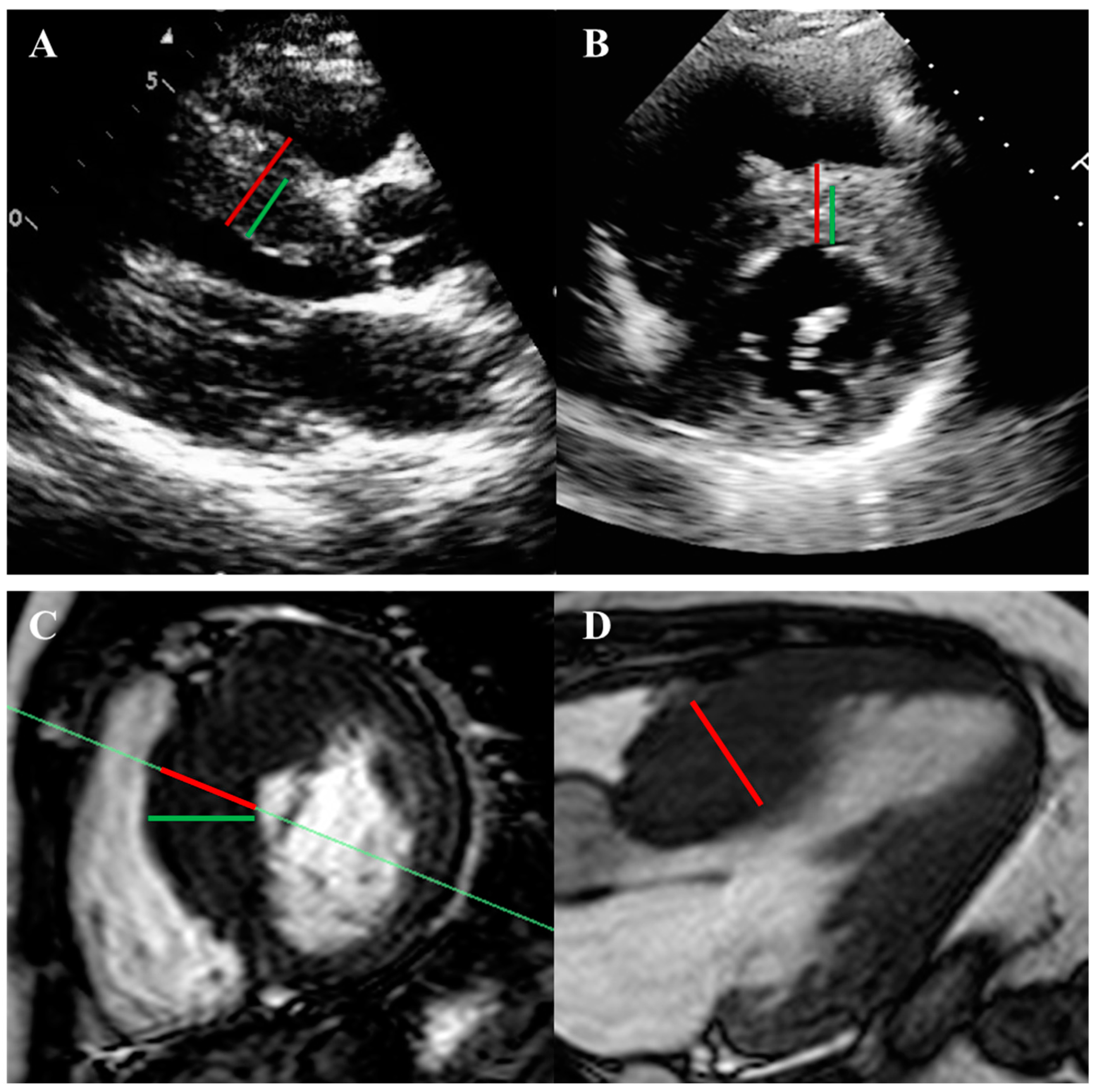
2.1. Left Ventricular Hypertrophy and Morphology

LVH is the primary diagnostic feature of HCM. In adults, a diagnosis is suggested by a maximal end-diastolic wall thickness ≥15 mm anywhere in the left ventricle or 13–14 mm with a family history of HCM or a known pathogenic/likely pathogenic sarcomere mutation, in the absence of other causes of LVH [1,2]. For children, a left ventricle (LV) wall thickness body surface area-adjusted z-score ≥ 2 standard deviations above the mean is used as a diagnostic criterion (Table 1) [9]. It is essential to recognize that reliance on fixed wall thickness thresholds may lead to underdiagnosis, especially in cases of subthreshold hypertrophy or apical HCM variants. Emerging research advocates using demographic-adjusted wall thickness cut-offs to enhance diagnostic accuracy and case finding [10]. Moreover, an indexed apical wall thickness exceeding 5.6 mm/m2, accompanied by the absence of normal apical tapering, has been proposed as a more sensitive marker for identifying apical HCM [10,11].
The pattern of hypertrophy is often asymmetric, typically involving less than half of the LV segments and occasionally confined to one or two segments [12]. The most common pattern is focal asymmetric hypertrophy of the basal anterior septum. Distinct LVH phenotypes have important clinical implications. LVOTO most likely accompanies the sigmoid phenotype, whereas the reverse curvature type is most likely to carry sarcomere mutations and exhibit late gadolinium enhancement (LGE) [13]. The apical variant is regarded as a more benign phenotype in terms of sudden cardiac death (SCD). Importantly, increased LV mass is not a diagnostic prerequisite, as focal hypertrophy can occur with a normal overall LV mass. Beyond diagnosis, LV wall thickness has prognostic value with a parabolic relationship between the degree of LV thickening and symptom severity. Furthermore, extreme LV wall thickness (>28–30 mm) is a strong predictor of SCD [2].

2.2. Systolic Function and Myocardial Deformation

The left ventricular ejection fraction (LVEF) is usually normal to hyperdynamic in patients with HCM, which is explained by an exaggerated contractile force generation in response to increased actin–myosin cross-bridges. Nonetheless, these patients demonstrate paradoxical contractile dysfunction, as demonstrated by an impaired global longitudinal strain (GLS), a more sensitive marker of contractile dysfunction than LVEF [9]. Lower (less negative) GLS values are of prognostic value and predict the occurrence of HCM-related adverse outcomes. It is unclear whether abnormal GLS reflects the underlying hypertrophy, myocardial disarray, or myocardial fibrosis. Finally, a reduced LVEF < 50% denotes progression to end-stage disease with poor prognosis and increased risk of SCD [14].

2.3. Diastolic Dysfunction

Diastolic dysfunction is present in approximately 80% of patients with HCM, irrespective of their morphological phenotype. This is characterized by elevated filling pressures and delayed myocardial relaxation due to myocardial hypertrophy, fibrosis, microvascular ischemia, and altered energetics [15]. In some cases, increased stiffness and severe hypertrophy significantly reduce the ventricular cavity size and stroke volume, potentially leading to restrictive physiology [16]. Notably, diastolic dysfunction can precede the development of overt LVH and independently contributes to reduced exercise capacity and adverse prognosis.

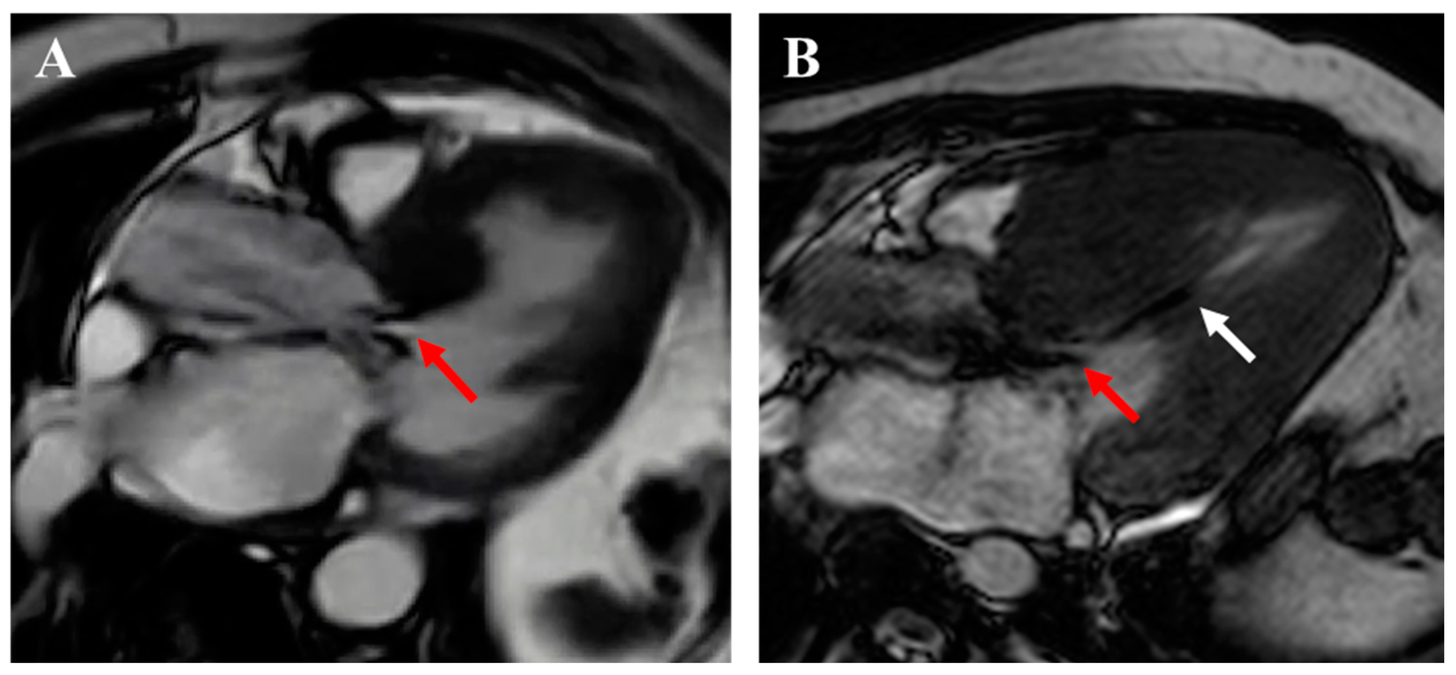
2.4. Apical Aneurysm

Left ventricular apical aneurysms, which occur in 2–5% of cases, particularly in apical and mid-cavitary phenotypes, are crucial imaging targets in HCM risk stratification [17]. These aneurysms result from repetitive myocardial ischemia, leading to transmural scarring and subsequent aneurysmal degeneration. Current guidelines consider apical aneurysms of any size a significant risk factor when evaluating for implantable cardioverter defibrillator (ICD) therapy for primary prevention [2]. However, recent evidence suggests that a larger aneurysm size (≥2 cm) is associated with a poorer prognosis and a higher risk of SCD [18].

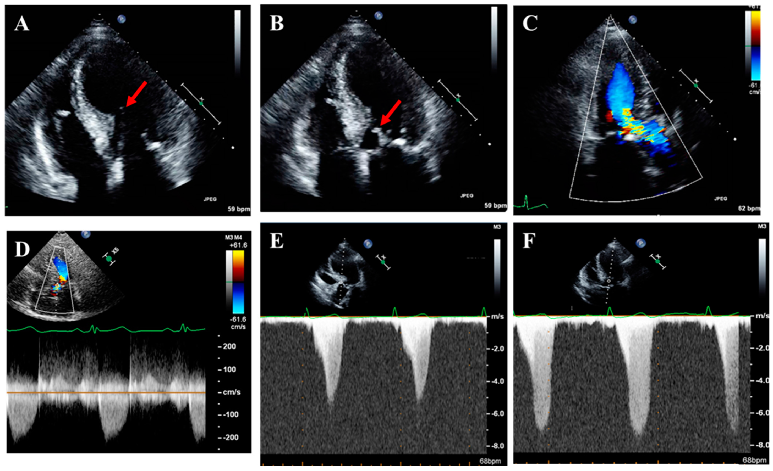
2.5. Left Ventricular Outflow Tract Obstruction

Approximately 70% of patients with HCM exhibit LVOTO, either at rest (35%) or with provocation (35%) [1,2]. The remaining 30% have non-obstructive HCM. While a peak LVOT gradient ≥ 30 mmHg defines obstruction, symptoms often manifest with gradients ≥ 50 mmHg. Notably, the presence of LVOTO is linked to an increased risk of SCD and heart failure progression in patients with HCM [19].
The primary mechanism of LVOTO in HCM is systolic anterior motion (SAM) of the mitral valve, a dynamic obstruction influenced by ventricular loading conditions and contractility. SAM results from a complex interplay of forces within the LVOT, often involving septal hypertrophy, LVOT narrowing, hyperdynamic LV, and mitral valve abnormalities [17]. Although common in HCM, SAM can occur in other conditions such as hypertensive heart disease and after mitral valve repair [20]. Mid-cavitary obstruction, alone or combined with LVOTO, can also occur due to hypertrophied or anomalous papillary muscles and a hyperdynamic LV with systolic cavity obliteration [21].

2.6. Abnormalities in Mitral Valve Apparatus and Mitral Regurgitation

Structural abnormalities of the mitral valve apparatus are common in HCM and can play a significant role in developing LVOTO and mitral regurgitation (MR). In patients with HCM, mitral valve abnormalities frequently include leaflet elongation, direct insertion of anomalous papillary muscles into the anterior mitral leaflet or LVOT, and anterior or apical displacement of the papillary muscles. MR in HCM can be secondary to SAM of the mitral valve or arise from intrinsic mitral valve pathology, such as mitral valve prolapse. Significantly, factors that exacerbate LVOTO severity often concurrently worsen the severity of SAM-related MR.

2.7. Myocardial Ischemia and Microvascular Dysfunction

Myocardial ischemia is common in patients with HCM, even without obstructive epicardial coronary artery disease, and is virtually universal in apical HCM [22]. This is primarily due to a supply–demand mismatch. Reduced myocardial oxygen supply in HCM results from decreased intramural arteriole density (rarefaction) with medial hypertrophy, microvascular dysfunction with impaired coronary flow reserve, and elevated filling pressures that reduce coronary perfusion [2,23]. Simultaneously, myocardial oxygen demand is increased by hypertrophy and intrinsically higher sarcomere-level force generation [23]. Myocardial bridging, more prevalent in HCM, can further exacerbate ischemia [1].

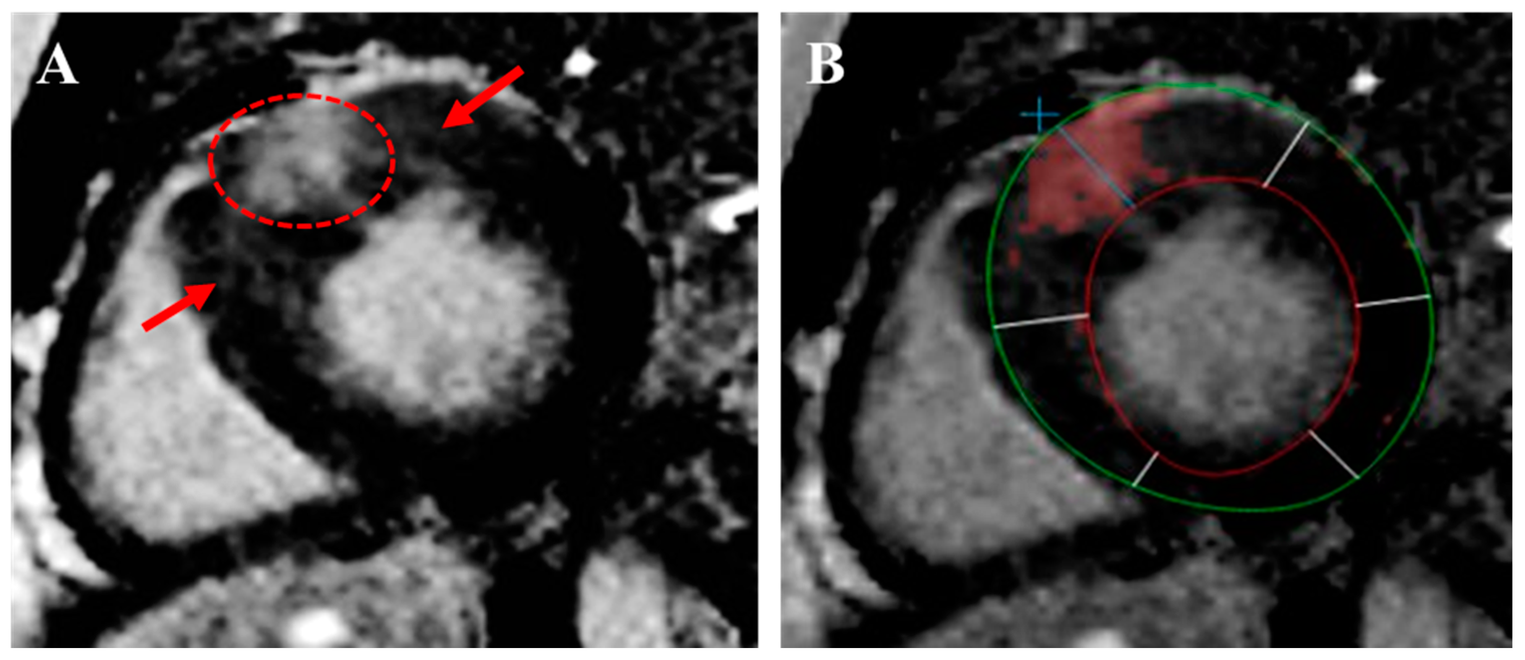
2.8. Myocardial Fibrosis

Myocardial fibrosis carries significant prognostic implications. It exists in two forms: interstitial and replacement. Interstitial fibrosis may represent a maladaptive response to chronic energy deprivation and supply–demand mismatch, attempting to preserve the cardiac structure. However, replacement fibrosis results from repetitive myocardial injury, leading to myocyte death and irreversible scarring [24]. This type of fibrosis is found in 50–60% of HCM patients, typically in the most hypertrophied segments, with a characteristic patchy, mid-myocardial distribution [17]. The extent of replacement fibrosis directly correlates with ventricular arrhythmias and SCD risk [25].

3. Multimodality Imaging in the Evaluation of HCM

3.1. Echocardiography

Transthoracic echocardiography (TTE) is the first-line imaging modality in evaluating all patients with known or suspected HCM (Table 2 and Table 3). Transesophageal echocardiography (TEE) is indicated in cases of suboptimal TTE imaging to elucidate LVOTO etiology or MR mechanism if uncertain, or intraoperatively during septal reduction therapies.
LVH assessment requires describing the pattern and distribution of hypertrophy and measuring maximal wall thickness. TTE can miss localized hypertrophy, especially in the basal anteroseptal, apical, and lateral areas, and ultrasound-enhancing agents (UEAs) can improve the detection of these patterns. Conversely, TTE can overestimate wall thickness due to foreshortening, tangential cuts, or inclusion of the RV trabeculae or moderator band (Figure 1) [1]. Integrating short- and long-axis views ensures accurate measurements.
LVEF should be evaluated at the initial diagnosis, with changes in clinical status, or during the surveillance of patients on cardiac myosin inhibitors (CMIs). Strain analysis and GLS measurements offer valuable prognostic data and should be considered in centers with experience in strain echocardiography. Strain may also aid in the discrimination of HCM from phenocopies such as cardiac amyloidosis (Figure 2). A comprehensive assessment of diastolic function should be routinely performed and include parameters such as the E/e’ ratio, left atrial volume index (LAVi), the duration difference between pulmonary vein atrial reversal (Ar) velocity and mitral inflow A-wave duration (Ar-A duration), and the peak tricuspid regurgitation (TR) jet velocity [9,26]. These parameters are applicable regardless of LVOTO; however, the E/e’ ratio and LAVi are unreliable with moderate or severe MR. Conventional E/e’ cut-off values may not directly apply to HCM; up to 25% of HCM patients with elevated E/e’ may have normal filling pressures. However, a restrictive filling pattern with elevated E/e’ ratio correlates with adverse outcomes, including heart failure hospitalizations, reduced exercise tolerance, and SCD [17].
Thorough LVOTO assessment, including provocative maneuvers, is needed to differentiate obstructive from non-obstructive phenotypes and distinguish dynamic LVOTO, which is characteristic of HCM, from fixed sub-valvular, valvular, or supravalvular aortic stenosis. Evaluation should identify and grade the SAM of the mitral valve using M-mode or 3D bi-plane imaging. The obstruction’s presence, severity, and location (i.e., LVOT, mid-cavitary, or multi-level) should be determined using color and pulse-wave Doppler (Figure 3). Factors contributing to obstruction, such as septal hypertrophy, LVOT narrowing, hyperdynamic LVEF, and mitral valve abnormalities, should be carefully assessed.
Continuous-wave (CW) Doppler is used to measure the peak instantaneous LVOT gradient, characteristically a late-peaking, “dagger-shaped” signal (Figure 3). Assessments should be performed at rest and if the resting gradient is <50 mmHg, with provocative maneuvers (Valsalva, squat-to-stand, or amyl nitrite) (Figure 3). Severe LVOTO (typically > 60 mmHg at rest) may show a “lobster-claw” spectral Doppler pattern due to a mid-systolic drop in LV ejection velocities. Differentiating LVOT signals from MR can be challenging, especially with late systolic MR. Velocities exceeding 5.5 m/s likely represent MR rather than LVOTO (Figure 3). Several techniques can aid this distinction (Table 4). In complex cases, the gradient can be indirectly estimated using the MR jet and systolic blood pressure (SBP) (LVOT gradient = SBP – 4 × [peak MR velocity]2 + 15–20 mmHg) but should be applied cautiously due to reliance on assumptions.
It is also essential to define the underlying mechanisms and severity of MR in HCM (SAM-related vs. primary MR) [27]. SAM-related MR typically, though not invariably, results in a posteriorly or laterally directed eccentric jet [28]. Quantifying the severity of eccentric MR jets can be challenging, often necessitating volumetric assessment of regurgitant volume and fraction.
Key features of apical aneurysms include a discrete, thin-walled (often ≤5 mm), dyskinetic, or akinetic region at the LV apex, clearly demarcated from the adjacent myocardium, frequently with a diastolic bulge [18]. Pitfalls such as foreshortening and misinterpretation of other conditions such as takotsubo cardiomyopathy must be avoided. A low threshold should be established for utilizing UEAs to improve the visualization of apical aneurysms and detect apical thrombi.

3.2. Stress Echocardiography

Exercise stress echocardiography is primarily indicated in symptomatic patients with HCM who exhibit a resting or provoked peak LVOT gradient < 50 mmHg on TTE to unmask latent LVOTO capable of producing symptoms (Table 2 and Table 3) [2]. Upright exercise is the preferred method as it provides the greatest physiological stimulus for provoking LVOTO and typically induces higher gradients than supine exercise [1]. Conversely, dobutamine stress echocardiography is not recommended for provoking LVOTO in HCM due to its non-physiological effects and potential for inducing dynamic gradients even in normal individuals [1,3].
Beyond its role in provoking LVOTO, exercise stress echocardiography may provide valuable insights into a patient’s functional capacity and symptom status under physiological stress. It also allows for assessing exercise-induced myocardial ischemia, changes in the severity of MR, and diastolic function parameters. While the role of diastolic parameters in exercise is still under investigation, assessing these parameters may help identify the mechanism of exercise intolerance and influence management decisions. It is reasonable to repeat exercise stress echocardiography if there is a decline in functional status or every 2–3 years in asymptomatic patients, especially if there is ambiguity regarding changes in their functional capacity [2].

3.3. Cardiac Magnetic Resonance Imaging

Cardiac magnetic resonance (CMR) is crucial for HCM diagnosis, risk stratification, and procedural planning, complementing echocardiography in most suspected or confirmed cases (Table 2 and Table 3). This modality more reliably identifies subtle or atypical patterns of LVH and apical aneurysms, often missed by echocardiography (Figure 4) [29,30]. It offers a more precise measurement of LV wall thickness, which is crucial for guiding ICD implantation by avoiding overestimation from tangential septal measurements or right ventricular trabeculae (Figure 1). CMR also differentiates HCM from phenocopies like cardiac amyloidosis, Fabry’s cardiomyopathy, athlete’s heart, and hypertensive heart disease, among others (Figure 5) [9,31,32].
CMR is the gold standard for evaluating LVEF, typically indicated when echocardiographic assessment is non-diagnostic or equivocal. This modality could also provide valuable insights into diastolic function by assessing LV mass, left atrial (LA) size, LV and LA strain, and parameters like peak filling rate (PFR) and time to peak filling rate (TPFR). Emerging evidence suggests that 4D flow techniques may allow the evaluation of mitral inflow patterns as part of the evaluation of diastolic function. CMR can further elucidate the factors contributing to LVOTO (Figure 6). While phase-contrast imaging within CMR can estimate peak LVOT gradients, echocardiography remains the preferred modality for this specific hemodynamic measurement. Similarly, CMR excels at clarifying the mechanism of MR, differentiating SAM-related MR from primary MR. Its volumetric assessment enables accurate and reproducible quantification of MR, particularly for the eccentric jets often seen in HCM [1,17,29,30,33].
CMR identifies and quantifies myocardial replacement fibrosis via LGE imaging. Replacement fibrosis, appearing as patchy, mid-wall, high-intensity signals on LGE within hypertrophied areas, is a critical SCD predictor (Figure 7) [34,35]. This is quantified using a > 6 SD threshold above the normal myocardium signal intensity. Although a burden of replacement fibrosis by LGE > 15% suggests increased SCD risk and potential ICD consideration, the risk associated with LGE is a continuum [36]. In contrast, interstitial fibrosis, seen as diffuse T1 elevation or faint lattice LGE, is associated with worse outcomes, including heart failure events, but has no definitive link to SCD (Figure 7) [37]. Native myocardial T1 and LGE entropies, reflecting myocardial tissue and fibrosis heterogeneity, are new and promising predictors of SCD in HCM [38,39]. Finally, abnormal myocardial perfusion due to microvascular dysfunction by CMR is an early disease marker, potentially preceding fibrosis, linked with increased non-sustained ventricular tachycardia [40,41].

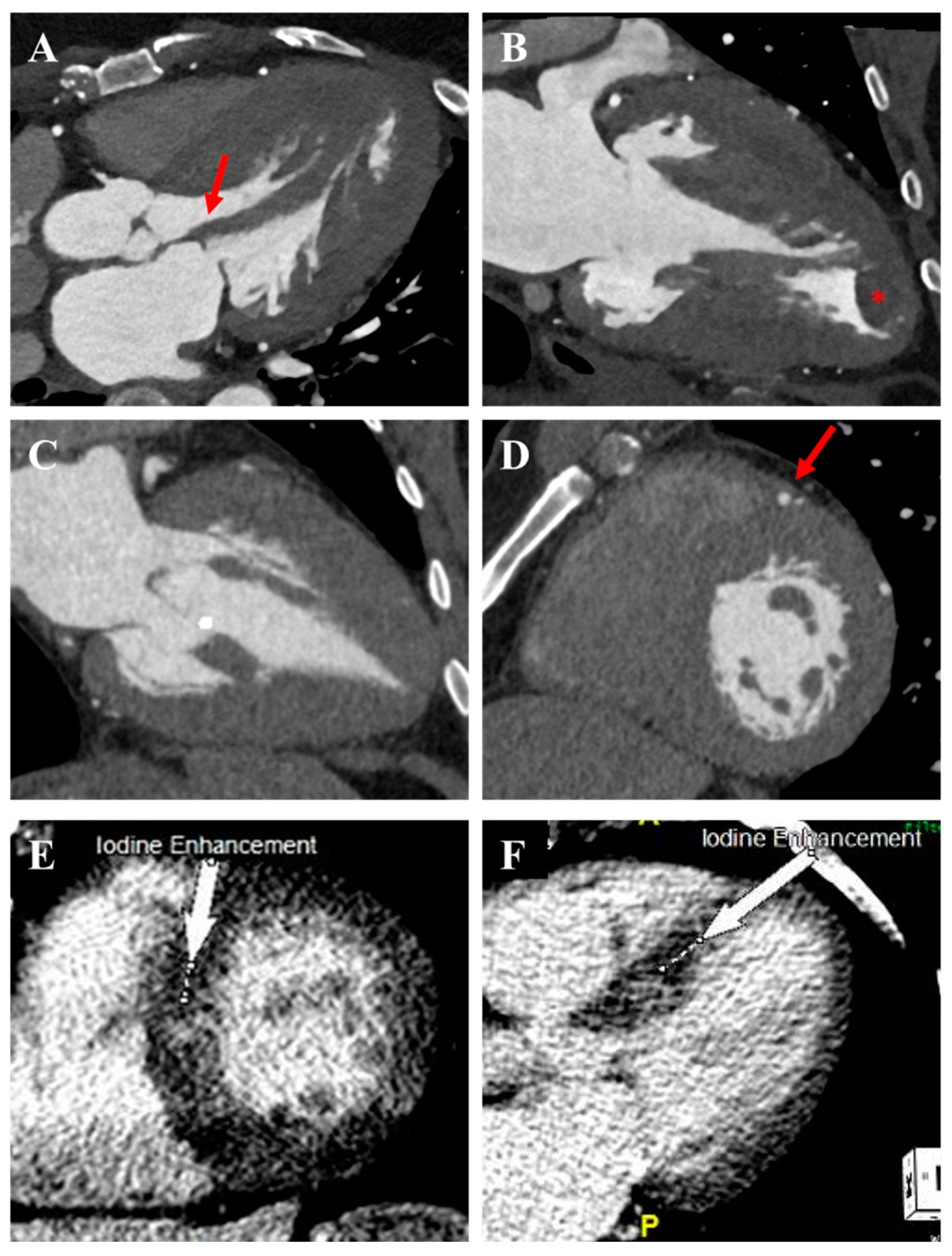
3.4. Cardiac Computed Tomography

Cardiac computed tomography (CCT) is the preferred non-invasive method for excluding epicardial coronary artery disease and evaluating myocardial bridging (Table 2 and Table 3) [42,43]. This modality can assess LVH and even LVEF when echocardiography is suboptimal and CMR is contraindicated. Furthermore, CCT can identify anatomical abnormalities of the sub-valvular apparatus, such as apically displaced or aberrant papillary muscles (Figure 8). Emerging data suggests late iodine enhancement (LIE) on CCT could assess replacement myocardial fibrosis [44]. While still under investigation, LIE may offer an alternative for fibrosis assessment in patients unable to undergo CMR. In the preprocedural setting, CCT can help visualize septal anatomy to identify target vessels for alcohol septal ablation and to provide safety considerations for surgical myomectomy [45,46]. The drawbacks of CCT include radiation exposure, use of iodine contrast, and inferior temporal resolution.

3.5. Nuclear Myocardial Perfusion Imaging

Nuclear myocardial perfusion imaging (MPI), particularly positron emission tomography (PET), is valuable in HCM for assessing microvascular dysfunction once epicardial coronary artery disease is excluded (Table 2 and Table 3). PET is preferred due to its superior temporal and spatial resolution and ability to quantify myocardial blood flow [9,47]. Furthermore, asymmetric resting tracer uptake, often more intense in hypertrophied areas, can be seen in HCM patients during MPI and should prompt consideration for further evaluation if the disease is not known (Figure 9) [1,48]. Nuclear MPI can assess LVEF and diastolic function parameters such as PFR and TPFR derived from the time–activity curve. However, this modality is not routinely indicated for this purpose.

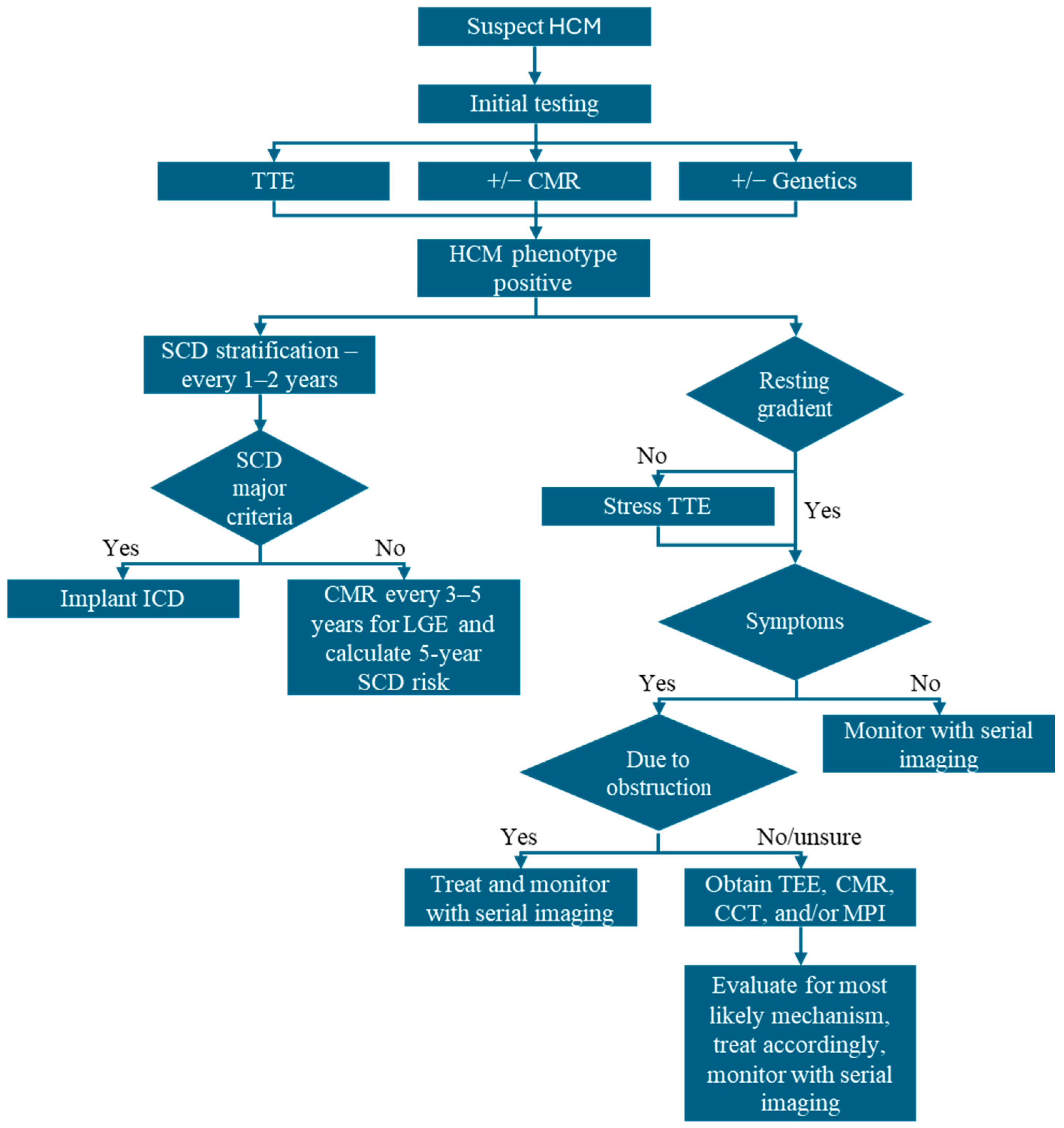
4. Imaging-Guided Management of HCM

4.1. Diagnosis and Classification

The accurate diagnosis of HCM relies heavily on a multimodality imaging approach, especially in differentiating HCM from phenocopies. Relying solely on echocardiography can lead to misdiagnosis, as these mimickers can present with similar features. Misdiagnosis can result in inappropriate treatment and potentially detrimental interventions. Similarly, determining the correct HCM phenotype, obstructive or non-obstructive, is crucial for appropriate management. Therefore, most patients with suspected HCM should undergo a comprehensive evaluation that includes TTE with complementary CMR and stress echocardiography to ensure accurate diagnosis and classification (Figure 10).
As HCM awareness continues to increase, there will be an increase in the detection of diseases before clinical symptoms, which often present with subtler changes on echocardiography requiring corroboration with other modalities. Early and accurate diagnosis, facilitated by this comprehensive approach, enables timely intervention, optimizes symptom management, allows for a broader range of treatment options, and improves risk stratification, ultimately reducing morbidity and mortality.

4.2. Symptom Management and Treatment of LVOTO

Patients with HCM often present with a variety of symptoms, including exertional dyspnea, fatigue, syncope, and chest pain. Diagnosing the root cause of these symptoms in HCM is particularly challenging due to the multiple potential contributing mechanisms, including systolic or diastolic dysfunction, LVOTO, MR, and ischemia from epicardial or microvascular disease. Crucially, these diverse mechanisms can present with similar symptoms, masking the underlying pathology. A multimodality imaging approach tailored to patients’ presentations can disentangle these overlapping presentations and pinpoint symptoms’ dominant contributing mechanism(s). As each of these etiologies requires a distinct management strategy, comprehensive imaging is not just about diagnosing but precisely defining the underlying pathophysiology to guide appropriate and effective treatment.
The presence of symptomatic LVOTO refractory to non-pharmacological strategies (e.g., avoidance of diuretic or pure vasodilators) informs the need to initiate beta-blockers or calcium channel blockers as an alternative [1,2,49]. As symptoms often manifest with LVOT gradients ≥ 50 mmHg at rest, during Valsalva, or with exercise, this threshold is generally used to guide treatment decisions. If symptomatic LVOTO persists despite initial therapy, escalation to CMI, disopyramide, or septal reduction therapies is recommended [50].
Comprehensive cardiac imaging is crucial when evaluating septal reduction therapies and mitral interventions for patients with HCM [1]. Simply identifying LVOTO or MR is insufficient; a thorough understanding of the underlying mechanisms is paramount. TTE and, when indicated, complementary TEE and CMR facilitate the identification of the complex interplay between anatomical and functional factors contributing to obstruction and regurgitation. Furthermore, when alcohol septal ablation is being considered, echocardiography with contrast can identify favorable coronary arterial anatomy supplying the septum [51]. By precisely characterizing these elements, imaging guides the heart team in tailoring the intervention to the patient’s needs. When surgery is considered, this may involve a combination of septal myectomy, mitral valve intervention, and papillary muscle reorientation to alleviate LVOTO and resolve MR.

4.3. Risk Stratification for SCD and ICD Implantation

Patients with HCM have an estimated SCD risk of approximately 0.5% per year, with younger individuals facing a higher risk than older patients [52,53,54]. Per current guidelines, all patients should undergo SCD risk stratification at initial diagnosis and every 1–2 years thereafter [2]. This involves evaluating established major clinical and imaging risk factors (Table 5). Multimodality imaging with echocardiography and CMR contributes to three major risk factors for primary prevention ICD consideration. These include a maximal wall thickness ≥ 28–30 mm, LVEF < 50%, and the presence of apical aneurysm [2]. Furthermore, CMR may guide ICD implantation when major risk factors are absent or indications for ICD remain uncertain. Specifically, extensive myocardial replacement fibrosis, quantified as ≥15% of myocardial mass on LGE imaging, may be considered (Class IIb) as an indication for primary prevention ICD implantation [36,55]. Finally, echocardiography contributes to estimating 5-year SCD risk calculation by providing measurements of left atrial diameter and peak LVOT gradient [19,56,57,58]. This risk estimation facilitates shared decision-making in primary prevention ICD implantation discussions.
Novel imaging parameters, including GLS, PET-assessed flow heterogeneity, and LGE entropy, demonstrate potential for refining SCD risk stratification [39,58,59,60,61,62]. However, further validation is required before their integration into clinical practice.

4.4. Assess Response to Therapy and Reverse Remodeling

TTE and stress echocardiography, when indicated, are essential for monitoring treatment response, particularly when evaluating LVOTO. Furthermore, echocardiography plays a crucial role in monitoring LVEF during CMI therapy. For example, when initiating or adjusting the dose of mavacamten, monthly TTE is recommended for the first three months (initiation phase), followed by quarterly assessments during maintenance [63]. A reduction in LVEF to <50% necessitates either dose reduction or discontinuation of the CMI [2,64]. Beyond LVEF and LVOT gradients, additional echocardiographic parameters such as LV mass index, E/e’ ratio, LAVi, and GLS can be utilized to assess for reverse remodeling during CMI use [64,65]. Although no evidence exists, the expectation is that improvements in these parameters will correlate with improved clinical outcomes. The utility of serial CMR in evaluating the effects of CMI on myocardial mass, myocardial native T1 time, and myocardial fibrosis remains unestablished.

5. Conclusions

Multimodality imaging is a cornerstone in the diagnosis and management of HCM. It allows for an earlier, more accurate diagnosis by having corroborating data, which also helps distinguish between morphological mimickers. A comprehensive imaging approach guides personalized treatment strategies by improving risk stratification and understanding underlying mechanisms contributing to patient symptoms. As novel imaging targets are identified and studied, more opportunities for advancing HCM care will be available.

Author Contributions

Conceptualization: A.L.S. and A.K.D.; writing—original draft: A.K.D. and A.L.S.; writing—review and editing: A.K.D., A.L.S., C.M.P., S.N., D.L., A.F., A.W.-S., M.J.G. and L.S. All authors have read and agreed to the published version of the manuscript.

Funding

This research received no external funding.

Conflicts of Interest

The authors have no conflicts of interest to declare.

References

  1. Nagueh, S.F.; Phelan, D.; Abraham, T.; Armour, A.; Desai, M.Y.; Dragulescu, A.; Gilliland, Y.; Lester, S.J.; Maldonado, Y.; Mohiddin, S.; et al. Recommendations for Multimodality Cardiovascular Imaging of Patients with Hypertrophic Cardiomyopathy: An Update from the American Society of Echocardiography, in Collaboration with the American Society of Nuclear Cardiology, the Society for Cardiovascular Magnetic Resonance, and the Society of Cardiovascular Computed Tomography. J. Am. Soc. Echocardiogr. 2022, 35, 533–569. [Google Scholar] [PubMed]
  2. Ommen, S.R.; Ho, C.Y.; Asif, I.M.; Balaji, S.; Burke, M.A.; Day, S.M.; Dearani, J.A.; Epps, K.C.; Evanovich, L.; Ferrari, V.A.; et al. 2024 AHA/ACC/AMSSM/HRS/PACES/SCMR Guideline for the Management of Hypertrophic Cardiomyopathy: A Report of the American Heart Association/American College of Cardiology Joint Committee on Clinical Practice Guidelines. Circulation 2024, 149, e1239–e1311. [Google Scholar]
  3. Maron, B.J.; Desai, M.Y.; Nishimura, R.A.; Spirito, P.; Rakowski, H.; Towbin, J.A.; Rowin, E.J.; Maron, M.S.; Sherrid, M.V. Diagnosis and Evaluation of Hypertrophic Cardiomyopathy: JACC State-of-the-Art Review. J. Am. Coll. Cardiol. 2022, 79, 372–389. [Google Scholar] [CrossRef]
  4. Semsarian, C.; Ingles, J.; Maron, M.S.; Maron, B.J. New perspectives on the prevalence of hypertrophic cardiomyopathy. J. Am. Coll. Cardiol. 2015, 65, 1249–1254. [Google Scholar] [CrossRef] [PubMed]
  5. Maron, M.S.; Hellawell, J.L.; Lucove, J.C.; Farzaneh-Far, R.; Olivotto, I. Occurrence of Clinically Diagnosed Hypertrophic Cardiomyopathy in the United States. Am. J. Cardiol. 2016, 117, 1651–1654. [Google Scholar] [CrossRef]
  6. Maron, B.J. Clinical Course and Management of Hypertrophic Cardiomyopathy. N. Engl. J. Med. 2018, 379, 655–668. [Google Scholar] [CrossRef] [PubMed]
  7. Desai, M.Y.; Owens, A.; Geske, J.B.; Wolski, K.; Naidu, S.S.; Smedira, N.G.; Cremer, P.C.; Schaff, H.; McErlean, E.; Sewell, C.; et al. Myosin Inhibition in Patients with Obstructive Hypertrophic Cardiomyopathy Referred for Septal Reduction Therapy. J. Am. Coll. Cardiol. 2022, 80, 95–108. [Google Scholar] [CrossRef]
  8. Alashi, A.; Desai, R.M.; Khullar, T.; Hodges, K.; Rodriguez, E.R.; Tan, C.; Popovic, Z.B.; Thamilarasan, M.; Wierup, P.; Lever, H.M.; et al. Different Histopathologic Diagnoses in Patients with Clinically Diagnosed Hypertrophic Cardiomyopathy After Surgical Myectomy. Circulation 2019, 140, 344–346. [Google Scholar] [CrossRef]
  9. Goldie, F.C.; Lee, M.M.Y.; Coats, C.J.; Nordin, S. Advances in Multimodality Imaging in Hypertrophic Cardiomyopathy. J. Clin. Med. 2024, 13, 842. [Google Scholar] [CrossRef]
  10. Shiwani, H.; Davies, R.H.; Topriceanu, C.C.; Ditaranto, R.; Owens, A.; Raman, B.; Augusto, J.; Hughes, R.K.; Torlasco, C.; Dowsing, B.; et al. Demographic-Based Personalized Left Ventricular Hypertrophy Thresholds for Hypertrophic Cardiomyopathy Diagnosis. J. Am. Coll. Cardiol. 2025, 85, 685–695. [Google Scholar] [CrossRef]
  11. Hughes, R.K.; Shiwani, H.; Rosmini, S.; Augusto, J.B.; Burke, L.; Jiang, Y.; Pierce, I.; Joy, G.; Castelletti, S.; Orini, M.; et al. Improved Diagnostic Criteria for Apical Hypertrophic Cardiomyopathy. JACC Cardiovasc. Imaging 2024, 17, 501–512. [Google Scholar] [CrossRef] [PubMed]
  12. Klues, H.G.; Schiffers, A.; Maron, B.J. Phenotypic spectrum and patterns of left ventricular hypertrophy in hypertrophic cardiomyopathy: Morphologic observations and significance as assessed by two-dimensional echocardiography in 600 patients. J. Am. Coll. Cardiol. 1995, 26, 1699–1708. [Google Scholar] [CrossRef] [PubMed]
  13. Bos, J.M.; Towbin, J.A.; Ackerman, M.J. Diagnostic, prognostic, and therapeutic implications of genetic testing for hypertrophic cardiomyopathy. J. Am. Coll. Cardiol. 2009, 54, 201–211. [Google Scholar] [CrossRef]
  14. Marstrand, P.; Han, L.; Day, S.M.; Olivotto, I.; Ashley, E.A.; Michels, M.; Pereira, A.C.; Wittekind, S.G.; Helms, A.; Saberi, S.; et al. Hypertrophic Cardiomyopathy with Left Ventricular Systolic Dysfunction: Insights from the SHaRe Registry. Circulation 2020, 141, 1371–1383. [Google Scholar] [CrossRef]
  15. Marstrand, P.; Han, L.; Day, S.M.; Olivotto, I.; Ashley, E.A.; Michels, M.; Pereira, A.C.; Wittekind, S.G.; Helms, A.; Saberi, S.; et al. Exercise response in hypertrophic cardiomyopathy: Blunted left ventricular deformational and twisting reserve with altered systolic-diastolic coupling. Circ. Cardiovasc. Imaging 2012, 5, 324–332. [Google Scholar]
  16. Kubo, T.; Gimeno, J.R.; Bahl, A.; Steffensen, U.; Steffensen, M.; Osman, E.; Thaman, R.; Mogensen, J.; Elliott, P.M.; Doi, Y.; et al. Prevalence, clinical significance, and genetic basis of hypertrophic cardiomyopathy with restrictive phenotype. J. Am. Coll. Cardiol. 2007, 49, 2419–2426. [Google Scholar] [CrossRef]
  17. Garcia, M.J. Multimodality Cardiovascular Imaging; Elsevier: Amsterdam, The Netherlands, 2025. [Google Scholar]
  18. Lee, D.Z.J.; Montazeri, M.; Bataiosu, R.; Hoss, S.; Adler, A.; Nguyen, E.T.; Rakowski, H.; Chan, R.H. Clinical Characteristics and Prognostic Importance of Left Ventricular Apical Aneurysms in Hypertrophic Cardiomyopathy. JACC Cardiovasc. Imaging 2022, 15, 1696–1711. [Google Scholar] [CrossRef]
  19. Elliott, P.M.; Gimeno, J.R.; Tome, M.T.; Shah, J.; Ward, D.; Thaman, R.; Mogensen, J.; McKenna, W.J. Left ventricular outflow tract obstruction and sudden death risk in patients with hypertrophic cardiomyopathy. Eur. Heart J. 2006, 27, 1933–1941. [Google Scholar] [CrossRef]
  20. Allen, R.D.; Edwards, W.D.; Tazelaar, H.D.; Danielson, G.K. Surgical pathology of subaortic septal myectomy not associated with hypertrophic cardiomyopathy: A study of 98 cases (1996–2000). Cardiovasc. Pathol. 2003, 12, 207–215. [Google Scholar] [CrossRef]
  21. Minami, Y.; Kajimoto, K.; Terajima, Y.; Yashiro, B.; Okayama, D.; Haruki, S.; Nakajima, T.; Kawashiro, N.; Kawana, M.; Hagiwara, N. Clinical implications of midventricular obstruction in patients with hypertrophic cardiomyopathy. J. Am. Coll. Cardiol. 2011, 57, 2346–2355. [Google Scholar] [CrossRef]
  22. Hughes, R.K.; Augusto, J.B.; Knott, K.; Davies, R.; Shiwani, H.; Seraphim, A.; Malcolmson, J.W.; Khoury, S.; Joy, G.; Mohiddin, S.; et al. Apical Ischemia Is a Universal Feature of Apical Hypertrophic Cardiomyopathy. Circ. Cardiovasc. Imaging 2023, 16, e014907. [Google Scholar] [CrossRef] [PubMed]
  23. Raphael, C.E.; Cooper, R.; Parker, K.H.; Collinson, J.; Vassiliou, V.; Pennell, D.J.; de Silva, R.; Hsu, L.Y.; Greve, A.M.; Nijjer, S.; et al. Mechanisms of Myocardial Ischemia in Hypertrophic Cardiomyopathy: Insights from Wave Intensity Analysis and Magnetic Resonance. J. Am. Coll. Cardiol. 2016, 68, 1651–1660. [Google Scholar] [CrossRef]
  24. Schlittler, M.; Pramstaller, P.P.; Rossini, A.; De Bortoli, M. Myocardial Fibrosis in Hypertrophic Cardiomyopathy: A Perspective from Fibroblasts. Int. J. Mol. Sci. 2023, 24, 14845. [Google Scholar] [CrossRef] [PubMed]
  25. Kiaos, A.; Daskalopoulos, G.N.; Kamperidis, V.; Ziakas, A.; Efthimiadis, G.; Karamitsos, T.D. Quantitative Late Gadolinium Enhancement Cardiac Magnetic Resonance and Sudden Death in Hypertrophic Cardiomyopathy: A Meta-Analysis. JACC Cardiovasc. Imaging 2024, 17, 489–497. [Google Scholar] [CrossRef]
  26. Nagueh, S.F.; Smiseth, O.A.; Appleton, C.P.; Byrd, B.F., 3rd; Dokainish, H.; Edvardsen, T.; Flachskampf, F.A.; Gillebert, T.C.; Klein, A.L.; Lancellotti, P.; et al. Recommendations for the Evaluation of Left Ventricular Diastolic Function by Echocardiography: An Update from the American Society of Echocardiography and the European Association of Cardiovascular Imaging. J. Am. Soc. Echocardiogr. 2016, 29, 277–314. [Google Scholar] [CrossRef] [PubMed]
  27. Schwammenthal, E.; Hagege, A.A.; Levine, R.A. Does the Flow Know? Mitral Regurgitant Jet Direction and Need for Valve Repair in Hypertrophic Obstructive Cardiomyopathy. J. Am. Soc. Echocardiogr. 2019, 32, 341–343. [Google Scholar] [CrossRef]
  28. Schwammenthal, E.; Nakatani, S.; He, S.; Hopmeyer, J.; Sagie, A.; Weyman, A.E.; Lever, H.M.; Yoganathan, A.P.; Thomas, J.D.; Levine, R.A. Mechanism of mitral regurgitation in hypertrophic cardiomyopathy: Mismatch of posterior to anterior leaflet length and mobility. Circulation 1998, 98, 856–865. [Google Scholar] [CrossRef]
  29. Moon, J.C.; Fisher, N.G.; McKenna, W.J.; Pennell, D.J. Detection of apical hypertrophic cardiomyopathy by cardiovascular magnetic resonance in patients with non-diagnostic echocardiography. Heart 2004, 90, 645–649. [Google Scholar] [CrossRef]
  30. Maron, M.S.; Rowin, E.J.; Maron, B.J. How to Image Hypertrophic Cardiomyopathy. Circ. Cardiovasc. Imaging 2017, 10, e005372. [Google Scholar] [CrossRef]
  31. Rickers, C.; Wilke, N.M.; Jerosch-Herold, M.; Casey, S.A.; Panse, P.; Panse, N.; Weil, J.; Zenovich, A.G.; Maron, B.J. Utility of cardiac magnetic resonance imaging in the diagnosis of hypertrophic cardiomyopathy. Circulation 2005, 112, 855–861. [Google Scholar] [CrossRef]
  32. Hindieh, W.; Weissler-Snir, A.; Hammer, H.; Adler, A.; Rakowski, H.; Chan, R.H. Discrepant Measurements of Maximal Left Ventricular Wall Thickness Between Cardiac Magnetic Resonance Imaging and Echocardiography in Patients with Hypertrophic Cardiomyopathy. Circ. Cardiovasc. Imaging 2017, 10, e006309. [Google Scholar] [CrossRef] [PubMed]
  33. Rowin, E.J.; Maron, B.J.; Maron, M.S. Reply: Hypertrophic Cardiomyopathy with Left Ventricular Apical Aneurysm. J. Am. Coll. Cardiol. 2017, 70, 407. [Google Scholar] [CrossRef]
  34. O’Hanlon, R.; Grasso, A.; Roughton, M.; Moon, J.C.; Clark, S.; Wage, R.; Webb, J.; Kulkarni, M.; Dawson, D.; Sulaibeekh, L.; et al. Prognostic significance of myocardial fibrosis in hypertrophic cardiomyopathy. J. Am. Coll. Cardiol. 2010, 56, 867–874. [Google Scholar] [CrossRef] [PubMed]
  35. Green, J.J.; Berger, J.S.; Kramer, C.M.; Salerno, M. Prognostic value of late gadolinium enhancement in clinical outcomes for hypertrophic cardiomyopathy. JACC Cardiovasc. Imaging 2012, 5, 370–377. [Google Scholar] [CrossRef] [PubMed]
  36. Mentias, A.; Raeisi-Giglou, P.; Smedira, N.G.; Feng, K.; Sato, K.; Wazni, O.; Kanj, M.; Flamm, S.D.; Thamilarasan, M.; Popovic, Z.B.; et al. Late Gadolinium Enhancement in Patients with Hypertrophic Cardiomyopathy and Preserved Systolic Function. J. Am. Coll. Cardiol. 2018, 72, 857–870. [Google Scholar] [CrossRef]
  37. Rahman, S.U.; Rowin, E.J. T1 Mapping and Interstitial Fibrosis as a Marker for Heart Failure in Hypertrophic Cardiomyopathy. Circ. Cardiovasc Imaging 2025, 18, e017938. [Google Scholar] [CrossRef]
  38. Wang, J.; Zhang, J.; Pu, L.; Qi, W.; Xu, Y.; Wan, K.; Zhu, Y.; Gkoutos, G.V.; Han, Y.; Chen, Y. The Prognostic Value of Left Ventricular Entropy from T1 Mapping in Patients with Hypertrophic Cardiomyopathy. JACC Asia 2024, 4, 389–399. [Google Scholar] [CrossRef]
  39. Ye, Y.; Ji, Z.; Zhou, W.; Pu, C.; Li, Y.; Zhou, C.; Hu, X.; Chen, C.; Sun, Y.; Huang, Q.; et al. Mean Scar Entropy by Late Gadolinium Enhancement Cardiac Magnetic Resonance Is Associated with Ventricular Arrhythmias Events in Hypertrophic Cardiomyopathy. Front. Cardiovasc. Med. 2021, 8, 758635. [Google Scholar] [CrossRef]
  40. Chiribiri, A.; Leuzzi, S.; Conte, M.R.; Bongioanni, S.; Bratis, K.; Olivotti, L.; De Rosa, C.; Lardone, E.; Di Donna, P.; Villa, A.D.; et al. Rest perfusion abnormalities in hypertrophic cardiomyopathy: Correlation with myocardial fibrosis and risk factors for sudden cardiac death. Clin. Radiol. 2015, 70, 495–501. [Google Scholar] [CrossRef]
  41. Camaioni, C.; Knott, K.D.; Augusto, J.B.; Seraphim, A.; Rosmini, S.; Ricci, F.; Boubertakh, R.; Xue, H.; Hughes, R.; Captur, G.; et al. Inline perfusion mapping provides insights into the disease mechanism in hypertrophic cardiomyopathy. Heart 2020, 106, 824–829. [Google Scholar] [CrossRef]
  42. Shariat, M.; Thavendiranathan, P.; Nguyen, E.; Wintersperger, B.; Paul, N.; Rakowski, H.; Crean, A.M. Utility of coronary CT angiography in outpatients with hypertrophic cardiomyopathy presenting with angina symptoms. J. Cardiovasc. Comput. Tomogr. 2014, 8, 429–437. [Google Scholar] [CrossRef] [PubMed]
  43. van der Velde, N.; Huurman, R.; Yamasaki, Y.; Kardys, I.; Galema, T.W.; Budde, R.P.; Zijlstra, F.; Krestin, G.P.; Schinkel, A.F.; Michels, M.; et al. Frequency and Significance of Coronary Artery Disease and Myocardial Bridging in Patients with Hypertrophic Cardiomyopathy. Am. J. Cardiol. 2020, 125, 1404–1412. [Google Scholar] [CrossRef]
  44. Gatti, M.; De Filippo, O.; Cura Cura, G.; Dusi, V.; Di Vita, U.; Gallone, G.; Morena, A.; Palmisano, A.; Pasinato, E.; Solano, A.; et al. Diagnostic accuracy of late iodine enhancement on cardiac CT for myocardial tissue characterization: A systematic review and meta-analysis. Eur. Radiol. 2024. [Google Scholar] [CrossRef]
  45. Takayama, H.; Yu, S.N.; Sorabella, R.; Leb, J.; Pulerwitz, T.C.; Cooper, C.; Argenio, M.; Shimada, Y.J.; Weiner, S.; Ginns, J.N. Virtual septal myectomy for preoperative planning in hypertrophic cardiomyopathy. J. Thorac. Cardiovasc. Surg. 2019, 158, 455–463. [Google Scholar] [CrossRef]
  46. Cooper, R.M.; Binukrishnan, S.R.; Shahzad, A.; Hasleton, J.; Sigwart, U.; Stables, R.H. Computed tomography angiography planning identifies the target vessel for optimum infarct location and improves clinical outcome in alcohol septal ablation for hypertrophic obstructive cardiomyopathy. EuroIntervention 2017, 12, e2194–e2203. [Google Scholar] [CrossRef] [PubMed]
  47. Bravo, P.E. Is there a role for cardiac positron emission tomography in hypertrophic cardiomyopathy? J. Nucl. Cardiol. 2019, 26, 1125–1134. [Google Scholar] [CrossRef] [PubMed]
  48. Camici, P.; Chiriatti, G.; Lorenzoni, R.; Bellina, R.C.; Gistri, R.; Italiani, G.; Parodi, O.; Salvadori, P.A.; Nista, N.; Papi, L.; et al. Coronary vasodilation is impaired in both hypertrophied and nonhypertrophied myocardium of patients with hypertrophic cardiomyopathy: A study with nitrogen-13 ammonia and positron emission tomography. J. Am. Coll. Cardiol. 1991, 17, 879–886. [Google Scholar] [CrossRef]
  49. Sherrid, M.V.; Shetty, A.; Winson, G.; Kim, B.; Musat, D.; Alviar, C.L.; Homel, P.; Balaram, S.K.; Swistel, D.G. Treatment of obstructive hypertrophic cardiomyopathy symptoms and gradient resistant to first-line therapy with beta-blockade or verapamil. Circ. Heart Fail 2013, 6, 694–702. [Google Scholar] [CrossRef]
  50. Mehra, N.; Veselka, J.; Smedira, N.; Desai, M.Y. Invasive therapies for symptomatic obstructive hypertrophic cardiomyopathy. Prog. Cardiovasc. Dis. 2023, 80, 46–52. [Google Scholar] [CrossRef]
  51. La Canna, G.; Scarfo, I.; Arendar, I.; Colombo, A.; Torracca, L.; Margonato, D.; Montorfano, M.; Alfieri, O. Targeting Alcohol Septal Ablation in Patients with Obstructive Hypertrophic Cardiomyopathy Candidates for Surgical Myectomy: Added Value of Three-Dimensional Intracoronary Myocardial Contrast Echocardiography. J. Clin. Med. 2021, 10, 2166. [Google Scholar] [CrossRef]
  52. Authors/Task Force members; Elliott, P.M.; Anastasakis, A.; Borger, M.A.; Borggrefe, M.; Cecchi, F.; Charron, P.; Hagege, A.A.; Lafont, A.; Limongelli, G.; et al. 2014 ESC Guidelines on diagnosis and management of hypertrophic cardiomyopathy: The Task Force for the Diagnosis and Management of Hypertrophic Cardiomyopathy of the European Society of Cardiology (ESC). Eur. Heart J. 2014, 35, 2733–2779. [Google Scholar]
  53. Dimitrow, P.P.; Chojnowska, L.; Rudzinski, T.; Piotrowski, W.; Ziolkowska, L.; Wojtarowicz, A.; Wycisk, A.; Dabrowska-Kugacka, A.; Nowalany-Kozielska, E.; Sobkowicz, B.; et al. Sudden death in hypertrophic cardiomyopathy: Old risk factors re-assessed in a new model of maximalized follow-up. Eur. Heart J. 2010, 31, 3084–3093. [Google Scholar] [CrossRef]
  54. Maron, B.J. Risk stratification and role of implantable defibrillators for prevention of sudden death in patients with hypertrophic cardiomyopathy. Circ. J. 2010, 74, 2271–2282. [Google Scholar] [CrossRef] [PubMed]
  55. Chan, R.H.; Maron, B.J.; Olivotto, I.; Pencina, M.J.; Assenza, G.E.; Haas, T.; Lesser, J.R.; Gruner, C.; Crean, A.M.; Rakowski, H.; et al. Prognostic value of quantitative contrast-enhanced cardiovascular magnetic resonance for the evaluation of sudden death risk in patients with hypertrophic cardiomyopathy. Circulation 2014, 130, 484–495. [Google Scholar] [CrossRef] [PubMed]
  56. Maron, M.S.; Olivotto, I.; Betocchi, S.; Casey, S.A.; Lesser, J.R.; Losi, M.A.; Cecchi, F.; Maron, B.J. Effect of left ventricular outflow tract obstruction on clinical outcome in hypertrophic cardiomyopathy. N. Engl. J. Med. 2003, 348, 295–303. [Google Scholar] [CrossRef] [PubMed]
  57. O’Mahony, C.; Jichi, F.; Pavlou, M.; Monserrat, L.; Anastasakis, A.; Rapezzi, C.; Biagini, E.; Gimeno, J.R.; Limongelli, G.; McKenna, W.J.; et al. A novel clinical risk prediction model for sudden cardiac death in hypertrophic cardiomyopathy (HCM risk-SCD). Eur. Heart J. 2014, 35, 2010–2020. [Google Scholar] [CrossRef]
  58. Hiemstra, Y.L.; Debonnaire, P.; Bootsma, M.; van Zwet, E.W.; Delgado, V.; Schalij, M.J.; Atsma, D.E.; Bax, J.J.; Marsan, N.A. Global Longitudinal Strain and Left Atrial Volume Index Provide Incremental Prognostic Value in Patients with Hypertrophic Cardiomyopathy. Circ. Cardiovasc. Imaging 2017, 10, e005706. [Google Scholar] [CrossRef]
  59. Tower-Rader, A.; Mohananey, D.; To, A.; Lever, H.M.; Popovic, Z.B.; Desai, M.Y. Prognostic Value of Global Longitudinal Strain in Hypertrophic Cardiomyopathy: A Systematic Review of Existing Literature. JACC Cardiovasc. Imaging 2019, 12, 1930–1942. [Google Scholar] [CrossRef]
  60. Lee, H.J.; Kim, H.K.; Lee, S.C.; Kim, J.; Park, J.B.; Hwang, I.C.; Choi, Y.J.; Lee, S.P.; Chang, S.A.; Lee, W.; et al. Supplementary role of left ventricular global longitudinal strain for predicting sudden cardiac death in hypertrophic cardiomyopathy. Eur. Heart J. Cardiovasc Imaging 2022, 23, 1108–1116. [Google Scholar] [CrossRef]
  61. Lu, D.Y.; Yalcin, H.; Yalcin, F.; Zhao, M.; Sivalokanathan, S.; Valenta, I.; Tahari, A.; Pomper, M.G.; Abraham, T.P.; Schindler, T.H.; et al. Stress Myocardial Blood Flow Heterogeneity Is a Positron Emission Tomography Biomarker of Ventricular Arrhythmias in Patients with Hypertrophic Cardiomyopathy. Am. J. Cardiol. 2018, 121, 1081–1089. [Google Scholar] [CrossRef]
  62. Zhao, X.; Jin, F.; Wang, J.; Zhao, X.; Wang, L.; Wei, H. Entropy of left ventricular late gadolinium enhancement and its prognostic value in hypertrophic cardiomyopathy a new CMR assessment method. Int. J. Cardiol. 2023, 373, 134–141. [Google Scholar] [PubMed]
  63. Desai, M.Y.; Owens, A.; Wolski, K.; Geske, J.B.; Saberi, S.; Wang, A.; Sherrid, M.; Cremer, P.C.; Lakdawala, N.K.; Tower-Rader, A.; et al. Mavacamten in Patients with Hypertrophic Cardiomyopathy Referred for Septal Reduction: Week 56 Results From the VALOR-HCM Randomized Clinical Trial. JAMA Cardiol. 2023, 8, 968–977. [Google Scholar] [PubMed]
  64. Olivotto, I.; Oreziak, A.; Barriales-Villa, R.; Abraham, T.P.; Masri, A.; Garcia-Pavia, P.; Saberi, S.; Lakdawala, N.K.; Wheeler, M.T.; Owens, A.; et al. Mavacamten for treatment of symptomatic obstructive hypertrophic cardiomyopathy (EXPLORER-HCM): A randomised, double-blind, placebo-controlled, phase 3 trial. Lancet 2020, 396, 759–769. [Google Scholar] [PubMed]
  65. Desai, M.Y.; Okushi, Y.; Gaballa, A.; Wang, Q.; Geske, J.B.; Owens, A.T.; Saberi, S.; Wang, A.; Cremer, P.C.; Sherrid, M.; et al. Serial Changes in Ventricular Strain in Symptomatic Obstructive Hypertrophic Cardiomyopathy Treated with Mavacamten: Insights From the VALOR-HCM Trial. Circ. Cardiovasc. Imaging 2024, 17, e017185. [Google Scholar]
Figure 1. Pitfalls in the measurement of left ventricular wall thickness in echocardiography and cardiac magnetic resonance (CMR) imaging in hypertrophic cardiomyopathy (HCM). (A,B) Left ventricular septal wall thickness measurements by echocardiography in the parasternal long-axis and short-axis views, respectively. The red line (28 mm) demonstrates incorrect measurement, resulting in overestimation due to the inclusion of right ventricular trabeculae compared to the correct measurement of the green line (17 mm). (C,D) A tangential cut red line (29 mm) on CMR that leads to an overestimation of the wall thickness when measured on a 3-chamber view, compared to the correct measurement green line (24 mm).
Jcm 14 02606 g001
Figure 2. Global longitudinal strain patterns for various HCM subtypes and common mimickers. (A) Asymmetric septal HCM with impaired strain along the septum. (B) Apical HCM with impaired strain in the apex. (C) Cardiac amyloidosis with impaired basal-mid strain and relative apical sparing (cherry-on-top pattern). (D) Severe aortic stenosis with diffuse impaired longitudinal strain.
Jcm 14 02606 g002
Figure 3. Representative echocardiography images in HCM for the comprehensive assessment of left ventricular outflow tract obstruction (LVOTO). (A) Depiction of an elongated anterior mitral valve (MV) leaflet (red arrow) and septal thickness as a mechanism for systolic anterior motion (SAM) of the MV. (B) Depiction of SAM of the MV (red arrow). (C) Color Doppler depicting flow acceleration in the left ventricular outflow tract (LVOT) at the level of SAM. (D) Continuous-wave (CW) Doppler of LVOT at rest with a gradient of 16 mmHg. (E) CW Doppler of LVOT with exercise showing dynamic LVOTO with a gradient of around 100 mmHg with the classic late-peaking, dagger shape. (F) CW Doppler with exercise and fanning of the transducer posteriorly depicting mitral signal contamination. Note the higher peak velocity, longer duration, and difference in shape compared to the LVOT signal.
Jcm 14 02606 g003
Figure 4. HCM phenotypes by CMR. (A) Reverse septum. (B) Sigmoid septum. (C) Typical apical. (D) Relative apical. (E) Mid-ventricular with apical aneurysm.
Jcm 14 02606 g004
Figure 5. Mimickers of HCM by CMR. (A,B) PRKAG2 cardiomyopathy masquerading as HCM with asymmetric septal hypertrophy and mid-myocardial late gadolinium enhancement (LGE). (C,D) Amyloid cardiomyopathy with concentric left ventricular hypertrophy (LVH) of septal predominance with typical diffuse sub-endocardial to transmural LGE. (E,F) Hypertensive heart disease with concentric LVH with basal septal predominance without LGE.
Jcm 14 02606 g005
Figure 6. Characterization of the mechanism of obstruction in HCM by CMR. (A) Pure LVOTO due to SAM of the MV (red arrow). (B) Multi-level obstruction with LVOTO (red arrow) with SAM and intracavitary gradient (white arrow) due to LVH, prominent papillary muscles, and hyperdynamic left ventricular ejection fraction (LVEF).
Jcm 14 02606 g006
Figure 7. Quantification of replacement fibrosis by CMR with LGE imaging of a patient with HCM. (A) The dashed red circle depicts replacement fibrosis as patchy, mid-wall, high-intensity signals, while the red arrows point towards interstitial fibrosis areas appearing as faint lattice signals. (B) The shaded red area depicts the 6-standard-deviation method used to quantify the amount of replacement fibrosis, noting that this should not include the areas of interstitial fibrosis.
Jcm 14 02606 g007
Figure 8. Cardiac computed tomography findings in HCM. (A) Asymmetric septal HCM with aberrant papillary muscle (red arrow) attached to the mitral valve contributing to LVOTO. (B) Mid-ventricular HCM with apical aneurysm and thrombus (*). (C) Apical HCM. (D) Myocardial bridge involving the left anterior descending artery (red arrow). (E,F) Asymmetric septal HCM with mid-myocardial late iodine enhancement (white arrows).
Jcm 14 02606 g008
Figure 9. Nuclear myocardial perfusion imaging (MPI) with positron emission tomography (PET) in apical HCM. (A,B) CMR images of a patient with apical HCM without epicardial coronary artery disease (not demonstrated in these images). (C) The corresponding MPI with PET depicting prominent resting tracer uptake at the apex consistent with the diagnosis of apical HCM with stress-induced apical ischemia due to microvascular disease. SAX: short axis; VLA: vertical long axis; HLA: horizontal long axis.
Jcm 14 02606 g009
Figure 10. Imaging algorithm for patients with suspected and confirmed HCM. CCT: cardiac computed tomography; CMR: cardiac magnetic resonance; HCM: hypertrophic cardiomyopathy; ICD: implantable cardioverter defibrillator; MPI: myocardial perfusion imaging; TEE transesophageal echocardiography; TTE: transthoracic echocardiography; SCD: sudden cardiac death.
Jcm 14 02606 g010
Table 1. Diagnostic criteria for HCM in adults.
Guideline-Supported CriteriaSupporting Findings
LV end-diastolic wall thickness ≥ 15 mm in any segment not entirely explained by another condition 
  • LVH that predominantly involves the basal septum or apex, sparing the posterior wall

  • SAM of MV and LVOTO

  • Apically displaced papillary muscle

  • Elongated anterior mitral valve leaflet
LV end-diastolic wall thickness 13–14 mm with a positive pathogenic genetic variant or in primary relatives of patients with HCM
In children: Indexed LV end-diastolic wall thickness z score ≥ 2
Emerging criteria
Demographically adjusted LVH
Indexed apical wall thickness > 5.6 mm/m2 in apical HCM
HCM: hypertrophic cardiomyopathy; LV: left ventricular; LVH: left ventricular hypertrophy; LVOTO: left ventricular outflow tract obstruction; MV: mitral valve; SAM: systolic anterior motion.
Table 2. Indication and frequency of imaging multimodality in HCM.
ModalityWhen to Obtain?When to Repeat?
TTE
  • Initial evaluation
  • Surveillance on cardiac myosin inhibitors
1–2 years, or functional decline
As per cardiac myosin inhibitor protocol
TEE
  • Uncertain LVOTO mechanism despite TTE
  • Uncertain MR mechanism/severity despite TTE
  • Procedural planning and intraprocedural
Personalized
Stress TTE
  • Initial evaluation if no LVOTO (<30 mmHg) at rest or with Valsalva
  • Symptomatic patients with LVOTO < 50 mmHg at rest or with Valsalva
2–3 years, or functional decline
CMR
  • Initial evaluation when TTE is non-diagnostic/equivocal or when differentiation from mimickers is needed
  • Uncertain LVOTO mechanism despite TTE
  • Uncertain MR mechanism/severity despite TTE
  • SCD risk remains uncertain
  • Procedural planning
3–5 years for SCD risk if no ICD present
CCT
  • Evaluation of obstructive epicardial CAD
  • Evaluation of LVH when TTE is non-diagnostic and CMR not available/contraindicated
  • Assessment of replacement fibrosis (late iodine enhancement) when CMR is not available/contraindicated
  • Procedural planning
Personalized
Nuclear MPI
  • Evaluation of microvascular ischemia after excluding epicardial CAD
Personalized
CAD: coronary artery disease; CCT: cardiac computed tomography; CMR: cardiac magnetic resonance; ICD: implantable cardioverter defibrillator; LVH: left ventricular hypertrophy; LVOTO: left ventricular outflow tract obstruction; MPI: myocardial perfusion imaging; MR: mitral regurgitation; TEE: transesophageal echocardiography; TTE: transthoracic echocardiography; SCD: sudden cardiac death.
Table 3. Imaging modalities and their capability to evaluate imaging targets *.
ModalityImaging Targets
LVHSystolic FunctionDiastolic FunctionApical
Aneurysm
LVOTO
(Gradient)
MR/LVOTO MechanismsEpicardial
CAD
MVDFibrosis
TTE+++++/++ with UEA+++
TEE++++++++++
Stress TTE+++++
(exercise diastology)
+/++ with UEA+++
(latent LVOTO)
++
(dynamic MR with exercise)
++
(LAD CFR)
CMR++++++++++++++++
(Stress CMR)
++++
(LGE)
CCT+++++++++/−
(anatomical abnormalities)
++++/−
(CCT perfusion)
+
(LIE)
Nuclear MPI+/−
(Asymmetric uptake)
++/−
(PFR/TPFR)
+++++
CAD: coronary artery disease; CCT: cardiac computed tomography; CFR: coronary flow reserve; CMR: cardiac magnetic resonance; LAD: left anterior descending artery; LIE: late iodine enhancement; LGE: late gadolinium enhancement; LVH: left ventricular hypertrophy; LVOTO: left ventricular outflow tract obstruction; MPI: myocardial perfusion imaging; MR: mitral regurgitation; MVD: microvascular disease; PFR: peak filling rate; quant: quantification; TEE transesophageal echocardiography; TPFR: time to peak filling rate; TTE: transthoracic echocardiography; UEA: ultrasound enhancing agent. * The number of + indicates that the imaging modality is superior in evaluating the target compared to other imaging modalities. Even if an imaging modality demonstrates capability or superiority (+++) for a particular target, it may not be the preferred initial choice or routinely recommended. Table 2 provides guidance on appropriate indications and repeat imaging frequency.
Table 4. Instructions on discrimination of mitral regurgitation from left ventricular outflow tract signal on echocardiography.
StepsPractical Guidance
1. Identify MR signal with CW DopplerBegin by placing the CW Doppler beam through the easily identifiable MR jet on color Doppler. Obtain and save the signal.
2. Maneuver toward the LVOTWith the CW Doppler, slowly sweep the transducer anteriorly toward the LVOT.
3. Observe for LVOT signalLook for a signal that:
● Starts later in systole
● Has a late-peaking, dagger-shaped appearance
● Has a shorter duration than the MR signal
● Has a lower peak velocity than the MR signal
4. PW Doppler mappingUse PW Doppler to map velocities around the LVOT, starting deep in the LV cavity and moving toward the aorta. A sudden increase in the velocity helps confirm the LVOT signal’s location.
5. Compare and contrastCompare the saved MR signal with the suspected LVOT signal. Note the differences in timing, shape, velocity, and duration.
6. ValsalvaIf uncertain, perform a Valsalva maneuver while monitoring the LVOT signal. An increase in velocity and a more pronounced dagger shape support the presence of a dynamic LVOTO.
CW: continuous wave; LVOT: left ventricular outflow tract; LVOTO: left ventricular outflow tract obstruction; MR: mitral regurgitation; PW: pulse wave.
Table 5. Specific imaging parameters for sudden cardiac death risk assessment.
ParameterCut-Off
Major
LV wall thickness≥28–30 mm
Apical aneurysmPresence
LVEF<50%
Non-major
LGE≥15%
5-year risk estimation
LVOT gradientContinuous, but higher risk at >30 mmHg
Left atrial diameterContinuous
Investigational
Left atrial volume index>34 mL/m2
GLS>−15%
Flow heterogeneity≥1.85
LGE entropy≥5.873
GLS: global longitudinal strain; LGE: late gadolinium enhancement; LV: left ventricular; LVEF: LV ejection fraction; LVOT: left ventricular outflow tract.
Disclaimer/Publisher’s Note: The statements, opinions and data contained in all publications are solely those of the individual author(s) and contributor(s) and not of MDPI and/or the editor(s). MDPI and/or the editor(s) disclaim responsibility for any injury to people or property resulting from any ideas, methods, instructions or products referred to in the content.

