Article

Food Authentication: The Detection of Arbutus unedo and Olea europaea Leaves as an Admixture of Oregano Using LAMP- and Duplex LAMP-Based Test Systems with Lateral-Flow Assays

Hamburg School of Food Science, Institute of Food Chemistry, University of Hamburg, Grindelallee 117, 20146 Hamburg, Germany
*
Author to whom correspondence should be addressed.
Agriculture 2024, 14(4), 597; https://doi.org/10.3390/agriculture14040597
Submission received: 25 February 2024 / Revised: 31 March 2024 / Accepted: 5 April 2024 / Published: 9 April 2024
(This article belongs to the Special Issue Feature Papers in Agricultural Product Quality and Safety)

Abstract

:
The Mediterranean herb oregano is one of the most frequently adulterated foods. Often morphologically similar leaf material is used as a filler, which can generally be detected using DNA-based methods. Loop-mediated isothermal amplification (LAMP) has high potential for point-of-care testing as it requires only a simple device for sample incubation and is less sensitive to inhibition by co-isolated metabolites compared to conventional PCRs (polymerase chain reactions). In this work, we have developed two LAMP assays for the specific detection of the adulterants olive (Olea europaea) and strawberry tree (Arbutus unedo). The combination with a rapid isolation protocol and LFAs (Lateral-flow assays) as a visualization technique provides a reliable indication of possible adulteration. It has also been shown that it is possible to estimate the level of contamination and to perform the LAMP/LFA assay with DNA isolation in less than 30 min. As a further option, a duplex LAMP/LFA assay was developed that allows both contaminants to be detected in parallel, making the rapid test system even more cost-effective and user-friendly.

1. Introduction

Although historically different species have been referred to as oregano, according to ISO 7925:1999, only dried leaves of plants of the genus Origanum may be sold as oregano spice, with the exception of the herb O. majorana, which is listed as marjoram [1]. This classification is further restricted by the European Pharmacopoeia, which only allows the species O. vulgare subsp. hirtum and O. onites to be traded as oregano [2]. It is also distinguished from Mexican oregano (Lippia graveolens), which, unlike Mediterranean oregano, belongs to the Verbenaceae family [3].
As spices are high-priced, low-volume products, the addition of lower-priced fillers can maximize profit or satisfy a sudden increase in demand. Food fraud for financial gain can occur at various stages of the supply chain and is a non-negligible problem that the spice processing industry and retailers must address in the interest of consumer protection. Oregano is particularly susceptible to food fraud because the Mediterranean herb enjoys ever-increasing popularity, which leads to high demand, making fraud profitable even at relatively low levels of stretch. In a study published in 2009, it was shown that 59% of the oregano samples tested contained more than 20% foreign plant material [4]. In 2021, the European Commission published a report, “Results of an EU wide coordinated control plan to establish the prevalence of fraudulent practices in the marketing of herbs and spices”, in which 48% of 295 reviewed oregano samples were suspected of being counterfeit. The samples were taken at different points in the supply chain, but no correlation was found between the frequency of contamination and the point in the supply chain from which the samples originated. The most frequently detected contaminant were Olea europaea leaves, which were found in 80 samples [5]. According to trade representatives, leaves of the strawberry tree (Arbutus unedo) are also among the more frequently found contaminants. Both strawberry trees and olive trees are widespread plants in the Mediterranean region, and accidental cross-contamination cannot be excluded [6,7].
Current analytical methods are mainly based on microscopic identification of the plant species by experienced staff, considering morphological differences in leaf shape, structure, and color [8]. This approach is limited by the processing state of the plant material and is also characterized by high time and thus financial expenditure. Impurities in already heavily processed materials (e.g., after grinding, in spice mixtures or already prepared foods) cannot be recognized with certainty [9]. Other quality control methods recognized by the ASTA (American Spice Trade Association) and the ESA (European Spice Association) include phytochemical profile analysis. Although phytochemical profile analysis is well suited for quality assurance, it is not possible to identify added non-aromatic impurities or clearly identify a species [7]. Molecular biological approaches such as SCAR (sequence characterized amplified region) PCR have been developed for the identification of olive leaves and other plant contaminants in oregano samples and are far superior to microscopy-based authentication in terms of their selectivity and sensitivity [4,10]. The critical point for the application of the previously mentioned analyses is the need for a laboratory infrastructure suitable for molecular biology, including special equipment such as a thermocycler, which is usually not available in spice-processing plants and intermediaries along the supply chain. Hence, sample material for such an analysis has to be transferred to an appropriately equipped laboratory. Due to the associated costs and, in particular, the time required, such analyses are not feasible in routine operations.
A molecular biological method that is capable of providing analytical results in a short time, at low cost, and without the use of complicated equipment, and one that can be integrated into routine operations, would therefore be valuable to the spice processing industry. The main disadvantage of already established biotechnological methods for admixture detection such as PCR is that a thermocycler is required, making isothermal amplification methods superior for on-site analysis. By performing amplification reactions in a battery-powered temperature control device, a location-independent assay can be performed.
Since its publication by Notomi et al. in 2000, the LAMP reaction has become one of the most widely used isothermal amplification methods and is characterized by high sensitivity, specificity, and product formation rate as well as versatile strategies for result visualization [11,12]. In addition, the reaction is less sensitive to co-isolated inhibitors compared to classical PCR, allowing the use of less complex methods for DNA isolation, thus reducing time and costs [13,14]. Previously published LAMP-based rapid test systems demonstrate the suitability of the LAMP reaction for in-field analytical applications, e.g., for COVID-19 testing [15,16,17], pathogen detection in food [18,19], and food authentication [20,21,22,23]. For the LAMP reaction, four to six primers, namely F3 and B3 (Forward and Backward outer primers), FIP and BIP (Forward and Backward Inner Primers), and the optional loopF and loopB (loop forward and loop backward primers), are used. The primers anneal to an approx. 400-base-pair-long target section of the DNA and are elongated by a polymerase with strong strand displacement activity. This produces a variety of reaction products with different molecular weights [11].
Although there are already portable real-time fluorometers for reaction monitoring available, e.g., a LAMP-based assay for the detection of COVID-19, the assays developed for this purpose require the investment in such a device as it is not yet standard equipment [24]. However, the use of different visual detection methods is sufficient to provide a clear yes/no answer to the question of the possible contamination of a sample. Among these, lateral-flow assays (LFAs) are particularly noteworthy due to their ease of use [25]. The principle of this method is based on the immobilization of a double-labeled reaction product by an antibody on the test strip with simultaneous binding of gold nanoparticles to the immobilized reaction product by a second antibody. The gold nanoparticles are responsible for the coloration of the test strip. For LFA detection in a LAMP assay, two of the oligonucleotides used as LAMP primers must be labeled with biotin or FAM/FITC (Fluorescein/Fluorescein-5-isothiocyanate), respectively. Suitable primer combinations for labeling are FIP/BIP, loopF/loopB, FIP/loopF, and BIP/loopB, while FIP/loopF and BIP/loopB are provide the highest sensitivity of all listed combinations [26,27,28]. The absence of the target sequence in the sample results in the absence of the colored test line on the test strip. Whether the LFA has worked properly can be verified by the presence of the control line.
It should be noted that the LAMP reaction has the property of generating products by non-specific amplification, and the probability of the occurrence of these products increases with incubation time [29]. Specific and non-specific amplification products differ in their molecular weight distribution as well as the time to the onset of the exponential amplification phase (time to positive). The difference in molecular weight distribution can be visualized, for example, by agarose gel electrophoresis (AGE), showing a typical ladder-like pattern for specific amplification products. In contrast, non-specific amplicons do not show clear bands. The difference in time to positive can be exploited in endpoint detection methods to distinguish specific from non-specific reaction and can be adjusted via the reaction parameters to maximize the time difference between the time to positive and the onset of non-specific amplification [21,22].
Another advantage of the LAMP/LFA system is the ability to multiplex reactions using differently labeled primer sets to distinguish between different reaction products in the same reaction tube, allowing specific detection of different targets in one assay [30,31]. Although the detection of different target products of mLAMP (multiplex LAMP) is generally more difficult and the risk of the occurrence of non-specific amplification increases with the number of primers in the reaction mixture, the development of mLAMP assays offers the possibility of making rapid test systems even more user-friendly and further reducing their costs [32,33].
To minimize the risk of workplace contamination by generated amplicons, detection methods that do not require opening of the reaction vessels are advantageous. Since a large amount of product is formed during LAMP reactions, cross-contamination of products is a commonly reported problem leading to false-positive results [34]. Colorimetric detection methods can be based, among other things, on the change in pH of the reaction solution, as one proton (and one pyrophosphate) is released for each dNTP incorporated in the amplicons. Due to the high product formation rate of LAMP reactions, a shift in the pH of the reaction medium can be followed over time if the reaction medium is only weakly buffered. This pH shift can be detected using various pH active dyes [35].
The aim of this study was to develop specific LAMP primer sets and reaction conditions for the detection of the two contaminants olive leaves and strawberry leaves in oregano. The developed method should provide a rapid qualitative answer (yes or no, true or false) to the question of contamination while saving cost and time. Ideally, the visualization method should enable an estimation of the contamination level so that a positive test result is only obtained when the relevant level of contamination in the spice is exceeded. To ensure that the method can be easily integrated into the routine analysis of the spice processing industry, the rapid tests should be applicable without laboratory infrastructure or personnel specifically trained in molecular biology techniques.

2. Materials and Methods

2.1. Sample Material

Fresh material was washed, freeze-dried for 24 h, and ground in liquid nitrogen with mortar and pestle to imitate industrial processing. Mixtures of A. unedo (1) and O. vulgare (8) and mixtures of O. europaea (4) and O. onites (6) (Table 1) were prepared in various ratios (0.1%, 1%, 5%, 10% (w/w) contamination in oregano). Oregano and admixture materials were combined based on their processing stage (see Table 1).

2.2. DNA Isolation

2.2.1. High-Quality DNA Isolation

The isolation of high-quality DNA from 50 mg of dried plant material was performed using the DNeasy Plant Pro Kit according to the manufacturer’s instructions (Qiagen GmbH, Hilden, Germany). The commercial dry samples were not further processed before extraction. Cleanup was conducted with the Monarch® PCR & DNA Cleanup Kit (New England Biolabs, Inc., Frankfurt am Main, Germany). Determination of the OD260/OD280 and OD260/OD230 values was performed with a Nanodrop™ oneC (Thermo Fischer Scientific Inc., Waltham, MA, USA) for checking the quality of DNA. For good quality of a DNA isolate to be confirmed, the first value must lay between 1.9 and 1.7 and the second value between 2.3 and 2.0. The DNA concentration was determined via photometric measurement with a Quantus™ Fluorometer and Quanti-Fluor® dsDNA System (Promega GmbH, Walldorf, Germany), and each DNA sample was diluted with water to a final working concentration of 1 ng/µL. The DNA isolates were used for assays either immediately or stored at −20 °C.

2.2.2. Point-of-Care (POC)-Suitable DNA Isolation

The parameters of the DNA isolation protocol and LAMP were optimized using design of experiment (DoE) aimed at shorter time to positive for the specific amplification and effective suppression of non-specific amplification. As the starting method, the previously published DNA extraction protocol [36] with minor modifications (guanidine thiocyanate concentration reduced from 5 M to 2.5 M, sample amount reduced from 50 mg to 20 mg) was used [21] The DNA extraction published by Zhang et al. was also adapted for easier application in a laboratory setting, so that centrifuges and spin columns could be used instead of a syringe system.
To 1 mL extraction buffer (2.5 M guanidine thiocyanate, 50 mM Tris(hydroxymethyl)aminomethane hydrochloride (Tris-HCl), 20 mM Ethylenediaminetetraacetic acid (EDTA), 21.3 mM Triton X-100, pH 6.4) 15 mg tissue was added, vortexed, and incubated for 5 min at room temperature. The sample was centrifuged, and 800 µL of the supernatant was applied to a silica spin column (EconoSpin™ by Epoch Life Science Inc., Missouri City, TX, USA). After centrifugation, the column was washed with 400 μL wash buffer (10 mM Tris-HCl, 100 mM NaCl, pH 8.0) and the eluate was discarded. DNA was eluted from the column with 50 μL Millipore water (20.2 MΩ·m) and used immediately or stored at −20 °C. The DNA concentration was determined via photometric measurement with a Quantus™ Fluorometer and Quanti-Fluor® dsDNA System (Promega GmbH, Walldorf, Germany). All centrifugation steps were conducted at 12,000× g for 1 min at room temperature.

2.3. Primer Design

The open-access software PrimerexplorerV5 by Eiken Chemical Co., Ltd. (Tokyo. Japan) was used for primer design. Nucleotide sequence data were obtained from the NCBI (National Center for Biotechnology Information) database. Of the nucleotide sequences available for A. unedo and O. europaea, those with the lowest sequence similarity to the homologous sequence segments of the matrix species O. vulgare and O. onites were selected for primer design to reduce the probability of non-specific primer annealing. Subsequently, the selected nucleotide sequences were compared with all nucleotide sequence data deposited in the NCBI for the genus Origanum using the BLAST 2.15.0 software. Initially, three primer sets per species were designed, and the primer set with the best performance for O. europaea and for A. unedo in terms of experimentally tested time to positive and specificity was selected for further optimization of the LAMP assay.
The final LAMP primer set for the detection of A. unedo was designed based on the internal transcribed spacer sequence (Acc.No. AF091952.1), and the final primer set for the detection of O. europaea was designed based on the trnL-trnF intergenic spacer (Acc.No. MG255765.1). With the chosen nucleotide sequences, the design of a loopB primer for O. europaea and of loopB as well as loopF primers for A. unedo was possible.

2.4. LAMP Assay

Bst 3.0 Polymerase, associated MgSO4 solution, and 10× isothermal reaction buffer were purchased from New England Biolabs GmbH (Frankfurt am Main, Germany). Designed primers were ordered from IDT DNA Inc. (Coralville, IA, USA). Primer stocks (25×) of the respective primer sets contained 40 μM FIP, 40 μM BIP, 5 μM F3, 5 μM B3, and, if desired, 10 μM loopF and 10 μM loopB. Desoxynucleosidtriphosphates (dNTPs) were purchased from Carl Roth GmbH (Karlsruhe, Germany). Fluorescent dye Syto™ 9 by Thermo Fisher Scientific (Waltham, MA, USA) was used for reaction monitoring in real-time measurements in a final concentration of 2 µM.
To prevent an early start of the amplification, the reactions were prepared on ice. For each LAMP assay, a total volume of 25 μL was used. The concentration of DNA polymerase Bst 3.0 was 0.32 U/µL, of dNTPs 1.2 mM each, and of MgSO4 4 mM in the final assay. The reactions contained either 1 ng of purified DNA or 5 μL of the point-of-care isolate. The no-template control (NTC) contained no added DNA. Reactions were carried out for 60 min at 67 °C followed by the enzyme inactivation at 80 °C for 5 min if not stated otherwise.

2.5. Optimization of LAMP Assay and Isolation Protocol with DoE

The LAMP assay and isolation procedure were optimized with DoE using real-time fluorescence measurement. The parameters sample amount, guanidine thiocyanat concentration in the extraction buffer, reaction temperature, and MgSO4 concentration in the reaction mix were refined based on the time-to-positive value and the occurrence of non-specific amplification products using a D-optimal custom design of JMP software version 16.2 from SAS Institute Inc. (Cary, CA, USA).

2.6. Lateral-Flow-Assay

5′-Biotin-labeled FIP, 5′-DIG (Digoxigenin)-labeled FIP, and 5′-6-FAM-labeled loopF-primer for A. unedo and 5′-Biotin-labeled BIP-primer and 5′-6-FAM-labeled loopB-primer for O. europaea were purchased from IDT DNA Inc.. The HybriDetect Universal Lateral-Flow Assay Kit and the HybriDetect 2T Lateral-Flow Assay Kit were purchased from Milenia Biotec GmbH (Gießen, Germany).
The respective primer stocks were prepared using the biotin and FAM-labeled primers as described in Section 2.4. For the duplex LAMP/LFA assays, the DIG-labeled FIP was used in the primer stock instead of the biotin-labeled FIP. The primer stock for the detection of A. unedo was prepared without the loopB primer. The LAMP assays were performed as described in Section 2.4, but without the final enzyme inactivation step. The reaction time varied between 7.5 and 15 min. For visual detection with LFAs, 7 µL of the obtained reaction product was added to the sample pads of the LFA strip, and the assay was carried out accordingly to the manufacturer’s instructions.

3. Results

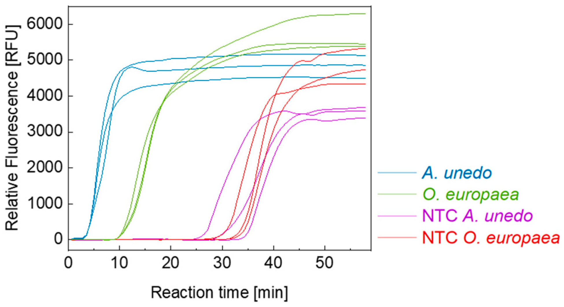
3.1. Primer Design and Selectivity

Designed primer sets (three primer sets per adulterant) were first tested using the standard protocol for LAMP assays proposed by NEB. The alignment of the target sequence and the homologous sequences of the species oregano (O. vulgare and O. onites), thyme, marjoram, and rockrose with indicated primer binding sites, as well as the phylogenetic trees generated from the target sequences, are listed in the supporting information (Figures S1 and S2). The selectivity of the final primer sets (one primer set per contaminant) was confirmed experimentally with 1 ng of high-quality DNA (see Section 2.2.1) from the species listed in Table 1. The amplification curves of negative samples are shown in the supporting information (Figure S3). Figure 1 shows the real-time amplification curve for the final A. unedo and O. europaea specific primer set prior to the optimization of LAMP reaction conditions.
In this case, the unspecific amplification becomes detectable after an approximately 30 min reaction time for both individual primer sets. As mentioned above, non-specific amplification is a common obstacle to overcome when developing assay methods based on LAMP. This issue is addressed in Section 3.2 Optimization of isolation protocol and reaction conditions. The final primer sets were selected due to the specificity and the already initially larger time gap between specific and non-specific amplification, compared to the other primer sets developed and tested. Table 2 shows the sequence data of the final primer sets.

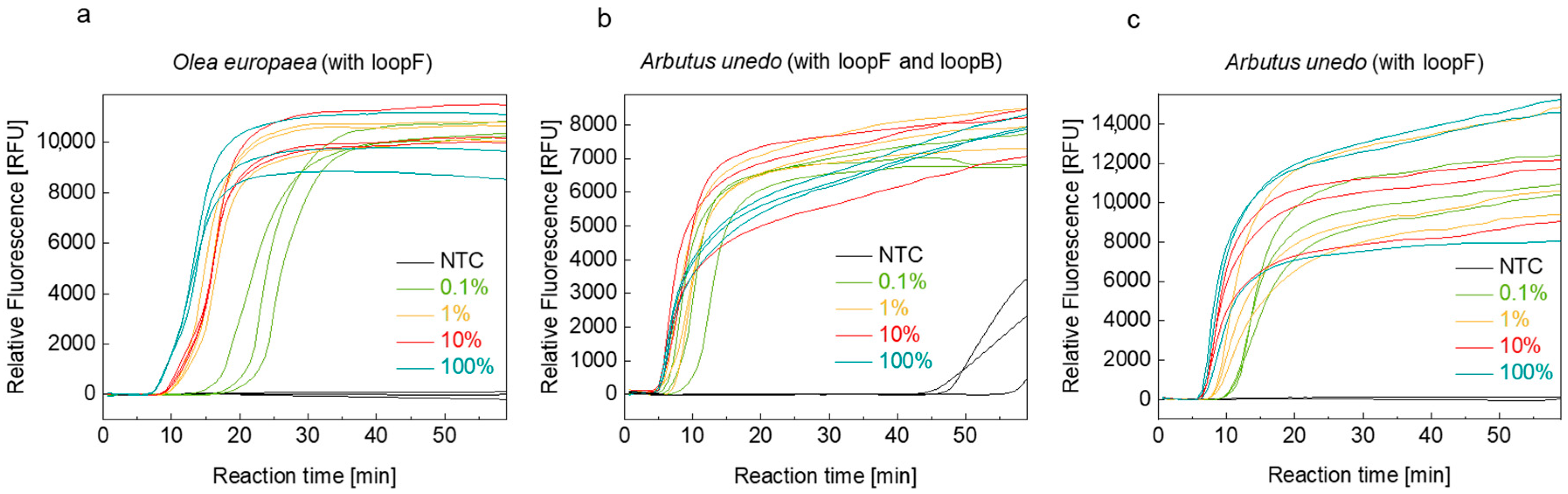
3.2. Optimization of Isolation Protocol and Reaction Conditions

First, reaction parameters optimized previously were transferred to the olive- and strawberry-tree-specific LAMP assays. Complete suppression of non-specific amplification was observed for both assays within the monitored time frame of 60 min as described earlier [21]. However, no amplification products could be detected when the POC isolate volume added to the LAMP reaction was increased from 2 µL to 5 µL. When using the standard LAMP protocol recommended by NEB, increasing the POC isolate volume resulted in an acceleration of the specific LAMP reaction. Consequently, the reaction conditions of the LAMP assay cannot be universally applied to DNA isolates prepared according to different extraction protocols.
Optimization of DNA isolation and amplification conditions using DoE and one factor at a time (OFAT) approaches were carried out using the olive-tree-specific assay as an example, and the resulting conditions were thereafter transferred to the strawberry-tree-specific assay. The initial conditions for the optimization were as previously described by Holz et al. [21]. The aim of optimizing reaction parameters was to suppress the occurrence of non-specific amplification and at the same time to achieve a short time-to-positive result.
The parameter values for the DNA isolation (buffer concentration of guanidine thiocyanate, washing steps, sample amount) and the LAMP assay (temperature, MgSO4 concentration) as well as the range of values for each variable parameter are listed in Table 3.
In agreement with the previous results, the optimization showed that reducing the magnesium ion concentration to 4 mM and increasing the temperature to 67 °C suppressed non-specific amplification. However, it was observed that after exceeding a temperature of 67 °C, an increase in time to positive occurred again. When optimizing the isolation method, an important aspect was to reduce the number of steps in order to make the method as user-friendly and time-saving as possible. It is therefore advantageous that the omission of the first washing step in the isolation protocol according to Zhang et al. led to equivalent amplification success [36]. Figure 2 shows the real-time fluorescence curves of the olive- and strawberry-tree-specific LAMP assays using DNA isolates obtained with the optimized POC-suitable isolation protocol from mixtures with different contaminant levels.
According to ESA, the recommended maximum level of foreign plant content in spice products is 1% for spices and 2% for herbs. Less than these amounts are considered inevitable, e.g., due to field contamination, and should not be considered adulterated. Therefore, our further work aimed to distinguish between oregano samples containing, for example, less than 0.1% or more than 1% of admixtures using the developed olive-tree- and strawberry-tree-specific LAMP assays. For the olive-tree-specific assay, discrimination of <0.1% and >z1% admixture was achieved using real-time LAMP (Figure 2a), allowing to estimate the relative level of contamination. In the case of the strawberry-tree-specific assay, discrimination of less than 0.1% and greater than 1% admixture level was achieved by omitting the loopB primer (Figure 2b,c). This also resulted in the very favorable suppression of non-specific amplification in the observed time frame. Therefore, the loopB primer was omitted for all further strawberry-tree-specific LAMP assays.

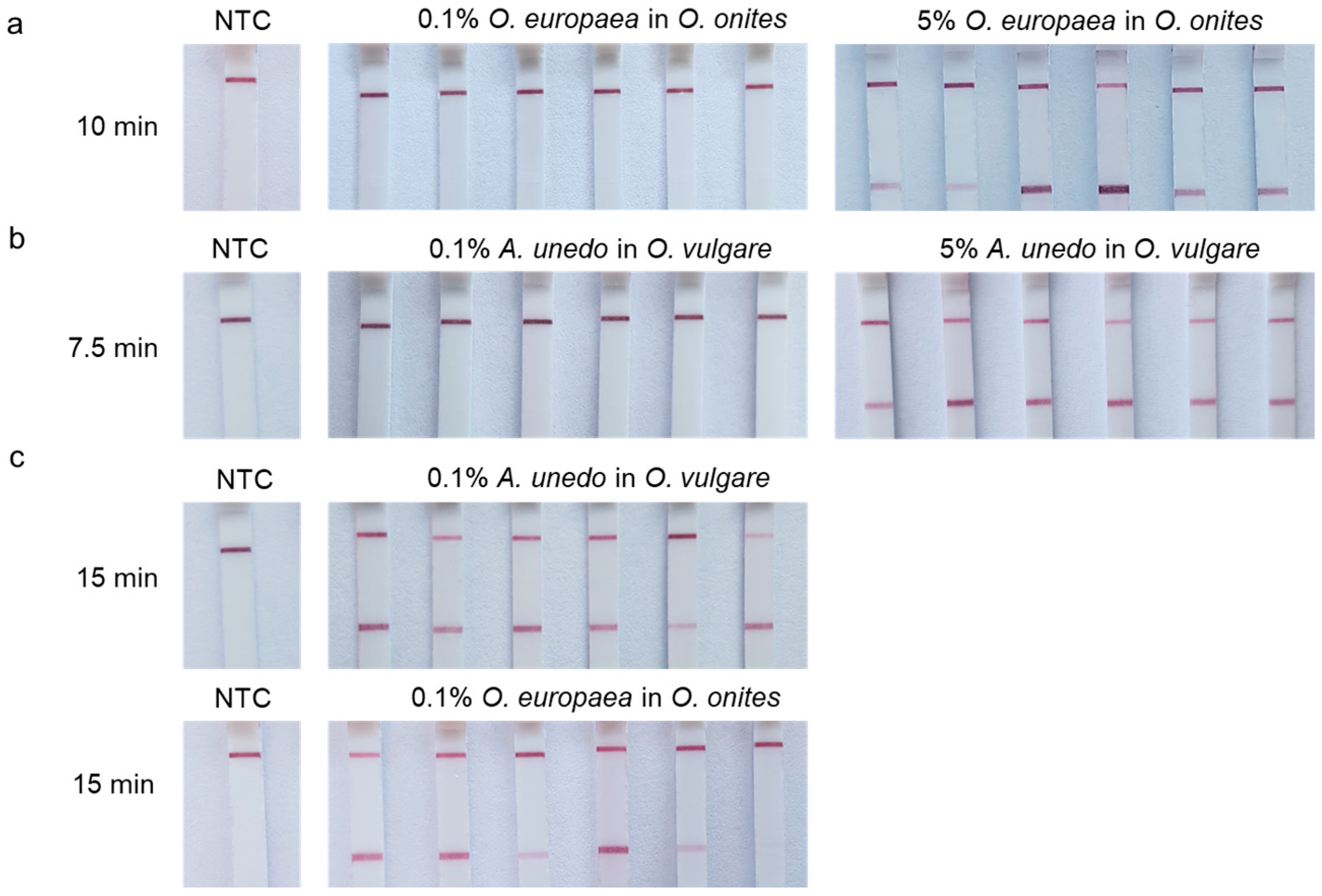
3.3. Lateral-Flow Assays

Mixtures with different admixture levels were initially used in duplicate in LAMP reactions to determine the shortest possible reaction time. Real-time measurements (Figure 2a,c) were used to estimate the time-to-positive result for the LAMP assay prior to LFA. Based on these figures, the time to amplicon formation for both olive- and strawberry-specific assays was set at 7.5 to 15 min. DNA isolations and LAMP assays were then performed to evaluate the reproducibility of the method and to determine the optimal reaction time for estimating the degree of mixing.
Figure 3 shows the LFA strips at different LAMP times for olive leaves (a) and strawberry tree leaves (b) as admixtures to oregano (0.1% or 5%), in 6-fold determination.
It was demonstrated that a relative estimation of the degree of impurity in oregano is possible. At reaction times of 7.5 min and 10 min, respectively, reliable positive results were obtained for admixture levels of 5% or more, while the results were negative for admixture levels of 0.1%. For samples with 1% admixture, the false-negative rate for both primer sets was 50% (supporting information, Figure S4). Therefore, the LFA method cannot be used to derive the exact contaminant level when it is between 1% and 5%. A LAMP reaction time of 7.5 min and 10 min is recommended for the detection of strawberry tree or olive leave contamination levels of more than 5% with the developed LAMP/LFA method. The total time for DNA isolation, LAMP reaction and the detection of the amplicons by LFA was less than 20 min, provided that the LAMP master mix was prepared in advance.

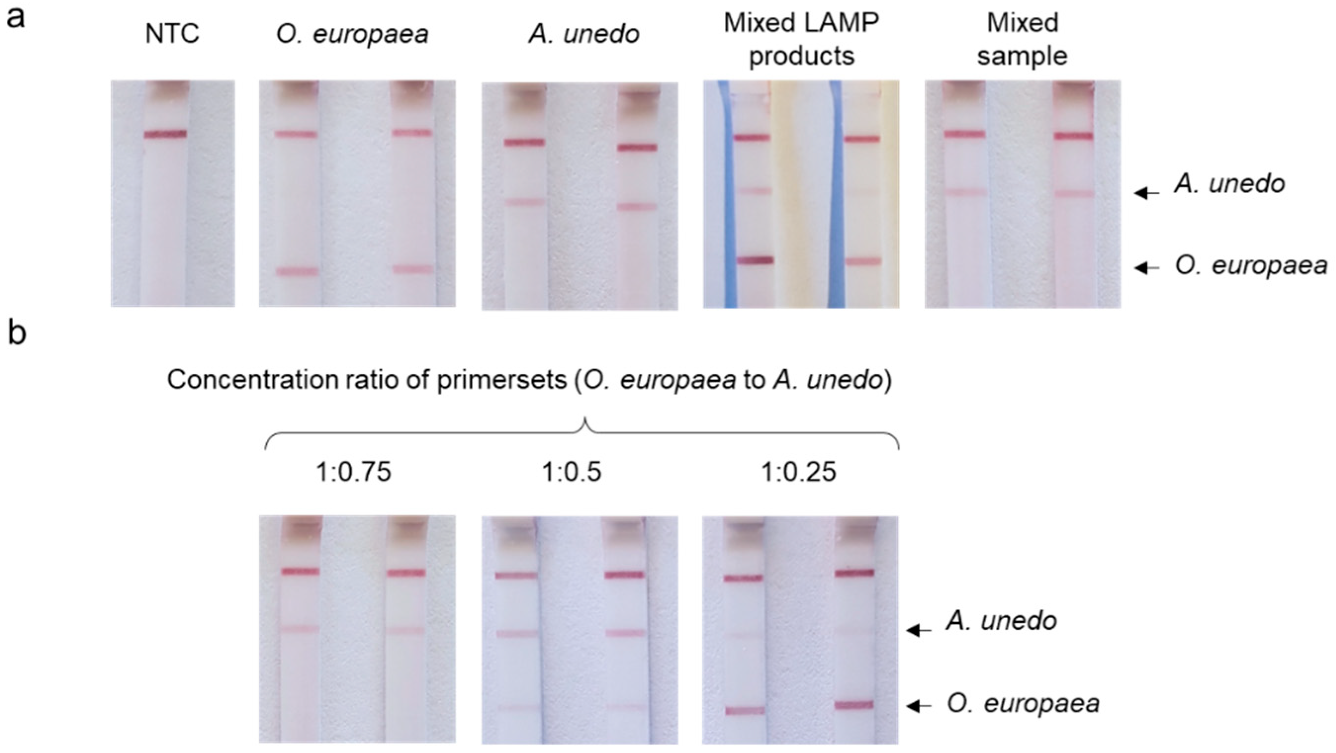
3.4. Duplex LAMP with LFA Detection

In order to further reduce the duration and costs of the assay and thus increase the efficiency of the test system, LFAs with the option of simultaneous detection of two different amplicon targets in a single LAMP and on one LFA strip were developed in this study. This requires that different amplicons (from specific amplification of O. europaea or A. unedo DNA) can be discriminated based on different primer affinity tags. For this reason, the FIP from the strawberry-tree-specific primer set was labeled with digoxigenin instead of the biotin tag and the primer set for the olive tree was left unchanged.
First, duplex LAMP reactions were performed and monitored in real-time to investigate whether the combination of two primer sets designed for two different target sequences in one reaction can affect amplicon formation or primer specificity. Under conditions optimized for high-quality DNA isolates, no significant changes could be observed in real-time LAMP (time to positive) or on AGE (amplicon pattern) (supporting information, Figures S5 and S6). Under the previously optimized DNA isolation and LAMP reaction conditions, non-specific amplification in the NTC sample started after a 40 min reaction time, but no effect of the additional primer set on the time to positive was observed for the analyzed samples. Therefore, using the optimized method (see Section 2.2.2), DNA was isolated from two samples, each containing only one species (either strawberry tree leaves or olive tree leaves), and from the third sample containing both in a 1:1 ratio. These three DNA samples served as templates in the duplex LAMP using both (olive and strawberry tree) primer sets in one reaction. As can be seen in Figure 4a, plain olive or strawberry tree samples resulted in staining of the corresponding bands on the LFA strip, indicating a positive reaction in each case. When isothermal amplification was carried out separately for each DNA sample and subsequently mixed prior to performing the LFA, two positive bands and the control band became visible. However, the use of the mixed composite sample initially resulted in a single positive band (A. unedo). Even when the reaction time was increased, the second positive band could not be observed for O. europaea amplicons. This effect of amplification bias is referred to as LAMP selection and can be compensated by varying the primer concentrations, as demonstrated by Liu et al. via melting curve analysis of the reaction products [37]. A plausible explanation for the phenomenon of LAMP selection, analogous to PCR selection, is the different affinity of the primer sets to the target sequences [38]. Due to the exponential nature of the reaction and competition for reagents, this ultimately leads to a suppression of amplification by the primer set with lower affinity to its target DNA. When the concentration of the A. unedo specific primer set was reduced to half of the initial value, both LAMP products could be detected within 15 min of LAMP incubation (Figure 4b). In the latter case, however, the primer set ratios had to be optimized in order to obtain a comparable signal intensity on the LFA strip and thus avoid a false-negative result.

4. Discussion

4.1. Primer Design and Uniplex LAMP/LFA Assays

The aim of this research project was to develop rapid test systems that enable a qualitative yes/no determination of potential adulteration of oregano samples directly at the point of care without the need for laboratory equipment. Established PCR-based methods for testing spice contaminants require significantly more time and laboratory equipment than the approaches developed here. Typically, PCR-based methods require much more highly pure DNA (up to 20 ng). Isolation of plant DNA using classical precipitation and adsorption methods can easily require more than 2 h [39,40]. A SCAR-PCR assay developed by Marieschi et al [10]. showed sensitivity for the determination of the O. europaea admixture that was comparable to that observed in this study, but took at least 1.5 h, depending on the heating rate of the thermocycler. If the amplicons are analyzed by AGE, additional time must also be allowed for the separation of the DNA [10]. Real-time PCR methods are time- and resource-efficient, but still require at least 1.5 h before the results are available and require sophisticated equipment, i.e., a thermocycler, that can perform fluorescence measurement.
Species-specific LAMP primers for olive and strawberry trees were designed for rapid DNA test development. The DNA isolation protocol and LAMP reaction conditions were then optimized using DoE to suppress non-specific amplification and ensure that target DNA could be amplified in the shortest possible time (compare Figure 1 and Figure 2). Both developed primer sets allow amplification of target DNA within 10 min.
An olive-specific LAMP was recently published by Sheu et al. for olive oil authentication [41]. Like the O. europaea specific primer set shown in our work (Table 2), the oleosin gene- specific LAMP primer set used by Sheu et al. included one loop primer. The authors described the appearance of specific LAMP products after 60 min when 20 ng of isolated genomic DNA was added to the reaction [41]. In comparison with the results published by Sheu et al., specific LAMP products were detectable in less than 10 min after reaction initiation in the course of this work when POC-optimized parameters for DNA isolation and LAMP were used. DNA isolates from olive leave samples used in this study (see Section 2.2.2) had an average DNA concentration of 0.02 ng/µL, so 0.1 ng of target DNA (5 µL of DNA isolate) was added per LAMP reaction with a total volume of 25 µL. It is obvious that the lower the proportion of olive leaves in the starting material, the lower the absolute content of target DNA in the isolates. The difference in the published time-to-positive values and those presented in this study can be explained by the nucleotide sequence data used for primer design. In this work, the olive-specific primer set was developed based on the trnL-trnF intergenic spacer, which is localized on the chloroplast DNA, and therefore is present in high copy numbers in the cell (up to 120 plastids per cell with an average of 15 copies cpDNA per chloroplast) [42]. In contrast, the sequence encoding oleosin in diploid O. europaea is present only twice in the nucleus, which can explain the increased time-to-positive value when the latter target DNA was used for the primer design [6,43].
Real-time fluorescence measurement was used during the development of the LAMP assay and isolation method to detect the successful amplification of the target DNA (olive or strawberry trees) or lack thereof. However, this detection method is not suitable for point-of-care detection, as conventional real-time fluorescence measuring devices are not portable. Portable devices for isothermal amplification that can measure fluorescence in real-time are already available for a few commercial LAMP-based test systems, i.e., the Doctor Vida SARS-CoV-2 assay [24]. Test systems with LFA detection represent a cost-effective alternative that is also attractive for consumers with a limited sample throughput.
In the present study, LFA detection was successfully established and evaluated for the point-of-care detection of amplicons. As part of the development of the LAMP/LFA method, we investigated whether it is possible to adjust the detection limit, i.e., the proportion of contamination in the oregano sample that results in a positive test line on the LFA after the LAMP, via the LAMP reaction time. This has already been shown in the publication by Holz et al. using the example of turmeric and safflower in saffron [21]. In the current study, it was shown that this is also possible for the detection of olive or strawberry tree leaves.
The suggested threshold for obtaining a positive result within 10 min is 5% olive leaves in the oregano sample. Within this reaction time, samples containing 0.1% olive leaves remained negative, and samples containing 1% were positive in 50% of cases; therefore, it is not possible to distinguish between 1% and 0.1% or 1% and 5% based on the reaction time. For the strawberry-tree-specific assay, a shorter time to positive was already determined in the preliminary real-time experiments than for the olive-specific assay. This was confirmed in the LAMP/LFA assays, where a reaction time of 7.5 min was found to be optimal. A reaction time of 7.5 min leads to analogous results as a reaction time of 10 min in the olive-specific LAMP/LFA assay. From this observation, it can be concluded that the LAMP reaction does not have to be complete in order to detect LAMP reaction products with LFAs. The termination of the reaction shortly after the start of the exponential phase already provides sufficient product for detection by LFA. However, to verify these results, further biological replicates and mixtures need to be examined before the method can be considered validated.
Carry-over contamination, i.e., re-amplification of amplicons from previous LAMP reactions leading to false-positive results, is a significant problem for the use of LAMP in routine analysis [34]. However, this obvious disadvantage of the LAMP/LFA test system can be compensated by using dUTP instead of dTTP and uracil DNA glycosylase (UDG) in the reaction mix. This procedure is already well known for the conventional PCR [44] and LAMP [45]. During amplification, the incorporation of dUTP results in amplicons that are digestible by the GDP and therefore cannot be amplified in a subsequent LAMP when a digestion step is performed beforehand. If a thermolabile UDG is used, the two steps (digestion and amplification) can be carried out without opening the reaction vessel [46]. Moreover, the optimal buffer capacity for the LAMP reaction can be used for LFA detection. In contrast, a weak buffer capacity is necessary to utilize a pH shift for detection [34].
With the LAMP/LFA rapid test, the time required to perform the test includes 5 min for DNA isolation, 10 min for the LAMP reaction, and a further 5 min for analyzing the amplicons using LFAs. This means that only 25 min are required to perform the test. However, this time estimation is only valid if the LAMP master mix has already been prepared and only the DNA isolate needs to be added. For the development of rapid test systems, it is therefore essential to find a way to transport and store the mastermixes at room temperature. To date, several approaches to freeze-drying and storing mastermixes have been pursued. In this context, Carter et al. were able to demonstrate the storage stability of lyophilized LAMP mastermixes at an ambient temperature over a period of 55 days with a minimal increase in time to positive [47]. Storage stability tests were also successfully carried out for the WarmStart® Colorimetric LAMP 2X Master Mix [48].

4.2. Duplex LAMP/LFA

Since user friendliness is a high priority in the development of LAMP/LFA tests, the combination of the two LAMP reactions into a one-pot reaction is desirable. Due to the complexity of the LAMP reaction products, a visualization method is required that can specifically distinguish between LAMP reaction products. Colorimetric detection or real-time detection using non-specific intercalation dye is not suitable for this purpose, as it only detects the formation of amplicons, but cannot distinguish between specific amplicons. Liu et al. showed that LAMP reaction products can be specifically distinguished by their melting curves [37]. Furthermore, it is possible to monitor multiplex LAMP reactions in real-time using probe-based approaches [49,50]. These techniques are not suitable for point-of-care use due to the laboratory equipment needed. A much simpler approach is the use of LFAs with two or more test lines to which specific LAMP reaction products can be bound.
The results presented show that the duplex LFA and the duplex LAMP work in principle with the primer sets developed but are unfortunately not suitable for the intended application in their current state of development. The fact that the ratio of the specific primer sets must be adapted to each other is problematic, as the test will otherwise not be able to identify both impurities, if present, in a sample of unknown composition. Even an optimization of the primer sets to achieve an equivalent affinity to the respective targets would not lead to the desired result if the levels of both contaminants in the oregano sample in question were very different. In this case, the LAMP selection leads to a false-negative response for the less concentrated target. If the requirements for the test system are limited to the question of possible contamination, further development of the test into a multiplex system with other known potential contaminants such as rockrose would be an interesting option [4]. Another possible modification of the application would be to carry out the LAMP reactions in different reaction vessels and then use a duplex LFA. This would lead to a reduction in the cost of the test system compared to using two LFAs.
Nevertheless, the fact that LAMP can be used in duplex, and possibly also in multiplex variants, is an encouraging result. Our results confirmed the general applicability of the developed LAMP primer sets for duplex LAMP and demonstrate that duplex LAMP reactions in combination with LFAs together form a promising tool for on-site analysis of spice samples, as they reduce resources and the time required to perform the analysis to a conceivable minimum.

5. Conclusions

In this work, two LAMP primer sets specific for the olive tree (O. europaea) or strawberry tree (A. unedo) were designed and their applicability for the development of point-of-care rapid test systems was demonstrated. Both assays, coupled with visualization through LFAs, reliably detected impurities as low as 0.1%. By using LFAs, it was possible to estimate the level of admixture content by varying the LAMP reaction time so that cases with a contamination level of 0.1% or less could be confidently distinguished from cases with a contamination level of 5% or more. The LAMP/LFA method cannot be used to derive the exact contaminant level when it is between 1% and 5%, as the false-negative rate for 1% samples is 50%.
In addition, a duplex method with LFA detection was introduced to visualize the test results for the samples containing both admixtures. Duplex LAMP reactions in combination with LFAs together form a promising tool for on-site analysis of spice samples, as they reduce resources and the time required to perform the analysis to a conceivable minimum. Due to the phenomenon known as LAMP selection, the method will provide a false-negative result for the lower concentrated contaminant, at the current state of development.

Supplementary Materials

The following supporting information can be downloaded at: https://www.mdpi.com/article/10.3390/agriculture14040597/s1, Figure S1: Multiple sequence alignment for the target sequences from A. unedo to homologous sequences listed in Table 1 and the respective phylogenetic trees; Figure S2: Multiple sequence alignment for the target sequences from O. europaea to homologous sequences listed in Table 1 and the respective phylogenetic trees; Figure S3: Real-time fluorescence LAMP reaction of the multi-species test during the initial primer set screening for both primer sets; Figure S4: Additional results of LFA optimization; Figure S5: Duplex real-time LAMP; Figure S6: AGE of duplex LAMP.

Author Contributions

Conceptualization, M.F.; methodology, N.H.; validation, N.H. and N.W.; formal analysis, N.H. and N.W.; investigation, N.H. and M.I.; resources, M.F.; writing—original draft preparation, N.H.; writing—review and editing, M.F., B.A.I., N.W., and M.I.; visualization, N.H.; supervision, M.F.; project administration, M.F. and B.A.I.; funding acquisition, M.F. All authors have read and agreed to the published version of the manuscript.

Funding

This IGF Project of the FEI was supported by AiF (project no. 21656 N) within the program for promoting the Industrial Collective Research (IGF) of the Federal Ministry of Economic Affairs and Climate Action (BMWK), based on a resolution of the German Parliament.

Institutional Review Board Statement

Not applicable.

Data Availability Statement

Further information and data on the project can be obtained from the authors upon request.

Acknowledgments

We would like to thank HELA GmbH and Husarich GmbH for being part of the project advisory committee and for the generous provision of the spice samples. For the latter, we also thank the Botanical Garden Hamburg and Marina Creydt. The graphical abstract (GA) was created with BioRender.com.

Conflicts of Interest

The authors declare no conflicts of interest. The funders had no role in the design of this study; in the collection, analyses, or interpretation of data; in the writing of this manuscript; or in the decision to publish the results.

References

  1. ISO 7925:1999; Dried Oregano (Origanum vulgare L.)—Whole or Ground Leaves—Specification. ISO: Geneva, Switzerland, 2015. Available online: https://www.iso.org/obp/ui/#iso:std:iso:7925:ed-2:v1:en (accessed on 20 February 2024).
  2. Marieschi, M.; Torelli, A.; Bianchi, A.; Bruni, R. Detecting Satureja montana L. and Origanum majorana L. by means of SCAR–PCR in commercial samples of Mediterranean oregano. Food Control 2011, 22, 542–548. [Google Scholar] [CrossRef]
  3. Kintzios, S.E. Oregano: The Genera Origanum and Lippia, 1st ed.; Medicinal and Aromatic Plants-Industrial Profiles; v. 25; Taylor and Francis: London, UK, 2002. [Google Scholar]
  4. Marieschi, M.; Torelli, A.; Poli, F.; Sacchetti, G.; Bruni, R. RAPD-based method for the quality control of Mediterranean oregano and its contribution to pharmacognostic techniques. J. Agric. Food Chem. 2009, 57, 1835–1840. [Google Scholar] [CrossRef]
  5. Maquet, A.; Lievens, A.; Paracchini, V.; Kaklamanos, G.; de La Calle, B.; Garlant, L.; Papoci, S.; Pietretti, D.; Zdiniakova, T.; Breidbach, A.; et al. Results of an EU Wide Coordinated Control Plan to Establish the Prevalence of Fraudulent Practices in the Marketing of Herbs and Spices; EUR30877EN; JRC126785; Publications Office of the European Union: Luxembourg, 2021. [Google Scholar]
  6. Besnard, G.; Garcia-Verdugo, C.; de Casas, R.R.; Treier, U.A.; Galland, N.; Vargas, P. Polyploidy in the olive complex (Olea europaea): Evidence from flow cytometry and nuclear microsatellite analyses. Ann. Bot. 2008, 101, 25–30. [Google Scholar] [CrossRef]
  7. Vannozzi, A.; Lucchin, M.; Barcaccia, G. cpDNA Barcoding by Combined End-Point and Real-Time PCR Analyses to Identify and Quantify the Main Contaminants of Oregano (Origanum vulgare L.) in Commercial Batches. Diversity 2018, 10, 98. [Google Scholar] [CrossRef]
  8. Mandrone, M.; Marincich, L.; Chiocchio, I.; Petroli, A.; Gođevac, D.; Maresca, I.; Poli, F. NMR-based metabolomics for frauds detection and quality control of oregano samples. Food Control 2021, 127, 108141. [Google Scholar] [CrossRef]
  9. Sasikumar, B.; Swetha, V.P.; Parvathy, V.A.; Sheeja, T.E. Advances in Adulteration and Authenticity Testing of Herbs and Spices. Adv. Food Authent. Test. 2016, 585–624. [Google Scholar] [CrossRef]
  10. Marieschi, M.; Torelli, A.; Bianchi, A.; Bruni, R. Development of a SCAR marker for the identification of Olea europaea L.: A newly detected adulterant in commercial Mediterranean oregano. Food Chem. 2011, 126, 705–709. [Google Scholar] [CrossRef]
  11. Notomi, T.; Okayama, H.; Masubuchi, H.; Yonekawa, T.; Watanabe, K.; Amino, N.; Hase, T. Loop-mediated isothermal amplification of DNA. Nucleic Acids Res. 2000, 28, E63. [Google Scholar] [CrossRef]
  12. Notomi, T.; Mori, Y.; Tomita, N.; Kanda, H. Loop-mediated isothermal amplification (LAMP): Principle, features, and future prospects. J. Microbiol. 2015, 53, 1–5. [Google Scholar] [CrossRef]
  13. Kaneko, H.; Kawana, T.; Fukushima, E.; Suzutani, T. Tolerance of loop-mediated isothermal amplification to a culture medium and biological substances. J. Biochem. Biophys. Meth. 2007, 70, 499–501. [Google Scholar] [CrossRef]
  14. Kiddle, G.; Hardinge, P.; Buttigieg, N.; Gandelman, O.; Pereira, C.; McElgunn, C.J.; Rizzoli, M.; Jackson, R.; Appleton, N.; Moore, C.; et al. GMO detection using a bioluminescent real time reporter (BART) of loop mediated isothermal amplification (LAMP) suitable for field use. BMC Biotechnol. 2012, 12, 15. [Google Scholar] [CrossRef]
  15. Augustine, R.; Hasan, A.; Das, S.; Ahmed, R.; Mori, Y.; Notomi, T.; Kevadiya, B.D.; Thakor, A.S. Loop-Mediated Isothermal Amplification (LAMP): A Rapid, Sensitive, Specific, and Cost-Effective Point-of-Care Test for Coronaviruses in the Context of COVID-19 Pandemic. Biology 2020, 9, 182. [Google Scholar] [CrossRef]
  16. Kashir, J.; Yaqinuddin, A. Loop mediated isothermal amplification (LAMP) assays as a rapid diagnostic for COVID-19. Med. Hypotheses 2020, 141, 109786. [Google Scholar] [CrossRef]
  17. Zhou, Y.; Wu, Y.; Ding, L.; Huang, X.; Xiong, Y. Point-of-care COVID-19 diagnostics powered by lateral flow assay. TrAC Trends Anal. Chem. 2021, 145, 116452. [Google Scholar] [CrossRef]
  18. Kampeera, J.; Pasakon, P.; Karuwan, C.; Arunrut, N.; Sappat, A.; Sirithammajak, S.; Dechokiattawan, N.; Sumranwanich, T.; Chaivisuthangkura, P.; Ounjai, P.; et al. Point-of-care rapid detection of Vibrio parahaemolyticus in seafood using loop-mediated isothermal amplification and graphene-based screen-printed electrochemical sensor. Biosens. Bioelectron. 2019, 132, 271–278. [Google Scholar] [CrossRef]
  19. Lalle, M.; Possenti, A.; Dubey, J.P.; Pozio, E. Loop-Mediated Isothermal Amplification-Lateral-Flow Dipstick (LAMP-LFD) to detect Toxoplasma gondii oocyst in ready-to-eat salad. Food Microbiol. 2018, 70, 137–142. [Google Scholar] [CrossRef]
  20. Focke, F.; Haase, I.; Fischer, M. Loop-mediated isothermal amplification (LAMP): Methods for plant species identification in food. J. Agric. Food Chem. 2013, 61, 2943–2949. [Google Scholar] [CrossRef]
  21. Holz, N.; Illarionov, B.; Wax, N.; Schmidt, C.; Fischer, M. Point-of-care suitable identification of the adulterants Carthamus tinctorius and Curcuma longa in Crocus sativus based on loop-mediated isothermal amplification (LAMP) and lateral-flow-assay (LFA). Food Control 2023, 148, 109637. [Google Scholar] [CrossRef]
  22. Wax, N.; Pförtner, L.S.; Holz, N.; Sterzl, S.; Melnik, M.; Kappel, K.; Bade, P.; Schröder, U.; Haase, I.; Fritsche, J.; et al. Fast and User-Friendly Detection of Flatfish Species (Pleuronectes platessa and Solea solea) via Loop-Mediated Isothermal Amplification (LAMP). J. Agric. Food Chem. 2023, 71, 14795–14805. [Google Scholar] [CrossRef]
  23. Zahradnik, C.; Martzy, R.; Mach, R.L.; Krska, R.; Farnleitner, A.H.; Brunner, K. Detection of the food allergen celery via loop-mediated isothermal amplification technique. Anal. Bioanal. Chem. 2014, 406, 6827–6833. [Google Scholar] [CrossRef]
  24. Doria, G.; Clemente, C.; Coelho, E.; Colaço, J.; Crespo, R.; Semikhodskii, A.; Mansinho, H.; Dinis, M.; Carvalho, M.F.; Casmarrinha, M.; et al. An isothermal lab-on-phone test for easy molecular diagnosis of SARS-CoV-2 near patients and in less than 1 hour. Int. J. Infect. Dis. 2022, 123, 1–8. [Google Scholar] [CrossRef]
  25. Posthuma-Trumpie, G.A.; Korf, J.; van Amerongen, A. Lateral flow (immuno)assay: Its strengths, weaknesses, opportunities and threats. A literature survey. Anal. Bioanal. Chem. 2009, 393, 569–582. [Google Scholar] [CrossRef]
  26. Allgöwer, S.M.; Hartmann, C.A.; Holzhauser, T. The Development of Highly Specific and Sensitive Primers for the Detection of Potentially Allergenic Soybean (Glycine max) Using Loop-Mediated Isothermal Amplification Combined with Lateral Flow Dipstick (LAMP-LFD). Foods 2020, 9, 423. [Google Scholar] [CrossRef]
  27. Ge, Y.; Wu, B.; Qi, X.; Zhao, K.; Guo, X.; Zhu, Y.; Qi, Y.; Shi, Z.; Zhou, M.; Wang, H.; et al. Rapid and sensitive detection of novel avian-origin influenza A (H7N9) virus by reverse transcription loop-mediated isothermal amplification combined with a lateral-flow device. PLoS ONE 2013, 8, e69941. [Google Scholar] [CrossRef]
  28. Yu, J.; Wang, F.; Zhan, X.; Wang, X.; Zuo, F.; Wei, Y.; Qi, J.; Liu, Y. Improvement and evaluation of loop-mediated isothermal amplification combined with a chromatographic flow dipstick assay and utilization in detection of Vibrio cholerae. Anal. Bioanal. Chem. 2019, 411, 647–658. [Google Scholar] [CrossRef]
  29. Rolando, J.C.; Jue, E.; Barlow, J.T.; Ismagilov, R.F. Real-time kinetics and high-resolution melt curves in single-molecule digital LAMP to differentiate and study specific and non-specific amplification. Nucleic Acids Res. 2020, 48, e42. [Google Scholar] [CrossRef]
  30. Wong, Y.-P.; Othman, S.; Lau, Y.-L.; Radu, S.; Chee, H.-Y. Loop-mediated isothermal amplification (LAMP): A versatile technique for detection of micro-organisms. J. Appl. Microbiol. 2018, 124, 626–643. [Google Scholar] [CrossRef]
  31. Yin, H.Y.; Fang, T.J.; Wen, H.W. Combined multiplex loop-mediated isothermal amplification with lateral flow assay to detect sea and seb genes of enterotoxic Staphylococcus aureus. Lett. Appl. Microbiol. 2016, 63, 16–24. [Google Scholar] [CrossRef]
  32. Liang, C.; Chu, Y.; Cheng, S.; Wu, H.; Kajiyama, T.; Kambara, H.; Zhou, G. Multiplex loop-mediated isothermal amplification detection by sequence-based barcodes coupled with nicking endonuclease-mediated pyrosequencing. Anal. Chem. 2012, 84, 3758–3763. [Google Scholar] [CrossRef]
  33. Tanner, N.A.; Zhang, Y.; Evans, T.C. Simultaneous multiple target detection in real-time loop-mediated isothermal amplification. BioTechniques 2012, 53, 81–89. [Google Scholar] [CrossRef]
  34. Karthik, K.; Rathore, R.; Thomas, P.; Arun, T.R.; Viswas, K.N.; Dhama, K.; Agarwal, R.K. New closed tube loop mediated isothermal amplification assay for prevention of product cross-contamination. MethodsX 2014, 1, 137–143. [Google Scholar] [CrossRef]
  35. Tanner, N.A.; Zhang, Y.; Evans, T.C. Visual detection of isothermal nucleic acid amplification using pH-sensitive dyes. BioTechniques 2015, 58, 59–68. [Google Scholar] [CrossRef]
  36. Zhang, M.; Liu, Y.; Chen, L.; Quan, S.; Jiang, S.; Zhang, D.; Yang, L. One simple DNA extraction device and its combination with modified visual loop-mediated isothermal amplification for rapid on-field detection of genetically modified organisms. Anal. Chem. 2013, 85, 75–82. [Google Scholar] [CrossRef]
  37. Liu, N.; Zou, D.; Dong, D.; Yang, Z.; Ao, D.; Liu, W.; Huang, L. Development of a multiplex loop-mediated isothermal amplification method for the simultaneous detection of Salmonella spp. and Vibrio parahaemolyticus. Sci. Rep. 2017, 7, 45601. [Google Scholar] [CrossRef]
  38. Wagner, A.; Blackstone, N.; Cartwright, P.; Dick, M.; Misof, B.; Snow, P.; Wagner, G.P.; Bartels, J.; Murtha, M.; Pendleton, J. Surveys of Gene Families Using Polymerase Chain Reaction: PCR Selection and PCR Drift. Syst. Biol. 1994, 43, 250. [Google Scholar] [CrossRef]
  39. Porebski, S.; Bailey, L.G.; Baum, B.R. Modification of a CTAB DNA extraction protocol for plants containing high polysaccharide and polyphenol components. Plant Mol. Biol. Rep. 1997, 15, 8–15. [Google Scholar] [CrossRef]
  40. Varma, A.; Padh, H.; Shrivastava, N. Plant genomic DNA isolation: An art or a science. Biotechnol. J. 2007, 2, 386–392. [Google Scholar] [CrossRef]
  41. Sheu, S.-C.; Wang, Y.-J.; Huang, P.-C.; Lien, Y.-Y.; Lee, M.-S. Authentication of olive oil in commercial products using specific, sensitive, and rapid loop-mediated isothermal amplification. J. Food Sci. Technol. 2023, 60, 1834–1840. [Google Scholar] [CrossRef]
  42. Sakamoto, W.; Takami, T. Chloroplast DNA Dynamics: Copy Number, Quality Control and Degradation. Plant Cell Physiol. 2018, 59, 1120–1127. [Google Scholar] [CrossRef]
  43. Giannoulia, K.; Banilas, G.; Hatzopoulos, P. Oleosin gene expression in olive. J. Plant Physiol. 2007, 164, 104–107. [Google Scholar] [CrossRef]
  44. Longo, M.C.; Berninger, M.S.; Hartley, J.L. Use of uracil DNA glycosylase to control carry-over contamination in polymerase chain reactions. Gene 1990, 93, 125–128. [Google Scholar] [CrossRef]
  45. He, L.; Xu, H. Development of a multiplex loop-mediated isothermal amplification (mLAMP) method for the simultaneous detection of white spot syndrome virus and infectious hypodermal and hematopoietic necrosis virus in penaeid shrimp. Aquaculture 2011, 311, 94–99. [Google Scholar] [CrossRef]
  46. Hsieh, K.; Mage, P.L.; Csordas, A.T.; Eisenstein, M.; Soh, H.T. Simultaneous elimination of carryover contamination and detection of DNA with uracil-DNA-glycosylase-supplemented loop-mediated isothermal amplification (UDG-LAMP). Chem. Commun. 2014, 50, 3747–3749. [Google Scholar] [CrossRef]
  47. Carter, C.; Akrami, K.; Hall, D.; Smith, D.; Aronoff-Spencer, E. Lyophilized visually readable loop-mediated isothermal reverse transcriptase nucleic acid amplification test for detection Ebola Zaire RNA. J. Virol. Methods 2017, 244, 32–38. [Google Scholar] [CrossRef]
  48. Song, X.; Coulter, F.J.; Yang, M.; Smith, J.L.; Tafesse, F.G.; Messer, W.B.; Reif, J.H. A lyophilized colorimetric RT-LAMP test kit for rapid, low-cost, at-home molecular testing of SARS-CoV-2 and other pathogens. Sci. Rep. 2022, 12, 7043. [Google Scholar] [CrossRef]
  49. Becherer, L.; Bakheit, M.; Frischmann, S.; Stinco, S.; Borst, N.; Zengerle, R.; von Stetten, F. Simplified Real-Time Multiplex Detection of Loop-Mediated Isothermal Amplification Using Novel Mediator Displacement Probes with Universal Reporters. Anal. Chem. 2018, 90, 4741–4748. [Google Scholar] [CrossRef]
  50. Crego-Vicente, B.; Fernández-Soto, P.; García-Bernalt Diego, J.; Febrer-Sendra, B.; Muro, A. Development of a Duplex LAMP Assay with Probe-Based Readout for Simultaneous Real-Time Detection of Schistosoma mansoni and Strongyloides spp.—A Laboratory Approach to Point-Of-Care. Int. J. Mol. Sci. 2023, 24, 893. [Google Scholar] [CrossRef]
Figure 1. Real-time fluorescence curves of final primer sets with 1 ng target DNA added, either specific for A. unedo or O. europaea, with corresponding NTC (no-template control).
Agriculture 14 00597 g001
Figure 2. Real-time fluorescence curves of the (a) olive-specific LAMP under POC-optimized conditions and strawberry-tree-specific LAMP assays under POC-optimized conditions with (b,c) without the added loopB primer.
Agriculture 14 00597 g002
Figure 3. Detection of admixture levels 0.1% or 5% in oregano with LAMP/LFA. (a) Olive or (b) strawberry tree; admixture content 0.1% or 5%; LAMP reaction time 10 min or 7.5 min. (c) Detection of admixture levels of 0.1% after LAMP incubation time of 15 min. All tests were performed in 6-fold determinations.
Agriculture 14 00597 g003
Figure 4. Detection of O. europaea and A. unedo admixtures using the elaborated duplex LAMP/LFA technique after 15 min LAMP reaction time and with varying primer set concentration ratios (O. europaea to A. unedo): (a) 1:1 (b) 1:0.75, 1:0.5, 1:0.25.
Agriculture 14 00597 g004
Table 1. Origin and condition of plant material used in this study.
No.SpeciesOrigin/SupplierProcessing Stage
1Arbutus unedoBotanical Garden, University of HamburgFreeze dried 24 h, ground
2Cistus creticusBotanical Garden, University of HamburgFreeze dried 24 h, ground
3Olea europaeaBotanical Garden, University of HamburgFreeze dried 24 h, ground
4Olea europaeaLocal drug storeDried, rubbed
5Origanum majoranaHusarich GmbHDried, rubbed, germinated
6Origanum onitesHusarich GmbHDried, rubbed, germinated
7Origanum onitesHela GmbHDried, rubbed, germinated
8Origanum vulgareBotanical Garden, University of HamburgFreeze dried 24 h, ground
9Thymus vulgarisBotanical Garden, University of HamburgFreeze dried 24 h, ground
10Carthamus tinctoriusBotanical Garden, University of HamburgFreeze dried 24 h, ground
11Ocimum basilicumLocal retailFreeze dried 24 h, ground
12Brassica oleracea var. italicaLocal retailFreeze dried 24 h, ground
13Crocus sativusHusarich GmbHDried, ground
14Curcuma longaHela GmbHDried, rubbed, germinated
15Apium graveolensLocal retailFreeze dried 24 h, ground
16Salvia officinalisLocal retailFreeze dried 24 h, ground
Table 2. Nucleotide sequences of the primer sets and labeled primers used for species-specific DNA detection by lateral-flow assay. The nucleotide sequences are shown in 5′ → 3′ direction.
Primer Set I: 5′-3′ Species: Olea europaea, Locus: trnL-trnF Intergenic Spacer
F3CACATGTGATATATAATACACATCC
B3CATTCCCAATGTAACATTAACATC
FIP (F2 + F1C)GGATCTTCAAAAAGACGACTTTGTC-TTAAGCAAGGAATCCCCAT
BIP (B2 + B1C)ATTCCAGGACTTGGAGAAAACTTTG-CCATCCTCATTTTATTAGATGACT
loopBCCCCCCTTGTCCTTTTAATTGACAT
6-FAM/loopB6-FAM/CCCCCCTTGTCCTTTTAATTGACAT
Biotin/BIPBiotin/ATTCCAGGACTTGGAGAAAACTTTG-CCATCCTCATTTTATTAGATGACT
Primer Set II: 5′-3′ Species: Arbutus unedo, Locus: Internal Transcribed Spacer
F3CCTCCGGGAACAATTGAGC
B3AACACAGCCCACGAATGG
FIP (F2 + F1C)GAACACGTTTCCCGAAGGACCG-CCAGTTGTCGCCTTCCATT
BIP (B2 + B1C)GTGAAATAACGAAACCCGGCGC-TGGGAGACGTGCATCTGTT
loopFACCCGCTCGAGGAGGAA
loopBAACCGCGCCAAGGAAACT
6-FAM/loopF6-FAM/ACCCGCTCGAGGAGGAA
Biotin/FIPBiotin/ATTCCAGGACTTGGAGAAAACTTTG-CCATCCTCATTTTATTAGATGACT
DIG/FIPDIG/ATTCCAGGACTTGGAGAAAACTTTG-CCATCCTCATTTTATTAGATGACT
Table 3. Parameters of DNA isolation method (buffer concentration of guanidine thiocyanate, washing steps, sample amount) and of the LAMP assay (temperature, MgSO4 concentration) prior and after optimization using the olive-tree-specific assay.
ParameterNew England Biolabs (NEB)Value Range for the ParameterHolz et al. 2023 [21]POC-Optimized
Parameters of DNA isolation optimized with DoE
Washing steps-one/twotwoone
Buffer conc. [mol/L]-1–2.52.52.5
Sample weight [mg]-5–252015
Optimized with OFAT
DNA sample per reaction [µL]-0.1–5.025
Parameters of LAMP reaction optimized with DoE
Temperature [°C]6562–727267
MgSO4 [mM]64–844
Disclaimer/Publisher’s Note: The statements, opinions and data contained in all publications are solely those of the individual author(s) and contributor(s) and not of MDPI and/or the editor(s). MDPI and/or the editor(s) disclaim responsibility for any injury to people or property resulting from any ideas, methods, instructions or products referred to in the content.

