Article

Akkermansia muciniphila Promotes Bone Development and Improves Eggshell Quality during the Sexual Maturity Period of Laying Hens by Increasing Osteogenesis

State Key Laboratory of Animal Nutrition and Feeding, College of Animal Science and Technology, China Agricultural University, Beijing 100193, China
*
Author to whom correspondence should be addressed.
Agriculture 2024, 14(4), 598; https://doi.org/10.3390/agriculture14040598
Submission received: 14 March 2024 / Revised: 7 April 2024 / Accepted: 8 April 2024 / Published: 10 April 2024
(This article belongs to the Section Farm Animal Production)

Abstract

:
Adequate bone development is imperative for maintaining the health and productivity of laying hens. Probiotics play a pivotal role in promoting bone formation and preventing osteoporosis. This study aimed to explore the effect of Akkermansia muciniphila (Akk) on the bone development and eggshell quality of laying hens during the rearing period. A total of 300 1-day-old Jingfen NO. 6 commercial pullets were categorized into two groups, one of which was fed a conventional diet for 20 weeks (Control group), the other group was fed a conventional diet with lyophilized Akk powder for 20 weeks (Akk group). During the first two weeks, pullets in the Akk group received live Akk inoculation, while birds in the Control group received normal saline administration. Micro-computed tomography analysis was employed to evaluate three bone microarchitectures: cortical bone (Cb), trabecular bone (Tb), and medullary bone (Mb). Our findings revealed that supplementation with Akk powder increased the thickness and bone mineral content of Cb and Tb, while simultaneously reducing the volume and bone surface area of Mb. The increased activity of alkaline phosphatase, a marker of osteogenesis, and the decreased activity of tartrate-resistant acid phosphatase, a marker of osteoclastic activity, were observed in the Akk group. Dietary supplementation of Akk powder improved the immune microenvironment in the bone marrow by increasing osteogenic-related CD8+ T cells and decreasing osteoclastogenesis-related CD4+ T cells. Additionally, Akk powder supplementation significantly enriched the Lactobacillaceae family in cecum. The enhancement of bone development by Akk contributed to increased eggshell strength and thickness. These findings demonstrate the osteomodulatory effects of Akk in laying hens and the connections between bone physiology and eggshell quality, highlighting the importance of gut–bone communications in laying hens.

1. Introduction

Eggs are a valuable and cost-effective source of animal protein. With growing demand for human consumption, the concept of “long-life laying hens”, capable of producing 500 eggs within a laying cycle of 100 weeks, was initially proposed in Europe to enhance persistency in egg production [1]. Extending laying cycles could significantly decrease the number of hens raised and reduce feed consumption, consequently lowering breeding costs, enhancing animal welfare, and alleviating environmental pressures associated with chicken farming [1].
The commercial poultry industry encounters several challenges as longer laying cycles necessitate sustainable egg quality and the long-term maintenance of health, particularly of the tissues and organs involved in egg production [1,2]. The bones of laying hens are among the organs closely associated with egg production, and osteoporosis has long been a significant welfare challenge in the egg industry [3]. Caged laying hens with high daily production are particularly susceptible to osteoporosis due to their unique bone microarchitecture [4]. Unlike mammals, the long bones of laying hens comprise three distinct types of microarchitectures: cortical bone (Cb), trabecular bone (Tb), and medullary bone (Mb) [5,6]. Bones undergo continuous renewal through bone remodeling, a process regulated by the balance between osteoclast-mediated bone resorption and osteoblast-mediated bone formation [7]. In most vertebrates, the growth plates close at puberty, halting long bone growth [8]. In young pullets, the growth of Cb and Tb persists until the onset of sexual maturity [9]. Following sexual maturity and under the influence of estrogen, osteoblasts form Mb rather than Cb or Tb [10,11,12,13]. During eggshell formation, calcium is absorbed from the blood at a higher rate than the average absorption rate from the diet, necessitating osteoclasts to mobilize calcium reserves from the Mb to maintain balance [1,14]. Studies have reported rapid changes in the microarchitecture of Mb and serum concentrations of bone resorption markers throughout the daily oviposition cycle [14,15]. Laying hens have a particularly high demand for calcium to support eggshell formation. However, during the early stages of egg production, the insufficient development of the Mb can lead to the resorption of Cb and Tb by osteoclasts to fulfill calcium requirements [4,16]. The traditional cage system further suppresses bone development by restricting movement and keeping hens standing for prolonged periods [17]. This inadequate development can ultimately lead to bone loss and increased fracture risk in laying hens [3,16,18]. Therefore, promoting proper bone development during the rearing period becomes crucial for ensuring the overall well-being and productivity of laying hens [19].
Bone development is regulated by a variety of factors, including nutrition, exercise, hormones, and immune cells [20,21,22,23,24]. In recent years, an increasing body of research has illuminated the pivotal role of gut microbiota and their metabolites in bone remodeling, which ultimately affects bone metabolism through gut–bone communication [25,26]. Some probiotics, such as Lactobacillus reuteri, Lactobacillus rhamnosus GG, Bifidobacterium longum, Bifidobacterium adolescentis, and Akkermansia muciniphila (Akk), have been shown to be beneficial for bone health [21,27,28,29,30]. Akk is a symbiotic bacterium in the gut of animals, utilizing mucus as a single nutrient source [31,32]. Given its safety in host health and its pivotal contributions to mitigating disease, Akk is increasingly recognized as a next-generation probiotic [32,33,34,35,36,37,38]. Studies have indicated that the abundance of Akkermansia in children’s gut microbiota surpasses that in older individuals, and fecal microbiota transplantation (FMT) from children offers superior protection against ovariectomized (OVX)-induced osteoporosis [30]. Akk also has a direct correlation with bone formation. For example, exposure to warmth (34 °C), as opposed to room temperature (RT), enhanced tibial bone volume and the abundance of Akkermansia in female mice [7]. Conversely, OVX mice exhibited a lower abundance of Akkermansia compared to mice undergoing sham operations [30]. Warm-exposed FMT augmented tibial breaking strength and bone volume in OVX mice compared with RT-exposed FMT [7]. Akk directly prevents OVX-induced osteoporosis by increasing osteogenic activity and inhibiting osteoclastogenesis through the secretion of extracellular vesicles [30]. Moreover, Akk has also been reported to promote the healing of bone fractures and mitigate Porphyromonas gingivalis-induced alveolar bone destruction [39,40,41]. Nevertheless, there is a dearth of research on the effects of Akk on bone development in laying hens.
Building upon the recognized benefits of Akk on bone physiology, the purpose of this study was to assess the impact of Akk on bone development in laying hens during the rearing period, along with investigating the underlying mechanisms. Furthermore, the study aimed to explore whether the alterations in bone physiology following Akk treatment affect eggshell quality. This study provides new perspectives for microbial-based interventions for enhancing the bone health of laying hens.

2. Materials and Methods

2.1. Preparation of Akkermansia muciniphila Suspensions and Lyophilized Powder

Akkermansia muciniphila ATCC BAA-835 (Akk) was cultured in modified Gifu Anaerobic Medium broth (GAM, HB8518, Hope bio Co., Ltd., Tsingtao, China) at 37 °C under anaerobic conditions (10% H2, 10% CO2, 80% N2; Don Whitley Scientific DG250, West Yorkshire, UK). The identity of Akk was confirmed by amplifying the 16S rRNA gene using primers 27F (5′-AGA GTT TGA TCA TGG CTC A-3′) and 1492R (5′-TAC GGT TAC CTT GTT ACG ACT T-3′). After 48 h, Akk were harvested by centrifugation at 8000 rpm for 10 min at 4 °C, resuspended in sterile PBS containing 20% glycerol, and finally stored at −80 °C until use. Bacterial suspensions were diluted with sterile PBS to 3.5 × 10⁸ CFU/mL (final glycerol concentration: 2%) and activated in a water bath at 37 °C for 10 min before use. For lyophilized powder preparation and Akk fermentation broth were centrifuged at 8000 rpm for 10 min and the supernatant was discarded. A solution containing 10% skimmed milk powder was then added to the precipitate, followed by mixing and vacuum freeze-drying for 24 h. The obtained lyophilized powder was stored at −20 °C.

2.2. Ethics Statement

All the work using animals was approved by the Animal Care and Use Committee of China Agricultural University (statement no. AW10204202-1-3). All procedures were conducted in accordance with the institutional animal ethics guidelines set by the Ministry of Agriculture and Rural Affairs of the People’s Republic of China.

2.3. Animals

A cohort of 300 1-day-old Jingfen NO. 6 commercial pullets were categorized into two groups based on body weight, with each group comprising 10 replicates of 15 birds each. Before reaching 10 weeks of age, groups of 15 pullets were accommodated in a 140 × 70 × 40 cm cage. Subsequently, after 10 weeks, groups of 3 pullets were housed in stainless-steel ladder cages (45 × 45 × 45 cm). All birds in this study had ad libitum access to feed and water. The environmental conditions, including room temperature and humidity, were automatically regulated in accordance with Jingfen NO. 6 Commercial Pullets Feeding Management protocol. The experiment spanned 20 weeks. The diet comprised a conventional diet for the Control group and a conventional diet supplemented with 107 CFU Akk powder per gram of feed (approximately 1 × 10⁹ CFU per bird per day) for the Akk group throughout the trial period (weeks 1–20). During the initial two weeks, birds in the Akk group received live Akk inoculation (1 mL/bird/day) at a concentration of 3.5 × 10⁸ CFU/mL every other day. Birds in the control groups were administered normal saline. The average feed intake was counted at weeks 6, 12, and 18, respectively. The average body weight and shank length of the flock were quantified at week 20. Hens with similar body weight and shank length from each group were selected for tissue collection. All the hens were euthanized by exsanguination. The liver and abdominal fat were weighed. The femur and cecal contents were immediately frozen in liquid nitrogen and stored at −80 °C for further analysis (Table 1).

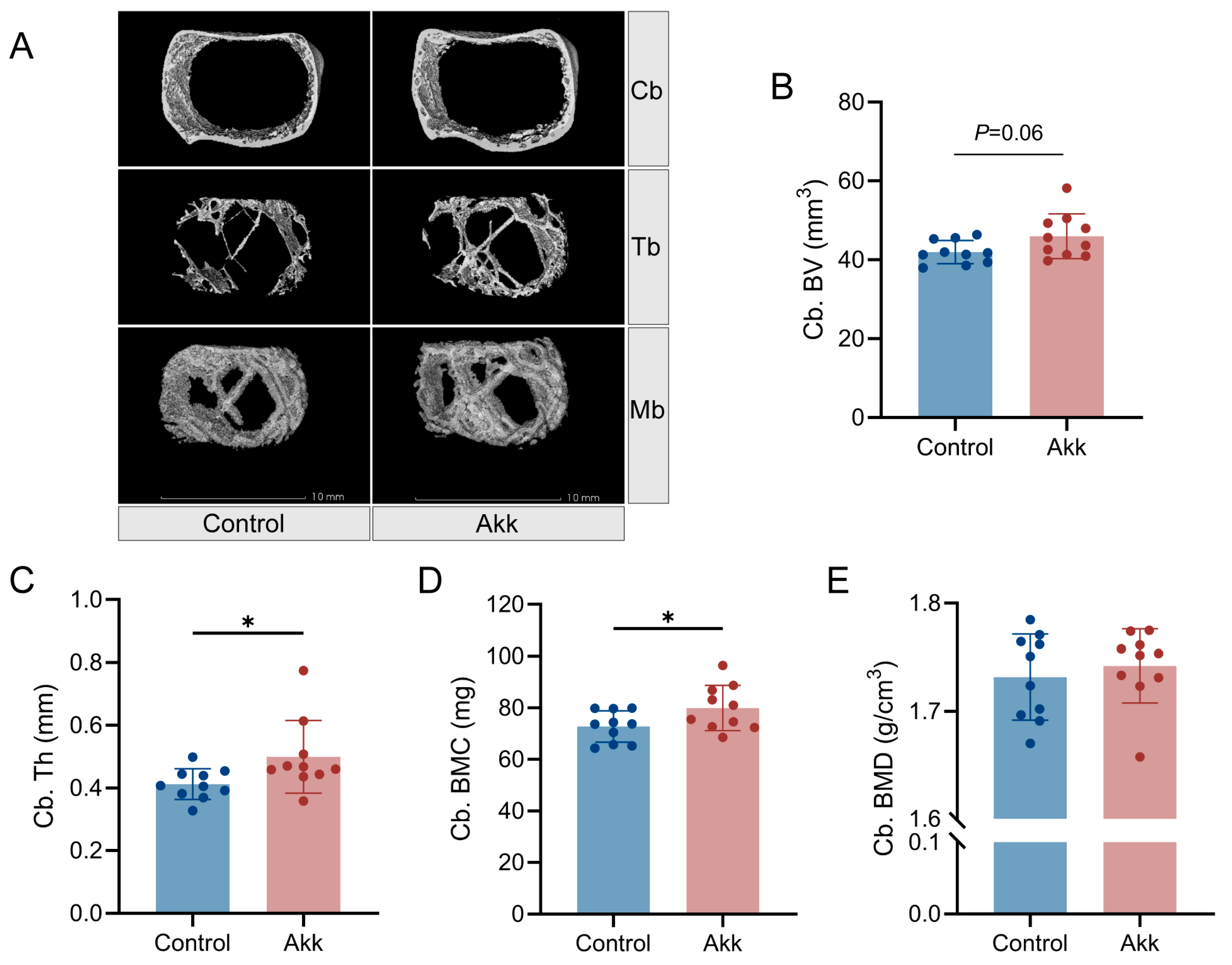
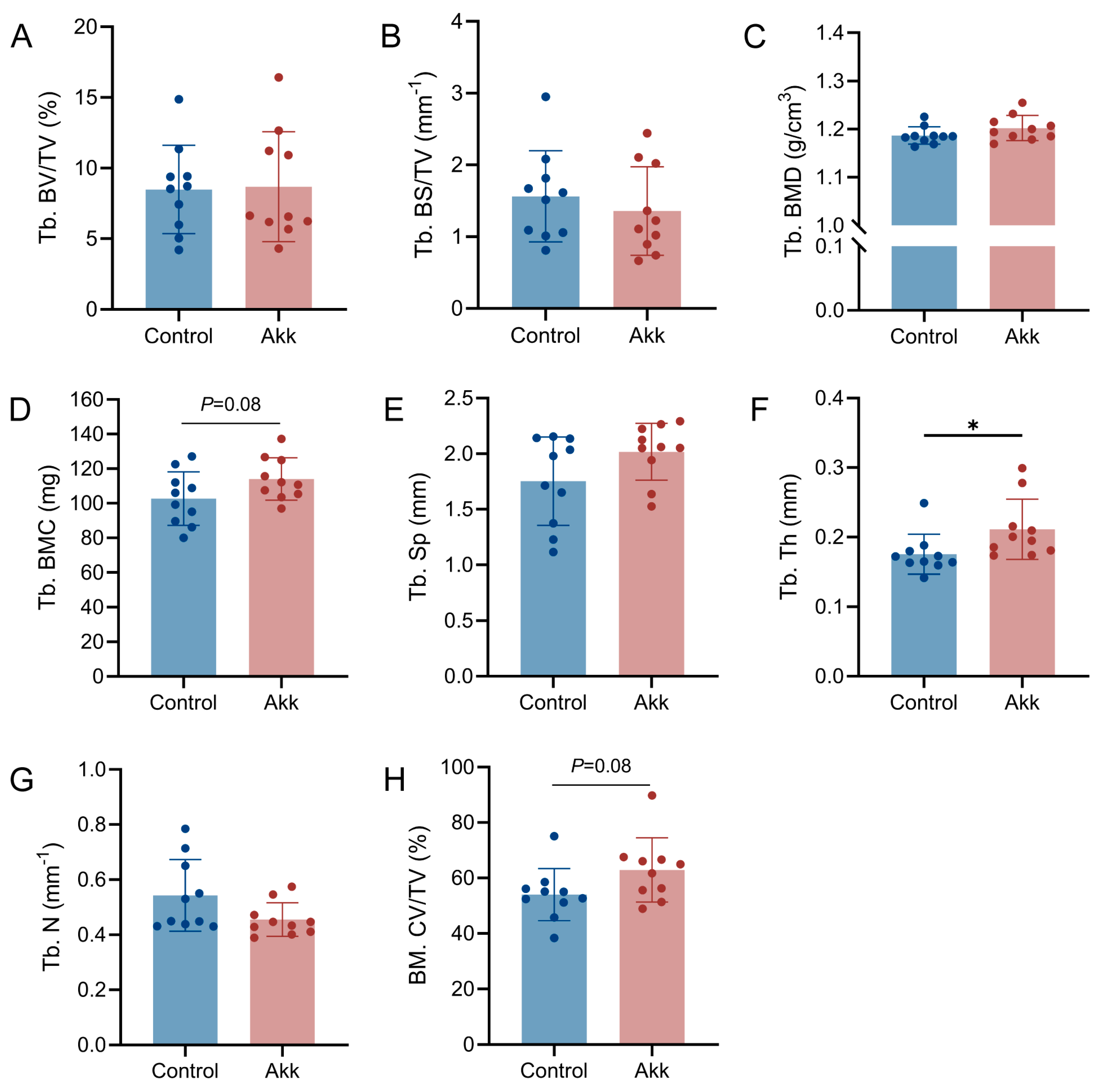
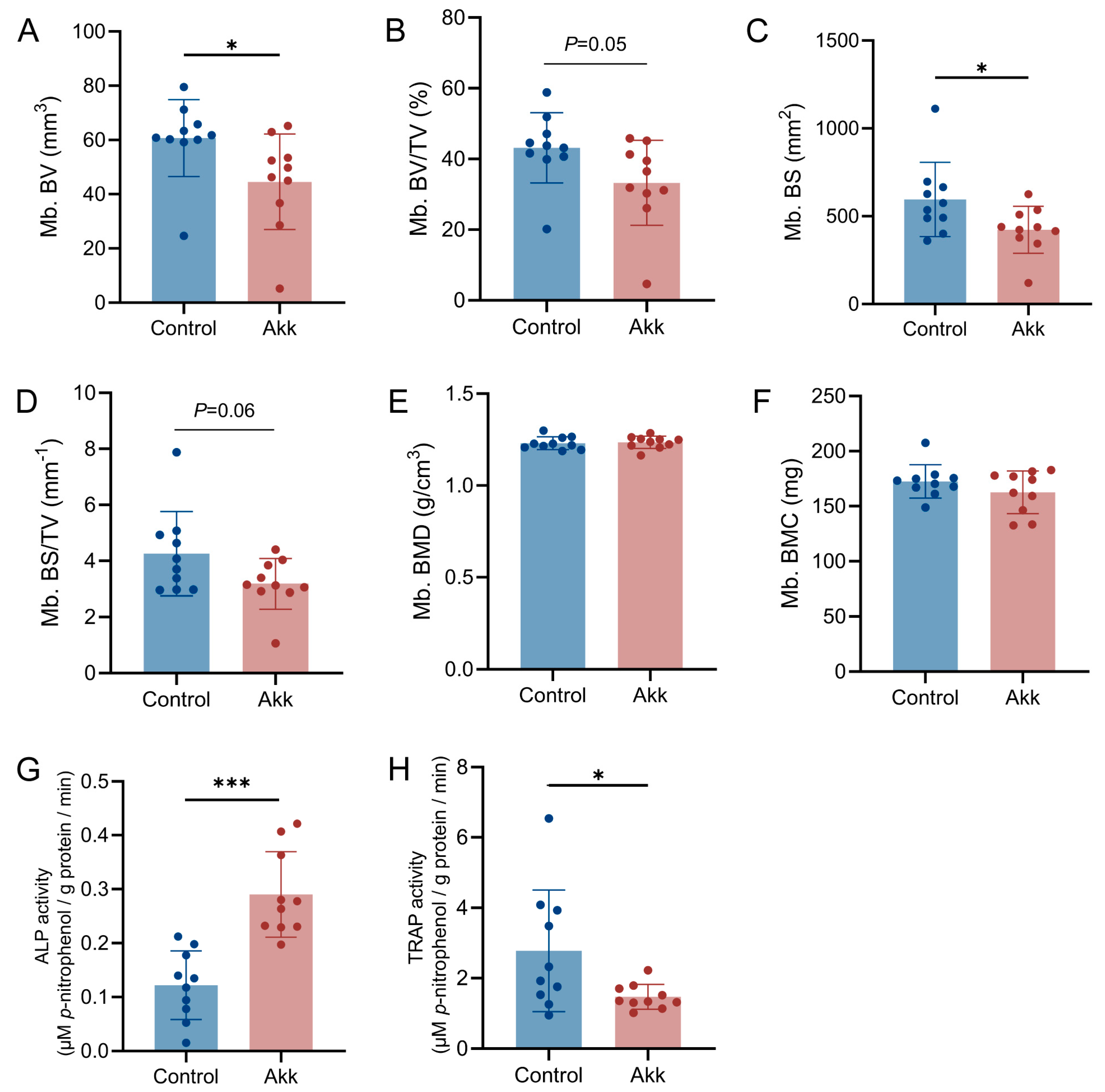
2.4. Micro-Computed Tomography Analysis

The right femurs of laying hens at 20 weeks were scanned using micro-computed tomography (micro-CT, NEMO, PINGSENG Healthcare Inc., Kunshan, China). Samples were scanned at 90 kV, 0.065 mA, in 3 cm-diameter holders. The CT reconstruction algorithm was FDK, the CT field of view was 15 mm, the pixel size was 0.0146 mm, and the slice thickness was 0.025 mm. Three-diameter reconstruction and analyses were conducted with the software Avatar (2.0.12.0, PINGSHENG Healthcare). For the femoral microarchitecture region, we analyzed 3 mm beginning with 3 mm below the landmark, as shown in Figure S1A. Three-dimensional images were reconstructed for visualization using the provided software. Image segmentation employed an adaptive-iterative threshold approach to differentiate between cortical bone (Cb), trabecular bone (Tb), and medullary bone (Mb). The different variables were measured and are described in Table 2 as defined by the previous study [42].

2.5. Determination of Alkaline Phosphatase Activity

Before alkaline phosphatase (ALP) activity assays, femurs were placed in ceramic crucibles and ground in liquid nitrogen. After grinding thoroughly, 0.1 g powders were collected in 2 mL tubes containing 1 mL cell lysis buffer (P0013J, Beyotime Biotechnology Co., Ltd., Shanghai, China). After a brief vortex lysis, samples were centrifuged at 12,000 rpm for 15 min at 4 °C. A total of 100 μL of the supernatant was collected for the determination of ALP activity. According to the manufacturer’s instructions (P0321S, Beyotime Biotechnology Co., Ltd., Shanghai, China), 50 μL of the reactive solution (para-nitrophenyl phosphate, pNPP) was mixed with 50 μL of sample and incubated at 37 °C for 30 min. The reaction was stopped with 100 μL stop solution. The absorbance of the sample was measured at 405 nm. The concentration of the total protein was measured using the BCA Protein assay kit (P0009, Beyotime Biotechnology Co., Ltd., Shanghai, China). The ALP activity was normalized to total protein, which was expressed as μM p-nitrophenol/g protein/min.

2.6. Determination of Tartrate-Resistant Acid Phosphatase Activity

Tissue lysis was performed as described above. According to the manufacturer’s instructions (P0321S, Beyotime Biotechnology Co., Ltd., Shanghai, China), 40 μL of the pNPP plus 5 μL tartaric acid solution was mixed with 40 μL sample and incubated at 37 °C for 30 min. The reaction was stopped with 160 μL stop solution. The absorbance of the sample was measured at 405 nm. The tartrate-resistant acid phosphatase (TRAP) activity was normalized to total protein, which was expressed as μM p-nitrophenol/g protein/min.

2.7. 16S rRNA Gene Sequencing and Analysis

The cecal contents of chickens were collected for microbiome analysis. 16S rRNA gene sequencing was conducted by Biomarker Technologies Co., Ltd. (Beijing, China). Shannon index, Simpson index, ACE index, and Chao1 index were used to measure community diversity. Beta diversity was evaluated by principal coordinate analysis to classify multiple samples and further demonstrate the differences in species diversity between samples. Relative abundances at the genus levels were statistically compared between the groups. Linear discriminant analysis (LDA) effect size (LEfSe) was used to identify the differences in microbial composition and to search for biomarkers with statistical differences between different groups. The Circos plot describes the correspondence between groups and genera, which not only reflects the proportion of dominant genera in each group but also reflects the distribution proportion of dominant genera in different groups. All the analysis was performed using BMKCloud (https://international.biocloud.net/zh/dashboard (accessed on 26 September 2023)).

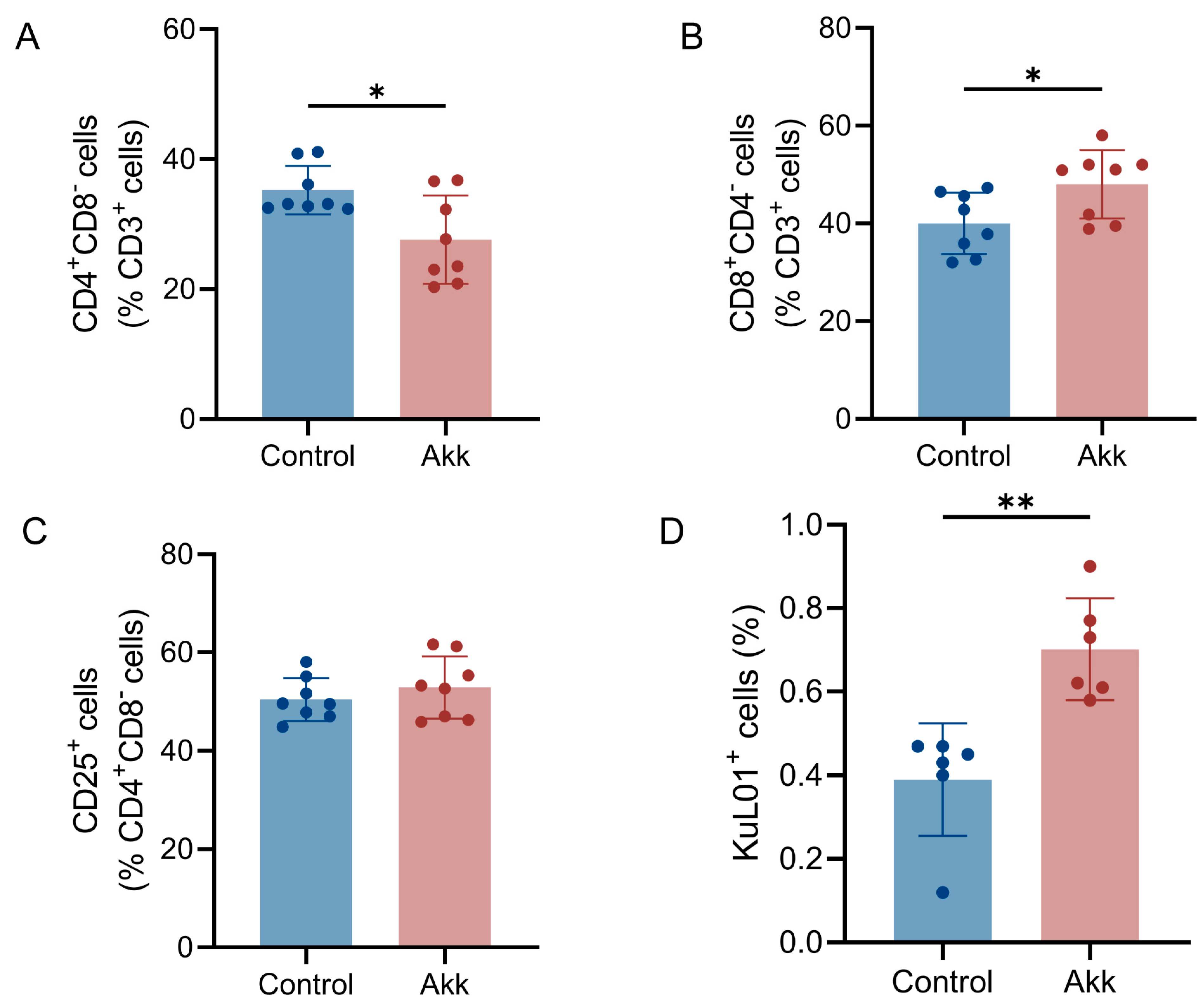
2.8. Preparation of Lymphocytes and Flow Cytometry

Single-cell suspensions of bone marrow were prepared as previously described [21]. Briefly, following the separation of the hen’s tibia and femur and the removal of muscles, the epiphysis was excised, and the bone marrow was flushed with RPMI 1640 medium (31800, Solarbio Technology Co., Ltd., Beijing, China) supplemented with 10% fetal bovine serum (v/v, NEWZERUM Ltd., Christchurch, New Zealand) until the bone became hollow. Subsequently, the bone marrow suspension was filtered through a 70-mesh cell sieve into a centrifuge tube. The bone marrow within the cell sieve was gently crushed using the syringe plunger. The bone marrow was further rinsed with RPMI medium and filtered in a centrifuge tube. The cell suspension was centrifuged at 600× g, 4 °C for 4 min, after which the supernatant was discarded, and approximately 6 mL of red blood cell lysis buffer (R1010, Solarbio Technology Co., Ltd., Beijing, China) was added to the tube. After 5 min of lysis, 40 mL of PBS was added to the tube, and the cell suspension was centrifuged at 600× g, 4 °C for 4 min. The cell suspension was then re-filtered through a 70-mesh sieve and stored in a centrifuge tube, resulting in single-cell suspensions of bone marrow. The concentration was adjusted to 2 × 10⁶ cells/mL using PBS before staining. Staining was performed for 30 min on ice in PBS containing 1% FBS. The antibodies used are listed in Table S1. Cells were acquired with a Coulter XL (Beckman Coulter, Brea, CA, USA), and analysis was performed with FlowJo (10.7.2) and FCS Express 6 Flow software.

2.9. Measurement of Egg Quality

At the conclusion of week 20, 5 eggs from each replicate (totaling 50 eggs per group) were randomly chosen for egg quality assessment. Egg quality was determined on three consecutive days, with egg collection conducted each day. Eggshell thickness, egg width, and egg length were assessed using a vernier caliper. The value of the egg shape index is defined as the egg’s width to length ratio. Egg weight, eggshell breaking strength, Haugh unit, and yolk color were determined using the Nabel DET-6000 egg analyzer (Kyoto, Japan). Eggshell strength was determined by measuring the maximum horizontal mechanical force experienced by the egg’s long axis when laid flat.

2.10. Statistical Analysis

Statistical analyses were conducted using SPSS version 20.0. Data are presented as means ± standard deviations (SD) unless indicated otherwise. Differences between the two groups were evaluated using a Two-tailed unpaired Student’s t-test. p < 0.05 was considered statistically significant.

3. Results

3.1. Effect of Akkermansia muciniphila Powder on Growth Performance of Laying Hens

Throughout the experiment, spanning weeks 0–6, 0–12, and 0–18, the introduction of Akkermansia muciniphila (Akk) powder into the diet did not yield significant alterations in the average daily feed intake (ADFI) among the experimental groups (Table 3). By the 20-week mark, comparisons between the groups revealed no notable changes in body weight, shank length, abdominal fat percentage, or hepatosomatic index (Figure 1A–D). These findings collectively suggest that Akk did not affect the growth performance of laying hens.

3.2. Dietary Supplementation of Akk Powder Promotes Bone Development

Ensuring sufficient bone development before sexual maturity is crucial for laying hens. The microarchitectures of cortical bone (Cb), trabecular bone (Tb), and medullary bone (Mb) were assessed and quantified using micro-CT (Figure 2A). As for Cb microarchitecture, Akk supplementation resulted in significant enhancements in cortical bone thickness (Cb. Th) and cortical bone mineral content (Cb. BMC) (Figure 2C,D), without significant effects on cortical bone volume (Cb. BV) and cortical bone mineral density (Cb. BMD) (Figure 2B,E). Regarding Tb parameters, the trabecular bone thickness (Tb. Th) exhibited a significant increase following Akk powder supplementation (Figure 3F). Significant differences were not observed in the ratio of trabecular bone volume to total femur volume (Tb. BV/TV), the ratio of trabecular bone surface area to total femur volume (Tb. BS/TV), trabecular bone mineral density (Tb. BMD), trabecular bone mineral content (Tb. BMC), trabecular bone spacing (Tb. Sp), and trabecular bone number (Tb. N) (Figure 3A–E,G). The ratio of the bone marrow cavity volume to the total femur volume (BM. CV/TV) did not differ significantly between the Control and Akk groups (Figure 3H). The quantitative analysis of Mb parameters revealed that hens in the Akk group exhibited lower medullary bone volume (Mb. BV) and medullary bone surface area (Mb. BS) compared to the Control group (Figure 4A,C). Both the ratio of Mb. BV to total femur volume (Mb. BV/TV) and the ratio of Mb. BS to total femur volume (Mb. BS/TV) showed no significant difference in the two groups (Figure 4B,D). Neither medullary bone mineral density (Mb. BMD) nor medullary bone mineral content (Mb. BMC) appeared to be affected by Akk powder supplementation (Figure 4E,F).
In the femur, the activities of alkaline phosphatase (ALP), indicative of osteoblast differentiation, and tartrate-resistant acid phosphatase (TRAP), a marker of osteoclastic activity, were evaluated. Dietary supplementation with Akk powder resulted in a significant increase in ALP activity, but the suppression of TRAP activity (Figure 4G,H), suggesting enhanced bone formation during bone remodeling, thereby facilitating the accumulation of bone mass.

3.3. Effect of Akk on Immune Cells Associated with Bone Remodeling in Bone Marrow

To explore the impact of Akk treatment on immune cells, the proportions of immune cells in the bone marrow were analyzed using flow cytometry, with the gating strategy outlined in Figure S1B. Regarding bone marrow immune cells, Akk led to a reduction in the proportion of CD4+ T cells (Figure 5A). Conversely, the number of CD8+ T cells notably increased following Akk supplementation (Figure 5B), while CD25+ T cells remained unchanged post-Akk powder supplementation (Figure 5C). Additionally, KuL01+ cells, representing osteoclast precursor cells in the bone marrow, exhibited a significant increase in the Akk powder-treated group (Figure 5D).

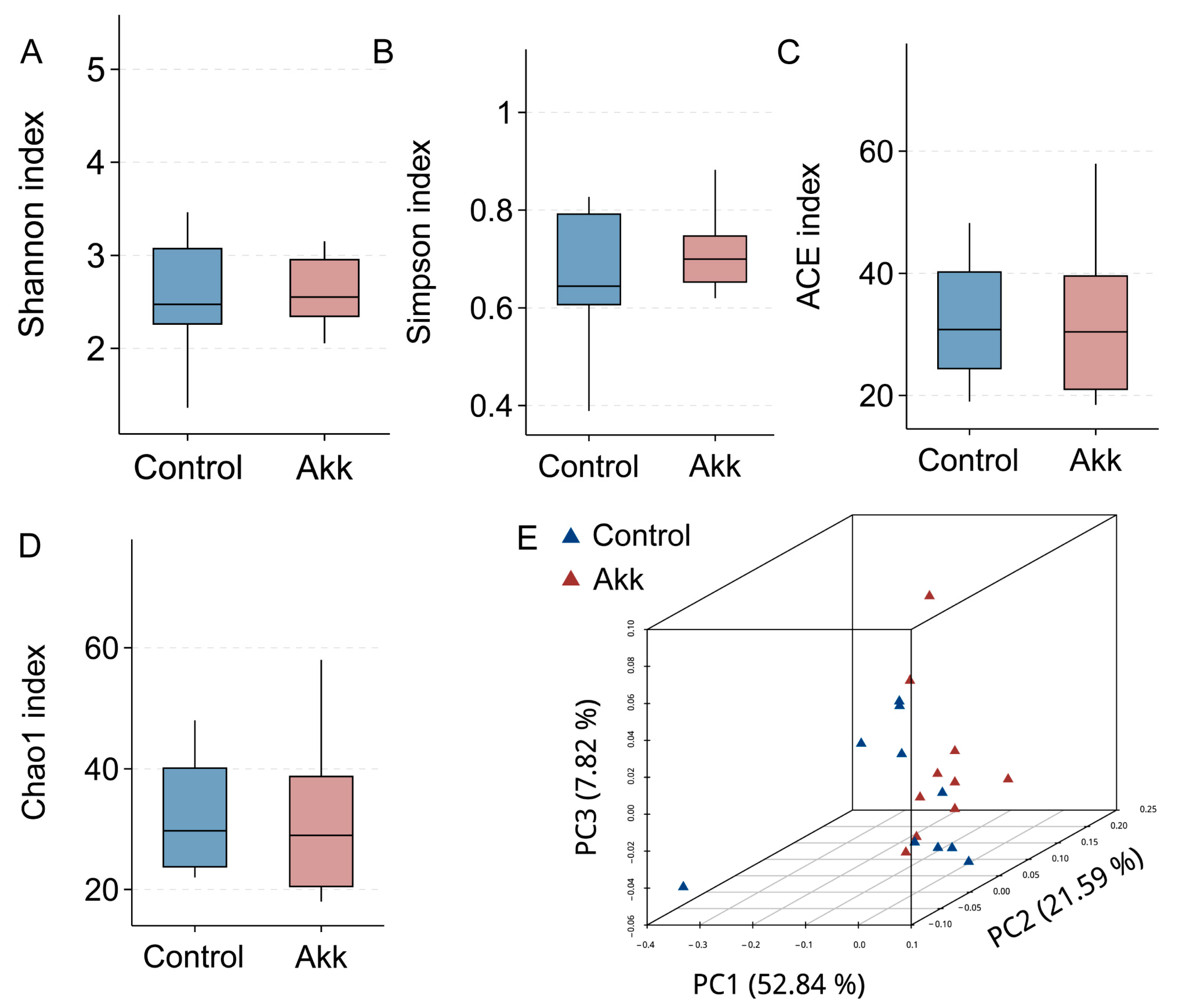
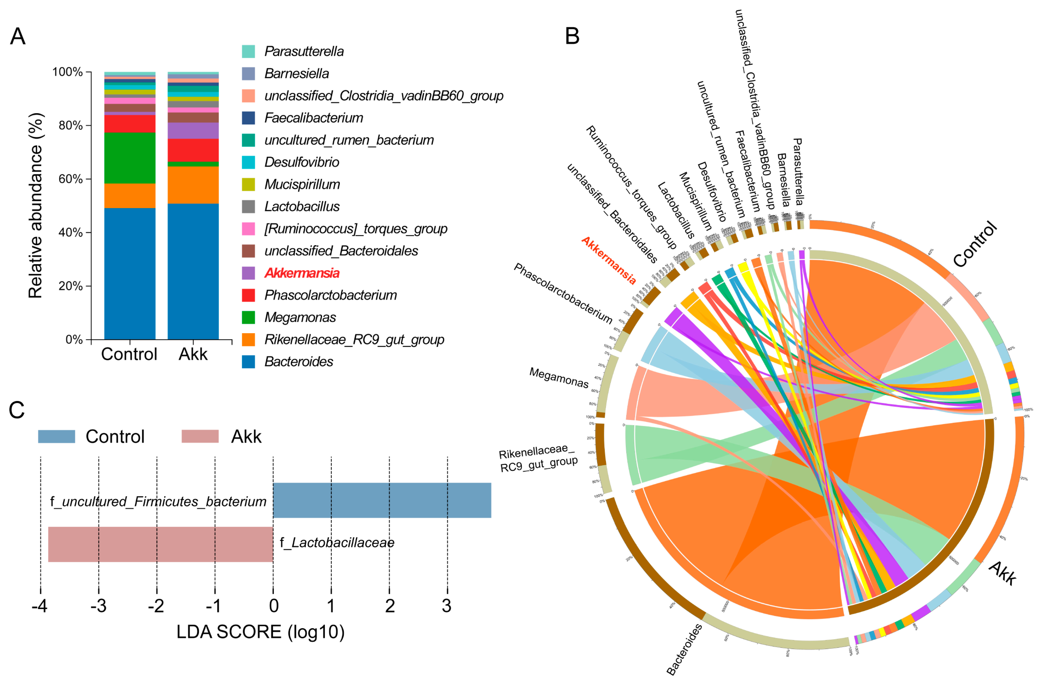
3.4. Effect of Akk on the Abundance and Diversity of Cecal Microbiota

To evaluate the influence of Akk supplementation on the cecal microbial communities, we conducted the 16S rRNA sequencing of cecal contents. No significant alterations were observed in the Shannon, Simpson, ACE, and Chao1 indices, suggesting that the diversity of cecal microbial communities remained stable (Figure 6A–D). Principal coordinate analysis (PCoA) also confirmed similar microbial diversity across the groups (Figure 6E). At the genus level, the top fifteen communities exhibited comparability between Control and Akk groups (Figure 7A,B). Linear discriminant analysis (LDA) effect size (LEfSe) analysis at the family level revealed that the uncultured_Firmicutes_bacterium predominated in the Control group, whereas Lactobacillaceae was enriched in the Akk group (Figure 7C).

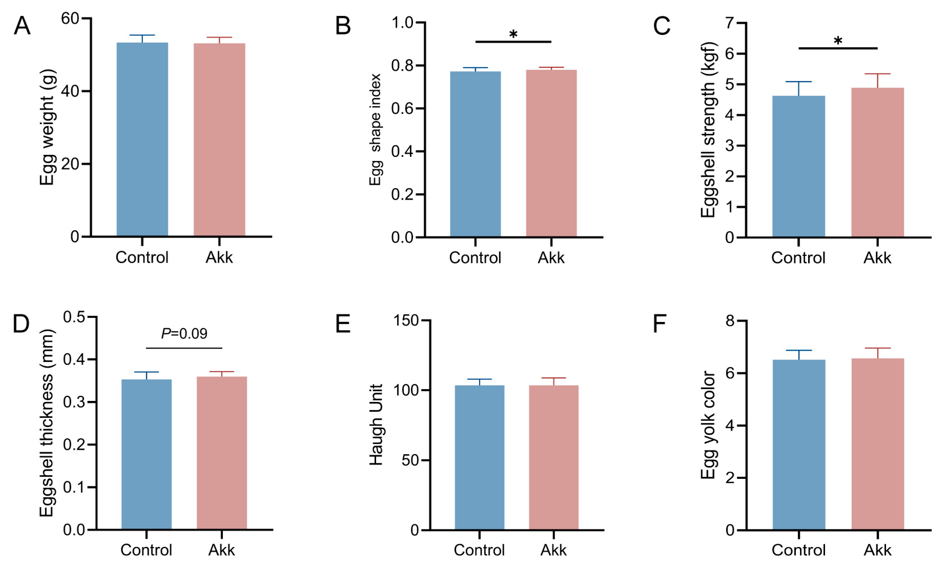
3.5. Dietary Supplementation of Akk Powder Increases Eggshell Quality

Egg weight showed no significant difference between the Control and Akk groups (Figure 8A). The egg shape index in the Akk group was significantly higher than that of the Control group (Figure 8B). The supplementation with Akk powder resulted in a considerable increase in eggshell strength and a trend towards thicker eggshells (Figure 8C,D). A stronger eggshell can help reduce the risk of breakage during transportation. Regarding internal quality, there were no changes observed in the Haugh unit or egg yolk color following Akk powder treatment (Figure 8E,F).

4. Discussion

The gut microbiota represents a key regulator of bone metabolism [43]. As a distinguished next-generation probiotic, Akk plays a significant role in modulating host bone remodeling and mitigating osteoporosis. Ensuring optimal bone development in laying hens is not only vital for the health of the host, but also for sustaining a productive laying cycle. This study provides novel insights into the gut-bone axis in laying hens. Dietary supplementation with lyophilized Akk powder facilitated postnatal bone development by enhancing bone formation, ultimately leading to improvements in eggshell quality.
The period preceding sexual maturity is critical for the skeletal development of laying hens. Previous studies have consistently demonstrated that neither live nor pasteurized Akk adversely affects host physiology [30,44]. Our findings further validate these results, indicating that Akk supplementation does not hinder the growth of laying hens. Notably, Akk supplementation significantly enhanced the development of Cb, characterized by increased volume and thickness, potentially conferring resistance to physical impacts. During the early stages of egg production, a higher mineral content in the Cb may contribute to enhanced eggshell formation. Tb serves as a crucial indicator of bone health in mammals, with degradation of Tb microarchitecture, manifested by reduced thickness and volume along with increased spacing, being a hallmark feature of osteoporosis onset [42,45]. Akk exhibited the most pronounced effects on Tb. Th, with greater thickness potentially offering mechanical protection. A previous study reported that Akk directly increases the bone mass of Cb and Tb by increasing osteogenic activity and inhibiting osteoclastogenesis [30]. The observed increase in ALP activity and decrease in TRAP activity in the Akk group were consistent with this, indicating that bone formation outweighs bone resorption following Akk powder supplementation, leading to bone mass accumulation.
Research on the development of Mb in birds remains relatively limited. Previous studies have indicated that the Mb of elderly laying hens gradually fills the bone marrow cavity, displaces the Tb, and ultimately increases the BMD and bone strength [46,47]. Additionally, within the daily egg-laying cycle, a negative correlation has been observed between the surface area of Mb and Tb [15]. Our findings suggest that lower Mb. BV and Mb. BS in the Akk group are correlated with higher bone formation and eggshell quality. Therefore, we speculate that the excessive formation of Mb during the sexual maturation of laying hens may compromise Cb and Tb development, adversely affecting eggshell formation. Further research is warranted to investigate the effects of medullary bone volume during sexual maturity on the subsequent oviposition performance of laying hens.
One of the mechanisms by which gut microbiota regulate bone remodeling is through the immune system. An imbalance in gut microbiota has been associated with bacterial translocation and chronic inflammation [21,48]. The inflammation often leads to an increased immune response and bone resorption in the bone marrow, consequently reducing bone formation, as observed in Eimeria-challenged or lipopolysaccharide-challenged broilers [49,50]. Significant bone loss has been noted in various models of intestinal inflammation, including dextran sulfate sodium-induced chemical injury, adoptive T cell transfer of colitis, and Salmonella enterica infection [51]. The skeletal and immune systems are intricately intertwined, sharing numerous cytokines, receptors, and transcription factors [23,24]. The receptor activator of NF-κB ligand (RANKL) plays an important role in osteoclastogenesis, primarily sourced from osteoblasts and activated T cells, effectively bridging these two systems [52]. The term “osteoimmunology” underscores the reciprocal interactions between the skeletal and immune systems [23,24,53]. Specifically, CD4+ T cells serve as one of the sources of RANKL in the bone marrow, and an increase in CD4+ T cells has been observed in osteoporosis associated with inflammatory bowel disease [54]. Following Akk supplementation, the lower CD4+ T cells in the bone marrow were associated with attenuated osteoclastogenesis, as evidenced by reduced TRAP activity. KuL01+ cells, representing monocytes (osteoclast precursor cells) in the bone marrow, exhibited a significant increase in the Akk powder-treated group, which may suggest that fewer osteoclast precursor cells differentiated into osteoclasts, thereby mitigating excessive bone resorption in the femur. Previous studies have indicated that butyrate stimulation can increase the expression of the Wnt ligand Wnt10b in CD8+ T cells, thereby promoting bone formation through the activation of Wnt signaling in osteoblasts [55]. Remarkably, Akk supplementation significantly upregulated the proportion of CD8+ T cells in the bone marrow. Collectively, dietary supplementation with Akk powder can improve the immune microenvironment in the bone marrow, leading to improved bone formation and reduced bone resorption in laying hens.
Feeding with Akk powder for 20 weeks did not induce any alterations in the diversity and abundance of cecal microbial communities, consistent with our previous findings [44]. However, Akk supplementation significantly enriched Lactobacillaceae, a family comprising numerous probiotic species, including Lactobacillus plantarum, Lactobacillus rhamnosus, Lactobacillus reuteri, and Lactobacillus ultunensis [56]. As conventional probiotics, Lactobacillus strains have a long history of use. Lactobacillus-fermented products, such as milk, soy skim milk, and Kefir, have been shown to have beneficial effects on bone health [57,58,59,60,61]. The administration of Lactobacillus rhamnosus GG has been reported to alter microbial diversity and increase the proportion of short-chain fatty acid-producing Clostridia in conventionally raised mice, thereby increasing bone volume and bone formation [55]. Lactobacillus reuteri could reduce bone loss in older women with low BMD [62]. Additionally, a mixture of Lactobacillus paracasei, Lactobacillus plantarum DSM 15312, and DSM 15313 increases femoral volume and BMC in OVX mice [63]. Collectively, Akk supplementation contributed to improving the cecal microenvironment by enriching Lactobacillaceae.
The decline in egg quality, particularly eggshell quality, presents a significant challenge for the egg industry. The physiological condition of bones during the early stages of the laying period may be closely linked to egg quality [64]. Therefore, we assessed egg quality at 20 weeks to investigate the relationship between bone physiology and eggshell quality. It has been reported that the egg shape index exhibited a positive correlation with the eggshell strength [65,66]. Our findings reveal that eggs in the Akk group exhibited improved egg shape index and eggshell strength. The high egg shape index means the eggs tend to be round [64]. The increased eggshell strength potentially mitigates physical damage during handling and transportation. Previous studies from our research group have demonstrated that Akk supplementation contributes to increased eggshell thickness and Haugh unit in older hens [44]. However, in this study, Akk had no significant effect on Haugh unit and yolk color, which could be attributed to the age of the laying hens.

5. Conclusions

This study elucidates the significant effects of Akk on bone physiology in laying hens, as well as the interplay between various bone microarchitectures and eggshell quality. Our findings show that the continuous dietary supplementation of Akk powder promoted bone formation and bone development by improving the bone marrow and intestinal microenvironment of laying hens, thereby improving eggshell quality. This study lays the groundwork for microbial-based interventions targeting bone-related diseases in laying hens. As a next-generation probiotic, lyophilized Akk powder holds promise as a potential additive in the poultry industry.

Supplementary Materials

The following supporting information can be downloaded at: https://www.mdpi.com/article/10.3390/agriculture14040598/s1, Figure S1: (A) The determination of femoral volume of interest in micro-CT analysis. (B) Gating strategy of flow cytometry; Table S1. Key antibody table.

Author Contributions

Conceptualization, D.L.; data curation, Z.L., G.Y. and D.L.; formal analysis, Z.L., Y.Z., G.Y., F.Z. and D.L.; funding acquisition, D.L.; investigation, Z.L., Y.Z., G.Y., F.Z. and D.L.; methodology, Z.L., Y.Z., F.Z., Y.H. and D.L.; project administration, D.L.; resources, D.L.; software, Z.L. and D.L.; supervision, D.L.; validation, Z.L. and D.L.; visualization, Z.L.; writing—original draft, Z.L.; writing—review and editing, Z.L., Y.H. and D.L. All authors have read and agreed to the published version of the manuscript.

Funding

This work was supported by the National Key Research and Development Program of China (2022YFD1300400) and the 2115 Talent Development Program of China Agricultural University.

Institutional Review Board Statement

All the experiments were reviewed and approved by the China Agricultural University Animal Care and Use Committee (9 July 2022, statement no. AW10204202-1-3).

Data Availability Statement

Data supporting the reported results can be requested from the corresponding author via email.

Conflicts of Interest

The authors declare no conflicts of interest.

Abbreviations

AkkAkkermansia muciniphila
CbCortical bone
TbTrabecular bone
MbMedullary bone
OVXOvariectomized
GAMGifu Anaerobic Medium
ALPAlkaline phosphatase
TRAPTartrate-resistant acid phosphatase
LEfSeLinear discriminant analysis effect size
ADFIAverage daily feed intake
RANKLReceptor activator of NF-κB ligand
PCoAPrincipal coordinate analysis
Cb. BVCortical bone volume
Cb. ThCortical bone thickness
Cb. BMCCortical bone mineral content
Cb. BMDCortical bone mineral density
FMTFecal microbiota transplantation
RTRoom temperature
Tb. BMCTrabecular bone mineral content
Tb. ThTrabecular bone thickness
Tb. BV/TVThe ratio of trabecular bone volume to total femur volume
Tb. BS/TVThe ratio of trabecular bone surface area to total femur volume
Tb. BMDTrabecular bone mineral density
Tb. SpTrabecular bone spacing
Tb. NTrabecular bone number
BM. CV/TVThe bone marrow cavity volume to the total femur volume
Mb. BVMedullary bone volume
Mb. BV/TVThe ratio of medullary bone volume to total femur volume
Mb. BSThe medullary bone surface area
Mb. BS/TVThe ratio of Mb. BS to total femur volume
Mb. BMDMedullary bone mineral density
Mb. BMCMedullary bone mineral content

References

  1. Bain, M.M.; Nys, Y.; Dunn, I.C. Increasing persistency in lay and stabilising egg quality in longer laying cycles. What are the challenges? Br. Poult. Sci. 2016, 57, 330–338. [Google Scholar] [CrossRef] [PubMed]
  2. Korver, D.R. Review: Current challenges in poultry nutrition, health, and welfare. Anim. Int. J. Anim. Biosci. 2023, 17 (Suppl. 2), 100755. [Google Scholar] [CrossRef] [PubMed]
  3. Sandilands, V. The laying hen and bone fractures. Vet. Rec. 2011, 169, 411–412. [Google Scholar] [CrossRef] [PubMed]
  4. Whitehead, C.C.; Fleming, R.H. Osteoporosis in cage layers. Poult. Sci. 2000, 79, 1033–1041. [Google Scholar] [CrossRef] [PubMed]
  5. van de Velde, J.P.; Vermeiden, J.P.; Bloot, A.M. Medullary bone matrix formation, mineralization, and remodeling related to the daily egg-laying cycle of Japanese quail: A histological and radiological study. Bone 1985, 6, 321–327. [Google Scholar] [CrossRef] [PubMed]
  6. Rodriguez-Navarro, A.B.; McCormack, H.M.; Fleming, R.H.; Alvarez-Lloret, P.; Romero-Pastor, J.; Dominguez-Gasca, N.; Prozorov, T.; Dunn, I.C. Influence of physical activity on tibial bone material properties in laying hens. J. Struct. Biol. 2018, 201, 36–45. [Google Scholar] [CrossRef] [PubMed]
  7. Chevalier, C.; Kieser, S.; Çolakoğlu, M.; Hadadi, N.; Brun, J.; Rigo, D.; Suárez-Zamorano, N.; Spiljar, M.; Fabbiano, S.; Busse, B.; et al. Warmth Prevents Bone Loss Through the Gut Microbiota. Cell Metab. 2020, 32, 575–590.e577. [Google Scholar] [CrossRef] [PubMed]
  8. Rauch, F. Bone growth in length and width: The Yin and Yang of bone stability. J. Musculoskelet. Neuronal Interact. 2005, 5, 194–201. [Google Scholar] [PubMed]
  9. Whitehead, C.C. Overview of bone biology in the egg-laying hen. Poult. Sci. 2004, 83, 193–199. [Google Scholar] [CrossRef]
  10. Squire, M.E.; Veglia, M.K.; Drucker, K.A.; Brazeal, K.R.; Hahn, T.P.; Watts, H.E. Estrogen levels influence medullary bone quantity and density in female house finches and pine siskins. Gen. Comp. Endocrinol. 2017, 246, 249–257. [Google Scholar] [CrossRef]
  11. Neijat, M.; Casey-Trott, T.M.; Robinson, S.; Widowski, T.M.; Kiarie, E. Effects of rearing and adult laying housing systems on medullary, pneumatic and radius bone attributes in 73-wk old Lohmann LSL lite hens1. Poult. Sci. 2019, 98, 2840–2845. [Google Scholar] [CrossRef] [PubMed]
  12. Reich, T.; Gefen, A. Effect of trabecular bone loss on cortical strain rate during impact in an in vitro model of avian femur. Biomed. Eng. Online 2006, 5, 45. [Google Scholar] [CrossRef] [PubMed]
  13. Casey-Trott, T.M.; Korver, D.R.; Guerin, M.T.; Sandilands, V.; Torrey, S.; Widowski, T.M. Opportunities for exercise during pullet rearing, Part II: Long-term effects on bone characteristics of adult laying hens at the end-of-lay. Poult. Sci. 2017, 96, 2518–2527. [Google Scholar] [CrossRef] [PubMed]
  14. Yan, J.; Wang, J.; Chen, J.; Shi, H.; Liao, X.; Pan, C.; Liu, Y.; Yang, X.; Ren, Z.; Yang, X. Adjusting phosphate feeding regimen according to daily rhythm increases eggshell quality via enhancing medullary bone remodeling in laying hens. J. Anim. Sci. Biotechnol. 2023, 14, 17. [Google Scholar] [CrossRef] [PubMed]
  15. Kerschnitzki, M.; Zander, T.; Zaslansky, P.; Fratzl, P.; Shahar, R.; Wagermaier, W. Rapid alterations of avian medullary bone material during the daily egg-laying cycle. Bone 2014, 69, 109–117. [Google Scholar] [CrossRef] [PubMed]
  16. Cransberg, P.H.; Parkinson, G.B.; Wilson, S.; Thorp, B.H. Sequential studies of skeletal calcium reserves and structural bone volume in a commercial layer flock. Br. Poult. Sci. 2001, 42, 260–265. [Google Scholar] [CrossRef] [PubMed]
  17. Casey-Trott, T.M.; Korver, D.R.; Guerin, M.T.; Sandilands, V.; Torrey, S.; Widowski, T.M. Opportunities for exercise during pullet rearing, Part I: Effect on the musculoskeletal characteristics of pullets. Poult. Sci. 2017, 96, 2509–2517. [Google Scholar] [CrossRef] [PubMed]
  18. Fleming, R.H. Nutritional factors affecting poultry bone health. Proc. Nutr. Soc. 2008, 67, 177–183. [Google Scholar] [CrossRef] [PubMed]
  19. Gautron, J.; Réhault-Godbert, S.; Van de Braak, T.G.H.; Dunn, I.C. Review: What are the challenges facing the table egg industry in the next decades and what can be done to address them? Anim. Int. J. Anim. Biosci. 2021, 15 (Suppl. 1), 100282. [Google Scholar] [CrossRef]
  20. Papadopoulou, S.K.; Papadimitriou, K.; Voulgaridou, G.; Georgaki, E.; Tsotidou, E.; Zantidou, O.; Papandreou, D. Exercise and Nutrition Impact on Osteoporosis and Sarcopenia-The Incidence of Osteosarcopenia: A Narrative Review. Nutrients. 2021, 13, 4499. [Google Scholar] [CrossRef]
  21. Li, J.Y.; Chassaing, B.; Tyagi, A.M.; Vaccaro, C.; Luo, T.; Adams, J.; Darby, T.M.; Weitzmann, M.N.; Mulle, J.G.; Gewirtz, A.T.; et al. Sex steroid deficiency-associated bone loss is microbiota dependent and prevented by probiotics. J. Clin. Investig. 2016, 126, 2049–2063. [Google Scholar] [CrossRef] [PubMed]
  22. Silva, B.C.; Bilezikian, J.P. Parathyroid hormone: Anabolic and catabolic actions on the skeleton. Curr. Opin. Pharmacol. 2015, 22, 41–50. [Google Scholar] [CrossRef] [PubMed]
  23. Okamoto, K.; Nakashima, T.; Shinohara, M.; Negishi-Koga, T.; Komatsu, N.; Terashima, A.; Sawa, S.; Nitta, T.; Takayanagi, H. Osteoimmunology: The Conceptual Framework Unifying the Immune and Skeletal Systems. Physiol. Rev. 2017, 97, 1295–1349. [Google Scholar] [CrossRef] [PubMed]
  24. Takayanagi, H. Osteoimmunology: Shared mechanisms and crosstalk between the immune and bone systems. Nat. Rev. Immunol. 2007, 7, 292–304. [Google Scholar] [CrossRef] [PubMed]
  25. Zaiss, M.M.; Jones, R.M.; Schett, G.; Pacifici, R. The gut-bone axis: How bacterial metabolites bridge the distance. J. Clin. Investig. 2019, 129, 3018–3028. [Google Scholar] [CrossRef] [PubMed]
  26. Lyu, Z.; Hu, Y.; Guo, Y.; Liu, D. Modulation of bone remodeling by the gut microbiota: A new therapy for osteoporosis. Bone. Res. 2023, 11, 31. [Google Scholar] [CrossRef] [PubMed]
  27. Britton, R.A.; Irwin, R.; Quach, D.; Schaefer, L.; Zhang, J.; Lee, T.; Parameswaran, N.; McCabe, L.R. Probiotic L. reuteri treatment prevents bone loss in a menopausal ovariectomized mouse model. J. Cell. Physiol. 2014, 229, 1822–1830. [Google Scholar] [CrossRef] [PubMed]
  28. Parvaneh, K.; Ebrahimi, M.; Sabran, M.R.; Karimi, G.; Hwei, A.N.; Abdul-Majeed, S.; Ahmad, Z.; Ibrahim, Z.; Jamaluddin, R. Probiotics (Bifidobacterium longum) Increase Bone Mass Density and Upregulate Sparc and Bmp-2 Genes in Rats with Bone Loss Resulting from Ovariectomy. Biomed. Res. Int. 2015, 2015, 897639. [Google Scholar] [CrossRef] [PubMed]
  29. Roberts, J.L.; Liu, G.; Darby, T.M.; Fernandes, L.M.; Diaz-Hernandez, M.E.; Jones, R.M.; Drissi, H. Bifidobacterium adolescentis supplementation attenuates fracture-induced systemic sequelae. Biomed. Pharmacother. 2020, 132, 110831. [Google Scholar] [CrossRef]
  30. Liu, J.H.; Chen, C.Y.; Liu, Z.Z.; Luo, Z.W.; Rao, S.S.; Jin, L.; Wan, T.F.; Yue, T.; Tan, Y.J.; Yin, H.; et al. Extracellular Vesicles from Child Gut Microbiota Enter into Bone to Preserve Bone Mass and Strength. Adv. Sci. 2021, 8, 2004831. [Google Scholar] [CrossRef]
  31. Derrien, M.; Vaughan, E.E.; Plugge, C.M.; de Vos, W.M. Akkermansia muciniphila gen. nov., sp. nov., a human intestinal mucin-degrading bacterium. Int. J. Syst. Evol. Microbiol. 2004, 54, 1469–1476. [Google Scholar] [CrossRef]
  32. Naito, Y.; Uchiyama, K.; Takagi, T. A next-generation beneficial microbe: Akkermansia muciniphila. J. Clin. Biochem. Nutr. 2018, 63, 33–35. [Google Scholar] [CrossRef] [PubMed]
  33. Derrien, M.; Belzer, C.; de Vos, W.M. Akkermansia muciniphila and its role in regulating host functions. Microb. Pathog. 2017, 106, 171–181. [Google Scholar] [CrossRef] [PubMed]
  34. Schneeberger, M.; Everard, A.; Gómez-Valadés, A.G.; Matamoros, S.; Ramírez, S.; Delzenne, N.M.; Gomis, R.; Claret, M.; Cani, P.D. Akkermansia muciniphila inversely correlates with the onset of inflammation, altered adipose tissue metabolism and metabolic disorders during obesity in mice. Sci. Rep. 2015, 5, 16643. [Google Scholar] [CrossRef] [PubMed]
  35. Everard, A.; Belzer, C.; Geurts, L.; Ouwerkerk, J.P.; Druart, C.; Bindels, L.B.; Guiot, Y.; Derrien, M.; Muccioli, G.G.; Delzenne, N.M.; et al. Cross-talk between Akkermansia muciniphila and intestinal epithelium controls diet-induced obesity. Proc. Natl. Acad. Sci. USA 2013, 110, 9066–9071. [Google Scholar] [CrossRef] [PubMed]
  36. Cani, P.D.; de Vos, W.M. Next-Generation Beneficial Microbes: The Case of Akkermansia muciniphila. Front. Microbiol. 2017, 8, 1765. [Google Scholar] [CrossRef] [PubMed]
  37. Zhai, Q.; Feng, S.; Arjan, N.; Chen, W. A next generation probiotic, Akkermansia muciniphila. Crit. Rev. Food Sci. Nutr. 2019, 59, 3227–3236. [Google Scholar] [CrossRef] [PubMed]
  38. Depommier, C.; Everard, A.; Druart, C.; Plovier, H.; Van Hul, M.; Vieira-Silva, S.; Falony, G.; Raes, J.; Maiter, D.; Delzenne, N.M.; et al. Supplementation with Akkermansia muciniphila in overweight and obese human volunteers: A proof-of-concept exploratory study. Nat. Med. 2019, 25, 1096–1103. [Google Scholar] [CrossRef] [PubMed]
  39. Mulhall, H.; DiChiara, J.M.; Deragon, M.; Iyer, R.; Huck, O.; Amar, S. Akkermansia muciniphila and Its Pili-Like Protein Amuc_1100 Modulate Macrophage Polarization in Experimental Periodontitis. Infect. Immun. 2020, 89, e00500-20. [Google Scholar] [CrossRef]
  40. Huck, O.; Mulhall, H.; Rubin, G.; Kizelnik, Z.; Iyer, R.; Perpich, J.D.; Haque, N.; Cani, P.D.; de Vos, W.M.; Amar, S. Akkermansia muciniphila reduces Porphyromonas gingivalis-induced inflammation and periodontal bone destruction. J. Clin. Periodontol. 2020, 47, 202–212. [Google Scholar] [CrossRef]
  41. Liu, J.H.; Yue, T.; Luo, Z.W.; Cao, J.; Yan, Z.Q.; Jin, L.; Wan, T.F.; Shuai, C.J.; Wang, Z.G.; Zhou, Y.; et al. Akkermansia muciniphila promotes type H vessel formation and bone fracture healing by reducing gut permeability and inflammation. Dis. Model. Mech. 2020, 13, dmm043620. [Google Scholar] [CrossRef] [PubMed]
  42. Sharma, M.K.; Liu, G.; White, D.L.; Tompkins, Y.H.; Kim, W.K. Graded levels of Eimeria challenge altered the microstructural architecture and reduced the cortical bone growth of femur of Hy-Line W-36 pullets at early stage of growth (0–6 wk of age). Poult. Sci. 2023, 102, 102888. [Google Scholar] [CrossRef] [PubMed]
  43. Sjögren, K.; Engdahl, C.; Henning, P.; Lerner, U.H.; Tremaroli, V.; Lagerquist, M.K.; Bäckhed, F.; Ohlsson, C. The gut microbiota regulates bone mass in mice. J. Bone Miner. Res. 2012, 27, 1357–1367. [Google Scholar] [CrossRef] [PubMed]
  44. Wei, F.; Yang, X.; Zhang, M.; Xu, C.; Hu, Y.; Liu, D. Akkermansia muciniphila Enhances Egg Quality and the Lipid Profile of Egg Yolk by Improving Lipid Metabolism. Front. Microbiol. 2022, 13, 927245. [Google Scholar] [CrossRef] [PubMed]
  45. Passi, N.; Gefen, A. Trabecular bone contributes to strength of the proximal femur under mediolateral impact in the avian. J. Biomech. Eng. 2005, 127, 198–203. [Google Scholar] [CrossRef] [PubMed]
  46. Chen, C.; Kim, W.K. The application of micro-CT in egg-laying hen bone analysis: Introducing an automated bone separation algorithm. Poult. Sci. 2020, 99, 5175–5183. [Google Scholar] [CrossRef] [PubMed]
  47. Wang, S.; Hu, Y.; Wu, Y.; Liu, Y.; Liu, G.; Yan, Z.; Li, Q.; Zhou, Z.; Li, Z. Influences of bioapatite mineral and fibril structure on the mechanical properties of chicken bone during the laying period. Poult. Sci. 2019, 98, 6393–6399. [Google Scholar] [CrossRef] [PubMed]
  48. Schepper, J.D.; Collins, F.L.; Rios-Arce, N.D.; Raehtz, S.; Schaefer, L.; Gardinier, J.D.; Britton, R.A.; Parameswaran, N.; McCabe, L.R. Probiotic Lactobacillus reuteri Prevents Postantibiotic Bone Loss by Reducing Intestinal Dysbiosis and Preventing Barrier Disruption. J. Bone Miner. Res. 2019, 34, 681–698. [Google Scholar] [CrossRef]
  49. Tompkins, Y.H.; Choi, J.; Teng, P.Y.; Yamada, M.; Sugiyama, T.; Kim, W.K. Reduced bone formation and increased bone resorption drive bone loss in Eimeria infected broilers. Sci. Rep. 2023, 13, 616. [Google Scholar] [CrossRef]
  50. Lv, Z.; Fan, H.; Gao, M.; Zhang, X.; Li, G.; Fan, Y.; Ning, Z.; Guo, Y. The accessible chromatin landscape of lipopolysaccharide-induced systemic inflammatory response identifying epigenome signatures and transcription regulatory networks in chickens. Int. J. Biol. Macromol. 2024, 266, 131136. [Google Scholar] [CrossRef]
  51. Peek, C.T.; Ford, C.A.; Eichelberger, K.R.; Jacobse, J.; Torres, T.P.; Maseda, D.; Latour, Y.L.; Piazuelo, M.B.; Johnson, J.R.; Byndloss, M.X.; et al. Intestinal Inflammation Promotes MDL-1(+) Osteoclast Precursor Expansion to Trigger Osteoclastogenesis and Bone Loss. Cell. Mol. Gastroenterol. Hepatol. 2022, 14, 731–750. [Google Scholar] [CrossRef] [PubMed]
  52. Arron, J.R.; Choi, Y. Bone versus immune system. Nature 2000, 408, 535–536. [Google Scholar] [CrossRef] [PubMed]
  53. Sharma, M.K.; Regmi, P.; Applegate, T.; Chai, L.; Kim, W.K. Osteoimmunology: A Link between Gastrointestinal Diseases and Skeletal Health in Chickens. Animals 2023, 13, 1816. [Google Scholar] [CrossRef] [PubMed]
  54. Ciucci, T.; Ibáñez, L.; Boucoiran, A.; Birgy-Barelli, E.; Pène, J.; Abou-Ezzi, G.; Arab, N.; Rouleau, M.; Hébuterne, X.; Yssel, H.; et al. Bone marrow Th17 TNFα cells induce osteoclast differentiation, and link bone destruction to IBD. Gut 2015, 64, 1072–1081. [Google Scholar] [CrossRef] [PubMed]
  55. Tyagi, A.M.; Yu, M.; Darby, T.M.; Vaccaro, C.; Li, J.Y.; Owens, J.A.; Hsu, E.; Adams, J.; Weitzmann, M.N.; Jones, R.M.; et al. The Microbial Metabolite Butyrate Stimulates Bone Formation via T Regulatory Cell-Mediated Regulation of WNT10B Expression. Immunity 2018, 49, 1116–1131.e1117. [Google Scholar] [CrossRef] [PubMed]
  56. Turroni, F.; Ventura, M.; Buttó, L.F.; Duranti, S.; O’Toole, P.W.; Motherway, M.O.; van Sinderen, D. Molecular dialogue between the human gut microbiota and the host: A Lactobacillus and Bifidobacterium perspective. Cell. Mol. Life Sci. 2014, 71, 183–203. [Google Scholar] [CrossRef] [PubMed]
  57. Chiang, S.S.; Pan, T.M. Antiosteoporotic effects of Lactobacillus -fermented soy skim milk on bone mineral density and the microstructure of femoral bone in ovariectomized mice. J. Agric. Food Chem. 2011, 59, 7734–7742. [Google Scholar] [CrossRef] [PubMed]
  58. Ong, A.M.; Kang, K.; Weiler, H.A.; Morin, S.N. Fermented Milk Products and Bone Health in Postmenopausal Women: A Systematic Review of Randomized Controlled Trials, Prospective Cohorts, and Case-Control Studies. Adv. Nutr. 2020, 11, 251–265. [Google Scholar] [CrossRef]
  59. Tu, M.Y.; Han, K.Y.; Chang, G.R.; Lai, G.D.; Chang, K.Y.; Chen, C.F.; Lai, J.C.; Lai, C.Y.; Chen, H.L.; Chen, C.M. Kefir Peptides Prevent Estrogen Deficiency-Induced Bone Loss and Modulate the Structure of the Gut Microbiota in Ovariectomized Mice. Nutrients 2020, 12, 3432. [Google Scholar] [CrossRef]
  60. Lee, C.S.; Kim, J.Y.; Kim, B.K.; Lee, I.O.; Park, N.H.; Kim, S.H. Lactobacillus-fermented milk products attenuate bone loss in an experimental rat model of ovariectomy-induced post-menopausal primary osteoporosis. J. Appl. Microbiol. 2021, 130, 2041–2062. [Google Scholar] [CrossRef]
  61. Tu, M.Y.; Chen, H.L.; Tung, Y.T.; Kao, C.C.; Hu, F.C.; Chen, C.M. Short-Term Effects of Kefir-Fermented Milk Consumption on Bone Mineral Density and Bone Metabolism in a Randomized Clinical Trial of Osteoporotic Patients. PLoS ONE 2015, 10, e0144231. [Google Scholar] [CrossRef] [PubMed]
  62. Nilsson, A.G.; Sundh, D.; Bäckhed, F.; Lorentzon, M. Lactobacillus reuteri reduces bone loss in older women with low bone mineral density: A randomized, placebo-controlled, double-blind, clinical trial. J. Intern. Med. 2018, 284, 307–317. [Google Scholar] [CrossRef] [PubMed]
  63. Ohlsson, C.; Engdahl, C.; Fåk, F.; Andersson, A.; Windahl, S.H.; Farman, H.H.; Movérare-Skrtic, S.; Islander, U.; Sjögren, K. Probiotics protect mice from ovariectomy-induced cortical bone loss. PLoS ONE 2014, 9, e92368. [Google Scholar] [CrossRef] [PubMed]
  64. Jiang, S.; Cui, L.Y.; Hou, J.F.; Shi, C.; Ke, X.; Yang, L.C.; Ma, X.P. Effects of age and dietary soybean oil level on eggshell quality, bone strength and blood biochemistry in laying hens. Br. Poult. Sci. 2014, 55, 653–661. [Google Scholar] [CrossRef] [PubMed]
  65. Gervais, O.; Nirasawa, K.; Vincenot, C.E.; Nagamine, Y.; Moriya, K. Effect of Long-Term Selection for Non-Destructive Deformation on Egg Shape in White Leghorns. J. Poult. Sci. 2016, 53, 249–256. [Google Scholar] [CrossRef] [PubMed]
  66. Sirri, F.; Zampiga, M.; Berardinelli, A.; Meluzzi, A. Variability and interaction of some egg physical and eggshell quality attributes during the entire laying hen cycle. Poult. Sci. 2018, 97, 1818–1823. [Google Scholar] [CrossRef]
Figure 1. The body weight and organ index of laying hens at week 20. (A) Body weight. (B) Shank length. (C) Abdominal percentage. (D) Hepatosomatic index.
Agriculture 14 00598 g001
Figure 2. Effect of Akk powder supplementation on cortical bone development. (A) Representative images of cortical bone (Cb), trabecular bone (Tb), and medullary bone (Mb) by micro-CT analysis. (BE) Quantification of cortical bone volume (Cb. BV) (B), cortical bone thickness (Cb. Th) (C), cortical bone mineral content (Cb. BMC) (D), and cortical bone mineral density (Cb. BMD) (E). Data are shown as mean ± SD (n = 10 per group). * p < 0.05.
Agriculture 14 00598 g002
Figure 3. Effect of Akk powder supplementation on trabecular bone development. Quantification of the ratio of trabecular bone volume to total femur volume (Tb. BV/TV) (A), the ratio of trabecular bone surface area (BS) to total femur volume (Tb. BS/TV) (B), trabecular bone mineral density (Tb. BMD) (C), trabecular bone mineral content (Tb. BMC) (D), trabecular bone spacing (Tb. Sp) (E), trabecular bone thickness (Tb. Th) (F), and trabecular bone number (Tb. N) (G). (H) Quantification of the volume of the medullary cavity versus total femur volume ratio (BM. CV/TV). Data are shown as mean ± SD (n = 10 per group). * p < 0.05.
Agriculture 14 00598 g003
Figure 4. Effect of Akk powder supplementation on medullary bone development, alkaline phosphatase activity, and tartrate resistant acid phosphatase activity. Quantification of medullary bone volume (Mb. BV) (A), the ratio of medullary bone volume to total femur volume (Mb. BV/TV) (B), medullary bone surface area (Mb. BS) (C), the ratio of medullary bone surface area to total femur volume (Mb. BS/TV) (D), medullary bone density (Mb. BMD) (E), and medullary bone mineral content (Mb. BMC) (F). (G) Measurement of alkaline phosphatase (ALP) activity in the femur. (H) Measurement of tartrate-resistant acid phosphatase (TRAP) activity in the femur. Data are shown as mean ± SD (n = 10 per group). * p < 0.05, *** p < 0.001.
Agriculture 14 00598 g004
Figure 5. Effect of Akk on immune cells associated with bone remodeling in bone marrow. (AD) The proportion of immune-related cells in the bone marrow of laying hens. n = 6–8 per group. * p < 0.05, ** p < 0.01.
Agriculture 14 00598 g005
Figure 6. Effect of Akk on the diversity of cecal microbiota. Shannon index (A), Simpson index (B), ACE index (C), and Chao1 index (D) of cecal microbiota. (E) A principal coordinate analysis (PCoA) of cecal microbiota. n = 10 per group.
Agriculture 14 00598 g006
Figure 7. Effect of Akk on the abundance of cecal microbiota. (A) Relative abundances of cecal microbiota at the genus level. (B) Circos plot of cecal microbiota at the genus level. (C) Differences in the bacterial communities at the family level were tested by linear discriminant analysis effect size, with linear discriminant analysis (LDA) score > 2 and p < 0.05. n = 10 per group.
Agriculture 14 00598 g007
Figure 8. Dietary supplementation with Akk powder increases eggshell quality. Measurement of egg weight (A), egg shape index (B), eggshell strength (C), eggshell thickness (D), Haugh unit (E), and egg yolk color (F) at week 22. Data are shown as mean ± SEM. * p < 0.05.
Agriculture 14 00598 g008
Table 1. Composition and nutrient levels of the experimental basal diets in laying hens.
ItemsContent, %
Week1–2 3–6 7–10 11–14 15–20
Corn (CP 7.8%)60.30763.89366.44667.87866.370
Soybean meal (CP 44%) 34.10431.38626.29818.17922.543
Soybean oil1.2000.3390.0000.0000.000
Corn gluten meal (60%) 0.0000.0000.0001.6061.393
Wheat bran (15.7%) 0.0000.0002.9317.5751.475
Calcium hydrogen phosphate 1.9002.1312.1802.0961.906
Limestone 1.3001.1651.1821.8655.150
Sodium chloride 0.3890.3880.2560.2180.352
Mineral premix a0.3000.3000.3000.3000.300
DL-Methionine, 98% 0.2200.2190.1790.1030.228
L-Lysine sulphate, 78% 0.1000.0000.0470.0000.104
Choline chloride, 50% 0.1000.1000.1000.1000.100
Vitamin premix b0.0400.0400.0400.0400.040
Ethoxyquinoline, 33% 0.0200.0200.0200.0200.020
Phytase 10,000 c0.0200.0200.0200.0200.020
Total 100100100100100
Nutrient levels
Metabolizable energy, Mcal/kg 2.8872.8602.8302.8002.750
Lysine, % 1.2361.0861.0000.7760.840
Methionine, % 0.5410.5290.4700.3840.490
Methionine + cystine, % 0.8770.8550.7800.6860.760
Crude protein, % 20.00019.00017.50015.80016.200
Nonphytate phosphorus, % 0.4050.4400.4500.4400.500
Calcium, % 1.0101.0001.0001.2002.500
Phosphorus, % 0.6710.6990.7030.6780.770
a: Mineral premix provided per kilogram of complete diet: iron 80 mg; copper 8 mg; manganese 60 mg; zinc 80 mg; iodine 1.2 mg; selenium 0.15 mg. b: Vitamin premix provided per kilogram of complete diet: vitamin A 12,000 IU; vitamin D3 2500 IU; vitamin E 30 IU; vitamin K3 2.65 mg; vitamin B12 0.025 mg; biotin 0.15 mg; folic acid 1.25 mg; nicotinic acid 50 mg. c: Phytase 10,000: enzymes of feed grade with the specification of 10,000 U/g.
Table 2. Definition and description of the microarchitecture of the femur from micro-CT.
AbbreviationDescription of VariablesUnit
BVBone volume of the bone segmentmm3
TVTotal volume of the entire region of interestmm3
BV/TVThe ratio of bone volume to total volume%
BSBone surface area of the bone segmentmm2
BS/TVThe ratio of bone surface area to total volume%
BMCBone mineral contentmg
BMDBone mineral densityg/cm3
ThThickness of the microarchitecturemm
Tb. SpMean distance between trabeculaemm
Tb. NTrabecular bone numbermm−1
BM. CV/TVThe volume of the medullary cavity versus total femur volume ratio%
Table 3. Effect of Akk supplementation on average daily feed intake of laying hens.
ItemsControlAkkSEMp Value
0–6 weeks, g19.9519.420.350.15
0–12 weeks, g36.2335.550.320.05
0–18 weeks, g45.8545.000.550.11
Disclaimer/Publisher’s Note: The statements, opinions and data contained in all publications are solely those of the individual author(s) and contributor(s) and not of MDPI and/or the editor(s). MDPI and/or the editor(s) disclaim responsibility for any injury to people or property resulting from any ideas, methods, instructions or products referred to in the content.

