Article

Novel Dental Low-Shrinkage-Stress Composite with Antibacterial Dimethylaminododecyl Methacrylate Monomer

1
PhD Program in Dental Biomedical Sciences, University of Maryland School of Dentistry, Baltimore, MD 21201, USA
2
Department of Restorative Dental Sciences, College of Dentistry, King Saud University, Riyadh 11451, Saudi Arabia
3
Department of Restorative Dental Sciences, Umm Al-Qura University, College of Dentistry, Makkah 24211, Saudi Arabia
4
American Dental Association Science and Research Institute, LLC., Gaithersburg, MD 20899, USA
5
Department of Operative Dentistry, Faculty of Dentistry, King Abdulaziz University, Jeddah 21589, Saudi Arabia
6
Biomaterials & Tissue Engineering Division, Department of Advanced Oral Sciences and Therapeutics, University Maryland School of Dentistry, Baltimore, MD 21201, USA
7
The Forsyth Institute, Harvard School of Dental Medicine Affiliate, Cambridge, MA 02142, USA
8
Center for Stem Cell Biology & Regenerative Medicine, University of Maryland School of Medicine, Baltimore, MD 21201, USA
9
Marlene and Stewart Greenebaum Cancer Center, University of Maryland School of Medicine, Baltimore, MD 21201, USA
*
Authors to whom correspondence should be addressed.
J. Funct. Biomater. 2023, 14(7), 335; https://doi.org/10.3390/jfb14070335
Submission received: 16 May 2023 / Revised: 13 June 2023 / Accepted: 17 June 2023 / Published: 25 June 2023

Abstract

:
Objectives: Current dental resins exhibit polymerization shrinkage causing microleakage, which has the potential to cause recurrent caries. Our objectives were to create and characterize low-shrinkage-stress (LSS) composites with dimethylaminododecyl methacrylate (DMADDM) as an antibacterial agent to combat recurrent caries. Methods: Triethylene glycol divinylbenzyl ether and urethane dimethacrylate were used to reduce shrinkage stress. DMADDM was incorporated at different mass fractions (0%, 1.5%, 3%, and 5%). Flexural strength, elastic modulus, degree of conversion, polymerization stress, and antimicrobial activity were assessed. Results: The composite with 5% DMADDM demonstrated higher flexural strength than the commercial group (p < 0.05). The addition of DMADDM in BisGMA-TEGDMA resin and LSS resin achieved clinically acceptable degrees of conversion. However, LSS composites exhibited much lower polymerization shrinkage stress than BisGMA-TEGDMA composite groups (p < 0.05). The addition of 3% and 5% DMADDM showed a 6-log reduction in Streptococcus mutans (S. mutans) biofilm CFUs compared to commercial control (p < 0.001). Biofilm biomass and lactic acid were also substantially decreased via DMADDM (p < 0.05). Conclusions: The novel LSS dental composite containing 3% DMADDM demonstrated potent antibacterial action against S. mutans biofilms and much lower polymerization shrinkage-stress, while maintaining excellent mechanical characteristics. The new composite is promising for dental applications to prevent secondary caries and increase restoration longevity.

1. Introduction

Currently, methacrylate-based composites are the most widely used restorative materials in dentistry [1]. These materials have several advantages, making them the materials of choice for dental practitioners, such as esthetics, improved wear resistance, and the capacity to bond to dentin and enamel [1]. Failure of resin composite restorations still occur, however, with the survival rate ranging between 5 and 10 years [2]. The major etiological factors for resin composite failures are recurrent caries and tooth fractures [3]. These failures could be due lack of bioactivity and the development of polymerization shrinkage stresses, respectively, at the tooth-restoration interface [3]. Polymerization shrinkage stress initiates during polymerization [4], wherein the composite shrinks and loses the ability to flow, leading to residual shrinkage stresses due to the bonding restraint between the composite and tooth structures [5,6,7]. As a result, the polymerization shrinkage stress may lead to debonding at tooth-restoration interfaces, resulting in marginal gap formation and micro-cracking [6,7,8,9]. The marginal gaps at the tooth-restoration interfaces may lead to recurrent caries, especially with the lack of bioactivity in resin-based dental materials [10].
Several methods have been investigated to reduce shrinkage stresses [11,12]. These strategies include changing the resin matrix’s chemistry by the use of resin systems with a unique polymerization behavior, such as epoxy resins [13], siloranes [14], and step-growth thiolene resins [15]. Furthermore, the use of epoxy oligomers or polymeric nanogels could be beneficial in reducing shrinkage stress [11,12]. Recently, a low-shrinkage-stress (LSS) resin was developed using urethane dimethacrylate (UDMA) as a base monomer and ether-based triethylene glycol divinylbenzyl ether (TEG-DVBE) as a diluent, which resulted in a lower polymerization rate by delaying the gel point phase [16]. As a result, this approach allows more time for the resin composite to reach the gel point, providing stress relaxation and preventing accumulation of excessive contraction stresses [17]. Additionally, UDMA can increase the resistance of the resin-based material to salivary hydrolysis, which can diminish material degradation [17]. TEG-DVBE can also resist esterase degradation and hydrolytic challenges [17]. In a recent study, the UDMA/TEG-DVBE system was incorporated into a dental adhesive, showing decreased water sorption and solubility [18]. In addition, the UDMA/TEG-DVBE dental adhesive formed more resin tags and thicker hybrid layers than commercially available adhesives [18].
Another approach to improving the longevity of resin-based materials involves using bioactive agents within the materials’ formulations [19,20,21]. Among several antibacterial materials, quaternary ammonium methacrylates (QAMs) had a potent antibacterial effect against dental biofilm and promising results for potential clinical applications [19]. Among the monomers derived from QAMs, DMADDM with 12-unit -CH2- chain length and dimethylaminohexadecyl methacrylate (DMAHDM) with 16-unit -CH2- chain length stood out [20]. These monomers showed a strong antibacterial effect against dental biofilm [20,21]. However, incorporating DMAHDM in resin composites showed inconsistent antibacterial properties in previous studies [22,23,24]. In addition, mixing DMAHDM in resins is a challenge due to the high viscosity of DMAHDM. In contrast, DMADDM can be more readily mixed with other resin monomers, thus reducing the processing and handling challenges. The DMADDM monomer can induce bacterial death by interacting with the positive charge quaternary amine N+ and the negative charge cell membrane, providing a contact-killing antimicrobial effect [25]. There were several attempts to incorporate DMADDM in dental materials [26,27,28]. In a previous study, DMADDM was incorporated in acrylic denture base material, which inhibited multi-species biofilm consisting of Candida albicans, S. mutans, Streptococcus sanguinis, and Actinomyces naeslundii [28]. Furthermore, the biofilm biomass was significantly reduced by incorporating 3.3% of DAMDDM [28]. In addition, there was an attempt to use DMADDM as a coating agent for dental implants, which reduced the metabolic activity and biofilm growth of multi-species biofilms [29]. Furthermore, they found that DMADDM-modified titanium implants could inhibit the biofilm growth of Neisseria and Actinomyces spp. [29]. Recently, the incorporation of DMADDM in resins showed a strong antibacterial effect against dental biofilms [26,27]. However, a literature search revealed no report on incorporating DMADDM in LSS resins.
Improving the longevity of dental restorations is a main goal in the dental material field; developing a bioactive LSS resin composite could enhance the marginal seal and prevent secondary caries [30,31,32]. Previously, it was shown that the sealing ability was enhanced with a low shrinkage-stress composite, which may inhibit secondary caries [31]. Other studies investigated the incorporation of DMADDM with resin-based material, showing promising antibacterial activity without compromising mechanical properties [27,33]. However, no report has investigated the antibacterial and mechanical characteristics of incorporating DMADDM with a LSS resin composite.
Therefore, this study aimed to elucidate the effect of DMADDM concentration dependence on mechanical properties, antibacterial response, and polymerization rate for LSS resin composite restorations. It was hypothesized that: (1) mechanical properties of a novel LSS resin composite would not be compromised with the incorporation of different mass fractions of DMADDM; (2) the novel composite with the incorporation of DMADDM would reduce biofilm viability, lactic acid production, and metabolic activity with increasing the DMADDM mass fraction; and (3) a reduction in the polymerization shrinkage stress would occur by utilizing the LSS composite.

2. Materials and Methods

2.1. Formation of Composites Containing Different Mass Fractions of DMADDM

The low-shrinkage-stress (LSS) resin was formulated using 55.8% UDMA (Esstech, Essington, PA, USA) and 44.2% TEG-DVBE (mass %), following previous studies [30,34]. This resin was designated as “UV”. The 0.2% camphorquinone (CQ, Millipore Sigma, Burlington, MA, USA) and 0.8% of 4-N, N-dimethylaminobenzoate (4EDMAB; Millipore Sigma, Burlington, MA, USA) were added as photoinitiators.
DMADDM was synthesized via the addition reaction of tertiary amines with organo-halides [35,36]. To synthesize DMADDM with CL of 12, 10 mmol of 2-(dimethylamino) ethyl methacrylate (DMAEMA, Aldrich, St. Louis, MO, USA), 10 mmol of 1-bromododecane (BDD) (TCI America, Portland, OR, USA), and 3 g of ethanol were added to a scintillation vial, which was capped and stirred at 70 °C for 24 h. Subsequently, ethanol was removed by evaporation [26]. The DMADDM was mixed with the UV resin to give final DMADDM concentrations in the resin composites of 0%, 1.5%, 3%, and 5% (weight %). The silanized barium boroaluminosilicate glass particles (d = 1.2 µm, Dentsply Sirona, Milford, DE, USA) were added at 70% mass fraction into the composite for mechanical reinforcement. The 70% glass filler level was chosen based on preliminary experiments to provide good handling properties (Table 1). In this study, Heliomolar (Ivoclar, Ontario, Canada) was used as a commercial control. It is a bioactive material that has the ability to release fluoride, according to previous studies [23,34]. The objective of this study was to investigate the antimicrobial efficacy and mechanical properties of the new formulations. To outline the experimental design process, a diagram depicting the procedures is shown in Figure 1.
The following were tested:
  • Heliomolar (designated as “Commercial Control”);
  • 30% UV + 0% DMADDM + 70% glass (designated as “Experimental UV control”);
  • 28.5% UV + 1.5% DMADDM + 70% glass (designated as “UV + 1.5% DMADDM”);
  • 27% UV + 3% DMADDM + 70% glass (designated as “UV + 3% DMADDM”);
  • 25% UV + 5% DMADDM + 70% glass (designated as “UV + 5% DMADDM”).

2.2. Mechanical Properties

For mechanical testing, a mold with specific dimensions of 2 × 2 × 25 mm3 was used to produce the composite bars [34]. Specimens were photopolymerized using a Labolight, DUO (GC, Tokyo, Japan) curing lamp for 1 min [34]. Flexural strength and elastic modulus (n = 6) were measured using a 3-point flexural test with a 10 mm span and a crosshead speed of 1 mm/min on a Universal Testing Machine (MTS, Insight 1, Cary, NC) [37].

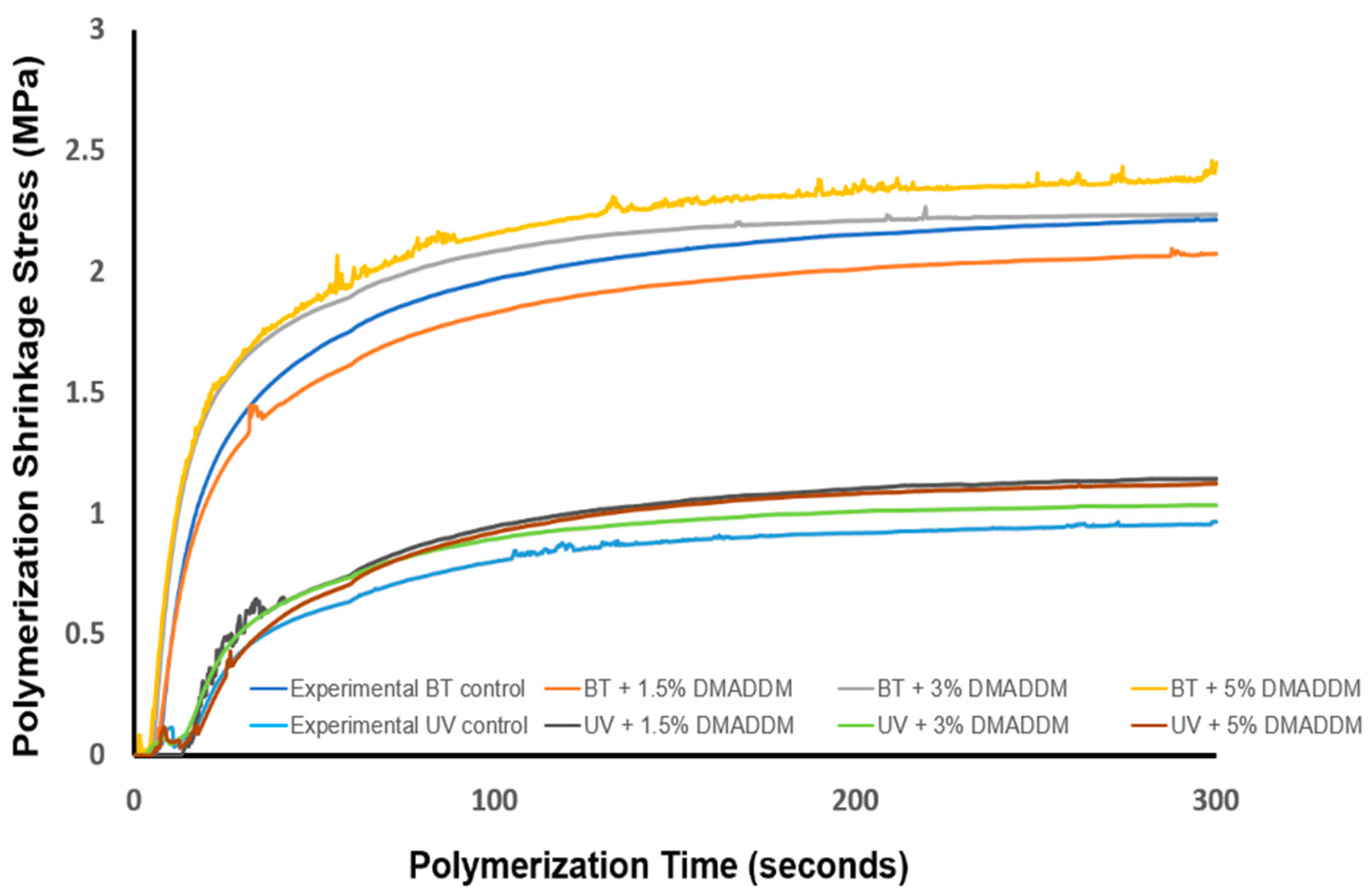
2.3. Polymerization Shrinkage Stress and Degree of Conversion

In preliminary experiments, it was determined that Heliomolar had a low degree of conversion and therefore would not be a suitable comparison for degree of conversion and polymerization stress measurements [30]. Therefore, a traditional, high-DC resin composite was used as a comparison to assess the degree of conversion and polymerization stress of the LSS resin composite. The high DC composite consisted of bisphenol A glycidyl dimethacrylate (BisGMA, Esstech) and triethylene glycol dimethacrylate (TEGDMA, Esstech), mixed at a mass ratio of 1:1 (referred to as BT resin). The chemical structure of these monomers are shown in Figure 2.
The following groups were tested for polymerization stress and degree of conversion:
(1)
30% BT + 70% glass (designated as “Experimental BT control”);
(2)
28.5% BT + 1.5% DMADDM + 70% glass (designated as “BT + 1.5% DMADDM”);
(3)
27% BT + 3% DMADDM + 70% glass (designated as “BT + 3% DMADDM”);
(4)
25% BT + 5% DMADDM + 70% glass (designated as “BT + 5% DMADDM”);
(5)
30% UV + 70% glass (designated as “Experimental UV control”);
(6)
28.5% UV + 1.5% DMADDM + 70% glass (designated as “UV + 1.5% DMADDM”);
(7)
27% UV + 3% DMADDM + 70% glass (designated as “UV + 3% DMADDM”);
(8)
25% UV + 5% DMADDM + 70% glass (designated as “UV + 5% DMADDM”).
Polymerization shrinkage stress was investigated using a cantilever beam-based tensometer [38]. This device was linked to a near-infrared (NIR) spectrometer, allowing for the simultaneous monitoring of the ongoing conversion of double bonds, as previously described [39].

2.4. Streptococcus mutans (S. mutans) Biofilm Model

2.4.1. Composite Samples for Biofilm Testing

Composite disks were fabricated (d = 9 mm, t = 2 mm) and each sample was photopolymerized for 60 s per side and stored at 37 °C for 24 h [34]. Samples were placed in distilled water and stirred for 1 h to facilitate removal of the uncured monomer [40]. Ethylene oxide (Anprolene AN 74i, Andersen, Haw River, NC, USA) was used to sterilize the resin composite disks (n = 6). Samples were de-gassed for seven days, following the manufacturer’s instructions.

2.4.2. Bacteria Inoculation and Biofilm Formation

Bacterial species were approved for use by the University of Maryland Baltimore Institutional Review Board. As a result of its association with dental caries, S. mutans (UA159) was chosen as the bacterial species for this study [41]. S. mutans was cultured overnight (16–18 h) in brain heart infusion (BHI) broth (Sigma-Aldrich, St. Louis, MO, USA) at 37 °C with 5% CO2 [34]. A spectrophotometer (Genesys 10S, Thermo Scientific, Waltham, MA) was used to adjust the inoculum to 107 colony-forming unit counts CFU/mL, based on the standard curve of OD600 nm versus the CFU/mL graph [42]. Each composite disk was set in the well of 24-well plates, covered with 1.5 mL BHI culture medium supplemented with 2% sucrose, and incubated for 24 h. Subsequently, the composite disks were transferred to 24-well plates, covered with 1.5 mL of fresh medium with sucrose, and incubated for another 24 h. According to a previous study, incubation for 48 h was adequate to form extensive biofilms on dental composites [43].

2.4.3. Live/Dead Staining of Biofilms

The biofilm-covered composite disks were washed with phosphate buffered saline (PBS) to remove planktonic bacteria. The resin composite samples were stained with the BacLight live/dead kit (Molecular Probes, Eugene, OR, USA). Each disk was incubated with 2.5 µM SYTO 9 and 2.5 µM propidium iodide for 15 min. The presence of live bacteria was stained with SYTO9 and emitted green fluorescence. Bacteria with damaged membranes were stained with propidium iodide and emitted red fluorescence. A fluorescence microscope (Eclipse TE2000-S, Nikon, Melville, NY, USA) was used to assess the biofilms on the disks [44].

2.4.4. Biofilm Colony-Forming Units Counts

Biofilm-attached disks were moved to a dish containing PBS and biofilms were harvested by a combination of scraping and sonication/vortexing [30]. The bacterial suspensions were serially diluted (101–106-fold) and spread on BHI agar plates. Agar plates were incubated for 48 h at 37 °C and 5% CO2, the colony number was counted, and the biofilm colony-forming units (CFU) counts were determined [43]. The CFU experiment was performed in triplicate.

2.4.5. Biofilm Metabolic Activity

The biofilm metabolic activity was measured by a 3-[4,5- dimethylthiazol-2-yl]-2,5- diphenyltetrazolium bromide (MTT) assay [45]. Composite samples with attached biofilms for 48 h were transferred to a 24-well plate containing 1 mL of MTT dye (0.5 mg/mL MTT in PBS) and incubated at 37 °C in 5% CO2 for 1 h [45]. Subsequently, each disk was moved into a new 24-well plate containing 1 mL DMSO in each well and incubated at room temperature in the dark for 20 min. For the absorbance calculation, 200 µL of the DMSO solution was added to a 96-well plate, and the absorbance was measured at 540 nm [45]. Higher absorbance values denote an increased biofilm metabolic activity [45]. The metabolic activity experiment was performed in triplicate.

2.4.6. Lactic Acid Production by Biofilms

The resin composite samples with attached biofilms for 48 h were transferred to 24-well plates filled with buffered peptone water (BPW, Aldrich, St. Louis, MO, USA), supplemented with 0.2% sucrose and incubated at 37 °C in 5% CO2 for 3 h [35]. The lactate dehydrogenase enzymatic was used to determine lactate concentrations in BPW thru measuring optical density at 340 nm using a microplate reader (Spectra-Max M5) as previously described [40]. The lactic acid production experiment was performed in triplicate.

2.4.7. Scanning Electron Microscopy (SEM)

The composites were investigated in SEM (Quanta 200, FEI Company, Hillsboro, OR, USA). The specimens were polished with 4000 grit sandpaper, followed by sputter-coating with gold (n = 6).
For SEM examination of biofilms, the biofilms on the composite disks (n = 6) at 48 h were cleaned with PBS and then soaked in 1% glutaraldehyde at 4 °C overnight. Then they were washed with PBS and subjected to dehydration using a sequence of ethanol solutions. They were then washed with hexamethyldisilazane and allowed to air-dry overnight. The samples were sputter-coated with platinum and examined in SEM.

2.5. Statistical Analysis

Statistical analyses were performed using Sigma Plot (SYSTAT, Chicago, IL, USA). One-way analyses of variance (ANOVA) and Tukey’s comparison tests were performed to detect the significant differences between groups. Results were considered statistically significant at a p-value of less than 0.05.

3. Results

3.1. Mechanical Properties

Flexural strength and elastic modulus of the composites are shown in Figure 3  ( mean   ±   sd ;   n   = 6 ) . The addition of 1.5%, 3%, and 5% DMADDM in LSS resin composite resulted in an increase in flexural strength compared to the commercial group (p < 0.05). The experimental group and 3% DMADDM composite had the highest flexural strength among experimental groups and was significantly higher than the commercial control. However, they demonstrated similar values compared to other groups (p > 0.05). This suggests that incorporating up to 5% of DMADDM in LSS composite had minimal effect on the flexural strength.
The elastic modulus values of the commercial control group were significantly greater than all other experimental groups (p < 0.05). Increasing the DMADDM mass fraction in LSS resin composite increased the elastic modulus compared to the experimental group (p < 0.05).

3.2. Polymerization Shrinkage Stress and Degree of Conversion

Degree of conversion results are shown in Figure 4 (n = 3). The degree of conversion of all of the groups gradually increased with time until they reached the plateau. The result showed that UV resin takes a long time to reach its maximum compared to BT resin. The UV-containing composites with up to 3% DMADDM reaches a degree of conversion of approximately 67%. However, the increase of DMADDM to 5% in UV resin decreased the degree of conversion to 62%. Furthermore, our findings demonstrated that there is no significant difference in the degree of conversion after incorporating 3% DMADDM into UV resin and BT resin (p < 0.05). The polymerization shrinkage stress is plotted in Figure 5 (n = 3). The BT resin composite groups showed greater polymerization shrinkage stress than UV resin (p < 0.05). The addition of DMADDM up to 5% in UV-containing composites demonstrated comparable polymerization shrinkage stress ( 1.12   ±   0.06   MPa ) to experimental UV resin ( 0.97   ±   0.05   MPa ) (p > 0.05). In contrast, the incorporation of DMADDM up to 5% in BT resin resulted in increasing polymerization shrinkage stress ( 2.45   ±   0.14   MPa   ) compared to experimental BT resin ( 2.22   ±   0.12   MPa   ) (p > 0.05).

3.3. Live/dead Staining of S. mutans Biofilms

Live/dead images of 48 h biofilms on the composites are illustrated in Figure 6. The surface of control groups had biofilms containing primarily live bacteria. The incorporation of DMADDM in the UV resin composite effectively inhibited biofilm growth, as demonstrated by the increased red staining observed with higher DMADDM mass fractions in the composite.

3.4. Biofilm Colony-Forming Units Counts

The CFU results of the S. mutans biofilm are shown in Figure 7  ( mean   ±   sd ;   n = 6 ) . Incorporating 1.5% DMADDM in LSS resin composite significantly decreased the CFUs for S. mutans compared to control groups (p < 0.05). Conversely, increasing the DMADDM mass fraction to 3% and 5% in UV resin composite showed more reduction in the CFU count by 6–6.5 log compared to the commercial group (p < 0.05). However, there was no significant difference in reducing the CFU count among these groups (p > 0.05).

3.5. MTT Assay of Metabolic Activity of S. mutans Biofilms

The metabolic activity of 48 h biofilms on the composites are illustrated in Figure 8. Incorporating 1.5%, 3%, and 5% of DMADDM in UV resin significantly decreased the metabolic activities compared to the control groups (p < 0.05). However, there was no significant difference in the metabolic activities among these groups (p > 0.05).

3.6. Lactic Acid Production by S. mutans Biofilms

Lactic acid production of S. mutans biofilms adherent on the resin composites is depicted in Figure 9  mean   ±   sd ;   n = 6 . The control groups had the highest acid production (p < 0.5). Incorporating 3% and 5% DMADDM in UV resin composite resulted in a significant reduction of acid concentration compared to the commercial group (p < 0.05). Nevertheless, there was no significant difference in acid production among these groups (p > 0.05). The incorporation of 1.5% DMADDM showed less lactic acid than commercial groups (p < 0.05). However, it has higher lactic acid production than the 3% and 5% DMADDM groups (p < 0.05). This indicates that increasing DMADDM concentration to 3% and 5% resulted in the highest biofilm reduction.

3.7. SEM Examination of Composites and Biofilms

Representative SEM images of composites are shown in Figure 10 (n = 6). All experimental composites had a satisfactory distributions of filler particles with a few small voids, similar to the commercial control.
Representative SEM images of two-day S. mutans biofilms on composites are shown in Figure 11 (n = 6). The experimental control composite, the commercial control composite, and the 1.5% DMADDM composite exhibited substantial biofilm formation. In contrast, adding 3% and 5% DMADDM into the LSS composite led to minimal biofilm formation on the composites.

4. Discussion

This study explored the effect of the antimicrobial quaternary ammonium resin, DMADDM, in a (UDMA + TEG-DBVE) LSS composite. Incorporating of DMADDM in a LSS resin composite may reduce polymerization shrinkage stress and secondary caries, which could increase the clinical longevity of resin composite restoration [30,31,32]. Inhibition of S. mutans biofilm and reduction of polymerization shrinkage stress while maintaining excellent mechanical characteristics was accomplished, and the study hypotheses were proved. The adding of 3% DMADDM in low-shrinkage composite generated a 6-log reduction in S. mutans biofilm growth while maintaining clinically acceptable mechanical characteristics. Moreover, as the mass fraction of DMADDM increased, the production of lactic acid and metabolic activity within the biofilm of S. mutans decreased.
There were several successful endeavors to improve the mechanical characteristics and aesthetics of the composite resin restoration [46,47]. However, the most significant drawbacks of resin composite are polymerization shrinkage volume, and the resulting shrinkage stress that occurs during the polymerization process [6,7,8,9]. Polymerization shrinkage stress can potentially result in debonding at the interface between tooth structure and restoration, resulting in micro-cracking of the tooth structure and gap formation [3]. Therefore, developing a low shrinkage stress composite is highly desirable, which could help reduce stresses on the tooth structure and increase the survival rate of the restoration [31]. In a previous study, an attempt was made to minimize polymerization stress by using a thiolene system, which reduced stress development in a Bis-GMA/TEGDMA composite resin from 2.8 MPa to 0.30 MPa [48]. However, incorporating the thiolene system in resin composite compromised mechanical properties [48]. Another attempt to minimize polymerization stress by incorporating tricyclo decanedimethanol diacrylate (SR833s) and isobornyl acrylate (IBOA) as a diluent monomer, resulting in reducing stress development [49]. However, this approach compromised the degree of conversion [49]. In addition, a resin composite that is based on silorane was investigated in a long-term 12 month study, which found the marginal retention was not significantly improved compared with traditional methacrylate’s-based composite resins [50]. Recently, a LSS composite was developed utilizing TEGDVBE and urethane dimethacrylate monomers, which resulted in reduced polymerization stress, good mechanical strength, and a high degree of conversion [30]. Furthermore, several attempts were made to incorporate DMAHDM in LSS resin to develop a bioactive composite resin [34,43]. However, the DMAHDM has a long chain length, which increases the resin composite’s viscosity and could reduce the filler load and degree of conversion. Also, several articles showed inconsistent antibacterial results of incorporating DMAHDM in resin composite, which could be due to the long chain length of DMAHDM being present in conformations that result in a reduction in quaternary ammonium charge density at the surface [22,23,34]. However, no report has studied the impact of integrating different mass fractions of DMADDM in a LSS composite resin.
This study thoroughly investigated the effects of incorporating different amounts of DMADDM in a composite containing the low shrinkage stress resin TEGDVBE. The primary objective of this study was to determine the optimal concentration of DMADDM incorporated into the composite that maximized the antimicrobial response while maintaining clinically-desirable mechanical properties. The incorporation of DMADDM mass fractions up to 5% achieved higher flexural strength than the commercial control and was comparable to the experimental control. On the other hand, LSS composite resin with and without incorporation of DMADDM showed a lower modulus of elasticity than commercial control composite resin. It is likely that the higher elastic modulus for the commercial control composite was due to the differences in the resin matrix when compared to the experimental LSS composites. The LSS resin composite undergoes a slower or more regulated polymerization process to facilitate stress reduction, which could potentially result in a reduced elastic modulus. In addition, it is possible that the elastic modulus increase with increasing DMADDM concentration could be due to a higher viscosity of the resin matrix from DMADDM addition. The mechanical properties of composites can be adversely affected by the poor distribution of fillers and the presence of large voids or agglomerates. In the present study, SEM analysis of composites revealed a relatively uniform distribution of filler particles within the resin matrix for all DMADDM composites, with a few small voids and the absence of large agglomerates or large flaws. These features are similar to those of the commercial control. Maintaining an excellent mechanical property with enhancing the marginal seal of the dental restoration could help improves longevity [31,32]. Previous studies showed that polymerization shrinkage stress affects the sealing ability of resin composite [31]. Therefore, in this study, we developed a bioactive low-shrinkage-stress resin composite, which demonstrated a lower polymerization shrinkage stress than the BT composite, which may help achieve a better marginal seal. The incorporation of up to 5% DMADDM in UV resin composite reduced polymerization shrinkage stress at 1.12 MPa compared to 2.45 MPa for BT resin with 5% DMADDM. The difference in polymerization shrinkage stress may be due to resin monomers’ polymerization rate [51]. The UV resin takes a longer time to reach the gel point, allowing for stress relaxation and preventing excessive contraction stress development [17]. Creating novel bioactive LSS resin composites with antibacterial properties could help suppress acidogenic bacteria and provide a better marginal seal, which may help in improving the longevity of dental restoration. Maximizing the degree of conversion is essential in improving the durability and longevity of dental composites [52]. The degree of conversion of most dimethacrylate-based composites ranges from 55% to 75% [53]. In the current study, a comparatively high degree of conversion was achieved for both the UV resin composite, ranging from 62% to 67%, and the BT resin composite, ranging from 65% to 69%. Incorporating 5% DMADDM in UV and BT resin composite reduced the degree of conversion, which could be due to an increase in monomer viscosity with increasing DMADDM concentration. Future studies are necessary to fully understand the reaction kinetics for this new resin composite.
Dental plaque could affect the longevity of dental restoration, resulting in cariogenic bacterial attachment, hydrolytic enzyme production, material degradation, and recurrent caries [54,55,56]. Therefore, developing an innovative bioactive material with antibacterial properties could increase the durability of the composite restoration. In a previous study, chlorhexidine was incorporated as an antibacterial agent in a low-shrinkage composite, resulting in a short-term antibacterial effect [57]. Another attempt investigated incorporating antibacterial fillers silver and zinc oxide nanoparticles in dental materials [58]. These nanoparticle fillers tended to be lost from the dental material surface in a short period, which led to increased surface porosity and compromised mechanical properties [58]. A new approach was developed to overcome this critical drawback by using QAMs, which could be copolymerized with dental resins by covalent bonding, providing long-term antibacterial effects [59]. Incorporating QAMs in methacrylate-based materials showed a potent antimicrobial effect through a contact-killing mechanism [25]. The QAMs have positively charged quaternary amine, which can bind to the negatively charged bacterial cell membrane, changing the essential ion balance and cause cytoplasmic leakage, leading to disruption of the bacterial membrane [25]. Previous studies showed that increased chain length of QAMs resulted in more potent antibacterial properties due to increased hydrophobicity, which may improve their efficacy in disrupting the hydrophobic bacterial cell membrane [20,21]. Furthermore, the incorporation of DMADDM in resin-based material achieved a durable antibacterial activity even after six months of water aging, with sustained antibacterial function and mechanical properties [60]. In this study, the incorporation of DMADDM into a LSS resin composite showed a potent antibacterial effect against S. mutans biofilm. The incorporation of 3% and 5% DMADDM into LSS resin composite significantly reduced the CFU count by 6 and 6.5 logs, respectively, compared to the commercial control (p < 0.05). In addition, our results showed that incorporating 3% and 5% of DMADDM in LSS resin composite achieved more than 85% reduction in lactic acid production and metabolic activities compared to the commercial control. Furthermore, live/dead staining images of S. mutans biofilms confirm the presence of living bacteria on the surface of experimental and commercial control groups. On the other hand, increasing DMADDM concentration in LSS composites reduces the live bacteria present on the surface. Moreover, SEM examination of S. mutans biofilms showed substantial biofilm growth on the experimental control composite, the commercial control composite, and the 1.5% DMADDM composite. In contrast, adding 3% and 5% DMADDM led to minimal biofilm growth on the LSS composite. However, our results observed no significant difference in antibacterial effect when incorporating 3% or 5% DMADDM against S. mutans biofilms (p > 0.05).
Regarding biocompatibility, a previous study showed that the incorporation of 10% DMADDM into a Scotchbond multi-purpose adhesive achieved excellent cell viability that matched that of the control group [21]. Another in vivo investigation showed that the incorporation of 5% DMADDM into the adhesive and composite showed biocompatibility matching the control group [61]. Moreover, another study investigated the cell viability of human gingival fibroblast cells of uncured traditional monomers and the new LSS monomers [30]. That study found no significant differences in cell viability and cytotoxicity between the LSS monomers and the control monomers already used clinically [30]. Therefore, the DMADDM composite has the capability to inhibit bacterial biofilms without compromising biocompatibility.
In summary, the present study systematically examined the effect of incorporating different concentrations of DMADDM on the mechanical and antibacterial properties of LSS resin composite for the first time. Incorporating 3% DMADDM into the LSS composite resulted in strong antibacterial properties without compromising mechanical properties, with comparable results to the LSS resin composite with 5% DMADDM. The incorporation of 3% DMADDM reduced biofilm CFU counts by six orders of magnitude compared to commercial resin composite. Therefore, developing an antibacterial LSS resin composite is a promising approach to overcoming secondary caries and increasing the clinical longevity of the resin composite restoration.
Additional studies are needed to explore the antibacterial property of LSS resin composite on multi-species biofilms that are more clinically relevant. Also, further studies are required to explore the long-term effect of DMADDM on the antibacterial and mechanical characteristics of LSS resin composites. In addition, future research is needed to investigate the thermal stability and long-term degradation behavior of the novel LSS composites.

5. Conclusions

This study developed a novel antibacterial low-shrinkage-resin composite. Incorporating 3% DMADDM into the resin composite provided a strong antibacterial effect against S. mutans biofilms which is commonly associated with secondary caries, without compromising the mechanical properties. Incorporating 3% DMADDM into this novel dental composite reduced the polymerization stress without negatively impacting the degree of conversion. In addition, this formulation achieved a 6-log reduction in biofilm CFUs with a significant decrease in lactic acid production and metabolic activity. These results indicate that a dental resin composite system composed of the low shrinkage stress resin TEGDVBE combined with the antimicrobial resin DMADDM may be a promising bioactive dental restoration that could reduce the onset of secondary caries.

Author Contributions

A.A. contributed to data curation, investigation, writing—original draft; R.A. contributed to data curation, writing—original draft; X.W. contributed to data curation; H.M. contributed to data curation; H.F. contributed to data curation; G.D.H. contributed to data curation; T.W.O. contributed to resources, Funding acquisition; J.S. contributed to supervision, resources, writing—review and editing; M.D.W. contributed to supervision, resources, funding acquisition, writing—review and editing; H.H.K.X. contributed to methodology, project administration, supervision, resources, writing—review and editing. All authors have read and agreed to the published version of the manuscript.

Funding

This research received no external funding.

Institutional Review Board Statement

Not applicable.

Informed Consent Statement

Not applicable.

Data Availability Statement

Not applicable.

Acknowledgments

We thank Dentsply Sirona (Milford, DE) for the donation of the glass fillers. This work was supported by the University of Maryland School of Dentistry bridge fund (HX) and the University of Maryland seed grant (HX).

Conflicts of Interest

The authors declare no conflict of interest.

References

  1. Bayne, S.C.; Ferracane, J.L.; Marshall, G.W.; Marshall, S.J.; van Noort, R. The Evolution of Dental Materials over the Past Century: Silver and Gold to Tooth Color and Beyond. J. Dent. Res. 2019, 98, 257–265. [Google Scholar] [CrossRef]
  2. Demarco, F.F.; Corrêa, M.B.; Cenci, M.S.; Moraes, R.R.; Opdam, N.J.M. Longevity of posterior composite restorations: Not only a matter of materials. Dent. Mater. 2012, 28, 87–101. [Google Scholar] [CrossRef]
  3. Braga, R.; Ballester, R.; Ferracane, J. Factors involved in the development of polymerization shrinkage stress in resin-composites: A systematic review. Dent. Mater. 2005, 21, 962–970. [Google Scholar] [CrossRef]
  4. Soares, C.J.; Faria-E-Silva, A.L.; Rodrigues, M.D.P.; Vilela, A.B.F.; Pfeifer, C.S.; Tantbirojn, D.; Versluis, A. Polymerization shrinkage stress of composite resins and resin cements–What do we need to know? Braz. Oral Res. 2017, 31, e62. [Google Scholar] [CrossRef]
  5. Pereira, R.; Valdívia, A.; Bicalho, A.; Franco, S.; Tantbirojn, D.; Versluis, A.; Soares, C. Effect of Photoactivation Timing on the Mechanical Properties of Resin Cements and Bond Strength of Fiberglass Post to Root Dentin. Oper. Dent. 2015, 40, E206–E221. [Google Scholar] [CrossRef]
  6. Bicalho, A.; Pereira, R.; Zanatta, R.; Franco, S.; Tantbirojn, D.; Versluis, A.; Soares, C. Incremental Filling Technique and Composite Material—Part I: Cuspal Deformation, Bond Strength, and Physical Properties. Oper. Dent. 2014, 39, e71–e82. [Google Scholar] [CrossRef] [PubMed]
  7. Bicalho, A.; Valdívia, A.; Barreto, B.; Tantbirojn, D.; Versluis, A.; Soares, C. Incremental Filling Technique and Composite Material—Part II: Shrinkage and Shrinkage Stresses. Oper. Dent. 2014, 39, e83–e92. [Google Scholar] [CrossRef] [PubMed]
  8. Rosatto, C.; Bicalho, A.; Veríssimo, C.; Bragança, G.; Rodrigues, M.; Tantbirojn, D.; Versluis, A.; Soares, C. Mechanical properties, shrinkage stress, cuspal strain and fracture resistance of molars restored with bulk-fill composites and incremental filling technique. J. Dent. 2015, 43, 1519–1528. [Google Scholar] [CrossRef]
  9. Soares, C.; Bicalho, A.; Verissimo, C.; Soares, P.; Tantbirojn, D.; Versluis, A. Delayed Photo-activation Effects on Mechanical Properties of Dual Cured Resin Cements and Finite Element Analysis of Shrinkage Stresses in Teeth Restored With Ceramic Inlays. Oper. Dent. 2016, 41, 491–500. [Google Scholar] [CrossRef] [PubMed]
  10. Balhaddad, A.; Garcia, I.; Mokeem, L.; Ibrahim, M.; Collares, F.; Weir, M.; Xu, H.; Melo, M. Bifunctional Composites for Biofilms Modulation on Cervical Restorations. J. Dent. Res. 2021, 100, 1063–1071. [Google Scholar] [CrossRef]
  11. Moraes, R.R.; Garcia, J.W.; Barros, M.D.; Lewis, S.H.; Pfeifer, C.S.; Liu, J.; Stansbury, J.W. Control of polymerization shrinkage and stress in nanogel-modified monomer and composite materials. Dent. Mater. 2011, 27, 509–519. [Google Scholar] [CrossRef]
  12. Shah, P.K.; Stansbury, J.W. Photopolymerization shrinkage-stress reduction in polymer-based dental restoratives by surface modification of fillers. Dent. Mater. 2021, 37, 578–587. [Google Scholar] [CrossRef] [PubMed]
  13. Hsu, S.-H.; Chen, R.-S.; Chang, Y.-L.; Chen, M.-H.; Cheng, K.-C.; Su, W.-F. Biphenyl liquid crystalline epoxy resin as a low-shrinkage resin-based dental restorative nanocomposite. Acta Biomater. 2012, 8, 4151–4161. [Google Scholar] [CrossRef] [PubMed]
  14. Arrais, C.A.G.; De Oliveira, M.T.; Mettenburg, D.; Rueggeberg, F.; Giannini, M. Silorane- and high filled-based"low-shrinkage" resin composites: Shrinkage, flexural strength and modulus. Braz. Oral Res. 2013, 27, 97–102. [Google Scholar] [CrossRef]
  15. Lu, H.; Carioscia, J.A.; Stansbury, J.W.; Bowman, C.N. Investigations of step-growth thiol-ene polymerizations for novel dental restoratives. Dent. Mater. 2005, 21, 1129–1136. [Google Scholar] [CrossRef] [PubMed]
  16. Yang, Y.; Urbas, A.; Gonzalez-Bonet, A.; Sheridan, R.J.; Seppala, J.E.; Beers, K.L.; Sun, J. A composition-controlled cross-linking resin network through rapid visible-light photo-copolymerization. Polym. Chem. 2016, 7, 5023–5030. [Google Scholar] [CrossRef]
  17. Wang, X.; Huyang, G.; Palagummi, S.V.; Liu, X.; Skrtic, D.; Beauchamp, C.; Bowen, R.; Sun, J. High performance dental resin composites with hydrolytically stable monomers. Dent. Mater. 2018, 34, 228–237. [Google Scholar] [CrossRef]
  18. Yamauchi, S.; Wang, X.; Egusa, H.; Sun, J. High-Performance Dental Adhesives Containing an Ether-Based Monomer. J. Dent. Res. 2020, 99, 189–195. [Google Scholar] [CrossRef] [PubMed]
  19. Cheng, L.; Zhang, K.; Zhang, N.; Melo, M.; Weir, M.; Zhou, X.; Bai, Y.; Reynolds, M.; Xu, H. Developing a New Generation of Antimicrobial and Bioactive Dental Resins. J. Dent. Res. 2017, 96, 855–863. [Google Scholar] [CrossRef]
  20. Han, Q.; Li, B.; Zhou, X.; Ge, Y.; Wang, S.; Li, M.; Ren, B.; Wang, H.; Zhang, K.; Xu, H.H.K.; et al. Anti-Caries Effects of Dental Adhesives Containing Quaternary Ammonium Methacrylates with Different Chain Lengths. Materials 2017, 10, 643. [Google Scholar] [CrossRef]
  21. Li, F.; Weir, M.D.; Xu, H.H.K. Effects of quaternary ammonium chain length on antibacterial bonding agents. J. Dent. Res. 2013, 92, 932–938. [Google Scholar] [CrossRef] [PubMed]
  22. de Oliveira, F.J.D.; Filho, P.S.F.d.S.; Costa, M.J.F.; Caldas, M.R.G.R.; Borges, B.C.D.; de Araújo, D.F.G. A comprehensive review of the antibacterial activity of dimethylaminohexadecyl methacrylate (DMAHDM) and its influence on mechanical properties of resin-based dental materials. Jpn. Dent. Sci. Rev. 2021, 57, 60–70. [Google Scholar] [CrossRef] [PubMed]
  23. Balhaddad, A.A.; Ibrahim, M.S.; Weir, M.D.; Xu, H.H.; Melo, M.A.S. Concentration dependence of quaternary ammonium monomer on the design of high-performance bioactive composite for root caries restorations. Dent. Mater. 2020, 36, e266–e278. [Google Scholar] [CrossRef] [PubMed]
  24. Bhadila, G.; Menon, D.; Wang, X.; Vila, T.; Melo, M.A.S.; Montaner, S.; Arola, D.D.; Weir, M.D.; Sun, J.K.; Xu, H.H.; et al. Long-term antibacterial activity and cytocompatibility of novel low-shrinkage-stress, remineralizing composites. J. Biomater. Sci. Polym. Ed. 2021, 32, 886–905. [Google Scholar] [CrossRef]
  25. Namba, N.; Yoshida, Y.; Nagaoka, N.; Takashima, S.; Matsuura-Yoshimoto, K.; Maeda, H.; Van Meerbeek, B.; Suzuki, K.; Takashiba, S. Antibacterial effect of bactericide immobilized in resin matrix. Dent. Mater. 2009, 25, 424–430. [Google Scholar] [CrossRef]
  26. Cheng, L.; Weir, M.D.; Zhang, K.; Arola, D.D.; Zhou, X.; Xu, H.H. Dental primer and adhesive containing a new antibacterial quaternary ammonium monomer dimethylaminododecyl methacrylate. J. Dent. 2013, 41, 345–355. [Google Scholar] [CrossRef]
  27. Zhou, C.; Weir, M.D.; Zhang, K.; Deng, D.; Cheng, L.; Xu, H.H. Synthesis of new antibacterial quaternary ammonium monomer for incorporation into CaP nanocomposite. Dent. Mater. 2013, 29, 859–870. [Google Scholar] [CrossRef]
  28. Zhang, K.; Ren, B.; Zhou, X.; Xu, H.H.K.; Chen, Y.; Han, Q.; Li, B.; Weir, M.D.; Li, M.; Feng, M.; et al. Effect of Antimicrobial Denture Base Resin on Multi-Species Biofilm Formation. Int. J. Mol. Sci. 2016, 17, 1033. [Google Scholar] [CrossRef]
  29. Li, B.; Ge, Y.; Wu, Y.; Chen, J.; Xu, H.H.K.; Yang, M.; Li, M.; Ren, B.; Feng, M.; Weir, M.D.; et al. Anti-Bacterial and Microecosystem-Regulating Effects of Dental Implant Coated with Dimethylaminododecyl Methacrylate. Molecules 2017, 22, 2013. [Google Scholar] [CrossRef]
  30. Bhadila, G.; Wang, X.; Zhou, W.; Menon, D.; Melo, M.A.S.; Montaner, S.; Oates, T.W.; Weir, M.D.; Sun, J.; Xu, H.H. Novel low-shrinkage-stress nanocomposite with remineralization and antibacterial abilities to protect marginal enamel under biofilm. J. Dent. 2020, 99, 103406. [Google Scholar] [CrossRef]
  31. Ferracane, J.L. Developing a more complete understanding of stresses produced in dental composites during polymerization. Dent. Mater. 2005, 21, 36–42. [Google Scholar] [CrossRef] [PubMed]
  32. Filemban, H.; Bhadila, G.; Wang, X.; Melo, M.A.S.; Oates, T.W.; Weir, M.D.; Sun, J.; Xu, H.H. Novel low-shrinkage-stress bioactive nanocomposite with anti-biofilm and remineralization capabilities to inhibit caries. J. Dent. Sci. 2021, 17, 811–821. [Google Scholar] [CrossRef] [PubMed]
  33. Chen, C.; Weir, M.D.; Cheng, L.; Lin, N.J.; Lin-Gibson, S.; Chow, L.C.; Zhou, X.; Xu, H.H. Antibacterial activity and ion release of bonding agent containing amorphous calcium phosphate nanoparticles. Dent. Mater. 2014, 30, 891–901. [Google Scholar] [CrossRef] [PubMed]
  34. Bhadila, G.; Wang, X.; Weir, M.D.; Melo, M.A.S.; Martinho, F.; Fay, G.G.; Oates, T.W.; Sun, J.; Xu, H.H.K. Low-shrinkage-stress nanocomposite: An insight into shrinkage stress, antibacterial, and ion release properties. J. Biomed. Mater. Res. Part B Appl. Biomater. 2021, 109, 1124–1134. [Google Scholar] [CrossRef] [PubMed]
  35. Antonucci, J.M.; Zeiger, D.N.; Tang, K.; Lin-Gibson, S.; Fowler, B.O.; Lin, N.J. Synthesis and characterization of dimethacrylates containing quaternary ammonium functionalities for dental applications. Dent. Mater. 2012, 28, 219–228. [Google Scholar] [CrossRef]
  36. Cheng, L.; Weir, M.D.; Xu, H.H.; Antonucci, J.M.; Kraigsley, A.M.; Lin, N.J.; Lin-Gibson, S.; Zhou, X. Antibacterial amorphous calcium phosphate nanocomposites with a quaternary ammonium dimethacrylate and silver nanoparticles. Dent. Mater. 2012, 28, 561–572. [Google Scholar] [CrossRef]
  37. Xu, H.H.; Moreau, J.L.; Sun, L.; Chow, L.C. Nanocomposite containing amorphous calcium phosphate nanoparticles for caries inhibition. Dent. Mater. 2011, 27, 762–769. [Google Scholar] [CrossRef]
  38. Chiang, M.Y.; Giuseppetti, A.A.; Qian, J.; Dunkers, J.P.; Antonucci, J.M.; Schumacher, G.E.; Gibson, S.-L. Analyses of a cantilever-beam based instrument for evaluating the development of polymerization stresses. Dent. Mater. 2011, 27, 899–905. [Google Scholar] [CrossRef]
  39. Wang, Z.; Landis, F.A.; Giuseppetti, A.A.; Lin-Gibson, S.; Chiang, M.Y. Simultaneous measurement of polymerization stress and curing kinetics for photo-polymerized composites with high filler contents. Dent. Mater. 2014, 30, 1316–1324. [Google Scholar] [CrossRef]
  40. Bhadila, G.; Baras, B.H.; Weir, M.D.; Wang, H.; Melo, M.A.S.; Hack, G.D.; Bai, Y.; Xu, H.H.K. Novel antibacterial calcium phosphate nanocomposite with long-term ion recharge and re-release to inhibit caries. Dent. Mater. J. 2020, 39, 678–689. [Google Scholar] [CrossRef]
  41. Wang, L.; Xie, X.; Qi, M.; Weir, M.D.; Reynolds, M.A.; Li, C.; Zhou, C.; Xu, H.H. Effects of single species versus multispecies periodontal biofilms on the antibacterial efficacy of a novel bioactive Class-V nanocomposite. Dent. Mater. 2019, 35, 847–861. [Google Scholar] [CrossRef] [PubMed]
  42. Wang, S.; Zhou, C.; Ren, B.; Li, X.; Weir, M.D.; Masri, R.M.; Oates, T.W.; Cheng, L.; Xu, H.K.H. Formation of persisters in Streptococcus mutans biofilms induced by antibacterial dental monomer. J. Mater. Sci. Mater. Med. 2017, 28, 178. [Google Scholar] [CrossRef] [PubMed]
  43. Bhadila, G.; Filemban, H.; Wang, X.; Melo, M.A.S.; Arola, D.D.; Tay, F.R.; Oates, T.W.; Weir, M.D.; Sun, J.; Xu, H.H. Bioactive low-shrinkage-stress nanocomposite suppresses S. mutans biofilm and preserves tooth dentin hardness. Acta Biomater. 2020, 114, 146–157. [Google Scholar] [CrossRef] [PubMed]
  44. Mitwalli, H.; Balhaddad, A.A.; AlSahafi, R.; Oates, T.W.; Melo, M.A.S.; Xu, H.H.K.; Weir, M.D. Novel CaF2 Nanocomposites with Antibacterial Function and Fluoride and Calcium Ion Release to Inhibit Oral Biofilm and Protect Teeth. J. Funct. Biomater. 2020, 11, 56. [Google Scholar] [CrossRef] [PubMed]
  45. Al-Dulaijan, Y.A.; Cheng, L.; Weir, M.D.; Melo, M.A.S.; Liu, H.; Oates, T.W.; Wang, L.; Xu, H.H. Novel rechargeable calcium phosphate nanocomposite with antibacterial activity to suppress biofilm acids and dental caries. J. Dent. 2018, 72, 44–52. [Google Scholar] [CrossRef]
  46. Beyth, N.; Domb, A.J.; Weiss, E.I. An in vitro quantitative antibacterial analysis of amalgam and composite resins. J. Dent. 2007, 35, 201–206. [Google Scholar] [CrossRef]
  47. Beyth, N.; Yudovin-Farber, I.; Bahir, R.; Domb, A.J.; Weiss, E.I. Antibacterial activity of dental composites containing quaternary ammonium polyethylenimine nanoparticles against Streptococcus mutans. Biomaterials 2006, 27, 3995–4002. [Google Scholar] [CrossRef]
  48. Carioscia, J.A.; Lu, H.; Stanbury, J.W.; Bowman, C.N. Thiol-ene oligomers as dental restorative materials. Dent. Mater. 2005, 21, 1137–1143. [Google Scholar] [CrossRef]
  49. He, J.; Cai, L.; Chen, Y.; He, Y.; Wang, M.; Tang, J.; Guan, H.; Wang, J.; Peng, X. Antitumor and radiosensitizing effects of SKLB-163, a novel benzothiazole-2-thiol derivative, on nasopharyngeal carcinoma by affecting the RhoGDI/JNK-1 signaling pathway. Radiother. Oncol. 2018, 129, 30–37. [Google Scholar] [CrossRef]
  50. Kruly, P.; Giannini, M.; Pascotto, R.C.; Tokubo, L.M.; Suga, U.S.G.; Marques, A.D.C.R.; Terada, R.S.S. Meta-analysis of the clinical behavior of posterior direct resin restorations: Low polymerization shrinkage resin in comparison to methacrylate composite resin. PLoS ONE 2018, 13, e0191942. [Google Scholar] [CrossRef]
  51. Imazato, S.; Ebi, N.; Takahashi, Y.; Kaneko, T.; Ebisu, S.; Russell, R.R.B. Antibacterial activity of bactericide-immobilized filler for resin-based restoratives. Biomaterials 2003, 24, 3605–3609. [Google Scholar] [CrossRef]
  52. Braga, R.R.; Ferracane, J.L. Contraction stress related to degree of conversion and reaction kinetics. J. Dent. Res. 2002, 81, 114–118. [Google Scholar] [CrossRef] [PubMed]
  53. Lovell, L.G.; Lu, H.; Elliott, J.E.; Stansbury, J.W.; Bowman, C.N. The effect of cure rate on the mechanical properties of dental resins. Dent. Mater. 2001, 17, 504–511. [Google Scholar] [CrossRef]
  54. Hansel, C.; Leyhausen, G.; Mai, U.; Geurtsen, W. Effects of Various Resin Composite (Co)monomers and Extracts on Two Caries-associated Micro-organisms in vitro. J. Dent. Res. 1998, 77, 60–67. [Google Scholar] [CrossRef]
  55. Khalichi, P.; Cvitkovitch, D.; Santerre, J. Effect of composite resin biodegradation products on oral streptococcal growth. Biomaterials 2004, 25, 5467–5472. [Google Scholar] [CrossRef]
  56. Barbosa, R.P.D.S.; Pereira-Cenci, T.; da Silva, W.M.; Coelho-De-Souza, F.H.; Demarco, F.F.; Cenci, M.S. Effect of cariogenic biofilm challenge on the surface hardness of direct restorative materials in situ. J. Dent. 2012, 40, 359–363. [Google Scholar] [CrossRef] [PubMed]
  57. Aljabo, A.; Xia, W.; Liaqat, S.; Khan, M.A.; Knowles, J.C.; Ashley, P.; Young, A.M. Conversion, shrinkage, water sorption, flexural strength and modulus of re-mineralizing dental composites. Dent. Mater. 2015, 31, 1279–1289. [Google Scholar] [CrossRef]
  58. Melo, M.A.; Guedes, S.F.; Xu, H.H.; Rodrigues, L.K. Nanotechnology-based restorative materials for dental caries management. Trends Biotechnol. 2013, 31, 459–467. [Google Scholar] [CrossRef] [PubMed]
  59. Wang, H.; Wang, S.; Cheng, L.; Jiang, Y.; Melo, M.A.S.; Weir, M.D.; Oates, T.W.; Zhou, X.; Xu, H.H. Novel dental composite with capability to suppress cariogenic species and promote non-cariogenic species in oral biofilms. Mater. Sci. Eng. C 2019, 94, 587–596. [Google Scholar] [CrossRef]
  60. Zhang, K.; Cheng, L.; Wu, E.J.; Weir, M.D.; Bai, Y.; Xu, H.H. Effect of water-ageing on dentine bond strength and anti-biofilm activity of bonding agent containing new monomer dimethylaminododecyl methacrylate. J. Dent. 2013, 41, 504–513. [Google Scholar] [CrossRef]
  61. Li, F.; Wang, P.; Weir, M.D.; Fouad, A.F.; Xu, H.H. Evaluation of antibacterial and remineralizing nanocomposite and adhesive in rat tooth cavity model. Acta Biomater. 2014, 10, 2804–2813. [Google Scholar] [CrossRef] [PubMed]
Figure 1. Schematic of the study design. The figure shows the optimization of antibacterial composites that incorporate varying mass fractions of DMADDM. Furthermore, the figure highlights the evaluation of both microbiological analysis and mechanical properties. Created via BioRender.com on 4 June 2023.
Jfb 14 00335 g001
Figure 2. Chemical structure of monomers used in the resins.
Jfb 14 00335 g002
Figure 3. Mechanical properties of tested composites (A) Flexural strength and (B) elastic modulus ( mean   ±   sd ;   n   = 6 ) . Incorporating 5% DMADDM in a LSS resin composite significantly increased flexural strength compared to commercial group (p < 0.05). The increase of DMADDM concentration by up to 5% increased the elastic of modules for UV resin compared to the experimental control (p < 0.05). LSS resin composite demonstrated significantly lower elastic of modules than commercial group (p < 0.05). Dissimilar letters indicate values that are significantly different from each other (p < 0.05).
Jfb 14 00335 g003
Figure 4. Degree of conversion as a function of polymerization time (n = 3). Incorporation of 5% DMADDM in LSS composite had the lowest polymerization rate. However, the degree of conversion for all groups is within the clinically acceptable level.
Jfb 14 00335 g004
Figure 5. Polymerization shrinkage stress vs. polymerization time (n = 3). The LSS composite groups exhibited a postponement in the progression of polymerization shrinkage stress (p < 0.05). The composite containing BT and 5% DMADDM exhibited a polymerization shrinkage stress measuring 2.21 MPa. On the other hand, the UV composite with 5% DMADDM experienced a decrease in shrinkage stress, reaching 1.12 MPa.
Jfb 14 00335 g005
Figure 6. Descriptive live/dead staining images of biofilms on resin composite disks. (A) Commercial control. (B) Experimental control. (C) Composite with 1.5% DMADDM. (D) Composite with 3% DMADDM. (E) Composite with 5% DMADDM. Commercial and experimental control resin composite were covered by live bacteria (green stain). In contrast, the addition of DMADDM to LSS composite resulted in more dead bacteria (red stain).
Jfb 14 00335 g006
Figure 7. The colony-forming unit (CFU) counts of biofilms on composite disks ( mean   ±   sd ;   n = 6 ) . Control groups had the highest biofilm growth. The increase in the DMADDM up to 5% significantly reduced the S. mutans biofilm growth compared to commercial control (p < 0.05). Adding 3% and 5% DMADDM into LSS resin composite significantly reduced the S. mutans biofilm growth by 6 orders of magnitude less than commercial control (p < 0.05). Dissimilar letters indicate values that are significantly different from each other (p < 0.05).
Jfb 14 00335 g007
Figure 8. Metabolic activity of biofilm MTT ( mean   ±   sd ;   n = 6 ) . Adding DMADDM to LSS resin composite reduced the metabolic activity of S. mutans compared with control groups (p < 0.05). Dissimilar letters indicate values that are significantly different from each other (p < 0.05).
Jfb 14 00335 g008
Figure 9. Lactic acid production by biofilms on resin composites ( mean   ±   sd ;   n = 6 ) . The commercial and experimental control groups had the highest lactic acid concentration (p < 0.05). Incorporating 3% and 5% of DMADDM in LSS resin composite significantly decreased lactic acid production compared to other groups (p < 0.05). Dissimilar letters indicate values that are significantly different from each other (p < 0.05).
Jfb 14 00335 g009
Figure 10. Representative SEM images of composites. (A,B) Commercial control composite. (C,D) Experimental control composite. (E,F) Composite with 1.5% DMADDM. (G,H) Composite with 3% DMADDM. (I,J) Composite with 5% DMADDM. All composites showed good filler particle distributions with a few small voids and absence of large agglomerates.
Jfb 14 00335 g010
Figure 11. Representative SEM images of two-day biofilms on composites. (A) Commercial control. (B) Experimental control. (C) Composite with 1.5% DMADDM. (D) Composite with 3% DMADDM. (E) Composite with 5% DMADDM. The incorporation of 3% and 5% DMADDM into the LSS composite greatly reduced biofilm formation.
Jfb 14 00335 g011
Table 1. Compositions of LSS composites.
CompositionExperimental UV Control (Weight %)UV + 1.5% DMADDM (Weight %)UV + 3% DMADDM (Weight %)UV + 5% DMADDM (Weight %)
Urethane dimethacrylate (UDMA)16.57%15.74%14.92%13.81%
Triethylene glycol divinylbenzyl ether (TEG-DVBE)13.13%12.46%11.82%10.94%
Camphorquinone0.06%0.06%0.06%0.06%
4-N, N-dimethylaminobenzoate0.24%0.24%0.24%0.24%
Dimethylaminododecyl methacrylate (DMADDM)0%1.5%3%5%
Silanized barium boroaluminosilicate glass particles70%70%70%70%
Disclaimer/Publisher’s Note: The statements, opinions and data contained in all publications are solely those of the individual author(s) and contributor(s) and not of MDPI and/or the editor(s). MDPI and/or the editor(s) disclaim responsibility for any injury to people or property resulting from any ideas, methods, instructions or products referred to in the content.

