Highlights
What are the main findings?
- Colloidal form of silver is biologically more reactive than the ionic form.
- Silver nanoparticles (AgNPs) can strongly bind to the spikes of the SARS-CoV-2 virus and prevent host cell attachment.
- Anti-viral activity of the AgNPs mainly depends on their size and structural features.
What is the implication of the main finding?
- AgNPs could be used to tackle the variants of SARS-CoV-2 virus effectively.
- Increased toxicity of AgNPs at the higher concentrations is a point of concern to be addressed.
Abstract
COVID-19 is an epizootic and life-threatening outbreak affecting millions of people globally. Coronavirus variants have emerged in different locations since their origin. Silver and its compounds, including silver nanoparticles (AgNPs), have been used in the medical field for a long period, especially in surgical treatments. The anti-microbial and anti-viral properties of silver are well documented. These properties depend on the size of the particles, concentration, precursor, method of preparation, and the presence of other benefiting compounds. Several experiments were conducted by researchers worldwide to prove the anti-bacterial and anti-viral properties of silver (Ag) and AgNPs, emphasizing that silver can be introduced to multiple organs in the human body and exhibit the expected antiviral characteristics. In this review article, use of silver nanoparticles to fight the COVID-19 pandemic according with the current information is discussed. The mechanisms involving antiviral activity and toxicity are discussed in detail. This article concludes that strong binding of AgNPs with SARS-CoV-2 virus prevents binding with the host cell, leading to the death of the virus. However, increased cytotoxic effect of the silver compounds at higher concentrations is a matter of concern.
1. Introduction
Public concerns regarding the emergence of infectious diseases are on the rise. Chronic respiratory diseases such as asthma and acute respiratory distress syndrome are long-term diseases characterized by the destruction of neutrophils or cycles of infection. The reports from 2010 show that, in India, acute respiratory diseases account for 15.9% of the total death rate in children under the age of 5 [1]. In a global scenario, 1.4 million infants (20% of mortality rate) were found to be the victims of acute lower respiratory infection (ALRI) [1,2]. In recent years, several variants of coronavirus have been imputed for the rapidly increasing severe respiratory diseases, with Severe Acute Respiratory Syndrome coronavirus (SARS-CoV) observed in 2002 and Middle East Respiratory Syndrome coronavirus (MERS CoV) observed in 2012 leading the table. The new version of SARS coronavirus, i.e., SARS-CoV-2, turned out to be more contagious and engendered a severe respiratory disease. The coronavirus disease (COVID-19) has deeply affected the world with high mortality rates [3]. As per the reports of the World Health Organization (WHO), this COVID-19 pandemic has caused more than 6.3 million deaths in the world (as of September 2022) [4] and has emerged as the most significant global health crisis after the Spanish flu pandemic of 1918 [5]. As stated by the World Health Organization (WHO), the outbreak of SARS-CoV-2 might have been initiated between mid-November and early December 2019. SARS-CoV-2 is the ninth documented coronavirus to torment human beings. Although bats, pangolins, and civets function as hosts for other coronaviruses, epidemiological and epizootic studies have revealed that there is no clear evidence of transmission of SARS-CoV-2 by the human–animal interface. SARS-CoV-2 is found in frozen food, packaging, and cold storage, indicating that the viruses’ transmission could occur between various surfaces [6].
The main reason for the transmission of coronaviruses is respiratory droplets [6,7,8]. Excrements from the infected animals and untreated wastewater are also proven to spread the infection [9]. Some studies [10,11] have demonstrated that blood plasma of SARS-CoV-2 infected people could also transmit the disease. However, these findings are not substantial [12]. Studies have also shown that the indiscretion of touching contaminated surfaces before contacting the mouth and other associated organs is the main cause of the mass transmission of viruses in a confined region [13]. Surface contamination raises concerns regarding transmission through food contamination, bioaccumulation of viruses in shellfish, and consumption of gobbets without proper cooking, even though transmission via food and water are not yet proven [14]. In an experiment conducted on a group of students, unintended touching of the nose and other facial organs was found to be an average of 23 times in an hour, which signifies the compulsory use of masks and standard clothing as a preventative measure to stop the spread of the coronavirus [15]. The lifetime of coronavirus was found to be unalike on different surfaces and under different conditions. Coronavirus was found to survive with an average of 672 h (28 days) at a temperature around 4 °C, while at higher temperatures (viz., 30 to 40 °C), the lifetime decreases to 2–9 h [16]. It was seen that, on steel surfaces, the coronavirus can survive for a period of about 120 h (5 days), whereas on aluminum surfaces, it can last for only about 2–8 h [17,18,19]. On the other materials, such as wood, paper, and glass, the coronavirus was found to survive for an average of 96 h [16]. Surfaces/materials made of metals, alloys, and metal oxides are highly effective in deactivating the coronavirus [20,21,22]. Especially, pure nanoparticles of copper [23,24,25,26], zinc [27,28,29], magnesium [30,31], and silver [32,33,34] and the corresponding oxides have proven to be highly effective against coronavirus. This article focuses on the antiviral and antimicrobial applications of silver ions and silver nanoparticles and their potential utility in tackling the life-threatening SARS-CoV-2 virus and its variants. Analysis of the impact of silver-based materials in the various facets of COVID-19 treatment is described. The different synthetic methods of AgNPs and their advantages and disadvantages are also discussed. Furthermore, toxicity profile of the AgNPs is assessed.
2. Role of Silver (Ag) in Medicine
Metallic nanoparticles/compounds/oxides are widely recommended materials for the medical field (silver, titanium, copper, zinc oxide, and iron oxide) for disinfectants, cosmetics, and personal protective equipment [35]. Silver nanoparticles (AgNPs) possesses special characteristics of targeting viruses and can easily attach to viral surface glycoproteins. Then, AgNPs can enter into virus cells to exhibit virucidal activity by attacking the viral genome [36]. Silver (Ag) is one of the seven metals of antiquity known to ancient humans. Although it was established as a precious metal, its use in human healthcare and medicine can be found in prehistoric data. The term silver has been derived from an Anglo-Saxon word siolfur, which means shiny substance, and its symbol ‘Ag’ was acquired from the Latin word argentum and Sanskrit term argunas [37]. Early applications of silver were in disinfection and storage of water. Figure 1 portrays various applications of silver from prehistoric to the present period.
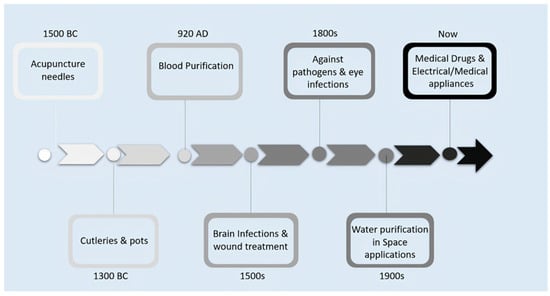
Figure 1.
Timeline of various applications of silver (Ag).
Silver has been extensively used in the medical field even before the advancements in medicine. Use of silver plates for cranioplasty [38,39], catheters [40], sutures [32,38], and silver wire for replacing fractured bones [41], and silver-coated endotracheal tubes [42] is well known. The anti-microbial property of silver is indirectly exploited in household applications and home appliances. Refrigerators and washing machines made by Samsung electronics use AgNPs, as they possess the features of fumigating and sterilizing apparel and help in the safekeeping of the machines from foreign microbes. The refrigerators manufactured by LG Electronics and Hitachi dishwashers also follow the same principles [32,38].
Various methods, including chemical, physical–biological, and hybrid techniques, can be adopted to manufacture nano- and micro-silver particles, as shown in Figure 2 by either a top-to-bottom or bottom-to-top approach. Physical methods of synthesis include plasma arcing [43], ball milling [44], pulsed laser deposition [45], spray pyrolysis [46], and lithographic techniques [47]. The chemical methods used to produce AgNPs are electro-deposition method, sol–gel process [48], chemical solution, chemical vapor deposition methods [49], hydrolysis co-precipitation, and wet chemical method [50]. Microwaves-assisted (frequency range 300 MHz to 300 GHz) nanoparticle synthesis from a blend of silver nitrate and carboxymethyl chitosan was also proven to be a good method for the efficient synthesis of silver nanoparticles [51]. Both physical and chemical methods produce high radiation and utilize stabilizing chemical agents that are harmful to human beings and the environment. Therefore, the biosynthetic route has gained much attention. AgNPs are manufactured with greater stability and precise dimension by biosynthetic routes, as they are made in a single step. In addition, this method provides an eco-friendly, non-toxic and safe route. Biosynthesis of AgNPs is normally achieved by treating the silver salts with plant extracts or microorganisms such as bacteria or fungi. The bio-reducing components of the plant extract polyphenols or proteins and enzymes of the microorganisms are found to play a key role in this bottom-to-top synthetic approach and serve as stabilizing agents.
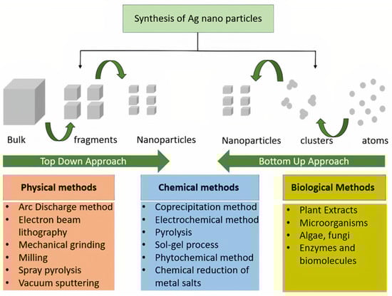
Figure 2.
Different methods of synthesizing silver nanoparticles (AgNPs).
Although silver possesses many advantages, its toxic effects cannot be neglected. A high concentration of silver in water bodies can affect the aquatic species and cause ecological imbalance [52,53]. Silver components enter the body through absorption, inhalation, and excretion. Ingestion causes the deposition of silver in the stomach and other intestinal regions, whereas inhalation deposits silver in the respiratory tract, such as the larynx trachea, and lungs [52,53,54,55]. Silver particles can induce alveolar inflammation and disrupt the heart and vascular function. Continuous inhalation of silver particles for more than 60 s can damage the lungs and cause many diseases [56]. Excessive consumption of the silver solution leads to a bluish-gray color on the skin known as Argyria [57]. The same effect that occurs in the eye is called Argyrosis. The use of silver-based creams for the treatment of wounds and burns is found to have severe side effects, such as methemoglobinemia, hypochloremia, hyponatremia, and eschars [58]. Aqueous solutions of silver compounds are known to damage various organs, including the liver and kidney, cause infections in the respiratory tract and colons, and affect platelet, white blood cells (WBCs), and red blood cells (RBCs) concentrations. Although the high antimicrobial efficacy of silver is beneficial, the lack of selectivity toward the particular strains of microbes also makes it dangerous for essential ecological bacteria [59]. Several experimental studies have validated the toxicity profile of the AgNPs. Experiments on the shrimp Penaeus monodon showed that AgNPs exhibit high anti-bacterial properties at lower concentrations along with a high survival rate. However, at higher concentrations, the toxic effect of the AgNPs becomes predominant, leading to the death of the specimen. Similar observations are made in the case of other aquatic animals and plants [60].
Silver (Ag) is a metal with high electrical and thermal conductivity and has been exploited to manufacture various utensils, jewelry, dental alloys, and explosives for a long time [61]. However, it also has good antimicrobial properties resulting in its ability to fight various infections [62,63,64,65,66]. Pure metallic silver is inert and usually does not interact with human tissues or microorganisms. However, in the presence of aqueous media such as wound fluid moisture, silver can readily form silver ions (i.e., Ag+) and exhibit various bio-activities [33]. The Ag+ form is found to be the active ionic form of silver, while the other possible oxidation states of silver, i.e., Ag2+, Ag3+, are unstable under regular conditions and hence, Ag2+, Ag3+ are generally not considered relevant in biological applications. Silver, being a heavy metal, exhibits oligodynamic effect and is therefore used in minimal quantities in said biological applications [67]. Studies have revealed that Ag+ ions form anionic complexes with the halide ions of the form [AgX2]−, where X could be Cl, Br, etc., and can combine with species containing nitrogen or oxygen donor groups to form stable compounds with improved bioactivity [67]. Silver nanoparticles are also known to form a colloidal solution or composite materials with the suitable polymeric matrix [68,69]. Impregnation, complexation, or blending of silver with suitable materials such as activated charcoal, phosphorous, zirconium lactate, allantoin, etc., are also found to be efficient ways of administration of silver in biological systems [70,71,72]. A trace amount of silver is found in all humans, with about 0.1 µg of silver in the blood itself. A general diet supplies about 40 µg of silver per day; however, about 99 percent of it would be readily excreted from the body. Silver in the blood level can increase to 23 μg/L when exposed to silver either as a medicine or in other forms. This absorption rate can be further increased during inflammation and cell growth [32]. Figure 3 shows the different parts of the human body where deposition of silver can occur. Although the effect of deposition of silver in the human brain is not investigated in detail, its effect on other organs such as the gastrointestinal tract, respiratory tract, skin, and genitals has been well documented [73]. Due to the divergent physical, chemical, and enzymatic barriers they encounter in the different organs and tissues of the body, silver particles can undergo drastic alterations in the structural properties and hence can exhibit unique and specific functional properties. The earlier use of silver particles for medicine has been associated with its significant burn and wound healing applications. Figure 4 shows the action of silver on the wound regions. The wound healing process of bandages containing silver particles is well understood. Diffusion of fluids from the wound triggers the release of silver ions (concentration of around 10–40 ppm) from the dressing material, and the free silver ions are found to be responsible for the observed anti-microbial action [74,75].
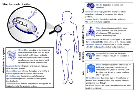
Figure 3.
Mode of action of silver ions in the human body. Reprinted from Elsevier [73]. 2022, Karthik V Shankar.
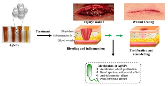
Figure 4.
Images showing the action of silver ions in wound dressing. Reprinted with permission from MDPI [76]. 2022, Karthik V Shankar.
Some studies show that silver and mercury ions hold the prime positions of all metal ions in antimicrobial action when used at concentrations less than 1 ppm in vitro [76]. These ions are absorbed into the bacterial membranes when compounded as silver sulfhydryl, which acts as a cytoplasmic poison. It was found that restrained strains of bacteria show a five-fold lower accumulation of silver as compared to the sensitive stains and produce a lower amount of hydrogen sulfide (about 33% only) [77,78]. The resistance observed in the restrained bacteria can be ascribed to the formation of intracellular protective systems. Figure 5 depicts the microbial action of silver from various devices against bacterial cells. In the sensitive bacterial strains, the silver ions are absorbed by phosphate and other electron-donating receptors on the membrane, which will then diffuse into the cell by endocytic vacuoles and phagocytosis. These silver particles inactivate membrane-related enzymes that distort the bacterial cell envelope, disrupting their ability to absorb nutrients. This also prevents the rejection of metabolic waste from the membrane, leading to pitting, which eventually becomes dangerous to bacteria. The inactivation of enzymes also prevents the replication of RNA and DNA in the cell [78]. The silver free radicals also exhibit excellent antimicrobial activity. Due to the presence of unpaired electrons, free radicals can readily react with several amino acids of cell protein such as arginine and glutamic acid and affect their regular functioning, ultimately leading to the destruction of bacterial cells [64,79,80].
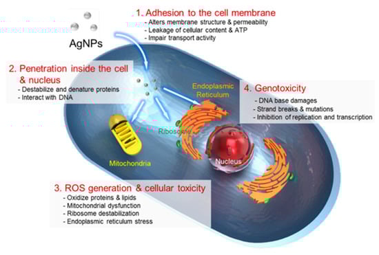
Figure 5.
Image showing the antimicrobial action of AgNPs. Reprinted with permission from MDPI [77]. 2022, Karthik V Shankar.
Nano-silver or silver nanoparticles (AgNPs) are an important form of silver with physical and chemical properties significantly different from those of normal silver particles [81]. Multiple studies have revealed that silver nanoparticles of the size range 50–100 nm exhibit a high order of anti-microbial properties [82,83]. Due to their smaller size, AgNPs can easily reach inaccessible organs and tissues and exhibit superior disinfection effects. AgNPs are found to be very effective against bacteria, fungi, leishmania, plasmodium, and neoplastic cells. AgNPs can activate the enzymes or interfere with the mitotic and meiotic stages of cell division of cancer cells and prevent their growth in various parts of the body. It is seen that in the presence of an appropriate amount of AgNPs, a vital nutrient present in acute lymphoblastic leukemia would exist in an inactive acidic form rather than in an active neutral form, which averts or slows down the growth and spreading of cancer [84]. Extensive experimental analyses conducted by Xiang et al. [85] demonstrated the potential of AgNPs against various viruses such as H1N1 Influenza A, and H3N2 Influenza. Both in vitro and in vivo results showed a significant decrease in the virus concentration and formation of lesions in the lung tissues of the test animals treated with AgNPs [86]. AgNPs were found to be useful in the treatment of genital herpes. From the detailed in vivo and in vitro studies, it was found that the AgNPs inhibit the growth and spread of the virus by boosting the virus immunological response system, which will then efficiently block the penetration of the virus into the cells and suppress inflammation [87]. Shahverdi et al. [88,89] identified that AgNPs can greatly enhance the inhibitory action of various antibiotics such as penicillin G, amoxicillin, erythromycin, clindamycin, and vancomycin against the bacteria E. coli and S. aureus. Detailed in vitro studies carried out on dengue virus (DEN-2) revealed that the mechanism of action of the prominent antiviral activity exhibited by the AgNPs originates from the effective inhibition of gene expression of the virus.
3. Variants of Coronaviruses
Since the outbreak, numerous variants of the SARS-CoV-2 virus have emerged in various parts of the world. Table 1 depicts the timeline of the critical features of SARS-CoV-2, which affected different countries. Several researchers worldwide have monitored and assessed the evolution of COVID-19 since January 2020. Based on the risk posed to the public, coronaviruses are classified under the specifications of ‘variants of interest’ (VOI) and ‘variants of concern’ (VOC). VOCs exhibit increased transmissibility, virulence, and threat to public health d. The salient features of these categories are shown in Figure 6. Based on these effects, many variants are classified as VOC. The primary variants under this category are alpha (B.1.1.7) and beta (B.1.35.1) variants, which were named on the 18 December 2020 [90,91]. The alpha variant was found in United Kingdom samples collected in September 2020, whereas the beta variant was discovered in South African samples procured in May 2020 [90,92]. The alpha variant was transmitted to more than 122 countries, including North American and European countries [93]. Due to continuous evolution, the alpha variant had undergone 13 mutations, making it difficult to track and detect using the RT-PCR method [91]. Furthermore, the beta variant has spread across 85 countries worldwide and is found at present in three different forms: B.1.35.1.1 (Botswana), B.1.35.1.2 (Mayotte), and B.1.35.1.3 (Bangladesh and Singapore) [91]. Over time, the beta variant had also undergone over 10 mutations, resulting in improved binding capability, immune resistance, and transmissibility of the virus [94]. The gamma (P.1) variant was the next major deadly variant (labeled on 11 January 2021) found in samples collected from Brazil in November 2020 [95,96]. This variant was also extensively found in the United States of America [43], Italy [44], and 46 other countries, including Canada [97]. This strain was found to have several mutations (approximately 11) and is said to possess a potential immune escape mutation (E484K) [96], causing severe complications, especially in people of age group 20 to 39 [98]. The variant that caused high distress in India was the delta variant (B.1.61.7.2; named on the 4 April 2021) [91]. The same delta variant was found responsible for Brazil’s increased percentage of stillbirths and placental dysfunction [99]. The variant omicron (B.1.1.529) is thus far the latest type of coronavirus found on the 24 November 2021 as Variants under Monitoring (VUM), which was later classified as a VOC on the 26 November 2021. This category was found in samples procured from various parts of the world and was found to have a higher transmissibility rate [91]. Another variant, epsilon (B.1.429), which was identified in the United States and another 31 countries in June 2020, was not considered under VOC owing to its lower virulent abilities [91,100].

Table 1.
Timeline of occurrence of various key events related to the SARS-CoV-2 outbreak.
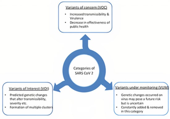
Figure 6.
Salient features of SARS-CoV-2 categories.
Variants of interest (VOI) are the category of coronavirus species anticipating to undergo genetic alterations over time that enhance their transmissibility, severity, as well as diagnostic or therapeutic escaping capacity, etc. Hence, they can emerge as threats to global public health [103]. The lambda (C.37) and mu (B.1.621) variants are the main examples of this category. The lambda variant was named on 14 June 2021 and mu on 30 August 2021 after the primary cases were reported from Peru in December 2020 and Colombia in January 2021, respectively [91,101]. Variants under monitoring (VOM) are the class of least concerned coronaviruses, which have not caused much trouble currently. Some of the variants in this category are C.1.2, B.1.617.1 (kappa), B.1.526 (iota), and B.1.525 (eta) [91]. However, these variants can undergo mutation and create havoc in the future and hence are monitored and assessed regularly [104].
B.1.1.529 (omicron) is a variant that must be considered carefully. These viruses were first reported in South Africa on 24 November 2021, the first of which was identified in a sample collected from a person on the 9 November 2021 [102]. These variants pose a significantly high transmissibility rate evident in reports collected from South Africa and many other countries [105]. This variant underwent 30 mutations in the genomic proteins, enhancing its permeability, transmissibility, and resistance to antibodies [102]. Of these 30 mutations, 23 were distinctive and were not found in other variants of coronaviruses. Evolutionary studies were conducted to identify the ancestral variant of these viruses, and it was found that the gamma variant (P.1) was closely related in the phylogenetic analysis [106]. Mutations such as E484A and Y505H are the primary causes of the absence of communication between S-RBD and antibodies. At the same time, Asn501Tyr improves ACE2 receptor binding, which is responsible for the high transmission of viruses. The low affinity of S-RBD suggests that the current vaccination processes cannot guarantee protection against these variants [107,108,109]. The WHO has created several guidelines to prevent the transmission of these strains, but their effectiveness in tackling the viruses is minimal. Due to researchers’ tireless efforts worldwide, several vaccines were designed for reducing the mortality rate worldwide. Some of the most widely used vaccines include mRNA-1273 (Moderna), BNT162b2 mRNA (Pfizer) [110], and Janssen Ad26.CoV2. S (Johnson & Johnson) [111] developed in the USA, AZD1222 (Oxford-AstraZeneca) [112] developed in the UK, BBIBP-CorV (Sinopharm) and CoronaVac (Sinovac Biotech) [113] developed in China, Sputnik [114] developed in Russia, and Covaxin and Covishield developed in India [55]. All major vaccines are listed in Table 2. Several other countries are making efforts to develop vaccines to ensure continuous availability to the public.
Table 2.
List of various vaccines developed to tackle COVID-19.
3.1. Treatment of COVID-19 Using Silver and Silver Nanoparticles (AgNPs)
Considering the tremendous success of silver ions and silver nanoparticles in the inhibition of varieties of microbes and in the treatment of infectious diseases (vide supra), their utilization in the successful inhibition of the SARS-Cov-2 virus was highly anticipated. The various stages of prevention and treatment methods connected with the coronavirus outbreak, where nanotechnology can play a major role, are depicted in Figure 7. It is well understood that viruses bind to cell receptors of the host and undergo rapid multiplication and ultimately mutilate the cell. The dead cells act as carriers of the virus in the host body and spread the virus to neighboring cells. Hence, strong inhibiting strategies would be necessary for preventing and treating viral infections. Reactive oxygen species (ROS) play a key role in the regulation of viral replication and functioning of organelles, potentially providing new insights into the prevention and treatment of coronavirus infections. Silver-based nanoparticles and composite materials are known to facilitate the production of ROS in cells and hence are extensively considered for COVID-19 treatments [116]. Although their action on SARS-CoV-2 remains unclear, due to the high order inhibition efficacy exhibited by the silver nanoparticles on other viruses, four classes of silver composite materials are identified, viz., Glutathione-capped silver sulfide nanoclusters (GSH-Ag2S NCs), PVP-coated silver nanomaterials (PVP-AgNMs), silver nanoparticle–anchored graphene oxide nanoparticles (GO-AgNPs) and PDDA-coated PVP-functionalized graphene oxide-silver nanocomposites (PDDA-PVP-GO-AgNCs) [117]. These materials promote and facilitate the formation of free radicals inhibiting the physiological processes of virus-cell [117]. It is observed that the binding of silver ion to the GP-120 protein subunit can prominently inhibit the normal functioning of the coronavirus. Binding of the silver ion results in the rendering of the S-protein subunits that are responsible for the binding of the beta variant of the SARS-CoV-2 virus spike to the host cell ACE 2 receptor, preventing the binding and fusion of the virus and host cell. Graphene oxide silver nano-composite ink was proven to have excellent inhibitory properties against influenza A virus and OC43 beta coronavirus, both of which are RNA viruses and come under the same lineage as SARS-CoV-2, indicating the potential use of these materials in the treatment of COVID-19 [118]. Inhalation of the AgNPs (average size of 2–10 nm) was recommended for first-line treatment for COVID-19 [119]. Suitably designed AgNPs with a higher negative zeta potential can bind to the viruses that have predominantly positively charged spike proteins. Considering the well-documented antibacterial efficacy of the AgNPs, the suggested inhalation treatment was also expected to control the biofilm formation in the upper respiratory system [119]. The size of the AgNPs is believed to have an important role in the binding process. Smaller particles, due to the larger surface area, were found to interact more effectively with the viral protein. The ideal size of the AgNPs for the optimum binding is around 2–15 nm [117]. Morita et al. showed that considering the stability of viral proteins, the most suitable size of AgNPs for this application is 10 nm. Larger nanoparticles were found to be less stable [120]. Further, it was shown that the stability of the nanoparticles can be enhanced by means of coating the nanoparticles with suitable polymers. Coated AgNPs exhibited better stability, lower agglomeration rate, and higher activities [117].
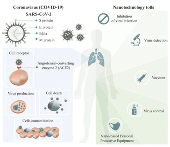
Figure 7.
Nanotechnology in the prevention and treatment of COVID-19. Reprinted with permission from Springer Nature [121]. 2022, Karthik V Shankar.
Several scientists have conducted experiments to validate the effect of silver compounds against COVID-19. Bui et al. [122] theoretically evaluated the inhibitory action of the mono silver-carbene and bis silver-carbene compounds on human protein ACE2 and SARS-CoV-2 protease PDB6LU7 with the help of molecular docking simulations. The simulations helped to understand the bond dissociation energy (BDE) of the hierarchical order of NHC–Ag > NHSi–Ag > NHGe–Ag for the mono silver compounds and NHSi–Ag-bis > NHGe–Ag-bis > NHC–Ag-bis for the bis-silver compounds. These simulations explain that NHC–Ag and NHC–Ag-bis compounds could inhibit SARS-CoV-2 receptors, with NHC–Ag-bis compounds showing superior inhibitory effects. These silver compounds’ inhibitory actions were comparable to the known antiviral drugs reported by Ribavirin and Remdesivir [122]. Hydroxychloroquine was used for the early treatment of COVID during the outbreak in 2019 [123]. Ghaffari et al., with the help of molecular dynamic simulations [123], showed that silver and gold nanoparticles could be used as drug delivery systems for medicines such as hydroxychloroquine or chloroquine without causing significant damage or side effects [124]. Hybrid components formed by combining silver and gold particles could also provide better options for this purpose [117]. During the pandemic, the streets of Milan in Italy were sterilized with a mixture containing silver ions (Ag+) and titanium dioxide [125]. This led to a significant decrease in the spread of coronaviruses during the initial outbreak of COVID-19 in these regions. Titanium dioxide nanoparticles are also good anti-viral agents used to inhibit the viral activity of influenza viruses (H3N2) [126].
Jeremiah et al. [120] experimented with viral pretreatment assay (VPrA), cell pretreatment assay (CprA), and cell post-treatment assay (CpoA) with AgNPs in VeroE6/TMPRSS2 to understand the mechanism of action of silver on intracellular and extracellular viruses. VprA results indicate the death and reduction of the viral load of extracellular viruses. From the CpoA results, the inhibitory properties of AgNPs were established. VeroE6/TMPRSS2 cells infected with SARS-CoV-2 virus, when washed with 2 ppm of PVP-AgNP10 silver nanocomposite solution, showed an excellent suppression rate. The CprA studies showed that the VeroE6/TMPRSS2 cells pretreated with 2 ppm PVP-AgNP10, when infected with SARS-CoV-2, showed partial inhibition only after 2 days, indicating the high order viral resistance provided by the silver nanocomposite material [116]. Sanea et al. [127] demonstrated the antiviral properties of AgNPs prepared using natural extracts against SARS-CoV-2. The silver nanoparticles synthesized from the strawberry extract exhibited better antiviral properties than the ginger extract silver nanoparticles. With the help of molecular docking and dynamic simulation studies, the authors monitored the interaction of about 30 possible natural products from both the extracts against seven SARS-CoV-2 protein targets (AAK1, Cathepsin L—human proteins; Mpro, ADP ribose phosphatase, NSP14, NSP16, PLpro—viral proteins). Among all the components of the extracts investigated, neohesperidin, a flavanone glycoside present in strawberry extract, exhibited a higher potential to bind the NSP16 protein of the SARS-CoV-2 virus and to a human AAK1 protein, indicating its key role in the antiviral properties exhibited by the nanoparticles synthesized by strawberry extracts alongside the virucidal effect of silver. Rodrigues et al. [122] with the help of DFT studies explained that the potential virucidal property of the silver originates from the spontaneous chemical reaction between the amino acids of the SARS-CoV-2 virus and Ag+ ions. Alveolar inflammation and lung fibrosis are the primary symptoms of COVID-19 patients initiated by a cytokine storm. AgNPs are effective protectors against inflammation and fibrosis because of their ability to change the transcriptional activity of cytokines [128]. Silver nanoparticles effectively reduce the number of inflammatory cytokines such as interleukin (IL)-1β, IL-6, and IL-17, transforming growth factor-beta (TGF-β), and tumor necrosis factor-alpha (TNF-α), thereby reducing inflammation and fibrosis via the NFκB and MAPKinase pathways. They further reduce collagen deposition by controlling the profibrotic gene expression of Col 1a1 and Col 1a2 [123]. Figure 8 shows a schematic representation of how AgNPs are effectively used in inhibiting SARS-CoV-2 viruses.
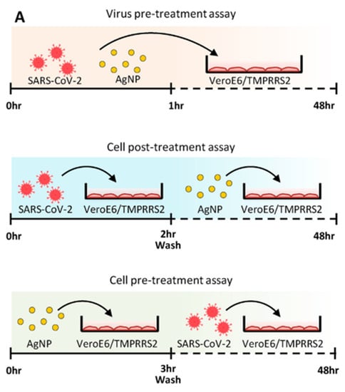
Figure 8.
Action of AgNPs on SARS-CoV-2 virus. Reprinted with permission from Elsevier [115]. 2022, Karthik V Shankar.
Inhalation of colloidal silver particles improves any form of respiratory disease [129,130]. However, inhalation could occur via either oral or nasal pathways. Zachar [131] observed a significant difference between them because the deposition of particles in the bronchial tree when consumed through nasal breathing was only 10%, which is minimal compared to the oral counterpart, which was about 30%. These findings led the authors to conclude that inhalation by oral breathing is the best method to introduce AgNPs into the system. Although silver can be introduced to the body in both colloidal or ionic forms, it is observed that the antiviral properties of the colloidal silver (AgNPs) are 10-fold greater than that of silver ionic particles, making the colloidal form more suitable for antiviral applications [132]. Nevertheless, the size of nanoparticles is a critical parameter for the appropriate therapeutic applications. It is known that smaller particles have higher specific weight fraction, and much higher particle density, which is a good characteristic for fighting different viral pathogens at a minimum inhibitory concentration (MIC) level [132]. Moreover, smaller particles can easily penetrate the cell wall and ultimately destroy the viral cells. In the case of HIV cells (cell size ~120 nm), the MIC estimate of silver nanoparticles was found to be 10 µg/mL [133]. Considering that the SARS-CoV-2 viral cell size is in the same range (~90–100 nm) [134], it is speculated that a similar MIC level of AgNPs would be suitable to treat COVID-19 [116].
3.2. Application of Silver Nanoparticles for Controlling COVID-19
As shown in Figure 9, AgNPs can be used in various forms to tackle COVID-19. AgNPs can be used as nano-based protective equipment, virus-controlling sanitizers, or nano vaccines to improve immunity and antigen carriers, etc. [135]. AgNPs can be used to manufacture clothes for protection against COVID-19 infection. Textile fibers containing AgNPs exhibit improved anti-bacterial and anti-viral properties [121]. Edible chitosan/pectin-supported silver nanoparticle films were found to enhance the antimicrobial properties and durability of the textiles significantly [136]. Composite materials containing lignin extracted from sugarcane bagasse and silver nanoparticles are successfully used in commercial antibacterial textiles. Cotton fibers combined with guanozole, zinc, and silver nanocomposites are also extensively utilized in manufacturing antimicrobial cloths [121].

Figure 9.
Application of silver nanoparticles in the medical field.
Radiochemical deposition of silver nanoparticles on cotton fiber was effective against Influenza A and Feline caliciviruses [121]. Kumar et al. [137] manufactured and analyzed a cloth with silver being photo-deposited over it and found that these clothes showed 97% efficiency in restraining the growth of the SARS-CoV-2 virus. The proposed mechanism of antiviral action, as shown in Figure 10, demonstrates that the binding of silver ions with the glycoprotein possessed by the virus is the main reason for the inactivation of the virus, which ultimately leads to the death of the virus [137]. Figure 10 shows the structure of the virus, which contains a lipid membrane and glycoprotein spikes comprising S1 and S2 subunits. The S1 spikes, as shown in Figure 10a, are exposed outside. A negatively charged CTD that combines with Ag+ ions alters the morphology and damages the cell walls, causing leakage of cell components and eventually leading to the virus’s death as depicted in Figure 10b [138].
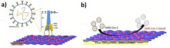
Figure 10.
Inactivation of SARS-CoV-2 virus in clothes using AgNPs. (a) Structure of virus containing lipid membrane and glycoprotein spikes, (b) Inactivating the COVID virus. Reprinted with permission from Elsevier [137]. 2022, Karthik V Shankar.
Even after the proper usage of personal protection equipment (PPE) by health workers, the spread of the SARS-CoV-2 virus among the categories has heavily risen [20]. This happened because of the prolonged existence of the virus in the air, environment, and PPE kits. To prevent this, the development of antibacterial and anti-viral surfaces is highly essential. It was found that metal-grafted graphene oxide (GO)-based fabrics could effectively reduce PPE contamination [139]. Of these, silver- or copper-based GO is found to be the best in performance. Embedding the silver/copper—GO composites in polymeric materials and utilizing them in the manufacturing of masks can also be considered. A higher level of viral inhibition was observed in the case of masks manufactured by fabric materials containing metals such as copper, silver, and gold [139]. Balagna et al. [140] studied the effects of coating masks with AgNPs. The coating was carried over a glass substrate, and it was found that there was a decreased possibility of infection for the coated mask as compared with the uncoated mask. The coating was sufficient to bring the titer value of SARS-CoV-2 to almost zero and improved the mask’s filtering effects significantly, which also helps to reduce unnecessary waste disposal [140].
Apart from clothing and masks, several other products used for COVID-19 treatments contain silver or AgNPs. SHEPROS, a Malaysian company, developed a suspension named ‘Nanosilver sanitizer’ [141] using AgNPs of a size ~25 nm, which is assured to destroy a wide variety of microorganisms and viruses. The oral and nasal cleaning agent ARGOVIT mouthwash containing AgNPs was found to prevent the spread of SARS-CoV-2 virus among health workers [142]. A similar product, Silvo Clean Spray, containing colloidal AgNPs, was developed by Weinnovate solutions, a startup company in collaboration with the Indian Department of Science and Technology and the Department of Biotechnology [143].
3.3. Future Scope
Vaccination is the only effective method for eradicating the uncontrollable spread of any virus. Nanomedicines could prove to be the best means of improving vaccine efficacy. Electrostatic binding of the S protein with gold nanoparticles result in s-AuNP nano vaccines showing high responses. Due to the enhanced anti-viral potency, there could be future chances of using silver-based nano vaccines instead of gold nano vaccines. Currently, even though detection of SARS-CoV-2 is performed through reverse transcription-polymerase chain reaction (RT-PCR) methods. Several nanoparticle-based detection methods have also been developed, while most of them include gold nanoparticles as their main constituent. However, silver nanoparticles can be a potential and economically viable replacement. To improve the efficiency of viral control, a careful and detailed study of the impact of various undetected protein enzymes on spreading and their subsequent inhibition must be performed. Based on these studies, the development of silver-based components can be developed to tackle future variants of coronavirus.
4. Conclusions
The COVID-19 outbreak that began in 2019 led to the rapid spread of the disease worldwide within a few months, resulting in an exponential death toll in most countries. Numerous variants of SARS-CoV-2 emerged during this period, including alpha, beta, gamma, and omicron, and continued with changes in the DNA sequences and mutations. The omicron variant is the current significant strain spread in various parts of the world. Several metals are identified with continuous research on metallo-medicines, and their effects on tackling diseases are established. Silver has been used in medicinal applications since ancient times. The anti-microbial properties of silver nanoparticles (AgNPs) depend on concentration, method of synthesis, size, presence of ‘helper molecules’, and various other factors. Prominent anti-viral properties of AgNPs could be utilized to tackle current variants as well as future mutants of coronavirus. Evidence collected from various experiments showed the inhibition of spikes of SARS-CoV-2 on ACE receptors when treated with AgNPs. AgNPs can also serve as a suitable drug delivery system for hydroxychloroquine and chloroquine medicines used to treat COVID-19. Administration of AgNPs via nasal or oral routes was found to help in controlling respiratory diseases. Silver-based textiles used for hospital requirements, including PPE kits and masks, are proven to limit the surface contact transmission of viruses to a great extent. Silver-based sanitizers and disinfectants have proven their high efficacy. However, high toxicity and adverse effect of AgNPs are still points of concern and should be addressed systematically. Even though extensive research and analysis are continuously being carried out with several materials for understanding their potential for tackling the pandemic, the development of superior inhibitory agents for the SARS-CoV-2 virus or its variants requires more time and effort.
Author Contributions
Conceptualization, K.V.S.; methodology, P.N.J.A. and B.S. (Bipin Sankar); software, K.V.S.; formal analysis, P.N.J.A., B.S. (Bipin Sankar) and K.V.S.; investigation, P.N.J.A., B.S. (Bipin Sankar), K.V.S. and N.V.K.; resources, P.N.J.A. and B.S. (Bipin Sankar); writing—original draft preparation, P.N.J.A., B.S. (Bipin Sankar) and K.V.S.; writing—review and editing, K.V.S., N.V.K., S.S. and B.S. (Balakrishnan Shankar); visualization, B.S. (Bipin Sankar) and P.N.J.A. supervision, N.V.K., S.S., B.S. (Balakrishnan Shankar) and K.V.S.; project administration, N.V.K., S.S., B.S. (Balakrishnan Shankar) and K.V.S.; funding acquisition, K.V.S., S.S. and N.V.K. All authors have read and agreed to the published version of the manuscript.
Funding
This research was funded by Amrita Vishwa Vidyapeetham, Amritapuri, India.
Institutional Review Board Statement
Not applicable.
Informed Consent Statement
Not applicable.
Data Availability Statement
The data presented in this study are available on request from the corresponding author.
Conflicts of Interest
The authors declare no conflict of interest.
References
- Murarkar, S.; Gothankar, J.; Doke, P.; Dhumale, G.; Pore, P.D.; Lalwani, S.; Quraishi, S.; Patil, R.S.; Waghachavare, V.; Dhobale, R. Prevalence of the acute respiratory infections and associated factors in the rural areas and urban slum areas of western Maharashtra, India: A community-based cross-sectional study. Front. Public Health 2021, 9, 723807. [Google Scholar] [CrossRef] [PubMed]
- Nair, H.; Simões, E.A.F.; Rudan, I.; Gessner, B.D.; Azziz-Baumgartner, E.; Zhang, J.S.F.; Feikin, D.R.; Mackenzie, G.A.; Moiïsi, J.C.; Roca, A. Global and regional burden of hospital admissions for severe acute lower respiratory infections in young children in 2010: A systematic analysis. Lancet 2013, 381, 1380. [Google Scholar] [CrossRef]
- Acter, T.; Uddin, N.; Das, J.; Akhter, A.; Choudhury, T.R.; Kim, S. Evolution of severe acute respiratory syndrome coronavirus 2 (SARS-CoV-2) as coronavirus disease 2019 (COVID-19) pandemic: A global health emergency. Sci. Total Environ. 2020, 730, 138996. [Google Scholar] [CrossRef] [PubMed]
- World Health Organization Website. Available online: https://covid19.who.int/ (accessed on 25 September 2021).
- AlSamman, M.; Caggiula, A.; Ganguli, S.; Misak, M.; Pourmand, A. Non-respiratory presentations of COVID-19, a clinical review. Am. J. Emerg. Med. 2020, 38, 2444. [Google Scholar] [CrossRef] [PubMed]
- Joint WHO-China Study Team. WHO-convened global study of origins of SARS-CoV-2: China part (text extract). Infect. Dis. Immun. 2021, 1, 125. [Google Scholar]
- Jayaweera, M.; Perera, H.; Gunawardana, B.; Manatunge, J. Transmission of COVID-19 virus by droplets and aerosols: A critical review on the unresolved dichotomy. Environ. Res. 2020, 188, 109819. [Google Scholar] [CrossRef]
- Burke, R.M.; Midgley, C.M.; Dratch, A.; Fenstersheib, M.; Haupt, T.; Holshue, M.; Ghinai, I.; Jarashow, M.C.; Lo, J.; McPherson, T.D. Active monitoring of persons exposed to patients with confirmed COVID-19—United States, January–February 2020. Morb. Mortal. Wkly. Rep. 2020, 69, 245. [Google Scholar] [CrossRef]
- Guo, Y.; Korteweg, C.; McNutt, M.A.; Gu, J. Pathogenetic mechanisms of severe acute respiratory syndrome. Virus Res. 2008, 133, 4. [Google Scholar] [CrossRef]
- Wang, W.; Xu, Y.; Gao, R.; Lu, R.; Han, K.; Wu, G.; Tan, W. Detection of SARS-CoV-2 in different types of clinical specimens. JAMA 2020, 23, 1843. [Google Scholar] [CrossRef]
- Chang, L.; Zhao, L.; Gong, H.; Wang, L.; Wang, L. Severe acute respiratory syndrome coronavirus 2 RNA detected in blood donations. Emerg. Infect. Dis. 2020, 26, 1631. [Google Scholar] [CrossRef]
- Chang, L.; Yan, Y.; Wang, L. Coronavirus disease 2019: Coronaviruses and blood safety. Transfus. Med. Rev. 2020, 34, 75. [Google Scholar] [CrossRef]
- Xu, J.; Zhao, S.; Teng, T.; Abdalla, A.E.; Zhu, W.; Xie, L.; Wang, Y.; Guo, X. Systematic Comparison of Two Animal-to-Human Transmitted Human Coronaviruses: SARS-CoV-2 and SARS-CoV. Viruses 2020, 12, 244. [Google Scholar] [CrossRef]
- Thippareddi, H.; Balamurugan, S.; Patel, J.; Singh, M.; Brassard, J. Coronaviruses–Potential human threat from foodborne transmission? Lebensm Wiss Technol. 2020, 134, 110147. [Google Scholar] [CrossRef]
- Kwok, Y.L.A.; Gralton, J.; McLaws, M.L. Face touching: Un hábito frecuente que tiene implicaciones para la higiene de las manos. Am. J. Infectar. 2015, 43, 112. [Google Scholar]
- Kampf, G.; Todt, D.; Pfaender, S.; Steinmann, E. A review and analysis Persistence of coronaviruses on inanimate surfaces and their inactivation with biocidal agents. J. Hosp. Infect. 2020, 104, 246. [Google Scholar] [CrossRef]
- Casanova, L.M.; Jeon, S.; Rutala, W.A.; Weber, D.J.; Sobsey, M.D. Effects of air temperature and relative humidity on coronavirus survival on surfaces. Appl. Environ. Microbiol. 2010, 76, 2712. [Google Scholar] [CrossRef]
- Sizun, J.; Yu, M.W.N.; Talbot, P.J. Survival of human coronaviruses 229E and OC43 in suspension and after drying onsurfaces: A possible source ofhospital-acquired infections. J. Hosp. Infect. 2000, 46, 55. [Google Scholar] [CrossRef]
- Duan, S.-M.; Zhao, X.-S.; Wen, R.-F.; Huang, J.J.; Pi, G.-H.; Zhang, S.-X.; Sheng, J.H.; Bi, L.; Ruan, L.; Dong, X.-P.; et al. Stability of SARS coronavirus in human specimens and environment and its sensitivity to heating and UV irradiation. Biomed. Environ. Sci. 2003, 16, 246. [Google Scholar]
- Sportelli, C.N. Materials Science Help the Fight against SARS-CoV-2. Nanomaterials 2020, 10, 802. [Google Scholar] [CrossRef]
- Govind, V.; Bharadwaj, S.; Sai Ganesh, M.R.; Vishnu, J.; Shankar, K.V.; Shankar, B.; Rajesh, R. Antiviral properties of copper and its alloys to inactivate covid-19 virus: A review. Biometals 2021, 34, 1217. [Google Scholar] [CrossRef]
- Wang, L.; Hu, C.; Shao, L. The antimicrobial activity of nanoparticles: Present situation and prospects for the future. Int. J. Nanomed. 2017, 12, 1227. [Google Scholar] [CrossRef] [PubMed]
- Wijesinghe, W.; Mantilaka, M.; Ruparathna, K.A.A.; Rajapakshe, R.; Sameera, S.A.L.; Thilakarathna, M. Filler matrix interfaces of inorganic/biopolymer composites and their applications. Interfaces Part. Fibre Reinf. Compos. 2020, 1, 95–112. [Google Scholar]
- Longano, D.; Ditaranto, N.; Sabbatini, L.; Torsi, L.; Cioffi, N. Synthesis and antimicrobial activity of copper nanomaterials. In Nano-Antimicrobials; Springer: Berlin/Heidelberg, Germany, 2012; p. 85. [Google Scholar]
- Bogdanovic, U.; Vodnik, V.; Mitric, M.; Dimitrijevic, S.; Skapin, S.D.; Zunic, V.; Budimir, M.; Stoiljkovic, M. Nanomaterial with High Antimicrobial Efficacy Copper/Polyaniline Nanocomposite. ACS Appl. Mater. Interfaces 2015, 7, 1955. [Google Scholar] [CrossRef] [PubMed]
- Galúcio, J.M.P.; de Souza, S.G.B.; Vasconcelos, A.A.; Lima, A.K.O.; da Costa, K.S.; de Campos Braga, H.; Taube, P.S. Synthesis, characterization, applications, and toxicity of green synthesized nanoparticles. Curr. Pharm. Biotechnol. 2022, 23, 420. [Google Scholar] [CrossRef]
- Chiriac, V.; Stratulat, D.N.; Calin, G.; Nichitus, S.; Burlui, V.; Stadoleanu, C.; Popa, M.; Popa, I.M. Antimicrobial property of zinc based nanoparticles. In IOP Conference Series: Materials Science and Engineering; IOP Publishing: Bristol, UK, 2016; p. 12055. [Google Scholar]
- Mishra, P.K.; Mishra, H.; Ekielski, A.; Talegaonkar, S.; Vaidya, B. Zinc oxide nanoparticles: A promising nanomaterial for biomedical applications. Drug Discov. Today 2017, 22, 1825. [Google Scholar] [CrossRef] [PubMed]
- Król, A.; Pomastowski, P.; Ranska, K.; Railean-Plugaru, V.; Buszewski, B. Zinc oxide nanoparticles: Synthesis, antiseptic activity and toxicity mechanism. Adv. Colloid Interface Sci. 2017, 249, 37. [Google Scholar] [CrossRef] [PubMed]
- Vimbela, G.V.; Ngo, S.M.; Fraze, C.; Yang, L.; Stout, D.A. Antibacterial properties and toxicity from metallic nanomaterials. Int. J. Nanomed. 2017, 12, 3941. [Google Scholar] [CrossRef]
- Balraj, B.; Senthilkumar, N.; Potheher, I.V.; Arulmozhi, M. Characterization, antibacterial, anti-arthritic and in-vitro cytotoxic potentials of biosynthesized magnesium oxide nanomaterial. Mater. Sci. Eng. B 2018, 231, 121. [Google Scholar] [CrossRef]
- Lansdown, A.B.G. Silver in health care: Antimicrobial effects and safety in use. Biofunctional Text. Ski. 2006, 33, 17. [Google Scholar]
- Melayie, A.; Youngs, J.W. Silver and its application on antimicrobial agents. Expert. Opin. Ther. Pat. 2005, 15, 125. [Google Scholar] [CrossRef]
- Walker, M.; Parsons, D. The biological fate of silver ions following the use of silver-containing wound care products–a review. Int. Wound J. 2014, 11, 496. [Google Scholar] [CrossRef]
- Ratan, Z.A.; Mashrur, F.R.; Chhoan, A.P.; Shahriar, S.M.; Haidere, M.F.; Runa, N.J.; Kim, S.; Kweon, D.-H.; Hosseinzadeh, H.; Cho, J.Y. Silver nanoparticles as potential antiviral agents. Pharmaceutics 2021, 13, 2034. [Google Scholar] [CrossRef]
- Galdiero, S.; Falanga, A.; Vitiello, M.; Cantisani, M.; Marra, V.; Galdiero, M. Silver nanoparticles as potential antiviral agents. Molecules 2011, 16, 8894. [Google Scholar] [CrossRef]
- Holden, N.E. History of the Origin of the Chemical Elements and Their Discoverers; Brookhaven National Lab. (BNL): Upton, NY, USA, 2019. [Google Scholar]
- Barillo, D.J.; Marx, D.E. Silver in medicine: A brief history BC 335 to present. Burns 2014, 40, S3. [Google Scholar] [CrossRef]
- Leone, G.; Pepi, S.; Consumi, M.; Mahdizadeh, F.F.; Lamponi, S.; Magnani, A. Phosphorylated xanthan gum-Ag (I) complex as antibacterial viscosity enhancer for eye drops formulation. Carbohydr. Polym. 2021, 267, 118196. [Google Scholar] [CrossRef]
- Kremer, J.R.; Mastronarde, D.N.; McIntosh, J.R. Computer visualization of three-dimensional image data using IMOD. J. Struct. Biol. 1996, 116, 71. [Google Scholar] [CrossRef]
- Ludwigson, D.C.; Mears, D.C. Today’s prosthetic metals. J. Miner. Met. Mater. Soc. 1964, 16, 226. [Google Scholar] [CrossRef]
- Konop, M.; Damps, T.; Misicka, A.; Rudnicka, L. Certain aspects of silver and silver nanoparticles in wound care: A minireview. J. Nanomater. 2016, 2016, 7614753. [Google Scholar] [CrossRef]
- Pietro Reverberi, A.; Vocciante, M.; Salerno, M.; Ferretti, M.; Fabiano, B. Green synthesis of silver nanoparticles by low-energy wet bead milling of metal spheres. Materials 2020, 13, 63. [Google Scholar] [CrossRef]
- Zakaria, M.A.; Menazea, A.A.; Mostafa, A.M.; Al-Ashkar, E.A. Ultra-thin silver nanoparticles film prepared via pulsed laser deposition: Synthesis, characterization, and its catalytic activity on reduction of 4-nitrophenol. Surf. Interfaces 2020, 19, 100438. [Google Scholar] [CrossRef]
- Semenova, A.A.; Ivanov, V.K.; Savilov, S.V.; Goodilin, E.A. Unusual silver nanostructures prepared by aerosol spray pyrolysis. CrystEngComm 2013, 15, 7863. [Google Scholar] [CrossRef]
- Colson, P.; Henrist, C.; Cloots, R. Nanosphere lithography: A powerful method for the controlled manufacturing of nanomaterials. J. Nanomater. 2013, 2013, 948510. [Google Scholar] [CrossRef]
- Rivero, P.J.; Urrutia, A.; Goicoechea, J.; Zamarreño, C.R.; Arregui, F.J.; Matías, I.R. An antibacterial coating based on a polymer/sol-gel hybrid matrix loaded with silver nanoparticles. Nanoscale Res. Lett. 2011, 6, 1. [Google Scholar] [CrossRef] [PubMed]
- Nasretdinova, G.R.; Fazleeva, R.R.; Mukhitova, R.K.; Nizameev, I.R.; Kadirov, M.K.; Ziganshina, A.Y.; Yanilkin, V.V. Electrochemical mediated synthesis of silver nanoparticles in solution. Russ. J. Electrochem. 2015, 51, 1029. [Google Scholar] [CrossRef]
- Raza, S. Shape-Dependent Antibacterial Studies of Silver Nanoparticles Synthesized by Wet Chemical Routes. Nanomaterials 2016, 6, 74. [Google Scholar] [CrossRef]
- Borase, H.P.; Salunke, B.K.; Salunkhe, R.B.; Patil, C.D.; Hallsworth, J.E.; Kim, B.S.; Patil, S.V. Plant extract: A promising biomatrix for ecofriendly, controlled synthesis of silver nanoparticles. Appl. Biochem. Biotechnol. 2014, 173, 1. [Google Scholar] [CrossRef]
- Maya, F.; Muhl, S.; Peña, O.; Miki-Yoshida, M. Synthesis and characterization of silver–carbon nanoparticles produced by high-current pulsed arc. Thin Solid Films 2009, 518, 1484. [Google Scholar] [CrossRef]
- Ratte, H.T. Bioaccumulation and toxicity of silver compounds: A review. Environ. Toxicol. Chem. Int. J. 1999, 18, 89. [Google Scholar] [CrossRef]
- Hollinger, M.A. Toxicological aspects of topical silver pharmaceuticals. Crit. Rev. Toxicol. 1996, 26, 255.2. [Google Scholar] [CrossRef]
- Parveen, K.; Banse, V.; Ledwani, L. Green synthesis of nanoparticles: Their advantages and disadvantages. In AIP Conference Proceedings; AIP Publishing LLC: Bristol, UK, 2016; p. 20048. [Google Scholar]
- Hayat, M.; Uzair, M.; Ali Syed, R.; Arshad, M.; Bashir, S. Status of COVID-19 vaccination around South Asia. Hum. Vaccin. Immunother. 2022, 18, 2016010. [Google Scholar] [CrossRef]
- Marshall, J.P.; Schneider, R.P. Systemic argyria secondary to topical silver nitrate. Arch. Dermatol. 1977, 113, 1077. [Google Scholar] [CrossRef]
- Fung, M.C.; Bowen, D.L. Silver products for medical indications: Risk-benefit assessment. J. Toxicol. Clin. Toxicol. 1996, 34, 119. [Google Scholar] [CrossRef]
- Tashi, T.; Gupta, N.V.; Mbuya, V.B. Silver nanoparticles: Synthesis, mechanism of antimicrobial action, characterization, medical applications, and toxicity effects. J. Chem. Pharm. Res. 2016, 8, 526. [Google Scholar]
- Griffitt, R.J.; Luo, J.; Gao, J.; Bonzongo, J.; Barber, D.S. Effects of particle composition and species on toxicity of metallic nanomaterials in aquatic organisms. Environ. Toxicol. Chem. Int. J. 2008, 27, 1972. [Google Scholar] [CrossRef]
- Rezvani, E.; Rafferty, A.; McGuinness, C.; Kennedy, J. Adverse effects of nanosilver on human health and the environment. Acta Biomater. 2019, 94, 145. [Google Scholar] [CrossRef]
- Ayrault, S.; Priadi, C.R.; Evrard, O.; Lefèvre, I.; Bonté, P. Silver and thallium historical trends in the Seine River basin. J. Environ. Monit. 2010, 12, 2177. [Google Scholar] [CrossRef]
- Rai, M.; Agarkar, G. Plant–fungal interactions: What triggers the fungi to switch among lifestyles? Crit. Rev. Microbiol. 2016, 42, 428. [Google Scholar] [CrossRef]
- Furno, F.; Morley, K.S.; Wong, B.; Sharp, B.L.; Arnold, P.L.; Howdle, S.M.; Bayston, R.; Brown, P.D.; Winship, P.D.; Reid, H.J. Silver nanoparticles and polymeric medical devices: A new approach to prevention of infection? J. Antimicrob. Chemother. 2004, 54, 1019. [Google Scholar] [CrossRef]
- Maillard, J.-Y.; Hartemann, P. Silver as an antimicrobial: Facts and gaps in knowledge. Crit. Rev. Microbiol. 2013, 39, 373. [Google Scholar] [CrossRef]
- Chernousova, S.; Epple, M. Silver as antibacterial agent: Ion, nanoparticle, and metal. Angew. Chem. Int. Ed. 2013, 52, 1636–1653. [Google Scholar] [CrossRef]
- Marx, D.E.; Barillo, D.J. Silver in medicine: The basic science. Burns 2014, 40 (Suppl. S1), S9–S18. [Google Scholar] [CrossRef] [PubMed]
- Damm, C.; Münstedt, H.; Rösch, A. The antimicrobial efficacy of polyamide 6/silver-nano-and microcomposites. Mater. Chem. Phys. 2008, 108, 61. [Google Scholar] [CrossRef]
- Anastasiadis, S.H.; Chrissopoulou, K.; Frick, B. Structure and dynamics in polymer/layered silicate nanocomposites. Mater. Sci. Eng. B 2008, 152, 33. [Google Scholar] [CrossRef]
- Chen, M.; Wang, M.-Y.; Han, J.-T.; Zhang, J.-Y.; Li, Z.-Y.; Qian, D.-J. Preparation and study of polyacryamide-stabilized silver nanoparticles through a one-pot process. J. Phys. Chem. B 2006, 110, 11224. [Google Scholar] [CrossRef] [PubMed]
- Rong, C.; Chen, Y.; Chen, C.; Hu, L.; Wang, H.; Li, Y. Toward simultaneous compatibilization and nucleation of fully biodegradabe nanocomposites: Effect of nanorod-assisted interfacial stereocomplex crystals in immiscible polymer blends. Compos. Part B Eng. 2022, 234, 109708. [Google Scholar] [CrossRef]
- Bragg, P.D.; Rainnie, D.J. The effect of silver ions on the respiratory chain of Escherichia coli. Can. J. Microbiol. 1974, 20, 883. [Google Scholar] [CrossRef]
- Kędziora, A.; Speruda, M.; Krzyżewska, E.; Rybka, J.; Łukowiak, A.; Bugla-Płoskońska, G. Similarities and differences between silver ions and silver in nanoforms as antibacterial agents. Int. J. Mol. Sci. 2018, 19, 444. [Google Scholar] [CrossRef]
- Möhler, J.S.; Sim, W.; Blaskovich, M.A.T.; Cooper, M.A.; Ziora, Z.M. Silver bullets: A new lustre on an old antimicrobial agent. Biotechnol. Adv. 2018, 36, 1391. [Google Scholar] [CrossRef]
- Lansdown, A.B.G.; Williams, A. How safe is silver in wound care? J. Wound Care 2004, 13, 131. [Google Scholar] [CrossRef]
- Lansdown, A.B.G.; Williams, A.; Chandler, S.; Benfield, S. Silver absorption and antibacterial efficacy of silver dressings. J. Wound Care 2005, 14, 155–160. [Google Scholar] [CrossRef]
- Nqakala, Z.B.; Sibuyi, N.R.S.; Fadaka, A.O.; Meyer, M.; Onani, M.O.; Madiehe, A.M. Advances in nanotechnology towards development of silver nanoparticle-based wound-healing agents. Int. J. Mol. Sci. 2021, 22, 11272. [Google Scholar] [CrossRef]
- Lee, S.H.; Jun, B.-H. Silver nanoparticles: Synthesis and application for nanomedicine. Int. J. Mol. Sci. 2019, 20, 865. [Google Scholar] [CrossRef]
- Lansdown, A.B.G. A review of the use of silver in wound care: Facts and fallacies. Br. J. Nurs. 2004, 13, S6. [Google Scholar] [CrossRef]
- Zhao, G.J.; Stevens, S.E. Multiple parameters for the comprehensive evaluation of the susceptibility of Escherichia coli to the silver ion. Biometals 1998, 11, 27–32. [Google Scholar] [CrossRef]
- Spacciapoli, P.; Buxton, D.; Rothstein, D.; Friden, P. Antimicrobial activity of silver nitrate against periodontal pathogens. J. Periodontal Res. 2001, 36, 108. [Google Scholar] [CrossRef]
- Teirumnieks, E.; Balchev, I.; Ghalot, R.S.; Lazov, L. Antibacterial and anti-viral effects of silver nanoparticles in medicine against COVID-19—A review. Laser Phys. 2020, 31, 13001. [Google Scholar] [CrossRef]
- Nazeruddin, G.M.; Prasad, N.R.; Prasad, S.R.; Shaikh, Y.I.; Waghmare, S.R.; Adhyapak, P. Coriandrum sativum seed extract assisted in situ green synthesis of silver nanoparticle and its anti-microbial activity. Ind. Crops Prod. 2014, 60, 212. [Google Scholar] [CrossRef]
- Mpenyana-Monyatsi, L.; Mthombeni, N.H.; Onyango, M.S.; Momba, M.N.B. Cost-effective filter materials coated with silver nanoparticles for the removal of pathogenic bacteria in groundwater. Int. J. Environ. Res. Public Health 2012, 9, 244. [Google Scholar] [CrossRef]
- Ahmadi, S. The importance of silver nanoparticles in human life. Adv. Appl. NanoBio-Technol. 2020, 1, 5. [Google Scholar]
- Xiang, D.; Zheng, Y.; Duan, W.; Li, X.; Yin, J.; Shigdar, S.; O’Connor, M.L.; Marappan, M.; Zhao, X.; Miao, Y. Inhibition of A/Human/Hubei/3/2005 (H3N2) influenza virus infection by silver nanoparticles in vitro and in vivo. Int. J. Nanomed. 2013, 8, 4103. [Google Scholar]
- Siadati, S.A.; Afzali, M.; Sayyadi, M. Could silver nano-particles control the 2019-nCoV virus?; An urgent glance to the past. Chem. Rev. Lett. 2020, 3, 9. [Google Scholar]
- Behbudi, G. Effect of silver nanoparticles disinfectant on covid-19. Adv. Appl. NanoBio-Technol. 2021, 2, 63. [Google Scholar]
- Shahverdi, A.R.; Fakhimi, A.; Shahverdi, H.R.; Minaian, S. Synthesis and effect of silver nanoparticles on the antibacterial activity of different antibiotics against Staphylococcus aureus and Escherichia coli, Nanomedicine Nanotechnology. Biol. Med. 2007, 3, 168. [Google Scholar]
- Fisman, D.N.; Tuite, A.R. Evaluation of the relative virulence of novel SARS-CoV-2 variants: A retrospective cohort study in Ontario, Canada. Cmaj 2021, 193, E1619. [Google Scholar] [CrossRef] [PubMed]
- Sokhansanj, B.A.; Zhao, Z.; Rosen, G.L. Interpretable and predictive deep modeling of the SARS-CoV-2 spike protein sequence. medRxiv 2021. [Google Scholar] [CrossRef]
- Caputo, V.; Calvino, G.; Strafella, C.; Termine, A.; Fabrizio, C.; Trastulli, G.; Ingrascì, A.; Peconi, C.; Bardini, S.; Rossini, A. Tracking the Initial Diffusion of SARS-CoV-2 Omicron Variant in Italy by RT-PCR and Comparison with Alpha and Delta Variants Spreading. Diagnostics 2022, 12, 467. [Google Scholar] [CrossRef]
- Boshier, F.A.T.; Venturini, C.; Stirrup, O.; Guerra-Assunção, J.A.; Alcolea-Medina, A.; Becket, A.H.; Byott, M.; Charalampous, T.; da Silva Filipe, A.; Frampton, D. The Alpha variant was not associated with excess nosocomial SARS-CoV-2 infection in a multi-centre UK hospital study. J. Infect. 2021, 83, 693. [Google Scholar] [CrossRef]
- Brejová, B.; Hodorová, V.; Boršová, K.; Čabanová, V.; Szemes, T.; Mišík, M.; Klempa, B.; Nosek, J.; Vinař, T. Sequencing SARS-CoV-2 in Slovakia: An Unofficial Genomic Surveillance Report. medRxiv 2021. [Google Scholar] [CrossRef]
- Salleh, M.Z.; Derrick, J.P.; Deris, Z.Z. Structural evaluation of the spike glycoprotein variants on SARS-CoV-2 transmission and immune evasion. Int. J. Mol. Sci. 2021, 22, 7425. [Google Scholar] [CrossRef]
- Islam, S.; Islam, T.; Islam, M.R. New coronavirus variants are creating more challenges to global healthcare system: A brief report on the current knowledge. Clin. Pathol. 2022, 15, 2632010X221075584. [Google Scholar] [CrossRef]
- Nangunoori, S.; Appavu, R.; Kethar, J. The Connection between COVID-19 Variants (Gamma and Delta Variant) and Demographics Using Python. J. Stud. Res. 2021, 10, 1. [Google Scholar] [CrossRef]
- CDC COVID-19 Vaccine Breakthrough Case Investigations Team; Birhane, M.; Bressler, S.; Chang, G.; Clark, T.; Dorough, L.; Fischer, M.; Watkins, L.F.; Goldstein, J.M.; Kugeler, K.; et al. COVID-19 vaccine breakthrough infections reported to CDC—United States, January 1–April 30, 2021. Morb. Mortal. Wkly. Rep. 2021, 70, 792. [Google Scholar]
- Johnsen, U.; Sutter, J.; Schulz, A.; Tästensen, J.; Schönheit, P. XacR—A novel transcriptional regulator of D-xylose and L-arabinose catabolism in the haloarchaeon H aloferax volcanii. Environ. Microbiol. 2015, 17, 1663. [Google Scholar] [CrossRef]
- Maggi, F.; Novazzi, F.; Genoni, A.; Baj, A.; Spezia, P.G.; Focosi, D.; Zago, C.; Colombo, A.; Cassani, G.; Pasciuta, R. Imported SARS-CoV-2 variant P. 1 in traveler returning from Brazil to Italy. Emerg. Infect. Dis. 2021, 27, 1249. [Google Scholar] [CrossRef]
- Carroll, T.; Fox, D.; van Doremalen, N.; Ball, E.; Morris, M.K.; Sotomayor-Gonzalez, A.; Servellita, V.; Rustagi, A.; Yinda, C.K.; Fritts, L. The B. 1.427/1.429 (epsilon) SARS-CoV-2 variants are more virulent than ancestral B. 1 (614G) in Syrian hamsters. PLoS Pathog. 2022, 18, e1009914. [Google Scholar] [CrossRef]
- Abrignani, M.G.; Murrone, A.; De Luca, L.; Roncon, L.; Di Lenarda, A.; Valente, S.; Caldarola, P.; Riccio, C.; Oliva, F.; Gulizia, M.M. COVID-19, Vaccines, and Thrombotic Events: A Narrative Review. J. Clin. Med. 2022, 11, 948. [Google Scholar] [CrossRef]
- Vaughan, A. Omicron emerges. New Sci. 2021, 252, 7. [Google Scholar] [CrossRef]
- Thakur, S.; Sasi, S.; Pillai, S.G.; Nag, A.; Shukla, D.; Singhal, R.; Phalke, S.; Velu, G.S.K. SARS-CoV-2 Mutations and Their Impact on Diagnostics, Therapeutics and Vaccines. Front. Med. 2022, 9, 815389. [Google Scholar] [CrossRef]
- Choi, J.Y.; Smith, D.M. SARS-CoV-2 variants of concern. Yonsei Med. J. 2021, 62, 961. [Google Scholar] [CrossRef]
- Torjesen, I. Covid-19: Omicron may be more transmissible than other variants and partly resistant to existing vaccines, scientists fear. BMJ 2021, 29, n2943. [Google Scholar] [CrossRef]
- Khandia, R.; Singhal, S.; Alqahtani, T.; Kamal, M.A.; Nahed, A.; Nainu, F.; Desingu, P.A.; Dhama, K. Emergence of SARS-CoV-2 Omicron (B. 1.1. 529) variant, salient features, high global health concerns and strategies to counter it amid ongoing COVID-19 pandemic. Environ. Res. 2022, 209, 112816. [Google Scholar] [CrossRef] [PubMed]
- Kannan, S.R.; Spratt, A.N.; Sharma, K.; Chand, H.S.; Byrareddy, S.N.; Singh, K. Omicron SARS-CoV-2 variant: Unique features and their impact on pre-existing antibodies. J. Autoimmun 2022, 126, 102779. [Google Scholar] [CrossRef] [PubMed]
- Twohig, K.A.; Nyberg, T.; Zaidi, A.; Thelwall, S.; Sinnathamby, M.A.; Aliabadi, S.; Seaman, S.R.; Harris, R.J.; Hope, R.; Lopez-Bernal, J. Hospital admission and emergency care attendance risk for SARS-CoV-2 delta (B. 1.617. 2) compared with alpha (B. 1.1. 7) variants of concern: A cohort study. Lancet Infect. Dis. 2022, 22, 35. [Google Scholar] [CrossRef]
- Saxena, S.K.; Kumar, S.; Ansari, S.; Paweska, J.T.; Maurya, V.K.; Tripathi, A.K.; Abdel-Moneim, A.S. Characterization of the novel SARS-CoV-2 Omicron (B. 1.1. 529) variant of concern and its global perspective. J. Med. Virol. 2022, 94, 1738. [Google Scholar] [CrossRef] [PubMed]
- Teo, S.P. Review of COVID-19 mRNA vaccines: BNT162b2 and mRNA-1273. J. Pharm. Pract. 2021, 12, 08971900211009650. [Google Scholar] [CrossRef]
- Oliver, S.E.; Wallace, M.; See, I.; Mbaeyi, S.; Godfrey, M.; Hadler, S.C.; Jatlaoui, T.C.; Twentyman, E.; Hughes, M.M.; Rao, A.K. Use of the Janssen (Johnson & Johnson) COVID-19 Vaccine: Updated Interim Recommendations from the Advisory Committee on Immunization Practices—United States, December 2021. Morb. Mortal. Wkly. Rep. 2022, 71, 90. [Google Scholar]
- Voysey, M.; Clemens, S.A.C.; Madhi, S.A.; Weckx, L.Y.; Folegatti, P.M.; Aley, P.K.; Angus, B.; Baillie, V.L.; Barnabas, S.L.; Bhorat, Q.E. Safety and efficacy of the ChAdOx1 nCoV-19 vaccine (AZD1222) against SARS-CoV-2: An interim analysis of four randomised controlled trials in Brazil, South Africa, and the UK. Lancet 2021, 397, 99. [Google Scholar] [CrossRef]
- Baraniuk, C. What do we know about China’s covid-19 vaccines? BMJ 2021, 373, n912. [Google Scholar] [CrossRef]
- Lawton, G. Sputnik V vaccine goes global. New Sci. 2021, 250, 10. [Google Scholar] [CrossRef]
- Knoll, M.D.; Wonodi, C. Oxford–AstraZeneca COVID-19 vaccine efficacy. Lancet 2021, 397, 72. [Google Scholar] [CrossRef]
- Das, C.; Paul, S.S.; Saha, A.; Singh, T.; Saha, A.; Im, J.; Biswas, G. Silver-based nanomaterials as therapeutic agents against coronaviruses: A review. Int. J. Nanomed. 2020, 15, 9301. [Google Scholar] [CrossRef]
- Bahrami, A.; Arabestani, M.R.; Taheri, M.; Farmany, A.; Norozzadeh, F.; Hosseini, S.M.; Nozari, H.; Nouri, F. Exploring the role of heavy metals and their derivatives on the pathophysiology of COVID-19. Biol. Trace Elem. Res. 2022, 200, 2639. [Google Scholar] [CrossRef]
- Crane, M.J.; Devine, S.; Jamieson, A.M. Graphene oxide/silver nanoparticle ink formulations rapidly inhibit influenza A virus and OC43 coronavirus infection in vitro. bioRxiv 2021. [Google Scholar] [CrossRef]
- Zachar, O. Nanomedicine formulations for respiratory infections by inhalation delivery: COVID-19 and beyond. Med. Hypotheses 2022, 159, 110753. [Google Scholar] [CrossRef]
- Jeremiah, S.S.; Miyakawa, K.; Morita, T.; Yamaoka, Y.; Ryo, A. Potent antiviral effect of silver nanoparticles on SARS-CoV-2. Biochem. Biophys. Res. Commun. 2020, 533, 195. [Google Scholar] [CrossRef]
- Campos, E.V.R.; Pereira, A.E.S.; De Oliveira, J.L.; Carvalho, L.B.; Guilger-Casagrande, M.; De Lima, R.; Fraceto, L.F. How can nanotechnology help to combat COVID-19? Opportunities and urgent need. J. Nanobiotechnol. 2020, 18, 1. [Google Scholar] [CrossRef]
- Bui, T.Q.; Loan, H.T.P.; My, T.T.A.; Quang, D.T.; Thuy, B.T.P.; Nhan, V.D.; Quy, P.T.; Van Tat, P.; Dao, N.D.Q.; Trung, T. A density functional theory study on silver and bis-silver complexes with lighter tetrylene: Are silver and bis-silver carbenes candidates for SARS-CoV-2 inhibition? Insight from molecular docking simulation. RSC Adv. 2020, 10, 30961. [Google Scholar] [CrossRef]
- Rezaee, P.; Akbari, M.; Morad, R.; Koochaki, A.; Maaz, M.; Jamshidi, Z. First principle simulation of coated hydroxychloroquine on Ag, Au and Pt nanoparticle as a potential candidate for treatment of SARS-CoV-2 (COVID-19). arXiv 2020, arXiv:2006.02343. [Google Scholar]
- Ghaffari, M.; Mollazadeh-Bajestani, M.; Moztarzadeh, F.; Uludağ, H.; Hardy, J.G.; Mozafari, M. An overview of the use of biomaterials, nanotechnology, and stem cells for detection and treatment of COVID-19: Towards a framework to address future global pandemics. Emergent Mater. 2021, 4, 19. [Google Scholar] [CrossRef]
- Ibrahim Fouad, G. A proposed insight into the anti-viral potential of metallic nanoparticles against novel coronavirus disease-19 (COVID-19). Bull. Natl. Res. Cent. 2021, 45, 36. [Google Scholar] [CrossRef]
- Nakano, R.; Ishiguro, H.; Yao, Y.; Kajioka, J.; Fujishima, A.; Sunada, K.; Minoshima, M.; Hashimoto, K.; Kubota, Y. Photocatalytic inactivation of influenza virus by titanium dioxide thin film. Photochem. Photobiol. Sci. 2012, 11, 1293. [Google Scholar] [CrossRef] [PubMed]
- Al-Sanea, M.M.; Abelyan, N.; Abdelgawad, M.A.; Musa, A.; Ghoneim, M.M.; Al-Warhi, T.; Aljaeed, N.; Alotaibi, O.J.; Alnusaire, T.S.; Abdelwahab, S.F. Strawberry and ginger silver nanoparticles as potential inhibitors for SARS-CoV-2 assisted by in silico modeling and metabolic profiling. Antibioticse 2021, 10, 824. [Google Scholar] [CrossRef] [PubMed]
- Allawadhi, P.; Singh, V.; Khurana, A.; Khurana, I.; Allwadhi, S.; Kumar, P.; Banothu, A.K.; Thalugula, S.; Barani, P.J.; Naik, R.R. Silver nanoparticle based multifunctional approach for combating COVID-19. Sens. Int. 2021, 2, 100101. [Google Scholar] [CrossRef] [PubMed]
- Wijnhoven, S.W.P.; Peijnenburg, W.J.G.M.; Herberts, C.A.; Hagens, W.I.; Oomen, A.G.; Heugens, E.H.W.; Roszek, B.; Bisschops, J.; Gosens, I.; Van De Meent, D. Nano-silver–a review of available data and knowledge gaps in human and environmental risk assessment. Nanotoxicology 2009, 3, 109. [Google Scholar] [CrossRef]
- Pilaquinga, F.; Morey, J.; Torres, M.; Seqqat, R.; de las Nieves Pina, M. Silver nanoparticles as a potential treatment against SARS-CoV-2: A review. Wiley Interdiscip. Rev. Nanomed. Nanobiotechnol. 2021, 13, e1707. [Google Scholar] [CrossRef]
- Zachar, O. Formulations for COVID-19 earlystage treatment via silver nanoparticles inhalation delivery at home and hospital. Sci. Prepr. 2020. [Google Scholar] [CrossRef]
- Dong, Y.; Zhu, H.; Shen, Y.; Zhang, W.; Zhang, L. Antibacterial activity of silver nanoparticles of different particle size against Vibrio Natriegens. PLoS ONE 2019, 14, e0222322. [Google Scholar] [CrossRef]
- Campbell, S.; Rein, A. In vitro assembly properties of human immunodeficiency virus type 1 Gag protein lacking the p6 domain. J. Virol. 1999, 73, 2270. [Google Scholar] [CrossRef]
- Swann, H.; Sharma, A.; Preece, B.; Peterson, C.A.; Eldredge, A.; Belnap, D.M.; Vershinin, M.; Saffarian, S. Minimal system for assembly of SARS-CoV-2 virus like particles. Sci. Rep. 2021, 11, 21877. [Google Scholar] [CrossRef]
- Abd Ellah, N.H.; Gad, S.F.; Muhammad, K.; Batiha, G.E.; Hetta, H.F. Nanomedicine as a promising approach for diagnosis, treatment and prophylaxis against COVID-19. Nanomedicine 2020, 15, 2085. [Google Scholar] [CrossRef]
- Vishnuvarthanan, M.; Rajeswari, N. Food packaging: Pectin–laponite–Ag nanoparticle bionanocomposite coated on polypropylene shows low O2 transmission, low Ag migration and high antimicrobial activity. Environ. Chem. Lett. 2019, 17, 439–445. [Google Scholar] [CrossRef]
- Kumar, A.; Nath, K.; Parekh, Y.; Enayathullah, M.G.; Bokara, K.K.; Sinhamahapatra, A. Antimicrobial silver nanoparticle-photodeposited fabrics for SARS-CoV-2 destruction. Colloid Interface Sci. Commun. 2021, 45, 100542. [Google Scholar] [CrossRef]
- Gulati, R.; Sharma, S.; Sharma, R.K. Antimicrobial textile: Recent developments and functional perspective. Polym. Bull. 2021, 79, 5747–5771. [Google Scholar] [CrossRef]
- Hewawaduge, C.; Senevirathne, A.; Jawalagatti, V.; Kim, J.W.; Lee, J.H. Copper-impregnated three-layer mask efficiently inactivates SARS-CoV2. Environ. Res. 2021, 196, 110947. [Google Scholar] [CrossRef]
- Balagna, C.; Perero, S.; Percivalle, E.; Nepita, E.V.; Ferraris, M. Virucidal effect against coronavirus SARS-CoV-2 of a silver nanocluster/silica composite sputtered coating. Open Ceram. 2020, 1, 100006. [Google Scholar] [CrossRef]
- Almanza-Reyes, H.; Moreno, S.; Plascencia-López, I.; Alvarado-Vera, M.; Patrón-Romero, L.; Borrego, B.; Reyes-Escamilla, A.; Valencia-Manzo, D.; Brun, A.; Pestryakov, A. Evaluation of silver nanoparticles for the prevention of SARS-CoV-2 infection in health workers: In vitro and in vivo. PLoS ONE 2021, 16, 0256401. [Google Scholar] [CrossRef]
- Tabrez, S.; Jabir, N.R.; Adhami, V.M.; Khan, M.I.; Moulay, M.; Kamal, M.A.; Mukhtar, H. Nano-encapsulated dietary polyphenols for cancer prevention and treatment: Successes and challenges. Nanomedicine 2020, 15, 1147. [Google Scholar]
- Vickers, N.J. Animal communication: When i’m calling you, will you answer too? Curr. Biol. 2017, 27, R713. [Google Scholar] [CrossRef]


Publisher’s Note: MDPI stays neutral with regard to jurisdictional claims in published maps and institutional affiliations. |
© 2022 by the authors. Licensee MDPI, Basel, Switzerland. This article is an open access article distributed under the terms and conditions of the Creative Commons Attribution (CC BY) license (https://creativecommons.org/licenses/by/4.0/).