Article

An Advanced Surface Treatment Technique for Coating Three-Dimensional-Printed Polyamide 12 by Hydroxyapatite

by
Abdulaziz Alhotan
1,
Saleh Alhijji
1,
Sahar Ahmed Abdalbary
2,
Rania E. Bayoumi
3,
Jukka P. Matinlinna
4,
Tamer M. Hamdy
5 and
Rasha M. Abdelraouf
6,*
1
Department of Dental Health, College of Applied Medical Sciences, King Saud University, P.O. Box 10219, Riyadh 12372, Saudi Arabia
2
Department of Orthopaedic Physical Therapy, Faculty of Physical Therapy, Nahda University, Beni Sueif 62521, Egypt
3
Biomaterials Department, Faculty of Dentistry (Girls), Azhar University, Cairo 11754, Egypt
4
Biomaterials Science, Division of Dentistry, Faculty of Biology, Medicine and Health, The University of Manchester, Manchester M13 9PL, UK
5
Restorative and Dental Materials Department, Oral and Dental Research Institute, National Research Centre (NRC), El Bohouth St., Dokki, Giza 12622, Egypt
6
Biomaterials Department, Faculty of Dentistry, Cairo University, Cairo 11553, Egypt
*
Author to whom correspondence should be addressed.
Coatings 2024, 14(9), 1181; https://doi.org/10.3390/coatings14091181
Submission received: 23 May 2024 / Revised: 28 August 2024 / Accepted: 3 September 2024 / Published: 12 September 2024
(This article belongs to the Special Issue Advanced Biomaterials and Coatings)

Abstract

:
Polymer 3D printing has is used in a wide range of applications in the medical field. Polyamide 12 (PA12) is a versatile synthetic polymer that has been used to reconstruct bony defects. Coating its surface with calcium phosphate compounds, such as hydroxyapatite (HA), could enhance its bonding with bone. The aim of this study was to coat 3D-printed polyamide 12 specimens with hydroxyapatite by a simple innovative surface treatment using light-cured resin cement. Polyamide 12 powder was printed by selective laser sintering to produce 80 disc-shaped specimens (15 mm diameter × 1.5 mm thickness). The specimens were divided randomly into two main groups: (1) control group (untreated), where the surface of the specimens was left without any modifications; (2) treated group, where the surface of the specimens was coated with hydroxyapatite by a new method using a light-cured dental cement. The coated specimens were characterised by both Fourier transform infrared spectroscopy (FTIR) and Transmission Electron Microscopy (TEM), (n = 10/test). The control and treated groups were further randomly subdivided into two subgroups according to the immersion in phosphate-buffered saline (PBS). The first subgroup was not immersed in PBS and was left as 3D-printed, while the second subgroup was immersed in PBS for 15 days (n = 10/subgroup). The surfaces of the control and treated specimens were examined using an environmental scanning electron microscope (SEM) and energy dispersive X-ray analysis (EDXA) before and after immersion in PBS. Following the standard American Society for Testing and Materials (ASTM D3359), a cross-cut adhesion test was performed. The results of the FTIR spectroscopy of the coated specimens were confirmed the HA bands. The TEM micrograph revealed agglomerated particles in the coat. The SEM micrographs of the control 3D-printed polyamide 12 specimens illustrated the sintered 3D-printed particles with minimal porosity. Their EDXA revealed the presence of carbon, nitrogen, and oxygen as atomic%: 52.1, 23.8, 24.1 respectively. After immersion in PBS, there were no major changes in the control specimens as detected by SEM and EDXA. The microstructure of the coated specimens showed deposited clusters of calcium and phosphorus on the surface, in addition to carbon, nitrogen, and oxygen, with atomic%: 9.5, 5.9, 7.2, 30.9, and 46.5, respectively. This coat was stable after immersion, as observed by SEM and EDXA. The coat adhesion test demonstrated a stable coat with just a few loose coating flakes (area removed <5%) on the surface of the HA-coated specimens. It could be concluded that the 3D-printed polyamide 12 could be coated with hydroxyapatite using light-cured resin cement.

1. Introduction

In subtractive manufacturing (SM), or conventional milling, a material is removed from a block until the desired item shape is achieved [1,2]. Despite still being extensively utilised in the creation of several medical and dental restorations, it has some drawbacks, among them the high amount of material waste [3]. This potentially raises material costs and may cause further environmental issues [4]. Additionally, this method is unsuitable for complex geometries. Additionally, in subtractive manufacturing, when milling tools age over time, they need to be replaced and maintained. This will add to the cost and duration of manufacturing [4].
The opposite of subtractive manufacturing is additive manufacturing (AM), sometimes referred to as 3D printing. The AM allows for customisation of complicated forms without the typical moulds and characteristic tools required for conventional milling processes. In addition, this technology enables the direct production of actual real products from virtual 3D images, saving time, material, and money [5].
Computer-aided design and computer-aided manufacturing (CAD/CAM) have been increasingly popular since the turn of the twenty-first century. These days, more and more materials are being printed in 3D. Using CAD software (https://www.autodesk.com/), a 3D digital model is created and sent to a 3D printer, which translates the digital model into a 3D product. At present, 3D printing is extensively utilised in a variety of industries and has advanced significantly [6].
Polymers, ceramics, and metals are used in dental 3D printing. The most utilised materials for 3D printing in dentistry are polymers because of their ease of processing, affordability, unique surface characteristics, mechanical and biological properties. They can be utilised to create functional casts, surgical guides, customised trays, and provisional restorations [7]. Polymethyl methacrylate (PMMA), polyurethane (PU), polyethylene (PE), polycarbonate (PC), polyetheretherketone (PEEK), polyethylene glycol (PEG), polydimethylsiloxane (PDMS), polylactic acid (PLA), poly(e-caprolactone) (PCL), acrylonitrile butadiene styrene (ABS), and polypropylene (PP) are some of the polymers that are commonly used in dental applications [8].
In orthopedics, polymer 3D-printing has also gained wide applications, which may aid in the reconstruction of complex bone structures with minimal waste [9]. Polyamide 12 (PA12) is a member of a large family of polyamides that are recognised for their exceptional toughness, strength, and impact resistance [10]. PA12 has been used in reconstructing bony defects and has given promising results. Its biocompatibility and versatility have been reported in several studies [11]. PA12 has become a successful orthopedic reconstructive material. It was successfully used in reconstructing defects in cranial and zygomatic bones [12,13].
A successful additive manufacturing technique for polyamides is selective laser sintering (SLS). The use of lasers raises the powdered material’s temperature to a point at which the particles agglomerate and form products with an exact dimension [14].
Coating its surface with calcium phosphate compounds could enhance biointegration, which is the adhesion of living tissue to the surface of a biomaterial or implant. For orthopedic and dental uses, hydroxyapatite (HA) is commonly used as a synthetic substitute for the calcium phosphate present naturally in the body because of its excellent biocompatibility and bioactive reactivity qualities [15,16]. It adheres to bone and encourages the growth of new bone, which is essential for the integration of prosthetics with bone, e.g., dental titanium implants [17].
Over the past few decades, calcium phosphate bioceramics have become commonly utilised in alloplastic bone transplants in dental applications. The components of calcium phosphate bioceramics include tricalcium phosphate (α- and β-TCP), hydroxyapatite (HA), or biphasic calcium phosphate (BCP), which is a combination of β- and HA-TCP. Bioceramics can be mixed to create composite scaffolds with improved mechanical qualities [18,19]. Among the calcium phosphate ceramics, hydroxyapatite (Ca10(PO4)6(OH)2) is one of the most used calcium phosphate compounds due to its structural and chemical similarity to teeth and bones. The composition of human bone is composed of approximately 70% inorganic matter (hydroxyapatite), 25% organic matter, and 5% water [19,20].
Several studies have investigated the effect of adding hydroxyapatite to light-curd dental materials such as glass ionomer and resin composites [20,21,22]. Its influence on the materials’ properties and the bond strength to the tooth structure was examined [20,21,22]. In these studies, the hydroxyapatite was added as fillers within the matrix of such restorative materials [20,21,22]. However, in the current study, hydroxyapatite was used as a coat rather than fillers, and the light-cured resin cement was used as an adhesive layer rather than a matrix, upon which the hydroxyapatite coat was applied.
The hydroxyapatite was also added previously as fillers to bone cements for bioactivity [15]. Regarding dental implants, a common technique to increase the bond strength of dental implant materials to bone was to coat them with hydroxyapatite [23]. It has been demonstrated that hydroxyapatite exhibited high cell affinity, which influenced osteoblast adhesion, proliferation, and direct bone integration [24]. It had better biomimetic properties concerning performance, structural, and functional aspects when it interacted with human bones [19,20].
When hydroxyapatite is implanted, a carbonated apatite layer will be precipitated due to the partial solubility of calcium and phosphorous ions in hydroxyapatite. Moreover, the formed layer acts as a platform for osteoblastic cells, promoting their growth and differentiation and impeding the formation of a fibrous capsule around the implant material. The greater production of this apatite-like layer improves the quality of the contact at the bone–implant interface. The primary challenge lies in achieving a proper attachment between hydroxyapatite and the surface of the polymeric material [25].
There are several methods for coating hydroxyapatite on implant surfaces, including electrophoretic deposition, sol–gel methods, biomimetics, and plasma spraying [26]. Yet, most of these methods require several stages, such as a high temperature, specialised equipment, and demanding circumstances [26].
Resin cements are used to bond indirect dental restorations to tooth structures. Resin cements containing 10-methacryloyloxydecyldihydrogenphosphate (10-MDP) have shown superior bonding to the hydroxyapatite of the enamel, dentin, ceramic and some metal surfaces [27]. This laboratory study introduces an innovative surface treatment technique using light-cured resin cement containing 10-MDP to coat 3D-printed polyamide 12 with hydroxyapatite. To simulate the behaviour of materials in a human body, solutions having a composition comparable to that of blood plasma can be utilised, such as phosphate-buffered saline (PBS).
Thus, the aim of the current laboratory study was to examine and assess the microstructure and elemental analysis of 3D-printed polyamide 12 coated with hydroxyapatite using an innovative technique (light-cured resin cement) versus an uncoated one (control). The effect of immersion in phosphate-buffered saline on the previous analysis was examined. The adhesion of the coating to the underlying substructure was also investigated. The null hypothesis postulated was that there was no difference between the control and coated specimens in microstructure and elemental analysis either before or after immersion in PBS.

2. Materials and Methods

2.1. Specimens Preparation

Polyamide 12 powder (Franz Eckert Gmbh, Waldkirch, Germany) was used for printing by selective laser sintering using a 3D-printing laser (Sintratec, Brugg, Switzerland). The dimensions of the specimens were inserted as input data into computerised software attached to the 3D-printed device (computer-aided design). A total of 80 disc-shaped specimens (15 mm in diameter × 1.5 mm in thickness) were fabricated.

2.2. Specimens Grouping

After the 3D-printing process, the obtained specimens were divided randomly into the following two main study groups: (1) the control group (untreated), where the surface of the specimens was left as 3D-printed without any modifications; and (2) the treated group, where the surface of the specimens was coated with hydroxyapatite. Each group was further randomly subdivided into two subgroups according to its immersion in phosphate-buffered saline (PBS). The first subgroup was not immersed in PBS, while the second subgroup was immersed in PBS for 15 days.
The study was approved by the Medical Research Ethical Committee (MREC) of the National Research Centre (NRC), Cairo, Egypt (Ref. number: 1287112022). The G*Power (version 3.1.9.7) sample size calculator was used to determine the sample size based on means and standard deviations [26]. The coated specimens were characterised by both Fourier-Transform Infrared spectroscopy and Transmission Electron Microscopy (n = 10/test). For the microstructure and elemental analysis, the projected sample size for each group was 20 (10/subgroup). The adhesion of the coat to treated specimens was exposed to a coat adhesion test before and after immersion in PBS (10 each). Eighty is the total number of specimens in the study.

2.3. Surface Treatment

Figure 1 represents the steps of the novel surface treatment. One type of dental resin cement was used that was dual cured, i.e., light and chemical cure (Panavia™ F2.0, Kuraray, Japan). The resin cement consisted of two pastes. The first paste contained 10-methacryloyloxydecyldihydrogenphosphate (MDP), dimethacrylate, silica, an initiator, a catalyst, and camphoroquinone. The second paste was made of hydrophobic aromatic and aliphatic dimethacrylate, sodium aromatic sulphinate, N,N-diethanol-p-toluidine, functionalised sodium fluoride, and silanized barium glass [28]. Barium was added to the glass to increase its radiopacity [29].
The resin cement was mixed following the manufacturer’s instructions. Being a two-paste system, one drop from each tube was dispensed into a mixing paper pad and mixed immediately using a plastic spatula for 20 s. The mixed resin was applied to the specimens using a disposable brush tip. Then, the hydroxyapatite powder (Ossila, Sheffield, UK) was applied to the uncured resin cement using a spatula. The cement holding the hydroxyapatite powder was light-polymerised for 20 s using a light-curing device (Mini LED, Satelec, Acteon, France). According to the hydroxyapatite manufacturer, its chemical formula was HCa5O13P3, and its average particle size was 10–20 µm.
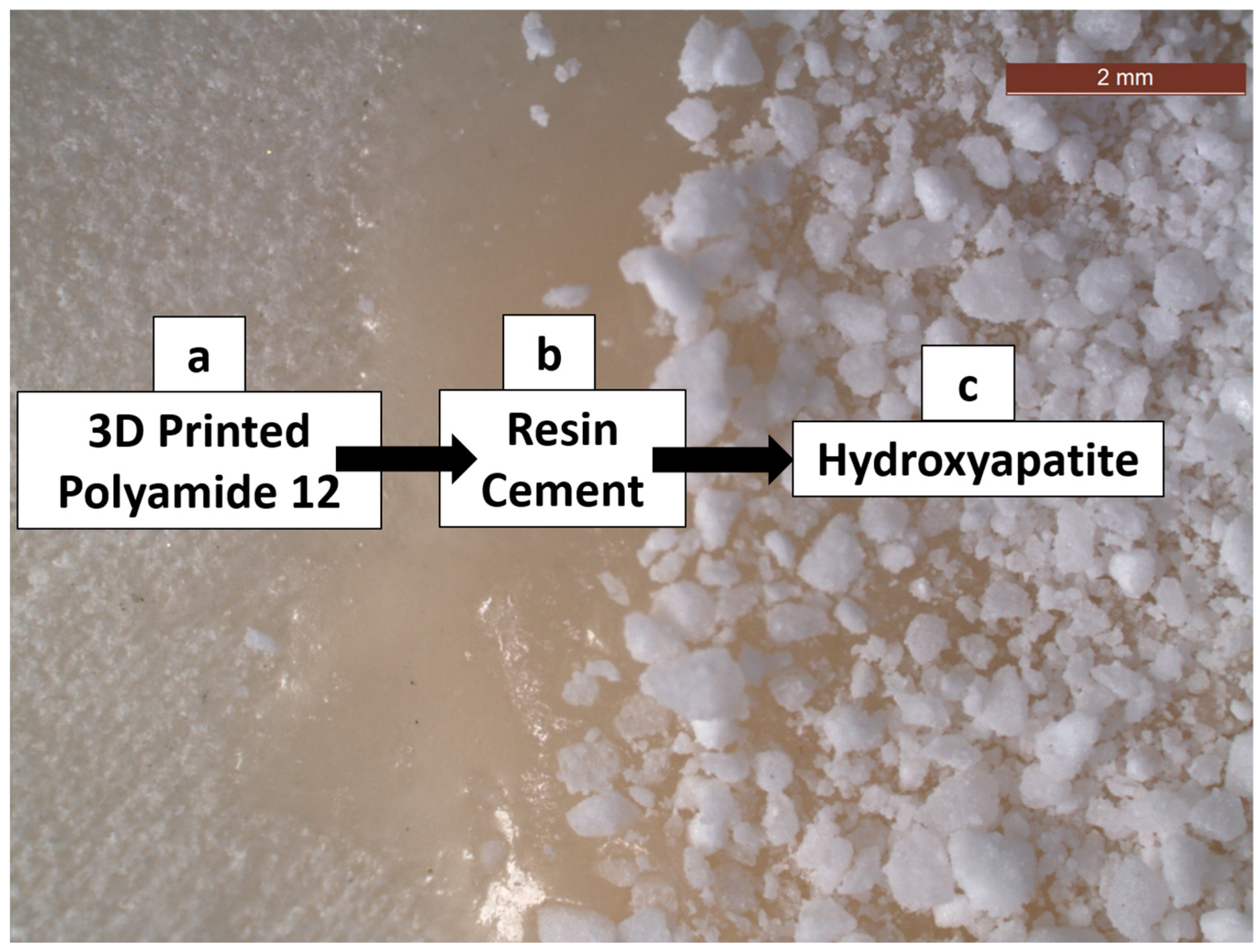
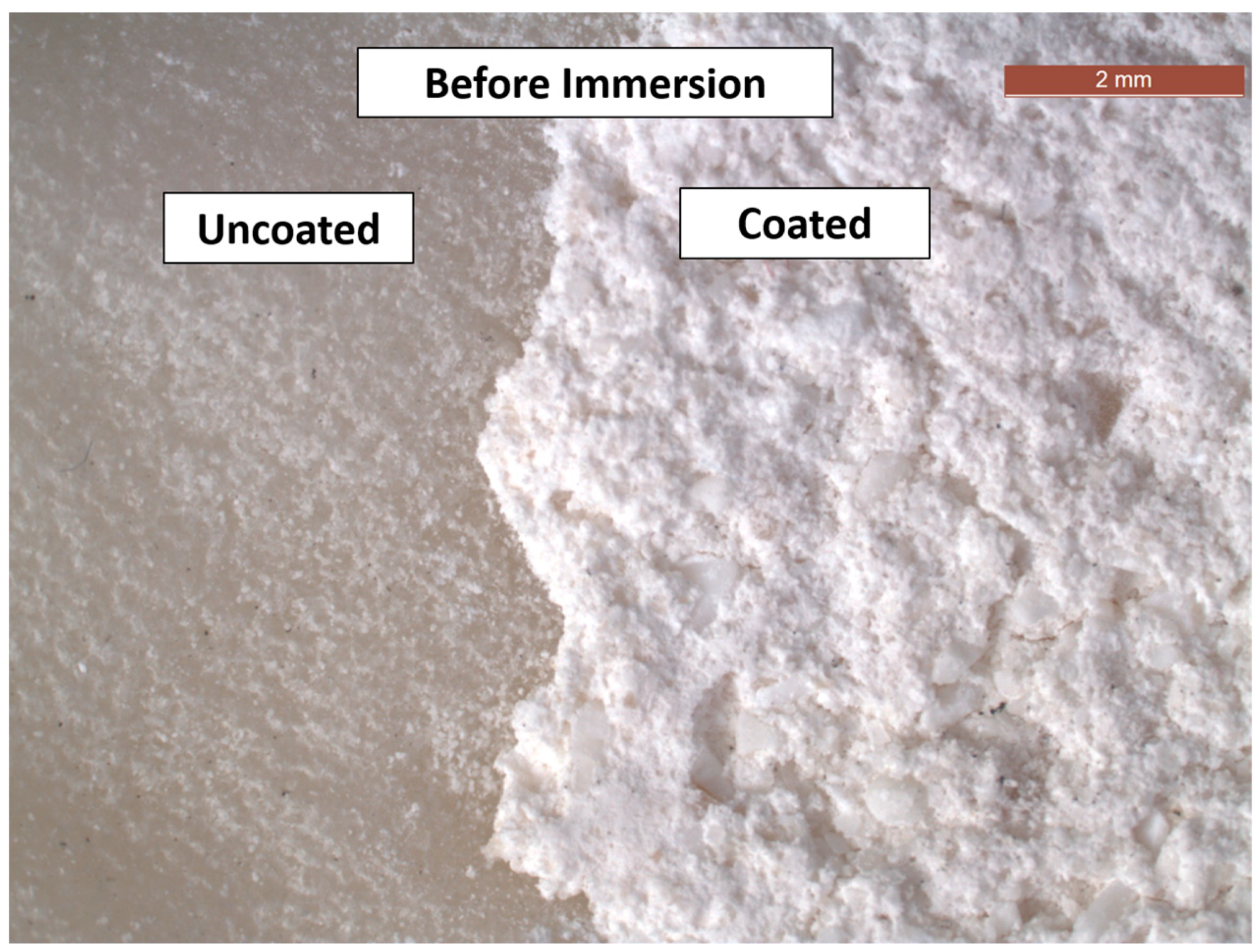
Stereomicroscopic images were taken at magnification 12.5× to illustrate the steps of coating (Figure 2 and Figure 3) using a stereomicroscope (Leica, Allendale, NJ, USA). The left-hand side part of Figure 2a shows the untreated 3D-printed polyamide 12, the middle portion (b) illustrates the resin cement, and the right part (c) demonstrates the application of the hydroxyapatite on the resin cement. Figure 3 is a stereomicroscopic image showing coated and uncoated surfaces before immersion in phosphate-buffered saline.

2.4. Characterisation by Fourier-Transform Infrared and Transmission Electron Microscopy

The coated specimens were characterised by Fourier-Transform Infrared (FTIR) spectroscopy (FT/IR-4000 Series Spectrometer, JASCO, Tokyo, Japan) to detect the functional groups on the specimens’ surface. In addition, the coated specimens were examined using transmission electron microscopy (TEM) (JEM-2100 HR TEM, JEOL Tokyo, Japan) at a 200 kV accelerating voltage and 22 Å imaging resolution.

2.5. Storage in Phosphate-Buffered Saline

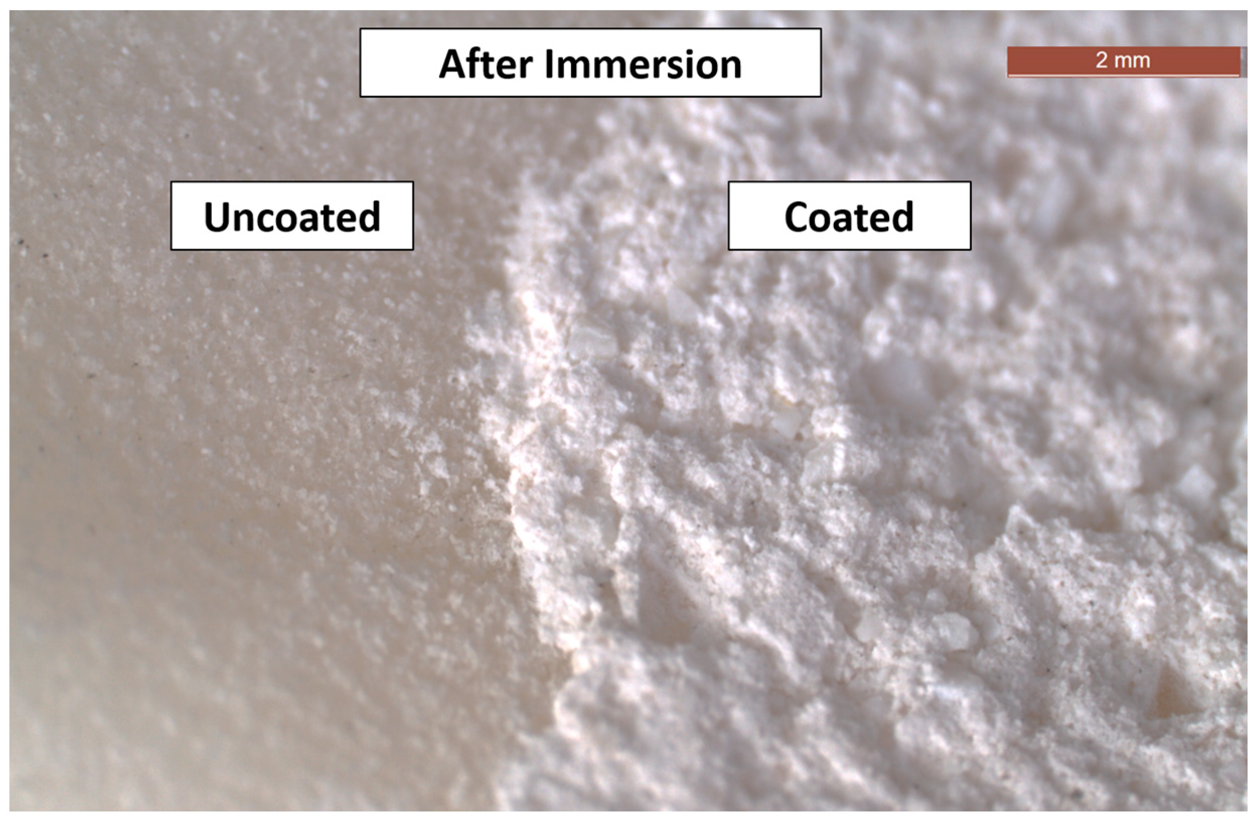
Both the control and treated specimens were next randomly subdivided into two subgroups. Half of the specimens from each group were left without immersion, whereas the other half was immersed in PBS (Dulbecco’s Phosphate-Buffered Saline, Lonza, Verviers, Belgium) for 15 days at 37 °C in an incubator (BTC, Cairo, Egypt). The composition of PBS was 0.0095 M PO43−. The stereomicroscopic image was taken for the coated and uncoated surfaces after immersion in PBS at a magnification of 12.5×, as seen in Figure 4.

2.6. Examination of Surface Microstructure and Elemental Analysis

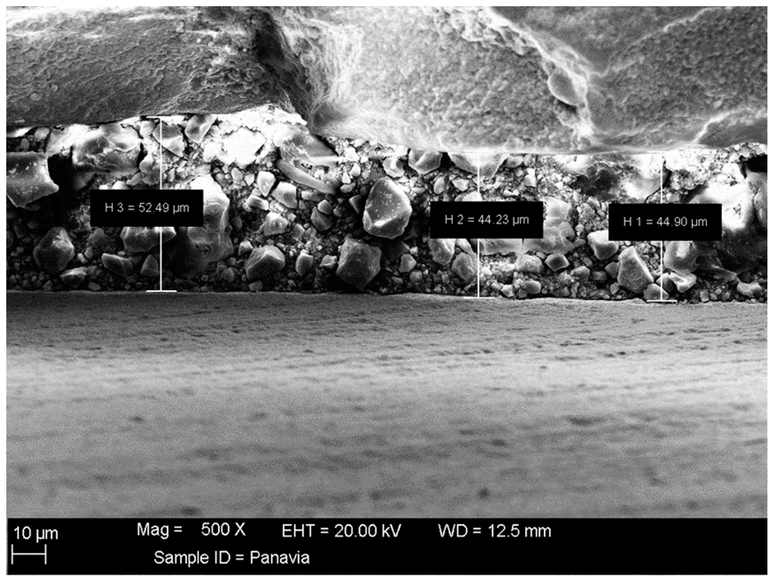
The surfaces of the control and treated specimens were examined using an environmental scanning electron microscope (SEM; JSM-5200, JEOL, Tokyo, Japan) before and after immersion in PBS. A magnification of 200× was used with a 30 kV accelerating voltage. An extra SEM micrograph was taken for the coated specimen (lateral view) to measure the coat thickness with 500× magnification.
Energy dispersive X-ray analysis (EDXA) was performed to detect the elements present on the surfaces of control and treated specimens both before and after immersion in PBS. Energy dispersive X-ray spectroscopy (EDX) (Oxford Inca Energy 350, Oxford Instruments, Abingdon, UK) was used with a 10 mm working distance, 3 nm resolution, and a 30 kV accelerating voltage. The quantification of the elements was presented in atomic%. The Ca/P ratio was calculated in the coated groups to determine the form of the calcium phosphate compound.

2.7. Coat Adhesion Test

Following the standard of the American Society for Testing and Materials (ASTM D3359) [30], the cross-cut adhesion test was performed. A sharp cutting blade (Blades for Adhesion Tester, GLTL, Changzhou, Jiangsu, China) was used to pierce the coating until it reached the substrate. Six uniformly spaced incisions (2 mm apart from each other) were made both vertically and horizontally to create a lattice pattern on the test area’s surface, and then an adhesive tape (ASTM D3359 Cross Hatch Adhesion Test Tape, GLTL, Changzhou, Jiangsu, China) was used to remove the coat following the standard steps of the tape test according to ASTM D3359 [30]. Figure 5 and Table 1 display the adhesion scale ranges.

3. Results

3.1. FTIR and TEM

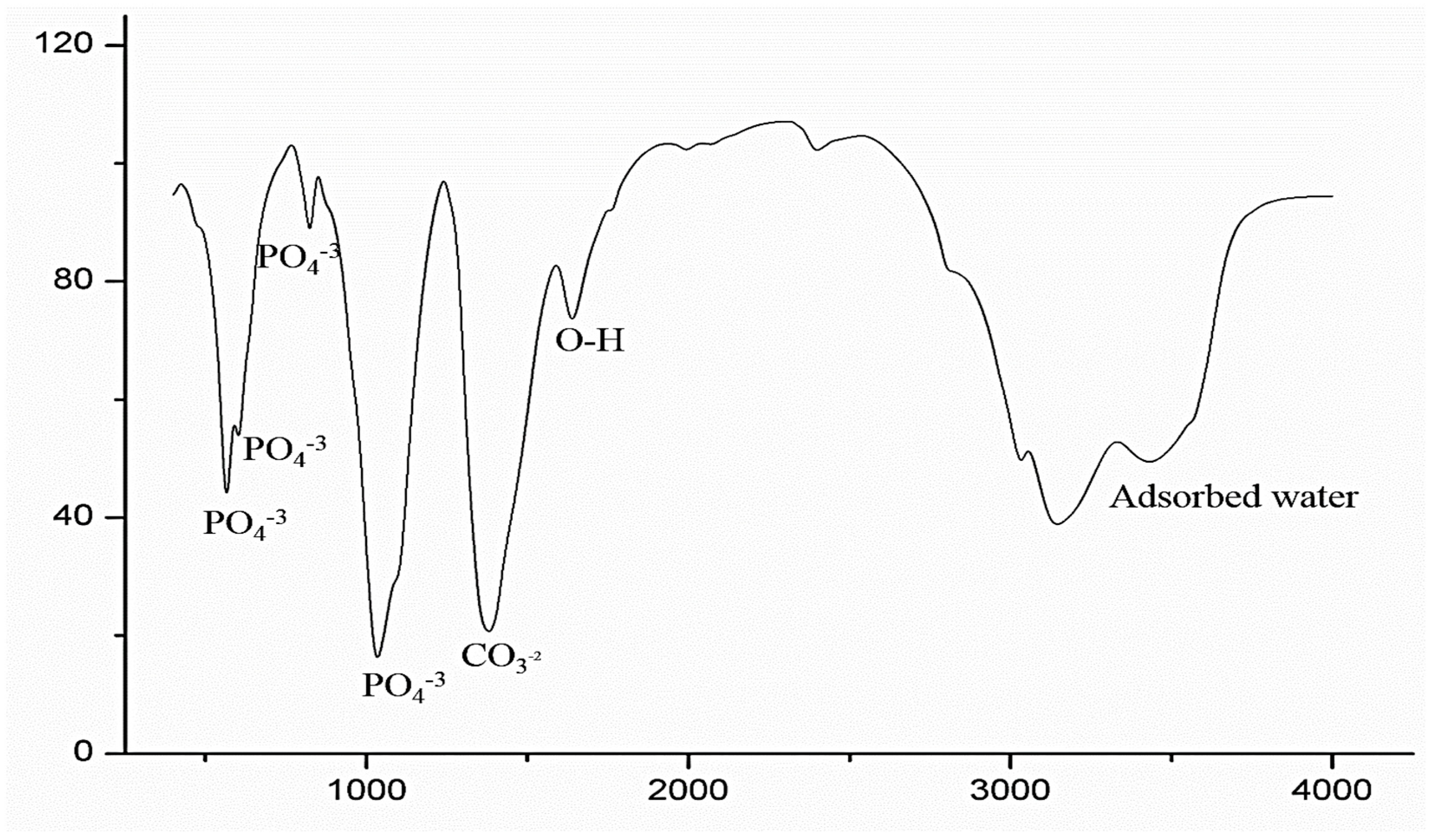
The spectra of the FTIR, seen in Figure 6, revealed the absorption peaks of hydroxyapatite on the coated specimens [31]. The vibration peaks of PO4−3 were shown by the sharp peaks at 566.005 and 601.682 cm−1. The PO4−3 group’s stretching mode was indicated by the bands at 823.455 and 1034.62 cm−1. The band that is seen at 1380.78 cm−1 indicated CO3−2. The O-H stretching mode was represented by the band at 1639.2 cm−1. Adsorbed water was indicated by the wide band at 3432.67 cm−1.
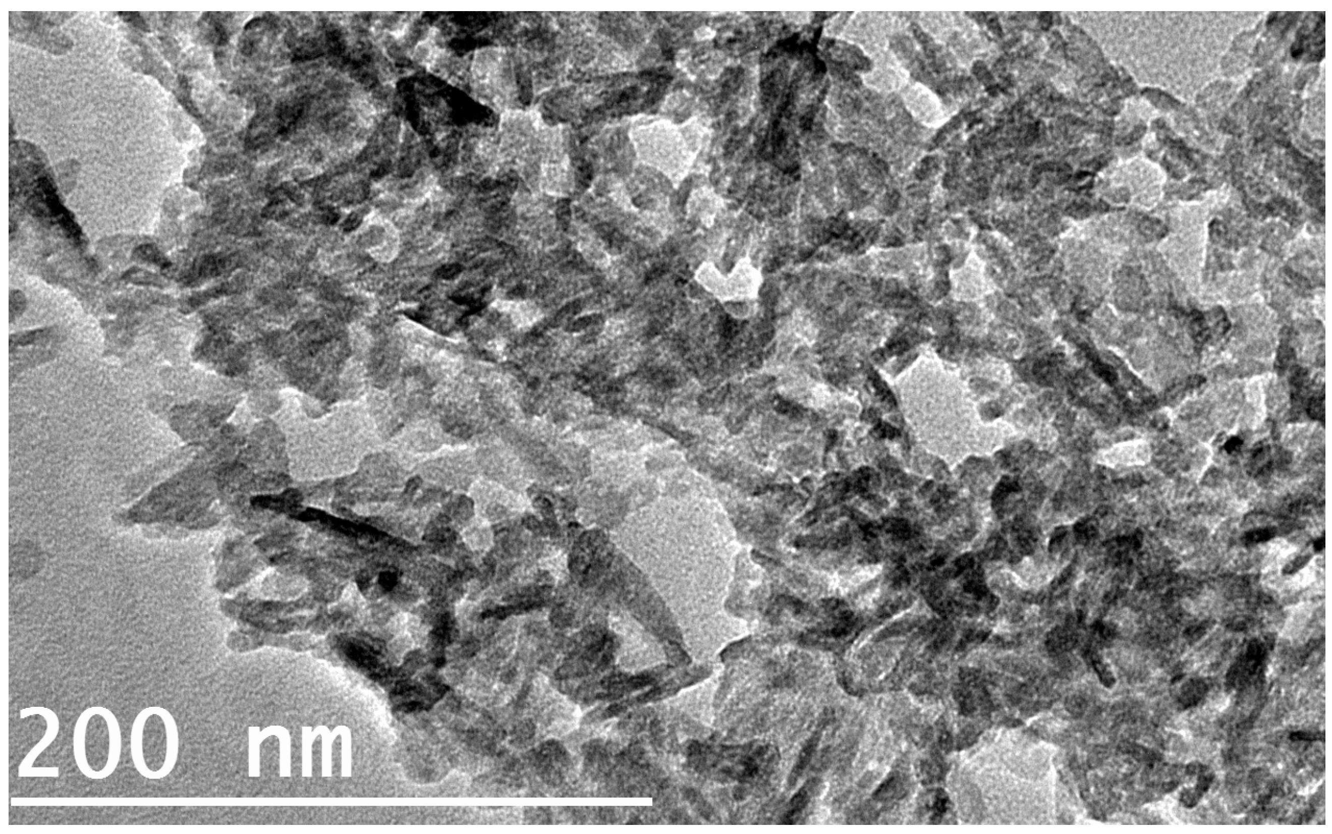
The TEM micrograph of the coated specimen is shown in Figure 7. The micrograph revealed non-uniform clusters representing the hydroxyapatite coat.

3.2. SEM and EDXA

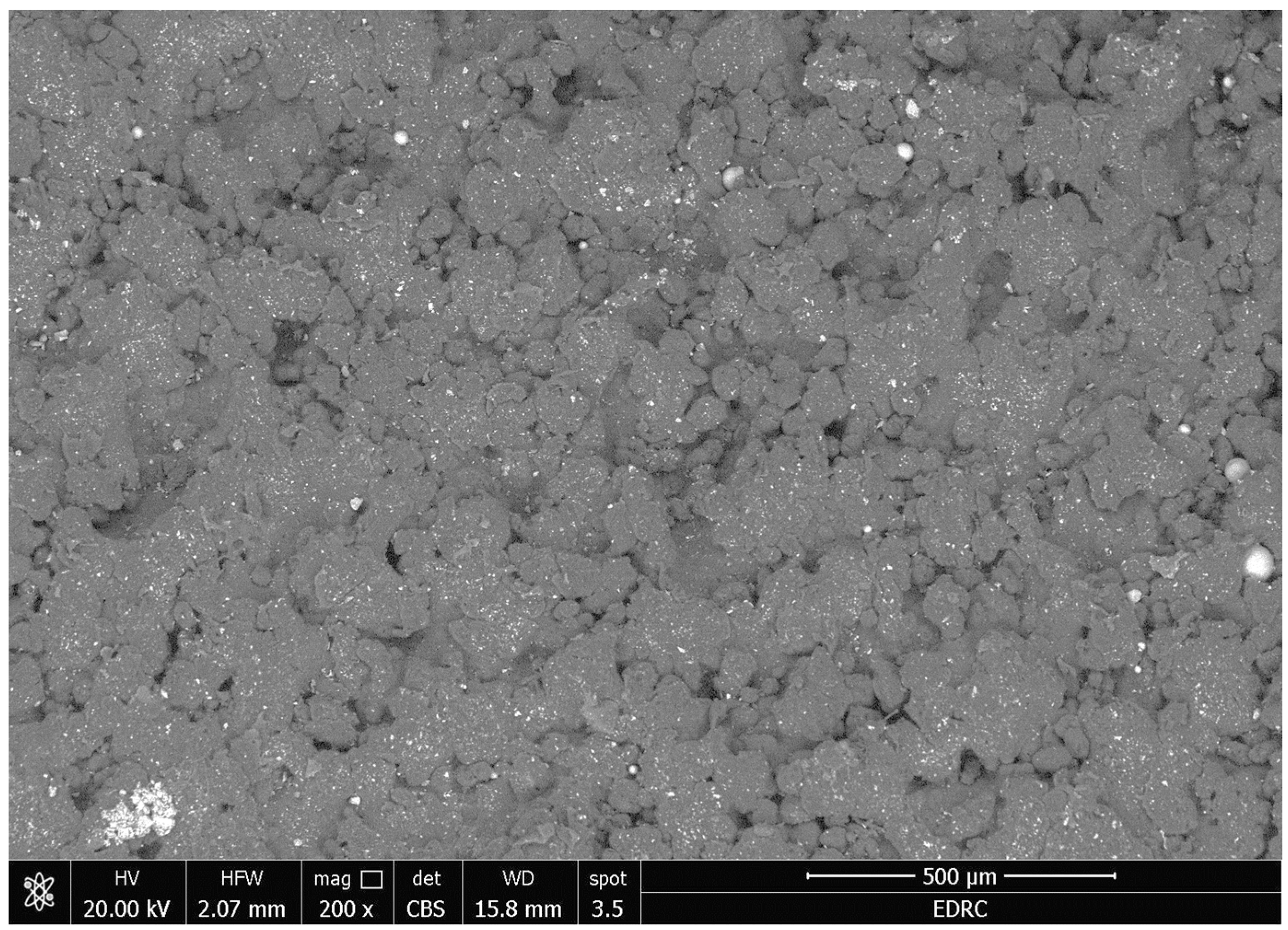
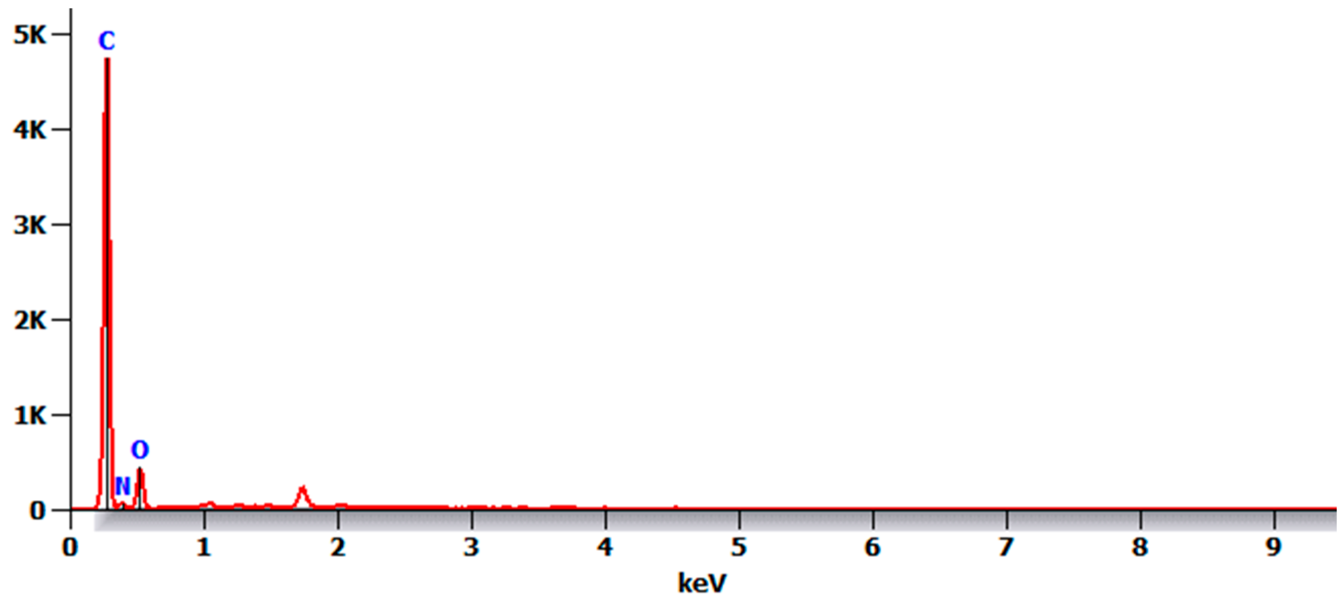
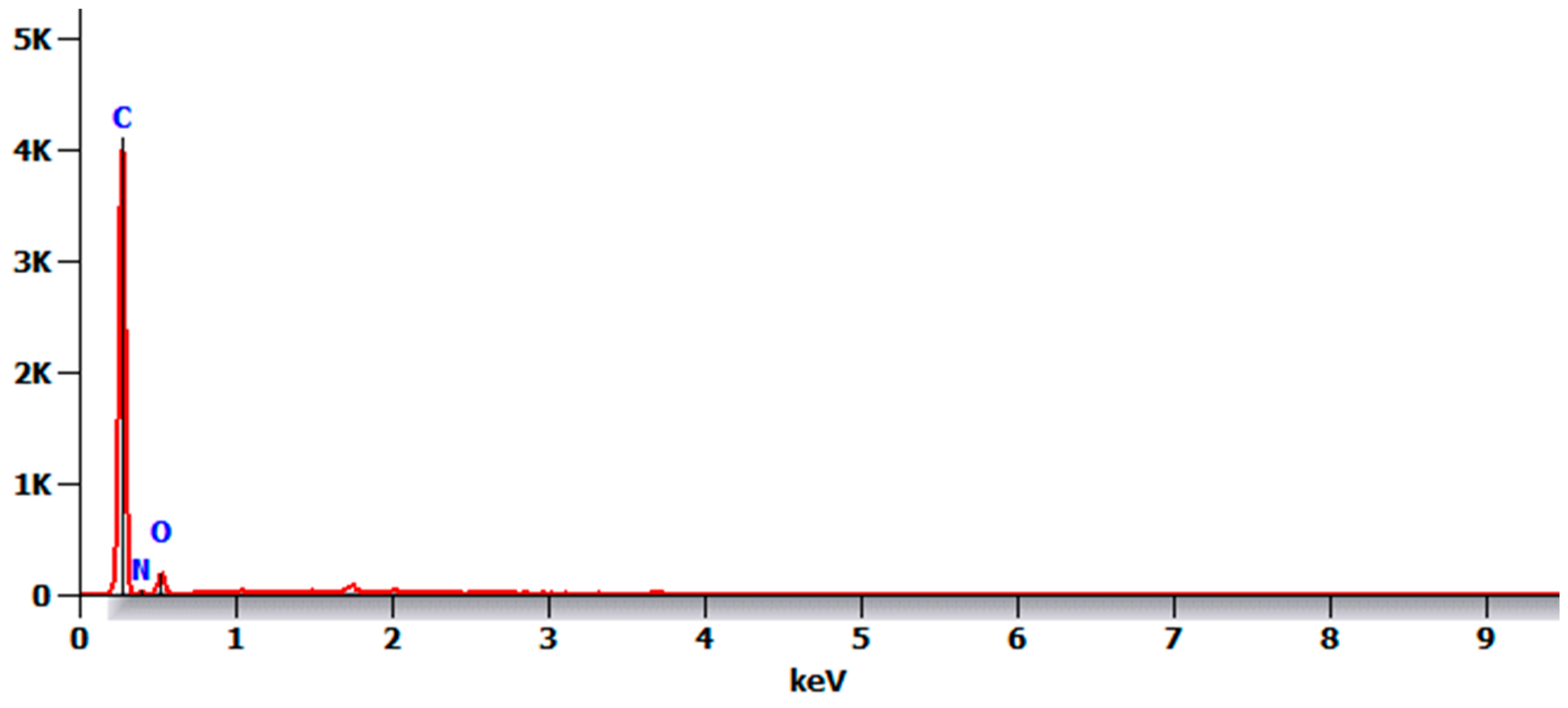
The SEM micrograph of the control 3D-printed polyamide 12 specimens (before immersion in PBS) illustrated the agglomerated 3D-printed particles with minimal porosity (Figure 8; magnification 200×). The EDX analysis of the control specimens (Figure 9) revealed the presence of carbon, nitrogen, and oxygen as atomic%: 52.1 ± 2.4, 23.8 ± 0.8, 24.1 ± 3.2, respectively.
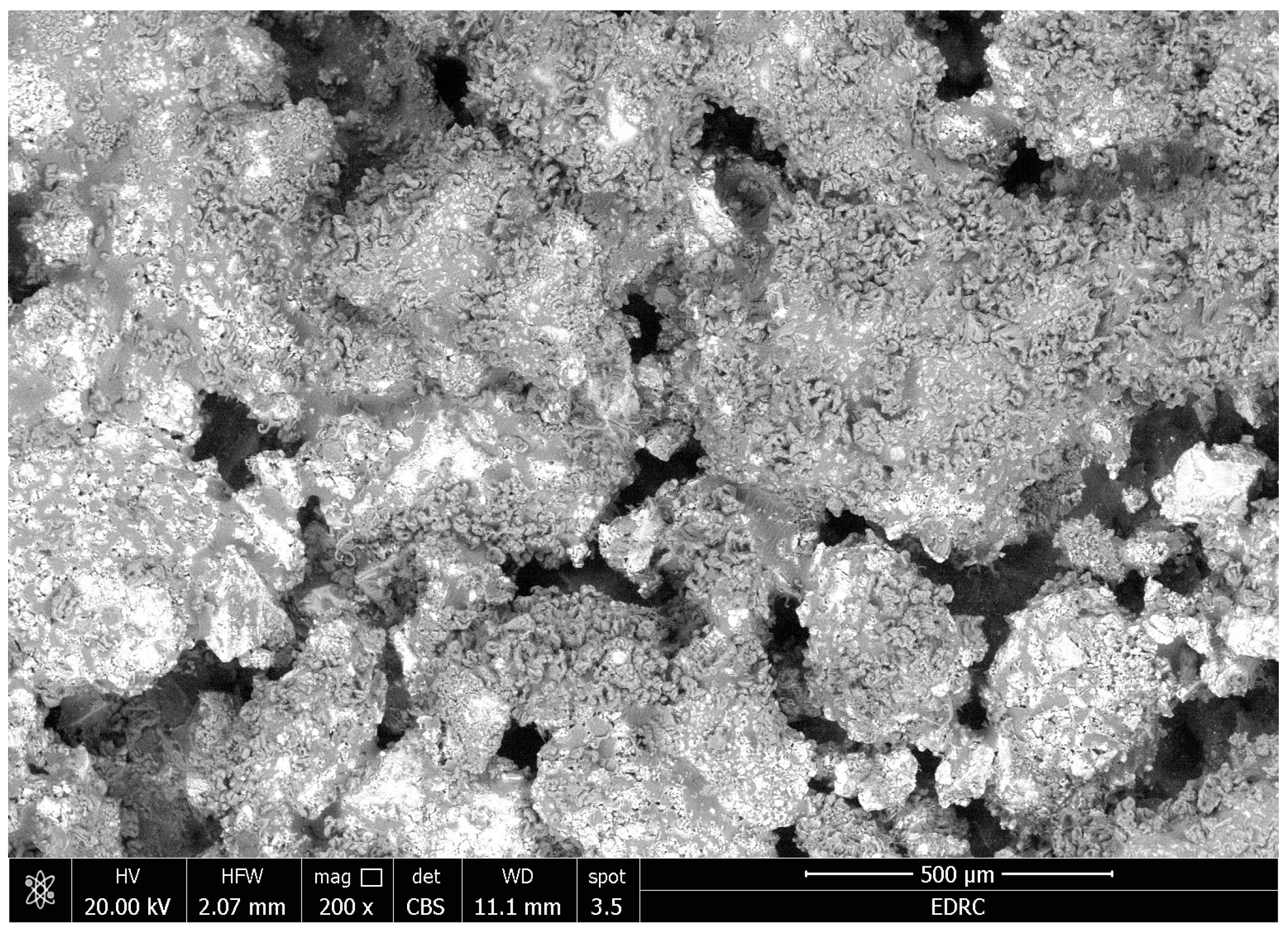
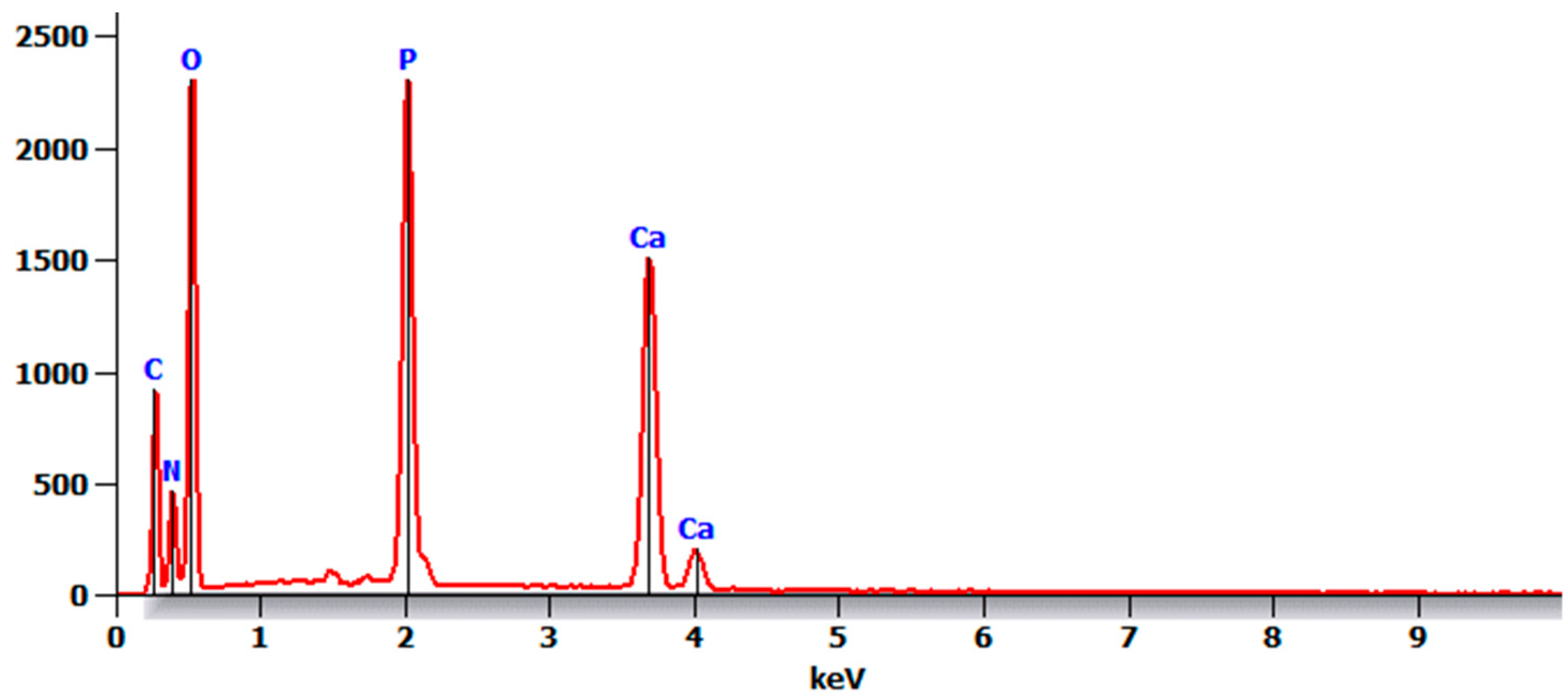
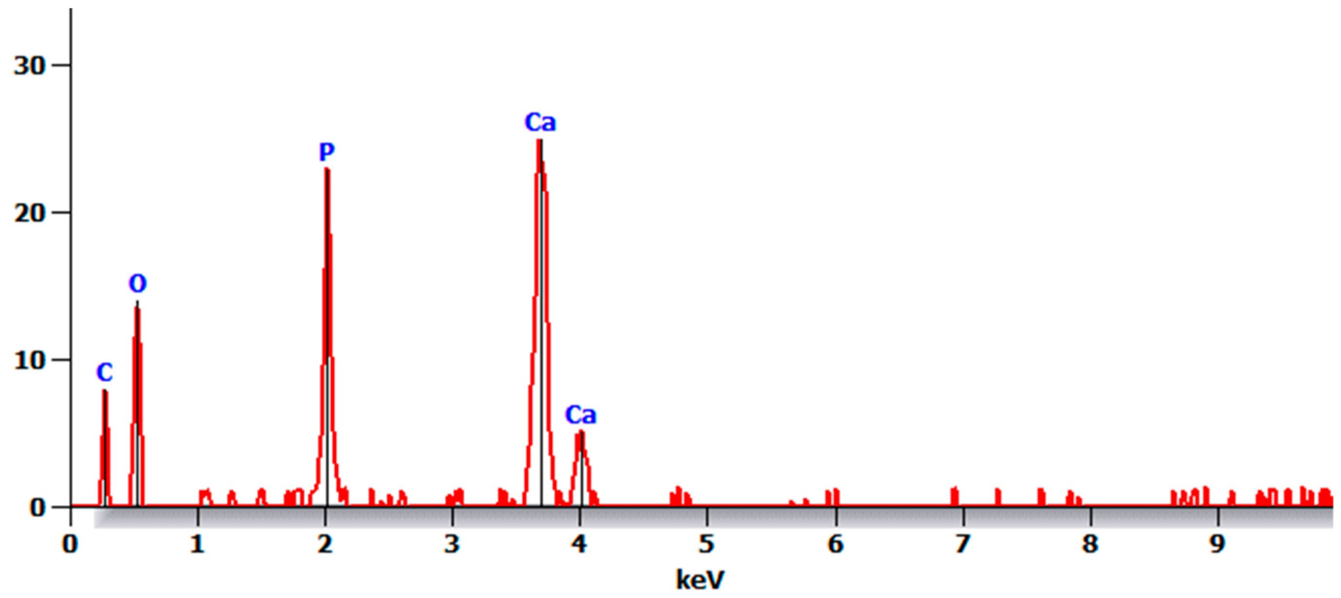
The SEM micrograph of the treated specimens (before immersion in PBS) illustrated the deposition of clusters on the surface (Figure 10). The EDX analysis of the coated specimens (Figure 11) showed the presence of calcium and phosphorus in addition to carbon, nitrogen, and oxygen with atomic%: 9.5 ± 3.1, 5.9 ± 1.6, 7.2 ± 3.8, 30.9 ± 2.0 and 46.5 ± 4.6 respectively. The Ca/P ratio was 1.6.
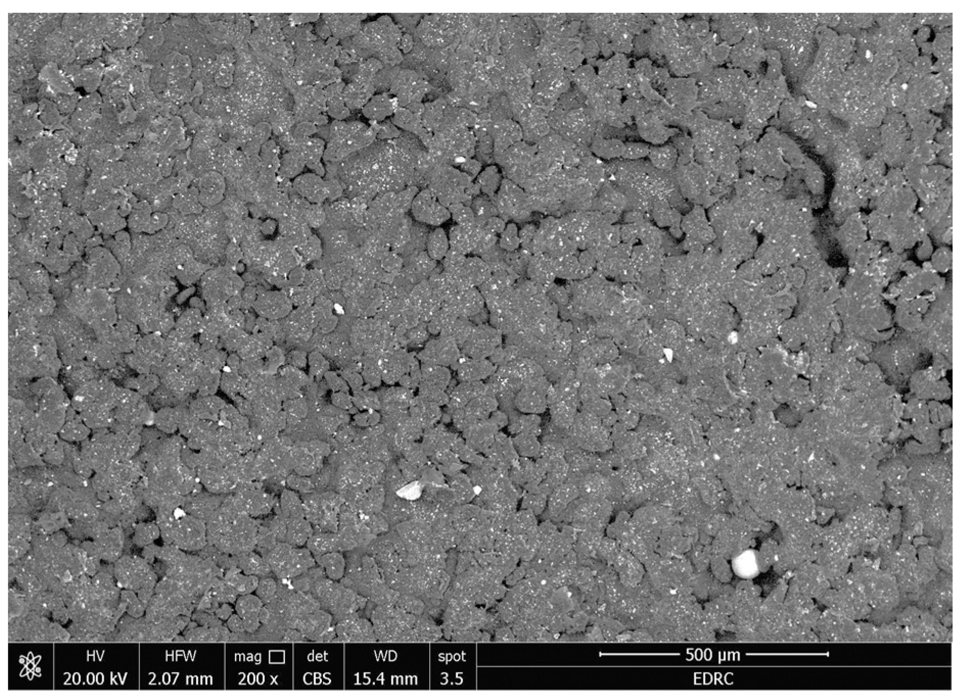
The SEM micrograph of the control 3D-printed polyamide 12 specimens after immersion in PBS (Figure 12) illustrated nearly the same microstructure as before immersion, which was agglomerated particles. The EDX analysis of the control specimens after immersion in PBS revealed the same elements detected before immersion, namely carbon, nitrogen, and oxygen with atomic%: 54.4 ± 2, 24.6 ± 1 and 20.9 ± 3, respectively (Figure 13).
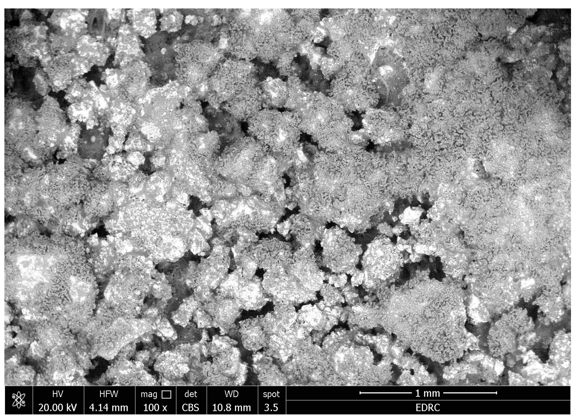
The SEM micrograph of the treated specimens after immersion in PBS illustrated the persistence of the deposited clusters upon the surface (Figure 14). The EDX analysis of the coated specimens (Figure 15) showed calcium and phosphorus in addition to carbon and oxygen with atomic%: 15.7 ± 2, 7.8 ± 1.5, 17.5 ± 3, 59 ± 4, respectively. The Ca/P ratio was 2.
To determine the coat thickness, the SEM micrograph of the lateral view of the coated specimen (Figure 16) displays that the average thickness of the coat was 100 ± 5 μm, the resin cement layer thickness was 47.2 ± 4.6 μm, and the hydroxyapatite was 52.8 ± 4.9 μm. The resin cement is represented by fillers (of different sizes) and the resinous matrix, and it is located as an intermediary layer between the substructure below and the hydroxyapatite above it.

3.3. Coat Adhesion Test

The surface of the coated specimens displayed the score 4B, which indicated minor coating flakes detached at the lines of intersections (area removed <5%) both before and after storage in PBS for 15 days at 37 °C.

4. Discussion

The 3D-printed polyamide 12 is a promising polymeric material that has been used in orthopedic reconstruction with successful results [32]. Its coating with hydroxyapatite (HA) could enhance its bonding with bone [19]. Resin cement containing 10-MDP has shown a durable bond with calcium and phosphate present in tooth structure [33,34]. Yet, it is noteworthy that this cement has not been previously used as an adhesive layer to coat the hydroxyapatite with the polymeric substructure.
The coated specimens were characterised by FTIR spectroscopy. Since each chemical bond absorbs infrared (IR) radiation with a unique wavenumber, FTIR allows the characterisation of many chemical bonds and functional groups found in a substance. The observed FTIR absorption spectrum’s shape and intensity matched the bands generated by HA particles. The obtained HA spectra agreed with those reported by previous studies [35,36,37]. The coated specimens were further characterised by TEM, which provided in-depth examination of the morphology and microstructure of the specimens. The TEM micrograph revealed non-uniform clusters representing the hydroxyapatite coat.
The current study examined the microstructure and elemental analysis of 3D-printed polyamide 12 coated with hydroxyapatite using light-cured resin cement versus an uncoated one (control). Now, the null hypothesis was rejected as there were differences between the control and coated specimens in the microstructure and elemental analysis either before or after immersion in PBS.
The SEM micrographs of the control 3D-printed polyamide 12 specimens illustrated agglomerated particles with minimal porosity due to the sintering process performed by the 3D printing process performed by the selective laser sintering device. The minimal porosity of PA12 in this study after 3D printing was not in agreement with a previous study that reported a non-homogenous surface with several particles as well as multiple pores and attributed this to the wavelength of the laser beam used [38].
The EDXA of the control specimens revealed the presence of carbon, nitrogen, and oxygen, as polyamide 12 is made up of amide group (-CO-NH-) repeating units joined by carbon atoms. Yet, the EDX was unable to detect hydrogen because of its solitary valence electron that makes up the K-shell of the hydrogen atom and participates in chemical bonding [39].
After immersion in PBS, there were no major changes in the control specimens, as detected by SEM and EDXA. This may be attributed to the surface properties of polyamide 12 and its resistance against deterioration as a result of its molecular structure with entangled long chains [40], in addition to its thermal stability due to the amide groups [41]. Moreover, polyamide 12 exhibits favourable characteristics such as excellent dimensional stability and low water sorption [42,43].
The microstructure of the coated specimens showed deposited calcium phosphate clusters on the surface, as the coating is mainly composed of hydroxyapatite. The molecular formula of hydroxyapatite is Ca5(PO4)3OH [44]. Its crystal unit cell contains ten calcium ions (10 Ca2+) and its chemical formula is frequently written as Ca10(PO4)6(OH)2 [44]. Its Ca/P atomic ratio is 10/6 or 1.67 in the unit cell [45]. Any deviation from the exact Ca/P ratio destabilises the crystal and enhances the dissolution of the material. In this research, the Ca/P ratio was 1.60, indicating calcium-deficient HA, which is slightly more bioactive than stoichiometric HA with a Ca/P ratio of 1.67 [46].
Several analytical techniques have been established in order to assess the success materials within the human body during their service life. Testing the materials for use in bone applications in a simulated testing medium is one of the most significant of these techniques [47]. It is reported that, to replicate the behaviour of materials, solutions having a composition comparable to that of human blood plasma can be utilised, such as phosphate-buffered saline (PBS) [48].
This current calcium phosphate coat did not show detachment from the surface after immersion, as revealed by SEM and EDXA. In addition, adhesion of the coating showed that the surface of the coated specimens displayed minor coating flakes. This might be due to the durable bond between the 10-MDP monomer and hydroxyapatite with the low rate of dissolution of resin cement in water [49]. After immersion in PBS, the Ca/P ratio was 2, which denoted tetra-calcium-phosphate (TTCP) with the chemical formula Ca4(PO4)2O. This conversion could be attributed to the chemical interaction between the hydroxyapatite and phosphate-buffered saline [50,51].
The surface treatment of hydroxyapatite using light-cured resin cement in PA12 could be considered a time-saving process. Contrary to the sol–gel technique used previously, this was a multi-step process. This started with the following preparation of the sol: tetra-ethyl-orthosilicate (4 mL), calcium-nitrate ethanolic solution (1 mL), and 85% phosphoric acid (0.13 mL) to ethanol (32.0 mL) under magnetic stirring. After agitation (30 min), an ammonia-ethanolic solution (2.4 mL) was added to the reaction mixture. This was followed by polyamide coating, where this sol was agitated for two hours to be deposited on PA by dip-coating. The substrates were left in the sols for 20 min; they were then dried for a day at 50 °C, and the resultant coat thickness was less than 3 μm [38].
On the other hand, the introduced coating method using light-cured resin cement was much simpler and seemed promising in this study, yet further studies are needed to assess and verify its interactions with osteoblasts, wettability, and the resultant surface roughness.
Although tribo-mechanical aspects of the coat were not measured in this study, it was expected that the coat could be stable during service due to the following several reasons: (1) the coated specimens were stored in phosphate-buffered saline for 15 days and no detachment was detected (examined by stereomicroscope, SEM and EDXA); (2) a coat adhesion test was performed following the standard American Society for Testing and Materials (ASTM D3359) after storage for 15 days in phosphate-buffered saline, and, in this test, mechanical stress was applied during cutting to penetrate the coat until reaching the substructure; it should be noted that in this study this test resulted a stable coat with just a few loose coating flakes (area removed <5%); (3) the used resin cement was the type containing 10-MDP, which was reported previously in the literature to bond chemically to various substructures and resist water degradation [52]. However, it is recommended to test the coat when subjected to wear or friction in future studies.

5. Conclusions

In summary, the 3D-printed polyamide 12 could be coated with hydroxyapatite using light-cured resin cement containing 10-methacryloyloxydecyldihydrogenphosphate (10-MDP). To check the optimal structure of the coated specimens, Fourier transform infrared spectroscopy (FTIR) and transmission electron microscopy (TEM) were used for characterisation. The stability of the coat was assessed after immersion for 15 days in phosphate-buffered saline (PBS) at 37 °C. The surfaces of the coated specimens were examined using a scanning electron microscope (SEM) and energy dispersive X-ray analysis (EDXA) both before and after immersion in PBS and compared to uncoated control specimens. In addition, a cross-cut adhesion test was performed following the standard American Society for Testing and Materials (ASTM D3359) before and after immersion in 37 °C PBS for 15 days. The main findings were presented as follows:
  • The results of the FTIR spectroscopy of the coated specimens confirmed the HA bands. The TEM micrographs revealed HA-agglomerated particles in the coat.
  • The coat was stable after immersion in PBS, as observed by SEM and EDXA as well as the coat adhesion test, which demonstrated a stable coat with just a few loose coating flakes (area removed <5%) on the surface of the HA-coated specimens.
  • There were no major changes in both the coated and uncoated (control) specimens before and after immersion in PBS. The SEM micrographs of the control 3D-printed polyamide 12 specimens illustrated the sintered 3D-printed particles with minimal porosity. Their EDXA revealed the presence of carbon, nitrogen, and oxygen. The microstructure of the coated specimens showed deposited clusters of calcium and phosphorus on the surface in addition to carbon, nitrogen, and oxygen.
Therefore, the light-cured resin cement resin with 10-MDP functional groups could be used for coating the 3D-printed polyamide 12 with hydroxyapatite, yet biological assessment is the further step to ensure its biocompatibility for biomedical applications.

Author Contributions

Conceptualisation, R.M.A. and T.M.H.; data curation, T.M.H., R.M.A., S.A.A. and A.A.; formal analysis, T.M.H., R.M.A. and A.A.; investigation, R.M.A. and S.A.A.; methodology, T.M.H., R.M.A., A.A. and S.A.A.; visualisation, A.A., S.A., S.A.A., R.E.B. and J.P.M.; resources, A.A., S.A., S.A.A. and R.E.B.; supervision, T.M.H., R.M.A., A.A. and J.P.M.; writing—original draft, R.M.A., T.M.H. and S.A.A.; writing—review and editing; T.M.H., R.M.A., J.P.M. and A.A. All authors have read and agreed to the published version of the manuscript.

Funding

This research was funded by Researchers Supporting Project number (RSPD2024R790), King Saud University, Riyadh, Saudi Arabia.

Institutional Review Board Statement

This study is approved by the Medical Research Ethical Committee (MREC) of National Research Centre (NRC), Cairo, Egypt (Ref. number: 1287112022).

Informed Consent Statement

Not applicable.

Data Availability Statement

The data presented in this study are available on request from the corresponding author.

Acknowledgments

The authors are grateful to the Researchers Supporting Project number (RSPD2024R790), King Saud University, Riyadh, Saudi Arabia.

Conflicts of Interest

The authors declare no conflicts of interest.

References

  1. Earar, K.; Iliescu, A.A.; Popa, G.; Iliescu, A.; Rudnic, I.; Feier, R.; Voinea-Georgescu, R.N. Additive vs. Subtractive CAD/CAM Procedures in Manufacturing of the PMMA Interim Dental Crowns a Comparative in Vitro Study of Internal Fit. Rev. Chim. 2020, 71, 405–410. [Google Scholar] [CrossRef]
  2. Hamdy, T.M.; Abdelnabi, A.; Othman, M.S.; Bayoumi, R.E. Alterations in Surface Gloss and Hardness of Direct Dental Resin Composites and Indirect CAD/CAM Composite Block after Single Application of Bifluorid 10 Varnish: An In Vitro Study. J. Compos. Sci. 2024, 8, 58. [Google Scholar] [CrossRef]
  3. Bhatnagar, A.; Bhardwaj, A.; Verma, S. Additive Manufacturing: A 3-Dimensional Approach in Periodontics. J. Adv. Med. Med. Res. 2020, 32, 105–117. [Google Scholar] [CrossRef]
  4. Moon, J.-M.; Jeong, C.-S.; Lee, H.-J.; Bae, J.-M.; Choi, E.-J.; Kim, S.-T.; Park, Y.-B.; Oh, S.-H. A Comparative Study of Additive and Subtractive Manufacturing Techniques for a Zirconia Dental Product: An Analysis of the Manufacturing Accuracy and the Bond Strength of Porcelain to Zirconia. Materials 2022, 15, 5398. [Google Scholar] [CrossRef]
  5. Jeong, M.; Radomski, K.; Lopez, D.; Liu, J.T.; Lee, J.D.; Lee, S.J. Materials and Applications of 3D Printing Technology in Dentistry: An Overview. Dent. J. 2023, 12, 1. [Google Scholar] [CrossRef]
  6. Cai, H.; Xu, X.; Lu, X.; Zhao, M.; Jia, Q.; Jiang, H.-B.; Kwon, J.-S. Dental Materials Applied to 3D and 4D Printing Technologies: A Review. Polymers 2023, 15, 2405. [Google Scholar] [CrossRef]
  7. Revilla-León, M.; Sadeghpour, M.; Özcan, M. An Update on Applications of 3D Printing Technologies Used for Processing Polymers Used in Implant Dentistry. Odontology 2020, 108, 331–338. [Google Scholar] [CrossRef]
  8. Rokaya, D.; Srimaneepong, V.; Sapkota, J.; Qin, J.; Siraleartmukul, K.; Siriwongrungson, V. Polymeric Materials and Films in Dentistry: An Overview. J. Adv. Res. 2018, 14, 25–34. [Google Scholar] [CrossRef]
  9. Nathanael, A.J.; Oh, T.H. Biopolymer Coatings for Biomedical Applications. Polymers 2020, 12, 3061. [Google Scholar] [CrossRef]
  10. Vidakis, N.; Petousis, M.; Tzounis, L.; Maniadi, A.; Velidakis, E.; Mountakis, N.; Kechagias, J.D. Sustainable Additive Manufacturing: Mechanical Response of Polyamide 12 over Multiple Recycling Processes. Materials 2021, 14, 466. [Google Scholar] [CrossRef]
  11. Priyadarshini, B.M.; Kok, W.K.; Dikshit, V.; Feng, S.; Li, K.H.H.; Zhang, Y. 3D Printing Biocompatible Materials with Multi Jet Fusion for Bioreactor Applications. Int. J. Bioprint. 2023, 9, 14–35. [Google Scholar] [CrossRef] [PubMed]
  12. Arafat, S.W.; Ibrahim, W.H.; Shaker, S.; Aldainy, D.G.; Salama, D.; Shaheen, H.A. Reconstruction of Cranial Bone Defects Using Polyamide 12 Patient-Specific Implant: Long Term Follow Up. J. Craniofac. Surg. 2022, 33, 1825–1828. [Google Scholar] [CrossRef] [PubMed]
  13. Shaheen, H.A.; Shaker, S.; Ibrahim, W.H.; AlDainy, D.G.; Salama, D.; Emara, A.S. Delayed Complex Fronto-Zygomatico-Orbital Reconstruction Using Patient Specific 3D Printed Implants; A Report of Three Complex Cases—A Short Communication. J. Plast. Reconstr. Aesthetic Surg. 2022, 75, 3877–3903. [Google Scholar] [CrossRef] [PubMed]
  14. Gomes, P.C.; Piñeiro, O.G.; Alves, A.C.; Carneiro, O.S. On the Reuse of SLS Polyamide 12 Powder. Materials 2022, 15, 5486. [Google Scholar] [CrossRef] [PubMed]
  15. Hamdy, T.M.; Saniour, S.H.; Sherief, M.A.; Zaki, D.Y. Effect of Incorporation of 20 Wt% Amorphous Nano-Hydroxyapatite Fillers in Poly Methyl Methacrylate Composite on the Compressive Strength. Res. J. Pharm. Biol. Chem. Sci. 2015, 6, 1136–1141. [Google Scholar] [CrossRef]
  16. Abdelnabi, A.; Hamza, M.K.; El-Borady, O.M.; Hamdy, T.M. Effect of Different Formulations and Application Methods of Coral Calcium on Its Remineralization Ability on Carious Enamel. Open Access Maced. J. Med. Sci. 2020, 8, 94–99. [Google Scholar] [CrossRef]
  17. Hamdy, T.M.; Mousa, S.M.A.; Sherief, M.A. Effect of Incorporation of Lanthanum and Cerium-Doped Hydroxyapatite on Acrylic Bone Cement Produced from Phosphogypsum Waste. Egypt. J. Chem. 2020, 63, 1823–1832. [Google Scholar] [CrossRef]
  18. Dutta, S.R.; Passi, D.; Singh, P.; Bhuibhar, A. Ceramic and Non-Ceramic Hydroxyapatite as a Bone Graft Material: A Brief Review. Ir. J. Med. Sci. 2015, 184, 101–106. [Google Scholar] [CrossRef]
  19. Hamdy, T.M. Dental Biomaterial Scaffolds in Tooth Tissue Engineering: A Review. Curr. Oral Health Reports 2023, 10, 14–21. [Google Scholar] [CrossRef]
  20. Chae, M.H.; Lee, Y.K.; Kim, K.N.; Lee, J.H.; Choi, B.J.; Choi, H.J.; Park, K.T. The Effect of Hydroxyapatite on Bonding Strength in Light Curing Glass Ionomer Dental Cement. Key Eng. Mater. 2006, 309, 881–884. [Google Scholar] [CrossRef]
  21. Razali, R.A.C.; Rahim, N.A.; Zainol, I.; Sharif, A.M. Preparation of Dental Composite Using Hydroxyapatite from Natural Sources and Silica. In Proceedings of the Journal of Physics: Conference Series; IOP Publishing: Bristol, UK, 2018. [Google Scholar]
  22. Domingo, C.; Arcs, R.W.; Lpez-Macipe, A.; Osorio, R.; Rodrguez-Clemente, R.; Murtra, J.; Fanovich, M.A.; Toledano, M. Dental Composites Reinforced with Hydroxyapatite: Mechanical Behavior and Absorption/Elution Characteristics. J. Biomed. Mater. Res. 2001, 56, 297–305. [Google Scholar] [CrossRef]
  23. Ong, J.L.; Chan, D.C.N. Hydroxyapatite and Their Use as Coatings in Dental Implants: A Review. Crit. Rev. Biomed. Eng. 2000, 28, 667–707. [Google Scholar] [CrossRef] [PubMed]
  24. Soleymani, S.; Naghib, S.M. 3D and 4D Printing Hydroxyapatite-Based Scaffolds for Bone Tissue Engineering and Regeneration. Heliyon 2023, 9, e19363. [Google Scholar] [CrossRef] [PubMed]
  25. Szcześ, A.; Hołysz, L.; Chibowski, E. Synthesis of Hydroxyapatite for Biomedical Applications. Adv. Colloid Interface Sci. 2017, 249, 321–330. [Google Scholar] [CrossRef] [PubMed]
  26. Abdulghafor, M.A.; Mahmood, M.K.; Tassery, H.; Tardivo, D.; Falguiere, A.; Lan, R. Biomimetic Coatings in Implant Dentistry: A Quick Update. J. Funct. Biomater. 2023, 15, 15. [Google Scholar] [CrossRef]
  27. Carvalho, P.C.K.; Almeida, C.C.M.S.; Souza, R.O.A.; Tango, R.N. The Effect of a 10-MDP-Based Dentin Adhesive as Alternative for Bonding to Implant Abutment Materials. Materials 2022, 15, 5449. [Google Scholar] [CrossRef]
  28. Al-Shehri, E.Z.; Al-Zain, A.O.; Sabrah, A.H.; Al-Angari, S.S.; Al Dehailan, L.; Eckert, G.J.; Özcan, M.; Platt, J.A.; Bottino, M.C. Effects of Air-Abrasion Pressure on the Resin Bond Strength to Zirconia: A Combined Cyclic Loading and Thermocycling Aging Study. Restor. Dent. Endod. 2017, 42, 206–215. [Google Scholar] [CrossRef]
  29. Pekkan, G. Radiopacity of Dental Materials: An Overview. Avicenna J. Dent. Res. 2016, 8, 8. [Google Scholar] [CrossRef]
  30. ASTM D3359/D3359M-17; Standard Test Methods for Measuring Adhesion by Tape Test. American Society for Testing and Materials, ASTM International: West Conshohocken, PA, USA, 2017.
  31. Torabinejad, B.; Mohammadi-Rovshandeh, J.; Davachi, S.M.; Zamanian, A. Synthesis and Characterization of Nanocomposite Scaffolds Based on Triblock Copolymer of L-Lactide, ε-Caprolactone and Nano-Hydroxyapatite for Bone Tissue Engineering. Mater. Sci. Eng. C Mater. Biol. Appl. 2014, 42, 199–210. [Google Scholar] [CrossRef]
  32. Zakręcki, A.; Cieślik, J.; Bazan, A.; Turek, P. Innovative Approaches to 3D Printing of PA12 Forearm Orthoses: A Comprehensive Analysis of Mechanical Properties and Production Efficiency. Materials 2024, 17, 663. [Google Scholar] [CrossRef]
  33. Pimentel de Oliveira, R.; de Paula, B.L.; Ribeiro, M.E.; Alves, E.; Costi, H.T.; Silva, C. Evaluation of the Bond Strength of Self-Etching Adhesive Systems Containing HEMA and 10-MDP Monomers: Bond Strength of Adhesives Containing HEMA and 10-MDP. Int. J. Dent. 2022, 2022, 5756649. [Google Scholar] [CrossRef] [PubMed]
  34. Hamdy, T.M. Polymerization Shrinkage in Contemporary Resin-Based Dental Composites: A Review Article. Egypt. J. Chem. 2021, 64, 3087–3092. [Google Scholar] [CrossRef]
  35. Gheisari, H.; Karamian, E.; Abdellahi, M. A Novel Hydroxyapatite -Hardystonite Nanocomposite Ceramic. Ceram. Int. 2015, 41, 5967–5975. [Google Scholar] [CrossRef]
  36. Chandrasekar, A.; Sagadevan, S.; Dakshnamoorthy, A. Synthesis and Characterization of Nano-Hydroxyapatite (n-HAP) Using the Wet Chemical Technique. Int. J. Phys. Sci. 2013, 8, 1639–1645. [Google Scholar]
  37. Bouropoulos, N.; Stampolakis, A.; Mouzakis, D.E. Dynamic Mechanical Properties of Calcium Alginate-Hydroxyapatite Nanocomposite Hydrogels. Sci. Adv. Mater. 2010, 2, 239–242. [Google Scholar] [CrossRef]
  38. Bandeira, L.C.; Ciuffi, K.J.; Calefi, P.S.; Nassar, E.J.; Silva, J.V.L.; Oliveira, M.; Maia, I.A.; Salvado, I.M.; Fernandes, M.H.V. Effect of Calcium Phosphate Coating on Polyamide Substrate for Biomaterial Applications. J. Braz. Chem. Soc. 2012, 23, 810–817. [Google Scholar] [CrossRef]
  39. Alhotan, A.; Abdelraouf, R.M.; El-Korashy, S.A.; Labban, N.; Alotaibi, H.; Matinlinna, J.P.; Hamdy, T.M. Effect of Adding Silver-Doped Carbon Nanotube Fillers to Heat-Cured Acrylic Denture Base on Impact Strength, Microhardness, and Antimicrobial Activity: A Preliminary Study. Polymers 2023, 15, 2976. [Google Scholar] [CrossRef]
  40. Chen, C.-W.; Ranganathan, P.; Mutharani, B.; Shiu, J.-W.; Rwei, S.-P.; Chang, Y.-H.; Chiu, F.-C. Synthesis of High-Value Bio-Based Polyamide 12,36 Microcellular Foams with Excellent Dimensional Stability and Shape Recovery Properties. Polymers 2024, 16, 159. [Google Scholar] [CrossRef]
  41. Guo, B.; Xu, Z.; Luo, X.; Bai, J. A Detailed Evaluation of Surface, Thermal, and Flammable Properties of Polyamide 12/Glass Beads Composites Fabricated by Multi Jet Fusion. Virtual Phys. Prototyp. 2021, 16, S39–S52. [Google Scholar] [CrossRef]
  42. Lewandowski, G.; Rytwińska, E.; Milchert, E. Physical Properties and Application of Polyamide 12. Polimery/Polymers 2006. [Google Scholar] [CrossRef]
  43. Morano, C.; Alfano, M.; Pagnotta, L. Effect of Strain Rates and Heat Exposure on Polyamide (PA12) Processed via Selective Laser Sintering. Materials 2023, 16, 4654. [Google Scholar] [CrossRef] [PubMed]
  44. Shi, H.; Zhou, Z.; Li, W.; Fan, Y.; Li, Z.; Wei, J. Hydroxyapatite Based Materials for Bone Tissue Engineering: A Brief and Comprehensive Introduction. Crystals 2021, 11, 149. [Google Scholar] [CrossRef]
  45. Vokhidova, N.R.; Ergashev, K.H.; Rashidova, S.S. Hydroxyapatite-Chitosan Bombyx Mori: Synthesis and Physicochemical Properties. J. Inorg. Organomet. Polym. Mater. 2020, 30, 3357–3368. [Google Scholar] [CrossRef]
  46. Beaufils, S.; Rouillon, T.; Millet, P.; Le Bideau, J.; Weiss, P.; Chopart, J.-P.; Daltin, A.-L. Synthesis of Calcium-Deficient Hydroxyapatite Nanowires and Nanotubes Performed by Template-Assisted Electrodeposition. Mater. Sci. Eng. C 2019, 98, 333–346. [Google Scholar] [CrossRef] [PubMed]
  47. Baino, F.; Yamaguchi, S. The Use of Simulated Body Fluid (SBF) for Assessing Materials Bioactivity in the Context of Tissue Engineering: Review and Challenges. Biomimetics 2020, 5, 57. [Google Scholar] [CrossRef]
  48. Yilmaz, B.; Pazarceviren, A.E.; Tezcaner, A.; Evis, Z. Historical Development of Simulated Body Fluids Used in Biomedical Applications: A Review. Microchem. J. 2020, 155, 104713. [Google Scholar] [CrossRef]
  49. Chen, Y.; Lu, Z.; Qian, M.; Zhang, H.; Chen, C.; Xie, H.; Tay, F.R. Chemical Affinity of 10-Methacryloyloxydecyl Dihydrogen Phosphate to Dental Zirconia: Effects of Molecular Structure and Solvents. Dent. Mater. 2017, 33, e415–e427. [Google Scholar] [CrossRef]
  50. Chen, C.; Lee, I.S.; Zhang, S.M.; Yang, H.C. Biomimetic Apatite Formation on Calcium Phosphate-Coated Titanium in Dulbecco’s Phosphate-Buffered Saline Solution Containing CaCl2 with and without Fibronectin. Acta Biomater. 2010, 6, 2274–2281. [Google Scholar] [CrossRef]
  51. Cieplik, F.; Rupp, C.M.; Hirsch, S.; Muehler, D.; Enax, J.; Meyer, F.; Hiller, K.-A.; Buchalla, W. Ca2+ Release and Buffering Effects of Synthetic Hydroxyapatite Following Bacterial Acid Challenge. BMC Oral Health 2020, 20, 85. [Google Scholar] [CrossRef]
  52. Shokry, M.; Al-Zordk, W.E.G.; Ghazy, M.H. Influence of Different Primer/Resin Cement Systems on Retention of Monolithic Zirconia Crowns. Mansoura J. Dent. 2021, 8, 53–57. [Google Scholar] [CrossRef]
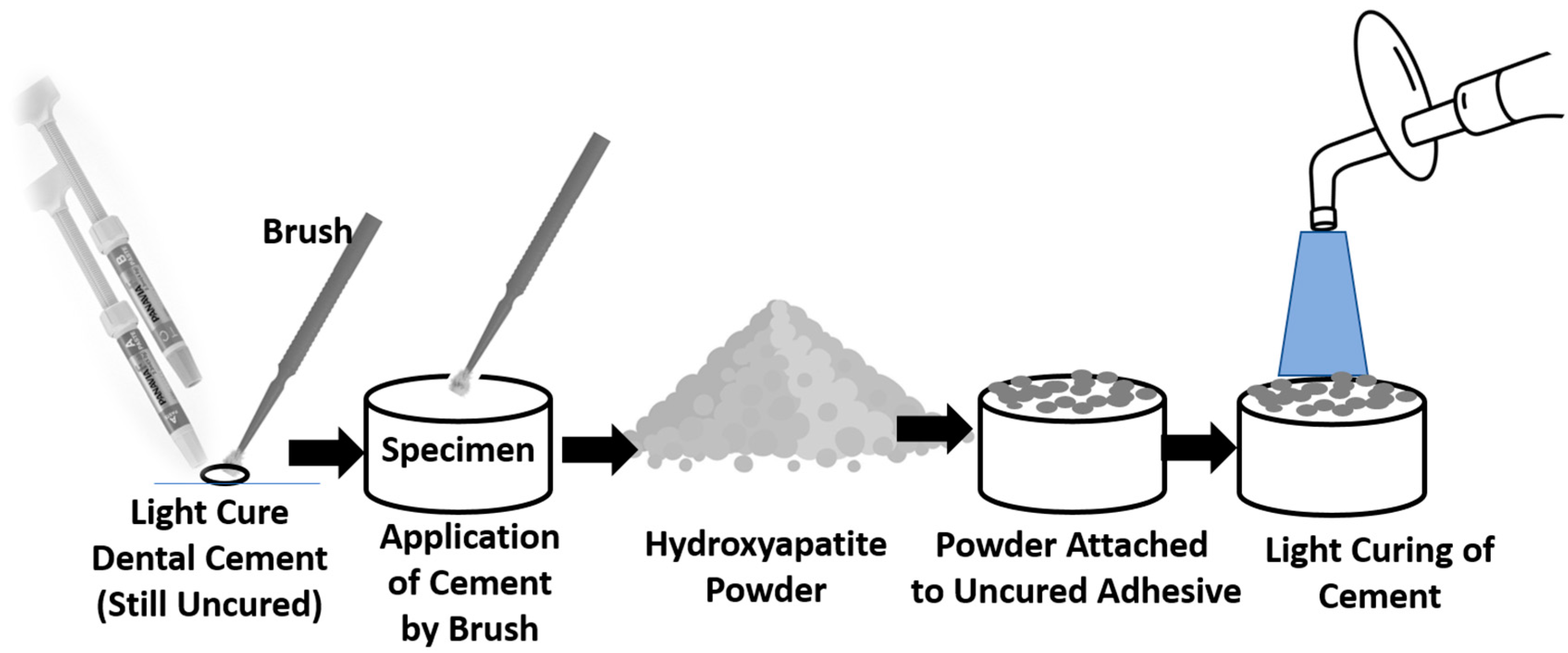
Figure 1. Diagram representing the surface treatment steps.
Coatings 14 01181 g001
Figure 2. Stereomicroscopic image illustrating the steps a–c of coating. Magnification: 12.5×.
Coatings 14 01181 g002
Figure 3. Stereomicroscopic image showing coated and uncoated surfaces (before immersion in PBS). Magnification: 12.5×.
Coatings 14 01181 g003
Figure 4. Stereomicroscopic image after immersion in PBS for coated and uncoated surfaces). Magnification: 12.5×.
Coatings 14 01181 g004
Figure 5. Diagram showing adhesion-scale according to ASTM D3359 standard.
Coatings 14 01181 g005
Figure 6. FTIR Spectra of the coated specimen.
Coatings 14 01181 g006
Figure 7. TEM micrographs of the coated specimen.
Coatings 14 01181 g007
Figure 8. SEM micrograph of control specimens before immersion in phosphate-buffered saline.
Coatings 14 01181 g008
Figure 9. EDXA of control specimens before immersion in phosphate-buffered saline.
Coatings 14 01181 g009
Figure 10. SEM micrograph of treated specimens before immersion in phosphate-buffered saline (magnification: 200×).
Coatings 14 01181 g010
Figure 11. EDXA spectrum of treated specimens before immersion in phosphate-buffered saline.
Coatings 14 01181 g011
Figure 12. SEM micrograph of control specimens after immersion in phosphate-buffered saline.
Coatings 14 01181 g012
Figure 13. EDXA spectrum of control specimens after immersion in phosphate-buffered saline.
Coatings 14 01181 g013
Figure 14. SEM micrograph of treated specimens after immersion in phosphate-buffered saline.
Coatings 14 01181 g014
Figure 15. EDXA spectrum of treated specimens after immersion in phosphate-buffered saline.
Coatings 14 01181 g015
Figure 16. SEM micrograph of lateral view of the coated specimens (magnification: 500×).
Coatings 14 01181 g016
Table 1. Adhesion-scale according to ASTM D3359 standard.
ScoreDescription and Area% removed
5BThe whole coat is attached, and the margins of the incisions are perfectly smooth (Area removed is zero).
4BMinor coating flakes detached at lines of intersections (Area removed <5%).
3BLittle coating flakes come off at cut intersections and around edges, (5 to 15% of the lattice).
2BParts of the squares and their edges have chipped off from the coat (15–35%).
1BWhole squares have come away from the coat, and enormous ribbons with cut edges have flaked (35–65%).
0BDetachment and flaking are worse than in Grade 1 (>65%).
Disclaimer/Publisher’s Note: The statements, opinions and data contained in all publications are solely those of the individual author(s) and contributor(s) and not of MDPI and/or the editor(s). MDPI and/or the editor(s) disclaim responsibility for any injury to people or property resulting from any ideas, methods, instructions or products referred to in the content.